Molecular Biology of the Cell Vol. 10, 2607–2618, August 1999 The Spindle Checkpoint of Budding Yeast Depends on a Tight Complex between the Mad1 and Mad2 Proteins Rey-Huei Chen,* ‡ D. Michelle Brady, § Dana Smith,* Andrew W. Murray,* and Kevin G. Hardwick* § *Department of Physiology, University of California, San Francisco, San Francisco, California 94143- 0444; ² Department of Molecular Biology and Genetics, Cornell University, Ithaca, New York 14853; and § Institute of Cell and Molecular Biology, University of Edinburgh, Edinburgh, EH9 3JR, United Kingdom Submitted February 18, 1999; Accepted May 18, 1999 Monitoring Editor: J. Richard McIntosh The spindle checkpoint arrests the cell cycle at metaphase in the presence of defects in the mitotic spindle or in the attachment of chromosomes to the spindle. When spindle assembly is disrupted, the budding yeast mad and bub mutants fail to arrest and rapidly lose viability. We have cloned the MAD2 gene, which encodes a protein of 196 amino acids that remains at a constant level during the cell cycle. Gel filtration and co-immunoprecipitation analyses reveal that Mad2p tightly associates with another spindle checkpoint component, Mad1p. This association is independent of cell cycle stage and the presence or absence of other known checkpoint proteins. In addition, Mad2p binds to all of the different phosphorylated isoforms of Mad1p that can be resolved on SDS-PAGE. Deletion and mutational analysis of both proteins indicate that association of Mad2p with Mad1p is critical for checkpoint function and for hyperphosphorylation of Mad1p. INTRODUCTION Cell cycle progression is a highly ordered and tightly regu- lated process. For example, mitosis occurs only after DNA synthesis has completed, and chromosome segregation does not begin until all the chromosomes have been correctly aligned on the mitotic spindle. These regulatory linkages are due to cell cycle checkpoints (Hartwell and Weinert, 1989; Elledge, 1996; Rudner and Murray, 1996), mechanisms that arrest the cell cycle if the preceding events have not been completed or if damage has occurred. Defects in checkpoints compromise the faithful transmission of genetic information and have been shown to play an important role in tumor progression (Hartwell and Kastan, 1994; Cahill et al., 1998). Mitosis in most eukaryotes is regulated by a cyclin-depen- dent kinase, which is activated by association with the mi- totic cyclins, and is encoded by CDC28 in the budding yeast Saccharomyces cerevisiae and the Cdc2 gene of other organ- isms. Activation of Cdc28 protein kinase leads to mitotic spindle formation. Proteolysis of the anaphase inhibitor Pds1p induces chromatids to separate and move to opposite spindle poles (Cohen-Fix et al., 1996), and the destruction of Clb2p and Ase1p are required for cells to exit from mitosis (Surana et al., 1993; Juang et al. 1997). Formation of an intact mitotic spindle and attachment of all sister chromatids to the spindle before anaphase occurs is crucial to proper chromosome segregation. Defects in spin- dle assembly or chromosome attachment prevent the onset of anaphase by activating the spindle checkpoint. Several components of the checkpoint have been identified through budding yeast genetics. Mutations in the MAD (mitotic ar- rest-deficient) (Li and Murray, 1991) and BUB (budding uninhibited by benzimidazole) (Hoyt et al., 1991) genes abol- ish this cell cycle arrest and allow cells to enter anaphase in the absence of a functional spindle, leading to cell death and massive chromosome mis-segregation (Hoyt et al., 1991; Li and Murray, 1991). Although the MAD and BUB genes are not essential for cell viability, mutations in these genes in- crease the chromosome loss rate even in the absence of spindle defects, suggesting that they regulate the metaphase to anaphase transition during normal cell cycles (Hoyt et al., 1991; Li and Murray, 1991). Many of the Mad and Bub proteins have now been identified and characterized (for review, see Rudner and Murray, 1996). Mad1p is a nuclear protein whose phosphorylation increases greatly upon spindle depolymerization and rises transiently during normal mitosis (Hardwick and Murray, 1995). Genetic and biochemical evidence suggests that Mad1p is phosphory- lated by Mps1p whose function is also required for the check- point (Hardwick et al., 1996; Weiss and Winey, 1996). ‡ Corresponding author. E-mail address: [email protected]. © 1999 by The American Society for Cell Biology 2607

Welcome message from author
This document is posted to help you gain knowledge. Please leave a comment to let me know what you think about it! Share it to your friends and learn new things together.
Transcript
Molecular Biology of the CellVol. 10, 2607–2618, August 1999
The Spindle Checkpoint of Budding Yeast Dependson a Tight Complex between the Mad1 and Mad2ProteinsRey-Huei Chen,*†‡ D. Michelle Brady,§ Dana Smith,* Andrew W. Murray,*and Kevin G. Hardwick*§
*Department of Physiology, University of California, San Francisco, San Francisco, California 94143-0444; †Department of Molecular Biology and Genetics, Cornell University, Ithaca, New York 14853;and §Institute of Cell and Molecular Biology, University of Edinburgh, Edinburgh, EH9 3JR, UnitedKingdom
Submitted February 18, 1999; Accepted May 18, 1999Monitoring Editor: J. Richard McIntosh
The spindle checkpoint arrests the cell cycle at metaphase in the presence of defects in the mitoticspindle or in the attachment of chromosomes to the spindle. When spindle assembly is disrupted,the budding yeast mad and bub mutants fail to arrest and rapidly lose viability. We have clonedthe MAD2 gene, which encodes a protein of 196 amino acids that remains at a constant levelduring the cell cycle. Gel filtration and co-immunoprecipitation analyses reveal that Mad2p tightlyassociates with another spindle checkpoint component, Mad1p. This association is independent ofcell cycle stage and the presence or absence of other known checkpoint proteins. In addition,Mad2p binds to all of the different phosphorylated isoforms of Mad1p that can be resolved onSDS-PAGE. Deletion and mutational analysis of both proteins indicate that association of Mad2pwith Mad1p is critical for checkpoint function and for hyperphosphorylation of Mad1p.
INTRODUCTION
Cell cycle progression is a highly ordered and tightly regu-lated process. For example, mitosis occurs only after DNAsynthesis has completed, and chromosome segregation doesnot begin until all the chromosomes have been correctlyaligned on the mitotic spindle. These regulatory linkages aredue to cell cycle checkpoints (Hartwell and Weinert, 1989;Elledge, 1996; Rudner and Murray, 1996), mechanisms thatarrest the cell cycle if the preceding events have not beencompleted or if damage has occurred. Defects in checkpointscompromise the faithful transmission of genetic informationand have been shown to play an important role in tumorprogression (Hartwell and Kastan, 1994; Cahill et al., 1998).
Mitosis in most eukaryotes is regulated by a cyclin-depen-dent kinase, which is activated by association with the mi-totic cyclins, and is encoded by CDC28 in the budding yeastSaccharomyces cerevisiae and the Cdc2 gene of other organ-isms. Activation of Cdc28 protein kinase leads to mitoticspindle formation. Proteolysis of the anaphase inhibitorPds1p induces chromatids to separate and move to oppositespindle poles (Cohen-Fix et al., 1996), and the destruction ofClb2p and Ase1p are required for cells to exit from mitosis(Surana et al., 1993; Juang et al. 1997).
Formation of an intact mitotic spindle and attachment ofall sister chromatids to the spindle before anaphase occurs iscrucial to proper chromosome segregation. Defects in spin-dle assembly or chromosome attachment prevent the onsetof anaphase by activating the spindle checkpoint. Severalcomponents of the checkpoint have been identified throughbudding yeast genetics. Mutations in the MAD (mitotic ar-rest-deficient) (Li and Murray, 1991) and BUB (buddinguninhibited by benzimidazole) (Hoyt et al., 1991) genes abol-ish this cell cycle arrest and allow cells to enter anaphase inthe absence of a functional spindle, leading to cell death andmassive chromosome mis-segregation (Hoyt et al., 1991; Liand Murray, 1991). Although the MAD and BUB genes arenot essential for cell viability, mutations in these genes in-crease the chromosome loss rate even in the absence ofspindle defects, suggesting that they regulate the metaphaseto anaphase transition during normal cell cycles (Hoyt et al.,1991; Li and Murray, 1991).
Many of the Mad and Bub proteins have now been identifiedand characterized (for review, see Rudner and Murray, 1996).Mad1p is a nuclear protein whose phosphorylation increasesgreatly upon spindle depolymerization and rises transientlyduring normal mitosis (Hardwick and Murray, 1995). Geneticand biochemical evidence suggests that Mad1p is phosphory-lated by Mps1p whose function is also required for the check-point (Hardwick et al., 1996; Weiss and Winey, 1996).‡ Corresponding author. E-mail address: [email protected].
© 1999 by The American Society for Cell Biology 2607

Homologues of spindle checkpoint components have beenidentified in fission yeast (Kim et al., 1998) and vertebrates(for review, see Hardwick, 1998). MAD2 homologues in thefrog Xenopus laevis (XMAD2) (Chen et al., 1996) and humans(HMAD2) (Li and Benezra, 1996) are essential for checkpointfunction in frog egg extracts and in cultured human cells,respectively (Chen et al., 1996; Li and Benezra, 1996). Unlikebudding yeast, vertebrate cells appear to require the check-point even when there is no perturbation of spindle assem-bly (Gorbsky et al., 1998). Kinetochores that are not attachedto microtubules recruit the vertebrate homologues of Mad2(Chen et al., 1996; Li and Benezra, 1996), Mad1, Mad3, Bub1,and Bub3 (Taylor and McKeon, 1997; Chan et al., 1998;Taylor et al., 1998), and a small fraction of the kinetochores inTaxol-treated cells recruit Mad2 (Waters et al., 1998). TheMad2 and Mad3 proteins bind to and are thought to inhibitthe activity of Cdc20p (Fang et al., 1998; Hwang et al., 1998;Kim et al., 1998), a substoichiometric component of the an-aphase-promoting complex (Fang et al., 1998), the large com-plex that initiates anaphase by catalyzing the ubiquitinationof cyclin B and proteins that regulate sister chromatid cohe-sion (King et al., 1995; Sudakin et al., 1995; Cohen-Fix et al.,1996; Zachariae and Nasmyth, 1996). The conservation of thespindle checkpoint proteins in eukaryotes indicates that thecheckpoint is an important regulator of cell division and thatits mechanism has been conserved throughout evolution.
We report the isolation of the budding yeast MAD2 geneand the characterization of the association between Mad1pand Mad2p that is essential for the function of the spindlecheckpoint.
MATERIALS AND METHODS
Yeast Strains and MediaTable 1 lists the strains used in this work, all of which are deriva-tives of W303 except the three original mad1 alleles, which are in theA364a background, and MAY 2072, which is in the S288c back-ground. Yeast media, growth conditions, stock solutions, and mo-lecular techniques were as previously described (Guthrie and Fink,1991; Hardwick and Murray, 1995).
Cloning of MAD2 and mad2 Gene DisruptionA 2.6-kb HindIII–SalI genomic fragment that resides upstream of thetranslational initiation codon of BET4 was subcloned into the cog-nate sites in the vector pRS316 (Sikorski and Hieter, 1989). Thisplasmid pRC2 was able to complement the benomyl-sensitive phe-notype of mad2-1 mutant. An ORF of 196 amino acids was identifiedin this region by DNA sequencing from both ends of the HindIII–SalI fragment.
To generate the mad2::URA3 disruption plasmid pRC10.1, a 1.2-kbfragment containing the URA3 gene was used to replace the frag-ment between the ApaI site located 20 base pairs upstream of theMAD2 translation initiation codon and the ScaI site that resides inamino acid position 148.
Preparation of Recombinant Mad2 Protein andMad2 AntibodiesThe coding region of MAD2 flanked by EcoRI sites was generated byPCR and cloned into pGEX1 at the EcoRI site. This GST fusionconstruct was transformed into Escherichia coli strain DH5a, and itsexpression was induced with 0.1 mM isopropyl-1-thio-b-d-galacto-pyranoside for 2 h at 37°C. Cells were pelleted and resuspended inPBS (2.7 mM KCl, 137 mM NaCl, 1.5 mM KH2PO4, 4.3 mMNa2HPO4, pH 7.2), and repelleted. The cell pellet was resuspendedin PBS containing 0.5% Triton X-100, 1 mM EGTA, 1 mM EDTA, 1mM PMSF, and 200 mg/ml lysozyme, and sonicated briefly. Thelysate was spun at 15 krpm in a Sorvall (Newton, CT) SS-34 rotor for30 min. The supernatant was loaded onto a 4-ml glutathione-agar-ose column. The column was washed with 40 ml of PBS, and theGST-Mad2 fusion protein was eluted with 5 mM reduced glutathi-one in 50 mM Tris, pH 8.0, and 0.5 mM DTT. Purified protein wasdialyzed into 50 mM HEPES, pH 7.6, 50 mM KCl, and 50% glycerol.The purified protein was used to raise antisera in rabbits (Babco,Berkeley, California). To affinity purify antibodies, the rabbit serumwas passed over a 50-ml column of GST protein coupled to Affi-Gel10 (Bio-Rad, Hercules, California) to remove anti-GST antibodies,before being loaded over a 3-ml column of GST-Mad2 proteincoupled to Affi-Gel 10. Elution of anti-Mad2 antibodies was per-formed as described (Chen et al., 1996).
Construction of mad2 DeletionsThe HindIII–XhoI fragment containing the MAD2 gene (Figure 1A)was subcloned into the corresponding sites in the vector pRS316 to
Table 1. Yeast strains
Strain Genotype
KH 34 MATa ura3-1, leu2,3-112, his3-11, trp1-1, ade2-1, can1-100RHC 1 MATa mad2-1, ura3-1, leu2,3-112, his3-11, trp1-1, ade2-1, can1-100RHC 15.1 MATa mad2D::URA3, ura3-1, leu2,3-112, his3-11, trp1-1, ade2-1, can1-100BEN 24 MATa mad1-1, ura3-52, leu2, his3, trp1-1, rad9D::LEU2BEN 27 MATa mad1-2, ura3-52, leu2, his3, trp1-1, rad9D::LEU2BEN 79 MATa mad1-3, ura3-52, leu2, his3, trp1-1, rad9D::LEU2KH 144 MATa mad1D.2::URA3, ura3-1, leu2,3-112, his3-11, trp1-1, ade2-1, can1-100KH 173 MATa mad3D.2::URA3, ura3-1, leu2,3-112, his3-11, trp1-1, ade2-1, can1-100KH 127 MATa bub1D::HIS3, ura3-1, leu2,3-112, his3-11, trp1-1, ade2-1, can1-100KH 128 MATa bub2D::URA3, ura3-1, leu2,3-112, his3-11, trp1-1, ade2-1, can1-100MAY 2072 MATa bub3D::LEU2, ura3-1, leu2,3-112, his3-11, trp1-1, ade2-1, can1-100RHC 88 MATa URA3::mad2-N5, mad2-1, ura3-1, leu2,3-112, his3-11, trp1-1, ade2-1, can1-100RHC 89 MATa URA3::mad2-C5, mad2-1, ura3-1, leu2,3-112, his3-11, trp1-1, ade2-1, can1-100RHC 91 MATa URA3::mad2-N10, mad2-1, ura3-1, leu2,3-112, his3-11, trp1-1, ade2-1, can1-100RHC 93 MATa URA3::mad2-C10, mad2-1, ura3-1, leu2,3-112, his3-11, trp1-1, ade2-1, can1-100KH 153 MATa URA3::GAL-MPS1, ura3-1, leu2,3-112, his3-11, trp1-1, ade2-1, can1-100
R.-H. Chen et al.
Molecular Biology of the Cell2608
give rise to the plasmid pRC4. The 39-untranslated region wasamplified by PCR, which also converted the EcoRI site following thestop codon to HindIII. This fragment was subcloned into pRS316 atHindIII–XhoI sites, giving rise to pRC66. All the deletion mutantswere made by PCR amplification and cloned at the HindIII site ofpRC66. The BamHI–XhoI fragments containing various deletionswere subcloned into pRS306 (Sikorski and Hieter, 1989). To inte-grate the plasmids into yeast at URA3 locus, the plasmids were cutat StuI in the URA3 gene.
Construction of mad1 Mutants, Deletions,Truncations, and Allele SequencingThree mutations were engineered into the MAD1 sequence by site-directed mutagenesis using the QuikChange site-directed mutagen-esis kit and Pfu DNA polymerase according to manufacturer’sinstructions (Stratagene, La Jolla, CA). A KpnI site was introducedjust before the first methionine of Mad1p using two primers, CT-TAAAATCGAGAGGTAATAGGGTACCATGGATGTGAGAGCG -GCATTG and its reverse complement. Two NotI sites were engi-neered at either side of the asparagine-rich stretch using the primersCCGGATAATCTCTTCAGGAGCGGCCGCTATGTTATTTTTGGT -TC with its reverse complement to introduce a site at position 974 ofthe coding sequence and GAACCAAAAATAACATAGCGGCCGC-CCCTGAAGAGATTATCCGG with its reverse complement to in-troduce a site at 1109. The other N- and C-terminal Mad1p deletionconstructs and the two-hybrid constructs were made by PCR am-plification (using VENT polymerase; New England Biolabs, Beverly,MA), followed by subcloning and sequencing of the resulting con-structs. pKH601 fuses full-length Mad1p (residues 1-749) to theGAL4 DNA binding domain of pAS1-CYH2; pKH602 fuses residues313-749; pKH603 fuses residues 529-749; pKH604 fuses residues1-318; pKH605 fuses residues 593-749; pKH609 fuses residues 529-718; pKH610 fuses 529-649.
The sequences of the mutations in the three original mad1 alleleswere determined by PCR amplification of the genomic loci followedby cycle sequencing of the PCR products (Applied Biosystems,Foster City, CA). Each allele was sequenced multiple times on bothstrands.
Immunoblotting, Immunoprecipitation, and GelFiltrationYeast extracts were made, and immunoblotting was performed aspreviously described (Hardwick and Murray, 1995). The affinity-purified anti-Mad2p antibody was used at a dilution of 1:500 in PBScontaining 2% BSA and 0.2% Tween 20, the anti-Mad1p antibody at1:2000 in Blotto (Harlow and Lane, 1988), and the anti-HA antibody(16B12, Babco) at 1:500 in Blotto.
For immunoprecipitations, yeast extracts were made by beadbeating in lysis buffer (50 mM HEPES, pH 7.6, 25 mM KCl, 50 mMNaF, 1 mM MgCl2, 1 mM EGTA, 0.1% Na-deoxycholate, 1 mMPMSF, 0.5 mM DTT, and 10 mg/ml leupeptin, pepstatin, and chy-mostatin) as previously described (Hardwick and Murray, 1995),except that in some cases the anti-Mad1p antibody was directlycoupled to the protein A-agarose (Harlow and Lane, 1988) beforeuse. Gel filtration using a Pharmacia (Piscataway, NJ) Superose 6fast performance liquid chromatography column was carried out asdescribed (Hardwick and Murray, 1995).
Transfection in COS CellsFor expression in COS7 cells, the coding regions of MAD1, MAD2,or MPS1 were subcloned into the vector SRa (Takebe et al., 1988) atthe EcoRI site. The sequence encoding the myc epitope was insertedat the amino terminus of MPS1 for detection with the anti-mycantibody 9E10. The plasmids were purified twice by standard ce-sium chloride gradient (Maniatis et al., 1982).
COS cells were maintained in Dulbecco’s modified Eagle’s me-dium plus 10% FBS, 100 U/ml penicillin, and 100 mg/ml strepto-mycin. Transfection was performed with standard calcium phos-phate precipitation as described (Chen et al., 1996).
RESULTS
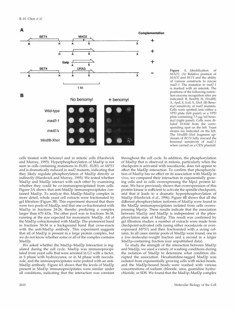
Identification and Characterization of MAD2The spindle checkpoint gene MAD2 in the budding yeast S.cerevisiae was originally identified (Li and Murray, 1991) asthe ORF YJL031C, which encodes a subunit of an essentialprenyltransferase (Li et al., 1993) and has been renamedBET4. However, sequencing this gene recovered from theoriginal mad2-1 strain failed to identify any mutation. Inaddition, a genomic DNA fragment outside of the prenyl-transferase coding region (HindIII–XhoI region in Figure 1A)fully rescued the benomyl sensitivity of mad2-1 (Figure 1; seecorrection in Li et al., 1994), suggesting that this fragmentencoded the bona fide MAD2 gene. This was confirmed bysequencing a 196-amino acid ORF (YJL030W), recoveredfrom wild-type cells and from the mad2-1 mutant. This anal-ysis shows that the mad2-1 mutation lies within YJL030Wconverting Trp94 into a stop codon. Deleting most of thecoding region of YJL030W produced viable strains that havephenotypes similar to that of mad2-1 (Figure 1B), and ex-pression of the coding region of YJL030W from a galactose-inducible promoter rescued the benomyl sensitivity ofmad2-1 in a galactose-dependent manner (Figure 1A). Theseobservations unequivocally show that YJL030W is the bonafide MAD2 gene.
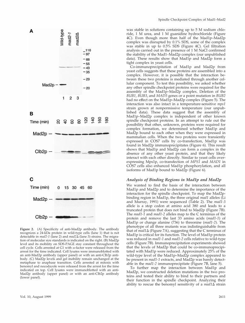
To characterize Mad2p, we generated antiserum againstrecombinant GST-Mad2. The affinity-purified antibodies rec-ognized Mad2p specifically on immunoblots (Figure 2A,lane 1), and the protein was missing in the mad2D strain, asexpected (Figure 2A, lane 3). We did not detect the truncatedform of Mad2p, which has a predicted molecular mass of 13kDa, in the mad2-1 strain, indicating that the truncated pro-tein is unstable or that the antibody recognizes epitopes inthe C-terminal half. We studied the protein by following itslevel during a synchronous cell cycle (Figure 2B). Althoughthe level of Clb2p, a mitotic cyclin, showed the expectedoscillation, there was no change in either the abundance orthe gel mobility of Mad2p during the cell cycle.
We examined the effect of activating the spindle check-point on Mad2p (Figure 2C). Cells were arrested in mitosisby depolymerizing their spindles with benomyl and nocoda-zole and then allowed to recover from their arrest. The levelof Clb2p fell as cells exited mitosis, but there was no changein either the abundance or the gel mobility of Mad2p. Ana-lyzing the behavior of Mad2p on two-dimensional gelsshowed a single spot whose mobility was unaffected byactivation of the spindle checkpoint (our unpublished data).These results suggest that the function of Mad2p is notregulated by post-translational modification, although wecannot exclude the possibility that only a very small fractionof the Mad2p molecules are modified.
Mad2p and Mad1p Bind Tightly to Each OtherIn VivoThe spindle checkpoint component Mad1p is a nuclearphosphoprotein, which becomes hyperphosphorylated in
Spindle Checkpoint Complex of Mad1–Mad2
Vol. 10, August 1999 2609
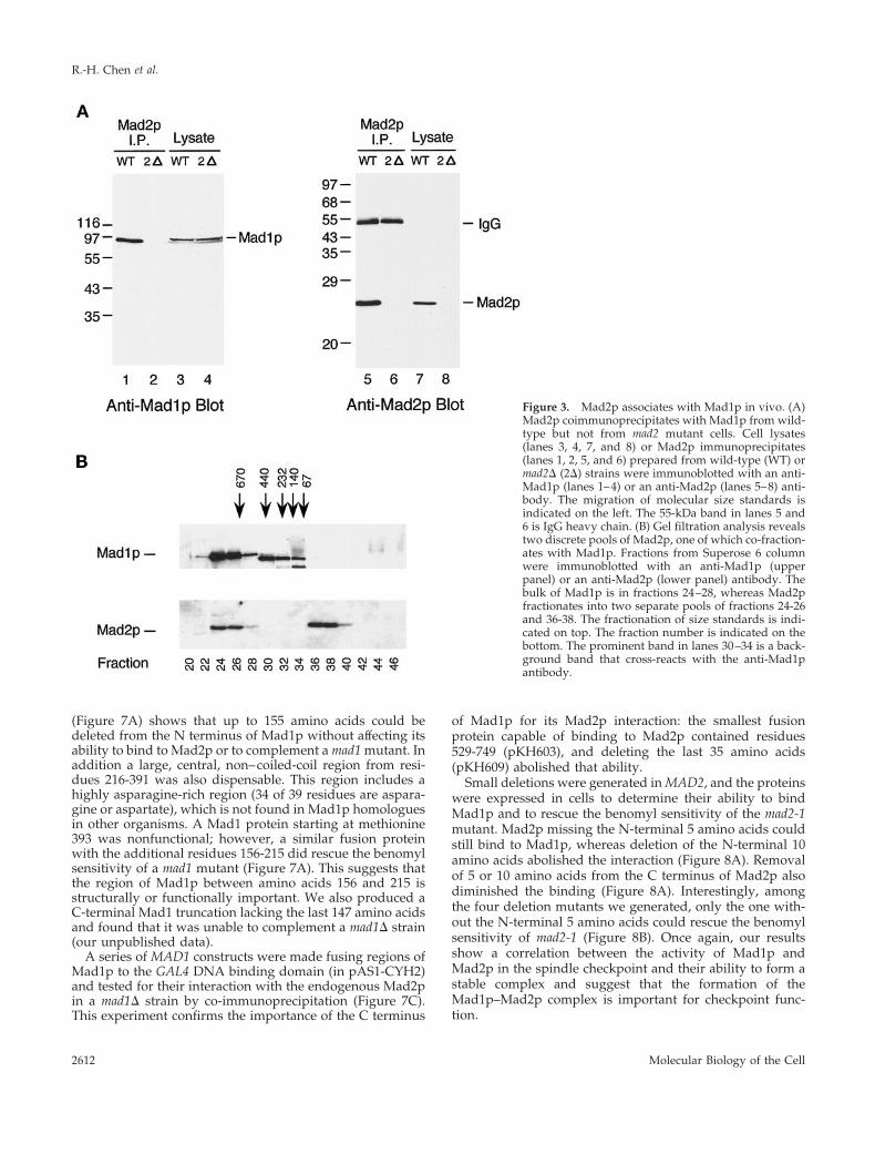
cells treated with benomyl and in mitotic cells (Hardwickand Murray, 1995). Hyperphosphorylation of Mad1p is notseen in cells containing mutations in BUB1, BUB3, or MPS1and is dramatically reduced in mad2 mutants, indicating thatthey likely regulate phosphorylation of Mad1p directly orindirectly (Hardwick and Murray, 1995). We tested whetherMad1p and Mad2p interact with each other by examiningwhether they could be co-immunoprecipitated from cells.Figure 3A shows that anti-Mad2p immunoprecipitates con-tained Mad1p. To analyze this Mad2p–Mad1p complex inmore detail, whole yeast cell extracts were fractionated bygel filtration (Figure 3B). This experiment showed that therewere two pools of Mad2p, and that one co-fractionated withMad1p in fractions 24-26, thereby predicting a complexlarger than 670 kDa. The other pool was in fractions 36-38,running at the size expected for monomeric Mad2p. All ofthe Mad1p cofractionated with Mad2p. The prominent bandin fractions 30-34 is a background band that cross-reactswith the anti-Mad1p antibody. This experiment suggeststhat all of Mad1p is present in a large protein complex, butwe do not know whether some or all of the complex containsMad2p.
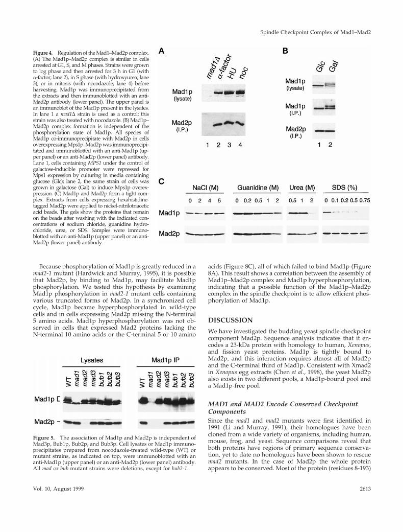
We asked whether the Mad1p–Mad2p interaction is reg-ulated during the cell cycle. Mad1p was immunoprecipi-tated from yeast cells that were arrested in G1 with a factor,in S phase with hydroxyurea, or in M phase with nocoda-zole, and the immunoprecipitates were probed with an anti-Mad2p antibody. Figure 4A shows that the levels of Mad2ppresent in Mad1p immunoprecipitates were similar underall conditions, indicating that the interaction was constant
throughout the cell cycle. In addition, the phosphorylationof Mad1p that is observed in mitosis, particularly when thecheckpoint is activated with nocodazole, does not appear toaffect the Mad2p interaction. To confirm that phosphoryla-tion of Mad1p has no effect on its association with Mad2p invivo, we compared their interaction in exponentially grow-ing cells and in cells overexpressing the Mps1 protein ki-nase. We have previously shown that overexpression of thisprotein kinase is sufficient to activate the spindle checkpoint,and that it leads to a dramatic hyperphosphorylation ofMad1p (Hardwick et al., 1996). Figure 4B shows that all thedifferent phosphorylation isoforms of Mad1p were found inthe Mad2p immunoprecipitates isolated from cells overex-pressing Mps1p. These results indicate that the associationbetween Mad1p and Mad2p is independent of the phos-phorylation state of Mad1p. This result was confirmed bygel filtration studies: a number of extracts were made fromcheckpoint-activated cells (using either nocodazole or over-expressed MPS1) and then fractionated with a sizing col-umn. In all cases similar pools of Mad2p were found, one ina low-molecular-weight fraction and a second in a largerMad1p-containing fraction (our unpublished data).
To study the strength of the interaction between Mad1pand Mad2p, we used a variety of washing conditions duringthe isolation of Mad2p to determine what condition dis-rupted the association. Hexahistidine-tagged Mad2p wasisolated from exponentially growing cells with nickel beads,and the Mad2p-bound beads were washed with variousconcentrations of sodium chloride, urea, guanidine hydro-chloride, or SDS. We found that the Mad1p–Mad2p complex
Figure 1. Identification ofMAD2. (A) Relative position ofMAD2 and BET4 and the abilityof various constructs to rescuemad2-1. The mutation in mad2-1is marked with an asterisk. Thepositions of the following restric-tion enzyme recognition sites areindicated: B, BamHI; H, HindIII;A, ApaI; S, ScaI; X, XhoI. (B) Beno-myl sensitivity of mad2 mutants.Cells were spotted onto either aYPD plate (left panel) or a YPDplate containing 7.5 mg/ml beno-myl (right panel). Cells were di-luted 10-fold from the corre-sponding spot on the left. Yeaststrains are indicated on the left.The HindIII–XhoI fragment up-stream of BET4 fully rescued thebenomyl sensitivity of mad2-1when carried on a CEN plasmid.
R.-H. Chen et al.
Molecular Biology of the Cell2610
was stable in solutions containing up to 5 M sodium chlo-ride, 1 M urea, and 1 M guanidine hydrochloride (Figure4C). Even though more than half of the Mad1p–Mad2pcomplex was disrupted by 0.1% SDS, some of the complexwas stable in up to 0.5% SDS (Figure 4C). Gel filtrationanalysis carried out in the presence of 1 M NaCl confirmedthe stability of the Mad1–Mad2p complex (our unpublisheddata). These results show that Mad1p and Mad2p form atight complex in yeast cells.
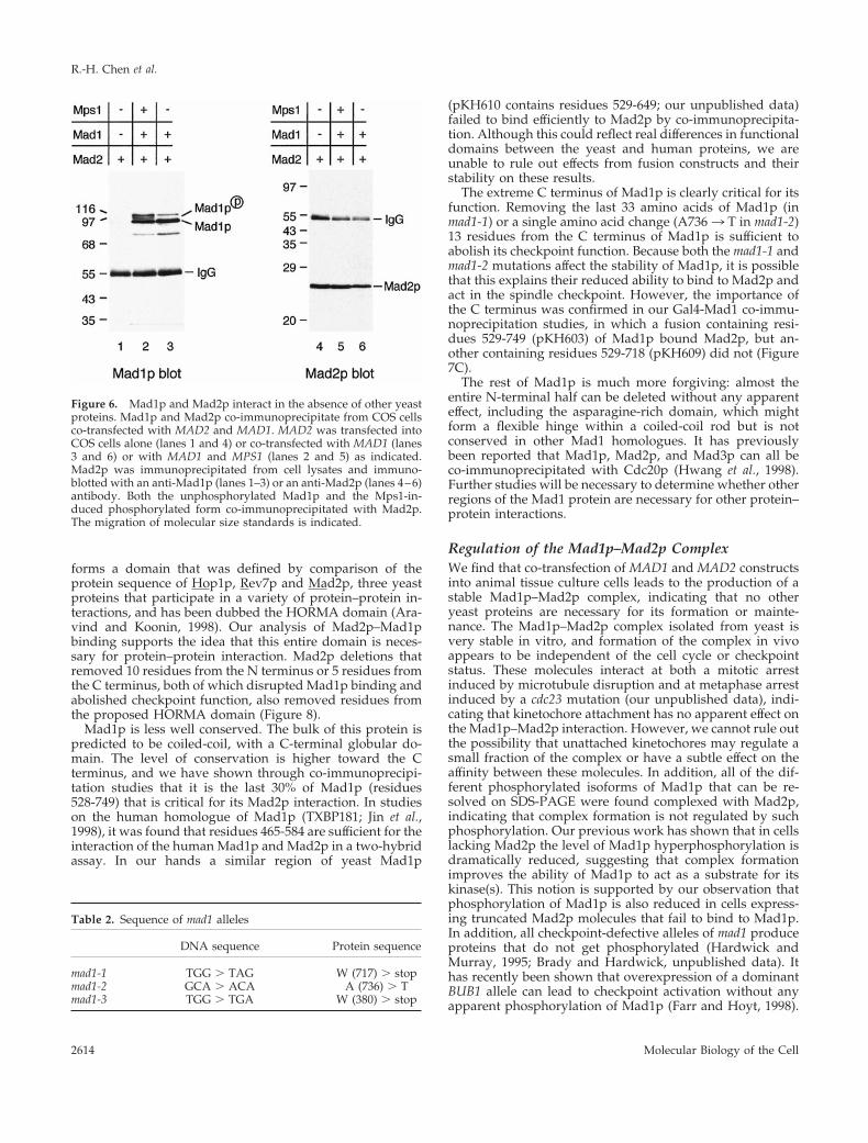
Co-immunoprecipitation of Mad1p and Mad2p fromyeast cells suggests that these proteins are assembled into acomplex. However, it is possible that the interaction be-tween these two proteins is mediated through another cel-lular component. To test this possibility, we asked whetherany other spindle checkpoint proteins were required for theassembly of the Mad1p–Mad2p complex. Deletion of theBUB1, BUB3, and MAD3 genes or a point mutation in BUB2had no effect on the Mad1p–Mad2p complex (Figure 5). Theinteraction was also intact in a temperature-sensitive mps1strain grown at nonpermissive temperature (our unpub-lished data). These data suggest that the assembly ofMad1p–Mad2p complex is independent of other knownspindle checkpoint proteins. In an attempt to rule out thepossibility that other, unknown, proteins were required forcomplex formation, we determined whether Mad1p andMad2p bound to each other when they were expressed inmammalian cells. When the two proteins were transientlyexpressed in COS7 cells by co-transfection, Mad1p wasfound in Mad2p immunoprecipitates (Figure 6). This resultshows that Mad1p and Mad2p can form a complex in theabsence of any other yeast protein, and that they likelyinteract with each other directly. Similar to yeast cells over-expressing Mps1p, co-transfection of MPS1 and MAD1 inCOS7 cells also enhanced Mad1p phosphorylation, and allisoforms of Mad1p bound to Mad2p (Figure 6).
Analysis of Binding Regions in Mad1p and Mad2pWe wanted to find the basis of the interaction betweenMad1p and Mad2p and to determine the importance of theinteraction for the spindle checkpoint. To map the Mad2p-binding region in Mad1p, the three original mad1 alleles (Liand Murray, 1991) were sequenced (Table 2). The mad1-3allele is a stop codon at amino acid 380 and leads to atruncated protein that does not bind to Mad2p (Figure 7B).The mad1-1 and mad1-2 alleles map to the C terminus of theprotein and remove the last 33 amino acids (mad1-1) ofMad1p or change alanine (736) to threonine (mad1-2). Thephenotype of all three mutants was indistinguishable fromthat of mad1D (Figure 7A), suggesting that the C terminus ofMad1p is critical for its function. The level of Mad1p proteinwas reduced in mad1-1 and mad1-2 cells relative to wild-typecells (Figure 7B). Immunoprecipitation experiments showedthat the levels of Mad2p that could be co-immunoprecipi-tated with Mad1p were reduced. Approximately 25% of thewild-type level of the Mad1p–Mad2p complex appeared tobe present in mad1-1 extracts, and Mad2p was barely detect-able in the mad1-2 immunoprecipitate (Figure 7B, lane 5).
To further map the interaction between Mad1p andMad2p, we constructed deletion mutations in the two pro-teins and tested their ability to bind to their partners andtheir function in the spindle checkpoint. Analyzing theirability to rescue the benomyl sensitivity of a mad1D strain
Figure 2. (A) Specificity of anti-Mad2p antibody. The antibodyrecognizes a 24-kDa protein in wild-type cells (lane 1) that is notdetectable in mad2-1 (lane 2) and mad2D (lane 3) strains. The migra-tion of molecular size standards is indicated on the right. (B) Mad2plevel and its mobility on SDS-PAGE stay constant throughout thecell cycle. Cells arrested at G1 with a-factor were released from thearrest for the time indicated. Cell lysates were immunoblotted withan anti-Mad2p antibody (upper panel) or with an anti-Clb2p anti-body. (C) Mad2p levels and gel mobility remain unchanged at themetaphase to anaphase transition. Cells arrested at mitosis withbenomyl and nocodazole were released from the arrest for the timeindicated on top. Cell lysates were immunoblotted with an anti-Mad2p antibody (upper panel) or with an anti-Clb2p antibody(lower panel).
Spindle Checkpoint Complex of Mad1–Mad2
Vol. 10, August 1999 2611
(Figure 7A) shows that up to 155 amino acids could bedeleted from the N terminus of Mad1p without affecting itsability to bind to Mad2p or to complement a mad1 mutant. Inaddition a large, central, non–coiled-coil region from resi-dues 216-391 was also dispensable. This region includes ahighly asparagine-rich region (34 of 39 residues are aspara-gine or aspartate), which is not found in Mad1p homologuesin other organisms. A Mad1 protein starting at methionine393 was nonfunctional; however, a similar fusion proteinwith the additional residues 156-215 did rescue the benomylsensitivity of a mad1 mutant (Figure 7A). This suggests thatthe region of Mad1p between amino acids 156 and 215 isstructurally or functionally important. We also produced aC-terminal Mad1 truncation lacking the last 147 amino acidsand found that it was unable to complement a mad1D strain(our unpublished data).
A series of MAD1 constructs were made fusing regions ofMad1p to the GAL4 DNA binding domain (in pAS1-CYH2)and tested for their interaction with the endogenous Mad2pin a mad1D strain by co-immunoprecipitation (Figure 7C).This experiment confirms the importance of the C terminus
of Mad1p for its Mad2p interaction: the smallest fusionprotein capable of binding to Mad2p contained residues529-749 (pKH603), and deleting the last 35 amino acids(pKH609) abolished that ability.
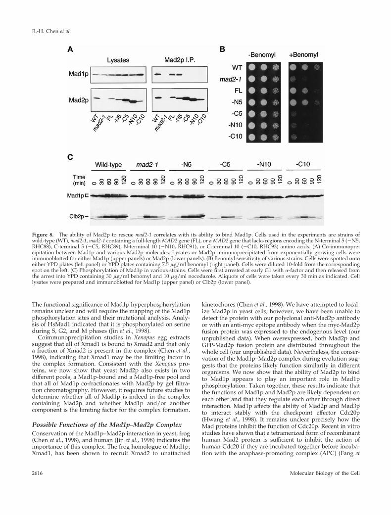
Small deletions were generated in MAD2, and the proteinswere expressed in cells to determine their ability to bindMad1p and to rescue the benomyl sensitivity of the mad2-1mutant. Mad2p missing the N-terminal 5 amino acids couldstill bind to Mad1p, whereas deletion of the N-terminal 10amino acids abolished the interaction (Figure 8A). Removalof 5 or 10 amino acids from the C terminus of Mad2p alsodiminished the binding (Figure 8A). Interestingly, amongthe four deletion mutants we generated, only the one with-out the N-terminal 5 amino acids could rescue the benomylsensitivity of mad2-1 (Figure 8B). Once again, our resultsshow a correlation between the activity of Mad1p andMad2p in the spindle checkpoint and their ability to form astable complex and suggest that the formation of theMad1p–Mad2p complex is important for checkpoint func-tion.
Figure 3. Mad2p associates with Mad1p in vivo. (A)Mad2p coimmunoprecipitates with Mad1p from wild-type but not from mad2 mutant cells. Cell lysates(lanes 3, 4, 7, and 8) or Mad2p immunoprecipitates(lanes 1, 2, 5, and 6) prepared from wild-type (WT) ormad2D (2D) strains were immunoblotted with an anti-Mad1p (lanes 1–4) or an anti-Mad2p (lanes 5–8) anti-body. The migration of molecular size standards isindicated on the left. The 55-kDa band in lanes 5 and6 is IgG heavy chain. (B) Gel filtration analysis revealstwo discrete pools of Mad2p, one of which co-fraction-ates with Mad1p. Fractions from Superose 6 columnwere immunoblotted with an anti-Mad1p (upperpanel) or an anti-Mad2p (lower panel) antibody. Thebulk of Mad1p is in fractions 24–28, whereas Mad2pfractionates into two separate pools of fractions 24-26and 36-38. The fractionation of size standards is indi-cated on top. The fraction number is indicated on thebottom. The prominent band in lanes 30–34 is a back-ground band that cross-reacts with the anti-Mad1pantibody.
R.-H. Chen et al.
Molecular Biology of the Cell2612
Because phosphorylation of Mad1p is greatly reduced in amad2-1 mutant (Hardwick and Murray, 1995), it is possiblethat Mad2p, by binding to Mad1p, may facilitate Mad1pphosphorylation. We tested this hypothesis by examiningMad1p phosphorylation in mad2-1 mutant cells containingvarious truncated forms of Mad2p. In a synchronized cellcycle, Mad1p became hyperphosphorylated in wild-typecells and in cells expressing Mad2p missing the N-terminal5 amino acids. Mad1p hyperphosphorylation was not ob-served in cells that expressed Mad2 proteins lacking theN-terminal 10 amino acids or the C-terminal 5 or 10 amino
acids (Figure 8C), all of which failed to bind Mad1p (Figure8A). This result shows a correlation between the assembly ofMad1p–Mad2p complex and Mad1p hyperphosphorylation,indicating that a possible function of the Mad1p–Mad2pcomplex in the spindle checkpoint is to allow efficient phos-phorylation of Mad1p.
DISCUSSION
We have investigated the budding yeast spindle checkpointcomponent Mad2p. Sequence analysis indicates that it en-codes a 23-kDa protein with homology to human, Xenopus,and fission yeast proteins. Mad1p is tightly bound toMad2p, and this interaction requires almost all of Mad2pand the C-terminal third of Mad1p. Consistent with Xmad2in Xenopus egg extracts (Chen et al., 1998), the yeast Mad2palso exists in two different pools, a Mad1p-bound pool anda Mad1p-free pool.
MAD1 and MAD2 Encode Conserved CheckpointComponentsSince the mad1 and mad2 mutants were first identified in1991 (Li and Murray, 1991), their homologues have beencloned from a wide variety of organisms, including human,mouse, frog, and yeast. Sequence comparisons reveal thatboth proteins have regions of primary sequence conserva-tion, yet to date no homologues have been shown to rescuemad2 mutants. In the case of Mad2p the whole proteinappears to be conserved. Most of the protein (residues 8-193)
Figure 4. Regulation of the Mad1–Mad2p complex.(A) The Mad1p–Mad2p complex is similar in cellsarrested at G1, S, and M phases. Strains were grownto log phase and then arrested for 3 h in G1 (witha-factor; lane 2), in S phase (with hydroxyurea; lane3), or in mitosis (with nocodazole; lane 4) beforeharvesting. Mad1p was immunoprecipitated fromthe extracts and then immunoblotted with an anti-Mad2p antibody (lower panel). The upper panel isan immunoblot of the Mad1p present in the lysates.In lane 1 a mad1D strain is used as a control; thisstrain was also treated with nocodazole. (B) Mad1p–Mad2p complex formation is independent of thephosphorylation state of Mad1p. All species ofMad1p co-immunoprecipitate with Mad2p in cellsoverexpressing Mps1p. Mad2p was immunoprecipi-tated and immunoblotted with an anti-Mad1p (up-per panel) or an anti-Mad2p (lower panel) antibody.Lane 1, cells containing MPS1 under the control ofgalactose-inducible promoter were repressed forMps1 expression by culturing in media containingglucose (Glc); lane 2, the same strain of cells wasgrown in galactose (Gal) to induce Mps1p overex-pression. (C) Mad1p and Mad2p form a tight com-plex. Extracts from cells expressing hexahistidine-tagged Mad2p were applied to nickel-nitrilotriaceticacid beads. The gels show the proteins that remainon the beads after washing with the indicated con-centrations of sodium chloride, guanidine hydro-chloride, urea, or SDS. Samples were immuno-blotted with an anti-Mad1p (upper panel) or an anti-Mad2p (lower panel) antibody.
Figure 5. The association of Mad1p and Mad2p is independent ofMad3p, Bub1p, Bub2p, and Bub3p. Cell lysates or Mad1p immuno-precipitates prepared from nocodazole-treated wild-type (WT) ormutant strains, as indicated on top, were immunoblotted with ananti-Mad1p (upper panel) or an anti-Mad2p (lower panel) antibody.All mad or bub mutant strains were deletions, except for bub2-1.
Spindle Checkpoint Complex of Mad1–Mad2
Vol. 10, August 1999 2613
forms a domain that was defined by comparison of theprotein sequence of Hop1p, Rev7p and Mad2p, three yeastproteins that participate in a variety of protein–protein in-teractions, and has been dubbed the HORMA domain (Ara-vind and Koonin, 1998). Our analysis of Mad2p–Mad1pbinding supports the idea that this entire domain is neces-sary for protein–protein interaction. Mad2p deletions thatremoved 10 residues from the N terminus or 5 residues fromthe C terminus, both of which disrupted Mad1p binding andabolished checkpoint function, also removed residues fromthe proposed HORMA domain (Figure 8).
Mad1p is less well conserved. The bulk of this protein ispredicted to be coiled-coil, with a C-terminal globular do-main. The level of conservation is higher toward the Cterminus, and we have shown through co-immunoprecipi-tation studies that it is the last 30% of Mad1p (residues528-749) that is critical for its Mad2p interaction. In studieson the human homologue of Mad1p (TXBP181; Jin et al.,1998), it was found that residues 465-584 are sufficient for theinteraction of the human Mad1p and Mad2p in a two-hybridassay. In our hands a similar region of yeast Mad1p
(pKH610 contains residues 529-649; our unpublished data)failed to bind efficiently to Mad2p by co-immunoprecipita-tion. Although this could reflect real differences in functionaldomains between the yeast and human proteins, we areunable to rule out effects from fusion constructs and theirstability on these results.
The extreme C terminus of Mad1p is clearly critical for itsfunction. Removing the last 33 amino acids of Mad1p (inmad1-1) or a single amino acid change (A7363 T in mad1-2)13 residues from the C terminus of Mad1p is sufficient toabolish its checkpoint function. Because both the mad1-1 andmad1-2 mutations affect the stability of Mad1p, it is possiblethat this explains their reduced ability to bind to Mad2p andact in the spindle checkpoint. However, the importance ofthe C terminus was confirmed in our Gal4-Mad1 co-immu-noprecipitation studies, in which a fusion containing resi-dues 529-749 (pKH603) of Mad1p bound Mad2p, but an-other containing residues 529-718 (pKH609) did not (Figure7C).
The rest of Mad1p is much more forgiving: almost theentire N-terminal half can be deleted without any apparenteffect, including the asparagine-rich domain, which mightform a flexible hinge within a coiled-coil rod but is notconserved in other Mad1 homologues. It has previouslybeen reported that Mad1p, Mad2p, and Mad3p can all beco-immunoprecipitated with Cdc20p (Hwang et al., 1998).Further studies will be necessary to determine whether otherregions of the Mad1 protein are necessary for other protein–protein interactions.
Regulation of the Mad1p–Mad2p ComplexWe find that co-transfection of MAD1 and MAD2 constructsinto animal tissue culture cells leads to the production of astable Mad1p–Mad2p complex, indicating that no otheryeast proteins are necessary for its formation or mainte-nance. The Mad1p–Mad2p complex isolated from yeast isvery stable in vitro, and formation of the complex in vivoappears to be independent of the cell cycle or checkpointstatus. These molecules interact at both a mitotic arrestinduced by microtubule disruption and at metaphase arrestinduced by a cdc23 mutation (our unpublished data), indi-cating that kinetochore attachment has no apparent effect onthe Mad1p–Mad2p interaction. However, we cannot rule outthe possibility that unattached kinetochores may regulate asmall fraction of the complex or have a subtle effect on theaffinity between these molecules. In addition, all of the dif-ferent phosphorylated isoforms of Mad1p that can be re-solved on SDS-PAGE were found complexed with Mad2p,indicating that complex formation is not regulated by suchphosphorylation. Our previous work has shown that in cellslacking Mad2p the level of Mad1p hyperphosphorylation isdramatically reduced, suggesting that complex formationimproves the ability of Mad1p to act as a substrate for itskinase(s). This notion is supported by our observation thatphosphorylation of Mad1p is also reduced in cells express-ing truncated Mad2p molecules that fail to bind to Mad1p.In addition, all checkpoint-defective alleles of mad1 produceproteins that do not get phosphorylated (Hardwick andMurray, 1995; Brady and Hardwick, unpublished data). Ithas recently been shown that overexpression of a dominantBUB1 allele can lead to checkpoint activation without anyapparent phosphorylation of Mad1p (Farr and Hoyt, 1998).
Figure 6. Mad1p and Mad2p interact in the absence of other yeastproteins. Mad1p and Mad2p co-immunoprecipitate from COS cellsco-transfected with MAD2 and MAD1. MAD2 was transfected intoCOS cells alone (lanes 1 and 4) or co-transfected with MAD1 (lanes3 and 6) or with MAD1 and MPS1 (lanes 2 and 5) as indicated.Mad2p was immunoprecipitated from cell lysates and immuno-blotted with an anti-Mad1p (lanes 1–3) or an anti-Mad2p (lanes 4–6)antibody. Both the unphosphorylated Mad1p and the Mps1-in-duced phosphorylated form co-immunoprecipitated with Mad2p.The migration of molecular size standards is indicated.
Table 2. Sequence of mad1 alleles
DNA sequence Protein sequence
mad1-1 TGG . TAG W (717) . stopmad1-2 GCA . ACA A (736) . Tmad1-3 TGG . TGA W (380) . stop
R.-H. Chen et al.
Molecular Biology of the Cell2614
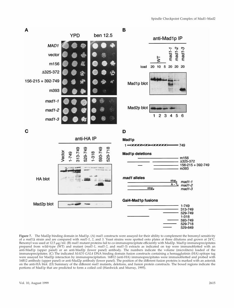
Figure 7. The Mad2p binding domain in Mad1p. (A) mad1 constructs were assayed for their ability to complement the benomyl sensitivityof a mad1D strain and are compared with mad1-1, 2, and 3. Yeast strains were spotted onto plates at three dilutions and grown at 24°C.Benomyl was used at 12.5 mg/ml. (B) mad1 mutant proteins fail to co-immunoprecipitate efficiently with Mad2p. Mad1p immunoprecipitatesprepared from wild-type (WT) and mutant (mad1-1, mad1-2, and mad1-3) extracts as indicated on top were immunoblotted with ananti-Mad1p (upper panel) or an anti-Mad2p (lower panel) antibody. The numbers indicate the volume (microliters) loaded of theimmunoprecipitates. (C) The indicated MAD1-GAL4 DNA binding domain fusion constructs containing a hemagglutinin (HA) epitope tagwere assayed for Mad2p interaction by immunoprecipitation. 16B12 (anti-HA) immunoprecipitates were immunoblotted and probed with16B12 antibody (upper panel) or anti-Mad2p antibody (lower panel). The position of the different fusion proteins is marked with an asteriskon the anti-HA blot. (D) Summary of the different mad1 mutants, deletions, and fusion protein constructs. The boxed regions indicate theportions of Mad1p that are predicted to form a coiled coil (Hardwick and Murray, 1995).
Spindle Checkpoint Complex of Mad1–Mad2
Vol. 10, August 1999 2615
The functional significance of Mad1p hyperphosphorylationremains unclear and will require the mapping of the Mad1pphosphorylation sites and their mutational analysis. Analy-sis of HsMad1 indicated that it is phosphorylated on serineduring S, G2, and M phases (Jin et al., 1998).
Coimmunoprecipitation studies in Xenopus egg extractssuggest that all of Xmad1 is bound to Xmad2 and that onlya fraction of Xmad2 is present in the complex (Chen et al.,1998), indicating that Xmad1 may be the limiting factor inthe complex formation. Consistent with the Xenopus pro-teins, we now show that yeast Mad2p also exists in twodifferent pools, a Mad1p-bound and a Mad1p-free pool andthat all of Mad1p co-fractionates with Mad2p by gel filtra-tion chromatography. However, it requires future studies todetermine whether all of Mad1p is indeed in the complexcontaining Mad2p and whether Mad1p and/or anothercomponent is the limiting factor for the complex formation.
Possible Functions of the Mad1p–Mad2p ComplexConservation of the Mad1p–Mad2p interaction in yeast, frog(Chen et al., 1998), and human (Jin et al., 1998) indicates theimportance of this complex. The frog homologue of Mad1p,Xmad1, has been shown to recruit Xmad2 to unattached
kinetochores (Chen et al., 1998). We have attempted to local-ize Mad2p in yeast cells; however, we have been unable todetect the protein with our polyclonal anti-Mad2p antibodyor with an anti-myc epitope antibody when the myc-Mad2pfusion protein was expressed to the endogenous level (ourunpublished data). When overexpressed, both Mad2p andGFP-Mad2p fusion protein are distributed throughout thewhole cell (our unpublished data). Nevertheless, the conser-vation of the Mad1p–Mad2p complex during evolution sug-gests that the proteins likely function similarily in differentorganisms. We now show that the ability of Mad2p to bindto Mad1p appears to play an important role in Mad1pphosphorylation. Taken together, these results indicate thatthe functions of Mad1p and Mad2p are likely dependent oneach other and that they regulate each other through directinteraction. Mad1p affects the ability of Mad2p and Mad3pto interact stably with the checkpoint effector Cdc20p(Hwang et al., 1998). It remains unclear precisely how theMad proteins inhibit the function of Cdc20p. Recent in vitrostudies have shown that a tetramerized form of recombinanthuman Mad2 protein is sufficient to inhibit the action ofhuman Cdc20 if they are incubated together before incuba-tion with the anaphase-promoting complex (APC) (Fang et
Figure 8. The ability of Mad2p to rescue mad2-1 correlates with its ability to bind Mad1p. Cells used in the experiments are strains ofwild-type (WT), mad2-1, mad2-1 containing a full-length MAD2 gene (FL), or a MAD2 gene that lacks regions encoding the N-terminal 5 (2N5,RHC88), C-terminal 5 (2C5, RHC89), N-terminal 10 (2N10, RHC91), or C-terminal 10 (2C10, RHC93) amino acids. (A) Co-immunopre-cipitation between Mad1p and various Mad2p molecules. Lysates or Mad2p immunoprecipitated from exponentially growing cells wereimmunoblotted for either Mad1p (upper panels) or Mad2p (lower panels). (B) Benomyl sensitivity of various strains. Cells were spotted ontoeither YPD plates (left panel) or YPD plates containing 7.5 mg/ml benomyl (right panel). Cells were diluted 10-fold from the correspondingspot on the left. (C) Phosphorylation of Mad1p in various strains. Cells were first arrested at early G1 with a-factor and then released fromthe arrest into YPD containing 30 mg/ml benomyl and 10 mg/ml nocodazole. Aliquots of cells were taken every 30 min as indicated. Celllysates were prepared and immunoblotted for Mad1p (upper panel) or Clb2p (lower panel).
R.-H. Chen et al.
Molecular Biology of the Cell2616

al., 1998). Perhaps Mad1p plays a role in the formation ofMad2p multimers at unattached kinetochores, in which casethe hyperphosphorylation of Mad1p may promote this ac-tivity.
Mad1p–Mad2p is one of several complexes known to beformed by spindle checkpoint components, although theprecise roles that the formation and interaction of thesecomplexes play in the checkpoint is currently unclear. Boththe localization and the activity of checkpoint componentscould be regulated by complex formation. As mentionedabove, in Xenopus Xmad1 recruits Xmad2 to kinetochores(Chen et al., 1998), and in mammalian cells the Bub3 proteinbinds to unattached kinetochores and appears to recruitboth Bub1 (Taylor et al., 1998) and a protein that has homol-ogy to Mad3 and Bub1 (Chan et al., 1998; Taylor et al., 1998).In budding yeast Bub1p binds to and phosphorylates Bub3p,and it has been suggested that the formation of this complexaffects the kinase activity of Bub1p (Roberts et al., 1994). TheMad1p–Mad2p complex could regulate both the localizationand/or the activity of other spindle checkpoint componentsby providing a structural framework for the assembly ofMad and Bub protein complexes at kinetochores that lackbound microtubules. This could regulate their ability tointeract with the APC and its associated regulators such asCdc20p. In so doing the Mad1p–Mad2p complex would playa crucial role in the inhibition of APC activity by the spindlecheckpoint.
ACKNOWLEDGMENTS
We thank all our lab members for their advice and encouragement.This work was supported by grants from National Institutes ofHealth and the Human Frontiers in Science Program (to A.W.M.),from National Institutes of Health (to R.-H.C.), and the WellcomeTrust (D.M.B. and K.G.H.). K.G.H. was a Special Research Fellow ofthe Leukemia Society of America. R.-H.C. was a Helen Hay Whitneypostdoctoral fellow.
REFERENCES
Aravind, L., and Koonin, E.V. (1998). The HORMA domain: a com-mon structural denominator in mitotic checkpoints, chromosomesynapsis and DNA repair. Trends Biochem Sci. 23, 284–286.
Cahill, D.P., Lengauer, C., Yu, J., Riggins, G.J., Willson, J.K.,Markowitz, S.D., Kinzler, K.W., and Vogelstein, B. (1998). Mutationsof mitotic checkpoint genes in human cancers. Nature 392, 300–303.
Chan, G.K., Schaar, B.T., and Yen, T.J. (1998). Characterization of thekinetochore binding domain of CENP-E reveals interactions withthe kinetochore proteins CENP-F and hBUBR1. J Cell Biol. 143,49–63.
Chen, R.-H., Juo, P.-C., Curran, T., and Blenis, J. (1996). Phosphor-ylation of c-Fos at the C-terminus enhances its transforming activity.Oncogene 12, 1493–1502.
Chen, R.-H., Shevchenko, A., Mann, M., and Murray, A.W. (1998).Spindle checkpoint protein xmad1 recruits xmad2 to unattachedkinetochores. J. Cell Biol. 143, 283–295.
Chen, R.-H., Waters, J.C., Salmon, E.D., and Murray, A.W. (1996).Association of spindle assembly checkpoint component XMAD2with unattached kinetochores. Science 274, 242–246.
Cohen-Fix, O., Peters, J.M., Kirschner, M.W., and Koshland, D.(1996). Anaphase initiation in Saccharomyces cerevisiae is controlledby the APC-dependent degradation of the anaphase inhibitorPds1p. Genes & Dev. 10, 3081–3093.
Elledge, S.J. (1996). Cell cycle checkpoints: preventing an identitycrisis. Science 274, 1664–1672.
Fang, G., Yu, H., and Kirschner, M.W. (1998). The checkpoint pro-tein MAD2 and the mitotic regulator CDC20 form a ternary complexwith the anaphase-promoting complex to control anaphase initia-tion. Genes & Dev. 12, 1871–1883.
Fang, G., Yu, H., and Kirschner, M.W. (1998). Direct binding ofCDC20 protein family members activates the anaphase-promotingcomplex in mitosis and G1. Mol. Cell 2, 163–171.
Farr, K.A., and Hoyt, M.A. (1998). Bub1p kinase activates the Sac-charomyces cerevisiae spindle assembly checkpoint. Mol. Cell. Biol.18, 2738–2747.
Gorbsky, G.J., Chen, R.H., and Murray, A.W. (1998). Microinjectionof antibody to mad2 protein into mammalian cells in mitosis in-duces premature anaphase. J. Cell Biol. 141, 1193–1205.
Guthrie, C., and Fink, G.R. (1991). Guide to Yeast Genetics andMolecular Biology, vol. 194, San Diego: Academic Press.
Hardwick, K., and Murray, A.W. (1995). Mad1p, a phosphoproteincomponent of the spindle assembly checkpoint in budding yeast.J. Cell Biol. 131, 709–720.
Hardwick, K.G. (1998). The spindle checkpoint. Trends Genet. 14,1–4.
Hardwick, K.G., Weiss, E., Luca, F.C., Winey, M., and Murray, A.W.(1996). Activation of the budding yeast spindle assembly checkpointwithout mitotic spindle disruption. Science 273, 953–956.
Harlow, E., and Lane, D. (1988). Antibodies. A Laboratory Manual,Cold Spring Harbor, NY: Cold Spring Harbor Laboratory.
Hartwell, L.H., and Kastan, M.B. (1994). Cell cycle control andcancer. Science 266, 1821–1828.
Hartwell, L.H., and Weinert, T.A. (1989). Checkpoints: controls thatensure the order of cell cycle events. Science 246, 629–634.
Hoyt, M.A., Trotis, L., and Roberts, B.T. (1991). S. cerevisiae genesrequired for cell cycle arrest in response to loss of microtubulefunction. Cell 66, 507–517.
Hwang, L.H., Lau, L.F., Smith, D.L., Mistrot, C.A., Hardwick, K.G.,Hwang, E.S., Amon, A., and Murray, A.W. (1998). Budding yeastCdc20: a target of the spindle checkpoint. Science 279, 1041–1044.
Jin, D.Y., Spencer, F., and Jeang, K.T. (1998). Human T cell leukemiavirus type 1 oncoprotein Tax targets the human mitotic checkpointprotein MAD1. Cell 93, 81–91.
Juang, Y.L., Huang, J., Peters, J.M., McLaughlin, M.E., Tai, C.Y., andPellman, D. (1997). APC-mediated proteolysis of Ase1 and the mor-phogenesis of the mitotic spindle. Science 275, 1311–1314.
Kim, S.H., Lin, D.P., Matsumoto, S., Kitazono, A., and Matsumoto,T. (1998). Fission yeast Slp1: an effector of the Mad2-dependentspindle checkpoint. Science 279, 1045–1047.
King, R.W., Peters, J.M., Tugendreich, S., Rolfe, M., Hieter, P., andKirschner, M.W. (1995). A 20S complex containing CDC27 andCDC16 catalyzes the mitosis-specific conjugation of ubiquitin tocyclin B. Cell 81, 279–288.
Li, R., Chen, R.-H., and Murray, A.W. (1994). Feedback control ofmitosis in budding yeast (correction). Cell 79, 388.
Li, R., Havel, C., Watson, J.A., and Murray, A.W. (1993). The mitoticfeedback control gene MAD2 encodes the a subunit of a prenyltransferase. Nature 336, 82–84.
Li, R., and Murray, A.W. (1991). Feedback control of mitosis inbudding yeast. Cell 66, 519–531.
Li, Y., and Benezra, R. (1996). Identification of a human mitoticcheckpoint gene: hsMAD2. Science 274, 246–248.
Spindle Checkpoint Complex of Mad1–Mad2
Vol. 10, August 1999 2617

Maniatis, T., Fritsch, E.F., and Sambrook, J. (1982). Molecular Clon-ing: a Laboratory Manual, Cold Spring Harbor, NY: Cold SpringHarbor Laboratory.
Roberts, R.T., Farr, K.A., and Hoyt, M.A. (1994). The Saccharomycescerevisiae checkpoint gene BUB1 encodes a novel protein kinase.Mol. Cell. Biol. 14, 8282–8291.
Rudner, A.D., and Murray, A.W. (1996). The spindle assemblycheckpoint. Curr. Opin. Cell Biol. 8, 773–780.
Sikorski, R.S., and Hieter, P. (1989). A system of shuttle vectors andyeast host strains designed for efficient manipulation of DNA inSaccharomyces cerevisiae. Genetics 122, 19–27.
Sudakin, V., Ganoth, D., Dahan, A., Heller, H., Hersko, J., Luca, F.,Ruderman, J.V., and Hershko, A. (1995). The cyclosome, a largecomplex containing cyclin-selective ubiquitination ligase activity,targets cyclins for destruction at the end of mitosis. Mol. Biol. Cell 6,185–198.
Surana, U., Amon, A., Dowzer, C., McGrew, J., Byers, B., andNasmyth, K. (1993). Destruction of the CDC28/CLB mitotic kinaseis not required for the metaphase to anaphase transition in buddingyeast. EMBO J. 12, 1969–1978.
Takebe, Y., Seiki, M., Fujisawa, J., Hoy, P., Yokota, K., Arai, K.,Yoshida, M., and Arai, N. (1988). SR alpha promoter: an efficient andversatile mammalian cDNA expression system composed of thesimian virus 40 early promoter and the R-U5 segment of humanT-cell leukemia virus type 1 long terminal repeat. Mol. Cell. Biol. 8,466–472.
Taylor, S.S., Ha, E., and McKeon, F. (1998). The human homologueof bub3 is required for kinetochore localization of bub1 and aMad3/Bub1-related protein kinase. J. Cell Biol. 142, 1–11.
Taylor, S.S., and McKeon, F. (1997). Kinetochore localization ofmurine Bub1 is required for normal mitotic timing and checkpointresponse to spindle damage. Cell 89, 727–735.
Waters, J.C., Chen, R.H., Murray, A.W., and Salmon, E.D. (1998).Localization of mad2 to kinetochores depends on microtubule at-tachment, not tension. J. Cell Biol. 141, 1181–1191.
Weiss, E., and Winey, M. (1996). The S. cerevisiae SPB duplicationgene MPS1 is part of a mitotic checkpoint. J. Cell Biol. 132, 111–123.
Zachariae, W., and Nasmyth, K. (1996). TPR proteins required foranaphase progression mediate ubiquitination of mitotic B type cy-clins in yeast. Mol. Biol. Cell 7, 791–801.
R.-H. Chen et al.
Molecular Biology of the Cell2618
Related Documents