Volume 4 • Issue 4 • 1000151 J Phys Chem Biophys ISSN: 2161-0398 JPCB, an open access journal Open Access Review Article Physical Chemistry & Biophysics Miyashita, J Phys Chem Biophys 2014, 4:4 http://dx.doi.org/10.4172/2161-0398.1000151 Keywords: Centrosome; Bilateral Symmetry; Cell geometry; Morphogenesis Abbreviations: MC: Mother Centriole; DC: Daughter Centriole; MT: MicroTubule; MTOC: MicroTubule Organizing Center; γ-TuRC: γ-Tubulin Ring Complex; PCM: Peri Centriolar Material Cell Order and Geometry A cell is not a bag of proteins, billions, and organelles, millions of ribosomes, thousands of mitochondria…: a careful order is imposed. Multicellular organisms are composed by many cells, trillions in Vertebrates. Metazoa develop large organs and organisms, able to run, fly, swim, well and fast, making use of huge numbers of small replicating units, the cells; from one cell, the fertilized oocyte, organisms develop made up of billions of cells: millions of different species, each one showing its own characteristic shape, which is achieved through a precise stereotypical positioning of cells. Indeed Vertebrates are able to build extremely sophisticated structures: our external ears (pinna), middle (little bones) and internal ear (cochlea and semicircular canals), besides bilaterally symmetric, are good examples. To assemble so complex organs, cells must know their position and operational directions: cells must know the real location of “up”, “down”, “front”, “rear” and these points of reference must be fine-tuned and shared with the neighboring cells. e biological mechanisms involved in metazoan tissue and organ development are in fact highly directional: division plane orientations, cell movements, stretching and bending of cylindrical structures, adhesions between cells, gradients of morphogens: what organizes “cell geometry” in Metazoa? e spatial resolution of diffusible molecules gradients in very small environments like cells is limited by the ability of receptors to discriminate small differences in ligand concentration: sensitivity to concentration changes in one part of the gradient comes at the cost of saturation in the rest of the gradient and global positional information might provide at most a low-resolution map of position within the cell: a more refined map must then exist What is the structural and molecular basis of a high-resolution map? Indeed microtubules (MTs) appear to be universally utilized to shape cells and organs: the synthesis of microfibrils of cellulose and *Corresponding author: Marco Regolini, AudioLogic, Department of Bioengineering and Mathematical Modeling, Via Francesco Ferrucci 6 – I-20145 Milano, Italy, Tel/Fax: +39 02 36 50 34 62; E-mail: [email protected] Received March 17, 2014; Accepted June 23, 2014; Published June 25, 2014 Citation: Regolini M (2014) The Spherical Reference System of Metazoan Cells. J Phys Chem Biophys 4: 151. doi:10.4172/2161-0398.1000151 Copyright: © 2014 Regolini M. This is an open-access article distributed under the terms of the Creative Commons Attribution License, which permits unrestricted use, distribution, and reproduction in any medium, provided the original author and source are credited. chitin is driven by sub-membrane microtubules in plants, fungi and arthropods [1]. Great differences appear in the way that different Clades follow to control microtubules: in order to arrange their microtubules, plants, fixed in the ground, can use extrinsic reference points or landmarks (cues like gravity, light and sun position: also compasses and GPSs utilize external cues); not so in motile Metazoa (and algae) which must generate their own shared (i.e. common to every cell of the same organism) points of reference: they manage MTs (aster) through a centrosome (intriguingly present in a single copy per cell), the main MTOC (MicroTubule Organizing Center); it operates like a geometrical tool which organizes, connects and labels the cell environment, generating a fine tuned intrinsic polarity. In addition, this tool is required to operate in a very easy mode: humans have about 10 14 cells, whereas in their DNA there are only 3,2.10 9 bases, then DNA morphogenetic-geometric instructions (made up of several bases) must be few but very clear and stereotyped, and need a simple but highly efficient tool capable of understanding themselves for positioning, correctly and precisely, adhesion and polarity factors, integrins, and membrane receptors, etc. Centrioles (and related cilia, flagella, basal bodies) are the only cell organelles which show a really surprising geometrical organization, an intriguing 9-fold symmetric structure very resembling to a goniometer (protractor); [2] supported the idea that the simplest and smallest device compatible with thermal fluctuations is a pair of cylinders oriented at right angles to each other: effectively, a pair of 9-fold symmetric cylindrical centrioles, oriented at right angles to each other and capable of irradiating an aster of radially directed MTs, is capable of organizing a spherical reference system, strongly resistant to thermal fluctuations The Spherical Reference System of Metazoan Cells Marco Regolini* AudioLogic, Department of Bioengineering and Mathematical Modeling, Via Francesco Ferrucci Milano, Italy Abstract Centrioles, through their 9-fold symmetry, circumferential polarity (non-equivalence of their 9 triplets) and orthogonal arrangement, may build a biological interface tool that recognizes topogenic molecular signals and translates them by delivering targeted complexes (polarity and adhesion factors, transmembrane receptors, mRNAs) into their expected locations, precisely connecting, like a wiring device, each targeted complex with the corresponding correctly-oriented microtubule. Through this tool (the centrosome and its aster of robust microtubules) DNA can draw, build and “label” an intrinsic finely tuned 3D grid line of the cell. Counterclockwise circumferential polarity of the mother centriole is the natural candidate to be the molecular basis of Metazoan bilateral symmetry. The following analysis aims to support the idea that there are well known molecular mechanisms capable of realizing the circumferential non-equivalence of the triplets and the reversal orientation of mother centriole rotational polarity: starting from experimental facts (the requirements of a spherical reference system based on two orthogonal protractors show a surprising correspondence with the evidence emerging from numerous experimental studies on centrioles and centrosomes), some plausible hypotheses are formulated (and logical consequent considerations deducted) to show that the proposed geometrical main role of the centrosome has robust and solid biophysical and biochemical foundations.

Welcome message from author
This document is posted to help you gain knowledge. Please leave a comment to let me know what you think about it! Share it to your friends and learn new things together.
Transcript
Research Article Open Access
Volume 4 • Issue 4 • 1000151J Phys Chem BiophysISSN: 2161-0398 JPCB, an open access journal
Open AccessReview Article
Physical Chemistry & BiophysicsMiyashita, J Phys Chem Biophys 2014, 4:4
http://dx.doi.org/10.4172/2161-0398.1000151
Keywords: Centrosome; Bilateral Symmetry; Cell geometry; Morphogenesis
Abbreviations: MC: Mother Centriole; DC: Daughter Centriole; MT: MicroTubule; MTOC: MicroTubule Organizing Center; γ-TuRC: γ-Tubulin Ring Complex; PCM: Peri Centriolar Material
Cell Order and GeometryA cell is not a bag of proteins, billions, and organelles, millions of
ribosomes, thousands of mitochondria…: a careful order is imposed. Multicellular organisms are composed by many cells, trillions in Vertebrates. Metazoa develop large organs and organisms, able to run, fly, swim, well and fast, making use of huge numbers of small replicating units, the cells; from one cell, the fertilized oocyte, organisms develop made up of billions of cells: millions of different species, each one showing its own characteristic shape, which is achieved through a precise stereotypical positioning of cells. Indeed Vertebrates are able to build extremely sophisticated structures: our external ears (pinna), middle (little bones) and internal ear (cochlea and semicircular canals), besides bilaterally symmetric, are good examples. To assemble so complex organs, cells must know their position and operational directions: cells must know the real location of “up”, “down”, “front”, “rear” and these points of reference must be fine-tuned and shared with the neighboring cells. The biological mechanisms involved in metazoan tissue and organ development are in fact highly directional: division plane orientations, cell movements, stretching and bending of cylindrical structures, adhesions between cells, gradients of morphogens: what organizes “cell geometry” in Metazoa?
The spatial resolution of diffusible molecules gradients in very small environments like cells is limited by the ability of receptors to discriminate small differences in ligand concentration: sensitivity to concentration changes in one part of the gradient comes at the cost of saturation in the rest of the gradient and global positional information might provide at most a low-resolution map of position within the cell: a more refined map must then exist What is the structural and molecular basis of a high-resolution map?
Indeed microtubules (MTs) appear to be universally utilized to shape cells and organs: the synthesis of microfibrils of cellulose and
*Corresponding author: Marco Regolini, AudioLogic, Department of Bioengineering and Mathematical Modeling, Via Francesco Ferrucci 6 – I-20145 Milano, Italy, Tel/Fax: +39 02 36 50 34 62; E-mail: [email protected]
Received March 17, 2014; Accepted June 23, 2014; Published June 25, 2014
Citation: Regolini M (2014) The Spherical Reference System of Metazoan Cells. J Phys Chem Biophys 4: 151. doi:10.4172/2161-0398.1000151
Copyright: © 2014 Regolini M. This is an open-access article distributed under the terms of the Creative Commons Attribution License, which permits unrestricted use, distribution, and reproduction in any medium, provided the original author and source are credited.
chitin is driven by sub-membrane microtubules in plants, fungi and arthropods [1]. Great differences appear in the way that different Clades follow to control microtubules: in order to arrange their microtubules, plants, fixed in the ground, can use extrinsic reference points or landmarks (cues like gravity, light and sun position: also compasses and GPSs utilize external cues); not so in motile Metazoa (and algae) which must generate their own shared (i.e. common to every cell of the same organism) points of reference: they manage MTs (aster) through a centrosome (intriguingly present in a single copy per cell), the main MTOC (MicroTubule Organizing Center); it operates like a geometrical tool which organizes, connects and labels the cell environment, generating a fine tuned intrinsic polarity. In addition, this tool is required to operate in a very easy mode: humans have about 1014 cells, whereas in their DNA there are only 3,2.109 bases, then DNA morphogenetic-geometric instructions (made up of several bases) must be few but very clear and stereotyped, and need a simple but highly efficient tool capable of understanding themselves for positioning, correctly and precisely, adhesion and polarity factors, integrins, and membrane receptors, etc.
Centrioles (and related cilia, flagella, basal bodies) are the only cell organelles which show a really surprising geometrical organization, an intriguing 9-fold symmetric structure very resembling to a goniometer (protractor); [2] supported the idea that the simplest and smallest device compatible with thermal fluctuations is a pair of cylinders oriented at right angles to each other: effectively, a pair of 9-fold symmetric cylindrical centrioles, oriented at right angles to each other and capable of irradiating an aster of radially directed MTs, is capable of organizing a spherical reference system, strongly resistant to thermal fluctuations
The Spherical Reference System of Metazoan CellsMarco Regolini*AudioLogic, Department of Bioengineering and Mathematical Modeling, Via Francesco Ferrucci Milano, Italy
AbstractCentrioles, through their 9-fold symmetry, circumferential polarity (non-equivalence of their 9 triplets) and orthogonal
arrangement, may build a biological interface tool that recognizes topogenic molecular signals and translates them by delivering targeted complexes (polarity and adhesion factors, transmembrane receptors, mRNAs) into their expected locations, precisely connecting, like a wiring device, each targeted complex with the corresponding correctly-oriented microtubule. Through this tool (the centrosome and its aster of robust microtubules) DNA can draw, build and “label” an intrinsic finely tuned 3D grid line of the cell. Counterclockwise circumferential polarity of the mother centriole is the natural candidate to be the molecular basis of Metazoan bilateral symmetry. The following analysis aims to support the idea that there are well known molecular mechanisms capable of realizing the circumferential non-equivalence of the triplets and the reversal orientation of mother centriole rotational polarity: starting from experimental facts (the requirements of a spherical reference system based on two orthogonal protractors show a surprising correspondence with the evidence emerging from numerous experimental studies on centrioles and centrosomes), some plausible hypotheses are formulated (and logical consequent considerations deducted) to show that the proposed geometrical main role of the centrosome has robust and solid biophysical and biochemical foundations.
J Phys Chem BiophysISSN: 2161-0398 JPCB, an open access journal
Citation: Regolini M (2014) The Spherical Reference System of Metazoan Cells. J Phys Chem Biophys 4: 151. doi:10.4172/2161-0398.1000151
Page 2 of 11
Volume 4 • Issue 4 • 1000151
centriole, “mother”, and a younger, newly assembled, centriole, the “daughter”, participate in mitosis and form the mitotic spindle, of which they constitute the poles. The newly duplicated centriole (DC) is assembled only close to a MC, used as a platform. Thus, the MC of a cell, after division, is inherited by only one of the two new arising cells, which, at its turn, will transmit this same MC only to one of its two daughters (the other cell receives the mother-cell’s DC matured into a new MC): so, in a clone of cells (derived from the same common ancestor cell) the MC of the ancestor survives in only one cell of the group; the age of each cell is the age of its MC. This is quite singular. In a sense it is possible to affirm that every cell division is asymmetric: one cell remains a mother cell (that one which conserves its old MC) whereas only the other one is a real daughter cell whose MC is a “new” MC derived from the old ex-DC of the mother-cell, just matured into a new MC: after each division there are not two equivalent cells (sisters), but a mother cell and a daughter cell.
As seen, in mitosis, the two centrosome constitute the pole of the spindle, playing a role in cell division: my personal opinion is that this “mitotic” role of the centrosome is not its “main” role, but only an additional role, put in the hands of the centrosome because of its convenient structure to nucleate polar and kinetochore MTs; in mitosis centrosomes are not indispensable: the Drosophila mutants defective for the centriolar protein DSas-4 [13] develop up to the adult stage, and, after using up maternal provisions of DSas-4, are almost completely lacking in centrioles, at least in the brain, starting from the third larval instar; cell divisions occur normally, but the images of the adult mutant fly (see on the Internet the comparative images of wild type vs. mutant in Basto’s free article “Fly without Centrioles”) show an individual with monstrous morphologic deformities; pathological anatomy of the general shape, the tilt and the anomalous curvature of the wings certainly impede flight, just as the abnormal angle between coxa and body cannot allow walking movements; a link between morphogenesis (literally “shape-creation”) and centrosome (astral MTs: see further) appears evident. In flies, imaginal discs for legs, antennae and wings, where geometry of appendages is established by the controlled 2D disposition of cells and will become 3D evident after extroflection, are formed in the very first developmental stages during which, if centrosomes are damaged, development stops immediately [14]. Metazoan cells behave as if they had at their disposal an intrinsic, autonomous (i.e. without external cues) biological geometric tool. This is, in my opinion, the fundamental main role of the centrosome, a geometric role.
In mice, first embryonic divisions occur with unpredictable cleavage plane positions and blastomeres lack centrioles but possess PCM and γ-TuRCs to form spindles (the “inner cell mass” does not have a stereotyped morphology; centrioles will appear only at the blastocyst stage, when cells begin to follow morphogenetic programs to orderly perform gastrulation): evidently the kernel or nucleus of the PCM necessary for mitosis is autonomously self-assembled in each new cell until the stage of 64-blastomeres, when centrioles begin to be formed and a complete centrosome is built. So during the first five rounds of cell division, cells assemble spindles in the absence of centrioles. Plk-4, a kinase controlling centriole formation, is essential for spindle assembly in cells lacking centrioles. Coelho et al. [15] have demonstrated that depletion of maternal Plk-4 prevents nucleation and growth of microtubules and results in monopolar spindle formation.
Plants, that do not have centrioles nor centrosomes, develop simple anatomical and histological architectures, cylindrical or laminar arrangement, repeated a large number of times (leaves, roots, flowers,
(much more noise-resistant than gradients of diffusible molecules) in order to finely map and wire cortical compartments.
The CentrosomeCentrosomes (for a review see: [3], The centrosome in cells and
organisms, Science; [4], Who needs a centrosome? BioMed Central Biology) are geometric organelles made up of two orthogonal centrioles (cylindrical structures composed of nine-fold orderly symmetric equi-spaced blades of three parallel MTs, “triplets”, linked to a central hub by nine radial spokes resembling, in transverse sections, and remembering a “cartwheel”, and so named): in the centrosome (during S-G2 and the first stages of mitosis) the centrioles are disposed like the letter “L”: an axial ”Mother” Centriole (MC) showing distal and subdistal appendages (9-fold symmetric) and an eccentric orthogonal “Daughter” Centriole (DC), without any appendages, are embedded in a protein matrix called PeriCentriolar Material (PCM), made up of coaxial layers subdivided into 9 sectors/wedges corresponding to the 9 MC blades [5-7]: the PCM is responsible for the nucleation and anchoring of microtubules: an “aster” of non-intersecting robust MTs irradiates radially and centrifugally from the centrosome to the cell cortex, like, from the central square of a city, many streets irradiate toward (and connect with) the periphery. MTs are nucleated by γ-TuRCs (γ-Tubulin Ring complexes), displaced on the centrosome surface and supported by protein scaffold (PCM); the 3D direction of each MT is evidently the consequence of the orientation and inclination of its own γ-TuRC; the ultrastructure of a γ-TuRC is similar to a cone, with a central/proximal apex and a distal base whose circumference is composed of γ-tubulin: each reference to the inclination and the orientation of γ-TuRCs should therefore be understood as referred to the inclination and to the orientation of the surface on which the γ-tubulin ring lies. Each centrosomal γ-TuRC is then oriented parallel to a plane tangent to the surface of the centrosome (supposed roughly spherical).
Kitagawa and colleagues, van Breugel and colleagues and Gönczy [8-10] have shown that the N-terminus domains of “Y-shaped” dimers of SAS-6 (a conserved protein indispensable for centriole building) naturally interact between themselves at 140° (interior angle of a regular nonagon or enneagon) to form polygonal oligomers, rings and helices (left- or right-handed); SAS-6 has been found in every model organism: Chlamydomonas, C.elegans, D. rerio, X. laevis, M. musculus, G. gallus, H. sapiens. If Drosophila SAS-6 is over expressed, it self-assembles (together other proteins) into well-ordered 9-fold symmetric tubules that bear a striking resemblance to the proximal region of centrioles (“cartwheel”) [11]. Then, SAS-6 is a powerful and quasi-autonomous self-assembling tool to build angles of 140°, the molecular basis of centriole 9-fold symmetry.
The centrosome shows unique and surprising characteristics: it is the only organelle that exists in a single copy per cell, together with the nucleus and the primary cilium (whose basal body is just the MC of the centrosome itself); it is the only organelle that does not have a membrane; it is in contact through its microtubules with each other organelle and each cytoplasmic and cortical location. When a cell enters the S-phase [12], the two centrioles (mother and daughter) separate (disengagement) and the DC matures into an MC (with 9 distal and subdistal appendages): then, orthogonally to both, old and new, MCs, a DC is assembled, in order to build two similar “diplosomes” (structures composed of two orthogonal not identical centrioles –MC and DC- not yet surrounded by a completely structured PCM); after assembling the PCM in G2/M, the two new centrosomes, each containing an old
J Phys Chem BiophysISSN: 2161-0398 JPCB, an open access journal
Citation: Regolini M (2014) The Spherical Reference System of Metazoan Cells. J Phys Chem Biophys 4: 151. doi:10.4172/2161-0398.1000151
Page 3 of 11
Volume 4 • Issue 4 • 1000151
changing (fine-tuning) the 3D disposal of their adhesion junctions, in order to move inside the embryo: the fibers of the basement membrane (built by ectodermal cells before invagination of bottle cells in Xenopus) or the fibers of the extracellular matrix organized by the first mesenchyme cells that invaginate into the see-urchin blastocoele (blastocyst cavity) play a main role due to their geometrical disposition: when cells lack centrosomes, tissues lose the capability of managing the coordinated geometry of adhesion junctions and extra-cellular fibers: epiboly, convergent extension, planar cell polarity are no longer possible. I am tempted to answer Bettencourt-Dias’ ask “Who needs a centrosome?”: Gastrulating organisms!
Thus, Planarians, like plants and Diploblasts, without centrosomes perform only simple anatomical and histological structures, with cylindrical or laminar arrangement. The geometric role played by the centrosome as the organizer of the spherical reference system of metazoan cells seems undeniable.
A “Biological Reference System Organizer”Let’s start imagining an horizontal protractor (or a clock dial)
with nine equidistant, different Braille marks like the nine different equidistant triplets of the centriole: it allows a blind person to organize the space, dividing the plane into nine ordered, labeled, identifiable (and then non-equivalent) sectors like a dart board or a clock dial. After orienting its horizontal protractor (the “0°” mark of the protractor or the numeral “12” of the clock dial, in the farthest segment opposite to the user) a blind person that receives a vocal signal (a number), through his fingers matches the signal with the corresponding Braille character and individuates the sector of the plane corresponding to the signal: so, a signal (intended for a desired location and containing the corresponding numeric information) can be “translated” into its real location on a plane through the interaction between the signal and the corresponding Braille character operated by the blind person’s fingers. The blind and the Braille clock dial are the elements of a set which symbolizes the biological-protractor/centriole: the MC is responsible for longitude (φ coordinate), the DC for latitude (θ coordinate), the MT aster for distance (ρ coordinate): this implies that centrosomal γ-TuRCs are labelled, i.e. distinguishable by own receptors corresponding to their inclination and orientation and capable of interacting with the corresponding targeting signals (Figures 1 and 2); this question will be faced and discussed in the next paragraphs.
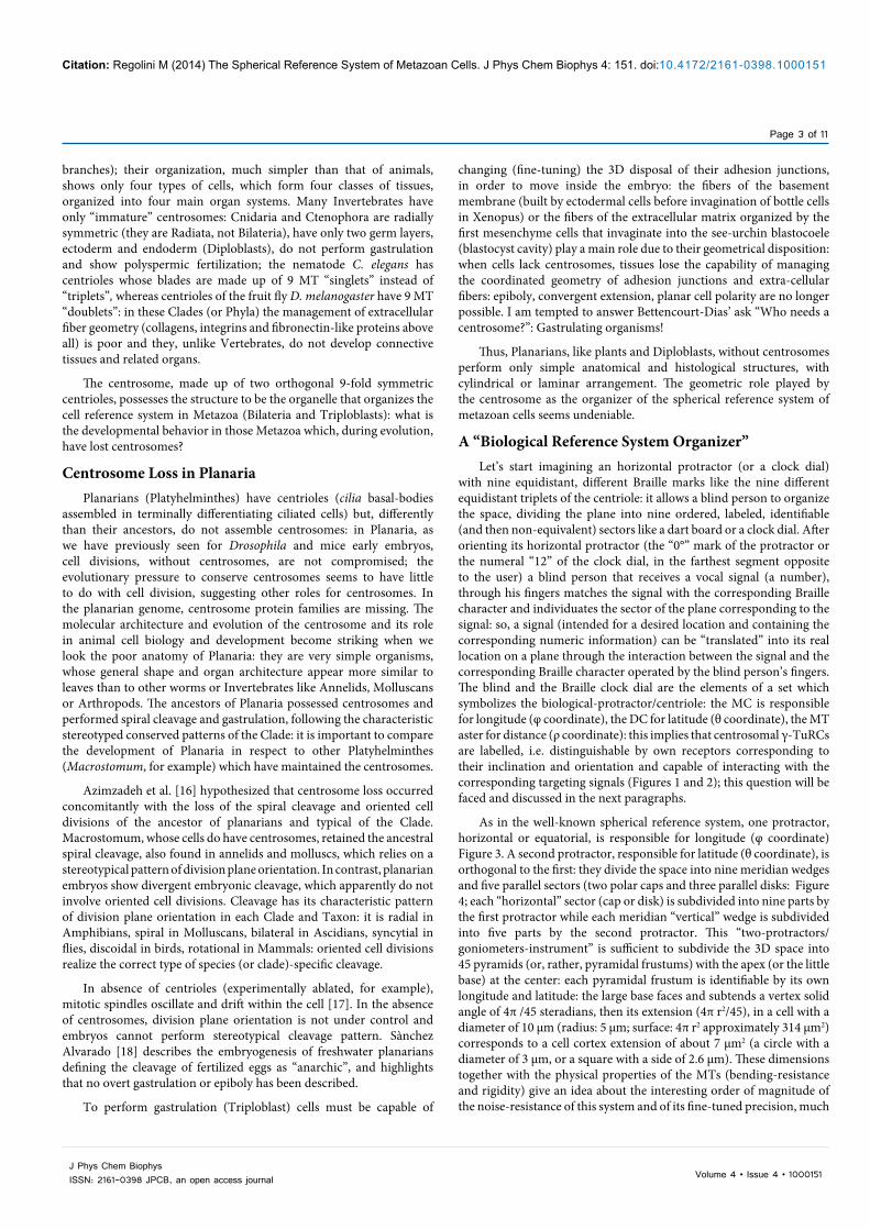
As in the well-known spherical reference system, one protractor, horizontal or equatorial, is responsible for longitude (φ coordinate) Figure 3. A second protractor, responsible for latitude (θ coordinate), is orthogonal to the first: they divide the space into nine meridian wedges and five parallel sectors (two polar caps and three parallel disks: Figure 4; each “horizontal” sector (cap or disk) is subdivided into nine parts by the first protractor while each meridian “vertical” wedge is subdivided into five parts by the second protractor. This “two-protractors/goniometers-instrument” is sufficient to subdivide the 3D space into 45 pyramids (or, rather, pyramidal frustums) with the apex (or the little base) at the center: each pyramidal frustum is identifiable by its own longitude and latitude: the large base faces and subtends a vertex solid angle of 4π /45 steradians, then its extension (4π r2/45), in a cell with a diameter of 10 μm (radius: 5 μm; surface: 4π r2 approximately 314 μm2) corresponds to a cell cortex extension of about 7 μm2 (a circle with a diameter of 3 μm, or a square with a side of 2.6 μm). These dimensions together with the physical properties of the MTs (bending-resistance and rigidity) give an idea about the interesting order of magnitude of the noise-resistance of this system and of its fine-tuned precision, much
branches); their organization, much simpler than that of animals, shows only four types of cells, which form four classes of tissues, organized into four main organ systems. Many Invertebrates have only “immature” centrosomes: Cnidaria and Ctenophora are radially symmetric (they are Radiata, not Bilateria), have only two germ layers, ectoderm and endoderm (Diploblasts), do not perform gastrulation and show polyspermic fertilization; the nematode C. elegans has centrioles whose blades are made up of 9 MT “singlets” instead of “triplets”, whereas centrioles of the fruit fly D. melanogaster have 9 MT “doublets”: in these Clades (or Phyla) the management of extracellular fiber geometry (collagens, integrins and fibronectin-like proteins above all) is poor and they, unlike Vertebrates, do not develop connective tissues and related organs.
The centrosome, made up of two orthogonal 9-fold symmetric centrioles, possesses the structure to be the organelle that organizes the cell reference system in Metazoa (Bilateria and Triploblasts): what is the developmental behavior in those Metazoa which, during evolution, have lost centrosomes?
Centrosome Loss in PlanariaPlanarians (Platyhelminthes) have centrioles (cilia basal-bodies
assembled in terminally differentiating ciliated cells) but, differently than their ancestors, do not assemble centrosomes: in Planaria, as we have previously seen for Drosophila and mice early embryos, cell divisions, without centrosomes, are not compromised; the evolutionary pressure to conserve centrosomes seems to have little to do with cell division, suggesting other roles for centrosomes. In the planarian genome, centrosome protein families are missing. The molecular architecture and evolution of the centrosome and its role in animal cell biology and development become striking when we look the poor anatomy of Planaria: they are very simple organisms, whose general shape and organ architecture appear more similar to leaves than to other worms or Invertebrates like Annelids, Molluscans or Arthropods. The ancestors of Planaria possessed centrosomes and performed spiral cleavage and gastrulation, following the characteristic stereotyped conserved patterns of the Clade: it is important to compare the development of Planaria in respect to other Platyhelminthes (Macrostomum, for example) which have maintained the centrosomes.
Azimzadeh et al. [16] hypothesized that centrosome loss occurred concomitantly with the loss of the spiral cleavage and oriented cell divisions of the ancestor of planarians and typical of the Clade. Macrostomum, whose cells do have centrosomes, retained the ancestral spiral cleavage, also found in annelids and molluscs, which relies on a stereotypical pattern of division plane orientation. In contrast, planarian embryos show divergent embryonic cleavage, which apparently do not involve oriented cell divisions. Cleavage has its characteristic pattern of division plane orientation in each Clade and Taxon: it is radial in Amphibians, spiral in Molluscans, bilateral in Ascidians, syncytial in flies, discoidal in birds, rotational in Mammals: oriented cell divisions realize the correct type of species (or clade)-specific cleavage.
In absence of centrioles (experimentally ablated, for example), mitotic spindles oscillate and drift within the cell [17]. In the absence of centrosomes, division plane orientation is not under control and embryos cannot perform stereotypical cleavage pattern. Sànchez Alvarado [18] describes the embryogenesis of freshwater planarians defining the cleavage of fertilized eggs as “anarchic”, and highlights that no overt gastrulation or epiboly has been described.
To perform gastrulation (Triploblast) cells must be capable of
J Phys Chem BiophysISSN: 2161-0398 JPCB, an open access journal
Citation: Regolini M (2014) The Spherical Reference System of Metazoan Cells. J Phys Chem Biophys 4: 151. doi:10.4172/2161-0398.1000151
Page 4 of 11
Volume 4 • Issue 4 • 1000151
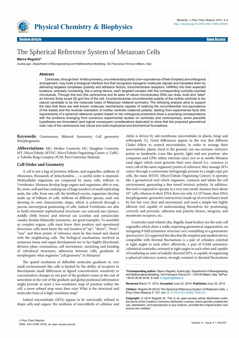
Figure 1: Centrosome theoretical geometrical model: orientation and labeling of -TuRCs. There is an absolute univocal and unambiguous one-to-one correspondence between targeting signal, γ-TuRC receptor, γ-TuRC orientation, MT direction, cell cortex compartment. Signal: undulating line (A) linked to a “twisted-chain” protein; the signal, also named in the text “geometric address” or “ targeting sequence”, is encoded in DNA and, after being transcribed or translated, is contained and displayed in the polypeptide or in the polyribonucleotide (twisted tube) destined for a pre-established cortical localization and recognized by the γ-TuRC receptor (undulating line at the base of the hemi-circle structure, that models the scaffold of the γ-TuRC); different γ-TuRC receptors (B,C) do not fit in with the “undulating” line/signal: they mark γ-TuRCs having orientations corresponding to different directions; orientation of the γ-TuRC itself, from which an MT (D arrow) originates, with a precise direction, points to the desired cell cortex location (E) (thus implicitly encoded in each signal/address). A kinesin carrier (wheel) transports the complex centrifugally. γ-TuRC orientation, MT direction and cortical compartments are the molecular “hardware” of the system whereas signals and γ-TuRC receptors represent the molecular “software”.(From: M. Regolini Centrosome: a geometrical model Lambert Academic Publishing Germany 2014)
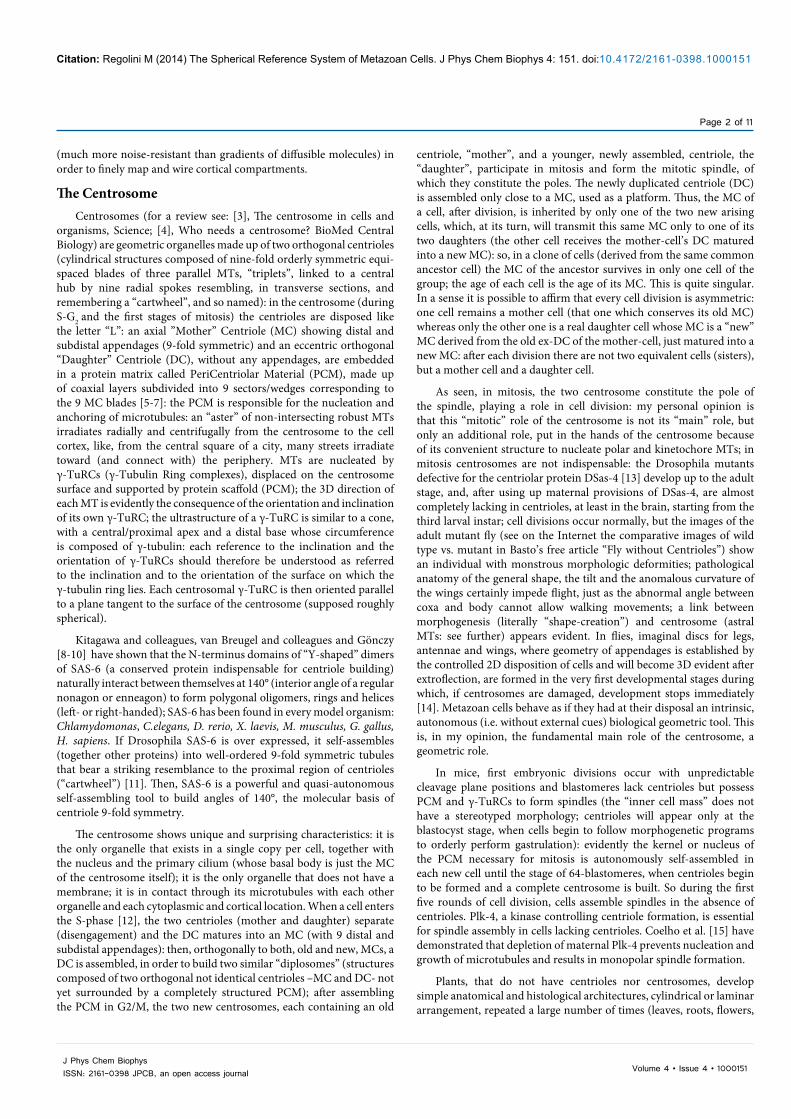
Figure 2: Centrosome theoretical geometrical model: functioning. Small ellipses represent γ-TuRCs on the centrosome (large sphere): each one is identified by its own private receptor specific for its longitude and latitude(capital letters: A, B, C) which recognizes only the corresponding targeting sequence. Each γ-TuRC has the orientation of the plane which, in that point, is tangent to the centrosome “spherical” surface. MTs (arrows) are nucleated with directions imposed by the orientation of the corresponding γ-TuRC: like orientation, like direction (one “discrete” orientation, one “discrete” direction). So, a molecular complex (twisted tube) through its “geometric” targeting sequence, recognizes and links exclusively the receptor (A or B or C) that marks that γ-TuRC which has the correct orientation to nucleate a microtubule directed to the desired (corresponding: A or B, or C) destination, reached through a kinesin carrier (wheel): one targeting sequence, one γ-TuRC receptor, one cortical compartment: one-to-one univocal correspondence.(From: M. Regolini Centrosome: a geometrical model Lambert Academic Publishing Germany 2014)
Figure 3: Centrosome theoretical geometrical model: a spherical reference system composed of two orthogonal protractors/goniometers. A: frontal view of two orthogonal protractors/goniometers, subdivided into nine sectors, which schematizes the two orthogonal centrioles: the first (horizontal) represents the base of the MC, arranged on the equatorial “x y“ plane; its “0° ” mark is used to orient the protractor/centriole; the second, the DC (vertical, orthogonal to the first), is closer to the reader: both “0°“ marks coincide; it is convenient to consider the second protractor divided, by its “vertical” diameter crossing the “0°” mark, into two halves (two opposite symmetrical hemi-protractors).B: schematic lateral view of the proximal end of both centrioles (during S, G2) to show the respective position of the above two sections.(From: M. Regolini Centrosome: a geometrical model Lambert Academic Publishing Germany 2014)
better than that of chemical gradients: 45 cell cortex compartments (or rather poles) are much more than six poles (anterior, posterior, dorsal, ventral, left and right).
Left-Right Let’s now imagine what happens by reverting (clockwise >
counterclockwise) the order of the sequence of the Braille characters of the equatorial protractor (responsible for longitude or φ coordinate): it becomes symmetric in respect to the original one and indicates symmetric directions. If the blind person is equipped with a “reverse” protractor (horizontal and orientated like the original one: the “0°” mark in the farthest segment opposite to him) receiving the same vocal signal as before, matches the signal with the (same) corresponding Braille character but individuates a sector of the plane which is symmetric, compared to that he would find through a non-reverse Braille protractor: the same coded signal has been “translated” into a symmetric location. By only a simple reversion of the sequence of the marks, it is possible to realize bilateral symmetry: it is not necessary to change an enormous number of topogenic instructions; this is particularly interesting from an evolutionary point of view: one single change in place of thousands of changes is much more likely.
Mirror symmetry of two 3D objects is very much easier than one might suppose: it consists of the opposite sign of only one coordinate; any point P (with coordinates “x, y, z”) belonging to one object, is symmetrical to the point P’ belonging to the symmetric object (in this case the plane of symmetry is, arbitrarily, the “y z” plane) whose coordinates are “ –x, y, z ” (note the sign “–“ before the “x” only); in a spherical reference system, only the sign of the φ coordinate changes. The MC of the centrosome, axial and then responsible for longitude, if built with reverse polarity (mirror symmetric in respect to a “default” MC: imagine a clock with the numeral “1” at the place of the numeral “11” and vice versa, the “2” at the place of the “10” and vice versa, and so on) matches each targeting sequence with the same corresponding
J Phys Chem BiophysISSN: 2161-0398 JPCB, an open access journal
Citation: Regolini M (2014) The Spherical Reference System of Metazoan Cells. J Phys Chem Biophys 4: 151. doi:10.4172/2161-0398.1000151
Page 5 of 11
Volume 4 • Issue 4 • 1000151
mark which is symmetrically positioned in respect to the default MC, and assembles aster and cytoskeleton with the same reverse polarity of its triplets, and this is so for the disposition of cell landmarks, polarity complexes, receptors and signaling molecules and, above all, for the direction of extracellular fibers: the whole polarization of cells and tissues is symmetric and the morphogenetic processes of paired and unpaired organs are carried out mirror-symmetrically: the reverse circumferential polarity of the MC is transmitted to tissues and organs that grow bilaterally symmetric. Anatomy of asymmetric organs (heart, liver, etc.) shows that left- or right-handed cells prevail over the opposite-handed ones (see [19], for an interesting review of bilateral symmetry and symmetry breaking in development of Echinoderms); in snails, only one maternal dominant allele is responsible for the left- or right-handed spiral cleavage: it is surprising that only one gene is able to completely reverse (with perfect mirror symmetry) the arrangement and orientation of cleavage planes in many many cells, however mirror symmetry, as seen, consists only in the sign “-“ before the coordinate “φ”; a reversed protractor/centriole performs the same task of a protractor with negative values whose biochemical realization is very difficult. A simple molecular mechanism must necessarily exist capable of reversing MC circumferential polarity.
Unlike animals, plants (that lack centrioles and centrosomes) do not have evident and true bilateral symmetry, a left and a right half or side; only the shape of leaves and flowers could seem nearly symmetric (although 2D only), but the curvature of the two leaf edges (particularly near the petiole and apex), the edge indentation, the position of petiole, the apex tilt, the arrangement of the veins that start alternately from the central vein and, above all, meristem histology and developmental biology exclude the existence of a true bilateral symmetry. Also in Diploblasts and Planaria bilateral symmetry is only apparent. Centrosomes appear to be capable of easily realizing bilaterally symmetric organs, confirming their main geometric role [20].
Centrosome geometry is then well defined: through the assembly of an oriented aster of MTs (and their associated motor proteins kinesins
and dyneins) with centrifugal radial direction, two 9-fold symmetric orthogonal centrioles are really able to organize oriented scaffolds for γ-TuRCs (pericentrin and SAS-4 are proteins involved in assembling γ-TuRC scaffolds), transmitting their circumferential polarity: each scaffold and its γ-TuRC (one or more per scaffold) assumes a double inclination imparted by two orthogonal centrioles (longitude and latitude) so that γ-TuRC rings acquire their own orientation, parallel to the local centrosome surface tangent plane and receive from centrioles also the marks (molecular receptors) corresponding to its orientation and to the relative targeting sequences (Figures 1 and 2).
About Angles and DirectionsThe surface of the centrosome is compartmentalized into
oriented and distinguishable (targeted or labelled) docking platforms for γ-TuRCs: pericentrin fibrils [5] and SAS-4 [6] connect radially centriolar blades and γ-TuRC scaffolds, organizing the PCM into nine sectors. Cell cortex is similarly compartmentalized, mapped and wired through the aster of PCM-MTs [21].
To create the correct stereotypical pattern of cell division orientation, the species-specific genetic instructions for positioning the spindle poles in each cell division are encoded and stored in DNA: in effect we have already seen that only one (maternal) gene is responsible for left- or right-handed spiralian cleavage in snails. The centrosome receives any recognizable signal from DNA and, through its MTs, nucleated by properly oriented γ-TuRCs, positions the poles of the spindle into the programmed cortical compartments in such a way that cleavage is, as forecast by DNA, radial, spiral, bilateral, syncytial, discoidal or rotational. During cleavage, the size of blastomeres can vary in a continuous fashion (starvation, abundance of nutrients, temperature) but the angle between two subsequent division planes is precisely and discretely fixed, always constant for each species and not continuously distributed in an interval of values: to give an example, the angle between the planes of two successive early divisions in sea urchin embryos (from 2 to 4 cells, and continuing to 60 cells) does not vary around a main value, it always has the same identical precise value, whatever the size of the zygote; after the surgical separation of the two first blastomeres of a sea urchin or a frog embryo, two complete, but smaller (about half-size) embryos develop, nonetheless the plane of each cleavage division occurs following and maintaining the same precise stereotyped pattern (orientation), characteristic of the species: size changes, directions do not, like vectors; and so it is during the first stages of organ development. It seems that metazoan cells possess a “discrete” instrument able to precisely read angle values from DNA instructions (species specific, then genetic, pattern of cleavage division orientations) and realize 3D angles, bilaterally symmetric when necessary, having the programmed value: in addition, angle amplitude appears to be chosen in a limited number of discrete values; consider for example left and right semicircular canals and their striking directional precision: the macroscopic result (good precision of anatomical structures) descends from the addition of many many microscopic arrangement of cells, developmental microprograms or modules which organize the assembly of a few cells with an extraordinary level of precision (as in cleavage); the addition of many different modules is necessarily affected and conditioned by different factors (e.g. diverse cell size due to random localized abundance/deficiency of nutrients). Indeed about the word “precision” a comment is necessary: 45 cell cortex compartments are much more than 10 thousand dots per inch (dpi) that is a high graphic precision; however the surface of each cortical compartment (a square with a side of 2.6 μm) is enormous if compared to the dimensions of a protein (the average diameter size
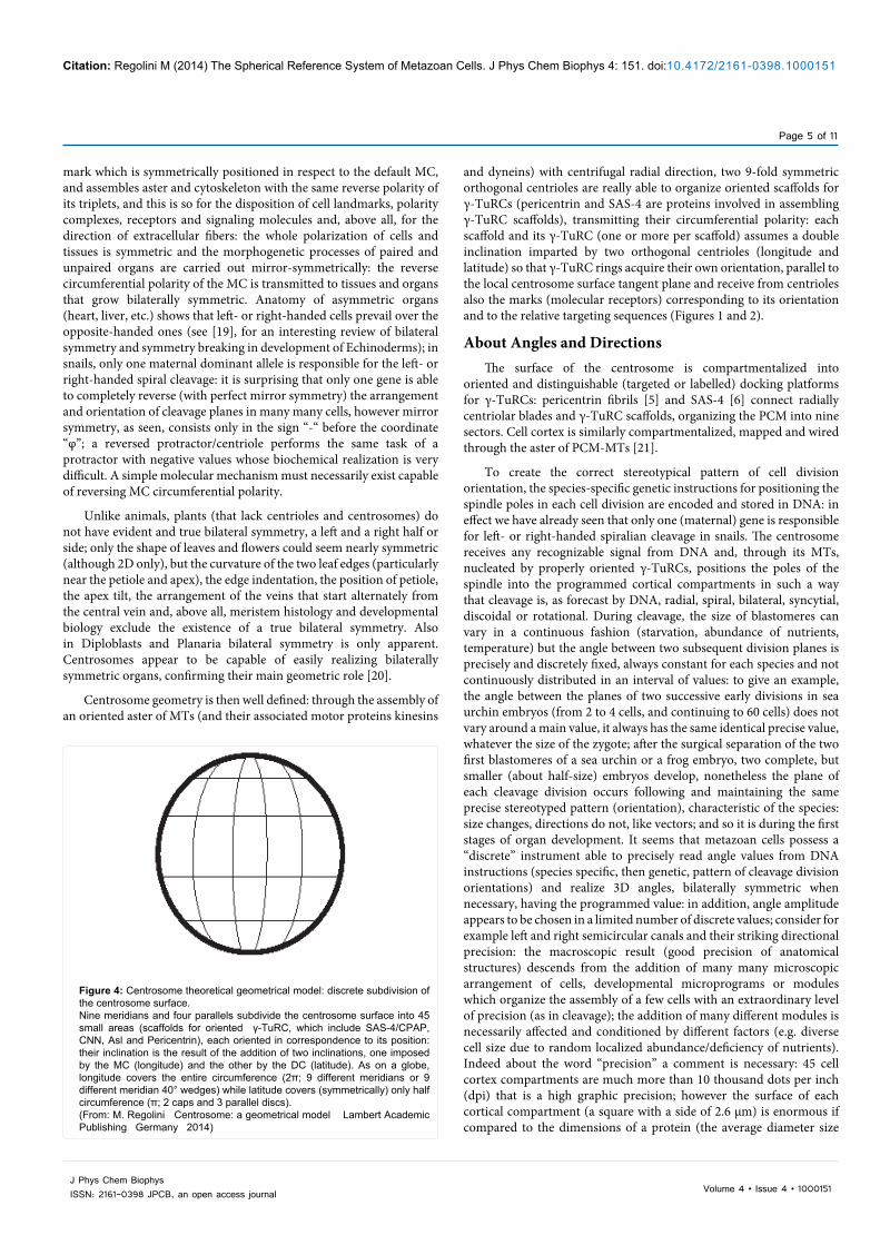
Figure 4: Centrosome theoretical geometrical model: discrete subdivision of the centrosome surface. Nine meridians and four parallels subdivide the centrosome surface into 45 small areas (scaffolds for oriented γ-TuRC, which include SAS-4/CPAP, CNN, Asl and Pericentrin), each oriented in correspondence to its position: their inclination is the result of the addition of two inclinations, one imposed by the MC (longitude) and the other by the DC (latitude). As on a globe, longitude covers the entire circumference (2π; 9 different meridians or 9 different meridian 40° wedges) while latitude covers (symmetrically) only half circumference (π; 2 caps and 3 parallel discs).(From: M. Regolini Centrosome: a geometrical model Lambert Academic Publishing Germany 2014)
J Phys Chem BiophysISSN: 2161-0398 JPCB, an open access journal
Citation: Regolini M (2014) The Spherical Reference System of Metazoan Cells. J Phys Chem Biophys 4: 151. doi:10.4172/2161-0398.1000151
Page 6 of 11
Volume 4 • Issue 4 • 1000151
intrinsic and stand-alone map, orienting its protractor in a defined modality, disposing a particular mark (the “start” or “0°” mark) in a fixed position (close to him, for example): so, the space is no longer homogeneous but subdivided into different and recognizable sectors and becomes a “meaningful” space, a useful room, a detailed and usable environment; now, exploring the room (divided in sectors by the protractor) he is able to map and make use of the room: its parts (doors, windows, walls, furniture) are easily localized and reachable. Similarly DNA, through the centrosome, generates ex novo a 3D (autonomously oriented) labeled environment, a grid line in a previously homogenous cell, transforming and converting it in a mapped, wired, “webbed” and polarized-in-detail cell: a useless homogenous cell cortex becomes a useful and usable compartmentalized space. Through the centrosome, a “blind” DNA becomes a “sighted” DNA: by the centrosome DNA, so to say, turns on the light in the cell (Figures 1 and 2).
Architectural, Biophysical and Molecular Structure of a Biological Spherical Reference System Organizer
A spherical reference system tool based on and built by two orthogonal protractors/goniometers with nine notches, which is what the centrosomes precisely are, in order to recognize (input) coded geometric signals, often at the N-terminus of the newly synthesized proteins or in the 3’UTR of mRNAs, and translate them (output) by delivering each targeted complex into the desired final location, must possess biological structures (centrioles) equipped with: 1) nine different marks (non-equivalent centriolar triplets) transmittable to the PCM corresponding compartments (meridian wedges and parallel caps and disks); 2) a start mark; 3) a constant and ordered sequence of marks; 4) a controlled orientation.
Non-equivalence of the centriolar triplets
Many studies on Protists have demonstrated that the nine triplets of centrioles are different (not-equivalent) and arranged in an ordered sequence: the circumferential (sequential) polarity of the disposal of triplets is accorded with the disposition of the cytoskeleton. Beisson and Jerka-Dziasdosz [22] found that, in Flagellates and Ciliates, the centriolar appendages are diverse, biochemically and morphologically, microtubule appendages show highly complex shapes as in Physarum or in Ochromonas. “It seems that in Protists and in Metazoa the triplets of basal bodies are not-equivalent…Is it possible to confirm this idea that the circumferential, morphological, structural and molecular asymmetry of centrioles can be inferred from Mammals ciliated epithelia? While the circumferential anisotropy of centrioles cannot be ascertained within the centrosome, its existence can be inferred from the properties they express during ciliogenesis, be it the formation of a primary cilium or of bona fide 9+2 cilia in ciliated epithelia, some of which at least derive directly from the centrioles. As in Ciliates and flagellates, these basal bodies nucleate appendages of various molecular compositions (basal foot, striated rootlets, alarm sheets, etc, which anchor the basal body to the membrane and to the cytoskeleton) and these nucleations arise at specific sites of the basal body cylinder; in particular, the basal foot is located on triplets 5 and 6 corresponding to the side of the effective stroke of the cilium. What is remarkable is that basal feet develop before the basal bodies reach their membrane site and before they acquire their functional orientation [22].
Electron microscopy has clearly highlighted the circumferential rotational asymmetry of Ciliates basal bodies/centrioles: a system for numbering the basal body triplets can be established by tracing axonemal doublets (cilia and flagella have nine MT “doublets” instead of “triplets”) in serial sections of the transition zone from the
is about 3-6 nm); so in metazoan organisms, composed of thousands billion cells (1014), we can see high precision of shapes but not absolute identity; Metazoa utilizes that level of precision which necessity and sufficiency require to achieve stereotyped reproducibility of high quality, but not identity.
DNA, through the Centrosome, Organizes a 3D Oriented Environment in a Previously Homogenous Cell
A “Braille marked” protractor is not used to measure, rather it is a useful translator of address-signals which allows a blind person to find locations (in 2D) corresponding to received signals; two orthogonal protractors perform the same identical task in 3D. As seen, in a cell, such instrument is an interface, a biological geometric tool that receives molecular coded signals (input: targeting or topogenic sequences), each one intended for a particular cortical compartment, recognizes them through their tertiary 3D structure and matches each targeting sequence with the corresponding mark (ligand-receptor interaction), returning (output) the spatial position of the desired locations to carry and delivery molecular complexes precisely where they are expected: any location can be easily reached through an oriented ray (an MT and motor protein like kinesins) arising from the selected mark (an oriented and “labeled” γ-TuRC) (Figures 4 and 5).
For a blind person, the space of an empty unknown room is obviously homogeneous, without any point of reference, everywhere identical and then “meaningless” and useless; through an oriented 9-graduated “Braille” protractor, the same space acquires nine intrinsic, autonomous and useful points of reference, relative to the room itself and without external cues: the blind person ignores where is the door or the window (external points of reference) and defines an autonomous,
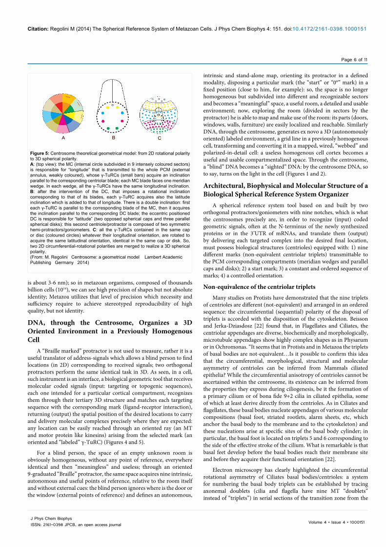
Figure 5: Centrosome theoretical geometrical model: from 2D rotational polarity to 3D spherical polarity.A: (top view): the MC (internal circle subdivided in 9 intensely coloured sectors) is responsible for “longitude” that is transmitted to the whole PCM (external annulus, weakly coloured), whose γ-TuRCs (small bars) acquire an inclination parallel to the corresponding centriolar blade; each MC blade faces one meridian wedge. In each wedge, all the γ-TuRCs have the same longitudinal inclination. B: after the intervention of the DC, that imposes a rotational inclination corresponding to that of its blades, each γ-TuRC acquires also the latitude inclination which is added to that of longitude. There is a double inclination: first each γ-TuRC is parallel to the corresponding blade of the MC, then it acquires the inclination parallel to the corresponding DC blade; the eccentric positioned DC is responsible for “latitude” (two opposed spherical caps and three parallel spherical disks): this second centriole/protractor is composed of two symmetric hemi-protractors/goniometers. C: all the γ-TuRCs contained in the same cap or disc (coloured circles) whatever their longitudinal orientation, are rotated to acquire the same latitudinal orientation, identical in the same cap or disk. So, two 2D circumferential-rotational polarities are merged to realize a 3D spherical polarity.(From: M. Regolini Centrosome: a geometrical model Lambert Academic Publishing Germany 2014)
J Phys Chem BiophysISSN: 2161-0398 JPCB, an open access journal
Citation: Regolini M (2014) The Spherical Reference System of Metazoan Cells. J Phys Chem Biophys 4: 151. doi:10.4172/2161-0398.1000151
Page 7 of 11
Volume 4 • Issue 4 • 1000151
axoneme down into the basal body [23]. So, in Ciliates it is possible to observe the connection between each triplet with particular fibers of the cytoskeleton: in the green alga Chlamydomonas reinhardtii the so named “striated” fibers of the “distal connector”, tie and fasten the triplets 9-1-2 of both basal bodies; the ”thick cruciform” fibers are attached to the triplets N° 3 and 4, whereas the “thin cruciform” ones are linked to the triplet N° 8; the newly-forming procentriol arises (orthogonally) always in front of the triplet N° 9. Geimer and Melkonian have also described, in Chlamydomonas, an “acorn-like” structure in the inner distal part of the basal body, adhering in a highly stereotyped manner to the triplets 2-1-9-8-7, and a second structure, shaped like the capital letter “V” in contact with the triplets 9, 5 and 4; Geimer and Melkonian sustain that the “cartwheel-shaped structure” realize the 9-fold symmetry of the triplets, whereas the acorn-shaped structure might play an equally important role imposing rotational molecular asymmetry on the microtubular triplets, controlling the asymmetric assembly of basal-body-associated fibers and cellular asymmetry in general.
The shape of any structure is the consequence of its molecular composition: different (non-equivalent) shapes imply different molecular composition (quaternary structure): each triplet possesses its own molecular distinct identifying mark (an “address” receptor). In Chlamydomonas the polypeptide VFL1 coded by the gene vfl1 (Variable number of FLagella), binds only to the triplet N° 1 [24] confirming the biochemical nature of circumferential asymmetry, i.e. each triplet has its own characteristic receptor (“address” label) which allows the triplet to interact only with its own ligand. Surprisingly Yoshinori and Yukio [25] found a human ortholog of Chlamydomonas Vfl1 protein and other correlated centrosomal proteins, highlighting that database searches provide evidence that the human proteome contains at least seven centrosomal leucine-rich repeat proteins. The question posed by Beisson and Jerka-Dziasdosz “Is it possible to confirm this idea that the circumferential, morphological, structural and molecular asymmetry of centrioles can be inferred from Mammals ciliated epithelia?” might receive an affirmative answer.
9-fold symmetry is not a characteristic of only axonemes, basal bodies and centrioles: also the PCM is 9-fold symmetric; in mammalian cells, the PCM is divided into nine sectors by radially disposed linear molecules Pericentrin like proteins [5] and SAS-4 [6], reproducing the 9-fold symmetry of the MC.
Biochemical non-equivalence of triplets gives a strong support to the idea of molecular receptors able to distinguish each triplet and the corresponding sector of the PCM: so, each γ-TuRC scaffold is equipped with proper molecular receptors by the geometrically corresponding MC and DC triplets and, consequently, to its spatial inclination Figures 1 and 2. Proteins and mRNAs are transported individually to particular locations in cell space because of an appropriate sequence. Transport vesicles recognize specific target membranes, such as the Golgi, vacuole, or plasma membrane, with the aid of SNARE proteins. In metazoans cells, targeted vesicle fusion requires, in addition to the SNAREs, both a delivery system and a secretory apparatus [26]. The centrosome and its aster of MTs map, label and wire the whole cell for building a precise, noise-resistant, delivery system.
A few words about the orthogonality of centrioles: one centriole, the MC, is a central hub while the second cannot form a cross with it but must assume any eccentric configuration: the simplest arrangement appears to be the known juxtaposition of the ends of both centrioles so that the MC is not a barrier and does not impede the DC from reaching every PCM wedge and transmitting to each wedge its
inclination latitudinal information (Regolini, 2014); furthermore this type of eccentricity allows the MC to control the orientation (shared and coordinated tissue polarity) of the DC at the time of its maturation into a new MC: thus DC rotational polarity can be orderly arranged so that the start marks (see next paragraph) of both centrioles coincide.
Start mark
The triplet N° 1 of C. reinhardtii (in the transition zone from basal-body to axoneme, easily recognized by the absence of the outer dynein arm in the axoneme) is continuous with the middle of the three triplets (9-1-2) to which the distal connecting fiber is attached [23] and with the triplet N° 9, in front of which the newly arising centriole is assembled: this site (between triplets N° 9 and N°1) could be the “start mark”, i.e. the starting mark from which the ordered sequence of nine marks begins and then used to orientate the acorn-shaped ring which Geimer and Melkonian consider to be responsible for the non-equivalence of the triplets (as just seen, this site is in contact with the “acorn-shaped” ring and the “V-shaped” structure). Controlling the orientation of the acorn-ring and fixing a recognizable “start mark” in a position imposed by the centriole which is used as platform (also in Ciliates centriole duplication occurs at right angles between the parent and nascent centriole) cells are capable of disposing orderly the sequence of the nine structure responsible for identifying each triplet and generating rotational polarity.
Constant and ordered sequence of marks
The symbols for the hours on a clock are orderly sequenced (circumferential polarity) and the symbol for “12 o’clock” (the “start mark”) is always at the top to guarantee the global orientation of the whole structure: which known biochemical mechanism can create the circumferential polarity of the nine centriole triplets, ordering, arranging and aligning them in accordance to a predefined sequence and a pre-established global orientation?
Non-equivalence of the triplets can be imposed by their quaternary structure, rather than by the tertiary structure, i.e. the nine triplets have the same tertiary structure (they are identical) and their difference consists in the juxtaposition of different macromolecules; a ring is certainly the most likely tool and the “acorn-shaped” ring described by Geimer and Melkonian is particularly appealing; a ring of an informational macromolecule able to contain a linearly ordered sequence of equi-spaced different 3D markers appears to be the most suitable structure. What kind of ring? An informational polypeptide or a polynucleotide ring: the last one has been effectively observed by Dippel [27] within the core of the basal body as a twisted or looped 90 Å diameter fiber, or more probably pair of fibers, RNase-sensitive, in association with dense granules. Some researchers consider the centrosome and spindle even a ribonucleoprotein complex [28,29].
Orientation of centrioles and centrosomes
The unique, very complicated and costly, centriole and centrosome cycle and duplication is part of the mechanism by which the cytoskeleton of the daughter cell is patterned upon that of the mother [26]: through this sophisticated process, the MC, before disengagement, is able to transmit to its old DC the information of orientation, and physically orients its old DC (before maturation into a new MC) in respect to the cytoskeleton; effectively also in Ciliates and Flagellates, as already seen, the process of centriole duplication occurs at right angles and utilizes a pre-existing centriole as a platform to orientate the arising centriole polarity in order to insert it correctly in the complex cytoskeleton; Paramecium (Ciliates) has a high number of basal bodies and cilia,
J Phys Chem BiophysISSN: 2161-0398 JPCB, an open access journal
Citation: Regolini M (2014) The Spherical Reference System of Metazoan Cells. J Phys Chem Biophys 4: 151. doi:10.4172/2161-0398.1000151
Page 8 of 11
Volume 4 • Issue 4 • 1000151
about 4,000: this Protist organizes and connects each basal body with cytoskeletal fibers in about 70 regular meridian rows, always with the same correct orientation of the triplets, something like a new trolley-bus (whose two sprung trolley poles must be correctly connected to the two polarized electric wires) is orientated and correctly (front-rear) positioned in respect to the electric “city-skeleton” made of aerial-suspended wires. So the cells in a tissue become coordinately polarized by coordinately oriented centrosomes: in Metazoa the centrosome is the “intrinsic” (no external cues) reference system; as “extrinsic” reference points are the same for each receiver and the related reference system is common to every receiver, similarly an “intrinsic” reference system must be the same (identically oriented) in each cell; multicellular organisms must possess a mechanism to transmit, share and coordinate their inside points of reference (as we have seen, the centrosome increases cell polarity up to 45 poles) and this function is performed through the orientation imprinted by the MC to its “old” DC before this one disengages and matures into a new MC, so that two co-oriented MCs build two co-oriented DCs and assemble two co-oriented centrosomes before cell division: the dividing cell, entering prophase, controls the “insertion” of the new centrosome into the cytoskeleton orienting its ordered sequence of triplets like the (non-equivalent!) pins of a microchip are connected to precise conductive tracks of a printed circuit board; so all the cells of a tissue inherit the same orientation (not having any common external points of reference) to correctly coordinate their global polarity (the cell grid-line or graticule of compartments) for building complex 3D organs: planar cell polarity components (Frizzled, Vangogh, Dishevelled and Prickle) are asymmetrically positioned in cells by oriented MTs before receiving Wnt signaling ligands. This may be (and very likely is) the reason for the complicated centrosome duplication process: before cell duplication, new centrioles are not produced de novo, on the contrary they arise using an MC as a platform to tune in their circumferential polarity with that of the MC [20].
Proposal of a Model of Centrosome FunctioningBefore modeling how the centrosome could carry out its geometric
role, organize a spherical reference system in metazoan cells, assemble a radial aster of MTs to map and wire the whole cortex, it is convenient to deal with two important macromolecular complexes: microtubules and centrosomal-RNA.
Microtubules
MTs are long and strong tubes composed of 13 parallel filaments of oriented dimers made of an α- and a β-tubulin monomer (-αβαβ…αβ+: the proximal end beginning with α-tubulin is called minus “-“ and starts from the nucleating unit, a γ-TuRC or another cytoplasmic MTOC as the Golgi complex, whereas the opposite β plus “+” growing end is distal and pushed toward the cell cortex).
Many images of interphase cells show the cytoplasm crowded with much more than 45 astral MTs (it is conceivable that more than only one MT originate from each oriented scaffold, but the number of cytoplasmic MTs remain too high); however many free (not linked to the centrosome) MTs, differently than astral or centrosome-made MTs, do not start from PCM γ-TuRCs, but are nucleated by other free cytoplasmic MTOC; quantitative analysis and model systems, in which all the MT minus-ends are attached to the centrosome while the plus-ends are at different distances from it, show that the astral MT density is described by the exponential f(x) = ae−bx. Introducing free MTs into the system leads to a change of the character of this dependence, and the system in which the concentration of free MTs
with minus ends located at different distances from the centrosome is 5 times higher than that of the centrosome-attached MTs and the system is described by the linear regression equation f(x) = kx + b, which corresponds to the experimentally obtained data [30]. Astral MTs, nucleated by centrosomal γ-TuRCs, constitute a small, limited, stable sub-population of the whole MT set, whereas other free MTs, built by cytoplasmic MTOCs, appear more dynamic and instable: astral MTs are quasi-stable microtubules that do not exchange subunits as rapidly as the majority of other microtubules (polar, kinetochore, structural, neuronal) and may have specialized functions in the cell [31,32] described perinuclear microtubules which constitute another “stable subpopulation of cytoplasmic MTs and may serve a function different from that of the more variable not perinuclear microtubules”.
So, astral MTs (stable, centrosome-made and less dynamic than cytoplasmic MTs) are only 1/5 (or 1/6) of the total interphase MT set, showing a good correspondence and agreement with the expected number of “wiring and mapping” centrosomal astral MTs.
Centrosomal RNA
“An intriguing morphological and developmental parallel between certain procentriolar elements and an RNA-containing component of the Paramecium kinetosome occurs during centriolar production and development in embryonic chick tracheal epithelial cells” [27]. This observation is much more important than it might appear: RNA presence in centrosomes is a matter of discussion, likely because its quantity is very small and, as Dippel observed, temporary: thus, functional, instead chemical, analysis are more meaningful.
Hiedemann and colleagues [33] reported their observations on the formation of an aster (although incomplete and non-functional) following the implant of Chlamydomonas and Tetrahymena basal bodies into Xenopus eggs: after the treatment of the basal bodies with weak concentrations of RNase, the formation of the partial aster was found to be completely suppressed; γ-TuRC nucleation of astral MTs appeared to be sensitive to the RNase, whereas only the centriolar MTs of the triplets (centriole wall) after removal of the CP110 cap, could still grow in length (in vitro. What is the functional association between RNA and PCM γ-TuRCs? Laane and Haugli [34] had already studied the association of RNA with centrosomes in Physarum, and Hartman and colleagues [35] in Tetrahymena pyriformis: together to Snyder (1980), they showed that RNase inhibited MT nucleation. Finally, clear and distinct centrosomal RNAs (cnRNAs), not coding and absent from the remaining cytoplasm have been identified in the centrosome of Spisula Solidissima (organism clearly bilaterally symmetric) [36]. Recently Ishigaki and colleagues [37] have found that RBM8A-MAGOH localizes in centrosomes in addition to nuclei (RBM8A possesses an RNA-binding motif and forms a tight heterodimer with MAGOH: this heterodimer is known to be a member of exon junction complex on exporting mRNA and is required for mRNA metabolisms such as splicing, mRNA export and nonsense-mediated mRNA decay). Dos Santos (2013) reports that centrosomal proteins tend to be larger than generic human proteins since their genes contain in average more exons (20.3 versus 14.6). They are rich in predicted disordered regions, which cover 57% of their length, compared to 39% in the general human proteome: as known, disordered protein domains frequently bind nucleic acids. Also proteins with leucine-rich repeats as the centrosomal protein CLERC (Centrosomal Leucine-Rich repeat and Coiled-coil containing protein) which is a human ortholog of Chlamydomonas Vfl1 protein (Yoshinori and Yukio, 2010) and other leucine-rich centrosomal protein bind nucleic acids.
J Phys Chem BiophysISSN: 2161-0398 JPCB, an open access journal
Citation: Regolini M (2014) The Spherical Reference System of Metazoan Cells. J Phys Chem Biophys 4: 151. doi:10.4172/2161-0398.1000151
Page 9 of 11
Volume 4 • Issue 4 • 1000151
Centrosomal RNA is particularly interesting because an RNA ring seems to be the best solution for many centrosome requirements: which known biochemical mechanism can create the circumferential polarity of the nine centriole triplets, ordering, arranging and aligning them in accordance to a predefined sequence and a pre-established global orientation? how is it possible to assemble in an opposite sequence (clockwise > counterclockwise) nine radial spokes and nine centriolar blades? A ring of different RNA loops or hairpins (as in the well known t-RNA secondary structure), orderly clockwise sequenced and equidistant, juxtaposed to the centriolar wall, can decorate and make different and distinguishable each one of the nine identical centriolar triplets; an RNA ring can also be easily reverted to assume a counter-clockwise disposition (a biochemical easy basis for left/right or bilateral symmetry; imagine to reverse a t-RNA molecule: the canonical clockwise sequence “CCA on the acceptor stem > T loop > anticodon loop > D loop”, after the reversion, becomes counter-clockwise opposite and then bilaterally symmetrical in comparison with the non reversed molecule. Reversion might be realized by an event of retrotransposition [20]: Alliegro [36] has identified in centrosomal RNA a sequence of a highly conserved 200 amino acid-long reverse transcriptase domain.
The geometrical model of centrosome functioning
Centrosome geometry and architecture must necessarily imply its function: as a technical designer first squares a sheet, similarly, during the last period of each cell division, through the centrosome, DNA first maps and wires the non-polarized and homogeneous cell cortex: so, DNA builds the intrinsic 3D map of the cell, transforming a (DNA-coded) “virtual” grid line into a real “actual” cellular grid with intrinsic points of reference which dictate and orientate the position of membrane polarity factors; DNA uses the centrosome to polarize the whole cellular cortex and membrane in order to assume the spatial control and the mastery of the cellular and extracellular environment. (Pay attention to the expressions “through the centrosome, DNA first maps” and “DNA uses the centrosome”: the centrosome is only a sophisticated instrument in the hands of, and controlled by, DNA, then it does not work autonomously, on the contrary it is strictly directed and managed by DNA).
Thus it is possible to delineate the fundamental properties of this organelle and the principles of its geometrical role.
- Centrioles have a circumferential ordered and discrete asymmetry as a consequence of macromolecular differences of the quaternary structure of their triplets.
- Centrioles are platforms for a set of regulatory molecules that assist and facilitate the semi-self-assembly of the PCM.
- The MC transmits its 9-fold symmetry (longitude-dependent inclination: φ coordinate of a spherical reference system) to PCM γ-TuRC scaffolds /docking platforms.
- The DC transmits its 9-fold symmetry (latitude-dependent inclination: θ coordinate of a spherical reference system) to PCM γ-TuRC scaffolds /docking platforms.
- Each γ-TuRC on a centrosomal scaffold/docking platform is oriented in accord with the tilt of the local tangent plane: scaffold’s (and γ -TuRC’s) inclination is the addition of the latitudinal (DC) to the longitudinal tilt (MC).
- Both centrioles transmit to the PCM their circumferential asymmetry and impress their molecular non-equivalence.
- Each γ-TuRC (or its centrosomal scaffold / docking platform) is “labeled” by the receptors, (longitude and latitude) corresponding to its own position and orientation, received from both centrioles.
- Each γ -TuRC (or its centrosomal docking platform) displays its receptors (longitude and latitude) to recognize the signal (a molecular ligand with a particular targeting sequence) corresponding to its own orientation and to the intended location in the cell cortex.
- The direction of PCM astral MTs depends on the orientation (tilt) of the γ -TuRCs by which they are nucleated.
- Each signal (ligand) has a 3D shape that recognizes only the receptors of that γ-TuRC that is oriented in order to nucleate an MT with the desired direction: there is a precise and invariable one-to-one correspondence between geometric signals and γ-TuRC receptors.
- The correspondence between centrosomal and cortical compartments is univocal (one-to-one) but time-variable, since it depends on the position and orientation of the centrosome: a same γ-TuRC can be connected (under the strict control of DNA), at different times, to diverse cortical compartments. This is the consequence of the different position of the centrosome: in dividing cells, during G1 (interphase) the centrosome is near the nucleus, at the center of the cytoplasm, whereas in non dividing cells it moves to the cell cortex where the MC is the basal body of the primary non-motile cilium.
- The one-to-one correspondence between targeting signals and γ-TuRC receptors is constant and invariable, whereas the one-to-one correspondence between targeting signals and cortical compartments is time-variable.
- The centrosome is a geometric discrete interface that receives geometric coded signals (input), matches each one with the corresponding γ-TuRC receptor (decoding: ligand-receptor interaction) and nucleates oriented MTs (translation) to reach the required locations (output). It is an attractive hypothesis that several centrosome targeting sequences exist, placed on those macromolecular complexes (RNA 3’ UTR or N-terminus of polypeptides) that must be routed towards prefixed cell locations through the appropriate MTs. Such a mechanism is quite similar to that used for translocation of proteins in mitochondria, where different targeting sequences, present on the proteins that must enter the mitochondrion, are sequentially decoded in order to reach the expected location (the external mitochondrial membrane, the internal one, the inter-membrane space or the matrix); it is well known that in fly oocyte bicoid and nanos mRNA are localized through their 3’UTR sequence and delivered by MT transport.
- Reverse or opposite rotational polarity of the MC is the base of Left-Right patterning and bilateral symmetry.
- Global architecture: to use a spherical reference instrument based on two orthogonal protractors/goniometers, like a globe, a two-step process is necessary: first the longitude of a point (φ coordinate) must be found, then, after a rotation of the vertical protractor around its axis for aligning the protractor inner border in correspondence with the found longitude, its latitude (θ coordinate) can be identified; this appears particularly complex and difficult (and frankly unlikely) in a cell; on the contrary, if a little sphere (always maintained in a controlled position and orientation) like the centrosomal PCM, is organized only once and for all in each cell, the problem of spatial orientation in the cell environment has a very easy solution. The PCM surface is subdivided and compartmentalized into 45 small areas, each one oriented and labelled by molecular receptors that recognize
J Phys Chem BiophysISSN: 2161-0398 JPCB, an open access journal
Citation: Regolini M (2014) The Spherical Reference System of Metazoan Cells. J Phys Chem Biophys 4: 151. doi:10.4172/2161-0398.1000151
Page 10 of 11
Volume 4 • Issue 4 • 1000151
the geometric (molecular) signals intended for the corresponding longitude and latitude (and, consequently, for the corresponding cytoplasmic sector too). Like on a globe parallels and meridians are marked on its surface once and for all to facilitate the task of finding a point of given coordinates, so the 9-fold symmetry of two orthogonal cylindrical centrioles, transmitted and impressed on the centrosome surface once and for all, permits an easy translation of molecular geometric signals (in which the intended coordinates are coded) into their corresponding localizations in the cytoplasm, only by the usual signal-receptor interaction between targeting sequences and γ-TuRC (or scaffold/docking platform) receptors, located on the centrosome surface. Obviously, this must happen maintaining fixed (or variable but, in any case under control that is never random) the orientation of the centrosome in the cell [38-44].
ConclusionCiliates have cylindrical centrioles, Metazoa have two orthogonal
cylindrical centrioles embedded in a “spherical” centrosome: the second centriole, orthogonal to the first, adds the capability of completely controlling the third dimension and building an accurate spherical reference system, indispensable for managing the arrangement of cells in multicellular organisms. The idea is that Metazoa have developed new pathways to adapt Ciliates’ sophisticated molecular mechanisms in order to transmit the 9-fold symmetry and polarity of two orthogonal cylindrical centrioles to the spherical wedges and sectors of the centrosome (or rather of the PCM): so Metazoa could assemble once and for all, a tool through which they can polarize (finely and coordinately) their cells, control, direct and organize their 3D anisotropic growth.References
1. Schwarz H, Moussian B (2007) Electron-microscopic and genetic dissection of arthropod cuticle differentiation. Modern research and educational topics in microscopy.
2. Albrecht-Buehler G (1981) Does the geometric design of centrioles imply their function? Cell Motil 1: 237-245.
3. Bornens M (2012) The centrosome in cells and organisms. Science 335: 422-426.
4. Bettencourt-Dias M (2013) Q&A: Who needs a centrosome? BMC Biol 11: 28.
5.
6. Sonnen KF, Schermelleh L, Leonhardt H, Nigg EA (2012) 3D-structured illumination microscopy provides novel insight into architecture of human centrosomes. Biol Open 1: 965-976.
7. Mennella V, Keszthelyi B, McDonald KL, Chhun B, Kan F, et al. (2012) Subdiffraction-resolution fluorescence microscopy reveals a domain of the centrosome critical for pericentriolar material organization. Nat Cell Biol 14: 1159-1168.
8. Gopalakrishnan J, Mennella V, Blachon S, Zhai B, Smith AH, et al. (2011) Sas-4 provides a scaffold for cytoplasmic complexes and tethers them in a centrosome. Nat Commun 2: 359.
9. Kitagawa D, Vakonakis I, Olieric N, Hilbert M, Keller D, et al. (2011) Structural basis of the 9-fold symmetry of centrioles. Cell 144: 364-375.
10. van Breugel M, Hirono M, Andreeva A, Yanagisawa HA, Yamaguchi S, et al. (2011) Structures of SAS-6 suggest its organization in centrioles. Science 331: 1196-1199.
11. Gönczy P (2012) Towards a molecular architecture of centriole assembly. Nat Rev Mol Cell Biol 13: 425-435.
12. Cottee MA, Raff JW, Lea SM, Roque H (2011) SAS-6 oligomerization: the key to the centriole? Nat Chem Biol 7: 650-653.
13. Vorobjev IA, Chentsov YuS (1982) Centrioles in the cell cycle. I. Epithelial cells. J Cell Biol 93: 938-949.
14. Basto R, Lau J, Vinogradova T, Gardiol A, Woods CG, et al. (2006) Flies without centrioles. Cell 125: 1375-1386.
15. Baker J, Theurkauf WE, Schubiger G (1993) Dynamic changes in microtubule configuration correlate with nuclear migration in the preblastoderm Drosophila embryo. J Cell Biol 122: 113-121.
16. Coelho PA, Bury L, Sharif B, Riparbelli MG, Fu J, et al. (2013) Spindle formation in the mouse embryo requires Plk4 in the absence of centrioles. Dev Cell 27: 586-597.
17. Azimzadeh J, Wong ML, Downhour DM, Sánchez Alvarado A, Marshall WF (2012) Centrosome loss in the evolution of planarians. Science 335: 461-463.
18. Azimzadeh J, Marshall WF (2010) Building the centriole. Curr Biol 20: R816-825.
19. Sànchez Alvarado A (2004) Planarians – Cell.
20. Morris VB (2007) Origins of radial symmetry identified in an echinoderm during adult development and the inferred axes of ancestral bilateral symmetry. Proc Biol Sci 274: 1511-1516.
21. Regolini M (2014) Centrosome: a geometrical model Lambert Academic Publishing Germany.
22. Dodgson J, Chessel A, Yamamoto M, Vaggi F, Cox S, et al. (2013) Spatial segregation of polarity factors into distinct cortical clusters is required for cell polarity control. Nat Commun 4: 1834.
23. Beisson J, Jerka-Dziadosz M (1999) Polarities of the centriolar structure: morphogenetic consequences. Biol Cell 91: 367-378.
24. Geimer S, Melkonian M (2004) The ultrastructure of the Chlamydomonas reinhardtii basal apparatus: identification of an early marker of radial asymmetry inherent in the basal body. J Cell Sci 117: 2663-2674.
25. Silflow CD, LaVoie M, Tam LW, Tousey S, Sanders M, et al. (2001) The Vfl1 Protein in Chlamydomonas localizes in a rotationally asymmetric pattern at the distal ends of the basal bodies. J Cell Biol 153: 63-74.
26. Yoshinori M, Yukio O (2010) CLERC and centrosomal leucine-rich repeat proteins. Cent Eur J Biol 5: 1-10.
27. Harold FM (2005) Molecules into cells: specifying spatial architecture. Microbiol Mol Biol Rev 69: 544-564.
28. Dippell RV (1976) Effects of nuclease and protease digestion on the ultrastructure of Paramecium basal bodies. J Cell Biol 69: 622-637.
29.
30. Alliegro MC (2011) The centrosome and spindle as a ribonucleoprotein complex. Chromosome Res 19: 367-376.
31. Chichinadze K, Lazarashvili A, Tkemaladze J (2013) RNA in centrosomes: structure and possible functions. Protoplasma 250: 397-405.
32. Smurova KM, Alieva IB, Vorob’ev IA (2007) [Free and centrosome-attached microtubules: quantitative analysis and the modeling of two-component system]. Tsitologiia 49: 270-279.
33. Schulze E, Kirschner M (1986) Microtubule dynamics in interphase cells. J Cell Biol 102: 1020-1031.
34. Park RS, Légier MF, Cartwright J Jr, Goldstein MA (1984) Perinuclear microtubules in postnatal rat heart. J Morphol 179: 13-19.
35. Heidemann SR, Sander G, Kirschner MW (1977) Evidence for a functional role of RNA in centrioles. Cell 10: 337-350.
36. Laane MM, Haugli FB (1974) Division centers in mitotic nuclei of Physarum polycephalum plasmodia. Norw J Bot 21: 309-318.
37. Hartman H, Puma JP, Gruney T Jr (1974) Evidence for the association of RNA with the ciliary basal bodies of Tetrahymena. J Cell Sci 16: 241-259.
38. Alliegro MC, Alliegro MA, Palazzo RE (2006) Centrosome-associated RNA in surf clam oocytes. Proc Natl Acad Sci U S A 103: 9034-9038.
J Phys Chem BiophysISSN: 2161-0398 JPCB, an open access journal
Citation: Regolini M (2014) The Spherical Reference System of Metazoan Cells. J Phys Chem Biophys 4: 151. doi:10.4172/2161-0398.1000151
Page 11 of 11
Volume 4 • Issue 4 • 1000151
39. Ishigaki Y, Nakamura Y, Tatsuno T, Hashimoto M, Iwabuchi K, et al. (2014) RNA-binding protein RBM8A (Y14) and MAGOH localize to centrosome in human A549 cells. Histochem Cell Biol 141: 101-109.
40. Dos Santos HG, Abia D, Janowski R, Mortuza G, Bertero MG, et al. (2013) Structure and non-structure of centrosomal proteins. PLoS One 8: e62633.
41. Lodish AB (2012) Molecular Cell Biology. WH Freeman & Co., New York.
Submit your next manuscript and get advantages of OMICS Group submissionsUnique features:
• User friendly/feasible website-translation of your paper to 50 world’s leading languages• Audio Version of published paper• Digital articles to share and explore
Special features:
• 350 Open Access Journals• 30,000 editorial team• 21 days rapid review process• Quality and quick editorial, review and publication processing• Indexing at PubMed (partial), Scopus, EBSCO, Index Copernicus and Google Scholar etc• Sharing Option: Social Networking Enabled• Authors, Reviewers and Editors rewarded with online Scientific Credits• Better discount for your subsequent articles
Submit your manuscripts as E- mail: www.omicsonline.org/submission/Citation: Regolini M (2014) The Spherical Reference System of Metazoan Cells. J Phys Chem Biophys 4: 151. doi:10.4172/2161-0398.1000151
42. Marshall WF (2011) Origins of cellular geometry. BMC Biol 9: 57.
43. Regolini MF (2013) Centrosome: is it a geometric, noise resistant, 3D interface that translates morphogenetic signals into precise locations in the cell? Ital J Anat Embryol 118: 19-66.
44. Snyder JA (1980) Evidence for a ribonucleoprotein complex as a template for microtubule initiation in vivo. Cell Biol Int Rep 4: 859-868.
Related Documents