ORIGINAL RESEARCH ARTICLE published: 26 May 2014 doi: 10.3389/fpsyg.2014.00494 The specificity of action knowledge in sensory and motor systems Christine E. Watson 1,2 *, Eileen R. Cardillo 2 , Bianca Bromberger 2 and Anjan Chatterjee 2 1 Moss Rehabilitation Research Institute, Einstein Healthcare Network, Elkins Park, PA, USA 2 Department of Neurology and Center for Cognitive Neuroscience, University of Pennsylvania, Philadelphia, PA, USA Edited by: Guy Dove, University of Louisville, USA Reviewed by: Emiliano Ricciardi, University of Pisa, Italy Anna M. Borghi, University of Bologna, Italy *Correspondence: Christine E. Watson, Moss Rehabilitation Research Institute, 50 Township Line Rd., Elkins Park, PA 19027, USA e-mail: [email protected] Neuroimaging studies have found that sensorimotor systems are engaged when participants observe actions or comprehend action language. However, most of these studies have asked the binary question of whether action concepts are embodied or not, rather than whether sensory and motor areas of the brain contain graded amounts of information during putative action simulations. To address this question, we used repetition suppression (RS) functional magnetic resonance imaging to determine if functionally-localized motor movement and visual motion regions-of-interest (ROI) and two anatomical ROIs (inferior frontal gyrus, IFG; left posterior middle temporal gyrus, pMTG) were sensitive to changes in the exemplar (e.g., two different people “kicking”) or representational format (e.g., photograph or schematic drawing of someone “kicking”) within pairs of action images. We also investigated whether concrete versus more symbolic depictions of actions (i.e., photographs or schematic drawings) yielded different patterns of activation throughout the brain. We found that during a conceptual task, sensory and motor systems represent actions at different levels of specificity. While the visual motion ROI did not exhibit RS to different exemplars of the same action or to the same action depicted by different formats, the motor movement ROI did. These effects are consistent with “person-specific” action simulations: if the motor system is recruited for action understanding, it does so by activating one’s own motor program for an action. We also observed significant repetition enhancement within the IFG ROI to different exemplars or formats of the same action, a result that may indicate additional cognitive processing on these trials. Finally, we found that the recruitment of posterior brain regions by action concepts depends on the format of the input: left lateral occipital cortex and right supramarginal gyrus responded more strongly to symbolic depictions of actions than concrete ones. Keywords: actions, functional magnetic resonance imaging (fMRI), motor system, semantic memory, visual motion INTRODUCTION A growing body of research suggests that our knowledge about the world is tightly intertwined with the brain’s systems for percep- tion and action (Barsalou, 1999; Gallese and Lakoff, 2005; Decety and Grèzes, 2006; see Barsalou, 2008 for a review). On these “embodied” accounts of semantic memory, sensory and motor states from real-world experiences are re-activated, or simulated, when we understand the meaning of words or other symbols (Barsalou, 1999, 2003; Gallese and Lakoff, 2005). In part because of the discovery of neurons in monkeys that fire both during action execution and observation (e.g., Di Pellegrino et al., 1992), researchers have been particularly interested in understanding the way in which the meanings of human actions and events are represented within the semantic system (Pulvermüller, 1999; Vigliocco et al., 2004; Gallese and Lakoff, 2005; Aziz-Zadeh and Damasio, 2008; Gallese and Sinigaglia, 2011). The extant evi- dence indicates that when we comprehend language referring to actions or think about the actions depicted in photographs or drawings, we engage, at least in part, sensory and motor systems in the brain (e.g., Kable et al., 2002; Hauk et al., 2004; Assmus et al., 2007; Raposo et al., 2009; Saygin et al., 2010). For example, reading words referring to actions performed with different body parts (e.g., “pick,” “lick,” “kick”) activates primary motor and pre- motor cortex in a somatotopic way (Hauk et al., 2004; see also Boulenger et al., 2009). Similarly, when participants view or make semantic decisions about actions in drawings or photographs (Kable et al., 2002; Assmus et al., 2007), or comprehend sen- tences describing motion events (Pirog Revill et al., 2008; Saygin et al., 2010 see Gennari, 2012 for a review), activation is observed within area MT+, a part of the visual system specialized for pro- cessing motion (Huk et al., 2002). Thus, action concepts may be represented within the same areas of the brain involved in actually executing and perceiving dynamic actions (see Watson et al., 2013 for a meta-analysis of this literature). (Throughout the manuscript, we will use “action concepts” as shorthand for “the semantic representations of actions”.) However, most studies on the neural basis of action concepts have asked the binary question of whether action concepts are www.frontiersin.org May 2014 | Volume 5 | Article 494 | 1

Welcome message from author
This document is posted to help you gain knowledge. Please leave a comment to let me know what you think about it! Share it to your friends and learn new things together.
Transcript

ORIGINAL RESEARCH ARTICLEpublished: 26 May 2014
doi: 10.3389/fpsyg.2014.00494
The specificity of action knowledge in sensory and motorsystemsChristine E. Watson1,2*, Eileen R. Cardillo2, Bianca Bromberger2 and Anjan Chatterjee2
1 Moss Rehabilitation Research Institute, Einstein Healthcare Network, Elkins Park, PA, USA2 Department of Neurology and Center for Cognitive Neuroscience, University of Pennsylvania, Philadelphia, PA, USA
Edited by:
Guy Dove, University of Louisville,USA
Reviewed by:
Emiliano Ricciardi, University ofPisa, ItalyAnna M. Borghi, University ofBologna, Italy
*Correspondence:
Christine E. Watson, MossRehabilitation Research Institute, 50Township Line Rd., Elkins Park, PA19027, USAe-mail: [email protected]
Neuroimaging studies have found that sensorimotor systems are engaged whenparticipants observe actions or comprehend action language. However, most of thesestudies have asked the binary question of whether action concepts are embodied ornot, rather than whether sensory and motor areas of the brain contain graded amountsof information during putative action simulations. To address this question, we usedrepetition suppression (RS) functional magnetic resonance imaging to determine iffunctionally-localized motor movement and visual motion regions-of-interest (ROI) andtwo anatomical ROIs (inferior frontal gyrus, IFG; left posterior middle temporal gyrus,pMTG) were sensitive to changes in the exemplar (e.g., two different people “kicking”)or representational format (e.g., photograph or schematic drawing of someone “kicking”)within pairs of action images. We also investigated whether concrete versus moresymbolic depictions of actions (i.e., photographs or schematic drawings) yielded differentpatterns of activation throughout the brain. We found that during a conceptual task,sensory and motor systems represent actions at different levels of specificity. Whilethe visual motion ROI did not exhibit RS to different exemplars of the same action orto the same action depicted by different formats, the motor movement ROI did. Theseeffects are consistent with “person-specific” action simulations: if the motor system isrecruited for action understanding, it does so by activating one’s own motor program foran action. We also observed significant repetition enhancement within the IFG ROI todifferent exemplars or formats of the same action, a result that may indicate additionalcognitive processing on these trials. Finally, we found that the recruitment of posteriorbrain regions by action concepts depends on the format of the input: left lateral occipitalcortex and right supramarginal gyrus responded more strongly to symbolic depictions ofactions than concrete ones.
Keywords: actions, functional magnetic resonance imaging (fMRI), motor system, semantic memory, visual
motion
INTRODUCTIONA growing body of research suggests that our knowledge about theworld is tightly intertwined with the brain’s systems for percep-tion and action (Barsalou, 1999; Gallese and Lakoff, 2005; Decetyand Grèzes, 2006; see Barsalou, 2008 for a review). On these“embodied” accounts of semantic memory, sensory and motorstates from real-world experiences are re-activated, or simulated,when we understand the meaning of words or other symbols(Barsalou, 1999, 2003; Gallese and Lakoff, 2005). In part becauseof the discovery of neurons in monkeys that fire both duringaction execution and observation (e.g., Di Pellegrino et al., 1992),researchers have been particularly interested in understandingthe way in which the meanings of human actions and eventsare represented within the semantic system (Pulvermüller, 1999;Vigliocco et al., 2004; Gallese and Lakoff, 2005; Aziz-Zadeh andDamasio, 2008; Gallese and Sinigaglia, 2011). The extant evi-dence indicates that when we comprehend language referring toactions or think about the actions depicted in photographs ordrawings, we engage, at least in part, sensory and motor systems
in the brain (e.g., Kable et al., 2002; Hauk et al., 2004; Assmuset al., 2007; Raposo et al., 2009; Saygin et al., 2010). For example,reading words referring to actions performed with different bodyparts (e.g., “pick,” “lick,” “kick”) activates primary motor and pre-motor cortex in a somatotopic way (Hauk et al., 2004; see alsoBoulenger et al., 2009). Similarly, when participants view or makesemantic decisions about actions in drawings or photographs(Kable et al., 2002; Assmus et al., 2007), or comprehend sen-tences describing motion events (Pirog Revill et al., 2008; Sayginet al., 2010 see Gennari, 2012 for a review), activation is observedwithin area MT+, a part of the visual system specialized for pro-cessing motion (Huk et al., 2002). Thus, action concepts maybe represented within the same areas of the brain involved inactually executing and perceiving dynamic actions (see Watsonet al., 2013 for a meta-analysis of this literature). (Throughout themanuscript, we will use “action concepts” as shorthand for “thesemantic representations of actions”.)
However, most studies on the neural basis of action conceptshave asked the binary question of whether action concepts are
www.frontiersin.org May 2014 | Volume 5 | Article 494 | 1

Watson et al. Specificity of action knowledge
embodied or not, rather than whether action concepts containgraded amounts of sensory and motor information duringputative action simulations (see Chatterjee, 2010; Willems andFrancken, 2012 for similar critiques). One possible scenario isthat action concepts typically evoke the same simulation: differentexemplars of an action (e.g., different photographs of someonediving) or different representational formats (e.g., photographs,drawings, or words) produce the same response within sensoryand motor systems. Alternatively, neural activity in sensory andmotor systems may differ each time an action concept is engaged,preserving details specific to the particular exemplar of an actionor format of the input.
In the present study, we addressed this question by examiningneural responses to action concepts evoked by different exem-plars of actions and by distinct visual formats. First, we used arepetition suppression (RS) paradigm (Grill-Spector and Malach,2001; Maccotta and Buckner, 2004; Grill-Spector et al., 2006) todetermine whether functionally-localized motor movement andvisual motion (area MT+) regions-of-interest (ROIs) were sen-sitive to changes in the exemplar (different people performingthe same action) or format (perceptually-rich photographs vs.pared-down, schematic drawings) between pairs of action images.If visual motion or motor areas exhibit decreases in activation(RS) to pairs of images depicting different exemplars of the sameaction or the same action in different formats, relative to pairsof different action images, it would suggest that an action con-cept (e.g., running) always evokes the same embodied response.On the other hand, an absence of RS for changes in exemplaror format would be consistent with the hypothesis that sensoryand motor simulations preserve instance-specific details aboutactions.
In addition to these functional ROIs, we also looked for RSwithin left posterior middle temporal gyrus (pMTG) and bilat-eral inferior frontal gyri (IFG), two areas of the brain consis-tently implicated in the representation of semantic knowledgeof actions (e.g., Kilner et al., 2009; Kalénine et al., 2010). Theproximity of pMTG and IFG to visual motion and motor sys-tems, respectively, enabled us to test the claim that areas of thebrain adjacent to modality-specific regions may represent moreabstract information derived from those modalities (Plaut, 2002;Thompson-Schill, 2003; Chatterjee, 2008, 2010).
Examining RS within these ROIs allowed us to determinethe specificity of action knowledge represented in sensory andmotor systems. Additionally, we tested whether photographs ofactions and schematic drawings of actions elicited different pat-terns of activation throughout the brain; we refer to these twotypes of visual depictions of actions as different “representa-tional formats.” In contrast to perceptually-rich photographs,schematic drawings preserve the fundamental analog structureof the things they represent while eliminating specific perceptualdetails (Peirce, 1955; Deacon, 1997). As a result, schematic draw-ings represent meaning more symbolically than photographs, butless symbolically than words. Consequently, schematic drawingsmay also engage more abstract mental representations than thoseengaged by concrete percepts, and less abstract representationsthan those engaged by purely-symbolic language (Chatterjee,2001). Recent evidence from stroke patients (Amorapanth et al.,
2012; Kranjec et al., 2013) implicates the right supramarginalgyrus as harboring such pared-down schematic visual represen-tations.
Additionally, on a graded view of conceptual representationin the brain (Thompson-Schill, 2003; Chatterjee, 2008, 2010),more abstract representations of knowledge are located adjacentto primary sensory and motor cortices. Given that schematicdrawings are a more symbolic representational format than pho-tographs, we predict that they will activate brain regions adjacentto those activated by more concrete photographs. Alternatively,areas of the brain involved in representing action concepts maynot distinguish between these different representational formats.
MATERIALS AND METHODSPARTICIPANTSSixteen participants (7 male; Mage = 25.3 years, range: 20–34years) participated in the study. All participants were right-handed, native speakers of English with normal or corrected-to-normal vision and no history of neurologic or psychiatric illness.All participants gave informed consent in accordance with theprocedures of the University of Pennsylvania Institutional ReviewBoard and were paid $20/h for their participation. One partici-pant was excluded from the study for having average task accuracyless than 2.5 standard deviations from the group’s mean accuracy.
STIMULIStimuli were 30 photographs (hereafter, “pictures”) and 30schematic drawings (hereafter, “drawings”) of humans per-forming common transitive or intransitive actions. We createdschematic drawings by tracing with a thick red line the config-uration of the actor’s body in each picture. Drawings of transitiveactions contained a simple black shape or line representing therecipient object; drawings of intransitive actions contained a blackline representing the ground or other relevant background indica-tor. To ensure that pictures and drawings were equally recogniz-able, we collected name agreement measurements from 20 pilotparticipants. The two image formats did not differ on averagename agreement [Mpictures = 97.9%, SDpictures = 2.5; Mdrawings =97.7%, SDdrawings = 2.9; t(29) = 0.43, p > 0.8].
Pictures and drawings depicted six unique actions: three tran-sitive actions (“kick”, “pull”, “push”) and three intransitive actions(“stretch”, “dive”, “walk”). Each action was represented in thestimulus set by five pictures and five corresponding drawingsshowing different exemplars of the action (e.g., five differentpeople diving).
Each experimental trial contained a prime image and a tar-get image. We paired the 30 pictures and 30 drawings in differentways to form the two conditions of interest (Figure 1). First,we manipulated the representational format of the prime andtarget (“format type”). The prime and target could both bepictures (Picture/Picture), both drawings (Drawing/Drawing),or the prime could be a picture and the target, a drawing(Picture/Drawing). Critically, we did not examine statistically thefourth combination of format types, Drawing/Picture trials; thesetrials served as filler trials. We adopted this approach to avoidunnecessarily testing conditions with no unique hypotheses. Byexamining Picture/Drawing trials, we could assess whether RS
Frontiers in Psychology | Cognitive Science May 2014 | Volume 5 | Article 494 | 2

Watson et al. Specificity of action knowledge
FIGURE 1 | Examples of experimental stimuli. Each trial consisted of aprime and target image presented in succession. Images on “Same”trials depicted the same instance of the same action. Images on“Alternate” trials depicted different instances of the same action. Images
on “Different” trials depicted different actions. Image pairs were eithertwo photographs (“Picture/Picture”), two schematic drawings(“Drawing/Drawing”), or a photograph followed by a schematic drawing(“Picture/Drawing”).
occurred between format types. If we used Drawing/Picture tri-als to address the same question a second time, we would increasethe likelihood of a finding a false positive result.
Second, we manipulated the perceptual and/or conceptualsimilarity between the prime and target (“action similarity”),where “conceptual similarity” refers to the same action (e.g.,“kicking”). On “Same” trials, the prime and target depicted thesame exemplar of the same action; thus, prime and target weresimilar perceptually and conceptually. On “Alternate” trials, theprime and target depicted different exemplars of the same action;thus, the prime and target were similar conceptually but not per-ceptually. On “Different” trials, the prime and target depicteddifferent actions and so were unrelated both perceptually andconceptually. Note that although prime and target were alwaysperceptually similar on Same trials, the degree of this perceptualsimilarity was greater for Picture/Picture and Drawing/Drawingtrials (i.e., the identical picture or drawing as prime and tar-get) relative to Picture/Drawing trials (i.e., the picture and theschematic drawing derived from it as prime and target).
In sum, we manipulated the format type (3) and action simi-larity (3) of the image pairs. Each cell of our design contained 30behavioral trials, yielding 270 trials of interest. Given our initialset of 30 pictures and 30 drawings, only 30 prime-target pairingswere possible for Same trials of each format type (Picture/Picture,Drawing/Drawing, Picture/Drawing). To create Alternate andDifferent trials, we selected randomly 30 prime-target pairs fromall possible pairings at each level of format type and action sim-ilarity. We used these same procedures to select Drawing/Picturefiller trials.
PROCEDUREDuring the experiment, participants decided if the prime andtarget images depicted “the same or different actions” at a
conceptual level. The correct response for Same and Alternatetrials was “yes” (e.g., prime and target both depict the same exem-plar, or different exemplars, of “diving”). The correct response forDifferent trials was “no” (e.g., prime and target depict “diving”and “kicking”). Prior to entering the scanner, participants com-pleted 5 min of practice trials to ensure that they understood thetask. To prevent participants from exploiting low-level visual cuesto make their decisions (e.g., correspondences between the imageboundaries of prime and target), prime and target images werepresented at different random locations on the screen.
On each trial, participants viewed the prime image for1000 ms, followed by a 250 ms fixation cross. Then, the targetimage appeared for 1750 ms, during which the participant madehis or her response. In total, each trial lasted 3000 ms. On nulltrials, participants viewed a fixation cross for 3000 ms. Trialswere separated by a 500 ms blank screen. The experiment waspresented using E-Prime software (Psychology Software Tools,Pittsburgh, PA) on a computer connected to a projector. Manualresponses and reaction times (RTs) were recorded with a buttonbox held by participants with both hands. “Yes” or “no” responseswere made by pressing a button with the left or right thumb. Halfof the participants indicated “yes” responses with a right but-ton press and “no” responses with a left button press; the otherhalf of participants were assigned the reverse pattern. While inthe scanner, participants completed 270 trials of interest, 90 fillertrials, and 90 null trials. Trials were presented in five scanningruns of 5.4 min each. Each run began with 9 s of introductoryscreens. Following these “ready screens,” experimental, filler, andnull trials occurred randomly within and across runs for eachparticipant.
After the experimental trials, participants completed two func-tional localizer scans. During the visual motion (area MT+)localizer, participants passively viewed four 32.5-s blocks each of
www.frontiersin.org May 2014 | Volume 5 | Article 494 | 3

Watson et al. Specificity of action knowledge
moving (flow fields) or stationary white dots on a black back-ground (Bavelier et al., 2001; Saygin et al., 2010). During themotor movement localizer, participants were instructed via com-puter screen to move the right hand, left hand, right foot, and leftfoot continuously for 20 s, or to rest for 20 s (Hauk et al., 2004;Boulenger et al., 2009; Raposo et al., 2009). Each type of blockwas presented 4 times.
DATA ACQUISITIONWe collected structural and functional data on a 3.0 Tesla SiemensTrio scanner using an eight-channel head coil. We acquiredhigh-resolution T1-weighted structural images using a MP-RAGEpulse sequence and near-isotropic voxels (0.98 × 0.98 × 1 mm).T2∗-weighted echo-planar images were collected during the fiveexperimental scanning runs (104 volumes each), the MT+ local-izer (91 volumes), and the motor localizer (102 volumes) (repeti-tion time = 3 s; echo time = 30 ms; flip angle = 90◦; field of view= 220 mm; slice thickness = 3 mm; matrix size = 64 × 64; voxelsize = 3.4 × 3.4 × 3 mm). Each functional volume consisted of50 axial slices that covered the whole cerebral cortex.
fMRI DATA PREPROCESSINGImaging data was preprocessed and analyzed using the FMRIBSoftware Library (FSL version 4.1; http://www.fmrib.ox.ac.uk/fsl). The first three volumes of each functional run were discardedto allow for steady state magnetization. Functional data were slicetiming corrected using sinc interpolation, motion corrected, andhigh-pass filtered (0.01 Hz). For each participant, functional datafrom each run were registered to a participant’s high-resolutionstructural image using FMRIB’s Linear Registration Tool with7◦ of freedom. One set of functional data for use in region-of-interest analyses was kept in each participant’s native space andsmoothed with a Gaussian kernel of 4 mm (full-width at half-maximum). A second copy of functional data for use in group-level analyses was registered to Montreal Neurological Institutestandard space (MNI-152) using linear registration with 12◦ offreedom and smoothed with a Gaussian kernel of 8 mm.
FIRST-LEVEL ANALYSESWe first modeled each functional scanning run separately for eachparticipant with FMRIB’s FEAT (fMRI Expert Analysis Tool). Weused an event-related model in which the events of interest beganwith the onset of the prime image and ended with the offset ofthe target image. Events were modeled as single impulses con-volved with FSL’s double-gamma hemodynamic response func-tion (HRF), along with the event’s temporal derivative. Regressorswere created for each format type/action similarity combi-nation [e.g., Picture/Picture(Same), Picture/Picture(Alternate),etc.], and for filler trials and null trials. Contrasts of interest werecomputed at the first level using linear combinations of theseregressors.
HIGHER-LEVEL ANALYSESFor each participant, contrasts between conditions modeledwithin a run were combined at the second-level using a fixedeffects model within FMRIB’s Local Analysis of Mixed Effects(FLAME). Finally, contrasts intended for third-level, group anal-yses were combined across participants using a mixed effects
model (FLAME1+2). Resulting group-level maps of z-statisticswere thresholded at z > 2.3 with a corrected cluster significancethreshold of p < 0.05 (Worsley et al., 1992). In order to com-pare the location of the visual motion ROI with our group-levelresults, we also computed the location of the visual motion ROI atthe group level. To more precisely determine the anatomical loca-tion of this region, we thresholded this analysis using voxel-based,rather than cluster-based, thresholding (GRF-theory-based max-imum height thresholding with p < 0.05, corrected) (Worsleyet al., 1992).
REGION-OF-INTEREST ANALYSESFor region-of-interest (ROI) analyses, we used FMRIB’sFeatquery tool to compute, for each participant, the mean con-trast of parameter estimates in each ROI for each condition [i.e.,Picture/Picture (Same), Picture/Picture (Alternate), etc.] minusnull (fixation) trials. With this data, within-subject RS effectswere evaluated using SPSS software. We looked for RS within eachROI by looking for effects of action similarity (Same, Alternate,Different) and format type (Picture/Picture, Drawing/Drawing,Picture/Drawing) using a two-way repeated measures ANOVA.When we observed an interaction between action similarity andformat type, p-values from tests of simple effects were correctedfor multiple comparisons using the Holm-Sidak method.
Our two ROIs of primary interest were defined functionally foreach participant. Visual motion ROIs were defined by contrastingblocks in which participants perceived moving vs. stationary dots(see above). The resulting map of z-values for this contrast wasthresholded first at a False Discovery Rate (FDR) (Nichols andHolmes, 2002) of q = 0.000001. (Here, we used the FDR methodgiven that it controls the family-wise error rate without beingoverly conservative for low smoothness data with few degreesof freedom, Nichols and Hayasaka, 2003.) We then selected thelargest cluster in each hemisphere that survived this threshold andfell within lateral occipital cortex. This anatomical constraint wasapplied rarely and excluded clusters that emerged in the occipi-tal poles. Using this procedure, visual motion ROIs were localizedfor 10 participants. For 2 participants, no voxels survived at thisthreshold, so we used a more lenient threshold of q = 0.05. Wenote that using a more lenient threshold to identify ROIs insome participants does not bias us to find differences between theexperimental conditions. On the contrary, by using voxels thatrespond less strongly to visual motion, we may have increasednoise in our analyses, making it more difficult to detect effects. For3 participants, no visual-motion-preferring voxels were detectedeven at a relaxed threshold. The average visual motion ROI had avolume of 7995 mm3 (SD = 5420).
Motor movement ROIs were defined in each participant bycontrasting the movement of each effector (left hand, right hand,left foot, right foot) with rest (see above). Resulting z-maps foreach of these contrasts were thresholded with the same generalprocedure described for the visual motion ROI. For each effector,we selected the largest cluster that survived the threshold. Clustersfor each of the four effectors were then combined to form a partic-ipant’s entire motor movement ROI. In 10 participants, a motorROI was identified at q = 0.0000001; for 2 other participants, thethreshold was relaxed to q = 0.05. We were unable to identify
Frontiers in Psychology | Cognitive Science May 2014 | Volume 5 | Article 494 | 4
Watson et al. Specificity of action knowledge
a motor movement ROI in 3 participants. The average motormovement ROI had a volume of 22813 mm3 (SD = 11111).
Figure 2 depicts the overlap of participants’ visual motionand motor movement ROIs transformed into MNI-152 standardspace. The location of visual motion ROIs within lateral temporo-occipital cortex agrees with previous localizations of area MT+(e.g., Dumoulin et al., 2000). Motor movement ROIs primarilycovered lateral and medial pre- and post-central gyri.
To ensure that RS within the motor movement ROI could notbe attributed to lower-level processes, we made a further adjust-ment to analyses performed within each participant’s motormovement ROI. In the experimental task, trials on which a par-ticipant responds “yes” (i.e., Same and Alternate trials) occurredmore frequently than “no” trials (i.e., Different trials). Since par-ticipants used one hand more often throughout the experiment,it is possible that we could observe a decrease in neural activityfor Same/Alternate trials relative to Different trials within motorregions due to manual response priming (i.e., repeated use ofone hand for responding). Therefore, we calculated the effects ofSame/Alternate trials (relative to null trials) and Different trials(relative to null trials) only within the hemisphere ipsilateral to themanual response for each condition. In other words, for analyseswithin the motor movement ROI, we only considered activationwithin the hemisphere not responsible for a participant’s buttonpress. For participants who responded “yes” with the right hand(to Same/Alternate trials), mean contrast of parameter estimatesfor Same and Alternate trials relative to null were computed onlywithin the right hemisphere motor movement ROI; mean con-trast of parameter estimates for Different trials (“no” responsesmade with the left hand) were computed only within the lefthemisphere motor movement ROI. In using this procedure, weensured that RS effects observed within motor regions could beattributable only to the experimental manipulations rather thanpriming of manual responses.
In addition to these two functionally-defined ROIs, we createdtwo anatomical ROIs: bilateral IFG and left pMTG. Each area wastaken from the Harvard-Oxford Cortical Atlas that is registeredto MNI-152 standard space and included in the FSL distribution.ROIs in standard space were transformed into each participant’snative space using linear registration (FLIRT). For each ROI, weexcluded any voxels that were also included in a participant’s
FIGURE 2 | Overlap of visual motion and motor movement
regions-of-interest across participants. Each participant’s ROIs havebeen transformed into standard MNI space. Color bars denote the numberof participants having a given ROI at each voxel. Overlap is displayed at asearch depth of 3 mm.
functionally-defined visual motion and motor movement ROIsto ensure that observations within the ROIs were independentof each other. Similarly, participants for whom visual motion(n = 3) and motor movement (n = 3) ROIs could not be locatedwere excluded from IFG and pMTG ROI analyses given that wecould not rule out overlap between functionally-responsive andanatomically-localized areas in these participants. Finally, giventhe contribution of IFG to action execution (e.g., Caspers et al.,2010; Press et al., 2012), we analyzed activation with the IFG ROIin the same manner as the motor movement ROI (see above).
RESULTSBEHAVIORAL ANALYSESWe used a two-way repeated measures ANOVA to look for effectsof action similarity (Same, Alternate, and Different) and formattype (Picture/Picture, Drawing/Drawing, and Picture/Drawing)on accuracy. We found a significant effect of action similarity[F(2, 28) = 28.7, p < 0.001] and a marginal effect of format type[F(2, 28) = 2.7, p = 0.08] (Figure 3A). The interaction betweenaction similarity and format type was not significant. Pairwisecomparisons revealed that participants were significantly lessaccurate on Alternate trials relative to Different (p = 0.02) andSame (p = 0.02) trials, and significantly less accurate on Differenttrials than Same trials (p = 0.01). Pairwise comparisons betweenformat types showed that participants were significantly less accu-rate on Drawing/Drawing trials than Picture/Picture trials (p =0.03); however, the mean difference in accuracy between theseconditions was very small (1.6%). No other pairwise differencesbetween format types reached significant.
Reaction time analyses were conducted only for correct trials.There was a significant effect of action similarity on partici-pants’ RTs [F(2, 28) = 67.2, p < 0.001] and a significant interac-tion between action similarity and format type [F(4, 56) = 30.0,p < 0.001] (Figure 3B). The effect of format type was not sig-nificant. To explore the interaction, we calculated simple effectsbetween levels of action similarity for each format type. For everyformat type, participants responded to Same trials significantlyfaster than either Alternate trials (all p < 0.001) or Different tri-als (all p < 0.001). For Picture/Picture trials, participants alsoresponded more quickly to Alternate trials than Different trials(p = 0.005). For Drawing/Drawing and Picture/Drawing trials,however, there was no significant difference between RTs toAlternate and Different trials. When jointly considering partici-pants’ RTs and accuracy, we note that participants’ lower accuracyon Alternate trials may not reflect errors, per se, but individual dif-ferences in whether a participant believed the two images indeeddepicted the same action. On the other hand, reaction time anal-yses were only carried out on trials in which participants acceptedidentical and alternate exemplars and rejected images of differentactions as depicting the same action; RTs thus reflect the time toaccumulate sufficient information to make each type of decision(e.g., Ratcliff, 1978).
ROI ANALYSESVisual motion and motor movement ROIs were functionally-localized for each participant. For each participant, we calcu-lated the mean contrast of parameter estimates between each
www.frontiersin.org May 2014 | Volume 5 | Article 494 | 5

Watson et al. Specificity of action knowledge
FIGURE 3 | Behavior on the experimental tasks while in the scanner. Mean accuracy (A) and reaction time (B) for each condition. Error bars denote plus orminus one standard error of the mean.
condition and null (fixation) trials within these regions. Then,we looked for effects of the action similarity (Same, Alternate,Different) and format type (Picture/Picture, Drawing/Drawing,and Picture/Drawing) of the prime and target images using a two-way repeated measures ANOVA. Within the visual motion ROI,there were significant effects of action similarity [F(2, 22) = 8.3,p = 0.002] and format type [F(2, 22) = 7.0, p = 0.005], and amarginally significant interaction between the two [F(4, 44) = 2.2,p = 0.08] (Figure 4A). Simple effects between levels of actionsimilarity for each format type showed significant suppressionfor Same trials relative to Different (p = 0.03) and relative toAlternate (p = 0.003) trials only for the Picture/Picture condi-tion. No other pairwise comparisons were significant. Thus, thevisual motion ROI exhibited RS only when the prime and targetimages were identical, perceptually-rich photographs of actions.
We evaluated RS effects within the motor movement ROI onlywithin the hemisphere ipsilateral to each condition’s expectedmanual response (see Materials and Methods). We observed asignificant effect of action similarity [F(2, 22) = 8.4, p = 0.002]but no effect of format type or interaction between the two(Figure 4B). Planned comparisons between each level of actionsimilarity showed significant suppression for Same trials rel-ative to Different (p = 0.006) and Alternate trials (p = 0.01).Suppression for Alternate trials relative to Different trials wasnot significant but showed a trend in that direction (p = 0.09).However, the main effect of action similarity was significantly fitby a linear contrast between Same, Alternate, and Different lev-els [F(1, 22) = 11.5, p = 0.006], suggesting that RS occurred inthe motor movement ROI when the prime and target imagesreferred to the same basic action, even if different exemplars orrepresentational formats.
Next, we looked for effects of action similarity and formattype within areas of the brain near to functionally-localizedvisual motion and motor movement ROIs. Within left pMTG,we observed significant effects of format type [F(2, 22) = 9.5,p = 0.001] and action similarity [F(2, 22) = 3.8, p = 0.04], butno significant interaction between the two (Figure 4C). Plannedcomparisons between each level of action similarity revealedsignificant suppression for Same trials relative to Alternate tri-als (p = 0.03) and marginally significant suppression for Same
trials relative to Different trials (p = 0.08). There was no dif-ference between Alternate and Different trials. Planned com-parisons between each format type indicated significantly lessactivation within left pMTG for Picture/Picture trials relativeto Drawing/Drawing (p = 0.01) or Picture/Drawing trials (p =0.001), and Drawing/Drawing and Picture/Drawing trials werenot significantly different from one another. Thus, left pMTGexhibited suppression when the prime and target were identicalbut not when they were merely different exemplars of the sameaction. And, this area of the brain was more strongly activatedoverall when the prime or target image was a schematic drawingof an action.
Finally, we examined RS effects within the IFG. As with themotor movement ROI, we analyzed activation within the hemi-sphere ipsilateral to each condition’s expected manual response(see Materials and Methods). Within IFG, we found a signifi-cant effect of action similarity [F(2, 22) = 8.1, p = 0.002]. Therewas no effect of format type or interaction (Figure 4D). Plannedcomparisons between levels of action similarity revealed no differ-ence between activation on Same and Different trials (p = 0.53).Surprisingly, we also observed significant enhancement (i.e., anincrease) for Alternate trials relative to both Different (p = 0.02)and Same (p < 0.001) trials. This result indicates that IFG exhib-ited not suppression, but increased activity when the imagesdepicted different exemplars of the same action.
Although these analyses examined the patterns of RS effectsbetween conditions, we note that the overall magnitude of valueswithin each ROI reflects the degree to which an ROI was moreactive during the task than fixation. For example, large meancontrasts of parameter estimates within the visual motion ROIlikely reflect the richer visual input present on experimental trialsrelative to fixation crosses.
WHOLE-BRAIN ANALYSESTo determine if concrete and more symbolic representationsof actions activate distinct areas throughout the brain, we alsoused a whole-brain, group-level analysis to compare activationfor perceptually-rich photographs of actions (Picture/Picturetrials) with activation for schematic drawings of actions(Drawing/Drawing trials). Because Same and Alternate trials were
Frontiers in Psychology | Cognitive Science May 2014 | Volume 5 | Article 494 | 6

Watson et al. Specificity of action knowledge
FIGURE 4 | Region-of-interest analyses. Visual motion (A) and motormovement (B) areas were functionally-localized in each participant. LeftpMTG (C) and bilateral IFG (D) were defined anatomically using the
Harvard-Oxford cortical atlas. Bars reflect the mean contrast of parameterestimates between each condition and null (fixation) trials. Error bars denoteplus or minus one standard error of the mean.
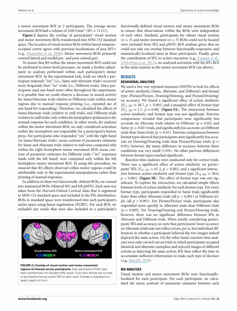
hypothesized to exhibit RS effects, we only compared Differenttrials for each of these two formats. Relative to Drawings, Picturesactivated a large, bilateral cluster that began in the occipital polesand extended into the fusiform gyri in both hemispheres (vol-ume = 32710 mm3; maximum z-value = 6.01; MNI coordinatesof maximum: x = 16, y = −96, z = −8) (Figure 5, red/yellow).Relative to Pictures, Drawings activated a cluster in the rightsupramarginal gyrus and superior parietal lobule (volume =3096 mm3; maximum z-value = 3.78; MNI coordinates of maxi-mum: x = 32, y = −52, z = 52) (Figure 5, light blue/dark blue).Drawings also activated a smaller cluster within left lateral occipi-tal cortex (volume = 1782 mm3; maximum z-value = 3.51; coor-dinates of maximum: x = −58, y = −66; z = −6). The majorityof voxels in this cluster were located anterior to the typical loca-tion of area MT+, as reported in other studies (Dumoulin et al.,2000) and within our own participant group (Figure 5, group-level visual-motion-preferring voxels shown in light green/darkgreen).
DISCUSSIONIn the present study, we used RS fMRI to determine the speci-ficity of information carried by sensory and motor systems during
conceptual processing of actions. Of primary interest was whetherbrain regions involved in performing movements and perceivingvisual motion, two areas of the brain often engaged by action con-cepts (Hauk et al., 2004; e.g., Kable et al., 2002), were sensitiveto changes in the exemplar or representational format of pairs ofaction images.
Our results reveal strikingly different response patternsbetween these two brain areas: while the visual motion ROI exhib-ited RS only for identical photographs of actions, suppressionoccurred in the motor movement ROI for repetitions of the sameand alternate exemplars of an action, irrespective of the format.This result suggests that neural activity within these sensorimo-tor regions during semantic tasks represents information aboutactions at different levels of specificity. On the one hand, duringcomprehension of static depictions of actions, voxels that respondstrongly to visual motion appear to encode information highlyspecific to a particular exemplar of an action or particular repre-sentational format: only when the prime and target images wereidentical and conveyed many perceptual details about the actor oraction context did we observe RS within the visual motion ROI.Because this region was strongly active for all conditions, it cannotbe the case that some conditions merely failed to activate visual
www.frontiersin.org May 2014 | Volume 5 | Article 494 | 7
Watson et al. Specificity of action knowledge
FIGURE 5 | Whole-brain analyses contrasting Picture/Picture(Different)
(red/yellow) and Drawing/Drawing(Different) (blue/light blue) trials.
The group-level location of visual-motion-preferring voxels is shown ingreen. Coordinates reported in MNI standardized space.
motion areas at all. Instead, neural responses to action conceptswithin this area preserve detailed information about the specificinstance of an action; different actors and/or representational for-mats activate different neural representations. Furthermore, wedid not observe RS when prime and target images were identicalschematic drawings. Thus, the absence of perceptually-rich detailsin schematic drawings may result in a more variable responsewithin areas specialized for visual motion, even across repeatedinstances of the same schematic drawing.
Although we focused on the activation of visual motion areasby conceptual processing of static action images, our resultsaccord with other studies on the response of area MT+ to dif-ferent types of visual motion. In particular, this area is sensitiveto changes in the speed, direction, and velocity of low-level visualmotion (Wall et al., 2008; Lingnau et al., 2009; Cardin et al., 2012;Weigelt et al., 2012). Thus, to the extent that different exemplarsof an action or different representational formats convey actionsperformed at different speeds, in different directions, etc., theresponse within visual motion regions may differ.
Yet, our results are at odds with two prior studies investigatingRS between pairs of dynamic action stimuli (i.e., videos) using asemantic task (Kable and Chatterjee, 2006; Wiggett and Downing,2010; but see Grossman et al., 2010). In both of these studies, areaMT+ was insensitive to changes in the actor and thus respondedsimilarly as long as the same action was repeated (e.g., “kick-ing”). Given that both of these studies used stimuli that containedactual visual motion, an alternative explanation of the presentresults is that area MT+ exhibits a narrower range of responses tostatic images than dynamic action stimuli. Although static imagesengage this area, they may do less strongly and with less variabil-ity than dynamic depictions of actions. If so, then the absenceof RS to alternate exemplars within the visual motion ROI in
the current study may reflect insufficient physiological power todetect differences between all conditions in this area.
In contrast to the highly-specific effects we observed withinthe visual motion ROI, the motor movement ROI exhibitedRS between pairs of images that depicted identical actions andpairs that depicted alternate exemplars of the same action. Thisresponse occurred both when the prime and target were the sameformat (Picture/Picture, Drawing/Drawing) or different formats(Picture/Drawing). This result suggests that a similar representa-tion is evoked within the motor system irrespective of the way inwhich an action concept is accessed; the same motor simulationis produced in response to different exemplars of the same actionor to actions presented in different formats.
One way in which this result could arise is if motor simulationsare grounded in person-specific motor programs for actions. Inother words, no matter who I perceive doing an action (e.g., Jackkicking, Jane kicking) or the format of the input (e.g., a photo-graph or schematic drawing of “kicking”), my motor simulationwill reflect the way in which I am inclined to kick. Indeed, thereis prior evidence that the involvement of motor regions in rep-resenting action concepts depends on an individual’s particularphysical experiences (Calvo-Merino et al., 2005, 2006; Beilocket al., 2008). For example, Calvo-Merino et al. (2005) found thatthe degree to which expert ballet and capoeira dancers recruitedmotor regions during action observation differed when watchingtheir own style of dance versus the other; the authors concludethat “. . . action observation evokes individual, acquired motorrepresentations. . . ” (p. 1247). Similarly, participants’ ability torecall actions depends on their motor expertise with those actions(Pezzulo et al., 2010). The present results extend these findings bysuggesting that an action evokes the same person-specific motorsimulation irrespective of the way in which an action concept isaccessed.
However, we note that the degree to which the motor systemparticipates in representing action concepts at all is also modu-lated by physical experience (described above) and task demands(Van Dam et al., 2012). Our recent meta-analysis of neuroimag-ing studies using action words and action images did not findconsistent involvement of premotor or primary motor cortex inconceptual processing of these stimuli (Watson et al., 2013). Inthe current study, we used a small set of very familiar actions,and we functionally-localized areas involved in performing move-ments within each participant. Therefore, we may have been morelikely than other studies to generate and detect effects within themotor system during conceptual processing of actions.
Even though participants made manual responses on eachtrial, our study design makes it unlikely that the RS we observedwithin the motor movement ROI reflects manual response prim-ing. First, for each participant, we only analyzed activation withinthe hemisphere that was ipsilateral to each condition’s expectedresponse. Thus, results from the motor movement ROI reflectactivation within the hemisphere not responsible for the but-ton press. Second, the RS effects were not entirely determinedby activation within hand-preferring parts of the motor sys-tem: we also functionally-localized areas active when performingfoot movements. Finally, we observed significantly different lev-els of activation within the motor movement ROI for Same and
Frontiers in Psychology | Cognitive Science May 2014 | Volume 5 | Article 494 | 8

Watson et al. Specificity of action knowledge
Alternate trials. If manual response priming was driving sup-pression effects, then we would expect no difference betweenconditions responded to with the same hand.
We used functionally-defined visual motion and motor move-ment ROIs rather than ROIs defined anatomically or from group-level results. However, since the tasks used to define these ROIsdid not require measurable behavioral responses, we cannot becertain that a given participant was paying attention or perform-ing the localizer task; indeed, differences in task engagement mayexplain why visual motion and motor movement ROIs could notbe identified, or required a more lenient threshold to be iden-tified, in some participants. Yet, given the potentially variablefunctional brain organization of each participant, using ROIsdefined in this way allowed us to more precisely test functionally-motivated hypotheses (see Saxe et al., 2006 for a similar argu-ment), i.e., that voxels that participate in more basic cognitivetasks (processing visual motion, executing body movements)would encode information at different levels of specificity duringa conceptual task.
We also examined RS effects in anatomically-defined ROIs.Within two brain areas neighboring visual motion and motormovement ROIs, we observed RS when the prime and targetimage depicted the same instance of the same action, but not dif-ferent instances of the same action. Instead, within left pMTG,we observed no differentiation between Alternate and Differenttrials, and within IFG, we observed enhancement for Alternate rel-ative to Different and Same trials. In some respects, these resultsare surprising: some researchers have suggested a “graded” viewof embodiment in which more abstract representations of actionmeaning are represented in brain areas adjacent to modality-specific cortices (Thompson-Schill, 2003; Kable et al., 2005;Chatterjee, 2008, 2010). Therefore, we expected to observe RS fordifferent exemplars of the same action within left pMTG and IFG.However, our pattern of results may be consistent with findingsof “repetition enhancement” rather than “repetition suppression”(Raposo et al., 2006; Kuperberg et al., 2008; see Segaert et al., 2013for a review). One hypothesis is that while suppression occurswhen the same cognitive process is performed on a prime andtarget, enhancement occurs when the target requires additionalprocesses, like explicit memory retrieval (Henson, 2003).
In the current study, we found significant enhancement forAlternate trials within IFG and non-significant but numericallyhigher activation for Alternate trials relative to Different trialsfor each format type within left pMTG. Alternate trials werealso the most difficult for participants. Therefore, it is possi-ble that verifying alternate exemplars of the same action (vs.the easier tasks of verifying an identical match or a completemismatch) required additional cognitive processing—and neu-ral activity—within IFG and left pMTG. IFG, in particular,has been shown to play a role in selecting among compet-ing representations in memory (Thompson-Schill et al., 1997;Moss et al., 2005). When determining whether two imageswere different exemplars of the same action, participants mayhave had to exert more cognitive effort to find the linkbetween two conceptually similar, but perceptually dissimilar,instances of an action. Lack of RS and numerical enhancementwithin pMTG may similarly reflect participants’ greater need
to retrieve explicit information about actions in the Alternatecondition.
Finally, we investigated at the whole-brain level the degreeto which the brain distinguishes between perceptually-rich pho-tographs of actions and more symbolic schematic drawings ofactions. Given that they contain more visual details than draw-ings, pictures unsurprisingly yielded greater activation through-out early visual cortex. The reverse comparison, however, yieldedgreater activation for schematic drawings in two areas of thebrain. First, drawings more strongly engaged the right supra-marginal gyrus and parts of the superior parietal lobe, a resultin agreement with a recent voxel-based lesion-symptom mapping(VLSM) study from our lab. In this study, stroke patients withdamage to the left or right hemisphere matched categorical spatialrelations among objects (e.g., “above,” “below”) across differentrepresentational formats (i.e., pictures, schematic drawings, andwords) (Amorapanth et al., 2012). Patients with damage to rightsupramarginal gyrus were particularly impaired matching spatialrelation words to their corresponding schematic drawings relativeto their corresponding pictures. A recent case study also supportsthe view that schematic drawings are processed differently thanperceptually-rich photographs: a patient with simultagnosia, acondition in which patients are characteristically unable to per-ceive more than a single object at a time (Luria, 1959), wasbetter able to comprehend spatial relations between objects (e.g.,“above,” “below”) when they were depicted as schematic draw-ings rather than as photographs (Kranjec et al., 2013). Given thepresent results as well as neuroimaging evidence for the activationof right supramarginal gyrus during the naming of spatial rela-tions between objects (e.g., Damasio et al., 2001), this part of thebrain may be responsible for recognizing the schematic structureof these pared-down percepts.
We also found greater activation for schematic drawings ofactions relative to photographs in left lateral occipital cortex;most voxels in this cluster were located anterior to visual motion-preferring areas, in lateral occipital cortex and the most posterioraspect of pMTG. This result is consistent with a graded viewof conceptual representation (Chatterjee, 2008, 2010; Watsonand Chatterjee, 2011). Action knowledge derived from visualmotion area MT+ is represented along a temporal posterior-to-anterior axis in which increasingly abstract information isrepresented more anteriorly. Accordingly, a brain area ante-rior to area MT+ responded more strongly to pared-down,more symbolic schematic drawings than to perceptually-richphotographs of actions. We also observed greater overall activa-tion of the left pMTG ROI for trials that included a schematicdrawing (Picture/Drawing or Drawing/Drawing trials). Together,these results suggest that more abstract or symbolic depictionsof actions recruit areas adjacent to modality-specific cortices.Consistent with this claim, we found using a meta-analysisapproach that words referring to actions consistently activated anarea within left middle temporal gyrus anterior to the area asso-ciated with visual depictions of actions (Watson et al., 2013). Theimplication of these findings for embodied accounts of seman-tic knowledge is that the recruitment of modality-specific—orother—regions depends on whether concepts are accessed bymore or less symbolic means. More symbolic depictions may
www.frontiersin.org May 2014 | Volume 5 | Article 494 | 9

Watson et al. Specificity of action knowledge
additionally, or instead, recruit information that is abstractedfrom direct experience and represented adjacent to modality-specific areas.
Finally, we acknowledge that participants’ did not need toaccess conceptual knowledge of actions on all trials. When theprime and target images were identical (Same trials), partici-pants’ decisions could be based solely on visual similarity. Wenote that the RS effects seen in the visual motion ROI suggestthat some inference about the images is being made even whenthey are perceptually identical insofar as neural activity in anarea sensitive to visual motion is influenced by static images. Avisual similarity strategy would not work on the Alternate andDifferent trials: though prime and target stimuli were visually dis-similar for both, these trial types required different behavioralresponses. Therefore, participants’ needed to access the mean-ing of the actions depicted in these images in order to make aresponse. Furthermore, the pattern of results suggests that par-ticipants drew upon action concepts even on Same trials: it isnot obvious why the repetition of visually similar images shouldyield decreased activation in the motor movement ROI. Instead,we suggest that the conceptual similarity of these images—andimages in the Alternate condition—produces RS within the motormovement ROI.
Understanding the specificity of brain regions to differ-ent exemplars of actions and representational formats makesembodied accounts of the semantic system more precise. Here,we found that sensory and motor systems carried differentamounts of information during conceptual processing of actions:while visual motion areas preserved exemplar- and format-specific details, regions involved in performing movementsresponded similarly as long as images referred to the samebasic action (e.g., “kicking”). Thus, when the motor systemparticipates in understanding an action, it may do so by acti-vating one’s own motor program for that particular action.Additionally, two brain regions (left lateral occipital cortexand right supramarginal gyrus) responded more strongly tomore symbolic representations of actions (i.e., schematic draw-ings) than to concrete ones (i.e., photographs). For embodiedaccounts, these data indicate that even outside of area MT+,the recruitment of posterior brain regions by action conceptsdepends on the format of the input. Within lateral occipi-totemporal cortex, in particular, more abstract representations ofactions may be represented adjacent to modality-specific corticalareas.
ACKNOWLEDGMENTSWe would like to thank Geoffrey Aguirre for his help with exper-imental design and data analysis and Matthew Lehet for hishelp running participants in pilot normative studies. This workwas supported by the National Institutes of Health (grant num-bers RO1 DC008779, RO1 DC012511 to Anjan Chatterjee, andT32-NS054575-04 to Christine E. Watson as a trainee).
REFERENCESAmorapanth, P., Kranjec, A., Bromberger, B., Lehet, M., Widick, P., Woods,
A. J., et al. (2012). Language, perception, and the schematic representa-tion of spatial relations. Brain Lang. 120, 226–236. doi: 10.1016/j.bandl.2011.09.007
Assmus, A., Giessing, C., Weiss, P. H., and Fink, G. R. (2007). Functional inter-actions during the retrieval of conceptual action knowledge: an fMRI study.J. Cogn. Neurosci. 19, 1004–1012. doi: 10.1162/jocn.2007.19.6.1004
Aziz-Zadeh, L., and Damasio, A. (2008). Embodied semantics for actions:findings from functional brain imaging. J. Physiol. 102, 35–39. doi:10.1016/j.jphysparis.2008.03.012
Barsalou, L. W. (1999). Perceptual symbol systems. Behav. Brain Sci. 22, 577–660.Barsalou, L. W. (2003). Abstraction in perceptual symbol systems. Philos. Trans. R.
Soc. Lond. B Biol. Sci. 358, 1177–1187. doi: 10.1098/rstb.2003.1319Barsalou, L. W. (2008). Grounded cognition. Annu. Rev. Psychol. 59, 617–645. doi:
10.1146/annurev.psych.59.103006.093639Bavelier, D., Brozinsky, C., Tomann, A., Mitchell, T., Neville, H., and Liu, G. (2001).
Impact of early deafness and early exposure to sign language on the cerebralorganization for motion processing. J. Neurosci. 21, 8931–8942.
Beilock, S. L., Lyons, I. M., Mattarella-Micke, A., Nusbaum, H. C., and Small,S. L. (2008). Sports experience changes the neural processing of action lan-guage. Proc. Natl. Acad. Sci. U.S.A. 105, 13269–13273. doi: 10.1073/pnas.0803424105
Boulenger, V., Hauk, O., and Pulvermüller, F. (2009). Grasping Ideas with themotor system: semantic somatotopy in idiom comprehension. Cereb. Cortex 19,1905–1914. doi: 10.1093/cercor/bhn217
Calvo-Merino, B., Glaser, D. E., Grèzes, J., Passingham, R. E., and Haggard, P.(2005). Action observation and acquired motor skills: an fMRI study withexpert dancers. Cereb. Cortex 15, 1243–1249. doi: 10.1093/cercor/bhi007
Calvo-Merino, B., Grèzes, J., Glaser, D. E., Passingham, R. E., and Haggard, P.(2006). Seeing or doing? Influence of visual and motor familiarity in actionobservation. Curr. Biol. 16, 1905–1910. doi: 10.1016/j.cub.2006.07.065
Cardin, V., Hemsworth, L., and Smith, A. T. (2012). Adaptation to heading direc-tion dissociates the roles of human MST and V6 in the processing of optic flow.J. Neurophysiol. 108, 794–801. doi: 10.1152/jn.00002.2012
Caspers, S., Zilles, K., Laird, A. R., and Eickhoff, S. B. (2010). ALE meta-analysisof action observation and imitation in the human brain. Neuroimage 50,1148–1167. doi: 10.1016/j.neuroimage.2009.12.112
Chatterjee, A. (2001). Language and space: some interactions. Trends Cogn. Sci. 5,55–61. doi: 10.1016/S1364-6613(00)01598-9
Chatterjee, A. (2008). The neural organization of spatial thought and language.Semin. Speech Lang. 29, 226–238. doi: 10.1055/s-0028-1082886
Chatterjee, A. (2010). Disembodying cognition. Lang. Cogn. 2, 79–116. doi:10.1515/LANGCOG.2010.004
Damasio, H., Grabowski, T. J., Tranel, D., Ponto, L. L. B., Hichwa, R. D., andDamasio, A. R. (2001). Neural correlates of naming actions and of namingspatial relations. Neuroimage 13, 1053–1064. doi: 10.1006/nimg.2001.0775
Deacon, T. W. (1997). The Symbolic Species: The Co-evolution of Language and theHuman Brain. New York, NY: W. W. Norton & Company.
Decety, J., and Grèzes, J. (2006). The power of simulation: imagining one’s own andother’s behavior. Brain Res. 1079, 4–14. doi: 10.1016/j.brainres.2005.12.115
Di Pellegrino, G., Fadiga, L., Fogassi, L., Gallese, V., and Rizzolatti, G. (1992).Understanding motor events: a neurophysiological study. Exp. Brain Res. 91,176–180. doi: 10.1007/BF00230027
Dumoulin, S. O., Bittar, R. G., Kabani, N. J., Baker, C. L., Le Goualher, G., Pike,G. B., et al. (2000). A new anatomical landmark for reliable identification ofhuman area V5/MT: a quantitative analysis of sulcal patterning. Cereb. Cortex10, 454–463. doi: 10.1093/cercor/10.5.454
Gallese, V., and Lakoff, G. (2005). The Brain’s concepts: the role of the Sensory-motor system in conceptual knowledge. Cogn. Neuropsychol. 22, 455–479. doi:10.1080/02643290442000310
Gallese, V., and Sinigaglia, C. (2011). What is so special about embodied simula-tion? Trends Cogn. Sci. 15, 512–519. doi: 10.1016/j.tics.2011.09.003
Gennari, S. P. (2012). Representing motion in language comprehension: lessonsfrom neuroimaging. Lang. Linguist. Compass 6, 67–84. doi: 10.1002/lnc3.317
Grill-Spector, K., Henson, R., and Martin, A. (2006). Repetition and the brain:neural models of stimulus-specific effects. Trends Cogn. Sci. 10, 14–23. doi:10.1016/j.tics.2005.11.006
Grill-Spector, K., and Malach, R. (2001). fMR-adaptation: a tool for studying thefunctional properties of human cortical neurons. Acta Psychol. (Amst.) 107,293–321. doi: 10.1016/S0001-6918(01)00019-1
Grossman, E. D., Jardine, N. L., and Pyles, J. A. (2010). fMR-adaptation revealsinvariant coding of biological motion on the human STS. Front. Hum. Neurosci.4:15. doi: 10.3389/neuro.09.015.2010
Frontiers in Psychology | Cognitive Science May 2014 | Volume 5 | Article 494 | 10

Watson et al. Specificity of action knowledge
Hauk, O., Johnsrude, I., and Pulvermüller, F. (2004). Somatotopic representation ofaction words in human motor and premotor cortex. Neuron 41, 301–307. doi:10.1016/S0896-6273(03)00838-9
Henson, R. N. A. (2003). Neuroimaging studies of priming. Prog. Neurobiol. 70,53–81. doi: 10.1016/S0301-0082(03)00086-8
Huk, A. C., Dougherty, R. F., and Heeger, D. J. (2002). Retinotopy and functionalsubdivision of human areas MT and MST. J. Neurosci. 22, 7195–7205.
Kable, J. W., and Chatterjee, A. (2006). Specificity of action representations inthe lateral occipitotemporal cortex. J. Cogn. Neurosci. 18, 1498–1517. doi:10.1162/jocn.2006.18.9.1498
Kable, J. W., Kan, I. P., Wilson, A., Thompson-Schill, S. L., and Chatterjee, A.(2005). Conceptual representations of action in the lateral temporal cortex.J. Cogn. Neurosci. 17, 1855–1870. doi: 10.1162/089892905775008625
Kable, J. W., Lease-Spellmeyer, J., and Chatterjee, A. (2002). Neural sub-strates of action event knowledge. J. Cogn. Neurosci. 14, 795–805. doi:10.1162/08989290260138681
Kalénine, S., Buxbaum, L. J., and Coslett, H. B. (2010). Critical brain regions foraction recognition: lesion symptom mapping in left hemisphere stroke. Brain133, 3269–3280. doi: 10.1093/brain/awq210
Kilner, J. M., Neal, A., Weiskopf, N., Friston, K. J., and Frith, C. D. (2009). Evidenceof mirror neurons in human inferior frontal gyrus. J. Neurosci. 29, 10153–10159.doi: 10.1523/JNEUROSCI.2668-09.2009
Kranjec, A., Ianni, G., and Chatterjee, A. (2013). Schemas reveal spatialrelations to a patient with simultanagnosia. Cortex 49, 1983–1988. doi:10.1016/j.cortex.2013.03.005
Kuperberg, G. R., Lakshmanan, B. M., Greve, D. N., and West, W. C. (2008). Taskand semantic relationship influence both the polarity and localization of hemo-dynamic modulation during lexico-semantic processing. Hum. Brain Mapp. 29,544–561. doi: 10.1002/hbm.20419
Lingnau, A., Ashida, H., Wall, M. B., and Smith, A. T. (2009). Speed encod-ing in human visual cortex revealed by fMRI adaptation. J. Vis. 9, 1–14. doi:10.1167/9.13.3
Luria, A. R. (1959). Disorders of “simultaneous perception” in a case of bilateraloccipito-parietal brain injury. Brain 82, 437–449. doi: 10.1093/brain/82.3.437
Maccotta, L., and Buckner, R. L. (2004). Evidence for neural effects of repe-tition that directly correlate with behavioral priming. J. Cogn. Neurosci. 16,1625–1632. doi: 10.1162/0898929042568451
Moss, H. E., Abdallah, S., Fletcher, P., Bright, P., Pilgrim, L., Acres, K., et al. (2005).Selecting among competing alternatives: selection and retrieval in the leftinferior frontal gyrus. Cereb. Cortex 15, 1723–1735. doi: 10.1093/cercor/bhi049
Nichols, T. E., and Holmes, A. P. (2002). Nonparametric permutation tests for func-tional neuroimaging: a primer with examples. Hum. Brain Mapp. 15, 1–25. doi:10.1002/hbm.1058
Nichols, T., and Hayasaka, S. (2003). Controlling the familywise error rate infunctional neuroimaging: a comparative review. Stat. Methods Med. Res. 12,419–446. doi: 10.1191/0962280203sm341ra
Peirce, C. S. (1955). Philosophical Writings of Peirce. New York, NY: Courier DoverPublications.
Pezzulo, G., Barca, L., Bocconi, A. L., and Borghi, A. M. (2010). When affor-dances climb into your mind: advantages of motor simulation in a memorytask performed by novice and expert rock climbers. Brain Cogn. 73, 68–73. doi:10.1016/j.bandc.2010.03.002
Pirog Revill, K., Aslin, R. N., Tanenhaus, M. K., and Bavelier, D. (2008). Neuralcorrelates of partial lexical activation. Proc. Natl. Acad. Sci. U.S.A. 105,13111–13115. doi: 10.1073/pnas.0807054105
Plaut, D. C. (2002). Graded modality-specific specialisation in semantics: a com-putational account of optic aphasia. Cogn. Neuropsychol. 19, 603–639. doi:10.1080/02643290244000112
Press, C., Weiskopf, N., and Kilner, J. M. (2012). Dissociable roles of human infe-rior frontal gyrus during action execution and observation. Neuroimage 60,1671–1677. doi: 10.1016/j.neuroimage.2012.01.118
Pulvermüller, F. (1999). Words in the brain’s language. Behav. Brain Sci. 22,253–279.
Raposo, A., Moss, H. E., Stamatakis, E. A., and Tyler, L. K. (2006).Repetition suppression and semantic enhancement: an investigation ofthe neural correlates of priming. Neuropsychologia 44, 2284–2295. doi:
Raposo, A., Moss, H. E., Stamatakis, E. A., and Tyler, L. K. (2009). Modulationof motor and premotor cortices by actions, action words and actionsentences. Neuropsychologia 47, 388–396. doi: 10.1016/j.neuropsychologia.2008.09.017
Ratcliff, R. (1978). A theory of memory retrieval. Psychol. Rev. 85, 59–108. doi:10.1037/0033-295X.85.2.59
Saxe, R., Brett, M., and Kanwisher, N. (2006). Divide and conquer:a defense of functional localizers. Neuroimage 30, 1088–1096. doi:10.1016/j.neuroimage.2005.12.062
Saygin, A. P., McCullough, S., Alac, M., and Emmorey, K. (2010). Modulationof BOLD response in motion-sensitive lateral temporal cortex by realand fictive motion sentences. J. Cogn. Neurosci. 22, 2480–2490. doi:10.1162/jocn.2009.21388
Segaert, K., Weber, K., de Lange, F. P., Petersson, K. M., and Hagoort, P.(2013). The suppression of repetition enhancement: a review of fMRIstudies. Neuropsychologia 51, 59–66. doi: 10.1016/j.neuropsychologia.2012.11.006
Thompson-Schill, S. L. (2003). Neuroimaging studies of semantic memory: infer-ring “how” from “where.” Neuropsychologia 41, 280–292. doi: 10.1016/S0028-3932(02)00161-6
Thompson-Schill, S. L., D’Esposito, M., Aguirre, G. K., and Farah, M. J. (1997).Role of left inferior prefrontal cortex in retrieval of semantic knowledge: a re-evaluation. Proc. Natl. Acad. Sci. U.S.A. 94, 14792–14797.
Van Dam, W. O., van Dijk, M., Bekkering, H., and Rüschemeyer, S.-A. (2012).Flexibility in embodied lexical-semantic representations. Hum. Brain Mapp. 33,2322–2333. doi: 10.1002/hbm.21365
Vigliocco, G., Vinson, D. P., Lewis, W., and Garrett, M. F. (2004). Representingthe meanings of object and action words: the featural and unitary semanticspace hypothesis. Cognit. Psychol. 48, 422–488. doi: 10.1016/j.cogpsych.2003.09.001
Wall, M. B., Lingnau, A., Ashida, H., and Smith, A. T. (2008). Selective visualresponses to expansion and rotation in the human MT complex revealedby functional magnetic resonance imaging adaptation. Eur. J. Neurosci. 27,2747–2757. doi: 10.1111/j.1460-9568.2008.06249.x
Watson, C. E., Cardillo, E. R., Ianni, G. R., and Chatterjee, A. (2013). Action con-cepts in the brain: an activation likelihood estimation meta-analysis. J. Cogn.Neurosci. 25, 1191–1205. doi: 10.1162/jocn_a_00401
Watson, C. E., and Chatterjee, A. (2011). The functional neuroanatomy of actions.Neurology 76, 1428–1434. doi: 10.1212/WNL.0b013e3182166e2c
Weigelt S., Singer W., and Kohler A. (2012). Feature-based attention affectsdirection-selective fMRI adaptation in hMT+. Cereb. Cortex 23, 2169–2178.doi: 10.1093/cercor/bhs192
Wiggett, A. J., and Downing, P. E. (2010). Representation of action in occipito-temporal cortex. J. Cogn. Neurosci. 23, 1765–1780. doi: 10.1162/jocn.2010.21552
Willems, R. M., and Francken, J. C. (2012). Embodied cognition: taking the nextstep. Front. Psychol. 3:582. doi: 10.3389/fpsyg.2012.00582
Worsley, K. J., Evans, A. C., Marrett, S., and Neelin, P. (1992). A three-dimensionalstatistical analysis for CBF activation studies in human brain. J. Cereb. BloodFlow Metab. 12, 900–918. doi: 10.1038/jcbfm.1992.127
Conflict of Interest Statement: The authors declare that the research was con-ducted in the absence of any commercial or financial relationships that could beconstrued as a potential conflict of interest.
Received: 28 January 2014; accepted: 06 May 2014; published online: 26 May 2014.Citation: Watson CE, Cardillo ER, Bromberger B and Chatterjee A (2014) The speci-ficity of action knowledge in sensory and motor systems. Front. Psychol. 5:494. doi:10.3389/fpsyg.2014.00494This article was submitted to Cognitive Science, a section of the journal Frontiers inPsychology.Copyright © 2014 Watson, Cardillo, Bromberger and Chatterjee. This is an open-access article distributed under the terms of the Creative Commons Attribution License(CC BY). The use, distribution or reproduction in other forums is permitted, providedthe original author(s) or licensor are credited and that the original publication in thisjournal is cited, in accordance with accepted academic practice. No use, distribution orreproduction is permitted which does not comply with these terms.
www.frontiersin.org May 2014 | Volume 5 | Article 494 | 11
10.1016/j.neuropsychologia.2006.05.017
Related Documents