The spatial tuning of adaption-based time compression Cognitive, Perceptual and Brain Sciences, Division of Psychology and Language Sciences, University College London, London, UK Inci Ayhan Cognitive, Perceptual and Brain Sciences, Division of Psychology and Language Sciences, University College London, London, UK Aurelio Bruno NTT Communication Science Laboratories, Nippon Telegraph & Telephone Corporation, Atsugi Kanagawa, Japan Shin’ya Nishida Cognitive, Perceptual and Brain Sciences, Division of Psychology and Language Sciences, University College London, London, UK,& CoMPLEX, University College London, London, UK Alan Johnston Temporal processing is traditionally dissociated from spatial vision. Recent evidence, however, has shown that adaptation to high temporal frequency (D. Burr, A.Tozzi, & M. C. Morrone, 2007; A. Johnston, D. H. Arnold, & S. Nishida, 2006; A. Johnston et al., 2008) induces spatially specific reductions in the apparent duration of subsecond intervals containing medium frequency drift or flicker. Here we examine the spatial tuning of these temporal adaptation effects. Our results show that duration compression is tightly tuned to the spatial location of the adaptor and can be induced by very narrow adaptors. We also demonstrate that the effects of adaptation on perceived duration are dissociable from those on apparent temporal frequency, which suggests early but separate influences of temporal frequency adaptation on time and speed perception. Keywords: temporal frequency, magnocellular pathway, duration perception Citation: Ayhan, I., Bruno, A., Nishida, S., & Johnston, A. (2009). The spatial tuning of adaption-based time compression. Journal of Vision, 9(11):2, 1–12, http://journalofvision.org/9/11/2/, doi:10.1167/9.11.2. Introduction Whereas our sense of space can be traced to the orderly spatial arrangement of neurons in the retina, there is no equivalent arrangement of neurons to support our sense of time and duration. The lack of an isomorphic representa- tion for duration shrouds the neural bases of time perception in mystery. Prevailing models of event time focus on the notion of an internal clock (Creelman, 1962; Treisman, 1963; Treisman, Faulkner, Naish, & Brogan, 1990), which assumes that timing is centralized and the same neural circuitry serves all modalities. Recent studies, however, have revealed that the apparent duration of brief intervals can be manipulated in a local region of visual space (Burr, Tozzi, & Morrone, 2007; Johnston, Arnold, & Nishida, 2006; Johnston et al., 2008). Thus, temporal processing has a spatial component. Johnston et al. (2006) showed a reduction in the apparent duration of a sub- second 10-Hz drifting grating following high temporal frequency motion adaptation. The magnitude of temporal compression is independent of the relative orientation of adaptor and test patterns, consistent with a pre-cortical locus. Additional support for an early locus comes from evidence of apparent duration compression after adapta- tion to invisible flicker (Johnston et al., 2008) and the finding that intermediate high-temporal contrast intervals are apparently compressed after a 1.5-s dynamic high contrast interval (Bruno & Johnston, 2007). These manipulations are thought to modify, adapt, or suppress transient (magnocellular) mechanisms. Although Johnston et al. (2006) demonstrated that adaptation-based reductions in apparent duration were space specific, the adapted and unadapted test regions were separated by at least a quadrant of the visual field. The extent of the spatial influence of temporal frequency adaptation remains unknown. The spatial tuning of these adaptation effects can provide a clue to the site of adaptation, since the average size of receptive fields increases system- atically from the retina through the lateral geniculate nucleus (Lee, Kremers, & Yeh, 1998; Xu et al., 2001) and primary visual cortex to extrastriate areas (Smith, Singh, Williams, & Greenlee, 2001; Zeki, 1978). In the Johnston et al. (2006) study, the stimuli are viewed in the near periphery. At 5 degrees of visual eccentricity mean receptive field diameter in macaque V1 is around 0.4 degrees (Dow, Journal of Vision (2009) 9(11):2, 1–12 http://journalofvision.org/9/11/2/ 1 doi: 10.1167/9.11.2 Received March 25, 2009; published October 2, 2009 ISSN 1534-7362 * ARVO

Welcome message from author
This document is posted to help you gain knowledge. Please leave a comment to let me know what you think about it! Share it to your friends and learn new things together.
Transcript

The spatial tuning of adaption-based time compressionCognitive, Perceptual and Brain Sciences,
Division of Psychology and Language Sciences,University College London, London, UKInci Ayhan
Cognitive, Perceptual and Brain Sciences,Division of Psychology and Language Sciences,
University College London, London, UKAurelio Bruno
NTT Communication Science Laboratories,Nippon Telegraph & Telephone Corporation,
Atsugi Kanagawa, JapanShin’ya Nishida
Cognitive, Perceptual and Brain Sciences,Division of Psychology and Language Sciences,
University College London, London, UK, &CoMPLEX, University College London, London, UKAlan Johnston
Temporal processing is traditionally dissociated from spatial vision. Recent evidence, however, has shown that adaptation tohigh temporal frequency (D. Burr, A.Tozzi, & M. C. Morrone, 2007; A. Johnston, D. H. Arnold, & S. Nishida, 2006; A.Johnston et al., 2008) induces spatially specific reductions in the apparent duration of subsecond intervals containingmedium frequency drift or flicker. Here we examine the spatial tuning of these temporal adaptation effects. Our results showthat duration compression is tightly tuned to the spatial location of the adaptor and can be induced by very narrow adaptors.We also demonstrate that the effects of adaptation on perceived duration are dissociable from those on apparent temporalfrequency, which suggests early but separate influences of temporal frequency adaptation on time and speed perception.
Keywords: temporal frequency, magnocellular pathway, duration perception
Citation: Ayhan, I., Bruno, A., Nishida, S., & Johnston, A. (2009). The spatial tuning of adaption-based time compression.Journal of Vision, 9(11):2, 1–12, http://journalofvision.org/9/11/2/, doi:10.1167/9.11.2.
Introduction
Whereas our sense of space can be traced to the orderlyspatial arrangement of neurons in the retina, there is noequivalent arrangement of neurons to support our sense oftime and duration. The lack of an isomorphic representa-tion for duration shrouds the neural bases of timeperception in mystery. Prevailing models of event timefocus on the notion of an internal clock (Creelman, 1962;Treisman, 1963; Treisman, Faulkner, Naish, & Brogan,1990), which assumes that timing is centralized and thesame neural circuitry serves all modalities. Recent studies,however, have revealed that the apparent duration of briefintervals can be manipulated in a local region of visualspace (Burr, Tozzi, & Morrone, 2007; Johnston, Arnold,& Nishida, 2006; Johnston et al., 2008). Thus, temporalprocessing has a spatial component. Johnston et al. (2006)showed a reduction in the apparent duration of a sub-second 10-Hz drifting grating following high temporalfrequency motion adaptation. The magnitude of temporalcompression is independent of the relative orientation ofadaptor and test patterns, consistent with a pre-cortical
locus. Additional support for an early locus comes fromevidence of apparent duration compression after adapta-tion to invisible flicker (Johnston et al., 2008) and thefinding that intermediate high-temporal contrast intervalsare apparently compressed after a 1.5-s dynamic highcontrast interval (Bruno & Johnston, 2007). Thesemanipulations are thought to modify, adapt, or suppresstransient (magnocellular) mechanisms.Although Johnston et al. (2006) demonstrated that
adaptation-based reductions in apparent duration were spacespecific, the adapted and unadapted test regions wereseparated by at least a quadrant of the visual field. Theextent of the spatial influence of temporal frequencyadaptation remains unknown. The spatial tuning of theseadaptation effects can provide a clue to the site of adaptation,since the average size of receptive fields increases system-atically from the retina through the lateral geniculate nucleus(Lee, Kremers, & Yeh, 1998; Xu et al., 2001) and primaryvisual cortex to extrastriate areas (Smith, Singh, Williams,& Greenlee, 2001; Zeki, 1978). In the Johnston et al.(2006) study, the stimuli are viewed in the near periphery.At 5 degrees of visual eccentricity mean receptive fielddiameter in macaque V1 is around 0.4 degrees (Dow,
Journal of Vision (2009) 9(11):2, 1–12 http://journalofvision.org/9/11/2/ 1
doi: 10 .1167 /9 .11 .2 Received March 25, 2009; published October 2, 2009 ISSN 1534-7362 * ARVO
Snyder, Vautin, & Bauer, 1981; Johnston, 1989) and itincreases systematically toward higher level areas of thebrain such as LIP, a suggested locus of temporal distortionsaround the time of saccadic eye movements (Morrone, Ross,& Burr, 2005), where receptive field size reaches 12 degreesat 5 degrees of eccentricity (Blatt, Andersen, & Stoner, 1990).Here we examine the spatial tuning functions of
temporal adaptation. Since the apparent duration of amoving stimulus depends to some extent upon its temporalfrequency (Kanai, Paffen, Hogendoorn, & Verstraten,2006), we first eliminated the influence of temporalfrequency adaptation on apparent temporal frequency.We then measured adaptation-induced changes in apparentduration for test patterns in the adapted and surroundingregions. We found that temporal compression is tightlytuned to the location of the adaptor. The spatial tuning ofshifts in apparent temporal frequency was similar to thatof adaptation-based duration compression perhaps indicat-ing that they share common components. There was someevidence of a long-range duration expansion effect but thiseffect was considerably weaker than the duration com-pression effect. We also showed that even narrow adaptinggratings could induce strong temporal compression. Theseresults suggest that the effects of adaptation on perceivedduration in the millisecond range can be attributed tolevels of the visual pathway where the receptive fields ofthe neurons remain relatively small.
General methods
Observers
Six adults participated (one female, five males), three ofwhich were naı̈ve to the purpose of the experiment. Twoof the authors participated in all experiments; in additionthere was one naı̈ve observer in Experiments 4 and 5 andthree naı̈ve observers in Experiment 6. Visual acuity wasnormal or corrected-to-normal for all subjects.
Apparatus
Observers were seated 57 cm from a 19-inch SonyTrinitron Multiscan 500PS monitor, with a refresh rate of100 Hz, driven by a VSG 2/5 visual stimulus generator(Cambridge Research Systems). The resolution of themonitor was 800 � 600 pixels. At this distance, themonitor subtended 40 � 30 degrees.
Procedure
In all experiments, subjects were presented with acentral fixation spot and an adaptor (1 cycle/deg sine
grating; 50% Michelson contrast). The duration of theadaptation phase was 32 s with 8 s top-ups between trials.The direction of motion of the adaptor reversed every 1 sto avoid the build up of a directional motion after effect.After the adaptation phase, the test stimuli, driftingsinusoidal gratings with a spatial frequency of 1 cycle/deg, were displayed sequentially on the adapted (standard)and unadapted (comparison) side of the fixation point. Theorder of presentation of the standard and comparison wasrandomized from trial to trial to control for time ordereffects (Jamieson & Petrusic, 1975). Subjects were askedto determine which of the test stimuli drifted faster (inperceived temporal frequency experiments) or for longer(in perceived duration experiments). Details of the stimuliin each experiment can be found in the methods of therelevant section. Whereas the duration or temporalfrequency of the standard stimulus was fixed across trials,the values of the comparison stimulus varied in 7 steps togenerate a psychometric function indicating the percent-age of trials in which the comparison was judged as longeror faster than the standard for each subject. The 50% pointon the psychometric function provided an estimate of theeffect of adaptation on the perceived duration or temporalfrequency of the standard.
The temporal frequency shift
The apparent duration of a moving stimulus increaseswith temporal frequency for low to mid-range frequencies(Kanai et al., 2006). Burr et al. (2007) have reported thatadaptation-induced duration compression disappears whenthe apparent speed of the two tests are matched. Johnstonet al. (2006) had previously shown that reductions inperceived duration induced by a 20-Hz dynamic adaptorpersist even after matching for perceived temporalfrequency, indicating that temporal compression is notmediated by changes in apparent temporal frequency.Here, we introduce a new procedure designed to eliminateany possible influence of temporal frequency adaptationon speed judgments. We varied the duty cycle ofinterleaved 5 and 20 Hz adaptors to find, for each subject,a critical ratio of 5 and 20 Hz adaptation for which nochange occurs in the perceived speed of a 10-Hz testpattern. We then use these critical ratios in our durationexperiments to ensure that any aftereffect we observecannot be attributed to changes in apparent temporalfrequency or speed.
Method
In Experiment 1, observers compared the temporalfrequency or speed of a drifting, Gaussian windowed,sinusoidal grating against a standard following a period of
Journal of Vision (2009) 9(11):2, 1–12 Ayhan, Bruno, Nishida, & Johnston 2

adaptation (Figure 1A). The adaptor was a vertical1 cycle/deg grating displayed in a circular aperture(diameter 5- of visual angle) centered 5- to the right ofa central fixation point. In the adaptation phase, thegratings drifted with a temporal frequency of either 5 or20 Hz. The two adaptors were interleaved on the basis ofone of 5 different duty cycles: 0% 5 Hz–100% 20 Hz,25% 5 Hz–75% 20 Hz, 50% 5 Hz–50% 20 Hz, 75% 5 Hz–25% 20 Hz, 100% 5 Hz–0% 20 Hz. The duty cycledetermined the proportion of time for which the twoadapting temporal frequencies were displayed. Each cycletypically lasted 4 s during the initial adaptation and 2 sduring the top-ups. After the adaptation phase, the teststimuli (sinusoidal gratings with a spatial frequency of1 cycle/deg drifting for 600 ms) were displayed sequen-tially on the adapted and unadapted side of the fixationpoint. Since the spatial frequency was always 1 cycle/deg,the speed and drift frequency had the same numericalvalue. The order of display was randomized for each trial.The temporal frequency of the standard stimulus, whichwas displayed in the same spatial position as the adaptor,was fixed across trials (10 Hz). The temporal frequency ofthe comparison stimulus, which was displayed in anunadapted spatial position (centered 5- to left of thecentral fixation point), was varied between 2 and 18 Hz in7 steps (2, 4.67, 7.33, 10, 12.67, 15.33, and 18 Hz). The
luminance contrast of test stimuli was 100% (Michelson),and both were presented in a Gaussian temporal window(amplitude 1.0; Standard: standard deviation = 100 ms;Comparison: standard deviation = comparison duration/6 ms). Observers reported which of the tests appeared todrift faster.
Results
Since the adaptors reversed direction at regular inter-vals, we were able to adapt temporal channels withoutgenerating a directional motion aftereffect. For eachsubject, the duty cycle of the two interleaved adaptors (5and 20 Hz) that had no effect on the perceived temporalfrequency of a subsequently displayed drifting stimuluscan be read from Figure 1B. The dashed line indicates thetemporal frequency of the standard grating. Data pointsshow the 50% point on the psychometric function (pointof subjective equality, PSE) plotted as a function of theproportion of the relative time of the 5- and 20-Hzadaptors. The straight lines are the linear fits to these datapoints for each subject. For two subjects, the criticalproportion of 5-Hz adaptation was around 0.25, for onesubject it was around 0.66 and for the remaining three itwas around 0.50.
Figure 1. Eliminating the influence of temporal frequency adaptation on speed judgments. (A) Time course of the binary choiceexperiments in which subjects made a speed judgment between two vertically oriented drifting gratings displayed in succession followingadaptation to an oscillating grating. (B) Results of the temporal frequency experiment for 6 different subjects (three authors and threenaïves). The dashed line indicates the temporal frequency of the standard grating. Data points show the PSEs plotted as a function of therelative duration of the 5- and 20-Hz adaptors. The straight lines are the linear fit of these data points for each subject. The adjustedcoefficient of determination R2 is reported for each subject separately. Error bars show T1 SE. Each point is derived from 140 trials.
Journal of Vision (2009) 9(11):2, 1–12 Ayhan, Bruno, Nishida, & Johnston 3
Spatial tuning of durationcompression
Local adaptation to high temporal frequency (20 Hz)oscillatory motion or flicker reduces the apparent durationof a subsequently displayed 10 Hz dynamic stimulus(Johnston et al., 2006) in the same region of the visualfield but the spatial tuning of this temporal aftereffect isunknown. In our earlier study, both the adaptor and testhad a diameter of 9- and their locations differed byapproximately 18- (across hemispheres) or approximately13- (within a hemisphere). Here we designed our stimulito cover a range of separations and sizes of adaptor andtest. The greatest separation was 11-, which was close tothe previously used separation and the largest apertureused was 5-, which was around half the size of theoriginal aperture. To study spatial specificity, we variedthe spatial separation of adaptor and test. In one experi-ment, we adapted along the horizontal midline and thenpositioned the standard above and below to measure aspatial tuning function. In the other experiments, wepresented the test patterns on the horizontal midline andpositioned the adaptor above and below. These proceduresgenerated similar spatial tuning functions.
Method
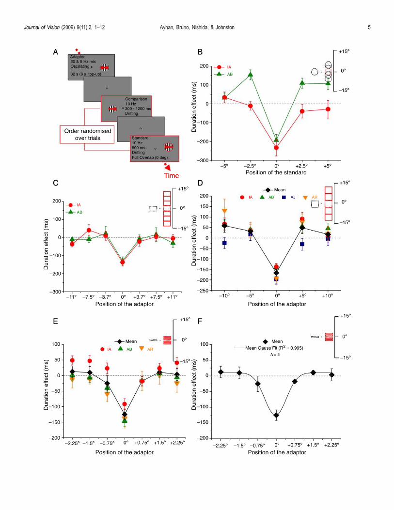
The temporal sequence of the adaptor and test patternsis shown in Figure 2A. In Experiment 2, subjects werepresented with a central fixation spot and the adaptor,which was always displayed at the same position (5- toright of a central fixation point) in a 5- diameter circularpatch. After the adaptation phase, the test stimuli (sinus-oidal gratings with a spatial frequency of 1 cycle/deg)drifting with a temporal frequency of 10 Hz weredisplayed sequentially on the adapted and unadapted sideof the fixation point. The standard appeared at the sameposition, half overlapping (2.5-) or completely (5-) aboveand below the adaptation location, while the comparisonappeared at the vertically symmetrical position on theunadapted side. The duration of the standard stimulus wasfixed across trials (600 ms). The duration of thecomparison stimulus was varied between 300 and 1200ms in 7 steps (300, 400, 500, 600, 800, 1000, and 1200 ms).The luminance contrast of both test stimuli was 100%.Observers reported which of the tests appeared to lastlonger. We compared the effects of adaptation against acontrol condition in which observers made temporal judg-ments between comparison and standard drifting gratingsin the absence of adaptation. In this experiment, trials forthe various positions of the standard were blocked.The procedure in the third and fourth experiment was
similar to the procedure we used in Experiment 2 exceptthat the adaptor and the test stimuli were sinusoidalgratings displayed within different sized rectangular or
square patches, 3.7- � 5- for the medium stimulus and5- � 5- for the larger stimulus. In this case there was nophysical overlap between the standard and the adaptor.Observers also made temporal judgments between com-parison and standard drifting gratings in the absence ofadaptation. Since the standard location was fixed indifferent adaptor locations, one baseline value was appliedto all conditions for each subject.In the fifth experiment we used narrow windows
(0.75- � 5-). The procedure was similar to that used inthe previous experiments except that in half of the trialsthe adaptor was displayed on the right of the fixation pointand in half of the trials it was displayed to the left of thefixation point. This refinement allowed us to eliminate anypossible hemispheric differences in duration judgments.
Figure 2. Effects of temporal frequency adaptation on durationjudgments as a function of the vertical displacement of thestandard or adaptor in the absence of temporal frequency shifts.The shape and the size of the stimuli relative to the monitor heightare shown on the upper right corner of each graph. Filled red linesindicate the position of the adaptor and dotted lines indicate theposition of the test patterns. (A) Time course of the binary choiceexperiments in which subjects made a duration judgment betweentwo vertically oriented drifting gratings displayed in successionfollowing adaptation to an oscillating grating. (B) Spatial tuningfunctions for temporal compression for two subjects. Apparentdurations relative to the baseline conditions are plotted as afunction of the vertical displacement of the standard gratingrelative to the position of the adaptor. Results are obtainedfollowing an adaptation phase consisted of the combination of 5and 20 Hz that elicited no change in temporal frequency (Figure 1B).Error bars show T1 SE. Each point is derived from 140 adaptationtrials and 140 control trials. (C) Spatial tuning functions formedium scale with the stimuli dimensions of 3.7- � 5- for2 subjects (2 authors; included in Figure 1B). The individualduration judgments comparative to the baseline conditions areplotted as a function of the vertical displacement of the standardgrating relative to the position of the adaptor. Error bars showT1 SE. Each point is derived from 140 adaptation trials and 140control trials. (D) Spatial tuning functions for large scale with thestimuli dimensions of 5- � 5- for 4 subjects (3 authors and 1 naïve;included in Figure 1B). The mean duration judgment comparativeto the baseline conditions is plotted as a function of the verticaldisplacement of the adapting field relative to the position of thestandard. Error bars show T1 SE of individuals and T1 SE ofthe mean. Each point is derived from 140 adaptation trials and140 control trials. (E) Results for narrow adaptors for 3 subjects.Mean duration judgment comparative to the baseline conditionsis plotted as a function of the vertical displacement of theadapting field relative to the position of the standard. Error barsshow T1 SE of individuals and T1 SE of the mean. Each point isderived from 140 adaptation trials and 280 control trials. (F) Thenarrow black line shows the Gaussian function fit to the meandata. Error bars show T1 SE of the mean. Each point is derivedfrom 140 adaptation trials and 280 control trials.
Journal of Vision (2009) 9(11):2, 1–12 Ayhan, Bruno, Nishida, & Johnston 4
Journal of Vision (2009) 9(11):2, 1–12 Ayhan, Bruno, Nishida, & Johnston 5
Results
In Experiments 2–4 (Figures 2B–2D), compressionrelative to the baseline conditions is highly spatiallylocalized, appearing only in the full overlapping con-dition. In Experiment 2 (Figure 2B), the two subjects,however, differed in the direction of the effect of temporalfrequency adaptation on apparent duration for the halfoverlap conditions. One subject AB shows evidence oftemporal expansion for overlapping and one of theeccentric locations. One might have expected the magni-tude of temporal compression in the half overlap conditionto be somewhat less than half that in the full overlapcondition. However, the data show no compression effector expansion. There may be a number of ways adaptedand non-adapted regions may combine. The presence of alateral expansion effect (subject AB) suggests that a nullresult in the partial overlap condition may reflect abalance between compression in the adapted region andexpansion in an adjacent unadapted region. The differ-ences in the shape of the functions for the two subjectsmay reflect individual differences in this balance.In the Experiments 3–5, we changed the location of the
adaptor rather than the standard. Now the control positionis the same for all adaptor positions. In this case, unlikeExperiment 2, the relative shape of the function isindependent of the baseline measurement. There were nopartial overlap conditions. As in Experiment 2, weobserved a very significant reduction in perceived durationfor the full overlap position. For positions adjacent to theadaptor, apparent duration recovered sharply to approachbaseline conditions. The results were qualitatively similarfor the medium (Figure 2C) and large (Figure 2D) stimuluswindows. Interestingly there was some evidence of anapparent duration expansion in the large-scale (5- � 5-)experiment. However, this expansion effect was onlyobserved in some conditions and some subjects. Figure 2Eshows the perceived duration relative to the baselineconditions as a function of the adaptor locations for narrow(0.75-) windows. Analysis at this finer scale provided abetter estimate of the shape and extent of the spatialtuning. The standard deviation of the best fitting Gaussian(Figure 2F) provided an estimate of the spatial extent ofthe spatial tuning of the duration compression effect (SD =0.59 degrees of visual angle, adjusted R2 = 0.995).Taken together, the results show that temporal duration
compression occurs in the absence of changes in temporalfrequency or speed and is highly specific to the location ofthe adaptor.
Lateral expansion
There was some indication of an apparent expansion ofduration at lateral spatial positions for large windows in
Experiment 2. In this stimulus configuration, adaptation in acentral location induced an apparent expansion in peripheraltest grating duration. This configuration induced the strongestduration expansion, although this was only seen in one of oursubjects (AB). There is little evidence for expansion fornarrow windows. The data suggest a long-range expansivesurround in combination with a short-range compressivemechanism. In Experiment 6, we attempted to investigate thislateral effect using a center-surround spatial arrangement.
Method
In this experiment, the adapting grating was displayedin a circular aperture and the test stimuli were displayed insurrounding annular patches. For the first experiment, wekept the diameter of the adaptor and the inner circle of theannuli test constant at 5-. We changed the outer diameterof the annuli to 6-, 7.5-, and 9- in different conditions. Inthe second experiment, we kept the diameter of the outercircle of the annular test constant at 9- and adjusted thediameter of the adaptor and the inner circle of the annulitests to 2-, 5-, and 8-. The experimental procedure wassimilar to the procedure we used in the previous experi-ments. The comparison was always presented on theunadapted side and the duration of the comparison wasvaried (range 350–850 ms) from trial to trial to determine apsychometric function. In the baseline conditions, observ-ers made temporal judgments in the absence of the adaptorfor each size of test stimulus in different conditions. Trialsfor the various sizes of the tests were blocked.
Results
The results of Experiment 6 can be seen in Figures 3Aand 3B. We found that while the adaptor produces areduction in the perceived duration in narrow surroundingregions, this effect disappears when the surroundingregion is wider. That the duration compression is onlyobserved in narrow surrounding regions is consistent withthe earlier experiments showing tight spatial tuning fortemporal compression. The lack of compression for largeannuli provides indirect evidence of a lateral expansioneffect since an expansion further from the adaptor may beexpected to null the compression close to the adaptor.
Spatial tuning of the temporalfrequency shift
Our working hypothesis is that the time pathway andthe motion pathway share some components. If this is thecase, the spatial tuning of the temporal frequency shiftshould be similar to the spatial tuning of apparent duration
Journal of Vision (2009) 9(11):2, 1–12 Ayhan, Bruno, Nishida, & Johnston 6

compression. To test this, we investigated the spatialtuning of perceived temporal frequency following 5 and20 Hz adaptation using narrow windows (0.75- � 5-).
Method
In Experiment 7, subjects compared the perceivedtemporal frequency of two drifting test gratings (0.75- � 5-)displayed sequentially on the adapted and unadapted sideof the fixation point following an adaptation phase. In halfof the trials, the adaptor was displayed on the right of thefixation point and in half of the trials it was displayed tothe left of the fixation point. The temporal frequency ofthe standard grating was fixed across trials (10 Hz). Thetemporal frequency of the comparison stimulus was variedbetween 2 and 18 Hz in 7 steps. Observers reported whichof the tests appeared to drift faster.
Results
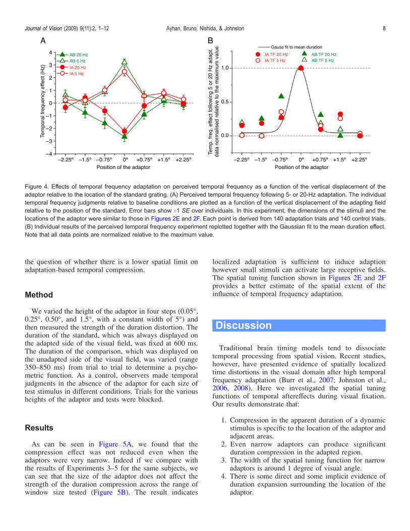
As expected, 5-Hz adaptation induced an increase and20-Hz adaptation induced a decrease in perceived tempo-ral frequency when the adaptor was presented at the same
location as the standard grating (Figure 4A). The spatialtuning is very similar to the tuning of duration compression(Figures 2E and 2F). For comparison, the results of theperceived temporal frequency experiment are plottedtogether with the Gaussian fit for the mean duration effectin Figure 4B. Similar spatial tuning of duration and temporalfrequency effects following temporal frequency adaptationprovides evidence that the time pathway and the motionpathway may share some components. However, given thatwe controlled for the perceived temporal frequencychanges in duration tasks and still found changes inapparent duration, we conclude that the effects ofadaptation on perceived duration are dissociable fromthose on temporal frequency. Adaptation can have morethan one effect on temporal mechanisms, e.g., a change insensitivity and a change in temporal tuning. We suggestthat these separate adaptations selectively influence appa-rent temporal frequency and apparent duration respectively.
Window size
The size of the duration compression effect does notappear to depend upon window size (Figure 2). This raises
Figure 3. Effects of temporal adaptation on duration judgments in the surrounding areas as a function of the relative size of the adaptor.Colored gratings indicate the adaptor locations. (A) Results of the surround effects experiment for four subjects (2 authors and 2 naïves;included in Figure 1B). The mean duration judgments comparative to the baseline conditions are plotted as a function of the outerdiameter of the surrounding area. Error bars show T1 SE of individuals and T1 SE of the mean. In this experiment, the diameter of theadaptor and the inner circle of the annuli are fixed at 5-. Each point is derived from 140 adaptation trials and 280 control trials. (B) Resultsof the surround effects experiment for four subjects (2 authors and 2 naïves; included in Figure 1B). As in Figure 3A except in thisexperiment, the outer diameter of the annuli is fixed at 9-. Each point is derived from 140 adaptation trials and 280 control trials.
Journal of Vision (2009) 9(11):2, 1–12 Ayhan, Bruno, Nishida, & Johnston 7
the question of whether there is a lower spatial limit onadaptation-based temporal compression.
Method
We varied the height of the adaptor in four steps (0.05-,0.25-, 0.50-, and 1.5-, with a constant width of 5-) andthen measured the strength of the duration distortion. Theduration of the standard, which was always displayed onthe adapted side of the visual field, was fixed at 600 ms.The duration of the comparison, which was displayed onthe unadapted side of the visual field, was varied (range350–850 ms) from trial to trial to determine a psycho-metric function. As a control, observers made temporaljudgments in the absence of the adaptor for each size oftest stimulus in different conditions. Trials for the variousheights of the adaptor and tests were blocked.
Results
As can be seen in Figure 5A, we found that thecompression effect was not reduced even when theadaptors were very narrow. Indeed if we compare withthe results of Experiments 3–5 for the same subjects, wecan see that the size of the adaptor does not affect thestrength of the duration compression across the range ofwindow size tested (Figure 5B). The result indicates
localized adaptation is sufficient to induce adaptionhowever small stimuli can activate large receptive fields.The spatial tuning function shown in Figures 2E and 2Fprovides a better estimate of the spatial extent of theinfluence of temporal frequency adaptation.
Discussion
Traditional brain timing models tend to dissociatetemporal processing from spatial vision. Recent studies,however, have presented evidence of spatially localizedtime distortions in the visual domain after high temporalfrequency adaptation (Burr et al., 2007; Johnston et al.,2006, 2008). Here we investigated the spatial tuningfunctions of temporal aftereffects during visual fixation.Our results demonstrate that:
1. Compression in the apparent duration of a dynamicstimulus is specific to the location of the adaptor andadjacent areas.
2. Even narrow adaptors can produce significantduration compression in the adapted region.
3. The width of the spatial tuning function for narrowadaptors is around 1 degree of visual angle.
4. There is some direct and some implicit evidence ofduration expansion surrounding the location of theadaptor.
Figure 4. Effects of temporal frequency adaptation on perceived temporal frequency as a function of the vertical displacement of theadaptor relative to the location of the standard grating. (A) Perceived temporal frequency following 5- or 20-Hz adaptation. The individualtemporal frequency judgments relative to baseline conditions are plotted as a function of the vertical displacement of the adapting fieldrelative to the position of the standard. Error bars show T1 SE over individuals. In this experiment, the dimensions of the stimuli and thelocations of the adaptor were similar to those in Figures 2E and 2F. Each point is derived from 140 adaptation trials and 140 control trials.(B) Individual results of the perceived temporal frequency experiment replotted together with the Gaussian fit to the mean duration effect.Note that all data points are normalized relative to the maximum value.
Journal of Vision (2009) 9(11):2, 1–12 Ayhan, Bruno, Nishida, & Johnston 8

5. The effects of adaptation on perceived duration aredissociable from those on temporal frequencyalthough the spatial tuning of these effects is similar.
The spatial specificity of the effects of temporal frequencyadaptation on apparent duration argues against a supramodalcentralized clock model (Creelman, 1962; Treisman, 1963;Treisman et al., 1990). Rather, the local distortions ofapparent duration suggest a modality specific mechanismthat shares some components with the motion pathway.It has been suggested that time might be encoded by a
population of metaneurons that are sensitive to the timecourse of neural processing in the brain (Buonomano &Merzenich, 1995; Eagleman et al., 2005; Karmarkar &Buonomano, 2007). This distributed neural account oftiming posits the translation of a temporal code into aplace code, with separate duration tuned units. An intervalcan then be encoded by reading out the distribution ofactivity over this population. The population response to astimulus can be biased in a direction opposite to anadaptor after prolonged viewing of a stimulus that isslightly different to the test along the relevant stimulusdimension. However, rather than adapting to intervals of aparticular duration, participants adapted to flicker of aparticular temporal frequency. Since participants are notadapted to duration per se, it is not clear how the changes inperceived duration we see after temporal frequency adapta-tion could be explained on a duration-tuned populationmodel. We therefore have to consider in more detail howtemporal frequency adaptation alters the visual system.
The narrow spatial tuning of both the temporalfrequency shift and apparent duration compression sug-gests an early locus in the visual pathway. The standarddeviation of spatial tuning for narrow windows was0.59 degrees at 5 degree of visual eccentricity. Closer tothe mean receptive field diameter of macaque V1 at around0.4 degrees (Dow et al., 1981; Johnston, 1989) than tohigher level cortical areas such as LIP, where receptivefield size reaches 12 degrees at 5 degrees of visualeccentricity (Blatt et al., 1990). Thus, the spatial tuning ofduration compression is more compatible with an earlythan late location in the visual pathway. A possiblecandidate for the site of the adaptation is the magnocel-lular pathway through the lateral geniculate nucleus(Johnston et al., 2006, 2008). Magnocellular neuronesare known to be sensitive to low spatial frequencyachromatic stimuli (Benardete & Kaplan, 1999). Theyare also responsive to high frequency (60 Hz) adaptors,which are invisible to cortical cells (Hawken, Shapley, &Grosof, 1996) but which can still induce durationcompression (Johnston et al., 2008). Solomon, Peirce,Dhruv, and Lennie (2004) have shown that magnocellular(M) cells in the LGN show slow adaptation at hightemporal frequencies (45 Hz) but not at low temporalfrequencies (1 Hz). M cells, but not P cells, are alsosubject to a fast adaptation phase referred to as contrastgain control which results in a sharpening of temporaltuning as a consequence of a loss of gain at low temporalfrequencies (Shapley & Victor, 1978). Recently Brunoand Johnston (2007) have demonstrated a reduction inperceived duration for intervals following a high contrast
Figure 5. Compression in apparent duration following temporal adaptation as a function of the height of the adaptor. The relativedimensions of the stimuli are shown below the abscissa. (A) Results of the adaptor size experiment for two subjects (included in Figure1B). The individual duration judgments comparative to the baseline conditions are plotted as a function of the height of the adapting area.Error bars show T1 SE. Each point is derived from 140 adaptation trials and 140 control trials. (B) The results of the lateral interactionexperiment in small, medium, and large scales (Experiments 3–5) are redrawn for two subjects for the condition in which the standard andthe adaptor gratings fully overlap. Error bars show T1 SE.
Journal of Vision (2009) 9(11):2, 1–12 Ayhan, Bruno, Nishida, & Johnston 9

context relative to a low contrast context, providingevidence that reductions in perceive duration may bemediated by a compression of the temporal impulseresponse in M cells, in this case following contrast gain.There is some evidence for duration expansion at
locations some distance from the adaptor. This suggeststhat adaption can have a local effect in the region of theadaptor and an opposite but weaker effect at a distance.This spatial pattern of adaptation could explain why thecompression effect is eliminated as test patterns extendbeyond the spatial location of the adaptor (Experiment 6)as if the duration percept resulted from the spatialsummation of a compression and expansion effect.Changes in apparent temporal frequency after temporal
frequency adaption can be dissociated from changes inapparent duration. Temporal frequency shifts are bidirec-tional, whereas a 10-Hz interval appears compressed after5-, 10-, and 20-Hz adaptation (Johnston et al., 2006), adifference we exploit in the current paper. Changes inapparent duration can also occur in the absence of anychange in apparent temporal frequency, as reportedabove. Dyslexics show normal temporal frequency shiftsbut do not show any duration compression after adaptionto invisible flicker (Johnston et al., 2008). We thereforeneed to identify separate mechanisms for changes inapparent temporal frequency and changes in apparentduration. We will first consider the change in apparentfrequency.The available evidence is that there are only two or
three temporal frequency channels in the human visualsystem (Fredericksen & Hess, 1998; Hess & Snowden,1992; Johnston & Clifford, 1995) and each filter has adifferent shape as well as a different pass-band. Johnstonand Clifford (1995) showed that the temporal filtersmeasured by Hess and Snowden (1992) were well fittedby a Gaussian in log time and its first and secondderivatives. While the zero order filter has the low-passtemporal property characteristic of P cells, the derivativefilters have the band-pass property associated with Mcells. Therefore, the available evidence does not supportthe idea that temporal frequency is encoded in terms of thepeak response of a set of identical narrow band filters.However, for sine functions, temporal frequency can berecovered from the relative activity of band-pass and low-pass filters (Smith & Edgar, 1994). On this model the shiftin apparent temporal frequency of a 10-Hz test patternafter high temporal frequency can be explained by arelative increase in the response of the low-pass channelas a result of reduction in sensitivity of the band-passchannel following adaptation. An increased ratio of low toband-pass channel activity signifies a lower temporalfrequency. The opposite shift arises after adaption to alow (e.g., 5 Hz) frequency.Since changes in perceived duration can be dissociated
from changes in perceived temporal frequency, a differentmechanism is required to explain changes in apparent
duration. In addition to changes in sensitivity, adaptationto high temporal frequency also sharpens band-passtemporal filters (Ibbotson, 2005), which as a consequenceintroduces a forward shift in the band-pass response. Wemay use this adaptation to explain the apparent durationcompression. Johnston (in press) has proposed a content-dependent clock. In this clock a forward model of thevisual signal is generated from the visual input. The newvisual input is then continuously compared to the predic-tion. When there is a match the system can tell the presetinterval has elapsed, the clock ticks and the cumulatedticks are a measure of the duration of the interval. Thisforward prediction needs to know the rate of change ofimage brightness and therefore relies on band-pass differ-entiating temporal filters (magnocellular neurones). Thecurrent brightness signal can be provided by a low-pass channel, which does not adapt to the same degree(parvocellular neurones). After adaptation, the phaseadvance in the magnocellular signal shifts the predictionforward in time, increasing the time to reset and therebyproducing the psychophysically observed time compres-sion. Therefore, we propose changes in responsivity ofthe M cells relative to P cells explain changes in apparenttemporal frequency, whereas sharpening of the M celltemporal impulse response explains apparent time com-pression (Johnston et al., 2006).In summary, adaptation-based apparent duration com-
pression was found to be tightly tuned to the location ofthe adaptor. For large adaptors compression extended to asmall region beyond the location of the adaptor. For smalladaptors the spatial extent of compression was around 1degree. For larger adaptors, compression was eliminatedand in some cases replaced by expansion, indicating aregion of apparent temporal expansion beyond theduration compression zone. These local distortions inapparent duration elicited by temporal frequency adapta-tion in the absence of changes in perceived speed orperceived temporal frequency leads us to conclude thattemporal channels in human vision mediate durationperception, as well as temporal frequency coding andmotion perception.
Acknowledgments
We are happy to acknowledge the support of theLeverhulme Trust and the Nippon Telegraph and Tele-phone Corporation.
Commercial relationships: none.Corresponding author: Inci Ayhan.Email: [email protected]: Cognitive, Perceptual and Brain Sciences, Divisionof Psychology and Language Sciences, University CollegeLondon, Gower Street, London, WC1E 6BT, UK.
Journal of Vision (2009) 9(11):2, 1–12 Ayhan, Bruno, Nishida, & Johnston 10
References
Benardete, E. A., & Kaplan, E. (1999). The dynamics ofprimate M retinal ganglion cells. Visual Neuro-science, 16, 355–368. [PubMed]
Blatt, G. J., Andersen, R. A., & Stoner, G. R. (1990).Visual receptive field organization and cortico-cortical connections of the lateral intraparietal area(area LIP) in the macaque. Journal of ComparativeNeurology, 299, 421–445. [PubMed]
Bruno, A., & Johnston, A. (2007). Contrast gain changesaffect the perceived duration of visual stimuli[Abstract]. Journal of Vision, 7(9):376, 376a, http://journalofvision.org/7/9/376/, doi:10.1167/7.9.376.
Buonomano, D. V., & Merzenich, M. M. (1995).Temporal information transformed into a spatial codeby a neural network with realistic properties. Science,267, 1028–1030. [PubMed]
Burr, D., Tozzi, A., & Morrone, M. C. (2007). Neuralmechanisms for timing visual events are spatiallyselective in real-world coordinates. Nature Neuro-science, 10, 423–425. [PubMed]
Creelman, C. D. (1962). Human discrimination of audi-tory duration. Journal of the Acoustical Society ofAmerica, 34, 582.
Dow, B. M., Snyder, A. Z., Vautin, R. G., & Bauer, R.(1981). Magnification factor and receptive field sizein foveal striate cortex of the monkey. ExperimentalBrain Research, 44, 213–228. [PubMed]
Eagleman, D. M., Tse, P. U., Buonomano, D., Janssen, P.,Nobre, A. C., & Holcombe, A. O. (2005). Time andthe brain: How subjective time relates to neural time.Journal of Neuroscience, 25, 10369–10371.[PubMed] [Article]
Fredericksen, R. E., & Hess, R. F. (1998). Estimatingmultiple temporal mechanisms in human vision.Vision Research, 38, 1023–1040. [PubMed]
Hawken, M. J., Shapley, R. M., & Grosof, D. H. (1996).Temporal-frequency selectivity in monkey visualcortex. Visual Neuroscience, 13, 477–492. [PubMed]
Hess, R. F., & Snowden, R. J. (1992). Temporal propertiesof human visual filters: Number, shapes and spatialcovariation. Vision Research, 32, 47–59. [PubMed]
Ibbotson, M. R. (2005). Contrast and temporal frequency-related adaptation in the pretectal nucleus of the optictract. Journal of Neurophysiology, 94, 136–146.[PubMed] [Article]
Jamieson, D. G., & Petrusic, W. M. (1975). Presentationorder effects in duration discrimination. Perception &Psychophysics, 17, 197–202.
Johnston, A. (in press). Visual time perception. In A. C.Nobre & J. T. Coull (Eds.), Attention and time.Oxford: Oxford University Press.
Johnston, A. (1989). The geometry of the topographicmap in striate cortex. Vision Research, 29, 1493–1500.[PubMed]
Johnston, A., Arnold, D. H., & Nishida, S. (2006).Spatially localized distortions of event time. CurrentBiology, 16, 472–479. [PubMed]
Johnston, A., Bruno, A., Watanabe, J., Quansah, B., Patel,N., Dakin, S., et al. (2008). Visually-based temporaldistortion in dyslexia. Vision Research, 48, 1852–1858.[PubMed]
Johnston, A., & Clifford, C. W. (1995). A unified accountof three apparent motion illusions. Vision Research,35, 1109–1123. [PubMed]
Kanai, R., Paffen, C. L., Hogendoorn, H., &Verstraten, F. A.(2006). Time dilation in dynamic visual display. Journalof Vision, 6(12):8, 1421–1430, http://journalofvision.org/6/12/8/, doi:10.1167/6.12.8. [PubMed] [Article]
Karmarkar, U. R., & Buonomano, D. V. (2007). Timing inthe absence of clocks: Encoding time in neuralnetwork states. Neuron, 53, 427–438. [PubMed][Article]
Lee, B. B., Kremers, J., & Yeh, T. (1998). Receptivefields of primate retinal ganglion cells studied with anovel technique. Visual Neuroscience, 15, 161–175.[PubMed]
Morrone, M. C., Ross, J., & Burr, D. (2005). Saccadic eyemovements cause compression of time as well asspace. Nature Neuroscience, 8, 950–954. [PubMed]
Shapley, R. M., & Victor, J. D. (1978). The effect ofcontrast on the transfer properties of cat retinal ganglioncells. The Journal of Physiology, 285, 275–298.[PubMed] [Article]
Smith, A. T., & Edgar, G. K. (1994). Antagonisticcomparison of temporal frequency filter outputs as abasis for speed perception. Vision Research, 34,253–265. [PubMed]
Smith, A. T., Singh, K. D., Williams, A. L., & Greenlee,M. W. (2001). Estimating receptive field size fromfMRI data in human striate and extrastriate visualcortex. Cerebral Cortex, 11, 1182–1190. [PubMed]
Solomon, S. G., Peirce, J. W., Dhruv, N. T., & Lennie, P.(2004). Profound contrast adaptation early in thevisual pathway. Neuron, 42, 155–162. [PubMed]
Treisman, M. (1963). Temporal discrimination and theindifference interval. Implications for a model of the“internal clock.” Psychological Monographs, 77, 1–31.[PubMed]
Journal of Vision (2009) 9(11):2, 1–12 Ayhan, Bruno, Nishida, & Johnston 11

Treisman, M., Faulkner, A., Naish, P. L., & Brogan, D.(1990). The internal clock: Evidence for a temporaloscillator underlying time perception with someestimates of its characteristic frequency. Perception,19, 705–743. [PubMed]
Xu, X., Ichida, J. M., Allison, J. D., Boyd, J. D., Bonds,A. B., & Casagrande, V. A. (2001). A comparison ofkoniocellular, magnocellular and parvocellular recep-
tive field properties in the lateral geniculate nucleusof the owl monkey (Aotus trivirgatus). The Journal ofPhysiology, 531, 203–218. [PubMed] [Article]
Zeki, S. M. (1978). Uniformity and diversity of structureand function in rhesus monkey prestriate visualcortex. The Journal of Physiology, 277, 273–290.[PubMed] [Article]
Journal of Vision (2009) 9(11):2, 1–12 Ayhan, Bruno, Nishida, & Johnston 12
Related Documents