Molecular Cell, Vol. 4, 983–993, December, 1999, Copyright 1999 by Cell Press The Spatial Position and Replication Timing of Chromosomal Domains Are Both Established in Early G1 Phase defined (Gilbert, 1998), the determinants of replication timing have proven complex. In at least one case, the determinant of replication timing appears to lie within the origin sequences themselves (Bousset and Diffley, 1998). In other cases, it is clearly mediated by cis-acting Daniela S. Dimitrova and David M. Gilbert* Department of Biochemistry and Molecular Biology State University of New York Health Science Center Syracuse, New York 13210 elements separate from the origins. Two types of chro- mosomal context effects have been found to confer Summary late initiation: proximity to a telomere (Ferguson and Fangman, 1992) and association with specific segments Mammalian chromosomal domains replicate at de- of chromosomal DNA (Friedman et al., 1996). In the ab- fined, developmentally regulated times during S phase. sence of these sequences, many late origins will fire The positions of these domains in Chinese hamster early in S phase. For one late-initiating origin located nuclei were established within 1 hr after nuclear enve- near a telomere, the signal for late replication is estab- lope formation and maintained thereafter. When G1 lished between metaphase (nocodazole arrest) and phase nuclei were incubated in Xenopus egg extracts, START (a factor arrest) (Raghuraman et al., 1997). Fi- domains were replicated in the proper temporal order nally, the ability of telomeres to delay replication of an- with nuclei isolated after spatial repositioning, but not other late-initiating origin requires SIR3 (Stevenson and with nuclei isolated prior to repositioning. Mcm2 was Gottschling, 1999), a silent chromatin component, sug- bound both to early- and late-replicating chromatin gesting that late replication of some origins is influenced domains prior to this transition whereas specification by chromatin structure. of the dihydrofolate reductase replication origin took We have developed a cell-free system that efficiently place several hours thereafter. These results identify initiates replication within mammalian nuclei isolated at an early G1 phase point at which replication timing is different stages of G1 phase. When intact late G1 phase determined and demonstrate a provocative temporal Chinese hamster ovary (CHO) nuclei are introduced into coincidence between the establishment of nuclear po- Xenopus egg extracts, replication initiates at the physio- sition and replication timing. logically relevant origins within the CHO dihydrofolate reductase (DHFR) locus (Dimitrova and Gilbert, 1998). Introduction DHFR origin recognition requires nuclei isolated from cells that have progressed beyond a distinct point dur- Chromosomal domains replicate in a defined spatiotem- ing G1 phase, the origin decision point (ODP) (Wu and poral program. Each domain occupies a specific nuclear Gilbert, 1996). The ODP takes place after the initial bind- position and replicates at a precise time during S phase ing of the pre–replication complex (pre-RC) Mcm pro- (reviewed in Berezney et al., 1999; Dimitrova and Gilbert, teins to chromatin (Dimitrova et al., 1999) and prior to the 1999). In general, Giemsa-light (R) bands, containing restriction point (Wu and Gilbert, 1997). The existence of transcriptionally active chromosomal domains, replicate an ODP was subsequently confirmed in cultured cells earlier, and Giemsa-dark (G) bands, comprising tran- without the use of Xenopus egg extract (Wu et al., 1998; scriptionally silent heterochromatin, replicate later in S D. S. D., unpublished data), demonstrating that novel phase (Gilbert, 1986; Hatton et al., 1988; Drouin et al., G1 phase regulatory events can be identified using this 1990; Manuelidis, 1990). Changes in replication timing heterologous cell-free system. accompany key stages of metazoan development (Holm- Here, we investigated whether replication timing in quist, 1987; Hatton et al., 1988; Selig et al., 1992) and mammalian cells is also established during G1 phase. are often coupled to changes in gene expression (Dhar To address this question, we developed methodology et al., 1989). Taken together, these results suggest that to differentially tag early- and late-replicating domains structural, transcriptional, and replicational domains of CHO cell chromosomes. This allowed us to monitor share topographical boundaries and represent basic both their spatial distribution within the nucleus and their units of chromosome organization. potential to be distinguished as early- or late-replicating It is not known whether components of replication domains in Xenopus egg extracts. Our results revealed origins dictate when domains replicate, or whether epi- that the temporal program for replication is established genetic mechanisms regulating domain structure and in early G1 phase, after the initial binding of Mcm2 pro- function dictate when origins will be activated. Translo- teins to chromatin but prior to the ODP. Intriguingly, cations and deletions can influence the replication tim- establishment of a replication timing program took place ing of large chromosomal domains (Calza et al., 1984; within the same time interval as the spatial repositioning Forrester et al., 1990). At some loci, the developmental of chromosomal domains within the nucleus. switch in replication timing is accompanied by a change in origin usage (Ermakova et al., 1999) whereas other Results loci initiate replication at the same origin whether early or late replicating (Kitsberg et al., 1993). Even in the Characterization of Spatiotemporal Replication yeast Saccharomyces cerevisiae, where origins are well Patterns in CHOC 400 Cells DNA replication takes place at discrete sites that can be visualized by pulse-labeling cells with 5-bromo-29- * To whom correspondence should be addressed (e-mail: gilbertd@ hscsyr.edu). deoxyuridine (BrdU) and staining nuclei with fluorescent

Welcome message from author
This document is posted to help you gain knowledge. Please leave a comment to let me know what you think about it! Share it to your friends and learn new things together.
Transcript
Molecular Cell, Vol. 4, 983–993, December, 1999, Copyright 1999 by Cell Press
The Spatial Position and Replication Timingof Chromosomal Domains Are Both Establishedin Early G1 Phase
defined (Gilbert, 1998), the determinants of replicationtiming have proven complex. In at least one case, thedeterminant of replication timing appears to lie withinthe origin sequences themselves (Bousset and Diffley,1998). In other cases, it is clearly mediated by cis-acting
Daniela S. Dimitrova and David M. Gilbert*Department of Biochemistry and Molecular BiologyState University of New York Health Science CenterSyracuse, New York 13210
elements separate from the origins. Two types of chro-mosomal context effects have been found to conferSummarylate initiation: proximity to a telomere (Ferguson andFangman, 1992) and association with specific segmentsMammalian chromosomal domains replicate at de-of chromosomal DNA (Friedman et al., 1996). In the ab-fined, developmentally regulated times during S phase.sence of these sequences, many late origins will fireThe positions of these domains in Chinese hamsterearly in S phase. For one late-initiating origin locatednuclei were established within 1 hr after nuclear enve-near a telomere, the signal for late replication is estab-lope formation and maintained thereafter. When G1lished between metaphase (nocodazole arrest) andphase nuclei were incubated in Xenopus egg extracts,START (a factor arrest) (Raghuraman et al., 1997). Fi-domains were replicated in the proper temporal ordernally, the ability of telomeres to delay replication of an-with nuclei isolated after spatial repositioning, but notother late-initiating origin requires SIR3 (Stevenson andwith nuclei isolated prior to repositioning. Mcm2 wasGottschling, 1999), a silent chromatin component, sug-bound both to early- and late-replicating chromatingesting that late replication of some origins is influenceddomains prior to this transition whereas specificationby chromatin structure.of the dihydrofolate reductase replication origin took
We have developed a cell-free system that efficientlyplace several hours thereafter. These results identifyinitiates replication within mammalian nuclei isolated atan early G1 phase point at which replication timing isdifferent stages of G1 phase. When intact late G1 phasedetermined and demonstrate a provocative temporalChinese hamster ovary (CHO) nuclei are introduced intocoincidence between the establishment of nuclear po-Xenopus egg extracts, replication initiates at the physio-sition and replication timing.logically relevant origins within the CHO dihydrofolatereductase (DHFR) locus (Dimitrova and Gilbert, 1998).IntroductionDHFR origin recognition requires nuclei isolated fromcells that have progressed beyond a distinct point dur-Chromosomal domains replicate in a defined spatiotem-ing G1 phase, the origin decision point (ODP) (Wu andporal program. Each domain occupies a specific nuclearGilbert, 1996). The ODP takes place after the initial bind-position and replicates at a precise time during S phaseing of the pre–replication complex (pre-RC) Mcm pro-(reviewed in Berezney et al., 1999; Dimitrova and Gilbert,teins to chromatin (Dimitrova et al., 1999) and prior to the1999). In general, Giemsa-light (R) bands, containingrestriction point (Wu and Gilbert, 1997). The existence oftranscriptionally active chromosomal domains, replicatean ODP was subsequently confirmed in cultured cellsearlier, and Giemsa-dark (G) bands, comprising tran-without the use of Xenopus egg extract (Wu et al., 1998;scriptionally silent heterochromatin, replicate later in SD. S. D., unpublished data), demonstrating that novel
phase (Gilbert, 1986; Hatton et al., 1988; Drouin et al.,G1 phase regulatory events can be identified using this
1990; Manuelidis, 1990). Changes in replication timingheterologous cell-free system.
accompany key stages of metazoan development (Holm- Here, we investigated whether replication timing inquist, 1987; Hatton et al., 1988; Selig et al., 1992) and mammalian cells is also established during G1 phase.are often coupled to changes in gene expression (Dhar To address this question, we developed methodologyet al., 1989). Taken together, these results suggest that to differentially tag early- and late-replicating domainsstructural, transcriptional, and replicational domains of CHO cell chromosomes. This allowed us to monitorshare topographical boundaries and represent basic both their spatial distribution within the nucleus and theirunits of chromosome organization. potential to be distinguished as early- or late-replicating
It is not known whether components of replication domains in Xenopus egg extracts. Our results revealedorigins dictate when domains replicate, or whether epi- that the temporal program for replication is establishedgenetic mechanisms regulating domain structure and in early G1 phase, after the initial binding of Mcm2 pro-function dictate when origins will be activated. Translo- teins to chromatin but prior to the ODP. Intriguingly,cations and deletions can influence the replication tim- establishment of a replication timing program took placeing of large chromosomal domains (Calza et al., 1984; within the same time interval as the spatial repositioningForrester et al., 1990). At some loci, the developmental of chromosomal domains within the nucleus.switch in replication timing is accompanied by a changein origin usage (Ermakova et al., 1999) whereas other Resultsloci initiate replication at the same origin whether earlyor late replicating (Kitsberg et al., 1993). Even in the Characterization of Spatiotemporal Replicationyeast Saccharomyces cerevisiae, where origins are well Patterns in CHOC 400 Cells
DNA replication takes place at discrete sites that canbe visualized by pulse-labeling cells with 5-bromo-29-* To whom correspondence should be addressed (e-mail: gilbertd@
hscsyr.edu). deoxyuridine (BrdU) and staining nuclei with fluorescent
Molecular Cell984
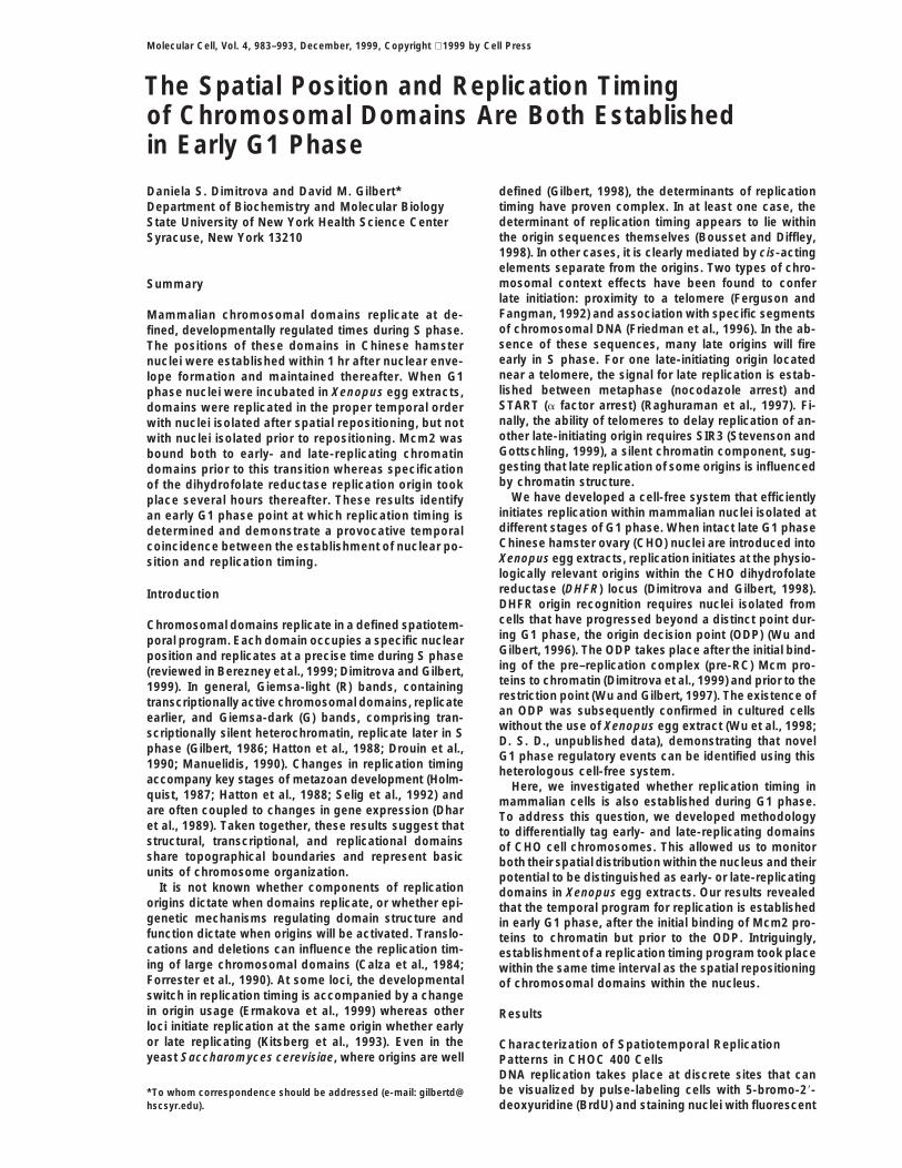
Figure 1. Spatiotemporal Patterns of DNAReplication in CHOC 400 Cells
(A) CHOC 400 cells were synchronized at theG1/S border and released into S phase. Atvarious intervals thereafter, cells were pulselabeled for 5 min with BrdU and stained withanti-BrdU antibodies. Shown are examples ofeach labeling pattern (I–V).(B) Ten asynchronous cultures of CHOC 400cells were pulse labeled for 3 min with IdU.One of these cultures was labeled simultane-ously with CldU (09) whereas the rest werechased for the indicated times before theywere pulse labeled with CldU. Sites of CldU(green) and IdU (red) incorporation were visu-alized using CldU- and IdU-specific antibod-ies. (c) is an expanded image of a single typeV focus.(C) The percentage of each type of nuclei(I/II–V) showing either complete colocaliza-tion (100% yellow foci), partial colocalization(mixture of red, green, and yellow foci), or littleto no colocalization (complete separation ofred, green, and yellow foci) was scored forchase periods less than 1 hr.(D) For chase periods longer than 1 hr, thepercentage of each type of IdU-labeled nuclei(I/II–V) that had proceeded to subsequentstages of S phase (or to G2 phase) during thechase was scored. In (C) and (D), the meanvalues are shown 6SEM for three indepen-dent experiments in which 100–200 nucleieach were scored.(E) Schematic diagram approximating the du-ration of each replication pattern, derivedfrom the independent approaches describedin (A) (scoring the percentages of each pat-tern at various time intervals) and (D).
antibodies to BrdU (Berezney et al., 1999). We reasoned within a small number of large internal or peripheral foci(type V) that likely represent large stretches of tandemlythat a detailed characterization of these sites would
provide the basis for an approach to rapidly evaluate repeated sequences, such as satellite DNA (Manuelidis,1990; O’Keefe et al., 1992).the replication timing of whole groups of domains within
individual nuclei replicating in our cell-free system. Fig- To confirm the distinct nature of these foci, we tookadvantage of the ability to distinguish DNA labeled withure 1A shows the spatiotemporal sequence of replica-
tion patterns in CHOC 400 cells. These patterns are either 5-chloro-29-deoxyuridine (CldU) or 5-iodo-29-deoxyuridine (IdU) using specific antibodies (Aten et al.,designated types I-V, in accordance with previous no-
menclature (O’Keefe et al., 1992). The type I pattern was 1992). Exponentially growing CHOC 400 cells were pulselabeled with IdU, chased for different periods of time,observed only during the first few minutes of S phase,
when the number of replication foci is relatively small. pulse labeled with CldU, and then stained with red (forIdU) and green (for CldU) fluorochrome-conjugated anti-New foci emerge continuously, until there are several
hundred foci throughout the internal, euchromatic re- bodies (Figure 1B). When visualized through a dual red/green filter, foci that synthesized DNA throughout thegions of the nucleus, defining the type II pattern. Since
type I is brief and is probably a direct precursor to the pulse/chase/pulse period incorporated both IdU andCldU and appear yellow, IdU-labeled foci having com-type II pattern, we refer to them collectively as type I/II.
This pattern of labeling is observed throughout the first pleted replication during the chase label only in red, andfoci that initiated replication during the chase label onlyhalf of S phase (0–6 hr). At mid–S phase (6–9 hr), replica-
tion takes place almost exclusively at the nuclear periph- in green. As expected, simultaneous IdU/CldU labelingrevealed exclusively yellow foci (Figure 1Ba). After a 10ery and nucleolar regions (type III). Late in S (9–12 hr),
replication occurs first within several relatively large foci min chase, type I/II foci were still yellow, but separatered and green foci were evident in some type III (Figurethroughout the nucleus (type IV) and subsequently
Establishing Mammalian Replication Timing985
1Bb) and many type IV and V (Figure 1Bc) patterns,possibly reflecting a higher rate of replication fork move-ment during late S (Housman and Huberman, 1975).Increasing chase times led to an increase in the numberof red- or green-labeled foci and a decrease in yellowfoci (Figures 1Bd–1Bh and 1C). These results generallyagree with previous reports concluding that early focicomplete replication within approximately 60 min (Man-ders et al., 1996; Ma et al., 1998) but reveal considerableheterogeneity among individual foci and suggest thatlate foci may complete replication more rapidly.
Chase periods longer than 1 hr frequently revealedclearly demarcated foci of the same pattern type. Sepa-rate green and red type I/II patterns were observed after1–6 hr chase periods (Figures 1Bg, 1Bk, and 1D). Sepa-rate type III patterns were observed in cell cultureschased for 1–3 hr (Figure 1Bh, 1Bi, and 1D). Separatetype IV patterns were found after a 1 hr chase but rarelywhen chased for 2 hr, while type V patterns were largelycompleted within 1 hr (Figure 1D). The presence of focilabeled only with CldU within nuclei that still containyellow foci (Figures 1Bb and 1Bd–1Bf) demonstratesthat activation of new replication sites does not requirethe completion of all previous sites. Furthermore, theonset of a subsequent pattern could often be seen priorto the completion of the previous pattern. These resultsare consistent with the continuous activation of replica-tion sites throughout S phase.
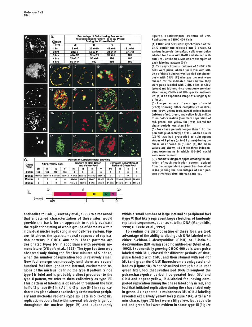
The length of the chase period sufficient for cells to Figure 2. The Spatial Positioning and Temporal Order of Replicationswitch from one type of replication pattern to subse- of Chromosomal Domains Are Maintained in Xenopus Egg Extractsquent patterns (Figure 1D) was consistent with the dura- Exponentially growing CHOC 400 cells were pulse labeled for 20
min with BrdU before intact nuclei from these cells were preparedtion of each pattern (Figure 1E). Hence, nuclei were notand introduced into a Xenopus egg extract. Aliquots of these nucleiobserved to progress to the next pattern (type II to typewere pulse labeled for 10 min with biotin-dUTP at the indicated timeIII, Figure 1Bn) or more than one pattern ahead (type IIIintervals in vitro. Nuclei were fixed and double stained for BrdUto type V, Figure 1Bl; type II to type V, Figure 1Bo) until(green) and biotin (red).
after the appropriate amount of time. Nuclei that werein G1 phase during the IdU label and in S phase duringthe CldU label stained only in green (Figures 1Bm and lag period and the absence of BrdU label. G2 phase1Bp), while nuclei that were in mid/late S phase during nuclei were not labeled with either nucleotide analogthe IdU label and had moved out of S phase prior to the since intact G2 nuclei are not competent to replicate inCldU label stained only in red (Figure 1Bj). These nuclei Xenopus egg extract (Leno et al., 1992).demonstrate the lack of cross-reactivity between the Exemplary results from these experiments are showntwo antibodies. Together, these data provide a thorough in Figure 2. The earliest in vitro DNA synthesis in all G1description of the temporal sequence of replication fo- phase nuclei resembled type I/II patterns (Figure 2A).cus activation in CHOC 400 cells (Figure 1E). Nuclear staining became more diffuse and uniform later
in the reaction (Figure 2B). Type III DNA synthesis (Figure2C) began after 60 min, and by 2 hr, 50%–80% of G1Replication in Xenopus Egg Extracts Respects
the Proper Temporal Order phase nuclei had progressed to the type III pattern.With S phase nuclei (BrdU-positive), in vitro biotin labelTo determine whether replication in CHO nuclei by Xen-
opus egg cytosol proceeds according to the proper tem- colocalized with sites of in vivo DNA synthesis during thefirst 20 min (Figures 2D and 2E) and became increasinglyporal program, exponentially growing CHOC 400 cells
were pulse labeled with BrdU, and intact nuclei from separated from BrdU label thereafter (Figures 2F–2J).Nuclei labeled in a type I/II pattern in vivo progressedthese cells were then introduced into a Xenopus egg
extract. At various times thereafter, the replication inter- to type III in vitro (Figure 2H), type III progressed to typeIV, and type IV progressed to type V (Figure 2G). Typemediates synthesized in vitro were briefly pulse labeled
with biotin-dUTP, and nuclei were stained with both IV and type V nuclei completed S phase in vitro (Figure2I) and ceased to incorporate biotin label thereafter (Fig-anti-BrdU antibodies (green) and Texas red–conjugated
streptavidin (red; streptavidin specifically recognizes ures 2K and 2L). At late time points, some nuclei hadprogressed through more than one pattern (type III tosites of biotin-dU incorporation). This protocol allowed
us to distinguish cells in all phases of the cell cycle. type V, Figure 2J). No nuclei were observed to skip fromearly to late patterns without passing through the middleNuclei at various stages of S phase could be identified
by their BrdU pattern. G1 phase nuclei could be identi- patterns, nor were any nuclei observed to repeat pat-terns already completed in vivo. As a control, aliquots offied by the appearance of biotin label after a 10–20 min
Molecular Cell986
these same nuclei were permeabilized under conditionsthat preserve replication efficiency (Dimitrova and Gil-bert, 1998) prior to introduction into Xenopus egg ex-tract. With these preparations, G2 phase nuclei wereable to initiate replication, and S phase nuclei were ob-served to reinitiate earlier patterns already replicated invivo (data not shown), consistent with the role of thenuclear membrane in preventing rereplication within onecell cycle (Leno et al., 1992). Importantly, replicationpatterns labeled in vivo, which reflect the relative posi-tions of those labeled domains, remained intact through-out the duration of the in vitro reaction. Hence, withintact CHO nuclei as a substrate, Xenopus egg extractdoes not reposition chromosomal domains, and replica-tion proceeds according to the proper temporal program.
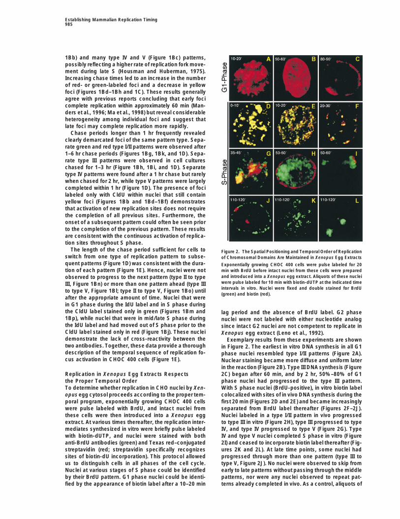
Monitoring Early and Late Replication Domainsthroughout the Cell CycleTo address whether the temporal order of replication isestablished at a specific point during G1 phase, wedifferentially tagged early- and late-replicating domainsin vivo with CldU and IdU and prepared nuclei fromthese cells at various times during the following G1phase. These nuclei were then introduced into Xenopusegg extracts and pulse labeled at various time pointswith biotin-dUTP. With this protocol, DNA synthesis tak-ing place either early or late in vitro could be localizedto either early- or late-replicating domains (Figure 3A).To verify that these manipulations do not disturb thereplication program, several control experiments wereperformed. First, we verified that the chromosomal do-mains labeled at the onset of S phase in the first cellcycle are the earliest to replicate in the following S phase Figure 3. DNA Replication Initiates within the Same Replication Do-
mains at the Beginning of Two Consecutive S Phases(Figure 3B). Cells synchronized at the G1/S border were(A) Pure populations of metaphase cells (95%–100%) were releasedpulse labeled with CldU (2–30 min pulses), collected infor 12–14 hr into fresh medium containing aphidicolin to accumulatemitosis, and released in the presence of aphidicolin tocells at the G1/S phase border. The earliest-replicating domainsaccumulate cells at the G1/S border of the second cellwere labeled briefly with CldU; cells were then chased into late Scycle. These cells were then pulse labeled with IdU (2–30phase and pulse labeled with IdU. Since cells do not remain com-
min, respectively) and double stained for CldU (green) pletely synchronous throughout the chase period, individual cellsand IdU (red). The number of double-labeled type I foci incorporate IdU into either type III, type IV, or type V late-replicating
patterns. These prelabeled cells were then resynchronized in theincreased with increasing pulse-labeling times, until focifollowing mitosis and collected after different time intervals for usebegan to resemble type II patterns, at which point therein the experiments described.was nearly complete colocalization of the two labels.(B) Cells synchronized at the G1/S border as described above wereShown is an example of a nucleus pulse labeled for 10pulse labeled for 10 min with CldU, collected in mitosis, and resyn-
min in both cell cycles, exhibiting an intermediate level chronized at the following G1/S border by a second aphidicolinof colocalization typical for this pulse length (Figure 3B). block. Cells were pulse labeled for 10 min with IdU, and the sites
of CldU (green) and IdU (red) incorporation were visualized as inWe conclude that the same subset of early replicationFigure 1B.domains initiates within a period of approximately 30(C) Aliquots of cells synchronized at the G1/S border of either themin at the onset of each S phase, consistent with previ-first (squares) or the second (circles) cell cycle in the experimentsous reports (Amaldi et al., 1973; Ma et al., 1998) anddescribed in (B) were used to analyze the distribution of early replica-
that this program is unperturbed by our protocol. tion intermediates at the DHFR locus. Nascent DNA strands wereTo verify that cells initiate at the appropriate origin labeled with [a-32P]dATP, and the distribution of the radioactive
replication intermediates was determined by hybridizing them to 17sites after this complex synchronization protocol, cellsunique DNA probes that encompass z120 kb of the DHFR locus.were synchronized as in Figure 3A, and the sites ofThe relative amounts of hybridization to each probe are plottedearliest DNA synthesis were mapped within the DHFRagainst the map position of each probe. The vertical shaded linelocus. Nascent replication forks within nuclei isolatedhighlights the positions of probes B–R, which encompass the region
at either the first or second G1/S boundaries were briefly of peak DHFR initiation activity.labeled with [a-32P]dATP. These short, pulse-labeled na-scent strands were hybridized to probes derived fromspecific positions within the DHFR locus. The relative replication initiated near each probe. Results (Figure 3C)
were indistinguishable between one cell cycle and theamounts of hybridization of these labeled strands wereplotted versus the map position of each DNA segment, next, demonstrating that our synchrony regime does not
alter the pattern of initiation sites at the DHFR locus.giving a distribution of the relative frequency at which
Establishing Mammalian Replication Timing987
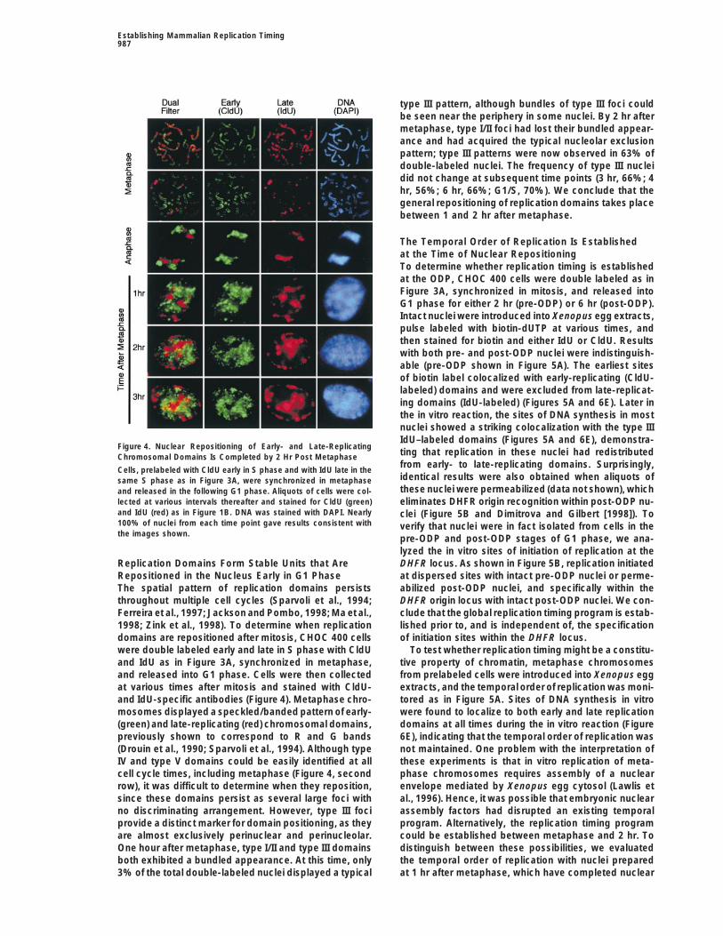
type III pattern, although bundles of type III foci couldbe seen near the periphery in some nuclei. By 2 hr aftermetaphase, type I/II foci had lost their bundled appear-ance and had acquired the typical nucleolar exclusionpattern; type III patterns were now observed in 63% ofdouble-labeled nuclei. The frequency of type III nucleidid not change at subsequent time points (3 hr, 66%; 4hr, 56%; 6 hr, 66%; G1/S, 70%). We conclude that thegeneral repositioning of replication domains takes placebetween 1 and 2 hr after metaphase.
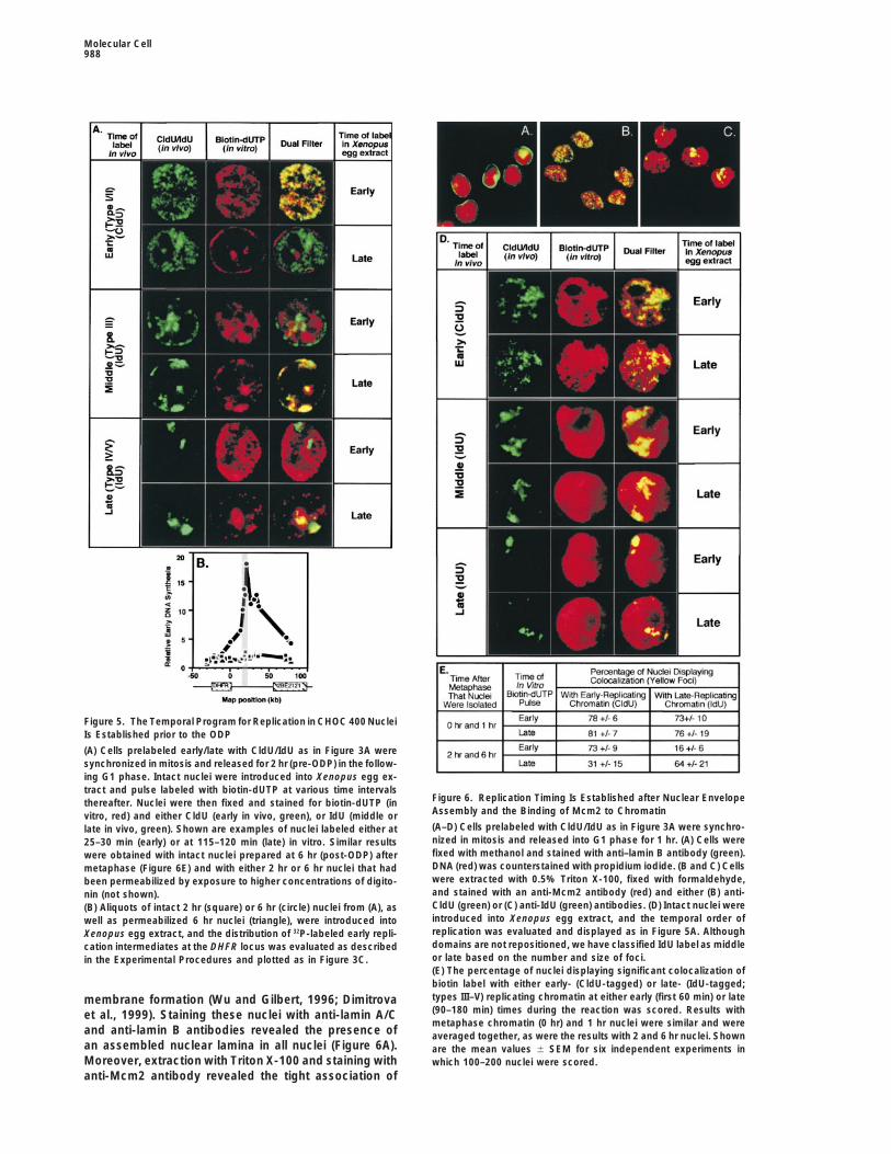
The Temporal Order of Replication Is Establishedat the Time of Nuclear RepositioningTo determine whether replication timing is establishedat the ODP, CHOC 400 cells were double labeled as inFigure 3A, synchronized in mitosis, and released intoG1 phase for either 2 hr (pre-ODP) or 6 hr (post-ODP).Intact nuclei were introduced into Xenopus egg extracts,pulse labeled with biotin-dUTP at various times, andthen stained for biotin and either IdU or CldU. Resultswith both pre- and post-ODP nuclei were indistinguish-able (pre-ODP shown in Figure 5A). The earliest sitesof biotin label colocalized with early-replicating (CldU-labeled) domains and were excluded from late-replicat-ing domains (IdU-labeled) (Figures 5A and 6E). Later inthe in vitro reaction, the sites of DNA synthesis in mostnuclei showed a striking colocalization with the type IIIIdU–labeled domains (Figures 5A and 6E), demonstra-
Figure 4. Nuclear Repositioning of Early- and Late-Replicatingting that replication in these nuclei had redistributedChromosomal Domains Is Completed by 2 Hr Post Metaphasefrom early- to late-replicating domains. Surprisingly,
Cells, prelabeled with CldU early in S phase and with IdU late in theidentical results were also obtained when aliquots ofsame S phase as in Figure 3A, were synchronized in metaphasethese nuclei were permeabilized (data not shown), whichand released in the following G1 phase. Aliquots of cells were col-
lected at various intervals thereafter and stained for CldU (green) eliminates DHFR origin recognition within post-ODP nu-and IdU (red) as in Figure 1B. DNA was stained with DAPI. Nearly clei (Figure 5B and Dimitrova and Gilbert [1998]). To100% of nuclei from each time point gave results consistent with verify that nuclei were in fact isolated from cells in thethe images shown. pre-ODP and post-ODP stages of G1 phase, we ana-
lyzed the in vitro sites of initiation of replication at theDHFR locus. As shown in Figure 5B, replication initiatedReplication Domains Form Stable Units that Are
Repositioned in the Nucleus Early in G1 Phase at dispersed sites with intact pre-ODP nuclei or perme-abilized post-ODP nuclei, and specifically within theThe spatial pattern of replication domains persists
throughout multiple cell cycles (Sparvoli et al., 1994; DHFR origin locus with intact post-ODP nuclei. We con-clude that the global replication timing program is estab-Ferreira et al., 1997; Jackson and Pombo, 1998; Ma et al.,
1998; Zink et al., 1998). To determine when replication lished prior to, and is independent of, the specificationof initiation sites within the DHFR locus.domains are repositioned after mitosis, CHOC 400 cells
were double labeled early and late in S phase with CldU To test whether replication timing might be a constitu-tive property of chromatin, metaphase chromosomesand IdU as in Figure 3A, synchronized in metaphase,
and released into G1 phase. Cells were then collected from prelabeled cells were introduced into Xenopus eggextracts, and the temporal order of replication was moni-at various times after mitosis and stained with CldU-
and IdU-specific antibodies (Figure 4). Metaphase chro- tored as in Figure 5A. Sites of DNA synthesis in vitrowere found to localize to both early and late replicationmosomes displayed a speckled/banded pattern of early-
(green) and late-replicating (red) chromosomal domains, domains at all times during the in vitro reaction (Figure6E), indicating that the temporal order of replication waspreviously shown to correspond to R and G bands
(Drouin et al., 1990; Sparvoli et al., 1994). Although type not maintained. One problem with the interpretation ofthese experiments is that in vitro replication of meta-IV and type V domains could be easily identified at all
cell cycle times, including metaphase (Figure 4, second phase chromosomes requires assembly of a nuclearenvelope mediated by Xenopus egg cytosol (Lawlis etrow), it was difficult to determine when they reposition,
since these domains persist as several large foci with al., 1996). Hence, it was possible that embryonic nuclearassembly factors had disrupted an existing temporalno discriminating arrangement. However, type III foci
provide a distinct marker for domain positioning, as they program. Alternatively, the replication timing programcould be established between metaphase and 2 hr. Toare almost exclusively perinuclear and perinucleolar.
One hour after metaphase, type I/II and type III domains distinguish between these possibilities, we evaluatedthe temporal order of replication with nuclei preparedboth exhibited a bundled appearance. At this time, only
3% of the total double-labeled nuclei displayed a typical at 1 hr after metaphase, which have completed nuclear
Molecular Cell988
Figure 5. The Temporal Program for Replication in CHOC 400 NucleiIs Established prior to the ODP
(A) Cells prelabeled early/late with CldU/IdU as in Figure 3A weresynchronized in mitosis and released for 2 hr (pre-ODP) in the follow-ing G1 phase. Intact nuclei were introduced into Xenopus egg ex-tract and pulse labeled with biotin-dUTP at various time intervals
Figure 6. Replication Timing Is Established after Nuclear Envelopethereafter. Nuclei were then fixed and stained for biotin-dUTP (inAssembly and the Binding of Mcm2 to Chromatinvitro, red) and either CldU (early in vivo, green), or IdU (middle or(A–D) Cells prelabeled with CldU/IdU as in Figure 3A were synchro-late in vivo, green). Shown are examples of nuclei labeled either atnized in mitosis and released into G1 phase for 1 hr. (A) Cells were25–30 min (early) or at 115–120 min (late) in vitro. Similar resultsfixed with methanol and stained with anti–lamin B antibody (green).were obtained with intact nuclei prepared at 6 hr (post-ODP) afterDNA (red) was counterstained with propidium iodide. (B and C) Cellsmetaphase (Figure 6E) and with either 2 hr or 6 hr nuclei that hadwere extracted with 0.5% Triton X-100, fixed with formaldehyde,been permeabilized by exposure to higher concentrations of digito-and stained with an anti-Mcm2 antibody (red) and either (B) anti-nin (not shown).CldU (green) or (C) anti-IdU (green) antibodies. (D) Intact nuclei were(B) Aliquots of intact 2 hr (square) or 6 hr (circle) nuclei from (A), asintroduced into Xenopus egg extract, and the temporal order ofwell as permeabilized 6 hr nuclei (triangle), were introduced intoreplication was evaluated and displayed as in Figure 5A. AlthoughXenopus egg extract, and the distribution of 32P-labeled early repli-domains are not repositioned, we have classified IdU label as middlecation intermediates at the DHFR locus was evaluated as describedor late based on the number and size of foci.in the Experimental Procedures and plotted as in Figure 3C.(E) The percentage of nuclei displaying significant colocalization ofbiotin label with either early- (CldU-tagged) or late- (IdU-tagged;types III–V) replicating chromatin at either early (first 60 min) or latemembrane formation (Wu and Gilbert, 1996; Dimitrova(90–180 min) times during the reaction was scored. Results withet al., 1999). Staining these nuclei with anti-lamin A/Cmetaphase chromatin (0 hr) and 1 hr nuclei were similar and were
and anti-lamin B antibodies revealed the presence of averaged together, as were the results with 2 and 6 hr nuclei. Shownan assembled nuclear lamina in all nuclei (Figure 6A). are the mean values 6 SEM for six independent experiments inMoreover, extraction with Triton X-100 and staining with which 100–200 nuclei were scored.anti-Mcm2 antibody revealed the tight association of
Establishing Mammalian Replication Timing989
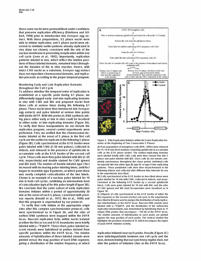
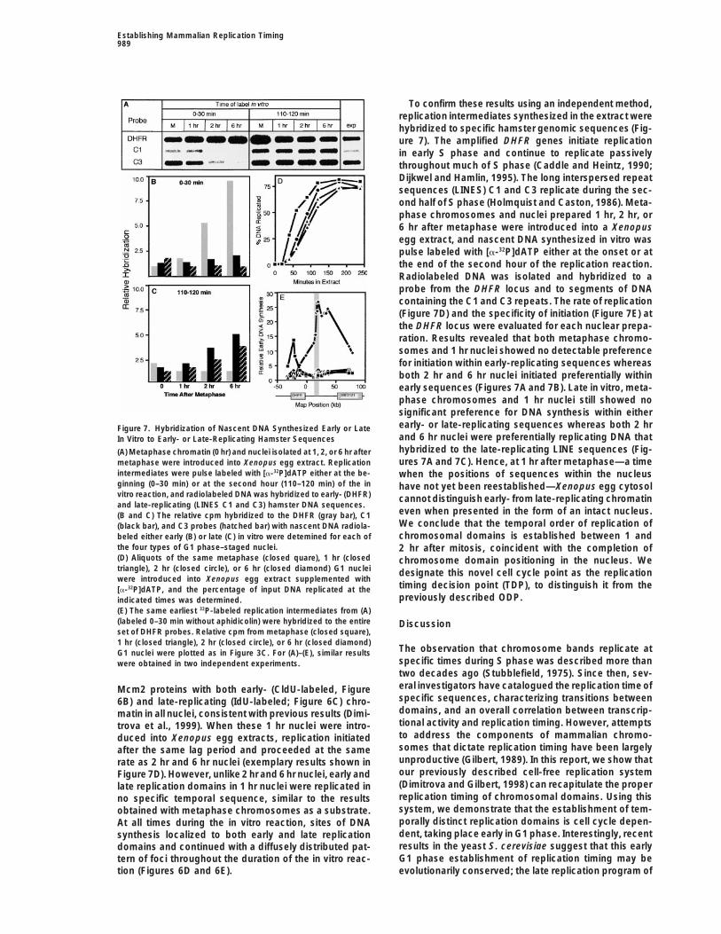
To confirm these results using an independent method,replication intermediates synthesized in the extract werehybridized to specific hamster genomic sequences (Fig-ure 7). The amplified DHFR genes initiate replicationin early S phase and continue to replicate passivelythroughout much of S phase (Caddle and Heintz, 1990;Dijkwel and Hamlin, 1995). The long interspersed repeatsequences (LINES) C1 and C3 replicate during the sec-ond half of S phase (Holmquist and Caston, 1986). Meta-phase chromosomes and nuclei prepared 1 hr, 2 hr, or6 hr after metaphase were introduced into a Xenopusegg extract, and nascent DNA synthesized in vitro waspulse labeled with [a-32P]dATP either at the onset or atthe end of the second hour of the replication reaction.Radiolabeled DNA was isolated and hybridized to aprobe from the DHFR locus and to segments of DNAcontaining the C1 and C3 repeats. The rate of replication(Figure 7D) and the specificity of initiation (Figure 7E) atthe DHFR locus were evaluated for each nuclear prepa-ration. Results revealed that both metaphase chromo-somes and 1 hr nuclei showed no detectable preferencefor initiation within early-replicating sequences whereasboth 2 hr and 6 hr nuclei initiated preferentially withinearly sequences (Figures 7A and 7B). Late in vitro, meta-phase chromosomes and 1 hr nuclei still showed nosignificant preference for DNA synthesis within eitherearly- or late-replicating sequences whereas both 2 hrFigure 7. Hybridization of Nascent DNA Synthesized Early or Lateand 6 hr nuclei were preferentially replicating DNA thatIn Vitro to Early- or Late-Replicating Hamster Sequenceshybridized to the late-replicating LINE sequences (Fig-(A) Metaphase chromatin (0 hr) and nuclei isolated at 1, 2, or 6 hr afterures 7A and 7C). Hence, at 1 hr after metaphase—a timemetaphase were introduced into Xenopus egg extract. Replication
intermediates were pulse labeled with [a-32P]dATP either at the be- when the positions of sequences within the nucleusginning (0–30 min) or at the second hour (110–120 min) of the in have not yet been reestablished—Xenopus egg cytosolvitro reaction, and radiolabeled DNA was hybridized to early- (DHFR) cannot distinguish early- from late-replicating chromatinand late-replicating (LINES C1 and C3) hamster DNA sequences.
even when presented in the form of an intact nucleus.(B and C) The relative cpm hybridized to the DHFR (gray bar), C1We conclude that the temporal order of replication of(black bar), and C3 probes (hatched bar) with nascent DNA radiola-chromosomal domains is established between 1 andbeled either early (B) or late (C) in vitro were detemined for each of
the four types of G1 phase–staged nuclei. 2 hr after mitosis, coincident with the completion of(D) Aliquots of the same metaphase (closed quare), 1 hr (closed chromosome domain positioning in the nucleus. Wetriangle), 2 hr (closed circle), or 6 hr (closed diamond) G1 nuclei designate this novel cell cycle point as the replicationwere introduced into Xenopus egg extract supplemented with
timing decision point (TDP), to distinguish it from the[a-32P]dATP, and the percentage of input DNA replicated at thepreviously described ODP.indicated times was determined.
(E) The same earliest 32P-labeled replication intermediates from (A)(labeled 0–30 min without aphidicolin) were hybridized to the entire Discussionset of DHFR probes. Relative cpm from metaphase (closed square),1 hr (closed triangle), 2 hr (closed circle), or 6 hr (closed diamond)
The observation that chromosome bands replicate atG1 nuclei were plotted as in Figure 3C. For (A)–(E), similar resultsspecific times during S phase was described more thanwere obtained in two independent experiments.two decades ago (Stubblefield, 1975). Since then, sev-eral investigators have catalogued the replication time ofMcm2 proteins with both early- (CldU-labeled, Figurespecific sequences, characterizing transitions between6B) and late-replicating (IdU-labeled; Figure 6C) chro-domains, and an overall correlation between transcrip-matin in all nuclei, consistent with previous results (Dimi-tional activity and replication timing. However, attemptstrova et al., 1999). When these 1 hr nuclei were intro-to address the components of mammalian chromo-duced into Xenopus egg extracts, replication initiatedsomes that dictate replication timing have been largelyafter the same lag period and proceeded at the sameunproductive (Gilbert, 1989). In this report, we show thatrate as 2 hr and 6 hr nuclei (exemplary results shown inour previously described cell-free replication systemFigure 7D). However, unlike 2 hr and 6 hr nuclei, early and(Dimitrova and Gilbert, 1998) can recapitulate the properlate replication domains in 1 hr nuclei were replicated inreplication timing of chromosomal domains. Using thisno specific temporal sequence, similar to the resultssystem, we demonstrate that the establishment of tem-obtained with metaphase chromosomes as a substrate.porally distinct replication domains is cell cycle depen-At all times during the in vitro reaction, sites of DNAdent, taking place early in G1 phase. Interestingly, recentsynthesis localized to both early and late replicationresults in the yeast S. cerevisiae suggest that this earlydomains and continued with a diffusely distributed pat-G1 phase establishment of replication timing may betern of foci throughout the duration of the in vitro reac-
tion (Figures 6D and 6E). evolutionarily conserved; the late replication program of
Molecular Cell990
a telomeric origin was shown to be determined at some IV and type V patterns in vitro (Figure 2), extracts areclearly capable of replicating these domains. Incompletepoint between metaphase and START (Raghuraman et
al., 1997). The ability to stage CHO cells at precise times replication of G1 nuclei could result from exhaustion ofextract components or the accumulation of a replicationin early G1 phase allowed us to demonstrate that replica-
tion timing is established after the binding of Mcm2 to inhibitor(s) in the extract. Alternatively, it is possible thata mid–S phase transition may be important for replica-chromatin and prior to specification of the DHFR origin
locus, which is upstream of the restriction point. Replica- tion of heterochromatin. In fact, remodeling of hetero-chromatin just prior to replication has been observed intion timing was established during the same early G1
phase interval in which chromosomal domains were re- cultured mammalian cells (Milner, 1969; Li et al., 1998).Further experiments will be required to distinguish thesepositioned in the nucleus after mitosis.possibilities.
A Cell-Free System to Study the Replication Timingof Mammalian Chromosomal Domains Relationship between Replication Timing
and Nuclear PositioningPrevious studies suggested that it might not be possibleto study the spatiotemporal order of replication using We demonstrate that nuclear envelope assembly, in-
cluding a morphologically complete nuclear lamina andXenopus egg extracts. With Xenopus sperm nuclei asa substrate (Mills et al., 1989), discrete replication foci an intact nuclear membrane, is completed prior to the
global repositioning of chromosomal domains. Consis-are evenly distributed throughout nuclei at all times dur-ing S phase. Experiments with Drosophila polytene tent with these results, completed decondensation of
chromonema fibers (Belmont and Bruce, 1994) and the(Sleeman et al., 1992), chicken erythrocyte (Leno andLaskey, 1991), or human fibroblast (Kill et al., 1991) nu- peripheral localization of human chromosome 18 (J.
Bridger and W. Bickmore, personal communication) re-clei observed exclusively type I/II patterns. These inves-tigators concluded that Xenopus egg extracts remodel quire 2–3 hr after mitosis. Although we cannot determine
whether each individual domain returns to precisely thesomatic cell nuclei, imposing upon them an embryonicreplication pattern. We found no evidence of remodeling same physical location within the nucleus, the overall
distribution of these domains was preserved after mito-(Figures 2 and 5), even after prolonged incubation times.An important difference is that the prior studies em- sis, and the same domains initiated replication at the
onset of two consecutive S phases.ployed nuclei prepared with detergents, which disruptsnuclear morphology and strongly inhibits DNA synthesis Genetic evidence in Drosophila suggests that position
effect variegation is correlated with a change in nuclear(Dimitrova and Gilbert, 1998). With nuclei prepared bycontrolled exposure of cells to digitonin, initiation of position of the variegated locus, mediated by the prox-
imity to heterochromatin (Dernburg et al., 1996). In mam-replication is rapid, synchronous (all nuclei initiate within10–30 min), and efficient. In addition, previous reports malian cells, silencing of certain genes during B and T
cell development requires a protein (Ikaros) that appearsemployed much lower concentrations of nuclei, whichcan also disrupt replication patterns (Dimitrova and Gil- to relocate these genes to centromeric heterochromatin
(Brown et al., 1999). Our finding that the temporal orderbert, 1998). We conclude that the conditions for nuclearpreparation and in vitro DNA replication described in of replication is not established until sequences are re-
positioned within the nucleus provides a provocativethis report preserve physiological properties of chromo-somes, allowing replication to proceed according to the coincidence between gene position, replication timing,
and transcriptional function. Indeed, Ikaros-mediatedprogram specified in vivo.Not all aspects of the in vivo replication pattern were gene silencing and repositioning is accompanied by a
change in replication timing of those domains (K. E.recapitulated in Xenopus extracts. First, the total rateof genomic replication is accelerated in Xenopus egg Brown, M. Merkenschlager, and A. G. Fisher, personal
communication). It is possible that replication timingextracts; nearly 80% of the genome is replicated within2–3 hr (Figure 7D), and mid/late replication begins within is a consequence of nuclear position, which is itself
a reflection of transcriptional potential. Accordingly, in2 hr (Figures 2, 5, and 7). Since replication fork elonga-tion rates in vitro are approximately one sixth of that Xenopus embryos there is no transcription and no evi-
dence for a spatiotemporal order to replication, whereasobserved in vivo (Dimitrova and Gilbert, 1998), the accel-erated rate of genome duplication is likely due to in- in mouse embryos transcription begins at the end of the
first cell cycle, and replication already follows a definedcreased origin activation. However, DHFR origin speci-ficity is indistinguishable from the in vivo situation, spatiotemporal pattern (Ferreira and Carmo-Fonseca,
1997). This model would imply that epigenetic regulationsuggesting that extracts do not activate origins that arenot typically utilized in vivo. One possibility is that the of chromosomal domains is dominant to the information
contained within origin sequences themselves in con-entire set of type I/II foci (which take 6 hr to replicatein vivo) may be activated within 1 hr in vitro. Indeed, we trolling replication timing. Although we have analyzed
only one replication origin, our finding that the overallobserved a greater number of labeled foci with G1 phasenuclei in Xenopus egg extracts as compared to early S replication timing program is maintained independently
of conditions that alter origin specification (Figure 5) isphase cells in culture (Figure 1 versus Figure 2B). Asecond difference is that G1 phase nuclei were rarely consistent with this notion.
How could nuclear position dictate replication timing?observed to progress to type IV or type V patterns denovo in the extract, and not more than 80% of the ge- Potentially, molecules recruited to the sites of specific
chromosomal domains could establish microenviron-nome was replicated (Figure 7 and Dimitrova and Gilbert[1998]). Since type III S phase nuclei progressed to type ments that modulate the ability of pre-RCs to compete
Establishing Mammalian Replication Timing991
a Xenopus egg extract supplemented with 100 mg/ml aphidicolin.for limiting concentrations of initiation factors. For ex-Nuclei were then washed free of aphidicolin, and the earliest-replicat-ample, transcriptional silencing at S. cerevisiae telo-ing nascent DNA chains were labeled with [a-32P]dATP. 32P-labeledmeres requires a microenvironment containing high lo-early replication intermediates were then hybridized to 17 unique
cal concentrations of Sir3p that form foci at the nuclear probes distributed over a 120 kb region of the DHFR locus. Relativeperiphery (Laroche et al., 1998), and nontelomeric loci cpm were obtained by phosphorimaging analysis and adjusted for
differences in probe size, deoxyadenine content, and hybridizationcan be silenced by anchorage to the nuclear peripheryefficiency by normalizing to the corresponding values for parallel(Andrulis et al., 1998). Sir3p is also required for the latehybridizations with labeled replication intermediates from exponen-replication of at least one origin near the telomeretially growing cells.(Stevenson and Gottschling, 1999), suggesting that theIn Cultured Cells
repressive chromatin structure created by this micro- Radioactive labeling of the earliest synthesized nascent DNAenvironment delays replication. The in vitro system de- strands was done as previously described (Gilbert et al., 1995),
except that intact nuclei were prepared by digitonin permeabili-scribed here should allow us to test hypotheses aboutzation.the molecules that establish replication timing differ-
ences.Replication Timing of Specific DNA Sequences in XenopusEgg Extracts
Experimental Procedures5 3 105 digitonin-permeabilized metaphase or 1 hr, 2 hr, and 6 hrG1 phase cells were resuspended in Xenopus egg extract, and early
Cell Culture and Synchronizationor late replication intermediates were labeled with 100 mCi of
CHOC 400 cells (CHO cell derivative containing z1000 amplified[a-32P]dATP during either the first 30 min or between 110 and 120 min
copies of the DHFR gene) were maintained and synchronized asof the in vitro reaction. Nuclei were washed, and labeled genomic
described (Gilbert et al., 1995; Dimitrova et al., 1999).DNA was purified as described (Gilbert et al., 1995). An equal numberof cpm from each sample was hybridized to a panel of DNA plasmids
Labeling of Nascent DNA with Nucleotide Analogs(1 mg each) immobilized on nylon membranes. Probe R (Gilbert et
In Cultured Cellsal., 1995) from the DHFR locus was used as an early-replicating
Exponentially growing CHOC 400 cells were pulse labeled with 30sequence, since it maps within the region of peak initiation activity.
mg/ml BrdU (Sigma) for 2–30 min. For differential tagging of early-Late-replicating hamster DNA sequences (LINES C1 and C3) were
and late-replicating DNA domains, CHOC 400 cells synchronized ata gift of G. Holmquist (Holmquist and Caston, 1986). Relative cpm
the G1/S border were released into S phase and labeled with CldUwere obtained by phosphorimaging analysis (Molecular Dynamics)
and IdU as described (Dimitrova et al., 1999).and normalized to the corresponding values from parallel hybridiza-
In Xenopus Egg Extractstions with replication intermediates from exponentially growing
Intact or permeabilized nuclei were prepared by digitonin treatmentCHOC 400 cells, labeled as described (Gilbert et al., 1995). This
and introduced into interphase Xenopus egg extracts at a concen-corrects for differences in probe size, deoxyadenine content, and
tration of 10,000 nuclei/ml as described (Dimitrova and Gilbert, 1998).hybridization efficiency. To express the relative preference for repli-
Aliquots were removed at different time points, biotin-11-dUTPcation of early or late sequences (Figures 7B and 7C), results for
(Sigma) was added to a final concentration of 50 mM, and reactionseach type of G1 phase–staged nuclei were normalized to the lowest
were further incubated for 5–10 min. Nuclei were fixed with 4%value among the three probes.
formaldehyde and transferred to polylysine-coated coverslips(Sigma) as described (Mills et al., 1989).
AcknowledgmentsRates and extent of genomic DNA replication were evaluated bymeasuring the amount of acid-precipitable [a-32P]dATP as described
We thank B. Knox for critical reading of the manuscript, K. Meiri(Dimitrova and Gilbert, 1998).and J. Schaefer for helpful discussions on the CldU/IdU stainingprocedure, I. Todorov for anti-Mcm2 antibody and C. J. Hutchison
Immunofluorescent Microscopyfor anti-lamin antibody, and G. Holmquist for providing late-replicat-
BrdU-substituted DNA was detected with mouse monoclonal anti-ing DNA probes. This work was supported by March of Dimes grant
BrdU antibody (Becton Dickinson, #347580) followed by FITC-conju-#1-FY96-0610 and NIH grant #GM57233-01 to D. M. G.
gated goat anti-mouse IgG (Jackson Labs, #115-095-146). The dif-ferential staining of DNA sites substituted with CldU or IdU was
Received March 8, 1999; revised September 17, 1999.performed as described (Dimitrova et al., 1999). Biotin-11-dUTPincorporation was detected with Texas red–conjugated streptavidin
References(Amersham #RPN1233). Mcm2 proteins were detected as described(Dimitrova et al., 1999). The nuclear lamina was detected using
Amaldi, F., Buongiorno-Nardelli, M., Carnevali, F., Leoni, L., Mariotti,monoclonal antibody LN43.2 (gift of C. Hutchison). DNA was stainedD., and Pomponi, M. (1973). Replicon origins in Chinese hamsterwith 0.1 mg/ml 49,6-diamidino-2-phenylindole (DAPI) or 0.01 mg/mlcell DNA. II. Reproducibility. Exp. Cell Res. 80, 79–87.propidium iodide (Sigma). All images shown were photographedAndrulis, E.D., Neiman, A.M., Zappulla, D.C., and Sternglanz, R.directly from a Nikon Labophot-2 microscope with a 1003 1.4 NA(1998). Perinuclear localization of chromatin facilitates transcrip-oil immersion Nikon PlanApo objective through either single FITCtional silencing. Nature 394, 592–595.and Texas red filters or a dual FITC 1 rhodamine fluorescence filter
(#51004V2, F/R 712) as described (Dimitrova et al., 1999). Slides Aten, J.A., Bakker, P.J.M., Stap, J., Boschman, G.A., and Veenhof,were scanned and figures assembled as described (Dimitrova et C.H.N. (1992). DNA double labelling with IdUrd and CldUrd for spatialal., 1999). No computer merging was performed, and only standard and temporal analysis of cell proliferation and DNA replication. His-brightness and contrast adjustments were made. All statistical data tochem. J. 24, 251–259.were collected using this microscope and filter sets. Confocal im- Belmont, A., and Bruce, K. (1994). Visualization of G1 chromosomes:aging (Dimitrova et al., 1999) of several selected images gave similar a folded, twisted, supercoiled chromonema model of interphaseresults. chromatid structure. J. Cell Biol. 127, 287–302.
Berezney, R., Dubey, D.D., and Huberman, J.A. (1999). Heterogene-Mapping Replication Origins by the Early Labeled Fragmentity of eukaryotic replicons, replicon clusters, and replication foci.Hybridization (ELFH) AssayChromosoma, in press.In Xenopus Egg ExtractsBousset, K., and Diffley, J.F. (1998). The Cdc7 protein kinase isSpecificity of initiation in the DHFR locus in pre- and post-ODPrequired for origin firing during S phase. Genes Dev. 12, 480–490.CHOC 400 nuclei was determined by the ELFH assay as described
(Dimitrova and Gilbert, 1998). In brief, nuclei were introduced into Brown, K.E., Baxter, J., Graf, D., Merkenschlager, M., and Fisher,
Molecular Cell992
A.G. (1999). Dynamic repositioning of genes in the nucleus of lym- Holmquist, G.P., and Caston, L.A. (1986). Replication time of inter-phocytes preparing for cell division. Mol. Cell. 3, 207–217. spersed repetitive DNA sequences in hamsters. Biochim. Biophys.
Acta 868, 164–177.Caddle, M.S., and Heintz, N.H. (1990). The replication timing of theamplified dihydrofolate reductase genes in the Chinese hamster Housman, D., and Huberman, J.A. (1975). Changes in the rate ofovary cell line CHOC 400. Biochem. Biophys. Res. Commun. 170, DNA replication fork movement during S phase in mammalian cells.134–139. J. Mol. Biol. 94, 173–181.
Calza, R.E., Eckardt, L.A., DelGiudice, T., and Schildkraut, C.L. Jackson, D.A., and Pombo, A. (1998). Replicon clusters are stable(1984). Changes in gene position are accompanied by a change in units of chromosome structure: evidence that nuclear organizationtime of replication. Cell 36, 689–696. contributes to the efficient activation and propagation of S phase
in human cells. J. Cell Biol. 140, 1285–1295.Dernburg, A.F., Broman, K.W., Fung, J.C., Marshall, W.F., Philips,J., Agard, D.A., and Sedat, J.W. (1996). Perturbation of nuclear archi- Kill, I.R., Bridger, J.M., Campbell, K.H., Maldonado, C.G., and Hutchi-tecture by long-distance chromosome interactions. Cell 85, son, C.J. (1991). The timing of the formation and usage of replicase745–759. clusters in S-phase nuclei of human diploid fibroblasts. J. Cell Sci.
100, 869–876.Dhar, V., Skoultchi, A.I., and Schildkraut, C.L. (1989). Activation andrepression of a b-globin gene in cell hybrids is accompanied by a Kitsberg, D., Selig, S., Keshet, J., and Cedar, H. (1993). Replicationshift in its temporal regulation. Mol. Cell. Biol. 9, 3524–3532. structure of the human b-globin gene domain. Nature 368, 588–590.Dijkwel, P.A., and Hamlin, J.L. (1995). The Chinese hamster dihydro- Laroche, T., Martin, S.G., Gotta, M., Gorham, H.C., Pryde, F.E., Louis,folate reductase origin consists of multiple potential nascent-strand E.J., and Gasser, S.M. (1998). Mutation of yeast Ku genes disruptsstart sites. Mol. Cell. Biol. 15, 3023–3031. the subnuclear organization of telomeres. Curr. Biol. 8, 653–656.Dimitrova, D.S., and Gilbert, D. (1998). Regulation of mammalian Lawlis, S.J., Keezer, S.M., Wu, J.-R., and Gilbert, D.M. (1996). Chro-replication origin usage in Xenopus egg extracts. J. Cell Sci. 111, mosome architecture can dictate site-specific initiation of DNA repli-2989–2998. cation in Xenopus egg extracts. J. Cell Biol. 135, 1–12.Dimitrova, D.S., and Gilbert, D.M. (1999). DNA replication and nu- Leno, G.H., and Laskey, R.A. (1991). The nuclear membrane deter-clear organization: prospects for a soluble in vitro system. Crit. Rev. mines the timing of DNA replication in Xenopus egg extracts. J. CellEuk. Gene Exp. 9, 353–361. Biol. 112, 557–566.Dimitrova, D.S., Todorov, I.T., Melendy, T., and Gilbert, D.M. (1999). Leno, G.H., Downes, C.S., and Laskey, R.A. (1992). The nuclearMcm2, but not RPA, is a component of the mammalian early G1- membrane prevents replication of human G2 nuclei but not G1 nucleiphase pre-replication complex. J. Cell Biol. 146, 709–722. in Xenopus egg extract. Cell 69, 151–158.Drouin, R., Lemieux, N., and Richer, C.L. (1990). Analysis of DNA Li, G., Sudlow, G., and Belmont, A.S. (1998). Interphase cell cyclereplication during S-phase by means of dynamic chromosome dynamics of a late-replicating, heterochromatic homogeneouslybanding at high resolution. Chromosoma 99, 273–280. staining region: precise choreography of condensation/deconden-Ermakova, O.V., Nguyen, L.H., Little, R.D., Chevillard, C., Riblet, R., sation and nuclear positioning. J. Cell Biol. 140, 975–989.Ashouian, N., Birshtein, B.K., and Schildkraut, C.L. (1999). Evidence Ma, H., Samarabandu, J., Devdhar, R.S., Acharya, R., Cheng, P.,that a single replication fork proceeds from early to late replicating Meng, C., and Berezney, R. (1998). Spatial and temporal dynamicsdomains in the IgH locus in a non-B cell line. Mol. Cell. 3, 321–330. of DNA replication sites in mammalian cells. J. Cell Biol. 143, 1415–Ferguson, B.M., and Fangman, W.L. (1992). A position effect on the 1425.time of replication origin activation in yeast. Cell 68, 333–339. Manders, E.M., Stap, J., Strackee, J., van Driel, R., and Aten, J.A.Ferreira, J., and Carmo-Fonseca, M. (1997). Genome replication in (1996). Dynamic behavior of DNA replication domains. Exp. Cell Res.early mouse embryos follows a defined temporal and spatial order. 226, 328–335.J. Cell Sci. 110, 889–897. Manuelidis, L. (1990). A view of interphase chromosomes. ScienceFerreira, J., Paolella, G., Ramos, C., and Lamond, A.I. (1997). Spatial 250, 1533–1540.organization of large-scale chromatin domains in the nucleus: a Mills, A.D., Blow, J.J., White, J.G., Amos, W.B., Wilcock, D., andmagnified view of single chromosome territories. J. Cell Biol. 139,
Laskey, R.A. (1989). Replication occurs at discrete foci spaced1597–1610.
throughout nuclei replicating in vitro. J. Cell Sci. 94, 471–477.Forrester, W.C., Epner, E., Driscoll, M.C., Enver, T., Brice, M., Papa-
Milner, G.R. (1969). Changes in chromatin structure during in-yannopoulou, T., and Groudine, M. (1990). A deletion of the human
terphase in human normoblasts. Nature 221, 71–72.beta-globin locus activation region causes a major alteration in chro-
O’Keefe, R.T., Henderson, S.C., and Spector, D.L. (1992). Dynamicmatin structure and replication across the entire beta-globin locus.organization of DNA replication in mammalian cell nuclei—spatiallyGenes Dev. 4, 1637–1649.and temporally defined replication of chromosome-specific alpha-Friedman, K.L., Diller, J.D., Ferguson, B.M., Nyland, S.V., Brewer,satellite DNA sequences. J. Cell Biol. 116, 1095–1110.B.J., and Fangman, W.L. (1996). Multiple determinants controllingRaghuraman, M., Brewer, B., and Fangman, W. (1997). Cell cycle-activation of yeast replication origins late in S phase. Genes Dev.dependent establishment of a late replication program. Science 276,10, 1595–1607.806–809.Gilbert, D.M. (1986). Temporal order of replication of Xenopus laevisSelig, S., Okumura, K., Ward, D.C., and Cedar, H. (1992). Delineation5S ribosomal RNA genes in somatic cells. Proc. Natl. Acad. Sci.of DNA replication time zones by fluorescence in situ hybridization.USA 83, 2924–2928.EMBO J. 11, 1217–1225.Gilbert, D.M. (1989). Temporal order of DNA replication in eukaryoticSleeman, A.M., Leno, G.H., Mills, A.D., Fairman, M.P., and Laskey,cells: its relationship to gene expression. Ph.D. thesis, StanfordR.A. (1992). Patterns of DNA replication in Drosophila polytene nucleiUniversity, Stanford, California.replicating in Xenopus egg and oocyte extracts. J. Cell Sci. 101,Gilbert, D.M. (1998). Replication origins in yeast vs. metazoa: separa-509–515.tion of the haves and the have nots. Curr. Opin. Gen. Dev. 8, 194–199.Sparvoli, E., Levi, M., and Rossi, E. (1994). Replicon clusters mayGilbert, D.M., Miyazawa, H., and DePamphilis, M.L. (1995). Site-form structurally stable complexes of chromatin and chromosomes.specific initiation of DNA replication in Xenopus egg extract requiresJ. Cell Sci. 107, 3097–3103.nuclear structure. Mol. Cell. Biol. 15, 2942–2954.Stevenson, J.B., and Gottschling, D.E. (1999). Telomeric chromatinHatton, K.S., Dhar, V., Brown, E.H., Iqbal, M.A., Stuart, S., Didamo,modulates replication timing near chromosome ends. Genes Dev.V.T., and Schildkraut, C.L. (1988). Replication program of active and13, 146–151.inactive multigene families in mammalian cells. Mol. Cell. Biol. 8,
2149–2158. Stubblefield, E. (1975). Analysis of the replication pattern of Chinesehamster chromosomes using 5-bromodeoxyuridine suppression ofHolmquist, G.P. (1987). Role of replication time in the control of
tissue-specific gene expression. Am. J. Hum. Genet. 40, 151–173. 33258 Hoechst fluorescence. Chromosoma 53, 209–221.
Establishing Mammalian Replication Timing993
Wu, J.-R., and Gilbert, D.M. (1996). A distinct G1 step required tospecify the Chinese hamster DHFR replication origin. Science 271,1270–1272.
Wu, J.-R., and Gilbert, D.M. (1997). The replication origin decisionpoint is a mitogen independent, 2-aminopurine sensitve, G1-phasestep that precedes restriction point control. Mol. Cell Biol. 17, 4312–4321.
Wu, J.-R., Keezer, S., and Gilbert, D. (1998). Transformation abro-gates an early G1-phase arrest point required for specification of theChinese hamster DHFR replication origin. EMBO J. 17, 1810–1818.
Zink, D., Cremer, T., Saffrich, R., Fischer, R., Trendelenburg, M.F.,Ansorge, W., and Stelzer, E.H. (1998). Structure and dynamics ofhuman interphase chromosome territories in vivo. Hum. Genet. 102,241–251.
Related Documents