1 An Analyzed Procedure Data Collection Form (DCF) is required for all suspected or diagnosed Lung and Esophageal Cancer Resections and one should be initiated every time the patient enters the operating room. These cases are risk adjusted and are included in the Data Analysis Reports. Fields that appear underlined and in blue are required for analyzed procedure record inclusion. If any of these fields are missing data, the entire record will be excluded from analysis. Completion of the Thymus/Mediastinal Mass, Tracheal Resection and Hiatal Hernia/GERD sections is optional for analyzed procedures. Procedures highlighted below, if performed as isolated procedures or with only other highlighted procedures, are not collected unless the Surgeon Participant chooses to track them. If collected, use the Non-analyzed Procedure DCF. Highlighted procedures done in conjunction with analyzed (major) procedures should be included on this Analyzed Procedure DCF. A. Demographics Patient ID: ___________________PatID (80) Medical Record #:_________________ MedRecN (90) First Name:__________________ PatFName (100) Middle Name:____________ PatMName(110) Last Name:___________________ PatLName (120) SSN#:______________ SSN (130) Patient participating in STS-related clinical trial: ClinTrial (140) None Trial 1 Trial 2 Trial 3 Trial 4 Trial 5 Trial 6 (If not “None” →) Clinical trial patient ID: _________________ ClinTrialPatID (150) Date of Birth:____/____/______ DOB (160) (mm/dd/yyyy) Age: ________ Age (170) Patient Postal Code:_________ PostalCode (180) Gender: Male Female Gender (190) Is the Patient's Race Documented? Yes No Patient Declined to Disclose RaceDocumented (200) Race: If Yes select all that apply White/Caucasian Yes No RaceCaucasian (210) Black/African American Yes No RaceBlack (220) Asian Yes No RaceAsian (230) American Indian/Alaskan Native Yes No RaceNativeAm (240) Native Hawaiian/Pacific Islander Yes No RacNativePacific (250) Other Yes No RaceOther (260) Hispanic or Latino Ethnicity: Yes No Not Documented Ethnicity (270) B. Admission Admission Status: Inpatient Outpatient / Observation AdmissionStat (280) If Inpatient → Admission Date: ____/___/_____ AdmitDt (290) Payor: Indicate the Primary payor: PayorPrim (300) If Primary Payor is not None/Self→ Indicate the Secondary (supplemental) payor: PayorSecond (320) None/self Medicare If Medicare → Fee For Service: Yes No PrimMCareFFS (310) Medicaid Military Health Indian Health Service Correctional Facility State Specific Plan Other Government Insurance Commercial Health Insurance Health Maintenance Organization Non U.S. Plan None/self Medicare If Medicare → Fee For Service: Yes No SecondMCareFFS (330) Medicaid Military Health Indian Health Service Correctional Facility State Specific Plan Other Government Insurance Commercial Health Insurance Health Maintenance Organization Non U.S. Plan Surgeon Name:_________________________________ Surgeon (340) Surgeon’s National Provider ID: _______________________ SurgNPI (350) The Society of Thoracic Surgeons General Thoracic Surgery Database Analyzed Procedure Data Collection Form Version 2.41 ©2018 The Society of Thoracic Surgeons Revised 5/31/2018

Welcome message from author
This document is posted to help you gain knowledge. Please leave a comment to let me know what you think about it! Share it to your friends and learn new things together.
Transcript

1
An Analyzed Procedure Data Collection Form (DCF) is required for all suspected or diagnosed Lung and Esophageal Cancer Resections and one should be initiated every time the patient enters the operating room. These cases are risk adjusted and are included in the Data Analysis Reports.
Fields that appear underlined and in blue are required for analyzed procedure record inclusion. If any of these fields are missing data, the entire record will be excluded from analysis.
Completion of the Thymus/Mediastinal Mass, Tracheal Resection and Hiatal Hernia/GERD sections is optional for analyzed procedures.
Procedures highlighted below, if performed as isolated procedures or with only other highlighted procedures, are not
collected unless the Surgeon Participant chooses to track them. If collected, use the Non-analyzed Procedure DCF.
Highlighted procedures done in conjunction with analyzed (major) procedures should be included on this Analyzed
Procedure DCF.
A. Demographics
Patient ID: ___________________PatID (80) Medical Record #:_________________ MedRecN (90)
First Name:__________________ PatFName (100)
Middle Name:____________ PatMName(110)
Last Name:___________________ PatLName (120)
SSN#:______________ SSN (130)
Patient participating in STS-related clinical trial: ClinTrial (140) None Trial 1 Trial 2 Trial 3 Trial 4 Trial 5 Trial 6 (If not “None” →) Clinical trial patient ID: _________________ ClinTrialPatID (150)
Date of Birth:____/____/______ DOB (160) (mm/dd/yyyy)
Age: ________
Age (170) Patient Postal Code:_________ PostalCode (180)
Gender: Male Female
Gender (190)
Is the Patient's Race Documented? � Yes � No � Patient Declined to Disclose RaceDocumented (200)
Race: If Yes select all that apply White/Caucasian Yes No RaceCaucasian (210)
Black/African American Yes No RaceBlack (220)
Asian Yes No RaceAsian (230)
American Indian/Alaskan Native Yes No RaceNativeAm (240)
Native Hawaiian/Pacific Islander Yes No RacNativePacific (250)
Other Yes No RaceOther (260)
Hispanic or Latino Ethnicity: Yes No Not Documented Ethnicity (270)
B. Admission Admission Status: Inpatient Outpatient / Observation
AdmissionStat (280) If Inpatient → Admission Date: ____/___/_____ AdmitDt (290)
Payor: Indicate the Primary payor: PayorPrim (300) If Primary Payor is not None/Self→ Indicate the Secondary (supplemental) payor: PayorSecond (320)
None/self Medicare If Medicare → Fee For Service: Yes No PrimMCareFFS (310) Medicaid Military Health Indian Health Service Correctional Facility State Specific Plan Other Government Insurance Commercial Health Insurance Health Maintenance Organization Non U.S. Plan
None/self Medicare If Medicare → Fee For Service: Yes No SecondMCareFFS (330) Medicaid Military Health Indian Health Service Correctional Facility State Specific Plan Other Government Insurance Commercial Health Insurance Health Maintenance Organization Non U.S. Plan
Surgeon Name:_________________________________ Surgeon (340)
Surgeon’s National Provider ID: _______________________
SurgNPI (350)
The Society of Thoracic Surgeons
General Thoracic Surgery Database Analyzed Procedure Data Collection Form
Version 2.41
©2018 The Society of Thoracic Surgeons Revised 5/31/2018

2
Taxpayer ID#: __________________________________
TIN (360) Hospital Name:______________________________________ HospName (370)
Hospital Postal Code:_________________ HospZIP (380)
Hospital Region:________ HospStat (390)
Hospital’s National Provider ID:_______________________ HospNPI (400)
C. Pre-Operative Evaluation
Height: ___________(cm) HeightCm (410) Weight: __________(kg) WeightKg (420)
Unintentional Wt loss over past 3 months? (Enter “0” if none) - _____________(kg) WtLoss3Kg (430)
CardioPulmonary History
Hypertension Hypertn (440) Yes No Congestive Heart Failure(CHF) CHF (450)
Yes No If Yes→ EF ______% EF (460)
Coronary Artery Disease (CAD) CAD (470)
Yes No Myocardial Infarction Yes No PreMI (480)
Afib per EKG within the last year; with or without treatment AFIB (490)
Yes No Valvular Heart Disease VHD (500)
Yes No If Yes→
Location – check all that apply:
AV Yes No VHDLocAV (510) MV Yes No VHDLocMV (520)
PV Yes No VHDLocPV (530) TV Yes No VHDLocTV (540)
Pulmonary Hypertension: PulmHypertn (550)
Yes No Unknown
Interstitial Fibrosis/ Interstitial Lung Disease InterstitialFib (560) Yes No
Vascular History
Major Vascular Disease MVD (580)
Yes No
DVT/PE DVTPE (590) Yes No
Cerebral Vascular Disease History
Cerebrovascular History: CerebroHx (610)
No CVD history Known disease, no events Transient Ischemic Attack (TIA)
Cerebrovascular Accident (CVA) If CVA→ Permanent Neurologic impairment Yes No
PNI (620)
Neuromuscular Disease
Neurologic symptoms present NeuroSymptPres (630)
Yes No
Myasthenia Gravis MyasGravis (640)
Yes No
Endocrine / GI / Renal History
Diabetes Diabetes (650)
Yes No If Yes→ Type of therapy: DiabCtrl (660)
None Diet Only Oral Insulin Other Subcutaneous Medication Other Unknown
Liver Dysfunction LiverDys (670)
Yes No
On Dialysis Dialysis (680)
Yes No
Cancer History
Coexisting Cancer CoexisCancer (690)
Yes No
Preoperative Chemotherapy / Immunotherapy PreopChemoCur (700)
Yes No If Yes →
Same disease, ≤ 6 months PreopChemoCurWhen (710) Same disease,> 6 months Unrelated disease, ≤ 6 months Unrelated disease, >6 months
Preop Thoracic Radiation Therapy PreopXRT (720)
Yes No If Yes →
Same disease, ≤ 6 months PreopXRTDisWhen (730) Same disease,> 6 months Unrelated disease, ≤ 6 months Unrelated disease, >6 months If Same disease, ≤ 6 months → Completion Date ______________
PreopXRTCompDt (740)
Prior Surgical History (check all that apply)
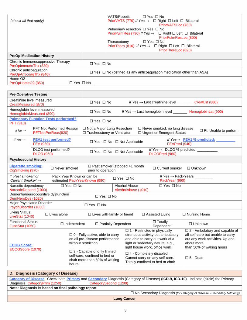
Prior Cardiothoracic Surgery PriorCTS (750)
Yes No If Yes → Sternotomy Yes No PriorStern (760)
3
(check all that apply) VATS/Robotic Yes No PriorVATS (770) If Yes → Right Left Bilateral PriorVATSLoc (780)
Pulmonary resection Yes No PriorPulmRes (790) If Yes → Right Left Bilateral PriorPulmResLoc (800)
Thoracotomy Yes No PriorThora (810) If Yes → Right Left Bilateral PriorThoraLoc (820)
PreOp Medication History
Chronic Immunosuppressive Therapy PreOpImmunoThx (830)
Yes No
Chronic anticoagulation PreOpAnticoagThx (840)
Yes No (defined as any anticoagulation medication other than ASA)
Home O2 PreOpHomeO2 (850) Yes No
Pre-Operative Testing
Creatinine level measured CreatMeasured (870)
Yes No If Yes → Last creatinine level ________ CreatLst (880)
Hemoglobin level measured HemoglobinMeasured (890)
Yes No If Yes → Last hemoglobin level _______ HemoglobinLst (900)
Pulmonary Function Tests performed?
PFT (910) Yes No
If No → PFT Not Performed Reason PFTNotPerReas(920)
Not a Major Lung Resection Tracheostomy or Ventilator
Never smoked, no lung disease Urgent or Emergent Status
Pt. Unable to perform
If Yes →
FEV1 test performed?
FEV (930) Yes No Not Applicable
If Yes→ FEV1 % predicted: _________
FEVPred (940)
DLCO test performed? DLCO (950)
Yes No Not Applicable If Yes→ DLCO % predicted: __________
DLCOPred (960)
Psychosocial History
Cigarette smoking:
CigSmoking (970) Never smoked
Past smoker (stopped >1 month prior to operation
Current smoker Unknown
If ‘Past smoker’ or ‘Current Smoker’ →
Pack Year Known or can be estimated PackYearKnown (980)
Yes No If Yes → Pack-Years _________ PackYear (990)
Narcotic dependency Yes No NarcoticDepend (1000)
Alcohol Abuse Yes No AlcoholAbuse (1010)
Dementia/neurocognitive dysfunction DemNeroDys (1020)
Yes No
Major Psychiatric Disorder PsychDisorder (1030)
Yes No
Living Status: LiveStat (1040)
Lives alone Lives with family or friend Assisted Living Nursing Home
Functional Status: FuncStat (1050) Independent Partially Dependent
Totally Dependent
Unknown
ECOG Score:
ECOGScore (1070)
0 - Fully active, able to carry on all pre-disease performance without restriction
1 - Restricted in physically strenuous activity but ambulatory and able to carry out work of a light or sedentary nature, e.g., light house work, office work
2 - Ambulatory and capable of all self-care but unable to carry out any work activities. Up and about more than 50% of waking hours
3 - Capable of only limited self-care, confined to bed or chair more than 50% of waking hours
4 - Completely disabled. Cannot carry on any self-care. Totally confined to bed or chair
5 - Dead
D. Diagnosis (Category of Disease)
Category of Disease: Check both Primary and Secondary Diagnosis (Category of Disease) (ICD-9, ICD-10). Indicate (circle) the Primary
Diagnosis. CategoryPrim (1250) CategorySecond (1280)
Note: Diagnosis is based on final pathology report.
No Secondary Diagnosis (for Category of Disease - Secondary field only)
Lung Cancer
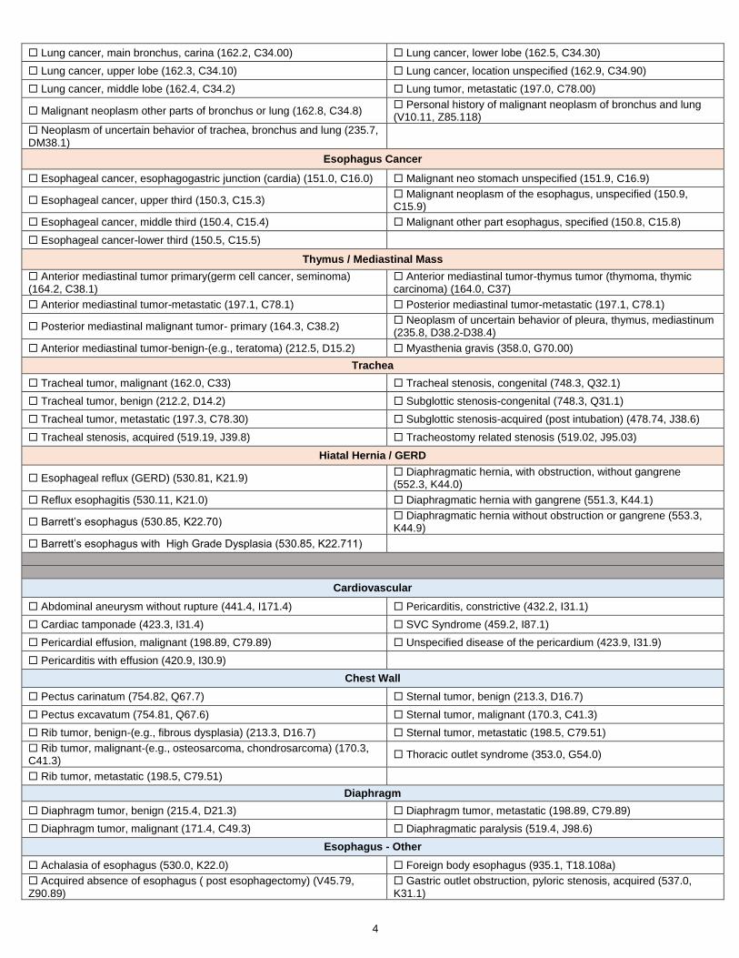
4
Lung cancer, main bronchus, carina (162.2, C34.00) Lung cancer, lower lobe (162.5, C34.30)
Lung cancer, upper lobe (162.3, C34.10) Lung cancer, location unspecified (162.9, C34.90)
Lung cancer, middle lobe (162.4, C34.2) Lung tumor, metastatic (197.0, C78.00)
Malignant neoplasm other parts of bronchus or lung (162.8, C34.8) Personal history of malignant neoplasm of bronchus and lung (V10.11, Z85.118)
Neoplasm of uncertain behavior of trachea, bronchus and lung (235.7, DM38.1)
Esophagus Cancer
Esophageal cancer, esophagogastric junction (cardia) (151.0, C16.0) Malignant neo stomach unspecified (151.9, C16.9)
Esophageal cancer, upper third (150.3, C15.3) Malignant neoplasm of the esophagus, unspecified (150.9, C15.9)
Esophageal cancer, middle third (150.4, C15.4) Malignant other part esophagus, specified (150.8, C15.8)
Esophageal cancer-lower third (150.5, C15.5)
Thymus / Mediastinal Mass
Anterior mediastinal tumor primary(germ cell cancer, seminoma) (164.2, C38.1)
Anterior mediastinal tumor-thymus tumor (thymoma, thymic carcinoma) (164.0, C37)
Anterior mediastinal tumor-metastatic (197.1, C78.1) Posterior mediastinal tumor-metastatic (197.1, C78.1)
Posterior mediastinal malignant tumor- primary (164.3, C38.2) Neoplasm of uncertain behavior of pleura, thymus, mediastinum (235.8, D38.2-D38.4)
Anterior mediastinal tumor-benign-(e.g., teratoma) (212.5, D15.2) Myasthenia gravis (358.0, G70.00)
Trachea
Tracheal tumor, malignant (162.0, C33) Tracheal stenosis, congenital (748.3, Q32.1)
Tracheal tumor, benign (212.2, D14.2) Subglottic stenosis-congenital (748.3, Q31.1)
Tracheal tumor, metastatic (197.3, C78.30) Subglottic stenosis-acquired (post intubation) (478.74, J38.6)
Tracheal stenosis, acquired (519.19, J39.8) Tracheostomy related stenosis (519.02, J95.03)
Hiatal Hernia / GERD
Esophageal reflux (GERD) (530.81, K21.9) Diaphragmatic hernia, with obstruction, without gangrene (552.3, K44.0)
Reflux esophagitis (530.11, K21.0) Diaphragmatic hernia with gangrene (551.3, K44.1)
Barrett’s esophagus (530.85, K22.70) Diaphragmatic hernia without obstruction or gangrene (553.3, K44.9)
Barrett’s esophagus with High Grade Dysplasia (530.85, K22.711)
Cardiovascular
Abdominal aneurysm without rupture (441.4, I171.4) Pericarditis, constrictive (432.2, I31.1)
Cardiac tamponade (423.3, I31.4) SVC Syndrome (459.2, I87.1)
Pericardial effusion, malignant (198.89, C79.89) Unspecified disease of the pericardium (423.9, I31.9)
Pericarditis with effusion (420.9, I30.9)
Chest Wall
Pectus carinatum (754.82, Q67.7) Sternal tumor, benign (213.3, D16.7)
Pectus excavatum (754.81, Q67.6) Sternal tumor, malignant (170.3, C41.3)
Rib tumor, benign-(e.g., fibrous dysplasia) (213.3, D16.7) Sternal tumor, metastatic (198.5, C79.51)
Rib tumor, malignant-(e.g., osteosarcoma, chondrosarcoma) (170.3, C41.3)
Thoracic outlet syndrome (353.0, G54.0)
Rib tumor, metastatic (198.5, C79.51)
Diaphragm
Diaphragm tumor, benign (215.4, D21.3) Diaphragm tumor, metastatic (198.89, C79.89)
Diaphragm tumor, malignant (171.4, C49.3) Diaphragmatic paralysis (519.4, J98.6)
Esophagus - Other
Achalasia of esophagus (530.0, K22.0) Foreign body esophagus (935.1, T18.108a)
Acquired absence of esophagus ( post esophagectomy) (V45.79, Z90.89)
Gastric outlet obstruction, pyloric stenosis, acquired (537.0, K31.1)

5
Dyskinesia/spasm of esophagus (530.5, K22.4) Mallory Weiss tear (530.7, K22.6)
Epiphrenic diverticulum (530.6, K22.5) Stricture and stenosis of esophagus (530.3, K22.2)
Esophageal perforation (530.4, K22.3) Tracheoesophageal fistula (530.84, J86.0)
Esophageal stricture (530.3, K22.2) Ulcer esophagus with bleeding (530.21, K22.11)
Esophageal tumor-benign (i.e., leiomyoma) (211.0, D13.0) Ulcer esophagus without bleeding (530.2, K22.10)
Esophagitis (530.1, K20.9) Zenkers diverticulum (530.6, K22.5)
Other disease of the esophagus (530.89, K22.8) Other digestive system complication (997.49, K91.XX)
Lung – Other
Acute respiratory failure (518.81, J96.00) Lung tumor, benign (e.g., hamartoma) (212.3, D14.30)
Aspergillosis (117.3, B44.9) Pneumonia (486.0, J18.9)
Bronchiectasis (494.0, J47.9) Post inflammatory pulmonary fibrosis (515, J84.89)
Cystic fibrosis with pulmonary manifestations (277.02, E84.0) Primary pulmonary hypertension ( 416.0, I 27.0)
Emphysema (492.8, J43.8) Pulmonary insufficiency following surgery/trauma (ARDS) (518.5, J95.82)
Emphysematous bleb (492.0, J43.9) Pulmonary sequestration (748.5, Q33.2)
Lung abscess (513.0, J85.2) Transplanted lung complication(s) (996.84, T86.8XX)
Interstitial lung disease/fibrosis (516.3, J84.1) Gangrene and necrosis of lung (513.0, J85.0)
Pneumothorax (512.8, J93.1) Hemothorax (511.8, J94.2)
Solitary pulmonary nodule (not a tumor, e.g., granuloma, subpleural lymph node, pulmonary infarct) (793.11, R91.1)
Mediastinum
Mediastinal nodes, metastatic (196.1, C77.1) Mediastinal cyst, Pericardial (519.3, J98.5)
Benign neoplasm of thymus (212.6, D15.0) Mediastinal cyst, Thymic (519.3, J98.5)
Lymphoma, intrathoracic (202.82, C85.92) Mediastinal nodes, benign (229.0, D36.0)
Mediastinal abscess (513.1, J85.3) Mediastinitis (519.2, J98.5)
Mediastinal cyst, Bronchogenic (519.3, J98.5) Posterior mediastinal tumor-benign(neurogenic)(212.5,D15.2)
Mediastinal cyst, Foregut duplication (519.3, J98.5) Unspecified disease of thymus gland (254.9, E32.9)
Pleura
Empyema with fistula (510.0, J86.0) Pleural thickening (511.0, J94.9)
Empyema without fistula (510.9, J86.9) Pleural tumor, benign (212.4, D19.0)
Empyema, tuberculosis (A15.6) Pleural tumor, metastatic (197.2, C78.2)
Pleural effusion, infected- (empyema) (511.1, J86.9) Malignant neoplasm other specified sites of pleura (163.8, C38.4)
Pleural effusion, malignant (197.2, C78.2) Malignant tumor of pleura, unspecified (e.g., mesothelioma) (163.9, C45)
Pleural effusion sterile (511.9, J90) Pleural effusion, TB; (Tuberculous pleurisy) (012.0, A15.6)
Pleural effusion, other specified, except TB (511.89, J90)
Thyroid
Goiter, nodular (241.9, E04.9) Thyroid neoplasm, malignant (193.0, C73)
Thyroid neoplasm, benign (226.0, D34)
Trachea & Larynx
Dysphagia, unspecified (787.2, R13.10) Vocal cord paralysis unspecified (478.3, J38.00)
Tracheomalacia-congenital (748.3, Q32.0) Vocal cord paralysis , unilateral (478.31, J38.01)
Tracheomalacia-acquired (519.1, J39.8) Vocal cord paralysis, bilateral (478.33, J38.02)
Tracheostomy-hemorrhage (519.09, J95.01)
Trauma
Flail chest (807.4, S22.5xxa) Sternal fracture (807.2, S22.20xa)
Rib fracture (807.0, S22.39xa) Tracheal injury (807.5, S12.8xxa)
Rib fractures, multiple (807.0, S22.49xa) Traumatic pneumothorax (860.0, S27.0xxa)
Miscellaneous

6
Abnormal radiologic finding (793.1, R91) Other non-infectious disorders of lymphatic channels (457.8, I89.8)
Chronic airway obstruction not elsewhere classified (496, J44.9) Malignant neoplasm of connective tissue and other soft tissue of the thorax (171.4, C49.3)
Chylothorax (457.8, 189.8) Malignant poorly differentiated neuroendocrine carcinoma, any site (209.3, C74.1)
Disruption of internal operation, surgical wound (998.31, T81.32XA) Non-healing surgical wound (998.83, T81.89XA)
Hemorrhage complicating a procedure (998.11, multiple codes) Other post- op infection (998.59, T81.4XXA)
Hematoma complicating a procedure (998.12, multiple codes) Persistent post-op fistula not otherwise classified (998.6, T81.83XA)
Hemoptysis unspecified (786.3, R04.2) Post-operative air leak (512.2, J95.812)
Hyperhidrosis, focal axilla (705.21, L74.510) Secondary malignant neoplasm of other specified sites (198.89, C79.89)
Hyperhidrosis, focal, face (705.21, L74.511) Shortness of breath (786.05, R06.02)
Hyperhidrosis, focal, palms (705.21, L74.512) Swelling, mass or lump in chest (786.6, R22.2)
Lymphadenopathy (785.6, R59.9) Other unlisted category of disease
Other Primary Specify: CategoryPrimOth (1260)
If diagnosis not listed, free text here:__________________________________________________
Other Primary ICD: CategoryPrimOthICD (1270)
Enter ICD-9 or ICD-10 of unlisted primary diagnosis, if known:______________________________
Secondary, Other Secondary Specify: CategorySecondOth (1290)
If secondary diagnosis not listed, free text here:____________________________________
Secondary, Other Secondary ICD: CategorySecondOthICD (1300)
Enter ICD-9 or ICD-10 of unlisted secondary diagnosis, if known :____________________________
E. Operative
Date of Surgery:______/______/_______
SurgDt (1310)
OR Entry Time: ______:_______
OREntryT (1320) Anesthesia Start Time: ______:_______ AnesthStartT (1340)
Procedure Start Time: ______:_______
ProcStartT (1360)
OR Exit Time: ______:_______
ORExitT (1330) Anesthesia End Time: _______:_______ AnesthEndT (1350)
Procedure End Time: _______:_______
ProcEndT (1370)
Multi-Day Operation (operation continued through midnight) MultiDay (1380)
Yes No
Planned, staged procedure? PlanStageProc (1390)
Yes No
Status of Operation Status (1400)
Emergent Urgent Elective Palliative
Reoperation (any prior cardiothoracic surgery
that affects operative field) Reop (1410)
Yes No Assisted by Robotic Technology Robotic (1420)
Yes No
Surgical Approach Conversion:
UnanticConv (1430) VATS→ Open Robotic → VATS Robotic→ Open No
If Yes→
Conversion Type: Elective Emergent UnanticConvTy (1440)
Conversion Reason: Vascular Anatomy Lymph Nodes Technical UnanticConvRsn (1450)
Blood transfusion intraoperatively (packed red blood cells) IntraopPRBC (1460)
Yes No If Yes→ #Red Blood Cell Units: _________ IntraopPRBCNum (1470)
ASA Classification:
ASA (1480)
I Normal, healthy
II Mild systemic disease
III Severe systemic disease
IV Life threatening severe systemic disease
V Moribund, not expected to survive without operation
VI Declared brain dead, organ donor
Check ALL of the procedures performed. Indicate (circle) the Primary Procedure.
Proc (1490) Primary (1500)
Analyzed Procedures
Lung Cancer Resection

7
Thoracoscopy, surgical; with lobectomy (32663) Removal of lung, single lobe (lobectomy) (32480)
Thoracoscopy with therapeutic wedge resection (eg mass or nodule, initial, unilateral (32666)
Removal of lung, two lobes (bilobectomy) (32482)
Thoracoscopy with therapeutic wedge resection(eg mass or nodule) each additional resection, ipsilateral (32667) List separately in addition to primary procedure code
Removal of lung, single segment (segmentectomy) (32484)
Thoracoscopy with diagnostic wedge resection followed by anatomic lung resection (32668), List separately in addition to primary procedure code
Removal of lung, sleeve lobectomy (32486)
Thoracoscopy with removal of a single lung segment (segmentectomy) (32669)
Removal of lung, completion pneumonectomy (32488)
Thoracoscopy with removal of two lobes (bilobectomy) (32670) Resection and repair of portion of bronchus (bronchoplasty) when performed at time of lobectomy or segmentectomy (32501)
Thoracoscopy with removal of lung, pneumonectomy (32671) Resection of apical lung tumor (e.g., Pancoast tumor), including chest wall resection, without chest wall reconstruction(s) (32503)
Thoracotomy with therapeutic wedge resection (eg mass nodule) initial (32505)
Resection of apical lung tumor (e.g., Pancoast tumor), including chest wall resection, with chest wall reconstruction (32504)
Removal of lung, total pneumonectomy; (32440) Thoracotomy with therapeutic wedge resection (eg mass nodule) each additional resection, ipsilateral (+32506)List separately in addition to primary procedure code
Removal of lung, sleeve (carinal) pneumonectomy (32442) Thoracotomy with diagnostic wedge resection followed by anatomic lung resection (+32507), List separately in addition to primary proc code
Thoracoscopy with mediastinal and regional lymphadenectomy (+32674) List separately in addition to primary procedure code
Thoracic lymphadenectomy, regional, including mediastinal and peritracheal nodes (38746)
Esophagus Resection
Transhiatal-Total esophagectomy, without thoracotomy, with cervical esophagogastrostomy (43107)
Partial esophagectomy, distal two-thirds, with thoracotomy only (43121)
Total esophagectomy without thoracotomy; with colon interposition or small intestine reconstruction (43108)
Thoracoabdominal-Partial esophagectomy, thoracoabdominal approach (43122)
Three Incision -Total esophagectomy with thoracotomy; with cervical esophagogastrostomy (43112)
Partial esophagectomy, thoracoabdominal with colon interposition or small intestine (43123)
Total esophagectomy with thoracotomy; with colon interposition or small intestine reconstruction (43113)
Total or partial esophagectomy, without reconstruction with cervical esophagostomy (43124)
Partial esophagectomy, cervical, with free intestinal graft, including microvascular anastomosis (43116)
Minimally invasive three incision esophagectomy (McKeown) (43288)
Ivor Lewis-Partial esophagectomy, distal two-thirds, with thoracotomy and separate abdominal incision (43117)
Minimally invasive esophagectomy, Ivor Lewis approach (43287)
Partial esophagectomy, with thoracotomy and separate abdominal incision with colon interposition or small intestine (43118)
Minimally invasive esophagectomy, Abdominal and neck approach (43286)
Hiatal Hernia / GERD Procedures
Laparoscopy, surgical, esophagogastric fundoplasty (e.g., Nissen, Toupet procedures) (43280)
Repair, paraesophageal hiatal hernia via laparotomy with mesh (43333)
Laparoscopy, surgical with repair of paraesophageal hernia (fundoplasty) without mesh (43281)
Repair, paraesophageal hiatal hernia via thoracotomy without mesh (43334)
Laparoscopy, surgical with repair of paraesophageal hernia (fundoplasty) with mesh (43282)
Repair, paraesophageal hiatal hernia via thoracotomy with mesh (43335)
Nissen fundoplasty- laparotomy (includes partial fundoplication/wrap) (43327)
Repair, paraesophageal hiatal hernia via thoracoabdominal approach without mesh (43336)
Transthoracic Fundoplication- open thoracotomy (includes Belsey/Nissen) (43328)
Repair, paraesophageal hiatal hernia via thoracoabdominal approach with mesh (43337)
Repair, paraesophageal hiatal hernia via laparotomy without mesh (43332)
Tracheal Resection
Carinal reconstruction (31766) Tracheal tumor or carcinoma excision; cervical (31785)
Excision tracheal stenosis, cervical (31780) Tracheal tumor or carcinoma excision; thoracic (31786)
Excision tracheal stenosis, thoracic (31781)
Thymus / Mediastinal Mass Resection
Thoracoscopy, surgical; with excision of mediastinal cyst, tumor, or mass (32662)
Thymectomy, transcervical approach (60520)
Thymus, resection via Thoracoscopy unilateral or bilateral (32673) Thymectomy, transthoracic approach (60521)
Mediastinal tumor, excision, open, Transthoracic approach (39220) Thymectomy, transthoracic approach, with radical mediastinal dissection (60522)

8
Non-analyzed Procedures Trachea, Bronchi, Larynx
Laryngectomy, partial (31370)
Tracheostomy replacement (tube change) prior to est. of fistula tract (31502)
Tracheal wound or injury suture repair; cervical (31800)
Tracheostomy, planned (31600) Tracheal wound or injury suture repair; intrathoracic (31805)
Tracheostomy revision simple, without flap (31613) Unlisted procedure, trachea, bronchi (31899)
Tracheostomy revision complex, with flap (31614) Bronchopleural fistula closure (32906)
Tracheoplasty; cervical (31750) Bronchogenic cyst removal
Tracheoplasty; intrathoracic (31760) Bronchial laceration suture
Bronchial sleeve resection
Bronchoplasty, graft repair (31770) Tracheostomy mediastinal
Bronchoplasty; excision stenosis and anastomosis (31775) Rigid stent removal
Bronchoscopy
Tracheobronchoscopy through established tracheostomy incision (31615)
Bronchoscopy, with transbronchial lung biopsy(s), each additional lobe (31632)
Endobronchial ultrasound (EBUS) during bronchoscopy diagnostic or therapeutic intervention(s) (31620)
Bronchoscopy, with transbronchial needle aspiration biopsy(s), each additional lobe (31633)
Bronchoscopy, diagnostic, with or without cell washing (31622) Bronchoscopy, with removal of foreign body (31635)
Bronchoscopy, with brushing or protected brushings (31623) Bronchoscopy, with placement of bronchial stent(s) (includes tracheal/bronchial dilation as required), initial bronchus (31636)
Bronchoscopy, with bronchial alveolar lavage (BAL) (31624) Bronchoscopy, each additional major bronchus stented (31637)
Bronchoscopy, with bronchial or endobronchial biopsy(s), single or multiple sites (31625)
Bronchoscopy, with revision of tracheal or bronchial stent inserted at previous session (31638)
Bronchoscopy, with placement of Fiducial markers (31626) Bronchoscopy, with excision of tumor (31640)
Bronchoscopy, navigational (31627) Bronchoscopy, with destruction of tumor or relief of stenosis by any method other than excision (e.g., laser therapy) (31641)
Bronchoscopy, with transbronchial lung biopsy(s), single lobe (31628)
Bronchoscopy, with placement of catheter(s) for intracavitary radioelement application (31643)
Bronchoscopy, with transbronchial needle aspiration biopsy(s) (31629)
Bronchoscopy, with therapeutic aspiration of tracheobronchial tree, initial (drainage of lung abscess) (31645)
Bronchoscopy, with tracheal/bronchial dilation or closed reduction of fracture (31630)
Bronchoscopy, with therapeutic aspiration of tracheobronchial tree, subsequent (31646)
Bronchoscopy, with placement of tracheal stent(s) (includes tracheal/bronchial dilation as required) (31631)
Pleural Space and Lung
Thoracostomy; with rib resection for empyema (32035) Insertion indwelling tunneled pleural catheter (32550)
Thoracostomy; with open flap drainage for empyema (32036) Thoracoscopy, diagnostic lungs and pleural space, without biopsy (32601)
Thoracotomy with biopsy(s) lung infiltrate(s) (e.g. wedge), unilateral (32096)
Thoracoscopy, diagnostic; with biopsy(s) of lung infiltrate(s) (e.g. wedge), unilateral (32607)
Thoracotomy with biopsy(s) lung nodule(s) or masses (e.g. incisional), unilateral (32097)
Thoracoscopy, diagnostic; with biopsy(s) of lung nodule(s) or mass(es) (eg incisional), unilateral (32608)
Thoracotomy with biopsy(s) of pleura (32098) Thoracoscopy, diagnostic; with biopsy(s) of pleura (32609)
Thoracotomy, with exploration (32100) Thoracoscopy, surgical; with pleurodesis (e.g., mechanical or chemical) (32650)
Thoracotomy, major; with control of traumatic hemorrhage and/or repair of lung tear (32110)
Thoracoscopy, surgical; with partial pulmonary decortication (32651)
Thoracotomy, major; for postoperative complications (32120) Thoracoscopy, surgical; with total pulmonary decortication (32652)
Thoracotomy with open intrapleural pneumolysis (32124) Thoracoscopy, surgical; with removal of intrapleural foreign body or fibrin deposit (32653)
Thoracotomy, major; with cyst(s) removal, with or without a pleural procedure (32140)
Thoracoscopy, surgical; with control of traumatic hemorrhage (32654)
Thoracotomy, major; with excision-plication of bullae, with or without any pleural procedure (32141)
Thoracoscopy, surgical; with excision-plication of bullae, including any pleural procedure (32655)
Thoracotomy, major; with removal of intrapleural foreign body or hematoma (32150)
Thoracoscopy, surgical; with parietal pleurectomy (32656)

9
Thoracotomy with cardiac massage (32160) Thoracoscopy with resection-plication for emphysematous lung (bullous or non-bullous) for lung volume reduction- LVRS, unilateral including any pleural procedure (32672)
Decortication, pulmonary, total (32220) Repair lung hernia through chest wall (32800)
Pleural scarification for repeat pneumothorax (32215) Closure of chest wall following open flap drainage for empyema (Clagett type procedure) (32810)
Decortication, pulmonary, partial (32225) Total lung lavage (for alveolar proteinosis) (32997)
Pleurectomy, parietal (32310) Radio-frequency ablation (RFA) lung tumor (32998)
Decortication and parietal pleurectomy (32320) Removal of lung, total pneumonectomy; extrapleural (32445)
Removal of lung, excision-plication of emphysematous lung(s) for lung volume reduction (LVRS) (32491)
Unlisted procedure, lung (32999)
Lung Other Procedures
Open closure of major bronchial fistula (32815) Double lung transplant (32853)
Single lung transplant (32851) Double lung transplant with CPB (32854)
Single lung transplant with CPB (32852) Thoracoplasty with closure of bronchopleural fistula (32906)
Mediastinum and Diaphragm
Thoracoscopy, diagnostic; mediastinal space, with biopsy (32606) Diaphragmatic hernia repair (other than neonatal), traumatic; acute (39540)
Mediastinotomy with exploration or biopsy; cervical approach (39000)
Diaphragmatic hernia repair (other than neonatal), traumatic; chronic (39541)
Mediastinotomy with exploration or biopsy; transthoracic approach (39010)
Diaphragm imbrication (i.e., plication) of (39545)
Mediastinal cyst, excision, open, Transthoracic approach (39200) Diaphragm; resection with simple repair (e.g., primary suture) (39560)
Mediastinoscopy, with or without biopsy (39400) Diaphragm; resection with complex repair (e.g., prosthetic material, local muscle flap) (39561)
Unlisted procedure, mediastinum (39499) Unlisted procedure, diaphragm (39599)
Diaphragm, laceration repair, any approach (39501)
Esophagoscopy
Esophagoscopy (43200) Upper gastrointestinal endoscopy with endoscopic ultrasound examination limited to the esophagus (43237)
Esophagoscopy with biopsy (43202) Upper gastrointestinal endoscopy with transendoscopic ultrasound-guided FNA (43238)
Esophagoscopy with removal of foreign body (43215) Upper gastrointestinal endoscopy with biopsy (43239)
Esophagoscopy with insertion of stent (43219) Upper gastrointestinal endoscopy with dilation of gastric outlet for obstruction (43245)
Esophagoscopy with balloon dilation (43220) Upper gastrointestinal endoscopy with directed placement of percutaneous gastrostomy tube (43246)
Esophagoscopy with insertion of guide wire followed by dilation over guide wire (43226)
Upper gastrointestinal endoscopy with removal of foreign body (43247)
Esophagoscopy with ablation of tumor (43228) Upper gastrointestinal endoscopy with insertion of guide wire followed by dilation of esophagus (43248)
Esophagoscopy with endoscopic ultrasound examination (EUS) (43231)
Upper gastrointestinal endoscopy with balloon dilation of esophagus (43249)
Esophagoscopy with transendoscopic ultrasound-guided fine needle aspiration (43232)
Upper gastrointestinal endoscopy with transendoscopic stent placement (43256)
Upper gastrointestinal endoscopy, diagnostic (43235) Upper gastrointestinal endoscopy with ablation of tumor (43258)
Esophagus Other Procedures
Thoracoscopy, surgical; with esophagomyotomy (Heller type) (32665)
Esophagostomy, fistulization of esophagus, external; cervical approach (43352)
Cricopharyngeal myotomy (43030) Gastrointestinal reconstruction for previous esophagectomy with stomach (43360)
Excision esophageal lesion with primary repair, cervical approach (43100)
Gastrointestinal reconstruction for previous esophagectomy with colon interposition or small intestine (43361)
Excision Esophageal lesion with primary repair, thoracic approach (eg: leiomyoma) (43101)
Suture of esophageal wound or injury; cervical approach (43410)
Diverticulectomy of hypopharynx or esophagus, with or without myotomy; cervical approach (43130)
Suture of esophageal wound or injury; transthoracic or transabdominal approach (43415)
Diverticulectomy of esophagus, with or without myotomy; thoracic approach (43135)
Closure of esophagostomy or fistula; cervical approach (43420)

10
Laparoscopic esophageal myotomy (Heller Myotomy, with or without fundoplication ) (43279)
Total gastrectomy with esophagoenterostomy (43620)
Laparoscopy, surgical, esophageal lengthening procedure (Collis) (43283) Secondary Procedure code
Total gastrectomy with Roux-en-Y reconstruction (43621)
Unlisted laparoscopy, esophagus (43289 ) Conduit revision s/p esophagectomy
Esophagoplasty with repair of TEF, cervical approach (43305) Per oral endoscopic myotomy (POEM)
Esophagoplasty with repair TEF, thoracic approach (43312) Trans oral fundoplication
Esophagomyotomy (Heller type); thoracic approach (43331) Esophageal lengthening procedure - open (Collis) Secondary Procedure code (43338)
Free jejunum transfer with microvascular anastomosis (43496) Ligation or stapling at gastroesophageal junction for esophageal perforation (43405)
Unlisted procedure, esophagus (43499)
Chest Wall and Neck
Muscle flap, neck (15732) Radical resection of sternum (21630)
Muscle flap; trunk (i.e., intercostal, pectoralis or serratus muscle) (15734)
Radical resection of sternum; with mediastinal lymphadenectomy (21632)
Excision of chest wall tumor including ribs (19260) Hyoid myotomy and suspension (21685) secondary procedure code
Excision of chest wall tumor involving ribs, with reconstruction (19271)
Division of scalenus anticus; without resection of cervical rib (21700)
Excision tumor, soft tissue of neck or thorax; subcutaneous (21555) Division of scalenus anticus; with resection of cervical rib (21705)
Excision tumor, soft tissue of neck or thorax; deep, subfascial, intramuscular (21556)
Reconstructive repair of pectus excavatum or carinatum; open (21740)
Radical resection of tumor (e.g., malignant neoplasm), soft tissue of neck or thorax (21557)
Reconstructive repair of pectus, minimally invasive approach (Nuss procedure), without thoracoscopy (21742)
Excision of rib, partial (21600) Open treatment of sternum fracture with or without skeletal fixation (21825)
Excision first and/or cervical rib (21615) Removal of sternal wire(s)
Excision first and/or cervical rib; with sympathectomy (21616) Reconstructive repair of pectus, minimally invasive approach (Nuss procedure), with thoracoscopy (21743)
Major reconstruction, chest wall (posttraumatic) (32820) Unlisted procedure, neck or thorax (21899)
Miscellaneous
Thoracoscopy, diagnostic pericardial sac, with biopsy (32604) SVC resection and reconstruction (34502)
Thoracoscopy, surgical; with removal of clot or foreign body from pericardial sac (32658)
Ligation thoracic duct (38381)
Thoracoscopy, surgical; with creation of pericardial window or partial resection of pericardial sac for drainage (32659)
Intraoperative jejunostomy (44015)
Thoracoscopy, surgical; with total pericardiectomy (32660) Omental flap (49904)
Thoracoscopy, surgical; with excision of pericardial cyst, tumor, or mass (32661)
Transthoracic thyroidectomy (60270)
Thoracoscopy, surgical; with thoracic sympathectomy (32664) Removal substernal thyroid, cervical approach (60271)
Stereotactic radiosurgery (SRS) and stereotactic body radiotherapy (SBRT),surgeon participation (32701)
Application of wound vac (97605, 97606)
Tube pericardiostomy (33015) Pericardial window (33025)
Other Minor Procedure Other
Enter Name of unlisted Procedure(s): ProcOth (1510)
Enter 5 digit CPT code(s) of unlisted procedure, if known: ProcOthCPT (1520)
Surgical Procedure for Lung Cancer or Suspected Lung Cancer? LungCancer (1580)
Yes No if yes, complete Section F
Surgical Procedure for Esophageal Cancer?
EsophCancer (1590) Yes No if yes, complete Section G
Are you collecting data for Thymus / Mediastinal Mass Resection? ThymusMediastinalData (1600)
Yes No if yes, complete Section H
Are you collecting data for Tracheal Resection?
TrachealData (1610) Yes No if yes, complete Section I
Are you collecting data for Hiatal Hernia / GERD?
HiatalHerniaData (1620) Yes No if yes, complete Section J

11
F. Lung Cancer
Diagnosis:
Was there a pathological diagnosis of lung cancer prior to the lung resection? (yes: lung cancer was diagnosed preoperatively; no: lung cancer was only suspected preoperatively) LungCancerSus (1630)
Yes No
How was lung cancer diagnosed?
Bronchoscopy ClinStagLungBronc (1640)
Yes No Needle Biopsy Attempted or Completed ClinStagLungNeedle (1650)
Yes No
Clinical Staging: Pre-treatment Lung cancer staging- to be completed if lung cancer suspected or documented AND lung resection performed. Clinical staging determines the treatment plan.
Clinical Staging Done ClinStagDoneLung (1660) Yes No
If Yes→
Pre-Op Positive Tissue diagnosis Obtained PreopPosTisOb (1670)
Yes No
Clinical Staging Methods : Choose all that apply
Radiographic Staging Procedures
PET or PET/CT ClinStagLungPET (1680)
Yes No Brain CT Scan ClinStagLungBrainCT (1700)
Yes No
CT ClinStagLungCT (1690)
Yes No Brain MRI ClinStagLungBMRI (1710)
Yes No
Was invasive mediastinal staging performed? ClinStagInvasive (1720)
Yes, reason documented Yes, reason not documented No
If Documented → Operative/Clinic Note indicates Invasive Mediastinal Staging performed for the following reasons: (check all that apply)
Lesion size > 3cm ClinStagInvasiveSize (1730) Yes No
Mediastinal Lymphadenopathy on CT > 1cm ClinStagInvasiveLymphCT (1740)
Yes No
Ipsilateral hilar mediastinal node FDG uptake on PET ClinStagInvasiveHilar (1750)
Yes No
Central Tumor ClinStagInvasiveTumor (1760) Yes No
Other ClinStagInvasiveOther (1770) Yes No
Mediastinal Tissue Sampling/Staging
EBUS ClinStagLungEBUS (1780)
Yes No VATS ClinStagLungVATS (1790)
Yes No
EUS ClinStagLungEUS (1800)
Yes No Other ClinStagLungOth (1810)
Yes No
Mediastinoscopy/Chamberlain ClinStagLungMedia (1830)
Yes No
Tumor size known? Yes No If Yes ↓ LungCaTumSzKnown (1850)
Lung CA tumor size in cm (the dominanat/most concerning lesion per CT Scan) LungCaTumSz (1860)
__________cm (ex. 2.3cm)
Invasion of Adjacent Structures LCInvAdjStr (1870) Yes No
Lung CA T Stage (tumor stage) ClinStageLungTumor (1880)
Tis T1 T2 T3 T4
Lung CA Nodes: ClinStageLungN (1890)
N0 No regional lymph node metastasis
N1 Metastasis in ipsilateral peribronchial or hilar and intrapulmonary nodes. Includes direct extension.
N2 Metastasis in ipsilateral mediastinal and/or subcarinal lymph nodes
N3 Metastasis in contralateral mediastinal or contralateral hilar nodes, ipsilateral or contralateral scalene or supraclavicular nodes
Lung CA Metastases: ClinStageLungM (1900)
M0 No distant metastasis
M1 Distant Metastasis

12
Lung - FINAL Pathological Staging
To be completed if lung cancer suspected or documented AND lung resection performed. (8th Edition)
Lung Cancer Results
ClinStageLungResult (1910)
No cancer found, benign tumor Lung Cancer Tumor present:
If Cancer Tumor Present→ PathStageLungT (1920)
TX Primary Tumor cannot be assessed, or tumor
proven by the presence of malignant cells in
sputum or bronchial washings but not visualized
by imaging or bronchoscopy
T0 No evidence of primary tumor
Tis Carcinoma in situ; squamous cell carcinoma in situ (SCIS); Adenocarcinoma in situ (AIS): adenocarcinoma with pure lepidic pattern, <3 cm in greatest dimension
T1mi Minimally invasive adenocarcinoma:
adenocarcinoma (<3 cm in greatest dimension)
with a predominantly lepidic pattern and <5 mm
invasion in greatest dimension.
T1a Tumor <1 cm in greatest dimension. A superficial, spreading tumor of any size whose invasive component is limited to the bronchial wall and may extend proximal to the main bronchus also is classified as T1a, but these tumors are uncommon.
T1b Tumor > 1 cm but < 2 cm in greatest dimension
T1c Tumor > 2 cm but <
3 cm in greatest
dimension
T2a Tumor > 3 cm but < 4 cm at greatest dimension, or having any of the
following features: 1. involves the main bronchus regardless of distance to
the carina, 2. but without involvement of the carina; invades visceral pleura
(PL1 or PL2); 3. associated with atelectasis or obstructive pneumonitis that
extends to the hilar region, involving part or all of the lung.
T2b Tumor > 4 cm but < 5 cm at greatest dimension
T3 Tumor > 5 cm but < 7 cm in greatest dimension or directly
invading any of the following: parietal pleura (PL3), chest
wall (including superior sulcus tumors), phrenic nerve,
parietal pericardium; or separate tumor nodule(s) in the
same lobe as the primary
T4 Tumor > 7 cm or tumor of any size invading one or more of the following: diaphragm, mediastinum, heart, great vessels, trachea, recurrent laryngeal nerve, esophagus, vertebral body, or carina; separate tumor nodule(s) in an ipsilateral lobe different from that of the primary
If tumor is T2a or T2b → Visceral Pleura Invasion Yes No VisPleuraInv (1930)
Lung CA Nodes: PathStageLungN (1940)
NX Regional lymph nodes cannot be assessed
N0 No regional lymph node metastasis
N1 Metastasis in ipsilateral peribronchial and/or ipsilateral hilar lymph nodes and intrapulmonary nodes, includes involvement by direct extension
N2 Metastasis in ipsilateral mediastinal and/or subcarinal lymph node(s)
N3 Metastasis in contralateral mediastinal, contralateral hilar, ipsilateral or contralateral scalene or supraclavicular lymph node(s)
If N2 → Multi-station N2 Yes No PathStageLungMultiN2 (1950)
Lung CA Metastases: PathStageLungM (1960)
M0 No distant metastasis
M1 Distant metastasis
Lung CA Histology: LungCAHist (1970)
Carcinoma in situ- Adenocarcinoma Squamous cell Large cell
Small cell Low Grade Neuroendocrine (typical carcinoid)
Intermediate grade neuroendocrine, atypical carcinoid
Mixed
Other
Grade: Low grade (well differentiated) LungCAHistGrade (1980)
Intermediate grade High grade (poorly differentiated)
Unknown / Not reported
Total # of Lymph Nodes sampled/harvested:_________ LungCANodes (1990)
Total # of Nodal Stations sampled/harvested: ___________ LungCANodStat (2000)

13
Lung CA Resection Margins Positive: LungCAPathMarg (2010)
Yes No
If Yes→ LungCAPathMargPosR (2020)
R1 (microscopic residual tumor present)
R2 (macroscopic (gross) residual tumor present)
G. Esophageal Cancer
Clinical Staging: Pre-treatment Esophageal cancer staging - to be completed if esophageal cancer suspected, documented OR esophagus resection performed. Clinical staging determines the treatment plan.
Clinical Staging Done ClinStagDoneEsoph (2030) Yes No
Radiologic / Endoscopic Staging Procedures
If Clinical Staging Done is Yes →
PET or PET/CT ClinStagEsophPET (2040)
Yes No CT ClinStagEsophCT (2050)
Yes No
Bronchoscopy ClinStagEsophBronc (2060)
Yes No EUS ClinStagEsophEUS (2090)
Yes No
Invasive Staging Procedures
VATS – for staging ClinStagEsophVATS (2100)
Yes No Laparoscopy – for staging ClinStagEsophLap (2110)
Yes No
Endoscopic Mucosal Resection ClinStagEsophEMR (2120)
Yes No Other: Yes No ClinStagEsophOth (2130)
Esophageal Tumor: ClinStageEsophT (2150)
T0 No evidence of primary tumor
Tis High grade dysplasia
T1 Tumor invades lamina propria, mucosa or submucosa
T2 Tumor invades muscularis propria
T3 Tumor invades adventitia
T4 Tumor invades adjacent structures
Clinical Diagnosis of Nodal Involvement: ClinStageEsophNode (2160)
Yes (N1, N2 or N3) No
Esophageal CA Metastases ClinStageEsophM (2170)
M0 No Distant Metastasis M1 Distant Metastasis
Tumor Location (check all that apply):
Cervical Esophagus (15 – < 20 cm) Yes No TumorEsopCervical (2180)
Upper Thoracic (20 - < 25 cm) Yes No TumorEsopUpThorac (2190)
Middle Thoracic (25 - < 30 cm) Yes No TumorEsopMidThorac (2200)
Lower Thoracic, including EG Junction (30 – 42 cm) Yes No TumorEsopLowThorac (2210)

14
Pathological Staging - Esophagus
Esophagus to be completed if esophageal cancer documented AND esophageal resection performed (Pre-Operative Evaluation – Esophageal Cancer = Yes) (8th Edition)
Esophageal Cancer Results ClinStageEsophResult (2220)
No cancer found, benign tumor Esophageal cancer present
If cancer present →
Esophageal Tumor: PathStageEsophT (2230)
TX Tumor cannot be assessed
T0 No evidence of primary tumor
Tis High Grade dysplasia, defined as malignant cells confined to the epithelium by the basement membrane
T1a Tumor invades lamina propria or
muscularis mucosa
T1b Tumor invades submucosa
T2 Tumor invades muscularis propria
T3 Tumor invades adventitia
T4a Tumor invades pleura, pericardium, azygos vein, diaphragm or peritoneum
T4b Tumor invades other adjacent structures such as aorta, vertebral body, or airway.
Esophageal CA Nodes: PathStageEsophN (2240)
NX Regional lymph nodes cannot be assessed
N0 No regional lymph node metastasis
N1 Metastasis in 1-2 regional nodes
N2 Metastasis in 3-6 regional lymph nodes
N3 Metastasis in 7 or more regional lymph nodes
Esophageal CA Metastases: PathStageEsophM (2250)
M0 No distant metastasis
M1 Distant metastasis
Esoph Histopathologic Type: PathStageEsophH (2260)
H1Squamous Carcinoma H2 Adenocarcinoma Other
Esophageal CA Histologic Grade: PathStageEsophG (2270)
GX Grade cannot be assessed
G1 Well differentiated
G2 Moderately differentiated
G3 Poorly differentiated, undifferentiated
Total # of Lymph Nodes sampled/harvested: _______ EsophCANodes (2280)
Esophageal CA Resection Margins Positive: Yes No EsophCAPathMarg (2290)
H. Thymus / Mediastinal Mass Resection Pre-Operative
Symptomatic myasthenia MyastheniaSympt (2300)
Yes No
If Yes → Chronic Medical Treatment: Mestinon TxMestinon (2310)
Yes No
Steroids TxSteroids (2320)
Yes No
Other Immunosuppressive Therapy TxImmunoSuppress (2330)
Yes No
Pre-operative management
IVIG IVIG (2340)
Yes No
Plasmaphereis Plasmaphereis (2350)
Yes No
Size of mass known: Yes No MassSizeKnown (2360) If yes → Largest diameter in mm derived from preop axial, coronal or sagittal image ___________mm MassSize (2370)
Initial Surgical Approach ThyInitSurgAp (2380)
Full Sternotomy Clamshell or Hemiclamshell Transcervical
Partial Sternotomy Robotic VATS
If “Robotic” or “VATS” → ThyRobVATSLoc (2390)
Location: Right Left Bilateral
If “Transcervical”, “Partial Sternotomy”, ”Robotic” or “VATS” → Conversion to open approach during procedure?

15
ThyConvToOpen (2400) Yes, Planned Yes, Unplanned No
If Yes → ThyConvAp (2410)
Sternotomy Clamshell Thoracotomy
Intentional resection of functioning phrenic nerve
PhrenicNerveResect (2420) Yes No
Pathologic Staging PathRptStage (2430)
Stage I Grossly and microscopically encapsulated. Also called a noninvasive thymoma. That is, it has not spread beyond the thymus.
Stage II The thymoma invades beyond the capsule (outer boundary of the thymus) and into the nearby fatty tissue or to the pleura (outer covering of the lung). Sometimes divided into:
Stage IIa Microscopic transcapsular invasion
Stage IIb Macroscopic capsular invasion
Stage III Macroscopic invasion of neighboring organs. The thymoma extends into the neighboring tissues or organs of the lower neck or upper chest area, including the pericardium (covering of the heart), the lungs, or the main blood vessels leading into or exiting from the heart.
Stage IVa Pleural or pericardial dissemination. The thymoma has spread widely throughout the pleura and/or pericardium.
Stage IVb Hematogenous or lymphatic dissemination. The thymoma has spread to distant organs.
WHO classification (from path report – Thymoma only) ThymomaType (2440)
Type A Type AB Type B1 Type B2
Type B3 Thymic Carcinoma or Type C Not Thymoma
Completeness of resection (from operative note or pathology report)
ResectCompleteness (2450) R0 R1 R2
Patient alive at 30 days post op Yes No PtAlive30Day (2460)
If Yes → Post – operative Event (30 day)
Myasthenic crisis requiring return to ICU or intervention (intubation, plasmapheresis) MYAL (2470)
Yes No
Unintentional phrenic nerve palsy PhrenicNervePalsy (2480)
Yes No
Patient alive at 90 days post op Yes No PtAlive90Day (2490)
If Yes → 90 Day Post – Operative Variable
Adjuvant thoracic radiation ThoracicRadiation (2500)
Yes No
Persistent unintentional phrenic nerve palsy PhrenNrvPalsyPersis (2510)
Yes No
I. Tracheal Resection
Pre-Operative
Current Airway AirwayCurr (2520) Native Oral ETT Trach T-Tube
Prior tracheostomy TracheostomyPrior (2530) Yes No
Prior intubation IntubatePrior (2540) Yes No
Prior Tracheal Resection TrachealResectPrior (2550) Yes No
Recent Bronchoscopic Intervention (within 6 weeks)
BronchInt6Wks (2560) Yes No (includes: core out, dilation, ablation, stent)
Recurrent nerves intact RecurrNervesIntact (2570) Yes No Unknown
If not intact → RecurrNervNotIntact (2580)
Which nerve? Right Left Both
Airway management during resection (check all that apply)
Cross – table ventilation CrossTableVent (2590)
Yes No VA ECMO Yes No VaECMO (2600)
Jet ventilation Yes No JetVent (2610)
VV ECMO VvECMO (2620)
Yes No Cardiopulmonary bypass Yes No CardoPulmBypass (2630)
Incision (check all that apply, must have at least one indicated)
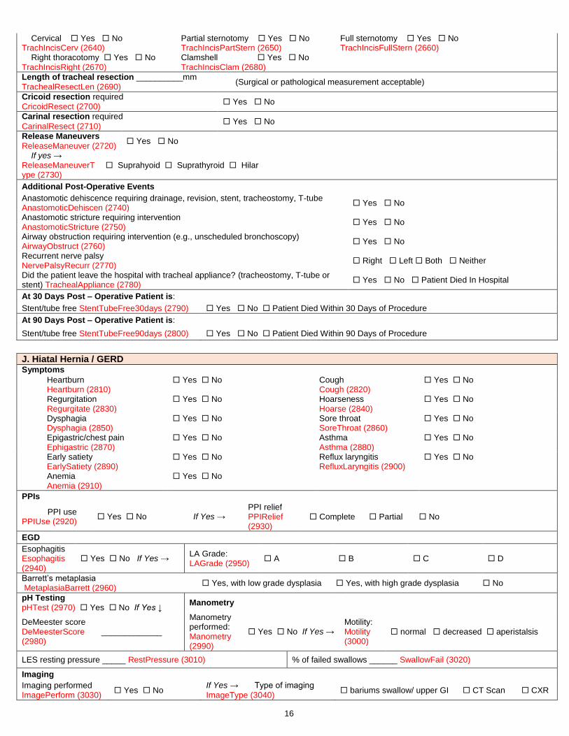
16
Cervical Yes No TrachIncisCerv (2640)
Partial sternotomy Yes No TrachIncisPartStern (2650)
Full sternotomy Yes No TrachIncisFullStern (2660)
Right thoracotomy Yes No TrachIncisRight (2670)
Clamshell Yes No TrachIncisClam (2680)
Length of tracheal resection __________mm
TrachealResectLen (2690) (Surgical or pathological measurement acceptable)
Cricoid resection required
CricoidResect (2700) Yes No
Carinal resection required
CarinalResect (2710) Yes No
Release Maneuvers
ReleaseManeuver (2720) Yes No
If yes → ReleaseManeuverType (2730)
Suprahyoid Suprathyroid Hilar
Additional Post-Operative Events
Anastomotic dehiscence requiring drainage, revision, stent, tracheostomy, T-tube AnastomoticDehiscen (2740)
Yes No
Anastomotic stricture requiring intervention AnastomoticStricture (2750)
Yes No
Airway obstruction requiring intervention (e.g., unscheduled bronchoscopy) AirwayObstruct (2760)
Yes No
Recurrent nerve palsy NervePalsyRecurr (2770)
Right Left Both Neither
Did the patient leave the hospital with tracheal appliance? (tracheostomy, T-tube or stent) TrachealAppliance (2780)
Yes No Patient Died In Hospital
At 30 Days Post – Operative Patient is:
Stent/tube free StentTubeFree30days (2790) Yes No Patient Died Within 30 Days of Procedure
At 90 Days Post – Operative Patient is:
Stent/tube free StentTubeFree90days (2800) Yes No Patient Died Within 90 Days of Procedure
J. Hiatal Hernia / GERD Symptoms
Heartburn Heartburn (2810)
Yes No Cough Cough (2820)
Yes No
Regurgitation Regurgitate (2830)
Yes No Hoarseness Hoarse (2840)
Yes No
Dysphagia Dysphagia (2850)
Yes No Sore throat SoreThroat (2860)
Yes No
Epigastric/chest pain Ephigastric (2870)
Yes No Asthma Asthma (2880)
Yes No
Early satiety EarlySatiety (2890)
Yes No Reflux laryngitis RefluxLaryngitis (2900)
Yes No
Anemia Anemia (2910)
Yes No
PPIs
PPI use PPIUse (2920)
Yes No If Yes → PPI relief PPIRelief (2930)
Complete Partial No
EGD
Esophagitis Esophagitis (2940)
Yes No If Yes → LA Grade: LAGrade (2950)
A B C D
Barrett’s metaplasia MetaplasiaBarrett (2960)
Yes, with low grade dysplasia Yes, with high grade dysplasia No
pH Testing pHTest (2970) Yes No If Yes ↓
Manometry
DeMeester score DeMeesterScore (2980)
_____________
Manometry performed: Manometry (2990)
Yes No If Yes → Motility: Motility (3000)
normal decreased aperistalsis
LES resting pressure _____ RestPressure (3010) % of failed swallows ______ SwallowFail (3020)
Imaging
Imaging performed ImagePerform (3030)
Yes No If Yes → Type of imaging
ImageType (3040) bariums swallow/ upper GI CT Scan CXR

17
Hiatal hernia size (cm) HerniaSize (3050)
_____________ Hiatal hernia type: HerniaType (3060)
I II III IV
Hernia repair status Primary repair Re-operation HerniaRepStat (3065)
If re-operation → Surgical approach used in the initial procedure: Laparoscopic Laparotomy Thoracotomy Not documented HerniaReopApp (3066)
Procedure Approach (check all that apply)
Laparoscopic GERDAppLaparoscopic (3070)
Yes No Robotic GERDAppRobotic (3080)
Yes No
Laparotomy GERDAppLaparotomy (3090)
Yes No Thoracotomy GERDAppThor (3100)
Yes No
Fundoplication ProcFundoplicate (3110)
Yes No If Yes → Type FundoplicateType (3120)
Partial Complete
Gastroplasty ProcGastroplasty (3130)
Yes No
Mesh ProcMesh (3140)
Yes No
Relaxing incision ProcRelaxIncision (3150)
Yes No
Is patient alive at 1 month post – Op?
GERDPtAliveMth (3160) Yes No
Is patient alive at 1 year post – Op?
GERDPtAliveYr (3210) Yes No
If Yes → 1 Month Post – Operative Follow Up If Yes → 1 Year Post – Operative Follow Up
Radiographic recurrence RadiographRecurr1Mon (3170)
Yes No Radiographic recurrence RadiographRecurr1Year (3220)
Yes No
Symptomatic recurrence SymptomRecurr1Mon (3180)
Yes No Symptomatic recurrence SymptomRecurr1Year (3230)
Yes No
Endoscopic Intervention EndoInt1Mon (3190)
Yes No Endoscopic Intervention EndoInt1Year (3240)
Yes No
Redo Operation RedoOperate1Mon (3200)
Yes No Redo operation RedoOperate1Year (3250)
Yes No
K. Disposition
Patient Disposition:
PatDisp (3260)
ICU Intermediate Care Unit Regular Floor Bed
Not Applicable (Expired in OR) Outpatient or Observation Status
ICU Admit this admission: Yes No ICUVisitInit (3270)
If Yes → Initial ICU Days: ______ ICUVisitInitDays (3280)
ICU Readmit: Yes No ICUVisitAdd (3290)
If Yes → Additional ICU Days: _______ ICUVisitAddDays (3300)
L. Post-Operative Events Indicate all adverse events that occurred within 1 month of surgery if discharged from the hospital or those that occur during the same admission, regardless of the length of stay.
Postoperative Events?
POEvents (3310) Yes No If Yes, select all that occurred: ↓
If Post-Operative Events Yes →
Unanticipated post-operative invasive procedure? Yes No PostOpInvProc (3330)
If unanticipated post-operative invasive procedure→
Primary Reason for Procedure: ReturnORRsn (3340)
Bleeding Bronchopleural Fistula Empyema Middle lobectomy for torsion
Conduit necrosis/failure following esophageal surgery Other
Anastomotic leak following esophageal surgery PosOpProcAL (3350)
Yes No If Yes → Surgical drainage and repair PosOpProcALRepair (3360)
Yes No
Stent placement PosOpProcALStent (3370)
Yes No
Additional chest tube placement PosOpProcALTube (3380)
Yes No

18
Chylothorax Present Yes No If Yes → ChyloPres (3390)
Chylothorax req. surgical ligation of thoracic duct Yes No PosOpProcChylotho (3400)
If No → Thoracic duct embolization attempted Yes No PosOpProcEmboli (3410)
If Yes → Was Thoracic duct embolization successful? Yes No PosOpProcDuctSucc (3420)
Pulmonary
Air leak > 5 days duration AirLeak5 (3430)
Yes No
Atelectasis req. bronchoscopy Atelectasis (3440)
Yes No
Pleural Effusion req. drainage CPlEff (3450)
Yes No
Pneumonia Pneumonia (3460)
Yes No
Acute Respiratory Distress Syndrome (ARDS) ARDS (3470)
Yes No Respiratory Failure RespFail (3480)
Yes No
Bronchopleural Fistula Bronchopleural (3490)
Yes No
Pulmonary Embolus PE (3500)
Yes No
Pneumothorax req. CT reinsertion Pneumo (3510)
Yes No
Initial Vent Support > 48 Hr Vent (3520)
Yes No Tracheostomy Trach (3530)
Yes No
Other Pulmonary Event OtherPul (3550)
Yes No
Cardiovascular
Atrial arrhythmia req. treatment AtrialArryth (3560)
Yes No Ventricular arrhythmia req. treatment VentArryth (3570)
Yes No Myocardial infarct MI (3580)
Yes No
Deep venous thrombosis (DVT) req. treatment DVT (3590)
Yes No Other CV event OtherCV (3600)
Yes No
Gastrointestinal
Ileus Ileus (3610)
Yes No Anastomotic leak requiring medical treatment only AnastoMed (3620)
Yes No Dilation esophagus DilationEsoph (3630)
Yes No
Conduit Necrosis Requiring Surgery CondNecSurg (3640)
Yes No Delayed conduit emptying requiring intervention (pyloric dilatation or botox) or maintenance of NG drainage > 7days post op DelayCondEmp (3650)
Yes No
Clostridium Difficile infection CDiff (3660)
Yes No Other GI event OtherGI (3670)
Yes No
Hematology
Packed red blood cells PostopPRBC (3680)
Yes No *transfusions documented here do not include blood given in OR*
If Yes→ # Units _________
PostopPRBCUnits (3690)
Urologic
Urinary tract infection UTI (3700)
Yes No
Urinary retention req. Catheterization UrinRetent (3710)
Yes No
Discharged with Foley catheter DischFoley (3720)
Yes No
Infection
Empyema req. treatment Empyema (3730)
Yes No Surgical Site Infection SurgSiteInfect (3740)
None Superficial Deep Organ space
Sepsis Sepsis (3750)
Yes No Another infection req. IV antibiotics OtherInfect (3760)
Yes No
Neurology

19
New central neurological event CentNeuroEvt (3770)
Yes No
Recurrent laryngeal nerve paresis -unexpected LaryngealNerve (3780)
Yes No
Delirium Delirium (3790)
Yes No Other neurological event OtherNeuro (3800)
Yes No
Miscellaneous
New renal failure per RIFLE criteria RenFailRIFLE (3810)
Yes No Chylothorax req. medical intervention ChyloMed (3820)
Yes No
Other events req. OR with gen. anesthesia OtherSurg (3830)
Yes No Unexpected Admission to ICU UnexpectAdmitICU (3840)
Yes No
M. Discharge
Patient is still in the hospital Yes No StillInHosp (3860)
If No → Date of Discharge: DischDt (3870)______/______/________
Discharge Status: MtDCStat (3880) Alive Dead
If Discharged Alive →
Discharge location: DisLoctn (3890)
Home Extended Care/Transitional Care Unit /Rehab
Other Hospital Nursing Home Hospice Other
Discharged with chest tube: CTubeDis (3900)
Yes No
Discharged with home O2 (new; not using O2 pre-op) DischHomeO2 (3910)
Yes No
If Yes → On O2 at 30 days postoperative? Yes No Unknown Patient Died Within 30 Days Post Op OnOxygen30DayPOp (3920)
Readmit to any hospital within 30 days of discharge: Readm30Dis (3930)
Yes No Unknown
If Yes → Readmission related to operative procedure?
Readm30DisRel (3940) Yes No Unknown
Status at 30 days after surgery: Mt30Stat (3950) Alive Dead Unknown
N. Follow Up Date of Last Follow-Up: ____/___/_____ LFUDate (3960) Mortality Status at Last Follow-Up: Alive Dead LFUMortStat (3970)
Mortality Date: ____/___/_____ MortDate (3980)
O. Quality Measures IV antibiotics ordered to be given within 1 hour before incision: IVAntibioOrdered (3990)
Yes No Not indicated for procedure
IV antibiotics given within 1 hour before incision: IVAntibioGiven (4000)
Yes No Not indicated for procedure
Cephalosporin Antibiotic Ordered CephalAntiOrdered (4010)
Yes No Not indicated for procedure Documented allergy or indication for therapeutic substitution
Prophylactic Antibiotic Discontinuation Ordered within 24 hour AntibioticDiscOrdered (4020)
Yes No Not indicated for procedure No, due to documented infection
Smoking Cessation Counseling SmokCoun (4030)
Yes No Patient refused Nonsmoker
DVT Prophylaxis Measures DVTProphylaxis (4040)
Yes No Not applicable
Related Documents