Molecular Biology of the Cell Vol. 20, 642– 657, January 15, 2009 The Slit/Robo System Suppresses Hepatocyte Growth Factor-dependent Invasion and Morphogenesis Maria Cristina Stella, Livio Trusolino, and Paolo M. Comoglio Division of Molecular Oncology, Institute for Cancer Research and Treatment, University of Torino School of Medicine, 10060 Candiolo, Torino, Italy Submitted March 27, 2008; Revised August 11, 2008; Accepted November 4, 2008 Monitoring Editor: Richard O. Hynes The Slit protein acts through the Roundabout receptor as a paracrine chemorepellent in axon guidance and as an inhibitor in leukocyte chemotaxis, but its role in epithelial cell motility and morphogenesis remains largely unexplored. We report that nontransformed epithelial cells and cancerous cells empower the Slit-2/Robo1 signaling system to limit outward migration in response to motogenic attractants and to remain positionally confined within their primitive location. Short hairpin RNA-mediated depletion of SLIT-2 or ectopic expression of a soluble decoy Robo enhance hepatocyte growth factor (HGF)-induced migration, matrix invasion, and tubulogenesis, concomitantly with the up-regulation of Cdc-42 and the down-modulation of Rac-1 activities. Accordingly, autocrine overexpression or exogenous administration of Slit-2 prevent HGF-triggered motile responses, reduce Cdc-42 activation, and stimulate Rac-1. This antimigratory activity of Slit-2 derives from the inhibition of actin-based protrusive forces and from an increased adhesive strength of cadherin- mediated intercellular contacts. These results disclose a novel function for Slit and Robo in the inhibition of growth factor-mediated epithelial cell motility and morphogenesis, invoking a critical role for both molecules as natural antagonists of neoplastic invasive growth. INTRODUCTION Morphogenetic events, such as formation of branched tubu- lar structures, angiogenesis, and neuronal networking, are modulated by a number of soluble and immobilized mole- cules that act as either attractants or repellents, including growth factors, extracellular matrix components, proteases, and morphogens (Lecuit and Lenne, 2007). There is now increasing evidence that cancer cells hijack the strategies by which the embryo grows and develops and that invading tumors coopt the genetic and signaling mechanisms under- lying tissue morphogenesis for the proper execution of neo- plastic dissemination (Huber et al., 2005). Therefore, insight into developmental programs is likely to illuminate essential aspects of cancer progression. Slit, acting through the transmembrane receptor Round- about (Robo), belongs to a recently identified family of se- creted repellents (Wong et al., 2002). Despite the consider- able body of knowledge gathered so far on the function of Slit and Robo in axon guidance (Dickson and Gilestro, 2006), neuronal migration (Wu et al., 1999), leukocyte chemotaxis (Wu et al., 2001) and angiogenesis (Wang et al., 2003), the information on the effects of the Slit/Robo system in normal and neoplastic epithelial cells is still fragmentary. If there is a general conservation of guidance mechanisms underlying cell migration and morphogenesis in different cell types, then Slit should limit the locomotion of Robo-expressing epithelial cells toward attractive stimuli. More importantly, it should sidetrack invading carcinomas away from attract- ants produced in the microenvironment. In this article, we sought to explore this issue using hepa- tocyte growth factor (HGF) as an attractive cue. HGF and its tyrosine kinase receptor Met play a crucial role both in development and cancer (Comoglio and Trusolino, 2002). During organogenesis HGF acts as a motogen and morpho- gen and stimulates, among other things, the directional mi- gration of myoblasts from the somites to the limbs as well as the guidance of motoneurons toward striated muscles (Birchmeier and Gherardi, 1998; Maina and Klein, 1999). In neoplastic contexts, activation of Met correlates with a met- astatic phenotype and a poor prognosis in several carcino- mas, and HGF stromal gradients favor tumor dissemination (Birchmeier et al., 2003). We show here that immortalized, nontransformed epithe- lial cells as well as carcinoma and melanoma cells frequently display a Slit/Robo signaling that endogenously counteracts HGF-driven migration, invasion, and morphogenesis. Mech- anistically, this Slit-dependent inhibitory activity resides in the reinforcement of the intercellular junctional apparatus (which impairs cell– cell dissociation) and in the weakening of protrusive forces (which decreases cell motility), possibly as a consequence of concomitant up-regulation of Rac-1 and down-regulation of Cdc-42 activities. Together, these results point to a fundamental role for Slit and Robo as anti-inva- sive cues during epithelial morphogenesis and neoplastic progression. MATERIALS AND METHODS Endpoint Polymerase Chain Reaction (PCR) and Primers ROBO-N and SLIT-2-Myc cDNAs were obtained by direct amplification of MDA-MB-435–retrotranscribed mRNAs. The primers used were the follow- ing: 5-ACT ATA TAT AGT ATT AAA CTA TTA ACT CTA GAA TGA AAT GGA AAC ATG TTC CTT TTT TGG-3 (sense Robo-N) and 5-AAC TAA This article was published online ahead of print in MBC in Press (http://www.molbiolcell.org/cgi/doi/10.1091/mbc.E08 – 03– 0321) on November 12, 2008. Address correspondence to: Maria Cristina Stella (mariacristina. [email protected]) or Livio Trusolino ([email protected]). 642 © 2009 by The American Society for Cell Biology http://www.molbiolcell.org/content/suppl/2008/11/12/E08-03-0321.DC1.html Supplemental Material can be found at:

Welcome message from author
This document is posted to help you gain knowledge. Please leave a comment to let me know what you think about it! Share it to your friends and learn new things together.
Transcript
Molecular Biology of the CellVol. 20, 642–657, January 15, 2009
The Slit/Robo System Suppresses Hepatocyte GrowthFactor-dependent Invasion and MorphogenesisMaria Cristina Stella, Livio Trusolino, and Paolo M. Comoglio
Division of Molecular Oncology, Institute for Cancer Research and Treatment, University of Torino School ofMedicine, 10060 Candiolo, Torino, Italy
Submitted March 27, 2008; Revised August 11, 2008; Accepted November 4, 2008Monitoring Editor: Richard O. Hynes
The Slit protein acts through the Roundabout receptor as a paracrine chemorepellent in axon guidance and as an inhibitorin leukocyte chemotaxis, but its role in epithelial cell motility and morphogenesis remains largely unexplored. We reportthat nontransformed epithelial cells and cancerous cells empower the Slit-2/Robo1 signaling system to limit outwardmigration in response to motogenic attractants and to remain positionally confined within their primitive location. Shorthairpin RNA-mediated depletion of SLIT-2 or ectopic expression of a soluble decoy Robo enhance hepatocyte growthfactor (HGF)-induced migration, matrix invasion, and tubulogenesis, concomitantly with the up-regulation of Cdc-42 andthe down-modulation of Rac-1 activities. Accordingly, autocrine overexpression or exogenous administration of Slit-2prevent HGF-triggered motile responses, reduce Cdc-42 activation, and stimulate Rac-1. This antimigratory activity ofSlit-2 derives from the inhibition of actin-based protrusive forces and from an increased adhesive strength of cadherin-mediated intercellular contacts. These results disclose a novel function for Slit and Robo in the inhibition of growthfactor-mediated epithelial cell motility and morphogenesis, invoking a critical role for both molecules as naturalantagonists of neoplastic invasive growth.
INTRODUCTION
Morphogenetic events, such as formation of branched tubu-lar structures, angiogenesis, and neuronal networking, aremodulated by a number of soluble and immobilized mole-cules that act as either attractants or repellents, includinggrowth factors, extracellular matrix components, proteases,and morphogens (Lecuit and Lenne, 2007). There is nowincreasing evidence that cancer cells hijack the strategies bywhich the embryo grows and develops and that invadingtumors coopt the genetic and signaling mechanisms under-lying tissue morphogenesis for the proper execution of neo-plastic dissemination (Huber et al., 2005). Therefore, insightinto developmental programs is likely to illuminate essentialaspects of cancer progression.
Slit, acting through the transmembrane receptor Round-about (Robo), belongs to a recently identified family of se-creted repellents (Wong et al., 2002). Despite the consider-able body of knowledge gathered so far on the function ofSlit and Robo in axon guidance (Dickson and Gilestro, 2006),neuronal migration (Wu et al., 1999), leukocyte chemotaxis(Wu et al., 2001) and angiogenesis (Wang et al., 2003), theinformation on the effects of the Slit/Robo system in normaland neoplastic epithelial cells is still fragmentary. If there isa general conservation of guidance mechanisms underlyingcell migration and morphogenesis in different cell types,then Slit should limit the locomotion of Robo-expressingepithelial cells toward attractive stimuli. More importantly,
it should sidetrack invading carcinomas away from attract-ants produced in the microenvironment.
In this article, we sought to explore this issue using hepa-tocyte growth factor (HGF) as an attractive cue. HGF and itstyrosine kinase receptor Met play a crucial role both indevelopment and cancer (Comoglio and Trusolino, 2002).During organogenesis HGF acts as a motogen and morpho-gen and stimulates, among other things, the directional mi-gration of myoblasts from the somites to the limbs as well asthe guidance of motoneurons toward striated muscles(Birchmeier and Gherardi, 1998; Maina and Klein, 1999). Inneoplastic contexts, activation of Met correlates with a met-astatic phenotype and a poor prognosis in several carcino-mas, and HGF stromal gradients favor tumor dissemination(Birchmeier et al., 2003).
We show here that immortalized, nontransformed epithe-lial cells as well as carcinoma and melanoma cells frequentlydisplay a Slit/Robo signaling that endogenously counteractsHGF-driven migration, invasion, and morphogenesis. Mech-anistically, this Slit-dependent inhibitory activity resides inthe reinforcement of the intercellular junctional apparatus(which impairs cell–cell dissociation) and in the weakeningof protrusive forces (which decreases cell motility), possiblyas a consequence of concomitant up-regulation of Rac-1 anddown-regulation of Cdc-42 activities. Together, these resultspoint to a fundamental role for Slit and Robo as anti-inva-sive cues during epithelial morphogenesis and neoplasticprogression.
MATERIALS AND METHODS
Endpoint Polymerase Chain Reaction (PCR) and PrimersROBO-N and SLIT-2-Myc cDNAs were obtained by direct amplification ofMDA-MB-435–retrotranscribed mRNAs. The primers used were the follow-ing: 5�-ACT ATA TAT AGT ATT AAA CTA TTA ACT CTA GAA TGA AATGGA AAC ATG TTC CTT TTT TGG-3� (sense Robo-N) and 5�-AAC TAA
This article was published online ahead of print in MBC in Press(http://www.molbiolcell.org/cgi/doi/10.1091/mbc.E08–03–0321)on November 12, 2008.
Address correspondence to: Maria Cristina Stella ([email protected]) or Livio Trusolino ([email protected]).
642 © 2009 by The American Society for Cell Biology http://www.molbiolcell.org/content/suppl/2008/11/12/E08-03-0321.DC1.htmlSupplemental Material can be found at:

ATG TAA TAC TAA TTT ATT AAA CGC GTT TAA GCG TAA TCT GGAACA TCA TAT GGG TAG TTG GCT CCA GAT GGC CGA TAG AGA ATTTTA TAT CCT TGT ATA TAC-3� (antisense ROBO-N, HA-tagged); 5�-ACCTTA GAC ATG CGC GGC GTT GGC TGG CAG ATG CTG TCC CTG-3�(sense Slit-2); 5�-TAT TAA TCT AGA TTA ATT CAG ATC CTC TTC TGAGAT GAG TTT TTG TTC GGA CAC ACA CCT CGT ACA GCC GC-3�(antisense Slit-2, Myc-tagged). Both ROBO-N-HA and SLIT-2-Myc werecloned by blunt-end ligation in the retroviral vector pLHCX (Clontech, Moun-tain View, CA). The sequences of the two SLIT-2-shRNAs, derived from ascreening performed using the SUPER RNA interference (RNAi) library(Brummelkamp et al., 2002) were the following: 5�-GAA CGT GTC CCG ATTAGA G-3� (sequence A) and 5�-CTG CCT TCG GGT AGA TGC T-3� (sequenceb); the scrambled, control short hairpin RNA (shRNA) used in MDA-MB-435cells was GAA GGT GGG GTA GAT GCT A; sequence B was used forsilencing SLIT-2 expression in Madin-Darby canine kidney (MDCK) cells aswell, whereas in this cell line sequence A, which does not target canine SLIT-2,was used as a control. The sequences of the three Robo-1 shRNA, derivedfrom the specific MISSION shRNA Gene Family Sets (Sigma-Aldrich, St.Louis, MO) were 5�-AGA AAT ACA GTC ACA TTA TCT C-3� (sequence A);5�-CCA CCA TTT CAT GGA AGA ACT C-3� (sequence B); 5�-CAC CAG CAAGGA TGT ATT TCT C-3� (sequence C). Sequence C was used for silencingRobo-1 in MDCK cells. Scramble MISSION shRNAs were provided by Sigma-Aldrich. The primers used for endpoint PCRs in human-derived cell lineswere 5�-TGC CGC AAG CTC TAC TGC CTG-3� (sense Slit-1); 5�-GCA CTGGCA GCT GTA GGA AAG A-3� (antisense Slit-1); 5�-GCA TTT TGC CTGGCT GTG AG-3� (sense Slit-2); 5�-CAT TGA TGG GCA AGC AGG TG-3�(antisense Slit-2); 5�-TGG AGA AGG ACA GCG TGG TG3� (sense Slit-3);5�-GAG CAG GCA TTG GCA GAG TC-3� (antisense Slit-3); 5�-CCT CGC ATTGTT GAA CAC C-3� (sense Robo-1); 5�-AAC ATT CGG TGT GAG CGAGG-3� (antisense Robo-1); 5�-AAG CTC TCT AGA GAG ACA AC-3� (senseRobo-2); 5�-TGG GCT TGC TAT AGG GCA CC-3� (antisense Robo-2); 5�-GGTCAC CCC ATC CCG AAG GG-3� (sense Robo-3); 5�-CAC CCA AGC CAGCAG GCC T-3� (antisense Robo-3); 5�-GCT GGC CCA GCT CTC CAG CC-3�(sense Robo-4); and 5�-ACT CAC AGG CCC GGA GCT CC-3� (antisenseRobo-4). The primers used for MDCK cell amplifications were 5�-TGC CGAAAG CTC TAC TGC CTG-3� (sense Slit-1); 5�-GCA CTG GCA GCT GTA GGAAAG-3� (antisense Slit-1); 5�-GCA TTT TGC CTG GCT GTG AG-3� (senseSlit-2); 5�-CGT TGA TGG GCA AGC AGG TG-3� (antisense Slit-2); 5�-CCAGCA GTA GGC ATC AAC AGC-3� (sense Slit-3); 5�-CAC CAC GCT GTC CTTCTC CA-3� (antisense Slit-3); 5�-ATG AGC AAT TTA GAG AAA TGT GG-3�(sense Robo-1); 5�-ggc gtg ggg cgg cct tca gct t-3� (antisense Robo-1); 5�-CCAGAC CCC TGA GAG CAC TA-3� (sense Robo-2); 5�-TGG GCT TGC TGT ATGGCA CC-3� (antisense Robo-2); 5�-CTT GGG ATG GAG GGA CCA AGC-3�(sense Robo-3); 5�-CAC CCA CGC CAG CAG GCC T-3� (antisense Robo-3);5�-GCT GGC CCG GCT CTC CAG CC-3� (sense Robo-4); and 5�-GGG AAAGGT TCT TGG AGC CTC T-3� (antisense Robo-4). Primers for semiquantita-tive real-time PCR were the following: for Slit-2, designed in two contiguousexons, 5�-GCA TTT TGC CTG GCT GTG AG-3� (sense Slit-2, MDA-MB-435);5�-CAT TGA TGG GCA AGC AGG TG-3� (antisense Slit-2, MDA-MB-435);5�-GCA TTT TGC CTG GCT GTG AG-3� (sense Slit-2, MDCK); and 5�-CGTTGA TGG GCA AGC AGG TG-3� (antisense Slit-2, MDCK). The housekeep-ing gene phospho-glycerate-kinase (PGK) was chosen as a standard reference.The primers were 5�-CTT ATG AGC CAC CTA GGC CG-3� (sense PGK, MDA-MB-435); 5�-CAT CCT TGC CCA GCA GAG AT-3� (antisense PGK, MDA-MB-435); 5�-ATC ACA GGT GGT GGA GAC AC-3� (sense PGK, MDCK); and 5�-CTAATG CCA ACC AGA GAT AG-3� (antisense PGK, MDCK). Total RNAs wereobtained using the RNeasy Mini kit (QIAGEN, Valencia, CA) and quantifiedusing RNA Nano Chips (Agilent Technologies, Santa Clara, CA) togetherwith a 2100 Bioanalyzer (Agilent Technologies). cDNAs were retro-tran-scribed with High Capacity cDNA Reverse Transcription kit (Applied Bio-systems, Foster City, CA). PCR amplifications were monitored using a 7009HT Analyzer (Applied Biosystems) under the following thermocycler condi-tions: stage 1, 95°C for 10 min for one cycle; and stage 2, 95°C for 15 s and 64°Cfor 1 min for 60 cycles.
AntibodiesWe used the following antibodies: anti-actin, anti-hemagglutinin (HA), biotin-conjugated anti-Myc (Santa Cruz Biotechnology, Santa Cruz, CA); anti-Rac 1,anti-Cdc42, anti-E-cadherin, and anti-�-catenin (BD Biosciences, San Jose,CA); anti-Myc (clone 9E10; Millipore, Billerica, MA); anti-vinculin and anti-FLAG (Sigma-Aldrich); and anti-Cdc-42 (Santa Cruz Biotechnology). Theanti-zona occludens (ZO)-1 monoclonal antibody (mAb), developed by D.Goodenough, was obtained from the Developmental Studies HybridomaBank (University of Iowa, Iowa City, IA).
Cell Culture and Viral InfectionMDA-MB-435, MDCK, U-87, RKO, HT-29, 769-P, and COS-7 were cultured inDMEM; Skov-3, DLD-1, HCT-116, and N-87 were cultured in RPMI 1640medium; LoVo was cultured in Ham’s F-12; Hec-1A and RT-112 were cul-tured in McCoy’s 5A; and HeLa was cultured in Iscove. All media weresupplemented with 10% FCS (Sigma). Expression of exogenous proteins wasobtained with LipofectAMINE 2000 (Invitrogen)-mediated transfection or
with viral infection. MISSION shRNA lentiviral particles were preparedaccording to the instruction of the manufacturer. Human cells were infectedwith a multiplicity of infection (MOI) of 30, whereas canine MDCK cells wereinfected with an MOI of 90. Retroviral hybrid vectors were produced bytransient transfection of 293T cells. Viral supernatants were filtered through a0.22-�m filter, and infections were performed in the presence of 4 �g/mlPolybrene (Sigma-Aldrich), followed by selection with puromycin (Sigma-Aldrich) or hygromycin-B (Invitrogen).
BiochemistryThe expression of Rac-1-gof-Myc, Rac-1-DN-Myc, Cdc42-DN-HA, Cdc-42-gof-FLAG, and Slit-2-Myc was analyzed by immunoprecipitation. For immu-noprecipitations, 5 � 106 cells were lysed for 20 min at 4°C with 1 ml of abuffer containing 50 mM HEPES, pH 7.4, 5 mM EDTA, 2 mM EGTA, 150 mMNaCl, 10% glycerol, and 1% Triton X-100, in the presence of protease andphosphatase inhibitors. Extracts were clarified at 12,000 rpm for 15 min,normalized with the BCA Protein Assay Reagent kit (Pierce Chemical, Rock-ford, IL), and incubated with different mAbs for 2 h at 4°C. Immune com-plexes were collected with either protein G- or protein A-Sepharose, washedin lysis buffer, and eluted. Total cellular proteins were extracted by solubi-lizing the cells in boiling SDS buffer (50 mM Tris-HCl, pH 7.5, 150 mM NaCl,and 1% SDS). Extracts were electrophoresed on SDS-polyacrylamide gels andtransferred onto nitrocellulose membranes (Hybond; GE Healthcare, LittleChalfont, Buckinghamshire, United Kingdom). Nitrocellulose-bound anti-bodies were detected by the enhanced chemiluminescence system (GEHealthcare). Western blot analyses to detect E-cadherin were performed usingprotein extracts derived from 6 � 104 MDCK cells suspended as hangingdrops (see below). Pool-down experiments were done using PAK-GST pro-tein beads (Cytoskeleton, Denver, CO), and 80% confluent cells starved for24 h (Sander et al., 1998). For Cdc-42 pool-down experiments, 50 �g ofPAK-GST proteins were incubated with 10 mg of whole cell lysates; for Rac-1,20 �g of PAK-GST proteins were incubated with 2 mg of whole cell lysates.
Biological AssaysIn all the biological assays, we used baculovirus-derived recombinant HGF(Naldini et al., 1995). If not otherwise indicated, HGF was used at a concen-tration of 25 ng/ml for the migration assays and at a concentration of 100ng/ml for tubulogenesis. Slit-2-Myc–tagged protein was obtained from thesupernatant of Chinese hamster ovary (CHO) cells retrovirally infected withSlit-2-Myc, grown in serum-free medium. Because Slit-2 is frequently associ-ated to heparan sulfate glycosaminoglycans (Liang et al., 1999; Ronca et al.,2001), the production and release of Slit-2 were optimized using CHO cellsdefective in proteoglycan synthesis (Esko et al., 1987). Conditioned mediumwas concentrated using Centricon Plus-20 (Millipore) centrifugal filter de-vices, with a cut-off of 100 kDa. The amount of Slit-2-Myc in each preparationwas quantified by silver staining. Eighty nanograms of concentrated Slit2-Myc polypeptide were used in combination with 20 ng of HGF. Always,soluble Slit 2-Myc polypeptide was administrated together with heparin, asdescribed previously (Hussain et al., 2006). Transwell migration and tubulo-genesis assays were performed and quantified as described previously (Stellaet al., 2005). Images were captured every with ImageReady software (AdobeSystems, Mountain View, CA) and arranged using Photoshop software(Adobe Systems). Scratch assays were performed as described previously(Michieli et al., 2004) and quantified by time-lapse microscopy (1 image every10 min), plotting the distance between the scratch margins measured as meanof 10 different measurements expressed as percentage of the initial distance.Aggregation assays were performed as described previously (Thoreson et al.,2000). After 12 h, aggregates were counted and stained with trypan blue tomonitor cell viability. Images were recorded as specified above. For motilityassays in the presence of an HGF isotropic gradient, cells were plated onto 24multiwell culture plates (MatTek, Ashland, MA), maintained in DMEM lack-ing phenol red (Sigma-Aldrich). Video images were recorded at 10-minintervals for 36 h; images were analyzed and arranged using Sony Vegassoftware (Sony Media Software, Tokyo, Japan) in combination with MicrosoftOffice Excel (Microsoft, Redmond, WA).
ImmunofluorescenceCells were seeded onto gelatin-coated glass coverslips. At the indicated times,cells were fixed, permeabilized, and blocked in 10% fetal calf serum (FCS).Primary antibodies were incubated for 12 h at 4°C followed by staining withfluorochrome-conjugated secondary antibodies (Invitrogen, Carlsbad, CA).F-actin was evidenced with tetramethylrhodamine B isothiocyanate (TRITC)-conjugated phalloidin. The stained cells were mounted in Mowiol and ob-served and photographed using a confocal laser scanning system.
Statistical and Densitometric AnalysisResults are means � SD or SEM. Comparisons were made using the two-tailed Student’s t test. p values �0.05 were considered to be statisticallysignificant. In each experimental point were scored 10 cells for isotropicmigration assays and 100 cysts together with 100 tubules for tubulogenesisanalysis. Blot images were captured using a ChemiDoc XRS molecular imager
Slit Limits HGF-dependent Invasion
Vol. 20, January 15, 2009 643

(Bio-Rad, Hercules, CA). Densitometric analysis was performed with a Quan-tity One one-dimensional analysis software installed on the imager.
RESULTS
Inhibition of Slit-2 or Robo-1 in Melanoma CellsEnhances HGF-mediated Motility and InvasionThere are three Slits and four Robos in mammals (Wonget al., 2002). Evidences from the literature indicate thatSlit-2, among all the Slit ligands, is the isoform mostfrequently expressed in melanomas and carcinomas(Wang et al., 2003). We decided to integrate this analysisby exploring, in parallel with Slit-2, Robo-1 expression inhuman cell lines. Reverse transcription (RT)-PCR analysisin 14 tumor cell lines derived from various tissues re-vealed that five lines (Skov-3, HeLa, MDA-MB-435, 769-P,and Hec-1A) displayed basal expression of both ROBO-1and SLIT-2 genes (Table 1). Detailed investigation of thefull panel of SLIT and ROBO transcripts in these five celllines indicated that, in addition to Slit-2 and Robo-1, HeLa,769-P, and Hec-1A cells express Slit-1, Slit-3, and Robo-3;Skov-3 express Slit-3 and Robo-3; and finally, MDA-MB-435cells express two Robo receptors, Robo-1 and Robo-3, butonly one Slit gene, Slit-2 (Figure 1 A).
We started our experiments by abrogating Slit-2 func-tion in MDA-MB-435, a melanoma cell line that has beenextensively characterized in terms of motile and invasiveresponses upon stimulation with motogenic cytokines,such as HGF (Trusolino et al., 2001; Michieli et al., 2004;Stella et al., 2005). Moreover, these cells have the uniqueproperty of expressing Slit-2 as the sole member of thisligand family, which alleviates concerns of redundancyand therefore facilitates initial biological investigation. Toinhibit Slit-2 expression, we used two short hairpin RNAs(shRNA-A and shRNA-B; see Supplemental Material) thatabated the expression of the SLIT-2 gene by 80 –90%, asassessed by quantitative real time RT-PCR (Figure 1B). Ascrambled shRNA was used as a control in all the exper-iments (Figure 1B).
Analysis of HGF-dependent cell motility in cells withnormal or diminished levels of Slit-2 was performed usingseveral in vitro assays meant to assess different parameters,including directional migration across transwell-permeablefilters toward a polarized source of ligand (anisotropic con-ditions), random migration on plastic dishes (isotropic con-ditions), and matrix invasion in three-dimensional cultures.Time course analyses of anisotropic cell migration revealed
that Slit-2–deficient cells displayed an enhanced chemotacticresponse to HGF in a time-dependent manner (Figure 1C).The rescue of Slit-2 expression through retroviral transduc-tion of a Myc-tagged variant of Slit-2 restored HGF-depen-dent cell migration at levels comparable with those observedin control cells (Figure 1, B and C). In isotropic conditions,cells lacking Slit-2 as well as control cells exhibited a highlyvariable motogenic behavior, moving around with frequentchanges of direction, in a somewhat erratic manner. How-ever, Slit-2–deficient cells were characterized by a statisti-cally significant increase in the total traveled distance (Fig-ure 1D). Finally, in a three-dimensional collagen invasionassay (tubulogenesis assay), cells with impaired expressionof Slit-2 were able to invade collagen much more efficientlythan control cells upon treatment with HGF (Figure 1E).Slit-2 silencing resulted in a modest, but clearly detectable,tubulogenic activity also under basal conditions, in the ab-sence of exogenous HGF (Figure 1E). This suggests that theSlit/Robo circuit chronically operative in these cells conveysconstitutive repelling signals that prevent cells from movingtoward attractive stimuli present in the adjacent matrix.
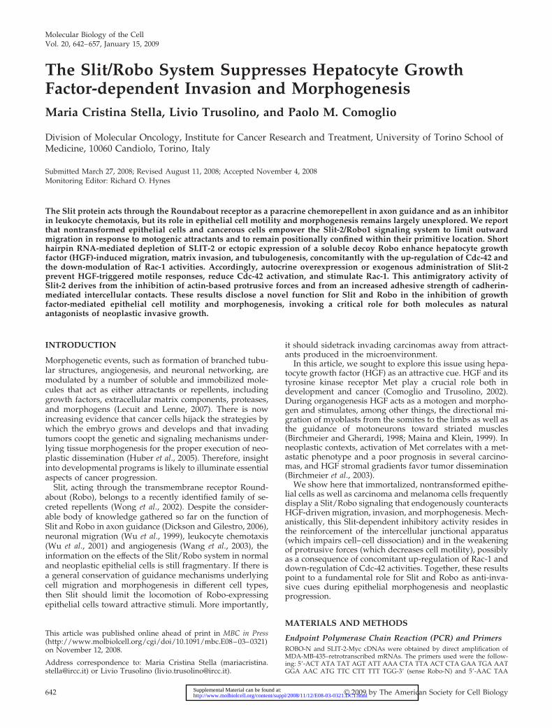
As a complementary approach to inhibit Slit-2 function,we transduced MDA-MB-435 cells with an HA-tagged vari-ant of Robo-N, a fragment of Robo-1 that contains only theextracellular part of the Robo protein and acts as a domi-nant-interfering decoy receptor (Wu et al., 1999) (Figure 2A).When subjected to a transwell migration assay (Figure 2B) orto a collagen invasion assay (Figure 2C) in the presence ofHGF, cells expressing Robo-N were more efficient than con-trol cells.
Finally, we validated these findings by inhibiting Robo-1instead of Slit-2. Indeed, reduction of Robo-1 by shRNAtechnology (Figure 2D) sensitized cells to HGF in both aniso-tropic cell migration (Figure 2E) and collagen invasion (Fig-ure 2F).
Together, these results indicate that genetic attenuation ofthe Slit/Robo pathway—through either silencing of the li-gand, silencing of the receptor, or overexpression of a dom-inant negative isoform of the receptor—exacerbates theHGF-dependent motile and invasive properties of neoplasticcells.
Hyperactivation of the Slit/Robo Pathway in MelanomaCells Suppresses HGF-dependent ResponsesIn parallel with inhibition of the Slit/Robo system, we at-tempted a gain-of-function approach by enhancing Slit/Robo signaling using three different experimental condi-tions: 1) potentiation of the endogenous loop by ectopicoverexpression of the SLIT-2 gene, 2) exogenous adminis-tration of the recombinant Slit-2 protein, and 3) cocultureexperiments using human embryonic kidney (HEK) cellsstably producing Slit-2.
In the first setting (Figure 3A), Slit-2 overexpression sub-stantially impaired the chemotactic responsiveness to HGFin transwell assays, which was restored upon shRNA-medi-ated down-modulation of Slit-2 itself (Figure 3, A and B).Under isotropic condition, Slit-2-overexpressing cells dis-played a static, nonmotile phenotype and were completelyinsensitive to HGF treatment (data not shown). Similarly, ina collagen invasion assay, the tubulogenic response to HGFwas much weaker in Slit-2–overexpressing cells than incontrol cells (Figure 3C).
For exogenous administration of the Slit-2 ligand, we firsttransduced CHO cells with the retroviral Slit-2-Myc con-struct and then recovered and concentrated the supernatantof these cells. In control experiments, we used mediumconditioned by CHO cells secreting Slit-2-Myc after deple-
Table 1. Expression of Slit-2 and Robo-1 in human cancer cells
Cell line Tumor derivation Slit-2 Robo-1
U-87 Glioblastoma � �RT-112 Bladder carcinoma � �RKO Colon carcinoma � �DLD-1 Colon carcinoma � �HCT-116 Colon carcinoma � �HT-29 Colon carcinoma � �LoVo Colon carcinoma � �N87 Gastric carcinoma � �A431 Squamous cell carcinoma � �Skov-3 Ovarian carcinoma � �HeLa Uterine cervix carcinoma � �MDA-MB-435 Melanoma � �Hec-1A Endometrial carcinoma � �769-P Renal carcinoma � �
M. C. Stella et al.
Molecular Biology of the Cell644

tion of the soluble Slit protein by anti-Myc immunoprecipi-tation (Figure 3D). The analysis of anisotropic cell migrationshowed that, in the presence of soluble Slit-2, wild-typeMDA-MB-435 cells did not migrate toward the HGF source.On the contrary, HGF stimulation in the presence of mockmedium or the Slit-2–depleted medium resulted in a signif-icant rate of migration, similar to that observable after ad-ministration of HGF alone (Figure 3E). In line with thatdetected with Slit-2–overexpressing MDA-MB-435 cells,HGF was unable to induce a consistent tubulogenic activityin the presence of Slit-2–conditioned medium, whereas itdid stimulate robust collagen invasion in the presence of theSlit-2–depleted medium (Figure 3F).
Next, we evaluated the ability of exogenous Slit-2 tohamper HGF-dependent cell migration in coculture ex-periments by using HEK cells expressing Slit-2. In thiscondition, HEK cells were transduced with the recombi-nant Slit-2-Myc retroviral construct (Figure 3G). Controlor Slit-2– expressing HEK cells were then seeded in thelower compartment of a transwell chamber, whereaswild-type MDA-MB-435 cells were positioned in the up-per compartment. Also in this setting, the migration ofwild-type MDA-MB-435 cells in response to HGF wasstrongly inhibited in the presence of Slit-2–secreting HEKcells but not in the presence of control HEK cells (Figure3H).
Figure 1. Slit-2 down-regulationenhances HGF-dependent motogen-esis and morphogenesis in MDA-MB-435 cells. (A) Endpoint RT-PCRexpression of SLIT and ROBO genes;(�): PCR reaction performed with-out template. (B) Variations ofSLIT-2 mRNA in MDA-MB-435 cellsin the following experimental condi-tions: wild-type cells (�); cells trans-duced with a scrambled shRNA se-quence (ctr); cells transduced withtwo different SLIT-2-specific shR-NAs (A and B); cells cotransducedwith shRNA B together with a Myc-tagged variant of SLIT-2. Transcriptquantitations are shown as –foldvariations of SLIT-2 mRNA contentwith respect to wild-type cells. Dataare the means � SD (error bars) offour independent experiments, per-formed in triplicate. (C) Time courseanalysis of the HGF-dependent aniso-tropic migration (transwell assay)in cells expressing a scrambledshRNA (�), the SLIT-2 shRNA B(�), or coexpressing the SLIT-2shRNA B together with Slit-2-Myc.Data are the means � SD (errorbars) of five independent experi-ments, performed in duplicate.*p � 0.05 at 6 h and **p � 0.01 at12 h. (D) Migratory trajectories ofcells expressing either a scrambledshRNA (�) or the SLIT-2 shRNA B(�) in the absence (�) or presence(�) of HGF. Images were recordedevery 10 min for 36 h by time-lapsevideomicroscopy. The diagramshows the trajectories of two repre-sentative cells for each experimen-tal condition. The lower panelquantitates the total distance cov-ered in 36 h. The average values �SEM, expressed in micrometers,derive from analysis of 10 cells foreach experimental condition. Thisexperiment was repeated threetimes in duplicate. (E) Collagen in-vasion (tubulogenesis) assay inMDA-MB-435 cells expressing thescrambled (�) or the SLIT-2shRNA B (�). Cells were treatedwith vehicle (�) or with HGF (�).The micrographs show representa-tive 4-d-old colonies. Bar, 20 �m.The bottom panel corresponds to the morphometric analysis of the collagen invasion assay presented in the top panel. The average numberof sprouting cells in each cyst and the average length of single tubules together � SEM are shown.
Slit Limits HGF-dependent Invasion
Vol. 20, January 15, 2009 645
Finally, we assessed whether the inhibitory effects causedby Slit-2 overactivation were in fact mediated by Robo re-ceptors. Accordingly, we treated cells transduced with ascrambled shRNA or with Robo-1–specific shRNAs withsupernatants derived from Slit-2–expressing CHO cells. Incontrol experiments, we used the same medium after Slit-2depletion by anti-Myc immunoprecipitation. In anisotropicconditions, lack of Robo-1 function potently rescued theimpairment of HGF-dependent cell migration produced byexogenous Slit-2 (Figure 3I). Similar results were obtained inthree-dimensional collagen assays (Figure 3L).
In summary, these findings indicate that either the auto-crine or paracrine potentiation of Slit signaling dramaticallyimpairs HGF-induced cell migration, invasion, and branch-ing morphogenesis. This inhibitory activity is predomi-nantly transduced by the Robo-1 receptor.
Effects of the Modulation of the Slit/Robo Pathway onHGF-dependent Responses in Carcinoma CellsWe decided to extend our observations to other neoplasticcell lines that, different from MDA-MB-435, are of epithelialorigin, express the junctional adhesion molecule E-cadherin
and grow as compact aggregates with well-defined intercel-lular contacts (Nishimura et al., 2003; DiFeo et al., 2006).Specifically, we examined Skov-3 (ovarian carcinoma) andHec-1A (endometrial carcinoma). Given the major role ofSlit-2 in dictating selective and dominant developmentalfates in mammalian tissues where other Slit and Robo mem-bers are coexpressed (Wong et al., 2002), we reasoned thatdecreasing or augmenting the specific activity of Slit-2would be sufficient to obtain a consistent biological readoutin these two cell lines, despite the presence of other Slit andRobo variants (Figure 1A).
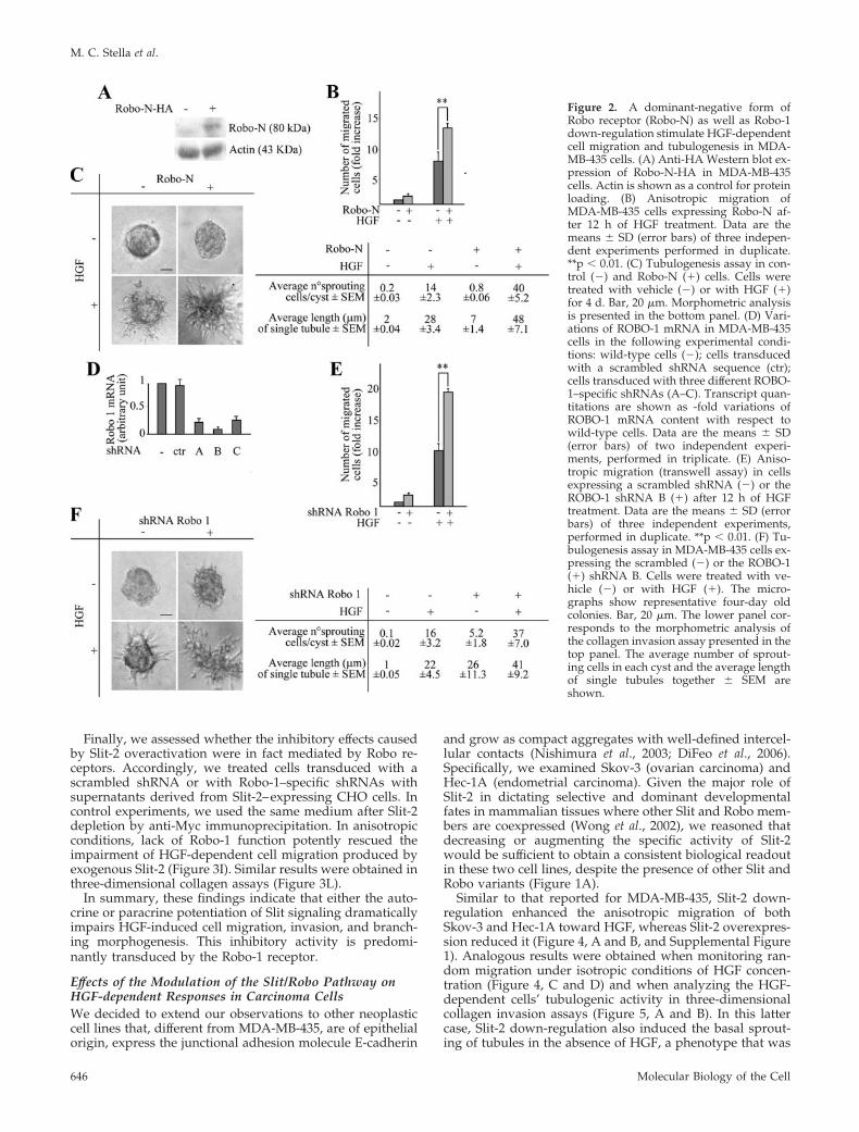
Similar to that reported for MDA-MB-435, Slit-2 down-regulation enhanced the anisotropic migration of bothSkov-3 and Hec-1A toward HGF, whereas Slit-2 overexpres-sion reduced it (Figure 4, A and B, and Supplemental Figure1). Analogous results were obtained when monitoring ran-dom migration under isotropic conditions of HGF concen-tration (Figure 4, C and D) and when analyzing the HGF-dependent cells’ tubulogenic activity in three-dimensionalcollagen invasion assays (Figure 5, A and B). In this lattercase, Slit-2 down-regulation also induced the basal sprout-ing of tubules in the absence of HGF, a phenotype that was
Figure 2. A dominant-negative form ofRobo receptor (Robo-N) as well as Robo-1down-regulation stimulate HGF-dependentcell migration and tubulogenesis in MDA-MB-435 cells. (A) Anti-HA Western blot ex-pression of Robo-N-HA in MDA-MB-435cells. Actin is shown as a control for proteinloading. (B) Anisotropic migration ofMDA-MB-435 cells expressing Robo-N af-ter 12 h of HGF treatment. Data are themeans � SD (error bars) of three indepen-dent experiments performed in duplicate.**p � 0.01. (C) Tubulogenesis assay in con-trol (�) and Robo-N (�) cells. Cells weretreated with vehicle (�) or with HGF (�)for 4 d. Bar, 20 �m. Morphometric analysisis presented in the bottom panel. (D) Vari-ations of ROBO-1 mRNA in MDA-MB-435cells in the following experimental condi-tions: wild-type cells (�); cells transducedwith a scrambled shRNA sequence (ctr);cells transduced with three different ROBO-1–specific shRNAs (A–C). Transcript quan-titations are shown as -fold variations ofROBO-1 mRNA content with respect towild-type cells. Data are the means � SD(error bars) of two independent experi-ments, performed in triplicate. (E) Aniso-tropic migration (transwell assay) in cellsexpressing a scrambled shRNA (�) or theROBO-1 shRNA B (�) after 12 h of HGFtreatment. Data are the means � SD (errorbars) of three independent experiments,performed in duplicate. **p � 0.01. (F) Tu-bulogenesis assay in MDA-MB-435 cells ex-pressing the scrambled (�) or the ROBO-1(�) shRNA B. Cells were treated with ve-hicle (�) or with HGF (�). The micro-graphs show representative four-day oldcolonies. Bar, 20 �m. The lower panel cor-responds to the morphometric analysis ofthe collagen invasion assay presented in thetop panel. The average number of sprout-ing cells in each cyst and the average lengthof single tubules together � SEM areshown.
M. C. Stella et al.
Molecular Biology of the Cell646

particularly evident in Hec-1A cells and paralleled that ob-served in Slit-2–deficient MDA-MB-435 cells (Figure 1E).
The fact that Skov-3 and Hec-1A form tightly packedepithelial sheets allowed us to scrutinize the effects of dosingSlit-2 levels in scratch assays. Again, abatement of Slit-2expression accelerated both the intrinsic and the HGF-in-duced motility of Skov-3 so that the scratch was completely
repaired after 12 h of HGF treatment. Conversely, increasedexpression of Slit-2 almost immobilized the cells, and woundrepair was not completed even after 24 h of HGF stimulation(Figure 4E and Supplemental Figure 2). The spontaneousmigration rate of Hec-1A seemed to be less intense than thatof Skov-3. However, also in this case, Slit-2–deficient cellstended to repair the wound more efficiently than mock cells,
Figure 3. Slit-2 inhibits HGF-de-pendent cell migration and tubulo-genesis in MDA-MB-435 cells. (A)Anti-Myc Western blot expressionof Slit-2-Myc in MDA-MB-435 cells.Cotransduction with the SLIT-2-specific shRNA B abrogates the ex-pression of Slit-2-Myc. (B) HGF-de-pendent anisotropic migration incells transduced with an emptyvector, a Slit-2-Myc vector, or co-transduced with Slit-2-Myc to-gether with the SLIT-2-specificshRNA-B. Cells were either left un-treated or stimulated with HGF for12 h. Data are the means � SD(error bars) of three independentexperiments performed in dupli-cate. **p � 0.01 at 12 h. (C) Tubu-logenesis assay in cells expressingthe empty (�) or the Slit-2-Myc (�)vector, in the presence of vehicle(�) or HGF (�). The micrographsshow representative 10-d old colo-nies. Bar, 20 �m. Morphometricanalysis is in the bottom panel. (D)Anti-Myc Western blot of condi-tioned media obtained from CHOcells infected with a control emptyvirus (�) or with a virus encodingfor Slit-2-Myc (�). Immunodeple-tion of Slit-2 from the culture su-pernatants using anti-Myc antibod-ies is also shown. (E) Anisotropicmigration of MDA-MB-435 cells inthe presence of control superna-tant, Slit-2–containing supernatant,or Slit-2–depleted supernatant.This analysis was performed after12 h of HGF treatment. Data are themeans � SD (error bars) of threeindependent experiments per-formed in duplicate. ***p � 0.001.(F) Tubulogenesis assay in MDA-MB-435 cells in the presence of Slit-2–immunodepleted (�) or Slit-2–containing (�) medium, with orwithout HGF. The micrographsshow representative 4-d-old colo-nies. Bar, 20 �m. Morphometricanalysis is in the bottom panel. (G)Western blot expression of Slit2-Myc in HEK cells. (H) Anisotro-pic migration of MDA-MB-435 cellscocultured with HEK cells express-ing or not expressing Slit-2-Myc.Cell migration was quantitated af-ter 12 h of HGF treatment. Data are the means � SD (error bars) of two independent experiments performed in duplicate. **p � 0.01. (I)Anisotropic migration of MDA-MB-435 cells expressing a scrambled shRNA (�) or the ROBO-1–specific shRNA B (�) in the presence ofSlit-2–containing (�) or Slit-2–depleted supernatant (�). This analysis was performed after 12 h of HGF treatment. Data are the means � SD(error bars) of three independent experiments, performed in duplicate. ***p � 0.001 and *p � 0.05. (L) Tubulogenesis assay in MDA-MB-435cells expressing a scrambled shRNA (�) or the ROBO-1–specific shRNA B (�) in the presence of Slit-2–containing (�) or Slit-2–depletedsupernatant (�), with or without HGF. The micrographs show representative 10-d-old colonies. Bar, 20 �m. Morphometric analysis is in thebottom panel. n.d., not determined.
Slit Limits HGF-dependent Invasion
Vol. 20, January 15, 2009 647
both under basal conditions and especially in the presence ofHGF. In contrast, Slit-2–overexpressing cells moved at a slowerpace (Figure 4F and Supplemental Figure 2). Together, theseresults indicate that Slit-2 can profoundly influence HGF-de-pendent cell migration and morphogenesis in different neo-plastic contexts and in different cellular typologies.
Effects of the Modulation of the Slit/Robo Pathway onHGF-dependent Responses in Polarized Epithelial CellsHGF can induce motility, invasion and tubulogenesis inseveral cell types. However, the best-studied cellular modelin which to assess the motogenic and morphogenetic prop-erties of this cytokine is represented by the canine kidneycell line MDCK (Stoker et al., 1987; Montesano et al., 1991).On such premises, we decided to include MDCK cells in ouranalysis. In these cells, the transcriptional profile of thevarious members of the Slit/Robo family, as assessed byRT-PCR, included all the three Slit isoforms (Slit-1, Slit-2,
and Slit-3) and three of four Robo homologues (Robo-1,Robo-3, and Robo-4) (Figure 6A).
In accordance with the data obtained in MDA-MB-435,Slit-2–deficient MDCK cells (Figure 6B) exhibited an en-hanced motogenic response to an anisotropic gradient ofHGF compared with cells expressing a control, nontargetingshRNA (Figure 6D). Similar results were obtained uponshRNA-mediated down-regulation of Robo-1 (Figure 6, Cand E). We then monitored the migration of MDCK cellsexpressing the control or the Slit-2–specific shRNA bytime-lapse videomicroscopy. Under isotropic conditionsof HGF concentration, Slit-2– deficient cells started dis-mantling cell-cell contacts earlier than control cells, emit-ted longer cytoplasmic protrusions and accelerated theirinitial detachment from the primary colony (Figure 6Fand Supplemental Movie 1).
These results suggest that Slit-2 silencing goes along witha structural fragility of intercellular junctions, which predis-
Figure 4. Manipulation of Slit-2 lev-els in Skov-3 and Hec-1A cells affectsHGF-dependent cell motility. (Aand B) Anisotropic migration ofSkov-3 (A) and Hec-1A (B) cells ex-pressing an empty control vector,the SLIT-2 shRNA or Slit-2-Myc af-ter 12 h of HGF treatment. Data arethe means � SD (error bars) ofthree independent experimentsperformed in duplicate. *p � 0.05and **p � 0.01. (C and D) Distancecovered in 36 h by Skov-3 (C) andHec-1A (D) expressing an emptycontrol vector, the SLIT-2 shRNAor Slit-2-Myc in the absence or pres-ence of isotropic concentrations ofHGF. The average values � SEM,expressed in micrometers, derivefrom analysis of 10 cells for eachexperimental condition. This exper-iment was repeated three times induplicate. (E and F) Rates of woundrepair in scratch assays of Skov-3(E) and Hec-1A (F) cells in the pres-ence (continuous lines) or absence(dotted lines) of HGF. Green, mockcells. Blue, cells expressing the anti-Slit-2 shRNA. Red, cells overex-pressing Slit-2-Myc. Also see Sup-plemental Figure 2.
M. C. Stella et al.
Molecular Biology of the Cell648

poses the cells to a more rapid acquisition of migratoryproperties in response to HGF motogenic signals. To test thishypothesis, we carried out aggregation assays in hangingdrop cultures (Thoreson et al., 2000), designed to comparethe strength of the intercellular adhesions in control or Slit-2–deficient MDCK cells (Figure 6G). As expected, controlcells formed tightly compacted aggregates, whereas Slit-2–deficient cells associated only loosely. Western blot analysisof E-cadherin expression in cell lysates obtained from thehanging drop cultures showed no substantial differences inthe expression levels of this protein in control versus Slit-2–deficient cells (Figure 6H). However, immunofluorescence
staining for E-cadherin localization in MDCK cell coloniesbefore administration of HGF revealed a discontinuous junc-tional pattern in cells with reduced levels of Slit-2, whichsuggests an impaired organization of intercellular adhesioncomplexes. Accordingly, HGF induced a prompt disappear-ance of E-cadherin from cell–cell contacts in Slit-2–deficientcells. Such delocalization occurred with slower kinetics incontrol cells (Figure 6I).
All in all, the general impression was that reduction ofSlit-2 levels induces a “premotile” phenotype that primescells for rapid and efficient cell–cell dissociation and scatter-ing in response to HGF; thus, Slit-2–deficient cells showbasal morphological characteristics similar to those of mockcells involved in the early phases of HGF-triggered motility.To extend this observation, we used confocal microscopy toanalyze the subcellular distribution of other molecularmarkers of lateral contacts (namely, �-catenin and the tightjunction protein ZO-1) and that of vinculin, a major compo-nent of cell-substrate adhesion. Again, we noticed that thejunctional immunoreactivity for �-catenin and ZO-1 wasalready weaker in quiescent Slit-2–deficient cells comparedwith mock cells, and the lateral enrichment of both mole-cules was further reduced after a brief (60-min) exposure toHGF (Figure 7, A and B). Accordingly, mock cells displayeda ventral dot-like pattern of vinculin localization, whichindicates association with focal contacts. As expected, HGF-induced cell spreading was accompanied by a cytoplasmicdispersal of vinculin, together with a minor redistribution atresidual cell–cell contacts and at leading edges. This topog-raphy was already present in nonstimulated Slit-2–deficientcells, and vinculin cytoplasmic diffusion was exacerbated byHGF (Figure 7C).
In an opposite perspective, we increased Slit-2 signalingby supplying this molecule in the extracellular environment,using either the conditioned medium derived from Slit-2-Myc-expressing CHO cells or cocultures with Slit-2-MycHEK cells. Wild-type MDCK cells treated with soluble,CHO-derived Slit-2 protein displayed a 12-fold reduction inHGF-dependent migration (across transwell filters) com-pared with cells exposed to mock or Slit-2–depleted super-natants (Figure 8A). When the migration assay was per-formed in the presence of Slit-2-Myc–expressing HEK cells,MDCK cells were threefold less efficient in the migratoryresponse to HGF than MDCK cells cocultured with controlHEK cells (Figure 8B). Similarly, HGF-induced invasion ofwild-type MDCK cells embedded in collagen gels was se-verely impaired upon concomitant administration of solubleSlit-2 (Figure 8C). Then, we monitored if the inhibition of cellmigration and branching morphogenesis due to Slit-2 over-expression was in fact mediated by Robo receptors. Accord-ingly, we treated cells transduced with a scrambled shRNAor with Robo-1–specific shRNAs (Figure 6C) with superna-tants derived from Slit-2–expressing CHO cells or with con-trol, Slit-2–depleted supernatants. Similar to MDA-MB-435cells, lack of Robo-1 function rescued the impairment ofHGF-dependent cell migration and collagen invasion pro-duced by exogenous Slit-2 (Figure 8, D and E). Finally, inscratch assays, Slit-2 down-regulation accelerated, whereasSlit-2 overexpression retarded, the time required to close thewounds, both under basal conditions and upon HGF stim-ulation (Figure 8F and Supplemental Figure 2).
In sum, modulation of Slit-2/Robo-1 function in MDCKcells recapitulates the findings obtained with the other celllines, extending the concept that Slit/Robo signaling limitsmotility, matrix invasion and morphogenesis of epithelialcells.
Figure 5. Manipulation of Slit-2 levels in Skov-3 and Hec-1A cellsaffects HGF-dependent collagen invasion. (A and B) Tubulogenesisassay in Skov-3 (A) and Hec-1A (B) cells expressing an emptycontrol vector, the SLIT-2 shRNA or Slit-2-Myc. Cells were treatedwith vehicle (�) or with HGF (�). The micrographs show represen-tative 4-d-old colonies for Skov-3 and 2-d-old colonies for Hec-1A.Bar, 10 �m. Morphometric analysis is in the bottom panel. n.d., notdetermined.
Slit Limits HGF-dependent Invasion
Vol. 20, January 15, 2009 649

Slit-2 Affects HGF-dependent Activation of Cdc-42and Rac-1Several mechanisms can be evoked to explain the inhibitoryrole that the Slit/Robo system exerts on HGF-induced cellmotility and branching morphogenesis. For example, a directinteraction between Slit and Met could down-modulate Metresponses after HGF treatment. However, by pool-down ex-periments using soluble tagged Slit-2 protein or by coimmu-noprecipitation experiments using cells overexpressing Slit-2-Myc, we were unable to demonstrate a physical associationbetween Slit and Met or HGF/Met. Moreover, Slit-2 did not
prevent Met activation as it did not affect Met phosphorylationafter HGF treatment (Supplemental Figure 3).
Another putative mechanism might rely on the capacityof Slit-2 to inhibit cell proliferation and/or to increaseapoptosis. This possibility can be excluded as well; in fact,Slit-2 displays a modest proapoptotic activity togetherwith a minor antiproliferative effect (Supplemental Figure4); however, these activities are not sufficient per se tojustify the almost complete abrogation of HGF-dependentcell motility and branching morphogenesis observed incells overexpressing Slit-2.
Figure 6. Down-regulation of Slit-2or Robo-1 in MDCK cells potentiatesHGF-dependent cell migration andweakens cell-cell contacts. (A) End-point RT-PCR expression of SLITand ROBO genes in MDCK cells;(�) PCR reaction performed with-out template. (B) Variations ofSLIT-2 mRNA in MDCK cells in thefollowing experimental conditions:wild-type cells (�); cells trans-duced with a retroviral constructexpressing the shRNA-A againsthuman SLIT-2, targeting a se-quence that does not match the cor-responding canine sequence (there-fore, shRNA-A was used as acontrol in MDCK cells); cells trans-duced with the ShRNA-B againsthuman SLIT-2, targeting a se-quence that is conserved in the cor-responding canine cDNA. Data arethe means � SD (error bars) of fiveindependent experiments, per-formed in triplicate. (C) Variationsof ROBO-1 mRNA in MDCK cellsin the following experimental con-ditions: wild-type cells (�); cellstransduced with a lentiviral con-struct expressing a scrambledshRNA sequence (ctr); cells trans-duced with the shRNA-C againsthuman ROBO-1, targeting a se-quence that is conserved in the cor-responding canine cDNA. Data arethe means � SD (error bars) of fourindependent experiments, per-formed in triplicate. (D) Anisotro-pic migration of MDCK cells ex-pressing the control (�) or theSLIT-2–specific (�) shRNA after12 h of HGF. Data are the means �SD (error bars) of three indepen-dent experiments performed in du-plicate. *p � 0.05. (E) Anisotropicmigration of MDCK cells express-ing the control (�) or the ROBO-1–specific shRNA C (�) after 12 h ofHGF. Data are the means � SD(error bars) of three independentexperiments performed in tripli-cate. **p � 0.01. (F) Scatter assay inMDCK cells expressing the control(�) or the Slit-2–specific shRNA
(�), treated with HGF and observed at different time points. Also see Supplemental Movie 1. Bar, 13 �m. (G) Hanging-drop adhesion assayin control (�) or Slit-2–deficient (�) MDCK cells. Bar, 20 �m. (H) Expression of E-cadherin and actin (as a loading control) in control (�) andSlit-2–deficient (�) MDCK cells. (I) Time course analysis of E-cadherin distribution in control (�) and Slit-2–deficient (�) MDCK cells afterHGF stimulation (40 ng/ml) for the indicated times. The intercellular pattern of E-cadherin in cells with reduced Slit-2 levels is basally weakerand discontinuous (arrowhead), with only a few cells displaying linear accumulation of E-cadherin (asterisk). Bar, 9 �m.
M. C. Stella et al.
Molecular Biology of the Cell650

Finally, one could speculate that the Slit/Robo and theHGF/Met pathways interact on common downstream targetmolecules directly involved in cytoskeletal dynamics. Anintriguing observation made on Slit-2–deficient MDCK cellwas that, in addition to the vulnerable structure of intercel-lular contacts and the prompt acquisition of HGF-inducedmotile properties, these cells formed filopodial extensions attheir leading edges shortly after HGF stimulation (Figure9A). In contrast, control MDCK cells responded to HGF withthe production of large lamellipodia exhibiting actin enrich-ment at the front, consistent with previous observations(Koch et al., 2005). An analogous phenotype could be alsodetected in MDA-MB-435 cells. In this cell line, unstimulatedcontrol cells seemed to be firmly spread to the substrate,with robust actin cables running through the cytoplasm.After a brief exposure to HGF, the cells became elongated
and redistributed their actin cytoskeleton in linear or dottedstructures close to the newly-formed front of migration. Thisphenotype was already present, under basal conditions, inSlit-2–deficient cells. In these cells, similar to that observedin MDCK, HGF treatment led to the generation of an arrayof filopodial spikes (Figure 9A).
During axon guidance the repulsive action of Slit-2 is medi-ated, among other factors, by changes in the activity of Cdc-42and Rac-1, two members of the Rho-like family of smallGTPases (Wong et al., 2001; Fan et al., 2003; Yang andBashaw, 2006). Because Rac-1 is required for production andstabilization of cell–cell contacts (Sahai and Marshall, 2002)and Cdc-42 is a well-known inducer of filopodia formation(Nobes and Hall, 1995), we speculated that the reducedstrength of intercellular junctions and the increased productionof filopodia observed in Slit-2–deficient cells could be due to a
Figure 7. Distribution of adhesionmarkers in MDCK cells with nor-mal or reduced levels of Slit-2. Dou-ble staining for actin and molecularmarkers of cell–cell contacts (A andB) or cell-substrate adhesion (C) inmock and shRNA-SLIT-2 cells inthe presence (�) or absence (�) ofHGF (40 ng/ml for 60 min). Thelateral enrichment of �-catenin (A)and ZO-1 (B) is basally reduced inSlit-2–deficient cells (arrowhead for�-catenin and arrows for ZO-1) andfurther decreases after short-termHGF stimulation. Similarly, vincu-lin immunoreactivity (C) at focalcontacts is impaired in Slit-2–defi-cient cells, with further cytoplasmicredistribution after HGF. �-Cateninand ZO-1 staining is from apicaloptical sections; vinculin staining isfrom basal sections. Bar, 4 �m(color) and 6 �m (black and white).
Slit Limits HGF-dependent Invasion
Vol. 20, January 15, 2009 651

Slit-2–dependent modulation of Rac-1 and Cdc-42 activities,which interferes with HGF-triggered motility.
To test this hypothesis, we monitored HGF-dependentactivation of Cdc-42 and Rac-1 in control and Slit-2–deficientMDA-MB-435 cells. We chose this cell line because thesecells express higher amounts of both Cdc-42 and Rac-1 com-pared with Skov-3, Hec-1A, and MDCK cells,. In their active,GTP-bound form, both Rac-1 and Cdc-42 bind directly top21-activated kinase-1 (PAK-1) (Burbelo et al., 1995). Wethus analyzed Cdc-42 and Rac-1 activation by pool-downexperiments using the Cdc42/Rac interactive binding do-main of PAK-1. In the absence of HGF, the basal activity ofCdc-42 was higher in Slit-2–deficient cells (Figure 9B),whereas the activity of Rac-1 was higher in control cells(Figure 9C). The HGF-dependent activation of Cdc-42 wasonly modestly augmented in control cells, whereas it was
potently increased in Slit-2–deficient cells (Figure 9B). Con-versely, HGF treatment further increased Rac-1 activation incontrol cells, but it was unable to induce any substantialstimulation of Rac-1 in cells with reduced levels of Slit-2(Figure 9C). Opposite (and complementary) results wereobtained when examining MDA-MB-435 cells overexpress-ing Slit-2-Myc. In this setting, autocrine overproduction ofSlit-2 almost totally suppressed Cdc-42 activation (Figure9D), whereas it hyperinduced the basal and (to a muchlower extent) HGF-triggered activity of Rac-1 (Figure 9E).
In an attempt to provide a causal link between the Slit-2-dependent regulation of Rho-like GTPases and HGF-depen-dent cell motility, we decided to perturb the activity ofCdc-42 in MDA-MB-435 cells with normal or reduced levelsof Slit-2. We reasoned that, if the increased motogenic andinvasive response observed in cells with reduced levels of
Figure 8. Exogenous Slit-2 inhibitsHGF-dependent cell migration, tu-bulogenesis, and wound repair inMDCK cells. (A) Anisotropic migra-tion of MDCK cells in the presence ofCHO-derived control supernatant,Slit-2-containing supernatant, or Slit-2–depleted supernatant. This analy-sis was performed after 12 h of HGFtreatment. Data are the means � SD(error bars) of three independent ex-periments performed in duplicate.***p � 0.001. (B) Anisotropic migra-tion of MDCK cells cocultured withHEK cells expressing or not express-ing Slit-2-Myc. Cell migration wasquantitated after 12 h of HGF treat-ment. Data are the means � SD (er-ror bars) of two independent experi-ments performed in duplicate. **p �0.01. (C) Tubulogenesis assay inMDCK cells in the presence of Slit-2–depleted (�) or Slit-2–containing (�)medium, with or without HGF. Themicrographs show representative7-d-old colonies. Bar, 30 �m. Mor-phometric analysis is in the rightpanel. (D) Anisotropic migration ofMDCK cells expressing a scrambledshRNA (�) or the ROBO-1–specificshRNA C (�) in the presence of Slit-2–containing (�) or Slit-2–depletedsupernatant (�). This analysis wasperformed after 12 h of HGF treat-ment. Data are the means � SD (er-ror bars) of three independent exper-iments performed in duplicate.***p � 0.001. (E) Tubulogenesis assayin MDCK cells expressing a scram-bled shRNA (�) or the ROBO-1–spe-cific shRNA C (�) in the presence ofSlit-2–containing (�) or Slit-2–de-pleted supernatant (�), with or with-out HGF. The micrographs showrepresentative 4-d-old colonies. Bar,20 �m. Morphometric analyses areshown. n.d., not determined. (F)Rates of wound repair in scratch as-says with (continuous lines) or with-out (dotted lines) HGF. Green,mock cells. Blue, cells expressingthe anti-Slit-2 shRNA. Red, cellsoverexpressing Slit-2. Also seeSupplemental Figure 2.
M. C. Stella et al.
Molecular Biology of the Cell652

Slit-2 is due to the up-regulation of Cdc-42, then inhibitionof Cdc-42 should reduce HGF-dependent cell motility inSlit-2– deficient cells. Conversely, constitutive activation ofCdc-42 in cells overexpressing Slit-2 should restore cell mi-gration. Accordingly, we transiently transfected Slit-2–defi-cient and Slit-2–overexpressing MDA-MB-435 cells with anHA-tagged dominant-negative variant of Cdc-42 (Cdc-42-DN-HA) or with a FLAG-tagged, constitutively active formof Cdc-42 (Cdc-42-gof-Flag), respectively (Supplemental Fig-
ure 5). In a transwell assay, alteration of Cdc-42 activityaffected HGF-dependent migration in both Slit-2–deficientand Slit-2–overexpressing cells, in accordance with the well-established role of Cdc-42 as a master regulator of cytoskel-etal dynamics and cell motility. However, although Cdc-42inhibition strongly impaired migration in Slit-2–deficientcells, the constitutively active Cdc-42 mutant was only par-tially effective in increasing the migratory ability of cellsoverexpressing Slit-2 (Figure 10, A and B). This suggests that
Figure 9. Slit-2 affects HGF-de-pendent actin reorganization andactivation of Cdc-42 and Rac-1. (A)Organization of the actin cytoskel-eton in MDCK (left) and MDA-MB-435 (right) expressing normal or re-duced levels of Slit-2, in thepresence or absence of HGF (for 90min). The actin cytoskeleton wasvisualized by TRITC-conjugatedphalloidin. Bar, 4 �m at low mag-nification and 1.5 �m at high mag-nification. (B–E) Time course anal-ysis of Cdc-42 (B and D) and Rac-1(C and E) activation in MDA-MB-435 cells in which Slit-2 expressionwas either down-regulated (B andC) or enhanced (D and E). In eachpanel, the top blot corresponds tothe pull-down experiment usingPAK-conjugate beads; the bottomblot represents total cell lysates asloading controls. The intensities ofeach band in the pool-down exper-iments were normalized with re-spect to the corresponding band inthe whole cell extracts. Numbersunder the representative blots arethe means � SD of three indepen-dent experiments.
Slit Limits HGF-dependent Invasion
Vol. 20, January 15, 2009 653
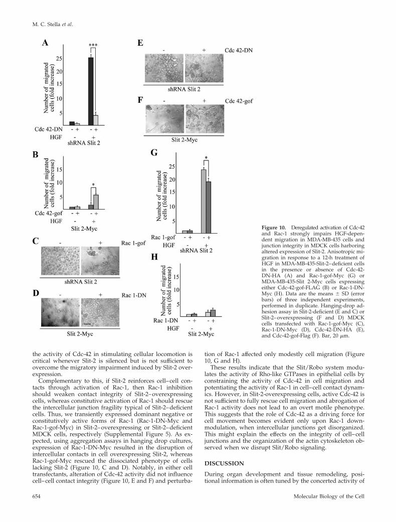
the activity of Cdc-42 in stimulating cellular locomotion iscritical whenever Slit-2 is silenced but is not sufficient toovercome the migratory impairment induced by Slit-2 over-expression.
Complementary to this, if Slit-2 reinforces cell–cell con-tacts through activation of Rac-1, then Rac-1 inhibitionshould weaken contact integrity of Slit-2–overexpressingcells, whereas constitutive activation of Rac-1 should rescuethe intercellular junction fragility typical of Slit-2–deficientcells. Thus, we transiently expressed dominant negative orconstitutively active forms of Rac-1 (Rac-1-DN-Myc andRac-1-gof-Myc) in Slit-2–overexpressing or Slit-2–deficientMDCK cells, respectively (Supplemental Figure 5). As ex-pected, using aggregation assays in hanging drop cultures,expression of Rac-1-DN-Myc resulted in the disruption ofintercellular contacts in cell overexpressing Slit-2, whereasRac-1-gof-Myc rescued the dissociated phenotype of cellslacking Slit-2 (Figure 10, C and D). Notably, in either celltransfectants, alteration of Cdc-42 activity did not influencecell–cell contact integrity (Figure 10, E and F) and perturba-
tion of Rac-1 affected only modestly cell migration (Figure10, G and H).
These results indicate that the Slit/Robo system modu-lates the activity of Rho-like GTPases in epithelial cells byconstraining the activity of Cdc-42 in cell migration andpotentiating the activity of Rac-1 in cell–cell contact dynam-ics. However, in Slit-2-overexpressing cells, active Cdc-42 isnot sufficient to fully rescue cell migration and abrogation ofRac-1 activity does not lead to an overt motile phenotype.This suggests that the role of Cdc-42 as a driving force forcell movement becomes evident only upon Rac-1 down-modulation, when intercellular junctions get disorganized.This might explain the effects on the integrity of cell–celljunctions and the organization of the actin cytoskeleton ob-served when we disrupt Slit/Robo signaling.
DISCUSSION
During organ development and tissue remodeling, posi-tional information is often tuned by the concerted activity of
Figure 10. Deregulated activation of Cdc-42and Rac-1 strongly impairs HGF-depen-dent migration in MDA-MB-435 cells andjunction integrity in MDCK cells harboringaltered expression of Slit-2. Anisotropic mi-gration in response to a 12-h treatment ofHGF in MDA-MB-435-Slit-2–deficient cellsin the presence or absence of Cdc-42-DN-HA (A) and Rac-1-gof-Myc (G) orMDA-MB-435-Slit 2-Myc cells expressingeither Cdc-42-gof-FLAG (B) or Rac-1-DN-Myc (H). Data are the means � SD (errorbars) of three independent experiments,performed in duplicate. Hanging-drop ad-hesion assay in Slit-2-deficient (E and C) orSlit-2–overexpressing (F and D) MDCKcells transfected with Rac-1-gof-Myc (C),Rac-1-DN-Myc (D), Cdc-42-DN-HA (E),and Cdc-42-gof-Flag (F). Bar, 20 �m.
M. C. Stella et al.
Molecular Biology of the Cell654
paracrine stimulatory cues, released distally, and autocrineinhibitory morphogens, which act locally to restrain migra-tion toward the external attractant source. In this article, weshow that immortalized, nontransformed epithelial cells andcancerous cell lines empower the Slit-2/Robo-1 signalingsystem to mitigate HGF-dependent migration, matrix inva-sion, and branching morphogenesis. Our results are in con-cordance with previous studies demonstrating that the Slit/Robo system is required for the spatial constraint and theproper obliteration of supernumerary structures during devel-opment, including the bronchial tree and the ureteric buds(Xian et al., 2001; Grieshammer et al., 2004).
The positional restriction exerted by the Slit/Robo systemloop might have important implications as a safeguardmechanism during cancer invasion and metastasis. Indeed,our results suggest that the xenophilic propensity of malig-nant cells could be negatively regulated, or at least spatiallymodulated, by the endogenous activity of the Slit/Robosystem. This line of thinking fits with the observations thatSlit-2 inhibits CXCL12-induced chemotaxis in breast carci-noma cells (Prasad et al., 2004) and that endogenous levels ofCXCL12 antagonize the repellent effects of Slit/Robo signal-ing during retinal axon pathfinding in zebrafish (Chalasaniet al., 2007). Moreover, the Slit genes frequently undergoepigenetic inactivation in a vast number of human cancers(Dallol et al., 2002, 2003a,b; Astuti et al., 2004; Dickinson et al.,2004). It is tempting to speculate that, during the evolution-ary trajectory of an incipient cancer cell, Slit silencing pro-vides a selective advantage for neoplastic progressionthrough disengagement of anti-invasive cues. This activity islikely to be corroborated by concomitant stimulation of cellaccretion, because Slit 2 has been demonstrated to exertantimitotic and proapoptotic effects (Dallol et al., 2002,2003b); indeed, we also found that Slit 2 down-regulationpartially enhances, whereas Slit 2 overexpression decreases,the viability of the cell lines tested in this study, although todifferent extents (Supplemental Figure 3).
The notion that the Slit genes might hinder full implemen-tation of the malignant phenotype is in contrast with thefinding that tumors expressing Slit-2 can attract endothelialcells expressing Robo-1, thus inducing angiogenesis andfavoring cancer growth (Wang et al., 2003). In the absence ofanimal models in which to investigate the heterotypic com-munications between tumor, endothelial, and stromal cellson conditional backgrounds of Slit and Robo expression, it isdifficult to assess the relative contribution of Slit as a repel-lent for the tumor and an attractant for endothelial cells.However, one could argue that the net effect of Slit activitywithin the tumor likely results from the reciprocal stoichi-ometry of autocrine versus paracrine stimulation, the ex-pression levels of the Robo receptors in the different celltypes, and the synergistic or antagonistic interactions withother morphogens such as ephrins, semaphorins, and angio-genic factors (Bissell and Radisky, 2001).
The anti-migratory activity of Slit-2 derives, at leastpartially, from the inhibition of actin-based protrusiveforces. Indeed, when Slit-2 is down-regulated, HGF stim-ulation results in the production of a copious amount offilopodia at the leading edge of cells as a prelude forefficient locomotion (Figure 9A). In response to HGF, thisenhanced filopodia formation in the absence of Slit/Robosignaling is accompanied by an increased activity of Cdc-42, whereas Slit-2 overexpression greatly reduces HGF-dependent activation of Cdc-42 (Figure 9, B and D). Con-sistent with these data, it has been demonstrated that therepelling activity of Slit during neuronal migration re-quires the interaction between the intracellular domain of
Robo-1 and a novel family of Rho GTPase-activating pro-teins (srGAPs) and that this interaction specifically inhib-its Cdc-42 (Wong et al., 2001). Similarly, it has been reportedthat Slit-2–dependent inhibition of medulloblastoma cell inva-sion goes along with Cdc-42 down-regulation (Werbowetski-Ogilvie et al., 2006).
Besides impairing actin-rich motility structures, Slit-2 alsopotentiates the adhesive strength of cadherin-mediated in-tercellular contacts, making multicellular colonies less proneto cell–cell dissociation and scattering. This activity is pos-sibly mediated by Slit-induced upregulation of Rac-1, as weobserved by genetic manipulation of Slit-2 expression levels(Figure 9, C and E, and Fig. 10, C and D). Accordingly, it hasbeen shown that in both mammalian cells and Drosophilaembryos Slit activates Rac-1 by recruiting the guanine-nu-cleotide exchange factor (GEF) Sos to Robo (Fan et al., 2003;Yang and Bashaw, 2006).
Although the general role of Rac-1 in the maturation andstabilization of intercellular contacts is well established, itsfunction in HGF-dependent cell motility is, at first sight,contradictory: on the one hand, overexpression of the Rac-specific GEF Tiam-1 inhibits HGF-induced scattering by in-creasing E-cadherin–mediated cell–cell adhesion and promot-ing actin polymerization at cell–cell contacts (Hordijk et al.,1997); on the other hand, HGF induces lamellipodia formationat the cell’s leading edges and cell locomotion by means ofRac-1 activation (Potempa and Ridley, 1998). One hypothesisfor this apparent discrepancy, also favored by others (Priceand Collard, 2001), is that the pre-existing threshold and thesubcellular compartmentalization of Rac-1 activity, as wellas the balance between cell–cell and cell–matrix adhesion,could dictate the biological outcome in response to HGF. IfRac-1 is basally very active at cell–cell contacts (a conditionthat occurs in the presence of elevated levels of Tiam-1 andin the inner part of compact epithelial colonies, where cell–cell interactions prevail over cell–matrix adhesions), thenintercellular junctions display a robust mechanical strengththat HGF is unable to weaken; conversely, in the presence ofa low and diffuse Rac-1 activity (for example, at the periph-ery of quiescent epithelial colonies, where cell–substrateadhesion dynamics dominate over cell–cell interactions),HGF triggers a Rac-1–dependent focalization of actin poly-merization at the free margins of the cells, which start de-veloping lamellipodia protrusions as a prerequisite for cellmigration. We postulate that the ability of Slit-2 to augmentthe basal activity of Rac-1, which cannot be substantiallysuperinduced by HGF stimulation (Figure 9E), shifts theequilibrium toward stabilization of intercellular contacts.This is in line with the observation that overexpression ofactivated Rac-1 alone (Ridley et al., 1995) is not sufficient torecapitulate HGF-induced cell scattering, possibly becauseof a more efficient activity on the stabilization of intercellularcontacts.
In conclusion, our findings reinforce the idea of funda-mental conservation of directional mechanisms for all so-matic cells (Wu et al., 2001; Rao et al., 2002) and support thenotion that the Slits and the Robos act as general guidancecues, tipping the balance between stimulatory and inhibi-tory morphogen gradients toward cellular repulsion. Thiscondition, in an autocrine context, leads to the induction ofa stationary phenotype which precludes cells from migrat-ing toward motogenic stimuli. It will be interesting to ana-lyze whether such positional constraint will impact tumormetastatization by using in vivo models of cancer progres-sion, and whether the frequently observed epigenetic silenc-ing of Slit-2 will be predictive of bad prognosis and tumordissemination in human malignancies.
Slit Limits HGF-dependent Invasion
Vol. 20, January 15, 2009 655

ACKNOWLEDGMENTS
We thank Carla Boccaccio, Andrea Bertotti, and Asha Balakrishnan for criticalreading of the manuscript; Damion Milne and Giorgio Giardina for imageanalysis; the Division of Molecular Angiogenesis for help and collaboration;Raffaella Albano, Laura Palmas and Lara Fontani for technical assistance; andAntonella Cignetto for secretarial assistance. This work was supported byAssociazione Italiana per la Ricerca sul Cancro grants (to P.M.C. and L.T.) anda Ministero dell’Istruzione, dell’Universita e della Ricerca (PRIN 2004) grant(to L.T.).
REFERENCES
Astuti, D., et al. (2004). SLIT2 promoter methylation analysis in neuroblas-toma, Wilms’ tumour and renal cell carcinoma. Br. J. Cancer 90, 515–521.
Birchmeier, C., and Gherardi, E. (1998). Developmental roles of HGF/SF andits receptor, the c-Met tyrosine kinase. Trends Cell Biol. 8, 404–410.
Birchmeier, C., Birchmeier, W., Gherardi, E., and Vande Woude, G. F.(2003). Met, metastasis, motility and more. Nat. Rev. Mol. Cell. Biol. 4,915–925.
Bissell, M. J., and Radisky, D. (2001). Putting tumours in context. Nat. Rev.Cancer 1, 46–54.
Burbelo, P. D., Drechsel, D., and Hall, A. (1995). A conserved binding motifdefines numerous candidate target proteins for both Cdc42 and Rac GTPases.J. Biol. Chem. 270, 29071–29074.
Chalasani, S. H., Sabol, A., Xu, H., Gyda, M. A., Rasband, K., Granato, M.,Chien, C. B., and Raper, J. A. (2007). Stromal cell-derived factor-1 antagonizesslit/robo signaling in vivo. J. Neurosci. 27, 973–980.
Brummelkamp et al. (2002). A system for stable expression of short interferingRNAs in mammalian cells. Science 296, 550–553
Comoglio, P. M., and Trusolino, L. (2002). Invasive growth: from develop-ment to metastasis. J. Clin. Invest. 109, 857–862.
Dallol, A., Da Silva, N. F., Viacava, P., Minna, J. D., Bieche, I., Maher, E. R.,and Latif, F. (2002). SLIT2, a human homologue of the Drosophila Slit2 gene,has tumor suppressor activity and is frequently inactivated in lung and breastcancers. Cancer Res. 62, 5874–5880.
Dallol, A., Krex, D., Hesson, L., Eng, C., Maher, E. R., and Latif, F. (2003a).Frequent epigenetic inactivation of the SLIT2 gene in gliomas. Oncogene 22,4611–4616.
Dallol, A., Morton, D., Maher, E. R., and Latif, F. (2003b). SLIT2 axon guidancemolecule is frequently inactivated in colorectal cancer and suppresses growthof colorectal carcinoma cells. Cancer Res. 63, 1054–1058.
Dickinson, R. E., Dallol, A., Bieche, I., Krex, D., Morton, D., Maher, E. R., andLatif, F. (2004). Epigenetic inactivation of SLIT3 and SLIT1 genes in humancancers. Br. J. Cancer 91, 2071–2078.
Dickson, B. J., and Gilestro, G. (2006). Regulation of commissural axonpathfinding by slit and its robo receptors. Annu. Rev. Cell Dev. Biol. 22,651– 675.
DiFeo, A., Narla, G., Camacho-Vanegas, O., Nishio, H., Rose, S. L., Buller,R. E., Friedman, S. L., Walsh, M. J., and Martignetti, J. A. (2006). E-cadherin isa novel transcriptional target of the KLF6 tumor suppressor. Oncogene 25,6026–6031.
Esko, J. D., Weinke, J. L., Taylor, W. H., Ekborg, G., Roden, L., Ananthara-maiah, and G., and Gawish, A. (1987). Inhibition of chondroitin and heparansulfate biosynthesis in Chinese hamster ovary cell mutants defective in galac-tosyltransferase I. J. Biol. Chem. 262, 12189–12195.
Fan, X., Labrador, J. P., Hing, H., and Bashaw, G. J. (2003). Slit stimulationrecruits Dock and Pak to the roundabout receptor and increases Rac activityto regulate axon repulsion at the CNS midline. Neuron 40, 113–127.
Grieshammer, U., Le Ma, Plump, A. S., Wang, F., Tessier-Lavigne, M., andMartin, G. R. (2004). SLIT2-mediated ROBO2 signaling restricts kidney induc-tion to a single site. Dev. Cell 6, 709–717.
Hordijk, P. L., ten Klooster, J. P., van der Kammen, R. A., Michiels, F., Oomen,L. C., and Collard, J. G. (1997). Inhibition of invasion of epithelial cells byTiam1-Rac signaling. Science 278, 1464–1466.
Huber, M. A., Kraut, N., and Beug, H. (2005). Molecular requirements forepithelial-mesenchymal transition during tumor progression. Curr. Opin. CellBiol. 17, 548–558.
Hussain, S. A., et al. (2006). A molecular mechanism for the heparan sulfatedependence of slit-robo signaling. J. Biol. Chem. 281, 39693–39698.
Koch, A., Mancini, A., El Bounkari, O., and Tamura, T. (2005). The SH2-domian-containing inositol 5-phosphatase (SHIP)-2 binds to c-Met directly
via tyrosine residue 1356 and involves hepatocyte growth factor (HGF)-induced lamellipodium formation, cell scattering and cell spreading. Onco-gene 24, 3436–3447.
Lecuit, T., and Lenne, P. F. (2007). Cell surface mechanics and the control ofcell shape, tissue patterns and morphogenesis. Nat. Rev. Mol. Cell Biol. 8,633–644.
Liang, Y., Annan, R. S., Carr, S. A., Popp, S., Mevissen, M., Margolis, R. K.,and Margolis, R. U. (1999). Mammalian homologues of the Drosophila slitprotein are ligands of the heparan sulfate proteoglycan glypican-1 in brain.J. Biol. Chem. 274, 17885–17892.
Maina, F., and Klein, R. (1999). Hepatocyte growth factor, a versatile signal fordeveloping neurons. Nat. Neurosci. 2, 213–217.
Michieli, P., Mazzone, M., Basilico, C., Cavassa, S., Sottile, A., Naldini, L., andComoglio, P. M. (2004). Targeting the tumor and its microenvironment by adual-function decoy Met receptor. Cancer Cell 6, 61–73.
Montesano, R., Schaller, G., and Orci, L. (1991). Induction of epithelialtubular morphogenesis in vitro by fibroblast-derived soluble factors. Cell66, 697–711.
Naldini, L., Vigna, E., Bardelli, A., Follenzi, F., Galimi, F., and Comoglio,P. M. (1995). Biological activation of pro-HGF (hepatocyte growth factor)by urokinase is controlled by a stoichiometric reaction. J. Biol. Chem. 270,603– 611.
Nishimura, M., Saito, T., Yamasaki, H., and Kudo, R. (2003). Suppression ofgap junctional intercellular communication via 5� CpG island methylation inpromoter region of E-cadherin gene in endometrial cancer cells. Carcinogen-esis 24, 1615–1623.
Nobes, C. D., and Hall, A. (1995). Rho, rac, and cdc42 GTPases regulate theassembly of multimolecular focal complexes associated with actin stressfibers, lamellipodia, and filopodia. Cell 81, 53–62.
Potempa, S., and Ridley, A. J. (1998). Activation of both MAP kinase andphosphatidylinositide 3-kinase by Ras is required for hepatocyte growthfactor/scatter factor-induced adherens junction disassembly. Mol. Biol. Cell 9,2185–2200.
Prasad, A., Fernandis, A. Z., Rao, Y., and Ganju, R. K. (2004). Slit protein-mediated inhibition of CXCR4-induced chemotactic and chemoinvasivesignaling pathways in breast cancer cells. J. Biol. Chem. 279, 9115–9124.
Price, L. S., and Collard, J. C. (2001). Regulation of the cytoskeleton byRho-like GTPases: implications for tumour cell invasion. Semin. Cancer Biol.11, 167–173.
Rao, Y., Wong, K., Ward, M., Jurgensen, C., and Wu, J. Y. (2002). Neuronalmigration and molecular conservation with leukocyte chemotaxis. Genes Dev.16, 2973–2984.
Ridley, A. J., Comoglio, P. M., and Hall, A. (1995). Regulation of scatterfactor/hepatocyte growth factor responses by Ras, Rac, and Rho in MDCKcells. Mol. Cell. Biol. 15, 1110–1122.
Ronca, F., Andersen, J. S., Paech, V., and Margolis, R. U. (2001). Character-ization of Slit protein interactions with glypican-1. J. Biol. Chem. 276, 29141–29147.
Sahai, E., and Marshall, C. J. (2002). RHO-GTPases and cancer. Nat. Rev.Cancer 2, 133–142.
Sander, E. E., van Delft, S., ten Klooster, J. P., Reid, T., van der Kammen,R. A., Michiels, F., and Collard, J. G. (1998). Matrix-dependent Tiam1/Racsignaling in epithelial cells promotes either cell-cell adhesion or cell mi-gration and is regulated by phosphatidylinositol 3-kinase. J. Cell Biol. 143,1385–1398.
Stella, M. C., Trusolino, L., Pennacchietti, S., and Comoglio, P. M. (2005).Negative feedback regulation of Met-dependent invasive growth by Notch.Mol. Cell. Biol. 25, 3982–3996.
Stoker, M., Gherardi, E., Perryman, M., and Gray, J. (1987). Scatter factor is afibroblast-derived modulator of epithelial cell mobility. Nature 327, 239–242.
Thoreson, M. A., Anastasiadis, P. Z., Daniel, J. M., Ireton, R. C., Wheelock,M. J., Johnson, K. R., Hummigbird, D. K., and Reynolds, A. B. (2000). Selectiveuncoupling of p120(ctn) from E-cadherin disrupts strong adhesion. J. CellBiol. 148, 189–202.
Trusolino, L., Bertotti, A., and Comoglio, P. M. (2001). A signaling adapterfunction for �6�4 integrin in the control of HGF-dependent invasive growth.Cell 107, 643–654.
Wang, B., et al. (2003). Induction of tumor angiogenesis by Slit-Robo signalingand inhibition of cancer growth by blocking Robo activity. Cancer Cell 4,19–29.
Werbowetski-Ogilvie, T. E., et al. (2006). Inhibition of medulloblastoma cellinvasion by Slit. Oncogene 25, 5103–5112.
M. C. Stella et al.
Molecular Biology of the Cell656

Wong, K., et al. (2001). Signal transduction in neuronal migration: roles ofGTPase activating proteins and the small GTPase Cdc42 in the Slit-Robopathway. Cell 107, 209–221.
Wong, K., Park, H. T., Wu, J. Y., and Rao, Y. (2002). Slit proteins: molecularguidance cues for cells ranging from neurons to leukocytes. Curr. Opin. GenetDev. 12, 583–591.
Wu, W., Wong, K., Chen, J. H., Jiang, Z. H., Dupuis, S. M., Wu, J. Y., and Rao,Y. (1999). Directional guidance of neuronal migration in the olfactory systemby the protein Slit. Nature 400, 331–336.
Wu, J. Y., Feng, L. L., Park, H. T., Havlioglu, N., Wen, L., Tang, H., Bacon, K. B.,Jiang, Z. H., Zhang, X. C., and Rao, Y. (2001). The neuronal repellent Slit inhibitsleukocyte chemotaxis induced by chemotactic factors. Nature 410, 948–952.
Xian, J., Clark, K. J., Fordham, R., Pannell, R., Rabbitts, T. H., and Rabbitts,P. H. (2001). Inadequate lung development and bronchial hyperplasia in micewith a targeted deletion in the Dutt1/Robo1 gene. Proc. Natl. Acad. Sci. USA98, 15062–15066.
Yang, L., and Bashaw, G. J. (2006). Son of sevenless directly links the Roboreceptor to rac activation to control axon repulsion at the midline. Neuron 52,595–607.
Slit Limits HGF-dependent Invasion
Vol. 20, January 15, 2009 657
Related Documents