Introduction to the Skeletal System

The skeletal system includes: The skeletal system includes: bones of the skeleton bones of the skeleton cartilages, ligaments and other stabilizing connective.
Dec 25, 2015
Welcome message from author
This document is posted to help you gain knowledge. Please leave a comment to let me know what you think about it! Share it to your friends and learn new things together.
Transcript

Introduction to the Skeletal System

Parts of the Skeletal SystemThe skeletal system includes:bones of the skeletoncartilages, ligaments and other stabilizing
connective tissues

Primary Functions
Support
Storage of minerals* and
lipids-Yellow Marrow
*calcium & phosphorus
Blood cell productionRed Marrow
Protection
Leverage

Types Of BonesLong
Flat
Sutural
Irregular
Short
Sesamoid

Long Bone StructureEpiphysis
Metaphysis
Diaphysis
Spongy Bone
Marrow Cavity
Cortex

Cells of BoneOsteocytes
Osteoblasts
Osteoclasts

Anatomy of Bone
Osseous tissue is supportive connective tissue containing specialized cells.
The matrix of bone tissue is solid because of the calcium salts deposited around protein fibers
The 4 characteristics of bone tissue are:1. Dense matrix containing deposits of calcium salts2. The matrix contains osteocytes in pockets organized
around blood vessels.3. Passageways form pathways for blood vessels to
exchange nutrients and wastes.4. Outer surfaces of bones are covered by periosteum
unless hyaline cartilage is present

Cellular Make-up About 1/3 of bone is protein
fibers (collagen).
Bone cells are only about 2% of bone mass.
Bone contains 4 types of cells:1. Osteocytes2. Osteoblasts3. Osteoprogenitor cells4. Osteoclasts

OsteocytesOsteocytes are mature bone cells that
maintain the bone matrix.
Each osteocyte lives in a lacuna (a pocket) between layers (lamellae) of matrix.
Canaliculi (narrow passageways) through the lamellae allow osteocytes to connect.
Osteocytes do not divide.
The main functions of osteocytes are:1. To maintain the protein and mineral
content of the matrix.2. To help repair damaged bone

Osteoblasts Osteoblasts are immature bone cells that
secrete the matrix by the process of osteogenesis (secretion of proteins and other inorganic compounds of the matrix).
Before calcium salts are deposited (forming bone), the matrix is called osteoid.
When osteoblasts are surrounded by bone, they become osteocytes.
Osteoprogenitor cells are stem cells that divide to produce osteoblasts.
Located in the inner, cellular layer of periosteum.
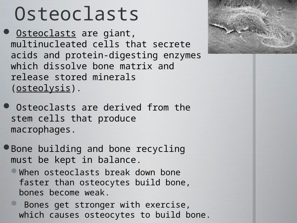
Osteoclasts Osteoclasts are giant, multinucleated
cells that secrete acids and protein-digesting enzymes which dissolve bone matrix and release stored minerals (osteolysis).
Osteoclasts are derived from the stem cells that produce macrophages.
Bone building and bone recycling must be kept in balance. When osteoclasts break down bone
faster than osteocytes build bone, bones become weak.
Bones get stronger with exercise, which causes osteocytes to build bone.

Structure of Compact Bone
The basic unit: osteocytes are arranged in circles around a central canal containing blood vessels.
Perpendicular to the central canal are perforating canals which carry blood vessels deep into the bone and bone marrow.
All osteons in long bones run the length of the bone, strengthening the bone in that direction.
A layer wraps around the circumference of the long bone and binds all together.

Structure of Spongy BoneSpongy bone matrix
forms an open network of trabeculae.
The space between trabeculae is filled with another tissue, red bone marrow, which has blood vessels and supplies nutrients to the osteocytes.
Red bone marrow is red because it forms red blood cells.
In other bones, spongy bone may hold yellow bone marrow, which is yellow because it stores fat. =>
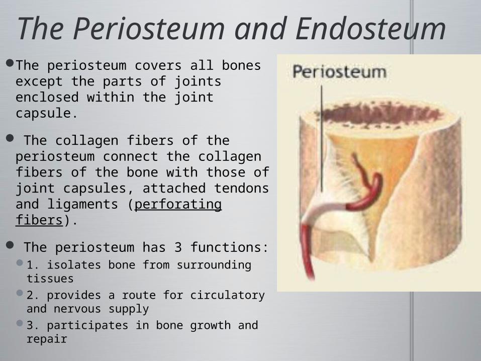
The Periosteum and Endosteum
The periosteum covers all bones except the parts of joints enclosed within the joint capsule.
The collagen fibers of the periosteum connect the collagen fibers of the bone with those of joint capsules, attached tendons and ligaments (perforating fibers).
The periosteum has 3 functions:1. isolates bone from surrounding
tissues2. provides a route for circulatory
and nervous supply3. participates in bone growth and
repair

The Periosteum and Endosteum
Endosteum contains osteoblasts, osteoprogenitor cells and osteoclasts, and is active in bone growth and repair.


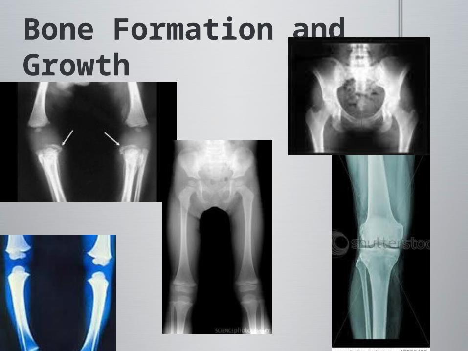
Bone Formation and Growth Human bones grow until about age 25. The process of replacing other tissues with bone is called ossification. The process of depositing calcium salts (calcification) occurs during ossification and in other tissues.
The 2 main forms of ossification1. intramembranous ossification2. endochondral ossification
Bone Formation and Growth

Intramembranous Ossification
Intramembranous ossification, also called dermal ossification because it occurs in the dermis, produces dermal bones such as the mandible and clavicle.
There are 3 main steps in intramembranous ossification:1. Stem cells collect in the area; differentiate into osteoblasts,
and begin ossification. The location where ossification begins is the ossification center, from which developing bone grows out in projections.
2. Blood vessels grow into the area to supply the osteoblasts. Projections connect, trapping blood vessels inside bone.
3. Spongy bone develops, which can be remodeled into osteons of compact bone, periosteum or marrow cavities.
Click icon to add picture
Endochondral OssificationMost bones originate as hyaline cartilage which becomes
ossified through the process of endochondral ossification.
Growth and ossification of a long bone occurs in 6 steps1. Chondrocytes in the center of the hyaline cartilage enlarge,
form struts which begin to calcify, then die, leaving cavities in the cartilage..
2. Blood vessels grow around the edges of the cartilage. Osteoblasts produce a layer of superficial bone around the shaft which will continue to grow and become compact bone.
3.Increased blood flow brings fibroblasts that become osteoblasts.
4. Remodeling creates a marrow cavity. Bone replaces cartilage at the metaphyses.
5. Capillaries and osteoblasts enter the epiphyses.6. The epiphyses fill with spongy bone. The cartilage
remaining within the joint cavity is the articulation cartilage. The cartilage at the metaphysis is the epiphyseal cartilage.


GrowthWhen the long bone stops growing, after
puberty, the epiphyseal cartilage disappears -- but its location is visible on X-rays as an epiphyseal line.
Appositional Growth: The growth of compact bone on the surface
(periosteum) of the bone, continues to thicken and strengthen the long bone with layers.

Proteus SyndromeProteus syndrome is a rare
overgrowth condition that can change the appearance and growth rate of various body parts.
This overgrowth is also typically asymmetric.
The word “Proteus” comes from the name of the ancient Greek god of change; this name was chosen because the overgrowth in Proteus syndrome can cause changes in the shapes of body structures over time

RemodelingThe adult skeleton must
maintain itself and replace mineral reserves. The process that recycles and renews bone matrix is remodeling.
Bone remodeling involves osteocytes, osteoblasts and osteoclasts.
Bone is continually remodeled, recycled and replaced. The rate of turnover varies. When deposition is greater than removal, bones get stronger. When removal is faster than replacement, bones get weaker.
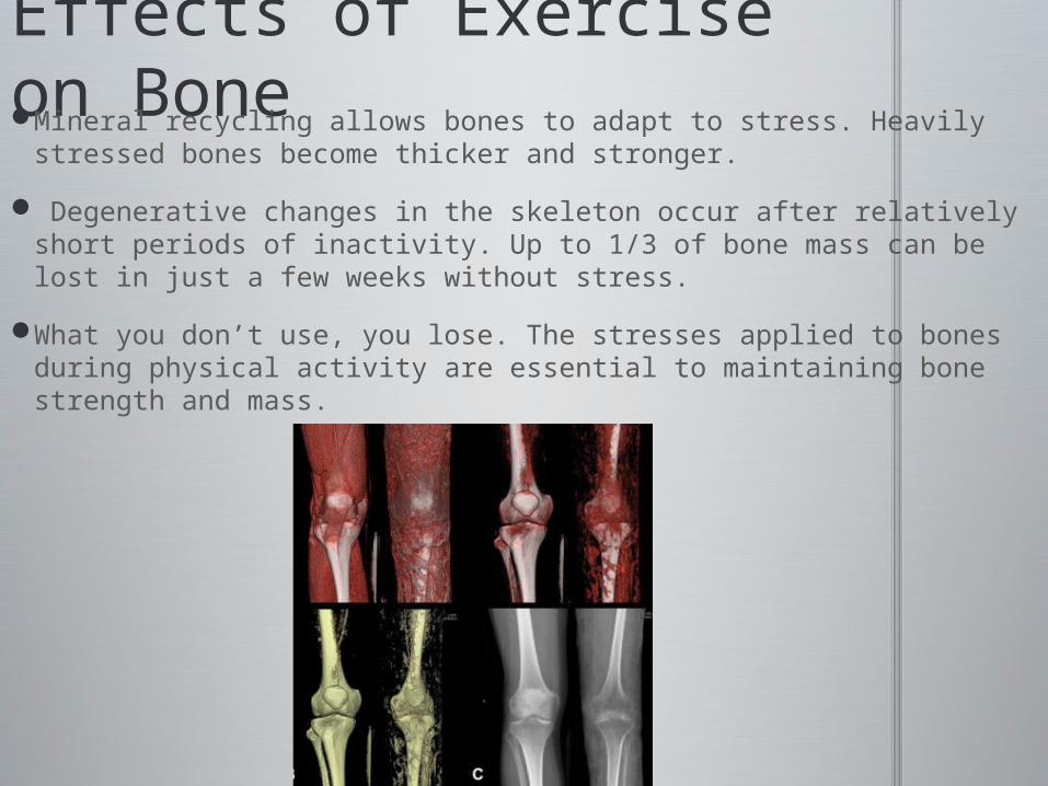
Effects of Exercise on BoneMineral recycling allows bones to adapt to stress. Heavily stressed
bones become thicker and stronger.
Degenerative changes in the skeleton occur after relatively short periods of inactivity. Up to 1/3 of bone mass can be lost in just a few weeks without stress.
What you don’t use, you lose. The stresses applied to bones during physical activity are essential to maintaining bone strength and mass.
Nutritional Effects on Bone1. A dietary source of calcium and phosphate salts, plus small amounts of magnesium, fluoride, iron and manganese.
2. The hormone calcitriol, made in the kidneys, is essential to proper absorption of calcium and phosphorus by the digestive tract. Calcitriol synthesis requires vitamin D3.
3. Vitamins: Vitamin C is required for collagen synthesis, and stimulates osteoblast differentiation. Vitamin A stimulates osteoblast activity. Vitamins K and B12 help synthesize bone proteins.

Hormonal Effects on Bone
4. Growth hormone and thyroxine stimulate bone growth.
5. Sex hormones: Estrogens and androgens stimulate osteoblasts for bone growth.
6. Other hormones regulate calcium and phosphate levels in body fluids.

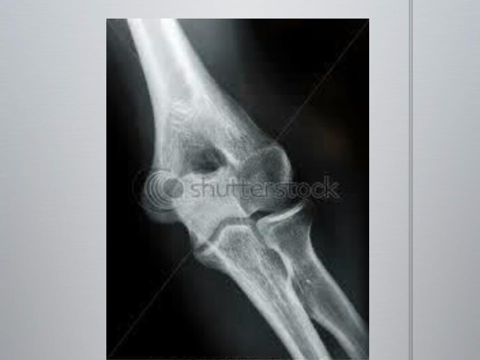
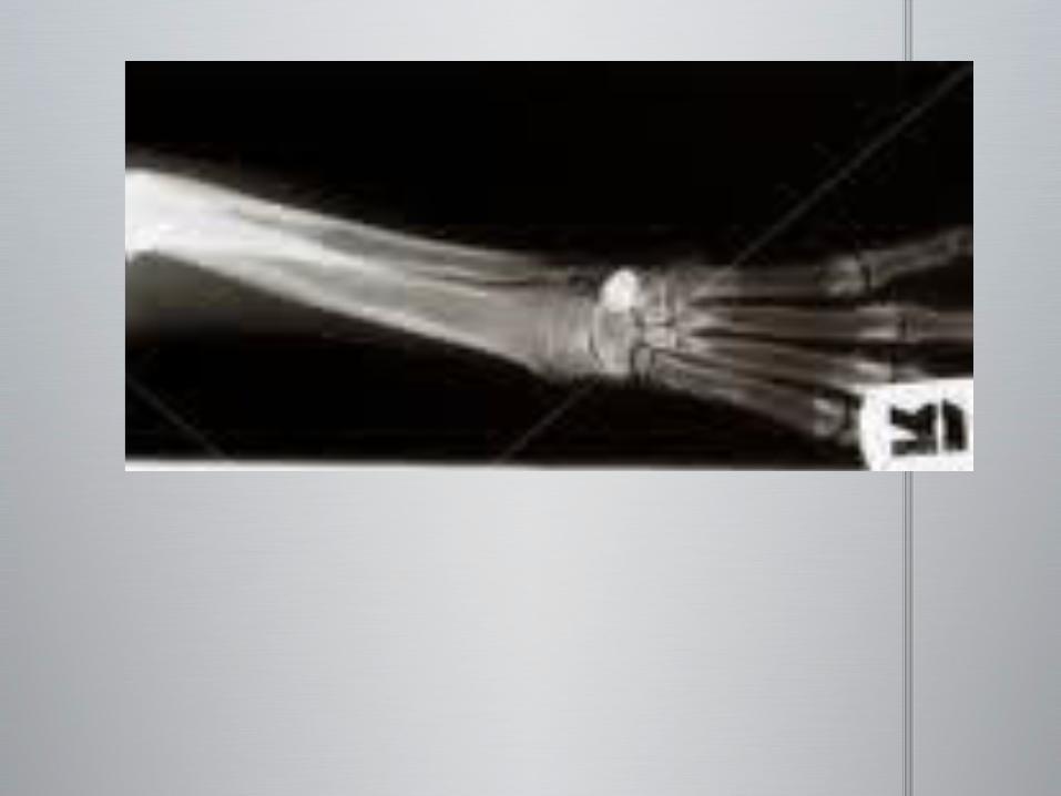
Fracture TypesSpiral
Comminuted
Compound
Stress
Longitudinal (linear)
Transverse
Oblique
Epiphyseal
Greenstick
Impacted

SpiralCreates a spiral
effect or S shape

ComminutedMore than two pieces
Often displaced
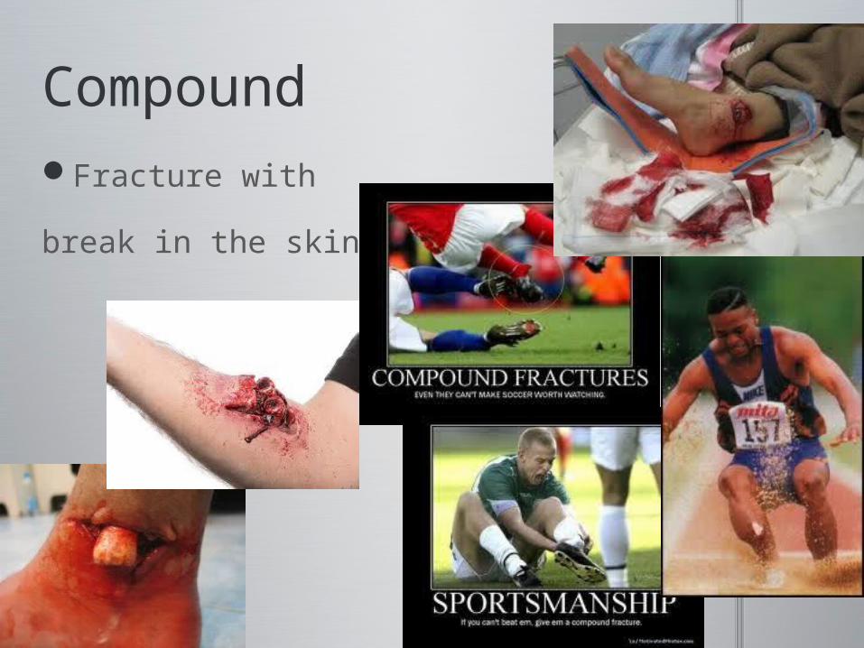
CompoundFracture with
break in the skin

StressMicrofractures
Occur over time

LongitudinalParallel to the shaft of the bone
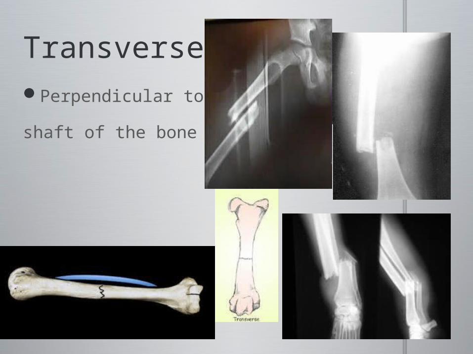
TransversePerpendicular to the
shaft of the bone

ObliqueDiagonal to the shaft of the
bone

EpiphysealThrough of across the
growth plate

GreenstickForce to one side splits
bone on opposite side

ImpactedBone is shortening in a crush-type
fracture
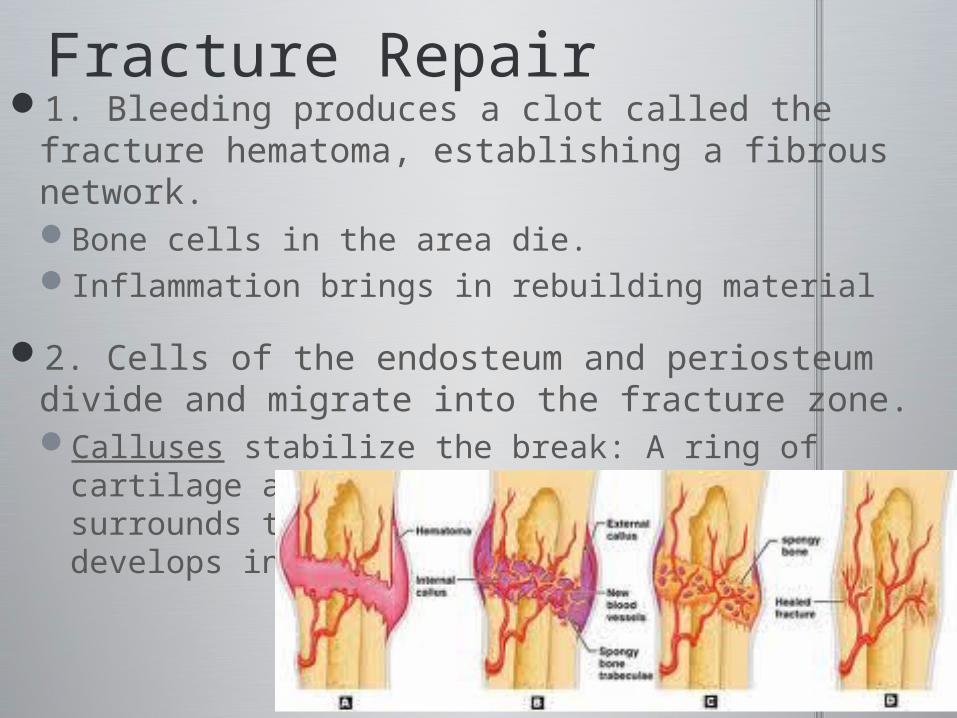
Fracture Repair1. Bleeding produces a clot called the fracture
hematoma, establishing a fibrous network. Bone cells in the area die.Inflammation brings in rebuilding material
2. Cells of the endosteum and periosteum divide and migrate into the fracture zone. Calluses stabilize the break: A ring of cartilage and
bone (external callus) surrounds the break, and an internal callus develops in the marrow cavity.

Fracture Repair3. New blood vessels are formed. Osteoblasts
replace the central cartilage of the external callus with spongy bone.
4. Osteoblasts and osteocytes continue to remodel the fracture for up to a year, reducing the bone calluses. Collagen and calcium are deposited.


Skeleton as a Calcium Reserve
Our bones are storage areas for many metabolically active minerals, particularly calcium.
Calcium (stored in bones) is the most abundant mineral in the body.
Calcium ions are important to membranes and to the intracellular activities of neurons and muscle cells, especially heart cells.
Calcium ion concentrations in body fluids must be closely regulated.
Calcium ion homeostasis is maintained by the hormones which control calcium ion storage, absorption and excretion.

Calcium and phosphate ions circulating in the blood are constantly being lost in the urine.
These ions must be replaced to maintain homeostasis. If they aren’t obtained from the diet, they will be released from storage in the skeleton, making bones weaker.
Exercise and a diet with plenty of calcium are necessary to keep bones strong.
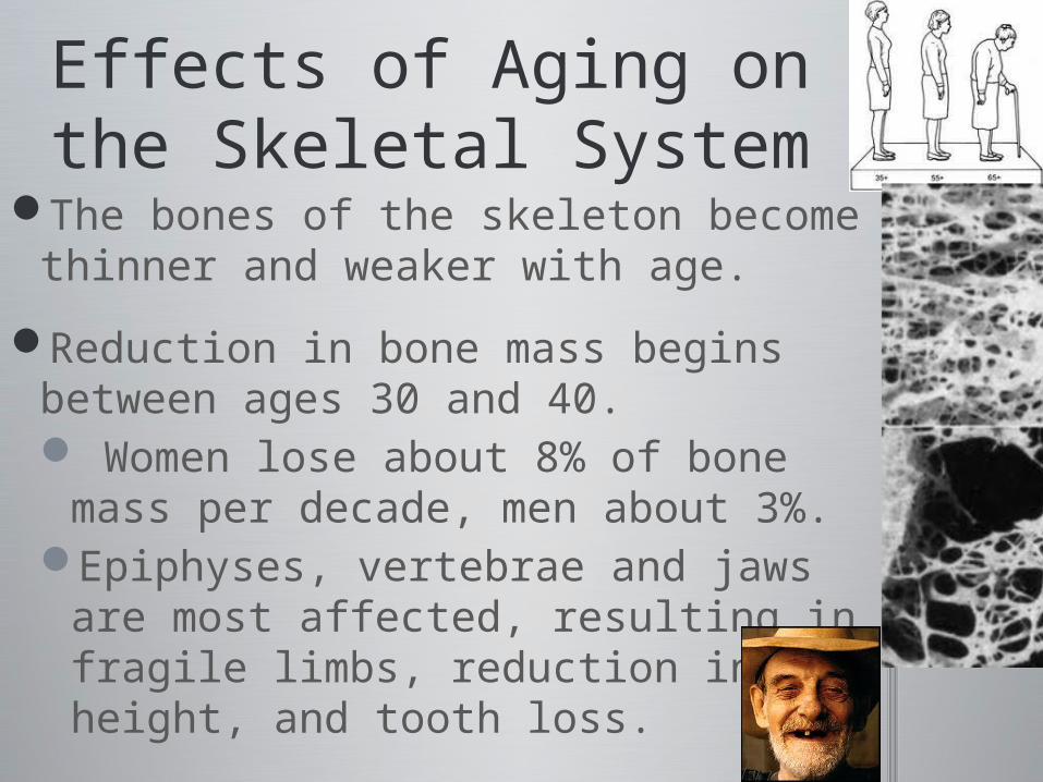
Effects of Aging on the Skeletal System
The bones of the skeleton become thinner and weaker with age.
Reduction in bone mass begins between ages 30 and 40. Women lose about 8% of bone
mass per decade, men about 3%.Epiphyses, vertebrae and jaws
are most affected, resulting in fragile limbs, reduction in height, and tooth loss.

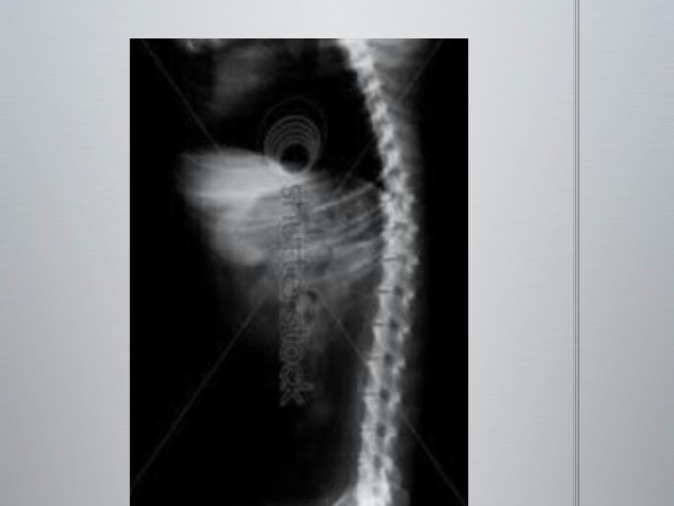
Effects of Aging on the Skeletal System
Osteoporosis is the condition of severe bone loss, extensive enough to impair normal function. Over age 45, 29% of women and 18% of men have osteoporosis.
Estrogens and androgens contribute to maintaining bone mass. Bone loss in women accelerates after menopause.
Cancerous tissues release a chemical (osteoclast-activating factor) that stimulates osteoclasts and produces severe osteoporosis.


Animal or Human


Related Documents