The Skeletal Muscle Pump During Contractile Transitions by Brian Steven Ferguson A dissertation submitted to the Graduate Faculty of Auburn University in partial fulfillment of the requirements for the Degree of Doctor of Philosophy Auburn, Alabama August 2, 2014 Key words: Blood flow, perfusion, exercise, hyperemia, vascular control Copyright 2014 by Brian Steven Ferguson Approved by L. Bruce Gladden, Chair, Professor of Kinesiology David D. Pascoe, Professor of Kinesiology Heidi A. Kluess, Associate Professor of Kinesiology Douglas C. Goodwin, Associate Professor of Chemistry and Biochemistry

Welcome message from author
This document is posted to help you gain knowledge. Please leave a comment to let me know what you think about it! Share it to your friends and learn new things together.
Transcript

The Skeletal Muscle Pump During Contractile Transitions
by
Brian Steven Ferguson
A dissertation submitted to the Graduate Faculty of Auburn University
in partial fulfillment of the requirements for the Degree of
Doctor of Philosophy
Auburn, Alabama August 2, 2014
Key words: Blood flow, perfusion, exercise, hyperemia, vascular control
Copyright 2014 by Brian Steven Ferguson
Approved by
L. Bruce Gladden, Chair, Professor of Kinesiology David D. Pascoe, Professor of Kinesiology
Heidi A. Kluess, Associate Professor of Kinesiology Douglas C. Goodwin, Associate Professor of Chemistry and Biochemistry

Abstract
The aim of this study was to characterize the contribution of skeletal muscle
contraction to the immediate hyperemic blood flow response as well as the continued
involvement in matching tissue perfusion to elevated metabolic rates. There exists a
substantial reserve for increased blood flow within skeletal muscle in response to
dynamic exercise however the interaction of neural regulation, vasoactive metabolites,
and mechanical characteristics are incompletely understood. To address questions
concerning blood flow in response to transitions from rest to various metabolic rates an
isolated canine gastrocnemius in situ model was employed. Seven canines were used for
this investigation with the gastrocnemius muscles isolated for isometric contractions with
tetanic stimulation. Measures were made for blood flow, blood pressure, force, and near
infrared spectrophotometric analyses under conditions of spontaneous blood flow. The
following transitions were investigated: from rest to tetanic contractions of 1/3 s, rest to
2/3 s, rest to 1/1 s and during the transition from 1/3 s to 2/3s all with spontaneous blood
flow response intact. Additionally, an estimation for the blood flow response with no
mechanical contribution from the muscle pump was made with determination for the
kinetics of the estimate. The time constant (tau) for the blood flow response was not
significantly different between the measured flow with contraction (Qwc) and the estimate
with no contraction (Qnc) for the 1/3 s stimulation rate (12.8 ± 5.5 s vs 11.8 ± 3.2 s
respectively), the transition from the high baseline (1/3 s) to a higher rate (2/3HB) (21.2 ±
ii

3.4 s vs 21.7 ± 4.8 s respectively), from rest to 2/3 s (25.6 ± 12.0 s vs 22.1 ± 1.3 s
respectively), or from rest to 1/1 s (16.7 ± 3.0 s vs 22.1 ± 1.3 s respectively). Initially, for
this model, there is a positive contribution to total blood flow provided by the contracting
skeletal muscle, however this diminishes within the first few contractions. At higher
stimulation rates the net effect of the contracting muscle is to limit local blood flow in the
exercising muscle. In conclusion, the muscle pump may contribute to local perfusion at
exercise onset with diminishing returns as rhythmic contractions continue. In the steady
state the main contributions of the muscle pump is to aid in the maintenance of central
hemodynamic
iii

Acknowledgments
I would first like to thank Dr. Bruce Gladden for providing me with the
opportunity to pursue a doctoral degree with his exceptional guidance and mentoring.
Dr. Gladden has been my academic mentor as well as life as an example of how to be a
successful research scientist, teacher, and man. Thank you to my committee members,
Dr. David Pascoe, Dr. Heidi Kluess, and Dr. Douglas Goodwin who have each
contributed to my development as an investigator, student, and teacher during my
graduate studies. Thank you to all of my fellow graduate students but especially my lab
mates Matthew Rogatzki and Yi Sun for contributing your time, effort and input to this
project. Thank you to Dr. Harry Rossiter, Dr. John Kowalchuk, and Dr. Rob W st for
their willingness to contribute time and knowledge to this project. I would like to extend
special thanks to Dr. Nicola Lai from Case Western University for working to develop
the Macro Programs making the contraction by contraction analysis possible for this
study. Finally I would like to thank all of my friends and family that have inspired and
supported me through my long journey to this point with their love and encouragement.
To my mom, dad and grandparents for encouraging me to pursue my dream.
iv

Table of Contents
Abstract ......................................................................................................................................... ii
Acknowledgments........................................................................................................................ iv
List of Tables .............................................................................................................................. vii
List of Figures ............................................................................................................................ viii
List of Abbreviations ................................................................................................................... ix
I. REVIEW OF LITERATURE .................................................................................................. 1
Exercise Hyperemia .......................................................................................................... 3
Potassium .......................................................................................................................... 5
Lactate ............................................................................................................................... 7
Hydrogen Ion .................................................................................................................... 8
Adenosine Triphosphate ................................................................................................... 9
Nitric Oxide and Prostanoids .......................................................................................... 11
Neural Control ................................................................................................................ 13
Acetylcholine Spillover .................................................................................................. 15
Myogenic Control ......................................................................................................... 16
Muscle Pump ................................................................................................................ 17
II. JOURNAL MANUSCRIPT ................................................................................................. 26
Abstract ......................................................................................................................... 26
Introduction ................................................................................................................... 27
v
Methods and procedures ............................................................................................... 34
Animals and Ethical Approval .............................................................................. 34
Surgical Preparation .............................................................................................. 35
Protocol ................................................................................................................. 36
Measurements ....................................................................................................... 37
Calculations and Kinetics Analysis ...................................................................... 38
Blood Flow Estimation ......................................................................................... 39
Statistical Analysis ................................................................................................ 43
Results ........................................................................................................................... 44
Discussion ..................................................................................................................... 60
Limitations ............................................................................................................ 65
Conclusions ........................................................................................................... 67
References ................................................................................................................................. 69
Appendices .................................................................................................................................. 78
Appendix A: Institutional Animal Care and Use Committee Approval .................................... 78
vi

List of Tables
Table 1: Blood gas and blood metabolites ................................................................................. 46
Table 2: Kinetics parameters for measured blood flow ............................................................. 48
Table 3: Kinetics parameters for estimated blood flow ............................................................. 49
vii

List of Figures
Figure 1: Measured blood flow first contraction ....................................................................... 40
Figure 2: Measured blood flow 80th contraction ........................................................................ 41
Figure 3: Estimated blood flow first contraction ....................................................................... 42
Figure 4: Estimated blood flow 80th contraction ....................................................................... 43
Figure 5: 1/3 s Qnc ..................................................................................................................... 50
Figure 6: 1/3 s Qwc ..................................................................................................................... 51
Figure 7: 2/3HB Qnc .................................................................................................................... 52
Figure 8: 2/3HB Qwc .................................................................................................................. 53
Figure 9: 2/3 s Qnc ..................................................................................................................... 54
Figure 10: 2/3 s Qwc ......................................................................................................................................................................... 55
Figure 11: 1/1 s Qnc ................................................................................................................... 56
Figure 12: 1/1 s Qwc ................................................................................................................... 57
Figure 13: 2/3 s muscle pump contribution ............................................................................... 58
Figure 14: 1/1 s muscle pump contribution ............................................................................... 59
viii
List of Abbreviations
Ach……Acetylcholine
NO…….Nitric Oxide
NOS…...Nitric Oxide Synthase
SNP……Sodium Nitroprusside
H+……...Hydrogen Ion
Ca2+ …...Calcium Ion
K+……...Potassium Ion
PKA…...Protein Kinase A
KCa…….Calcium-activated Potassium
Channel
PKG…..Protein Kinase G
PKC…..Protein Kinase C
AA……Arachidonic Acid
L-NMMA.. NG-monomethyl-L-arginine
SNP…..Sodium nitroprusside
Tmax…Maximal Tension
ATP……Adenosine Triphosphate
ADP……Adenosine Diphosphate
AMP……Adenosine Monophosphate
ADO……Adenosine
EDHF…..Endothelial Derived
Hyperpolarizing Factor
VSMC….Vascular Smooth Muscle Cells
cGMP…..Cyclic Guanosine
Monophosphate
PGI2……Prostacyclin
ET……...Endothelin
H2O2…...Hydrogen Peroxide
COX…...Cyclooxygenase Enzymes
L-NAME.. NG-nitro-L-arginine methyl-
ester
Hb……..Hemoglobin
ix

I. REVIEW OF LITERATURE
As eloquently stated by Loring B. Rowell, a historical perspective exposes a chain
of ideas that reveals the ability to build on the findings of others and thus provide that
strength that is the continuity of science (128). Our understanding of the work of our
predecessors guides us down an evolving path of discovery and investigation. The focus
of this investigation will be to discuss those factors which contribute to blood flow
regulation, particularly in exercising skeletal muscle. Blood flow through a skeletal
muscle vascular bed is determined by both the perfusion pressure as well as the vascular
tone. A large fraction of the vascular resistance in skeletal muscle is controlled at the
level of small muscular “feed” arteries which lie external to the muscle and therefore are
not directly exposed to the conditions within the skeletal muscle they perfuse. Upon
entering the muscle further blood flow control is mediated by small arteriolar networks
and finally terminal arterioles. Herein, many of the key regulators in matching systemic
perfusion to metabolic demand will be discussed. The primary aim of this investigation
was to determine muscle pump contribution to the rapid hyperemic response at exercise
onset as well as the ongoing contribution to total blood flow for various contractile
frequencies and transitions which in turn engendered various metabolic rates.
Research physiologists have long sought to define the mechanisms responsible for
precisely matching tissue perfusion to metabolic demand during transitions from rest to
1

dynamic exercise as well as the maintenance of adequate perfusion during steady state
activities. The pioneering work of Sir William Harvey first detailed the structure of the
heart as well as blood flow through the body in De Motu Cordis, 1628 (65). Since his
detailed works further investigations have sought to delineate all of the regulating factors
in blood flow control. Technological improvements as well sophisticated investigative
techniques systematically revealed an increasingly complex system responsible for the
regulation of whole body circulation. By the end of the 17th century Jan Swammerdam
had recorded in his notebooks microscopic descriptions of red blood cells (107), however
the first published descriptions came from the light microscopy work of Anton van
Leeuwenhoek (151). During this time blood transfusion was observed to be capable of
reviving a previously exsanguinated animal of the same species (98). Other investigators
sought to cure human maladies by transfusing blood of docile animals into humans (93).
This practice was soon halted after the death of Antoine Mauroy in 1667 following
transfusion twice with calf’s blood (93). Subsequently, for more than a century there was
little progress made in delineating the factors contributing to the components of blood or
the factors that regulated perfusion. However, near the turn of the 19th century Scottish
physician John Hunter, based on his observations in anatomy and surgery, insightfully
stated that “blood goes where it is needed” (73).
Since this simple, discerning statement, over two centuries of investigation have
vastly increased our understanding of skeletal muscle hyperemia. Pioneering work by
Gaskell examined the mechanical contribution of skeletal muscle, thus laying the
groundwork for the muscle pump hypothesis by stimulating motor nerves producing brief
contractions while collecting the venous effluent into a graduated cylinder for 5 s periods.
2

An initial expulsion of blood was observed, followed by a delayed increase in venous
blood flow peaking between 10 and 15 s (53). This “muscle pump” action was believed
to contribute to maintaining central blood pressure dynamics. This hypothesis would also
ignite more than a century of investigation into the mechanisms for matching perfusion as
well as the role contracting muscle plays in drawing increased blood flow to itself during
exercise.
Exercise Hyperemia
A precise matching of blood flow to tissue metabolism is required in most living
tissues. Generally, inadequate perfusion, whether due to insufficiencies in local flow or
limitations to whole body vascular dynamics, are more limiting than over perfusion.
Early efforts to quantify peak blood flow utilizing plethysmography or xenon washout
(59, 105) produced estimates of 50-60 ml∙100 g-1∙min-1. Based on these estimates
Mellander & Johansson (105) concluded that a healthy young heart could sufficiently
supply skeletal muscle with adequate blood flow while maintaining blood pressure. This
conclusion was contradicted by Secher et al. (133), when arm exercise was added to
ongoing leg exercise. Despite unaltered power output by the legs, lower limb blood flow
was reduced with the addition of arm exercise indicating a vasoconstriction of the feeding
arteries to the legs and pointing toward a limitation within the cardiovascular system to
maintain both adequate blood pressure and blood flow when a large skeletal muscle mass
is involved in dynamic exercise. Subsequently, investigations measuring arterial inflow
or venous outflow, utilizing a knee extensor model, found peak exercise hyperemic
responses to have been previously underestimated by a factor of four or five (5, 129). In
trained endurance athletes peak blood flow may reach 400ml ∙ 100g-1 ∙ min-1,
3

approximately eight times higher than originally posited (126). When examining muscle
V̇O2 at either the whole body level (132, 154) or across a single muscle group (5, 125)
increases based on extraction capabilities are similarly 2.5 to 3 fold higher than rest.
Therefore, the vast increases in V̇O2 are predominantly mediated by increases in
conductance facilitated by changes of vessel caliber.
The proportional matching of skeletal muscle blood flow to the metabolic needs
of tissue has now been well characterized. Utilizing isolated preparations in rat for both
twitch and tetanic stimulations, skeletal muscle blood flow was observed to increase in
proportion to contraction frequency (99). Additionally, the aforementioned investigation
found heterogeneities in blood flow distribution based on fiber type variations of whole
muscle, indicating qualitative differences in the blood flow response to exercise. At rest
and at walking speeds blood flow to deep slow-twitch fibers is three to four times greater
than that to peripheral fast muscles. Upon initiation of walking exercise, blood flow
increases proportionally to the oxidative muscles while it decreases to the glycolytic;
however as exercise intensities are increased to moderate running speeds, further blood
flow to all muscles increases in proportion to the exercise intensity (90). This
proportional increase in blood flow was observed across a wide range of running speeds
up to maximal running speed in rats (8). The heterogeneity has further been observed in
the fast type muscle of the gracilis versus slow type soleus of the cat; however, both
muscles exhibit an active hyperemic response that is linearly related to oxygen
consumption (16).
Vascular smooth muscle cells (VSMC) are responsible for integrating and
coordinating responses to multiple vascular control signals. The multitude of vasoactive
4
factors then exert an influence primarily by altering ion channel open/close properties
leading to direct depolarization of the VSMC’s or by triggering second messenger
systems leading to depolarization. In the following sections, a number of putative
vasoactive factors will be discussed.
Potassium (K+)
Potassium (K+) was recognized in early investigations to cause vasodilation in
response to intra-arterial injection, thus altering total vascular conductance (37). That
interstitial [K+] increases following muscular contraction was first observed in stimulated
muscle preparations (69, 85). These findings were subsequently reproduced during
human exercise utilizing micro dialysis to confirm increased [K+] in the venous effluent
(77, 97). The increase in [K+] is proportional to the total period of contractile activity
(69) as well as proportional to higher rates of metabolic activity (58, 77). During the first
minutes of rhythmic stimulations in either the hindlimb or individual gracilis muscle of
dogs, the blunting of vasoconstriction was attributed to increases in K+ release from the
muscle, which subsequently interferes with the movement of calcium (Ca2+) ions
attenuating the ability of resistance vessels to constrict (12). Hnik et al. additionally
reported that [K+] rapidly increased in the interstitial space following contraction. The
rapidity of K+ release following a single contraction (112, 149) makes this the only
muscle-derived activator of vasodilation that is likely capable of contributing directly to
the immediate hyperemic response. Although causative linkage remains elusive, the time
course and magnitude for the increases in venous [K+] have been correlated in both
animal (110) and human (83) models to the initiation of muscle hyperemia following
contraction.
5
Regulation of [K+] at the vascular smooth muscle cell involves four primary
regulatory channels: KATP, KCa, KIR and KV and these are important in the regulation of
both [K+] as well as [Ca2+] and subsequent contractile state (91). Potassium channels
play a central role in the maintenance and regulation of the cellular membrane potential
in smooth muscle. This in turn controls intracellular [Ca2+], as well as Ca2+ flux directly
affecting contractile tone of the vascular smooth muscle (115). Within the vascular
smooth muscle cells there are K+ channels that respond to changes in cellular energy
metabolism (KATP channels) as well as those which may stabilize the resting membrane
K+ conductance (KIR) (120, 144). Hyperpolarization of the membrane due to the outward
flux of K+ due to KATP activation decreases the influx of Ca2+ resulting in vasodilation.
Conversely closing of the KATP channels leads to membrane depolarization thereby
opening voltage-dependent calcium channels resulting in a Ca2+ influx and
vasoconstriction. KATP channel activity is modulated directly by the metabolic
conditions surrounding the vascular smooth cell with increased ADP/ATP ratio, acidosis,
and hypoxia all representing activators. Additionally, KATP channels are controlled by
prostacyclin (PGI2), adenosine and β2-adrenoreceptors that activate via cAMP/protein
kinase A (PKA), as well as nitric oxide (NO) activation through cGMP (120).
Calcium-activated potassium (KCa) channels are highly expressed in vascular
smooth muscle cells and when activated lead to cell hyperpolarization and closing of
voltage-sensitive calcium channels, resulting in vasodilation (19). This channel type
belongs to a family of conductance channels, each with characteristic structural and gate
properties. The primary stimulus to KCa channels is thought to be membrane
depolarization, thus these represent a mechanism of negative feedback to
6

vasoconstriction (19). Common vasodilatory substances: adenosine, endothelium-
derived hyperpolarizing factor (EDHF), nitric oxide (NO), norepinephrine and hydrogen
ion (H+) may partially exert their effect through stimulation of PKA and protein kinase G
(PKG), which phosphorylate KCa leading to vasodilation (19, 103, 115). Substances
associated with vasoconstriction such as endothelin (ET) and angiotensin-II reduce KCa
channel opening through PKC activation (72, 94, 106). Supplemental to the negative
feedback characteristics of the KCa channels are voltage-dependent K channels (KV
channels). These channels are similarly responsive to membrane potential fluctuations
such that depolarization causes their opening and resultant vasodilation (32, 34). Further,
KV channels respond to redox signaling, primarily through the formation of superoxide
(O2-˙) which, from a variety of cellular enzymes, inhibits Kv channels directly or by
dismutation to hydrogen peroxide (H202) which activates NADPH oxidase to release
superoxide and act on vascular smooth muscle cells (61). Because of conserved
functions, the redundant nature therein, and the ability to react to multiple vasoactive
stimuli it is likely that the effects of a complex group of factors are integrated to mediate
K+ channel contributions to vascular tone at any instant.
Lactate
Lactate is the end product of glycolysis and its accumulation occurs when its
production outstrips removal. There are several underlying mechanisms which together
promote lactate production, limit lactate removal, and thus lead to lactate accumulation.
The accumulation of lactate has been well documented during exercise (21, 54, 79, 80,
131). Classic thought stemming from the work of Gaskell persisted through the work of
Hill and viewed the accumulation of lactate as a potential moderator of vascular tone.
7

However, with regard to exercise hyperemia, the correlation between lactate
accumulation and blood flow at various exercise intensities is weak (97, 101) and the
time course for elevated lactate does not compare favorably with flow changes (97).
Finally, following exercise termination lactate levels remain elevated (131) after flow has
returned to near resting levels. In addition to poor correlation for the time course of
vascular changes and lactate, it is now recognized that there are exercise intensities that
elicit substantial changes in flow while lactate concentration [La_] remains minimally
elevated above resting levels. Lactate has been identified as an independent vasodilator
with arteriolar dilation dependent on H2O2-mediated activation of vascular smooth
muscle guanylate cyclase (30). Taken cumulatively, the evidence suggests that the role
of lactate in vasodilation may be as a synergist with other vasoactive substances.
Hydrogen ion (H+)
Independent of the presence of lactate in the vasculature of contracting skeletal
muscle, alterations in H+ ion concentration; i.e., pH changes, have been implicated in the
regulation of vascular tone. Elucidation of a single mechanistic effect associated with
[H+] changes is difficult to identify in the dynamic in vivo setting. To explore the effects
of lowered pH during hypercapnia as well as normocapnia with acidosis, isolated rat
cerebral arteries were examined for smooth muscle (Ca2+) concentration [Ca2+] and
membrane potential changes. Under hypercapnic and normocapnic conditions the
response to acidosis was similar, causing dilation. This effect was attributed to the
indirect effects of lowered pH on decreased [Ca2+] thus causing relaxation (117).
However, the mechanism of this response is unclear as hypercapnic acidosis caused
8

hyperpolarization of the smooth muscle membrane whereas normocapnic acidosis as well
as reduced pH in bicarbonate-free solution led to dilation through decreased [Ca2+].
It remains possible that during hypercapnia the CO2 molecules have a direct
effect on membrane potential that is separate from any effects of H+ on [Ca2+]; however
other investigations suggest a role for ATP-sensitive K+ channels. The effects of
acidification on the arteriolar response to a depolarizing extracellular [K+] showed that
cell membrane potential is linearly related to vessel diameter with low pH (~6.8) leading
to a hyperpolarization independent of [K+] (40). Direct effects of pH changes on KATP
channels have been observed both with and without the presence of ATP. Without ATP
present the K+ flux is minimally affected while the presence of ATP provides channel
inhibition leading to decreased open probability (Popen) times. The changes in the kinetic
behavior produced from decreasing pH had minimal effect on Popen (36). Taken together,
these results likely indicate that changes in pH have a nominal direct effect on vessel
diameter but interact synergistically with other vasoactive contributors.
Adenosine Triphosphate (ATP)
ATP functions as more than the energy intermediate for cellular work and its
vasoactive properties arise from the intact molecule as well as its metabolites, adenosine
diphosphate (ADP), adenosine monophosphate (AMP), and adenosine (ADO). ATP acts
as a potent vasodilator by activating P2Y purinergic receptors on vascular endothelial
cells triggering subsequent vasodilators such as nitric oxide (NO), prostaglandins, and
endothelial hyperpolarizing factor (EDHF) (27, 28, 122). ATP, ADP, AMP, and ADO
are found at relatively high concentrations in the venous effluent of exercising muscle.
Early investigations into the release of these metabolites found increased ATP in the
9

effluent of exercising frog muscle (17). Subsequent investigation determined that ATP is
added to the blood during its passage through the muscle bed as arterial ATP
concentrations remained stable while venous concentrations increased in proportion to
exercise intensity (50). Additionally, [ATP] increased in the venous effluent during light
forearm exercise with or without cuff occlusion, while no changes in venous [ATP] were
observed during occlusion alone, suggesting exercise as the key stimulus for increases in
[ATP] (49).
The “adenosine hypothesis” was first introduced by Berne and colleagues who
forwarded adenosine as a the primary regulator of vascular tone by showing that the
nucleoside can induce coronary vasodilation in the perfused heart (15). In cardiac tissue
the release of ADO has been shown to be phasic in nature and its effectiveness in
maintaining vasodilation diminishes with time (39). In skeletal muscle, both blood flow
and interstitial [ADO] increase proportionally to the intensity of muscle contraction,
associating its vasoactive properties with muscular work (66).
ATP may originate from several locations ultimately interacting with the
interstitium or at vascular smooth cells; these origins include: 1) endothelial cells (114),
2) sympathetic nerve terminals (18, 82), and 3) release from red blood cells (44, 45, 55,
141). Purinergic signaling is complicated by the variety of receptors and their
localization. The purinergic receptors are divided into the P1 and P2 families. P1
receptors with the four subtypes A1, A2A, A2B, and A3 selectively bind ADO and act
through G-protein coupled receptor signaling (52). The P2 receptor family is further
divided into the P2X receptor type with seven isoforms that act as ionotropic membrane
channels in response to ATP stimulus (116) and the P2Y receptor type with eight
10
isoforms operating as metabotropic membrane receptors in response to ATP binding
(152). Localization of specific subtypes can lead to varied responses with stimulation
from ATP.
It is likely that the involvement of ATP in matching tissue perfusion to metabolic
demand is tissue specific and arises from both ATP released to the lumen causing
vasodilation as well as interstitial ATP from contracting muscles and sympathetic nerve
terminals activating P2X receptors resulting in vasoconstriction. For example, ATP in
the lumen of blood vessels can bind to P2Y purinergic receptor subtypes in endothelial
cells which act through G-protein coupled receptors to release Ca2+, activating nitric
oxide synthase and leading to vasodilation (35, 123). Conversely ATP binding to P2X
receptor subtypes in vascular smooth muscle cells leads to channel opening and the direct
influx of Ca2+ resulting in vasoconstriction (13). Opposing responses to a single stimulus
suggests that the vascular response to purinergic signaling occurs in distinct local
domains where the subtype expressed varies depending on the tissue type examined (96).
Purinergic signaling plays an important role in local control of vessel tone however, they
appear to be complimentary to the hyperemic response and not wholly responsible for it
(26).
Nitric Oxide and Prostanoids
Downstream effectors of vessel caliber including NO, EDHF, and prostanoids are
all implicated as potential contributors to vasodilation and increased blood flow due to
stimulation by Ach, ATP and mechanical factors. Nitric oxide synthase (NOS) is the
enzyme responsible for catalyzing the formation of NO from L-arginine and is
constitutively expressed as endothelial NOS (eNOS) and neuronal NOS (nNOS). NOS
11

are expressed in skeletal muscles of all mammals and aid in regulating force production,
and autoregulation of blood flow, as well as redox related systems (143). For human
muscle these are localized to the endothelium where they contribute to blood flow
autoregulation (51) and the skeletal muscle where NO may influence contraction (60, 87,
113). NOS are responsive to several controlling mechanisms including receptor binding,
shear stress of the endothelial cells (118), Ca2+ concentrations and phosphorylation status
at a serine residue in the reductase domain (Ser1177) as well as a threonine residue in the
calmodulin-binding domain (Thr495) (46). Free NO has a short half-life unless it can be
transported via interaction with proteins to form s-nitrosothiols with hemoglobin, or form
nitrites or nitrates (1). NO affects vessel caliber by stimulating guanylate cyclase to
produce cyclic-guanylate monophosphate (cGMP) which activates specific
phosphodiesterases and ion channels leading to vasodilation (38). NO has been
characterized as a contributor to resting vascular tone possibly stimulating, in addition to
guanylate cyclase, prostaglandins (130). NO may also play a role in conducted
vasodilation along arteriolar endothelium (24). Not surprisingly, a variety of studies
relying on different NOS inhibitors, in different muscle groups, under different exercise
paradigms led to a variety of opinions as to the potential role of NO in exercise
hyperemia. An emergent theme is that blunting of NOS alone is not sufficient to blunt
the hyperemic response to exercise, suggesting compensatory action of other mechanisms
in response to skeletal muscle activity.
Production of prostanoids is regulated by the availability of arachidonic acid (AA)
and the activity of cyclooxygenase (COX) enzymes. COX catalyzes the conversion of
AA to prostaglandin H2, from which prostacyclin, prostaglandin E2, and thromboxane E2
12

are derived. Important to vessel dilation are prostacyclin and prostaglandin; the
production of both is influenced by purinergic signaling as well as the contraction of
skeletal muscles (146). Their concentrations in venous effluent following forearm
exercise of man (155) are elevated. Importantly, in canine hindlimb stimulation,
prostaglandin E2 release was elevated but found not to contribute to the hyperemic
response after inhibition with indomethacin (156). Further, within the interstitial space
of muscle prostaglandin E2 as well as prostacyclin have been found to increase in
proportion to exercise intensity (78). However, several investigations aimed at blocking
the hyperemic response through COX inhibition, as well as simultaneous COX and NOS
inhibition were unsuccessful, suggesting a non-essential role for these substances in the
response.
Neural Control
A neural regulatory mechanism has been viewed as an attractive mediator of
vascular caliber due to the rapidity with which such an effect could be mediated relative
to the supposed slower speed with which metabolic control could elicit a response. Early
investigations linking the proportional response of the vascular system to increases in
muscular activity found that the vasculature had a diminished response to adrenergic
stimulation during exercise compared with rest (124). Attenuation of sympathetic
vasoconstriction can potentially be mediated prejunctionally by reducing neurotransmitter
release as well as postjunctionally by interfering with receptor binding and/or action
within the smooth muscle cells (147). Limited support for a prejunctional mechanism is
provided by exercise-mediated attenuation of vasoconstriction in response to sympathetic
nerve stimulation but an absence of such a response to exogenous norepinephrine (25).
13

More recent investigations have identified that, in addition to norepinephrine release from
sympathetic nerves, ATP co-release occurs and acts as a neurotransmitter in vascular
smooth muscle (27, 71). In binding with P2X purinergic receptors located primarily in the
vascular smooth muscle, ATP could mediate a direct vasoconstriction through a different
mechanism than norepinephrine.
Several studies support postjunctional modulation of vasoconstrictor response to
exogenous norepinephrine and other α-adrenoreceptor agonists in exercising muscle (6,
12, 23, 41, 124, 147). At moderate levels of exercise, α2 constriction is preferentially
attenuated by local vasodilatory substances leading to small arteriolar dilation while at
higher exercise levels both α2 as well as α1 constriction are blunted (6). These results
were subsequently supported by infusion of the α1-specific agonist phenylephrine and α2-
selective agonist clonidine into surgically instrumented dogs during mild and heavy
exercise. Only α2-adrenergic-receptor responsiveness was affected during mild exercise
while both α1/α2-adrenergic receptor responsiveness were affected during heavy exercise
(23). To explore the role NO may contribute to the blunting of sympathetic
vasoconstriction in humans the same α1/α2-adrenergic receptor agonists were used along
with tyramine to stimulate endogenous release of norepinephrine and NG-monomethyl-L-
arginine (L-NMMA) or NG-nitro-L-arginine methyl ester (L-NAME) to inhibit NOS. By
antagonizing NOS during tyramine infusion it was determined that NO is not obligatory
for functional sympatholysis in contracting skeletal muscle (41). Further experimental
evidence suggests that NO is not obligatory in humans. With prior elevation of forearm
blood flow to levels seen during exercise with sodium nitroprusside (SNP), a precursor
for NO, subsequent α-adrenergic vasoconstriction remained intact further indicating NO
14

was not effective in blunting sympathetic constriction (127). By administration of a
ganglionic blocker prior to exercise onset Buckwalter & Clifford (22) were able to
observe blood flow in the external iliac arteries during treadmill walking in canines with
and without neural contributions. There were immediate increases in iliac blood flow in
both control and ganglionic blockade with no differences in hindlimb conductance over
the first 20 s. This provides further evidence that the immediate rise in conductance at
the onset of exercise is not dependent on the autonomic nervous system.
Acetylcholine (Ach) Spillover
The appeal for Ach as mechanism lies in its feed-forward potential as a vascular
regulator. In this hypothesis, Ach released during activation of the motor nerves escapes
the motor end plate and is capable of stimulating the vasculature of skeletal muscle (153).
This dilatory mechanism would then be directly correlated to increases in work rate
and/or metabolic rate, thus coupling blood flow to force development. One experimental
approach to this question utilized atropine as a muscarinic receptor blocker in the skeletal
muscle vasculature of the forearm in humans performing either a single hand grip or
exposure to a single blood pressure cuff inflation to 120 mmHg. The infusion of atropine
prior to either experimental protocol had no effect on peak forearm blood flow or total
hyperemia as compared to control (20). Alternative observations utilizing neuromuscular
blockade of nicotinic receptors in human forearm (43) as well as during sciatic nerve
stimulation in the instrumented canine hind limb (112) found no increase in blood flow
following attempted contraction or nerve stimulation. Nicotinic receptor blockade
prevents the contributing factors of the muscle contraction per se while still allowing for
normal Ach release. The lack of response in these separate investigations suggests that
15

Ach spillover is not the explanatory mechanism for rapid dilation in the vasculature of
skeletal muscle at the onset of contraction.
Myogenic Control
A link connecting the initiation of exercise with increased blood flow via a
singular putative vasoactive substance has, to this point, been impossible to identify.
This difficulty in resolving spatial and temporal issues relating to either metabolite or
neural control ultimately led to investigations of mechanical factors that could fill the gap
in explaining increased conductance as well as maintenance of flow during skeletal
muscle contraction. Hypotheses of this type suppose that the contraction of the muscle
itself contributes directly and independently to perfusion of the muscle while also
interacting with the vasculature to regulate flow. Similar in concept to the benefits of
neural control or Ach spillover, mechanical effects of muscle contraction would
constitute a feed-forward mechanism that could promote a rapid increase in blood flow
(76, 88).
It has been difficult to demonstrate a hyperemic response to extravascular
pressure in the absence of an actual muscular contraction (42,805). A creative
instrumentation of isolated canine hindlimb by Mohrman and Sparks (109) allowed for
interrogation of this question by placing an intramuscular pressure transducer deep within
the muscle and a cuff around the muscle belly. In comparing the vascular conductance
following tetanic contraction to that observed following the cuff occlusion it was
concluded that a portion, but not all, of the vasodilation could be accounted for in this
way. This response was observed following high extravascular pressures, raising the
question of their in vivo application. In humans exercising at moderate running speeds,
16
intramuscular pressures of 270 mmHg have been recorded (11) and during maximal
contraction pressures as high as 570 mmHg have been reported (134). To elicit similar
myogenic responses in rat soleus arterioles, pressures of 600 mmHg were required (31)
whereas for an observable response in canine gracilis, extravascular pressures of only 100
mmHg were required (10). Importantly, in the canine gastrocnemius in situ preparation
performing isometric contractions with tetanic stimulation via sciatic nerve, regional
pressure differences have been reported (3). The pressures were recorded superficially
and deep near the origin, mid-muscle, and insertion with deep pressures of 586, 1,676,
and 993 mmHg and superficial pressures of 170, 371, and 351 mmHg respectively. The
observations of increased interstitial pressure in proportion to contraction intensity (121)
as well as the intensity dependent magnitude of vasodilation (33, 148, 149) suggest a
graded response to the mechanical actions of skeletal muscle. These results suggest that
compressive changes within the vasculature contribute to the immediate adaptation of
blood flow rather than myogenic dilation following contraction.
Muscle Pump
The inadequacy of the previous mechanisms to fully resolve the spatial and
temporal alterations in local skeletal muscle blood flow at least partially led to a muscle
pump hypothesis and subsequent investigations of the muscle pump contribution.
According to the muscle pump hypothesis, skeletal muscle contraction effectively
compresses the contents of its blood vessels which in turn expels blood while also
increasing the arterio-venous pressure gradient increasing local blood flow (91). There
are several mechanisms through which the muscle pump may contribute to muscle blood
flow. However three predominate: 1) Within dependent limbs the local arterio-venous
17

pressure gradient may be increased by the transient reduction of hydrostatic pressure in
the venous circuit; 2) The addition of peripheral kinetic energy to the blood aids in the
return of blood to the heart in a healthy vasculature with functioning venous valves
(fighting gravity); and 3) The elastic recoil of the relaxing muscles may rapidly open the
tethered veins creating a suction of blood from the arterial inflow. Interestingly, even if
the muscle pump is not directly implicated in the immediate matching of metabolism to
perfusion in an exercising muscle the benefits could still be surmised indirectly. An
ancillary contribution of the muscle pump, separate from potential direct contributions to
muscle blood flow, is to reduce muscle blood volume and return it centrally which allows
for normal increases in cardiac output throughout exercise, a phenomenon that is absent
in persons lacking venous valves (128).
Observations of the contribution of muscular contraction to blood flow existed
prior to the early 1900’s and include those of Harvey (1628), Hunter (1794), and Gaskell
(1869) as previously mentioned. However, pioneering investigations that laid the
framework for a “muscle pump” hypothesis came from work in humans by Leonard Hill
(1909), D.R. Hooker (1911), and August Krogh (1929) (see references in Loring Rowell,
2004). In upright humans, as they begin to walk, there is a reduction in pressure in the
superficial veins below that of hydrostatic pressure. Pollack and Wood (119) noted that it
took six to seven steps to reduce ankle vein pressure from 90 to ~25 mmHg. Based on
the previous works it was thought that the slow reduction in venous pressure was due to
the superficial blood being forced from small perforating veins to deep veins. For this to
occur a high driving pressure would be necessary with a negative pressure in the deep
veins compared to superficial, a phenomenon which could be observed during the
18
skeletal muscle contraction/relaxation cycle. These early observations set the stage for
subsequent investigations into the exact mechanisms of the muscle pump as well as its
potential contribution to immediate versus sustained hyperemia.
Various investigations have found that skeletal muscle contraction can raise
intramuscular pressure. In some of the earliest, stimulated frog gastrocnemii were found
to increase pressure from 100 to 300 mmHg (68). Interestingly, sustained isometric
contractions of rat calf muscles produced increases in pressure linearly correlated with
increases in force of contraction. Similar to the results of Ameredes et al. (3), higher
average pressure was observed in the central area of the rat calf muscle (220+80 mmHg)
as opposed to the peripheral regions (85+56 mmHg) (84). The result of these local
increases in perfusion pressure, in conjunction with functioning venous valves oriented
toward the heart, is that blood flows from the compressed segment toward the heart.
During rhythmic contractions of skeletal muscle this unidirectional pumping action
imparts kinetic energy during the contraction phase while potentially enhancing refilling
during the relaxation phase.
An interesting study into the intra-thoracic and intra-abdominal pressures in man
while running in place found that the pressure developed in the abdomen to stabilize the
core raised inferior caval pressure sufficiently to impede venous return from the legs.
However, the force imparted to the blood from the contracting muscles was sufficient to
pump blood past this functional obstruction (145). This phenomenon was further
examined during a series brief tetanic contractions at a rate mimicking the stride
frequency of running in cat muscle (47). In this paradigm, arterial inflow occurred only
during the relaxation phase while venous outflow occurred primarily during the
19

contraction phase in the dependent limbs. The average blood flow through the muscle
was observed to be as great, or greater, than the flow in the immediate post-exercise
period. The venous pressure reduction between contractions suggests a gain in local
perfusion pressure attributed to the pumping action of the skeletal muscle (47).
Extension of these findings to humans resulted from experiments that were performed at
rest and during exercise while subjects were tilted from supine to upright. A shift from
supine to upright during post-exercise hyperemia did not significantly increase flow
through the muscles; however, the same shift performed during the exercise increased
maximal flow approximately 60% (48). This observation did not offer mechanistic
insight for the muscle pump but provided support for the role of muscle contraction in
dependent limbs to increase local perfusion.
These findings have been further evaluated more recently in both healthy and
diseased humans. Comparison of plantar flexion in the upright seated versus supine
position found differential increases in blood flow through the femoral artery for single as
well as repeated contractions that were proportional to increases in force production.
Femoral artery flow was increased to a greater degree in the upright seated position
relative to supine however, the changes in the venous volume due to hydrostatic pressure
changes were insufficient to fully explain the total blood flow, indicating that local
changes in vascular resistance are requisite (95). With continuous monitoring of femoral
artery inflow utilizing Doppler ultrasound, Rådegran and Saltin (121) were able to
establish temporal resolution of the incoming arterial versus the outflowing venous blood.
Their conclusion, based on curve fitting the increase in blood velocity through
contraction cycles, was that the elevation in blood flow during the initial contractions is
20

due to mechanical factors whereas the vasodilators that are triggered during the first
contractions potentiate further amplification in blood flow. In another study, the
reduction in venous pressure at the ankle previously seen in upright walking was tested in
upright cycling. Ankle vein pressures in healthy subjects declined 45 mmHg leading to a
5.3-fold increase in femoral artery blood flow while that of the CHF patients only
dropped 36mmHg and femoral artery blood flow increased only 1.7-fold during the same
exercise (138). These results indicate that there are mechanical contributions to blood
flow in dependent limbs, a dynamic relationship between central versus peripheral pump
contributions, and a likely interplay between muscle pumping and vasodilatory agents
previously discussed.
Recent investigations have shed important light on contributions of the muscle
pump during the initial hyperemic response from rest to activity as well as the impact on
maximal blood flow for exercising muscle. Thus far, a myriad of vasoactive substances
have been discussed collectively implying a necessity for vasodilation concurrent to an
observable muscle pump effect. In an inventive attempt to circumvent the identification
of a single vasoactive substance, Hamann et al. (62) reasoned that several of these
substances might cause vasodilation via a final common pathway; specifically by
hyperpolarizing the membrane of smooth muscle cells. With the working hypothesis that
rapid increases in blood flow at exercise onset require vasodilation this investigation
aimed to prevent dilation by holding the membrane potential in a depolarized state, thus
any immediate changes on blood flow following a single tetanic contraction could be
attributed to a muscle pump effect. An additional strength of this investigation was the
creative instrumentation of the canine subjects, thus allowing upright body positioning as
21

well as controlled sciatic nerve stimulation. Intra-arterial infusion of K+ effectively holds
the membrane potential of smooth muscle cells in a depolarized state (86), this reduces
resting blood flow as well as blunts the dilator response to hyperpolarizing substances.
For comparison, in separate trials, phenylephrine was infused to reduce baseline blood
flow to the same level as that observed during K+ infusion; phenylephrine is a selective
α1-adrenergic receptor agonist. Under control conditions a marked increase in blood
flow over baseline was observed immediately following a single contraction. During
phenylephrine infusion baseline flow was reduced approximately 50% but an immediate
increase in flow remained present following a single contraction. Conversely, K+
infusion reduced baseline flow approximately 50% while also ablating the hyperemic
response to a single stimulated muscle contraction. During all trial contractions force
was preserved indicating no intrinsic reduction in skeletal muscle activity due to infusion.
This would suggest that the muscle pump operating by itself is not responsible for the
immediate hyperemia following a single tetanic contraction and that both vasodilation
resulting from membrane hyperpolarization and muscle contraction generate the
increased flow in vivo.
The contribution of the muscle pump to skeletal muscle auto-perfusion has been
investigated with regard to both its role in the immediate response and its role in the
steady state of exercise/contraction hyperemia. Commonly, a transient reduction of the
hydrostatic column in venous circulation is posited as the primary benefit from the
muscle pump. This lowers venous pressure within the exercising muscle during
relaxation thus increasing the pressure gradient promoting additional blood flow to the
muscle. An experimental hurdle to quantifying this contribution is the requirement of a
22
sufficient hydrostatic column, normal exercise and recruitment characteristics, and the
instrumentation necessary to quantify force and conductance. In an elegantly
instrumented canine model, Hamann et al. measured canine hindlimb blood flow during
treadmill running with intra-arterial infusion of ADO (64). In control conditions the
experimental limb blood flow increased approximately 4-fold 10 s after the initiation of
moderate intensity exercise. When resting blood flows were raised to observed
exercising levels with ADO prior to exercise initiation there was no additional increase in
flow from the muscular contraction. One limitation of this investigation is that the results
are based solely on vasodilation from ADO infusion. However this still provides strong
evidence that vasodilation is sufficient by itself, independent of any muscle pump effect,
to cause the full effect of exercise hyperemia.
To identify the potential contributions of the muscle pump to maximal blood flow
experiments have been conducted with stimulated contractions following prior
vasodilation. To establish maximal vasodilation, Dobson et al. (42) first clamped venous
outflow for five minutes to allow any local vasodilating agents to accumulate. Additional
infusion of ADO and sodium nitroprusside with the release of the occlusion generated
baseline blood flows in this paradigm that were as great as flows observed during
maximal exercise. Importantly, no direct instrumentation of the muscle’s arterial supply
were used as this has been suggested to negatively affect the normal vascular response
under investigation (88). The gastrocnemius vasculature was fully dilated prior to
contractions as described above followed by four consecutive muscle contractions
stimulated at a rate of one contraction per second. In this model, the average venous
outflow was found to be lower across the four contraction period than the corresponding
23

averages during the induced hyperemia and post-contraction periods. If the muscle pump
were important in perfusion of the muscle, one would have expected an increase in blood
flow with contraction onset. Therefore, these findings argue against the efficacy of the
muscle pump to contribute to peak skeletal muscle blood flow in the presence of maximal
vasodilation.
As discussed, the muscle pump hypothesis predicts that if skeletal muscle begins
rhythmic contraction after maximal vasodilation is already achieved, blood flow should
increase further due to the mechanical contributions. This was examined with tetanic
contractions in addition to maximal vasodilation with sodium nitroprusside (SNP) (92).
Similar to the results of Dobson et al., tetanic contractions in an already maximally
vasodilated muscle bed did not significantly increase blood flow. Further, in this
preparation the ability of the muscle to autoregulate was abolished leading these
investigators to question the efficacy of in situ preparations for studies of exercise
hyperemia. Supporting these experimental findings, further evidence in several in situ
studies reported no significant muscle pump effect for skeletal muscle blood flow (102,
111, 140). Contrarily, other studies with conscious animals and conscious humans
exercising on treadmills, provide evidence for the muscle pump as a contributor to
maintaining skeletal muscle blood flow during exercise (136, 149). These findings call
into question the mechanisms by which the muscle pump may contribute as well as
inherent problems within the in situ design and instrumentation.
In describing the muscle pump it is important to consider that conductance or
resistance characteristics do not exist across a pump. For differences in conductance to
be attributed to a muscle pump effect the contraction must increase energy in the
24

circulation and not result from relaxation smooth muscle or resistance vessels (92). The
muscle pump may increase blood flow to exercising muscle by increasing total kinetic
energy or decreasing the venous pressure during contraction, thus increasing the energy
gradient forcing blood through the vascular bed. In support of the muscle pump
contribution to maintaining elevated flow during exercise reports have indicated higher
blood flows to deep, highly oxidative muscles during normal exercise than those that
could be achieved through vasodilation alone (89, 90). Additionally, there is evidence
that rhythmic tetanic stimulation is able to simulate the magnitude of blood flow that has
been observed during normal locomotory exercise, indicating efficacy for stimulated
muscle preparations in muscle pump investigations (100). Taken together these results
support the role of the muscle pump in blood flow maintenance during exercise as well as
the experimental potential for the in situ preparation.
The implication for the muscle pump as a contributor to both the immediate
hyperemic responses as well as continued conductance during rhythmic contractions
remains controversial. The purposes of the present study are to identify the contribution
of the muscle pump on a contraction by contraction basis from rest to a variety metabolic
rates as well as during the transition from a low metabolic rate to a higher rate. Further,
we aimed to identify a predicted conductive response by fitting the blood flow response
curve from initiation through steady state exercise
25

II. JOURNAL MANUSCRIPT
Abstract
The aim of this study was to characterize the contribution of skeletal muscle
contraction to the immediate hyperemic blood flow response as well as the continued
involvement in matching tissue perfusion to elevated metabolic rates. There exists a
substantial reserve for increased blood flow within skeletal muscle in response to
dynamic exercise however the interaction of neural regulation, vasoactive metabolites,
and mechanical characteristics are incompletely understood. To address questions
concerning blood flow in response to transitions from rest to various metabolic rates an
isolated canine gastrocnemius in situ model was employed. Seven canines were used for
this investigation with the gastrocnemius muscles isolated for isometric contractions with
tetanic stimulation. Measures were made for blood flow, blood pressure, force, and near
infrared spectrophotometric analyses under conditions of spontaneous blood flow. The
following transitions were investigated: from rest to tetanic contractions of 1/3 s, rest to
2/3 s, rest to 1/1 s and during the transition from 1/3 s to 2/3s all with spontaneous blood
flow response intact. Additionally, an estimation for the blood flow response with no
mechanical contribution from the muscle pump was made with determination for the
kinetics of the estimate. The time constant (tau) for the blood flow response was not
significantly different between the measured flow with contraction (Qwc) and the estimate
with no contraction (Qnc) for the 1/3 s stimulation rate (12.8 ± 5.5 s vs 11.8 ± 3.2 s
26

respectively), the transition from the high baseline (1/3 s) to a higher rate (2/3HB) (21.2 ±
3.4 s vs 21.7 ± 4.8 s respectively), from rest to 2/3 s (25.6 ± 12.0 s vs 22.1 ± 1.3 s
respectively), or from rest to 1/1 s (16.7 ± 3.0 s vs 22.1 ± 1.3 s respectively). Initially, for
this model, there is a positive contribution to total blood flow provided by the contracting
skeletal muscle, however this diminishes within the first few contractions. At higher
stimulation rates the net effect of the contracting muscle is to limit local blood flow in the
exercising muscle. In conclusion, the muscle pump may contribute to local perfusion at
exercise onset with diminishing returns as rhythmic contractions continue. In the steady
state the main contributions of the muscle pump is to aid in the maintenance of central
hemodynamics.
Introduction
The mechanical effect of muscular contraction to initiate an immediate increase
in local skeletal muscle perfusion as well as to facilitate increased blood flow during
rhythmic types of exercise are collectively referred to as the “muscle pump”. Indirectly,
muscle contraction in the dependent limbs leads to expulsion and central return of
peripheral venous blood which increases cardiac filling pressure, stroke volume, and thus
cardiac output (135). In such a manner the action of the muscle pump increases central
blood volume facilitating a normal rise in cardiac output by the Frank-Starling effect,
which can then be directed to the exercising muscle, thereby indirectly promoting muscle
hyperemia. In the current investigation, the focus is on direct effects by which the
muscle pump may contribute to immediate and sustained muscle perfusion.
27

The muscle pump hypothesis grew from observations of venous pressure
reductions in the ankles of men when transitioning from standing to walking (119).
These early observations were furthered by showing that blood flow during heavy
rhythmic contractions in human calf muscles was greater when the subjects were standing
than when they were in the supine position (48). Subsequent investigations aimed to
quantify a muscle pump contribution, reasoning that contraction-induced changes in
venous pressures would be greater with the limb below the heart than at or above the
heart. An enhanced flow from a single contraction with the limb below the heart (149) as
well as during rhythmic exercise (95, 139) were attributed to the benefit of the skeletal
muscle pump.
The muscle pump is believed to contribute to muscle perfusion through three
primary mechanisms: 1) Compression of the contents of skeletal muscle blood vessels,
transiently reducing hydrostatic pressure in the venous circulation during relaxation; 2)
Addition of peripheral kinetic energy to the blood in healthy vasculature, thus aiding in
the central return of blood (noted above); and 3) Elastic recoil of relaxing muscles to
rapidly open veins tethered to the muscle, thereby creating a suction of blood from
arterial inflow (47, 88, 91, 137). There is ample evidence for an immediate increase in
blood flow at the onset of exercise (33, 95, 112, 139, 149, 150). However there is
controversy as to whether these immediate increases in blood flow are accompanied by
an immediate vasodilation (33, 112, 139, 149) or responsive vasodilation that occurs after
a latency of up to five seconds (9, 56, 75, 104, 136).
Recent investigations have shed important light on contributions of the muscle
pump during the initial hyperemic response from rest to activity as well as its impact on
28

maximal blood flow for exercising muscle. In an inventive attempt to circumvent the
contribution of a single vasoactive substance, Hamann et al. (62) that several of these
substances might cause vasodilation via a final common pathway; specifically by
hyperpolarizing the membrane of smooth muscle cells. With the working hypothesis that
rapid increases in blood flow at exercise onset require vasodilation, this investigation
aimed to prevent dilation by holding the membrane potential in a depolarized state. By
clamping smooth muscle membrane potential, immediate changes in blood flow
following a single tetanic contraction could be attributed to a mechanical muscle pump
effect alone. An additional strength of this investigation was the creative instrumentation
of the canine subjects, thus allowing upright body positioning as well as controlled sciatic
nerve stimulation. Intra-arterial infusion of K+ effectively holds the membrane potential
of smooth muscle cells in a depolarized state (86), thus reducing resting blood flow as
well as blunting the dilator response to hyperpolarizing substances. For comparison, in
separate trials, phenylephrine (a selective α1-adrenergic receptor agonist) was infused to
reduce baseline blood flow to the same level as that observed during K+ infusion. Under
control conditions a marked increase in blood flow over baseline was observed
immediately following a single contraction. During phenylephrine infusion, baseline
flow was reduced by approximately 50% but an immediate increase in flow remained
present following a single contraction. However, K+ infusion reduced baseline flow
approximately 50% while also ablating the hyperemic response to a single stimulated
muscle contraction. During all trial contractions force was preserved, indicating no
intrinsic reduction in skeletal muscle activity due to infusion. This suggests that the
muscle pump operating by itself is not responsible for the immediate hyperemia
29

following a single tetanic contraction and that the combination of vasodilation resulting
from membrane hyperpolarization and muscle contraction act together to generate the
increased flow in vivo.
The contribution of the muscle pump to skeletal muscle auto-perfusion has been
investigated with regard to both its role in the immediate response and its role in the
steady state of exercise/contraction hyperemia. Commonly, a transient reduction of the
hydrostatic column in venous circulation is posited as the primary benefit from the
muscle pump. This lowers venous pressure within the exercising muscle during
relaxation thus increasing the pressure gradient across the muscle vasculature. This
increase in driving pressure should promote additional blood flow to the muscle. An
experimental hurdle to quantifying this contribution is the requirement of a sufficient
hydrostatic column, normal exercise and recruitment characteristics, and the
instrumentation necessary to quantify force and conductance. Again, in an elegantly
instrumented canine model, Hamann et al. (64) measured canine hindlimb blood flow
during treadmill locomotion with intra-arterial infusion of ADO. In control conditions
the experimental limb blood flow increased approximately 4-fold 10 s after the initiation
of moderate intensity exercise. When resting blood flows were raised to the observed
exercising levels with ADO prior to exercise initiation there was no additional increase in
flow from the muscular contraction; e.g., no muscle pump effect was observed. One
limitation of this investigation is that the results are based solely on vasodilation from
ADO infusion. However this still provides strong evidence that vasodilation is sufficient
by itself, independent of any muscle pump effect, to elicit the full effect of exercise
hyperemia.
30

To identify the potential contributions of the muscle pump to maximal blood
flow, experiments have been conducted with stimulated contractions following prior
vasodilation. To establish maximal vasodilation in the surgically isolated canine
gastrocnemius complex, Dobson et al. employed the combined effects of vasodilator
agents and vascular occlusion. First, the muscle was infused briefly with ADO and
sodium nitroprusside, then both the arterial inflow and venous outflow were clamped for
five minutes; this allowed further accumulation of local, endogenous vasodilating agents
(42). Upon release of the occlusion, blood flows reached the peak values observed in this
preparation during rhythmic isometric contractions that elicit V̇O2peak. Importantly, no
direct instrumentation of the muscle’s arterial supply was used as this has been suggested
to negatively affect the normal vascular response under investigation (88). With the
gastrocnemius vasculature fully dilated prior to contractions as described above, four
consecutive muscle contractions were stimulated at a rate of one contraction per second.
In this model, the average venous outflow was found to be lower across the four-
contraction period than the corresponding averages during the induced hyperemia and
post-contraction periods. If the muscle pump were important in maximal perfusion of the
muscle, one would expect an increase in blood flow when the contractions were elicited
during the peak hyperemic period following release of the occlusion. Therefore, these
findings argue against the efficacy of the muscle pump to contribute to peak skeletal
muscle blood flow in the presence of maximal vasodilation.
As discussed, the muscle pump hypothesis predicts that if skeletal muscle begins
rhythmic contraction after maximal vasodilation is already achieved, blood flow should
increase further due to the mechanical contributions. In a rat hindlimb preparation, by
31

performing tetanic contractions after achieving maximal vasodilation with sodium
nitroprusside (SNP), no additive effect was observed from the contractions (92). In this
preparation the ability of the muscle to autoregulate was abolished leading these
investigators to question the efficacy of in situ preparations for studies of exercise
hyperemia. The ablation of autoregulation in this experimental set up, and small animal
preparations like it, is likely due to the intrusion of measurement equipment into the
small vessels being studied. In the larger canine gastrocnemius in situ preparation,
Dobson et al. (42) also showed no increase in blood flow with muscle contraction after
prior maximal vasodilation had been achieved. In canine preparations instrumentation
has not reduced the muscles ability to autoregulate during dynamic upright exercise (64)
or following a single tetanic stimulation in the isolated preparation (62). Additional
evidence in several in situ studies reported no significant muscle pump effect for skeletal
muscle blood flow; however, in these studies resting as well as vasodilated blood flow
were substantially lower than those of other investigations (102, 111, 140). Despite the
reports described above indicating no muscle pump contribution, other studies performed
on conscious animals and conscious humans exercising on treadmills, provide evidence
for the muscle pump as a contributor to maintaining skeletal muscle blood flow during
exercise (136, 149). Clearly, controversy remains with regard to efficacy of the muscle
pump.
In describing the muscle pump it is important to consider that conductance or
resistance characteristics do not exist across a pump. For differences in conductance to
be attributed to a muscle pump effect the contraction must increase energy in the
circulation and not result from relaxation of smooth muscle or resistance vessels (92).
32

The muscle pump may increase blood flow to exercising muscle by increasing total
kinetic energy or decreasing the venous pressure during contraction. This increases the
driving pressure aiding blood flow into the vascular bed independently from smooth
muscle relaxation or increased vessel diameter. In support of the muscle pump
contribution to maintaining elevated flow during exercise, reports have indicated higher
blood flows to deep, highly oxidative muscles during normal exercise than those that
could be achieved through vasodilation alone (89, 90). While blood flows are typically
higher for dynamic exercise than for stimulated muscle, evidence that rhythmic tetanic
stimulation is able to simulate comparable blood flow was reported when comparing
blood flow to different muscle groups during tetanic stimulation in rat hindlimb (100).
Additional evidence supports the efficacy of the muscle pump influence on blood
flow in tetanically stimulated skeletal muscle. In an inventive study to separate muscle
contribution to auto-perfusion from increased arterial flow due to cardiac responses,
extra-corporeal tubing was used to allow venous blood from contracting muscle to be
selectively returned to the muscle’s own arterial inflow. In this way the mechanical
forces of contraction were observed to be sufficient for initiation and maintenance of
blood flow within the muscle with no cardiac contribution. In this preparation the blood
flow responses to a stimulation rate of 1/ s increased to approximately 80% of the values
observed during the normal cardiac-supported perfusion response (137). Collectively,
these results indicate a potential role for the muscle pump as a contributor to skeletal
muscle blood flow and further support the use of the large animal in situ preparation as a
means for investigation.
33

As the preceding discussion indicates, the role of the muscle pump as a
contributor to both the immediate hyperemic responses as well as continued conductance
during rhythmic contractions remains controversial. Therefore, the purposes of the
present study are to identify the contribution of the muscle pump on a contraction by
contraction basis from 1) rest to a variety of contraction rates that engender different
metabolic rates, and 2) from a low contraction rate to a higher rate. In order to estimate
the effects of the muscle pump, we collected blood flow data at a high frequency and then
made assumptions about the flow profile with regard to which portion was strictly due to
the mechanical effects of contraction versus a portion due to all other spontaneous
vasoactive, neural, metabolic and myogenic responses.
Methods
Animals and ethical approval
This study was conducted with the approval of the Institutional Animal Care and
Use Committee of Auburn University (Auburn, AL), where the experiments were
performed. Nine adult dogs of either sex were initially anesthetized with pentobarbital
sodium (30 mg∙kg-1) via injection into a prominent cephalic vein of the forelimb. After
confirming that a surgical plane of anesthesia had been attained, a jugular vein was
isolated, and all subsequent doses (65–100 mg) were delivered directly through this vein
to maintain a deep surgical plane of anesthesia. All dogs were treated with an initial
bolus of heparin (1,500 U∙kg-1) upon jugular isolation with two subsequent equal doses
given at two hour intervals to a total of 3,000 U∙kg-1. Dogs were intubated with an
endotracheal tube and mechanically ventilated (model 613, Harvard Apparatus, Holliston,
MA). Rectal temperature was maintained at 37°C with a heating pad and heating lamps.
34

Mechanical ventilation was initiated at 20 ml∙kg-1, 15 breaths∙minute-1, 50% inspiratory
cycle and adjusted from this baseline to maintain normal arterial Po2, Pco2, and pH
levels.
Some of the data from experiments on the animals used here have been previously
analyzed and reported (Wüst et al., 2014); however, that publication dealt with oxygen
uptake on-kinetics. In the current investigation, some data from these same animals have
been used in a separate, independent analysis for determination of muscle pump
contribution to the immediate and steady state blood flow at varying metabolic rates and
transitions.
Surgical preparation
The surgical preparation was similar to the methods previously described in detail
(67, 142). In short, the left gastrocnemius plus superficial digital flexor muscle group
(GS) was surgically isolated from surrounding muscles. All muscles that overlie the GS
(sartorius, gracilis, semitendinosus, and semimembranosus) were cut with a cauterizing
blade (electric soldering gun) at their insertions and laid back. A portion of the
calcaneus, with the two tendons from the GS attached, was cut away at the heel and
clamped around a metal rod for connection to an isometric myograph via a load cell
(Interface SM-250, Scottsdale, AZ, USA) and a universal joint coupler. The universal
joint coupler allowed the muscle generate force directly in line with the load cell while
generating minimal torque. The proximal end of the muscle remained attached to its
origin. The hind limb was fixed at the knee and ankle with bone nails placed into the
femur and tibial bones then attached to a fixed platform. A turnbuckle strut was inserted
between the tibial bone nail and the arm of the myograph to further minimize limb
35

movement during contractions. The muscle was covered with saline-soaked gauze and a
thin plastic sheet to reduce drying and cooling. The left sciatic nerve, which innervates
the GS, was isolated near the GS, doubly ligated, cut between the ties, and the distal end
secured in an epoxy-resin loop containing stimulating electrodes.
Venous outflow from the GS was isolated by ligating all veins draining into the
popliteal vein except the GS veins. Similarly, arterial inflow was only from the popliteal
artery with all other arterial branches ligated. The popliteal vein was cannulated, and this
venous blood flow was returned to the animal via a reservoir attached to a cannula in the
left jugular vein. An indwelling oximeter probe (Oximetrix 3, Abbott, North Chicago,
IL) and flowmeter (T206, Transonic Systems, Ithaca, NY) were inserted in-line with the
cannulated vein to continuously monitor the fraction of saturated hemoglobin (Hb) and
blood flow, respectively. Perfusion pressure was measured via a cannula in the right,
contralateral femoral artery.
After all experiments, the muscle was removed from the animal, cleared of
surface connective tissue, and weighed before (wet) as well as after drying. The wet
weight was used to normalize physiological variables to muscle mass (e.g., blood flow
and V̇O2). All dogs were euthanized at the end of the experimental series with an
overdose of pentobarbital sodium and saturated potassium chloride.
Protocol
At the start of each experiment, the muscle was set at an optimal length (Lo) by
progressive lengthening until a peak in developed tension was obtained during
stimulation at 0.2 Hz. Once Lo was determined, at least 5 min of rest was given before
36
experiments began. Before the start of each experimental condition the resting muscle
tension was checked, and the muscle reset to Lo as necessary. Isometric tetanic
contractions (0.2 ms pulses at 50 Hz; 200 ms duration) were elicited at different
frequencies via maximal nerve stimulation (6-8 V). Four different contractile transitions
were studied: 1) from rest to 0.33 Hz (1/3 s), 2) from rest to 0.67 Hz (2/3 s), 3) from rest
to 1.0 Hz (1/1 s), and 4) from 0.33 Hz to 0.67 Hz (2/3 from high baseline, 2/3HB). These
rates of tetanic trains are expected to result in metabolic rates that are ≈40% (0.33 Hz),
≈60% (0.67 Hz), and ≈100% (1.0 Hz) of V̇O2peak for this muscle (2, 81) although in
practice the absolute rates will vary somewhat due to previous stimulation history (e.g.,
fatigue). Between all trials the muscle was allowed to recover for 35 minutes at the
resting perfusion rate. For all trials muscle blood flow was spontaneous.
Measurements
Maximal tension (Tmax) was determined as the highest value during the
contraction (load cell 90% response time was <1 ms). The indwelling flowmeter was set
to its highest pulsatile cut-off frequency of 100 Hz, and manually calibrated with a
graduated cylinder before, during and after each series of contractions. Arterial samples
collected before and after each series of contractions were analyzed immediately at 37°C
for Po2, Pco2, pH, and lactate concentration ([La+]) by a blood gas, pH, metabolite
analyzer (GEM Premier 3000, Instrumentation Laboratories, Lexington, MA) and for
hemoglobin concentration ([Hb]), and percent saturation of Hb with O2 (SO2) by a CO-
Oximeter (IL 682, Instrumentation Laboratories, Lexington, MA) set for dog blood.
Venous O2 saturation was measured by an indwelling oximeter and along with
arterial O2 values and blood flow measurements was used to calculate contraction-by-
37

contraction V̇O2 as described previously (67). Muscle oxygenation was determined using
continuous-wave near-infrared spectroscopy (NIRS; Oxymon MkIII, Artinis Medical
Systems, Zetten, the Netherlands). Data for V̇O2 and muscle oxygenation on-kinetics
from some of the current experiments have been published previously and will only be
referred to secondarily in the present communication.
Calculations and kinetics analysis
Analysis of blood flows
The onset of contractions was determined with a Microsoft Excel macro written
in-house and blood flow data were fitted using OriginLab 9.1 (Northhampton, MA, USA)
from contraction onset with the following mono-exponential function:
y(t) = ybas + A [1 – e-(t-TD)/tau] (equation 1)
The variable value (y) any time (t) after the onset of contractions was characterized by the
steady state increment ([y]ss), above the baseline ([y]b), and TD and represent the delay
and time constant of the exponential response respectively. Ybas was held constant as the
30 s average value prior to the first contraction. The other parameters (A0, TD and tau)
were allowed to float until the “best fit” was seen. The criteria for “best fit” have been
described by Whipp et al. (74). Briefly, this is accomplished by moving the exponential
fitting window to minimize the 95% confidence interval (C95), maximize the flatness of
the residual, and minimize the reduced χ2 of the fit (in that order). Basically, Chi2,
residuals of the fit, and the 95% confidence interval for tau were observed until the best
combination of minimal values was achieved. Muscle blood flow was identified on a
38

contraction-by-contraction basis with the in-house macro and using the muscle weights,
blood flow data were expressed as:
Q̇ = ml·100g-1·min-1 (equation 2)
Blood flow estimation
Figures 1-4 illustrate the same two single contraction cycles at the 2/3 s
stimulation rate, the first contraction and the 80th contraction, representing the final
contraction during these data collection periods. Blood flow for each contraction cycle is
determined by integrating the entire flow curve and dividing by the time of the cycle
(e.g., ≈1.5 s for the 2/3 s stimulation rate); we refer to this as blood flow with contraction
(Qwc; see shaded area in Figures 1 and 2). In order to estimate what the blood flow might
have been in the absence of the mechanical effects of the contractile force, we assumed
that the blood flow would have traversed a route from its value just prior to each
contraction (specifically 0.04 s prior to each contraction cycle with the cycle onset being
determined by the rise in force) to the blood flow at the end of each contraction cycle (see
line labeled Qnc in Figures 3 and 4). Note that the ending blood flow of one contraction
cycle is the blood flow immediately prior to the following contraction. This “no
contraction” flow rate (Qnc) was then calculated by integrating the area under the line
connecting blood flow prior to the contraction cycle with blood flow at the end of that
cycle and then dividing by the time of the cycle (see shaded area in Figures 3 and 4).
39

Figure 1. Representative image for measured blood flow (Qwc) superimposed with
tension development during the first contraction of 2/3 s stimulation rate. Qwc line
indicates the actual blood flow tracing from a single dog during the 2/3 s trial.
Qwc Line
40

Figure 2. Representative image for measured blood flow (Qwc) superimposed with
tension development during the 80th contraction of 2/3 s stimulation rate. Qwc line
indicates the actual blood flow tracing from a single dog during the 2/3 s trial.
Qwc Line
41

Figure 3. Representative image for measured blood flow (Qnc) superimposed with
tension development during the first contraction of 2/3 s stimulation rate. Area under the
Qnc line indicates the estimated blood flow that would have occurred during this period
with only the mechanical effect of contraction pumping removed.
Qnc Line
42

Figure 4. Representative image for measured blood flow (Qnc) superimposed with
tension development during the 80th contraction of 2/3 s stimulation rate. Area under the
Qnc line indicates the estimated blood flow that would have occurred during this period
with only the mechanical effect of contraction pumping removed.
Statistical analysis
Data are presented as means ± SDs. Separate one-way repeated-measures
analysis of variance (ANOVA) was used to compare measures made within each trial, as
well as to compare the measurements in different trials. Data were collected on seven
total dogs for 1/3 s and 2/3HB trials with six used for analysis. Due to venous sampling
during the transition from 1/3 s to 2/3HB blood flow in one animal was not analyzed for
Qnc Line
43

blood flow. Six animals were included for the 2/3 s trial and three trials were performed
at the 1/1 s stimulation rate. The level of significance was set at p < 0.05. If a difference
was found, post-hoc analysis with Bonferroni correction was performed to identify the
locations of specific differences between pairs in the dataset. Where appropriate, a paired
t-test was performed and all statistical analyses were performed in SPSS 22.0 (IBM
Corp., Armonk, NY, USA).
Results
The average muscle weight (n = 7) of the gastrocnemius complex was 75 ± 15 g
and the average percentage of water was 77.1 ± 1.1%. These data are very similar to
previous findings from this lab utilizing this model. One of the muscles used for analysis
was slightly edematous following pump perfusion trials with perfusion pressure > 160
mmHg (data in a separate report); wet weight for this muscle was corrected to the
average percent water of the other six muscles and included with these as a group
average.
All trials were compared from the first contraction until the contraction occurring
at the two minute mark (e.g.-120 s data periods). Maximal tension (Tmax) during the 1/3 s
stimulation protocol tended to increase from the first (617 ± 231 N) to the last contraction
(653 ± 243 N) representing a modest change in force (106 ± 6%; P = 0.06). During the
2/3 s protocol Tmax showed no indication of fatigue from first (578 ± 233 N) to final
contraction (541 ± 188 N) maintaining force output (94 ± 6%; P = 0.23). The limited
number of samples from the 1/1 s protocol (n = 3) precluded adequate statistical analysis
however force declined from an initial (590 ± 293 N) to (459 ± 159 N), maintaining (78 ±
11%; P = 0.24) of initial force. A significant decline in force was observed in the 2/3HB
44
trial where force declined from (638 ± 233 N) to (548 ± 180 N) maintaining only (86 ±
4%; P =0.006). The observation of fatigue in this trial is likely a result of a time effect as
this trial was begun following the three minute elevated baseline period of 1/3 s
contractions.
Table 1 contains blood gas parameters for the different stimulation protocols.
There were no differences in the arterial measures at any time points, post-test venous
blood gasses were different than pre-test baseline as anticipated. Baseline SO2 in the 1/1
s trial (74 ± 5%) appear lower than the 1/3 s baseline (88 ± 3%), possibly due to an
elevated resting V̇O2 stemming from the prior contractile bouts but limited by the sample
number of the 1/1 s trials (n = 3). Lactate values were greater at post-test in the 2/3 s (2.3
± 0.3mM) relative to baseline (p = 0.01) and 2/3HB (2.7 ± 0.2mM) relative to its baseline
(p = 0.01) as anticipated with the increased work rate.
45

Table 1. Arterial and venous blood gas, pH, and blood metabolites for rest and at steady state for various stimulation rates
1/3 Contraction
2/3 Contraction
1/1 Contraction
2/3HB Contraction
BL Post BL Post BL Post BL Post
Arterial
SO2 (%) 97 ± 1 - 97 ± 0.3 97 ± 0.2 97 ± 0.1 97 ± 0.4 - 97 ± 1
PO2 (mmHg) 106 ± 11 - 105 ± 7 112 ± 10 104 ± 13 106 ± 13 - 108 ± 10
PCO2 (mmHg) 31 ± 1 - 30 ± 2 30 ± 2 30 ± 2 31 ± 1 - 30 ± 2
pH 7.41 ± 0.02 - 7.41 ±
0.03 7.40 ± 0.03
7.38 ± 0.01
7.37 ± 0.02 - 7.40 ±
0.02 [Lactate-]
(mM) 1.2 ± 0.5 - 1.4 ± 0.4 1.5 ± 0.6 1.7 ± 0.6 1.8 ± 0.5 - 1.3 ± 0.4
Venous BL SS BL SS BL SS BL SS
SO2 (%) 88 ± 3 22 ± 5a 84 ± 4 22 ± 6a 74 ± 5b 17 ± 7a - 25 ± 9a
PO2 (mmHg) 61 ± 5 17 ± 2a 51 ± 3 17 ± 3a 44 ± 6 14 ± 2a - 19 ± 4a
PCO2 (mmHg) 32 ± 1 50 ± 2a 32 ± 2 57 ± 3a 33 ± 0 60 ± 7a - 59 ± 3a
pH 7.39 ± 0.02
7.33 ± 0.01a
7.39 ± 0.03
7.27 ± 0.04a
7.38 ± 0.01
7.25 ± 0.02a -
7.26 ± 0.02a
[Lactate-] (mM) 1.4 ± 0.4 1.6 ± 0.3 1.6 ± 0.5 2.3 ± 0.3c 1.9 ± 0.5 2.7 ± 0.2a - 2.2 ± 0.3c
Values are mean + S.D for 1/3 (n = 7), 2/3 (n = 6), 1/1 (n = 3), and 2/3HB (n = 7). Abbreviations: SO2, oxygen saturation; P, partial pressure; mmHg, millimeters of mercury; mM, millimolar; BL, baseline prior to individual trial; SS, steady state value; Post, following final contraction; 1/3, one contraction per three seconds; 2/3, two contractions per three seconds; 1/1, one contraction per second; 2/3HB, two contractions per three seconds beginning from high baseline. Arterial values are baseline prior to contraction onset and following the final contraction. No significance found across all arterial measures at any time point sampled. Significant difference (P < 0.05) compared to (a) different from all baseline values, (b) different from 1/3 baseline, likely due to slightly elevated resting V̇O2 due to previous contractions. (c) different from 1/3 and 2/3 baselines.
46

The data for the average blood flow responses as well as the average estimated
blood flow responses without a mechanical effect (Q̇Rnc) for all stimulation frequencies
along with the respective mono-exponential fits are illustrated as group means for each
condition in Figures 5-12. Tables 2 and 3 contain the blood flow kinetics parameters
associated with the fitting for the Qwc and Qnc trials respectively, corresponding to four
different transitions; rest to 1/3 s, rest to 2/3 s, rest to 1/1s and from a lower contraction
(metabolic) rate to a higher one (1/3 s to 2/3HB). The responses were determined with the
mono-exponential fitting procedures previously described.
A repeated measures ANOVA with Greenhouse-Geisser correction indicated
differences in baseline (A0) between stimulation protocols (F(1.51, 7.54) = 103.74, p <
0.001). Post hoc analysis with Bonferroni correction showed that the A0 was higher for
2/3HB Qnc and Qwc than all other groups (59.0 ± 11.8 ml·100g-1·min-1 vs 58.7 ± 8.6
ml·100g-1·min-1 respectively, p < 0.005), though not different from each other (p = 1.0).
Additionally, A0 in Qnc 2/3 s (6.7 ± 3.7) was significantly lower than A0 in Qwc 2/3 s
(14.3 ± 2.2 ml·100g-1·min-1, p = 1.012). The repeated measures ANOVA analysis with
Greenhouse-Geisser correction indicated amplitude change (ΔQ̇Rss) based on stimulation
group (F(1.37, 6.84) = 23.62, p < 0.001). Post hoc analysis with Bonferroni correction
revealed ΔQ̇Rss in Qnc 1/3 s (49.9 ± 8.1 ml·100g-1·min-1) to be greater than ΔQ̇Rss in Qwc 1/3
s (45.5 ± 7.6 ml·100g-1·min-1, p = 0.03) and ΔQ̇Rss in Qnc 2/3 s (96.8 ± 29.5 ml·100g-
1·min-1) to be greater than ΔQ̇Rss in Qwc 2/3 s (78.1 ± 25.4 ml·100g-1·min-1, p = 0.003).
For the current experiments, these data suggest faster kinetics responses for blood flow at
the slowest stimulation frequency (e.g. -1/3 s). The key component of these results is the
contribution of the mechanical effect of force production; e.g., the muscle pump.
47
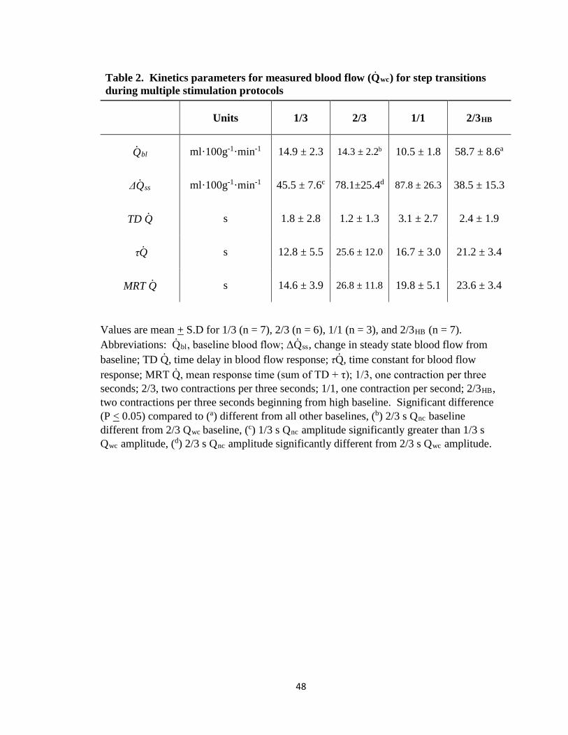
Table 2. Kinetics parameters for measured blood flow (Q̇Rwc) for step transitions during multiple stimulation protocols
Units 1/3 2/3 1/1 2/3HB
Q̇Rbl ml·100g-1·min-1 14.9 ± 2.3 14.3 ± 2.2b 10.5 ± 1.8 58.7 ± 8.6a
ΔQ̇Rss ml·100g-1·min-1 45.5 ± 7.6c 78.1±25.4d 87.8 ± 26.3 38.5 ± 15.3
TD Q̇ s 1.8 ± 2.8 1.2 ± 1.3 3.1 ± 2.7 2.4 ± 1.9
τQ̇ s 12.8 ± 5.5 25.6 ± 12.0 16.7 ± 3.0 21.2 ± 3.4
MRT Q̇ s 14.6 ± 3.9 26.8 ± 11.8 19.8 ± 5.1 23.6 ± 3.4
Values are mean + S.D for 1/3 (n = 7), 2/3 (n = 6), 1/1 (n = 3), and 2/3HB (n = 7). Abbreviations: Q̇Rbl, baseline blood flow; ΔQ̇Rss, change in steady state blood flow from baseline; TD Q̇, time delay in blood flow response; τQ̇, time constant for blood flow response; MRT Q̇, mean response time (sum of TD + τ); 1/3, one contraction per three seconds; 2/3, two contractions per three seconds; 1/1, one contraction per second; 2/3HB, two contractions per three seconds beginning from high baseline. Significant difference (P < 0.05) compared to (a) different from all other baselines, (b) 2/3 s Qnc baseline different from 2/3 Qwc baseline, (c) 1/3 s Qnc amplitude significantly greater than 1/3 s Qwc amplitude, (d) 2/3 s Qnc amplitude significantly different from 2/3 s Qwc amplitude.
48

Table 3. Kinetics parameters for estimated blood flow (Q̇Rnc) for step transitions during multiple stimulation protocols
Units 1/3 2/3 1/1 2/3HB
Q̇Rbl ml·100g-1·min-1 14.0 ± 4.0 6.7 ± 3.7b 5.8 ± 1.8 59.0 ± 11.8a
ΔQ̇Rss ml·100g-1·min-1 49.9 ± 8.1c 96.8±29.4d 90.2 ± 36.4 50.1 ± 13.1
TD Q̇ s 1.9 ± 1.5 0.6 ± 1.0 3.3 ± 1.7 3.2 ± 1.6
τQ̇ s 11.8 ± 3.2 26.9 ± 10.2 22.1 ± 1.3 21.7 ± 4.8
MRT Q̇ s 13.5 ± 3.6 27.5 ± 10.4 25.3 ± 1.8 25.0 ± 3.8
Values are mean + S.D for 1/3 (n = 7), 2/3 (n = 6), 1/1 (n = 3), and 2/3HB (n = 7). 1/1 were excluded from statistical analysis and reported here for reference. Abbreviations: Q̇Rbl, baseline blood flow; ΔQ̇Rss, change in steady state blood flow from baseline; TD Q̇, time delay in blood flow response; τQ̇, time constant for blood flow response; MRT Q̇, mean response time (sum of TD + τ); 1/3, one contraction per three seconds; 2/3, two contractions per three seconds; 1/1, one contraction per second; 2/3HB, two contractions per three seconds beginning from high baseline. Significant difference (P < 0.05) compared to (a) different from all other baselines, (b) 2/3 s Qnc baseline different from 2/3 Qwc baseline, (c) 1/3 s Qnc amplitude significantly greater than 1/3 s Qwc amplitude, (d) 2/3 s Qnc amplitude significantly different from 2/3 s Qwc amplitude.
49

Figure 5. Average blood flow from all 1/3 s trials for estimated blood flow (Qnc) with
fitted kinetics.
50
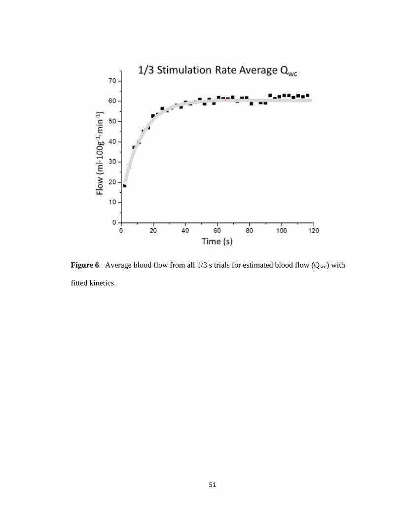
Figure 6. Average blood flow from all 1/3 s trials for estimated blood flow (Qwc) with
fitted kinetics.
51

Figure 7. Average blood flow from all 2/3HB s trials for estimated blood flow (Qnc) with
fitted kinetics.
52

Figure 8. Average blood flow from all 2/3HB s trials for estimated blood flow (Qwc) with
fitted kinetics.
53

Figure 9. Average blood flow from all 2/3 s trials for estimated blood flow (Qnc) with
fitted kinetics.
54
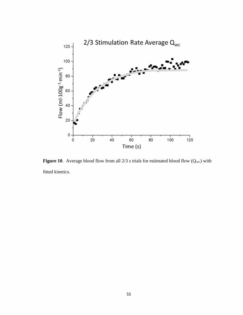
Figure 10. Average blood flow from all 2/3 s trials for estimated blood flow (Qwc) with
fitted kinetics.
55

Figure 11. Average blood flow from all 1/1 s trials for estimated blood flow (Qnc) with
fitted kinetics.
56

Figure 12. Average blood flow from all 1/1 s trials for estimated blood flow (Qwc) with
fitted kinetics.
Figures 13 and 14 highlight the frequency dependence in quantifying the muscle
pump contribution. The muscle pump contribution was converted to a percentage as
shown by the following equation:
% Muscle Pump Contribution = (Q̇Rwc - Q̇Rnc)/Q̇Rwc x 100 (equation 3)
By quantifying the muscle pump in this way, positive values represent a positive
contribution to total blood flow while negative values indicate a reduction in flow relative
to that which would have been observed with vasodilation alone. The adjustment of
blood flow in the 1/3 s trials indicated a reduced blood flow by the second contraction
57

(Qnc > Qwc) in all but one animal (fourth contraction) suggesting the muscle pump was
not contributing to the hyperemic response beyond this point. Conversely, in the 2/3 s
protocol during at least the first six contractions a positive muscle pump effect was
observed. In the 2/3 s protocol this effect declined rapidly (tau = 4.4 ± 2.1 s) and beyond
20 seconds total blood flow was reduced by contraction relative to the estimated blood
flow from vasodilation alone.
Figure 13. The kinetics response for the percent muscle pump contribution from onset to two minutes of contractions for the 2/3 s stimulation rate. Notice the slow component and negative values for muscle pump contribution in the 2/3 s trial. See text for discussion of frequency dependence in muscle pump contribution.
58

Figure 14. The kinetics responses for the percent muscle pump contribution from onset to two minutes of contractions for the 1/1 s stimulation rate. Notice the positive contribution attributed to the muscle pump at all points during the 1/1 s trials. See text for discussion of frequency dependence in muscle pump contribution.
This is likely due to the majority of blood flow occurring in the passive “vasoactive”
period during low frequency contractions relative to the blood flow ejected during the
rapid “contraction” phase. For the 1/1 s stimulation rate a slower adjustment in the
muscle pump contribution was observed (tau = 18 ± 10 s) as well as positive values
throughout the two minute stimulation period.
59

Discussion:
For this investigation we examined the spontaneous blood flow response of a
surgically isolated skeletal muscle during transitions from rest to contraction rates
eliciting various metabolic rates as well as from a low contraction rate to a higher rate
during tetanic stimulation. Further, based on the estimated spontaneous blood flow
response that excludes the mechanical effects of muscle contraction, we characterized the
kinetics response of the directly measured blood flow to the kinetics response of the
estimated “no contraction” blood flow on a contraction by contraction basis.
The contribution of the muscle pump to skeletal muscle perfusion has been a long
debated topic. Efforts to establish this contribution have generally relied on prior
vasodilation, preventing vasodilation, or limb position relative to the heart to determine if
the contracting muscle was capable of increasing its own blood flow. To our knowledge,
this is the first investigation to attempt to separate the spontaneous blood flow response to
a variety of contraction frequencies from an estimate of the blood flow response due only
to vasodilation. Additionally, based upon this estimate we aimed to characterize the
kinetics response for blood flow and the estimated blood flow from rest through two
minutes on a contraction by contraction basis. The muscle pump hypothesis posits that
skeletal muscle contraction aids in its own perfusion by translocating blood volume from
the muscle into the venous vasculature towards central circulation. This movement
transiently reduces the venous pressure during the relaxation phase of each contraction.
Veins, tethered to skeletal muscle that rapidly returns to resting size/shape, may quickly
create negative pressure in the veins and potentiate the flow from the arterial circuit (47,
88, 136). Our contraction by contraction approach to identify the muscle pump
60

contribution, from exercise onset through steady state, in spontaneously self-perfusing
skeletal muscle is a novel attempt to examine this hypothesis.
The pressure generated within the vasculature during skeletal muscle contractions
causes the forceful expulsion of blood from the muscle bed into the venous circulation
while concurrently creating an impediment to incoming flow from the arterial
vasculature. In this investigation it was assumed that with all vasoactive and myogenic
responses intact an estimate could be made to compare the blood flow from vasodilation
to that which occurs with the cyclic ejection/obstruction pattern observed during
rhythmic contractions. Greater measured blood flow than estimated “no contraction”
blood flow at any contraction was interpreted as a net benefit to total skeletal muscle
perfusion contributed by the muscle pump. During high frequency contractile stimuli, a
greater portion of the total blood flow during each contraction cycle is mobilized during
the fast “contraction” phase relative to the slow “vasoactive” phase. During a high
frequency contraction cycle the subsequent contraction is superimposed early on during
the vasoactive phase thus reducing its contribution to total flow. With slower contraction
frequency a greater portion of the total flow occurs during the vasoactive phase allowing
for faster adjustment to the steady state of blood flow. The impediment to flow that
occurs during skeletal muscle contraction outweighs the increased flow ejected therein;
this creates a limitation to total flow that is exacerbated by high frequency contraction
cycles.
Because no single vasodilatory substance has been identified that fully explains
the changes in blood flow following muscle contraction, alternative mechanisms have
been sought. The muscle pump hypothesis is appealing for the immediate hyperemic
61
response because with the first contraction a decrease in venous pressure could lead to an
increase in perfusion. According to this hypothesis, by emptying the volume of blood
present in resting skeletal muscle with contraction, the pressure gradient across that
muscle bed is increased, thus promoting additional arterial inflow (47, 88, 91, 136).
Evidence in support of a muscle pump effect comes from investigations in which
equivalent exercise bouts were initiated with limb position either above or below the
heart. Our data would indicate that at exercise onset the muscle pump does aid in
increasing blood flow beyond vasodilation alone, but that this benefit quickly diminishes
and is further largely dependent on contraction frequency.
With prior maximal vasodilation achieved, tilting subjects from supine to an
upright position during post-exercise hyperemia did not enhance blood flow while the
same shift performed during rhythmic exercise significantly raised flow (48). This
observation was attributed to the lowering of venous pressure by muscle contraction in
the dependent position and an increase in local perfusion pressure. Comparison of
hyperemic responses to repeated forearm cuff inflation, simulating only the mechanical
contribution to venous emptying, were greater when performed with the arm below the
heart compared with above (149). However, vasodilation in addition to muscle pumping
appeared requisite as blood flow responses to muscle contraction were greater for both
positions when compared to the cuff inflation trials. Importance of the vasodilation at
exercise onset is further illustrated by increases in mean blood velocity from rest to
exercise during handgrip exercise with the arm positioned below the heart in the supine
position. The gain in blood flow was not wholly explained by the difference in blood
62
pressure between the positions indicating concurrent vasodilation is needed with the
reduction in venous pressure to observe an immediate increase in blood flow (139).
Hypotheses aimed at separating the potential muscle pump contribution from
spontaneous adjustments in vessel diameter have reasonably assumed that for the pump
to be a positive contributor, blood flow should increase with the onset of contractions in a
previously dilated resting vascular bed. This hypothesis has been tested with the addition
of stimulated muscle contractions subsequent to vasodilation in canine diaphragm (111)
as well as in the isolated gastrocnemius (42, 111). By maintaining experimental control
over the arterial flow with constant pressure, Naamani et al. (111) concluded that the
muscle pump had little direct effect on muscle blood flow and primarily contributed to
the observed reductions in venous pressure. A possible limitation of that study was the
relatively low blood flow values attained in their vasodilation protocols. To achieve a
blood flow with vasodilation comparable to maximal exercise, Dobson et al. (42)
combined infusion of SNP with ADO and further occluded arterial as well as venous
flow. This method “trapped” all exogenous and endogenous vasodilating agents in the
muscle for a period of five minutes. Upon release, the flows achieved were similar to
those elicited during contraction rates inducing V̇O2peak for this muscle preparation. The
addition of contractions in the previously dilated skeletal muscle vasculature led to a
reduction in total blood flow indicating no benefit of the muscle pump in achieving
maximal blood flow. In the current study, we characterized the kinetic responses of
blood flow and found that Qwc was greater than Qnc in the 1/1 s trials while the inverse
was true for all other protocols. We interpret this as a frequency dependency for the
63

muscle pump effect because the imposition of rapid contractions precludes an adequate
filling time and thus a reduction in our estimate of total blood flow.
The potential for the muscle pump to contribute to muscle hyperemia is dependent
on both vasodilation within the tissue, allowing for increased blood volume as well as a
contraction frequency allowing for adequate filling time. It is well known that blood
flow to skeletal muscle increases in response to elevated contractile activity. In rats
performing treadmill running, blood flow was increased selectively in oxidative fiber
types at slower running speeds and in all fiber types with increasing running speed (90).
Further, blood flow increased across a variety of running speeds from 15 to 75 m/min
with increased flow directly related to high-oxidative fiber type content at greater speeds.
Fiber type specific differences in blood flow were confirmed in a subsequent
investigation indicating blood flow heterogeneity within a single muscle as well as
indicating a strong correlation between total blood flow and contraction frequency (99).
With a stimulation pattern of one per second, aimed at simulating running stride
frequency in the calf of the cat, blood flow through the rhythmically contracting muscle
was found to exceed that of spontaneous flow during the immediate post-exercise period.
This was taken as evidence for the muscle pump contribution (47). In examining the
influence of cycling cadence in relation to blood flow at a fixed workload, Gotshall et al.
(57) attributed greater vascular conductance at higher cadences to more effective muscle
pumping.
Additional evidence suggests that the relationship between contraction frequency
and metabolic cost may be more complex. Metabolic byproducts, including many
putative vasodilators, increase with increasing contractile work. The connection between
64

blood flow and V̇O2 has been demonstrated (2, 4, 5, 108); however, there appear to be
conditions related to contraction duration which can dissociate this relationship. With
matched contractile work performed, contractions of short duration elicited greater V̇O2
and rates of ATP use than long duration contractions (14, 29, 70). This potentially relates
to the ATP cost of ion transport during muscle activation and relaxation. In one of the
aforementioned studies Hogan et al. utilized a tetanic contraction-to-rest-ratio of
0.25s/0.75s for short duration versus 1s/3s for long duration with constant blood flow.
The short duration stimulation pattern resulted in higher V̇O2 than the long duration
under these conditions in spite of the matched tension-time integral. With spontaneous
perfusion the same stimulus protocol was utilized to determine if blood flow was more
closely matched to contractile work or metabolic rate. In this setting the spontaneous
blood flow response was more closely associated with muscle metabolism than the total
work performed (63). In the current study, as blood flow approached its steady state
within each protocol, the frequency of contraction was paramount in determining a
muscle pump contribution.
Limitations
Laughlin et al. (7) proposed that during investigations into the effect of the muscle
pump, the very instrumentation required for measurement may abolish the effect.
However, that supposition was based on investigations in the rat hindlimb where the
delicate vasculature no longer exhibited auto-regulatory control after instrumentation. It
is unlikely that large animal preparations similar to that utilized in the current study limit
the auto-perfusive response of the skeletal muscle vasculature. Concerns regarding the
use of isometric, tetanically stimulated muscle have been raised questioning the ability to
65

extrapolate these findings to those of dynamically recruited muscle in vivo. Hamann et
al. (64) addressed this limitation by using a chronically instrumented canine model in
which the animals were able to perform dynamic exercise on a treadmill with
simultaneous measures of blood flow and blood pressure. Prior to the onset of mild-
intensity treadmill exercise vasodilation was induced with ADO infusion. The onset of
normal, spontaneously recruited muscle contraction in the previously vasodilated
hindlimb failed to further increase blood flow indicating that without concurrent
vasodilation the mechanical action of contracting muscle was no benefit to muscle blood
flow.
Although we believe our model offers some significant advantages for the
assessment of potential muscle pump contributions to skeletal muscle blood flow, we
recognize its limitations. The complete activation of all fibers with tetanic stimulation
represents a stimulus that is not likely observed in a normal dynamic exercise condition.
Perfusion in skeletal muscle is heterogeneous and is directed preferentially to fiber types
based on the intensity of the exercise (90, 99). This response of normal perfusion in
dynamic exercise is eliminated during rhythmic tetanic stimulation and may influence the
muscle pump contribution. An additional consideration during dynamic muscle
activation is the presence of antagonistic contractions generating a “push/pull” of blood
between muscle groups, a characteristic that is absent in this context. Also, as has been
noted, the benefit of the muscle pump may only be fully realized when investigated in the
distal segments of animals with a significant hydrostatic column.
66

Conclusions
The results of the current investigation suggest that during simulated exercise, in
spontaneously perfused muscle, the contribution of the muscle pump to skeletal muscle
perfusion is limited. In this study we utilized an analytic technique in which all naturally
occurring vascular adaptations associated with exercise were included in our estimation
of the blood flow response to various metabolic rates. The time course for blood flow
response was not different between the measured flow and the estimated flow suggesting
that with vascular responses intact, vasodilation is obligatory. The time course in the
adjustment of blood flow between the actual flow and the estimated flow indicated no
significant differences. In all but one animal (fourth contraction), by the second
contraction of the 1/3 s stimulation protocol we observed no additional contribution to
blood flow as a result of skeletal muscle contraction, similarly in the 2/3 s protocol during
at least the first six contractions a positive muscle pump effect was observed. For the 2/3
s protocol this effect declined rapidly (tau = 4.4 ± 2.1 s) and beyond 20 seconds total
blood flow was reduced by contractions relative to the estimated blood flow from
vasodilation alone. At a stimulation frequency of 1/1 s there appears to be a positive
muscle pump contribution throughout the stimulation protocol. Similar to previously
mentioned investigations we have identified the frequency of contraction as a key
component to the efficacy of the muscle pump hypothesis. During high frequency
contraction rates the blood that is expelled during the forceful phase represents a greater
proportion of the total blood flow during each contraction cycle. During low frequency
contraction rates a greater fraction of the blood flow occurs in the post contraction
“vasoactive” period. Rapid skeletal muscle contraction rates quickly reduce the
67

contribution of the muscle pump to total blood flow during the steady state, actually
impeding blood flow rather than enhancing it.
68

References
1. Alderton W, Cooper C, and Knowles R. Nitric oxide synthases: structure, function and inhibition. Biochem J 357: 593-615, 2001. 2. Ameredes BT, Brechue WF, and Stainsby WN. Mechanical and metabolic determination ofV˙ o 2 and fatigue during repetitive isometric contractions in situ. Journal of Applied Physiology 84: 1909-1916, 1998. 3. Ameredes BT, and Provenzano MA. Regional intramuscular pressure development and fatigue in the canine gastrocnemius muscle in situ. Journal of Applied Physiology 83: 1867-1876, 1997. 4. Andersen P, and Henriksson J. Capillary supply of the quadriceps femoris muscle of man: adaptive response to exercise. The Journal of Physiology 270: 677-690, 1977. 5. Andersen P, and Saltin B. Maximal perfusion of skeletal muscle in man. The Journal of Physiology 366: 233-249, 1985. 6. Anderson KM, and Faber JE. Differential sensitivity of arteriolar alpha 1-and alpha 2-adrenoceptor constriction to metabolic inhibition during rat skeletal muscle contraction. Circulation research 69: 174-184, 1991. 7. Armstrong R, Delp M, Goljan E, and Laughlin M. Distribution of blood flow in muscles of miniature swine during exercise. Journal of applied physiology 62: 1285-1298, 1987. 8. Armstrong R, and Laughlin MH. Rat muscle blood flows during high-speed locomotion. Journal of applied physiology 59: 1322-1328, 1985. 9. Armstrong R, Vandenakker C, and Laughlin M. Muscle blood flow patterns during exercise in partially curarized rats. Journal of Applied Physiology 58: 698-701, 1985. 10. Bacchus A, Gamble G, Anderson D, and Scott J. Role of the myogenic response in exercise hyperemia. Microvascular research 21: 92-102, 1981. 11. Ballard RE, Watenpaugh DE, Breit GA, Murthy G, Holley DC, and Hargens AR. Leg intramuscular pressures during locomotion in humans. Journal of Applied Physiology 84: 1976-1981, 1998. 12. Beaty O, and Donald D. Role of potassium in transient redution in vasoconstricttive responses of muscle resistance vessels during rhythmic exercise in dogs. CIRCULATION RESEARCH 41: 452-460, 1977. 13. Benham C. ATP-activated channels gate calcium entry in single smooth muscle cells dissociated from rabbit ear artery. The Journal of Physiology 419: 689-701, 1989. 14. Bergstrom M, and Hultman E. Energy cost and fatigue during intermittent electrical stimulation of human skeletal muscle. J Appl Physiol 65: 1500-1505, 1988. 15. Berne RM. Cardiac nucleotides in hypoxia: possible role in regulation of coronary blood flow. American Journal of Physiology--Legacy Content 204: 317-322, 1963. 16. Bockman EL. Blood flow and oxygen consumption in active soleus and gracilis muscles in cats. American Journal of Physiology-Heart and Circulatory Physiology 244: H546-H551, 1983. 17. Boyd I, and Forrester T. The release of adenosine triphosphate from frog skeletal muscle in vitro. The Journal of physiology 199: 115-135, 1968. 18. Brain KL, Jackson VM, Trout SJ, and Cunnane TC. Intermittent ATP release from nerve terminals elicits focal smooth muscle Ca2+ transients in mouse vas deferens. The Journal of physiology 541: 849-862, 2002. 19. Brayden JE. Potassium channels in vascular smooth muscle. Clinical and experimental pharmacology and physiology 23: 1069-1076, 1996.
69
20. Brock R, Tschakovsky M, Shoemaker J, Halliwill J, Joyner M, and Hughson R. Effects of acetylcholine and nitric oxide on forearm blood flow at rest and after a single muscle contraction. Journal of Applied Physiology 85: 2249-2254, 1998. 21. Brooks GA. Intra-and extra-cellular lactate shuttles. Medicine and science in sports and exercise 32: 790-799, 2000. 22. Buckwalter JB, and Clifford PS. Autonomic control of skeletal muscle blood flow at the onset of exercise. American Journal of Physiology-Heart and Circulatory Physiology 277: H1872-H1877, 1999. 23. Buckwalter JB, Naik JS, Valic Z, and Clifford PS. Exercise attenuates α-adrenergic-receptor responsiveness in skeletal muscle vasculature. Journal of Applied Physiology 90: 172-178, 2001. 24. Budel S, Bartlett IS, and Segal SS. Homocellular conduction along endothelium and smooth muscle of arterioles in hamster cheek pouch unmasking an NO wave. Circulation research 93: 61-68, 2003. 25. Burcher E, and Garlick D. Antagonism of vasoconstrictor responses by exercise in the gracilis muscle of the dog. Journal of Pharmacology and Experimental Therapeutics 187: 78-85, 1973. 26. Burnstock G. Dual control of vascular tone and remodelling by ATP released from nerves and endothelial cells. Pharmacol Rep 60: 12-20, 2008. 27. Burnstock G. P2 purinoceptors: historical perspective and classification. P2 purinoceptors: localization, function and transduction mechanisms 1-29, 1996. 28. Burnstock G. Purine and pyrimidine receptors. Cellular and Molecular Life Sciences 64: 1471-1483, 2007. 29. Chasiotis D, Bergstrom M, and Hultman E. ATP utilization and force during intermittent and continuous muscle contractions. J Appl Physiol 63: 167-174, 1987. 30. Chen Y, Wolin MS, and Messina EJ. Evidence for cGMP mediation of skeletal muscle arteriolar dilation to lactate. Journal of Applied Physiology 81: 349-354, 1996. 31. Clifford PS, Kluess HA, Hamann JJ, Buckwalter JB, and Jasperse JL. Mechanical compression elicits vasodilatation in rat skeletal muscle feed arteries. The Journal of physiology 572: 561-567, 2006. 32. Cole WC, Chen TT, and Clément-Chomienne O. Myogenic regulation of arterial diameter: role of potassium channels with a focus on delayed rectifier potassium current. Canadian journal of physiology and pharmacology 83: 755-765, 2005. 33. Corcondilas A, Koroxenidis GT, and Shepherd JT. Effect of a brief contraction of forearm muscles on forearm blood flow. Journal of Applied Physiology 19: 142-146, 1964. 34. Cox RH. Molecular determinants of voltage-gated potassium currents in vascular smooth muscle. Cell biochemistry and biophysics 42: 167-195, 2005. 35. da Silva CG, Specht A, Wegiel B, Ferran C, and Kaczmarek E. Mechanism of purinergic activation of endothelial nitric oxide synthase in endothelial cells. Circulation 119: 871-879, 2009. 36. Davies N, Standen N, and Stanfield P. The effect of intracellular pH on ATP-dependent potassium channels of frog skeletal muscle. The Journal of physiology 445: 549-568, 1992. 37. Dawes GS. The vaso-dilator action of potassium. J Physiol 99: 224-238, 1941. 38. Derbyshire ER, and Marletta MA. Biochemistry of soluble guanylate cyclase. In: cGMP: Generators, Effectors and Therapeutic ImplicationsSpringer, 2009, p. 17-31. 39. DeWitt DF, Wangler RD, Thompson CI, and Sparks H. Phasic release of adenosine during steady state metabolic stimulation in the isolated guinea pig heart. Circulation research 53: 636-643, 1983.
70

40. Dietrich HH, and Dacey Jr RG. Effects of extravascular acidification and extravascular alkalinization on constriction and depolarization in rat cerebral arterioles in vitro. Journal of neurosurgery 81: 437-442, 1994. 41. Dinenno FA, and Joyner MJ. Blunted sympathetic vasoconstriction in contracting skeletal muscle of healthy humans: is nitric oxide obligatory? The Journal of physiology 553: 281-292, 2003. 42. Dobson JL, and Gladden LB. Effect of rhythmic tetanic skeletal muscle contractions on peak muscle perfusion. Journal of Applied Physiology 94: 11-19, 2003. 43. Dyke CK, Dietz NM, Lennon RL, Warner DO, and Joyner MJ. Forearm blood flow responses to handgripping after local neuromuscular blockade. Journal of Applied Physiology 84: 754-758, 1998. 44. Ellsworth M. The red blood cell as an oxygen sensor: what is the evidence? Acta physiologica scandinavica 168: 551-559, 2000. 45. Ellsworth ML, Forrester T, Ellis CG, and Dietrich HH. The erythrocyte as a regulator of vascular tone. American Journal of Physiology-Heart and Circulatory Physiology 269: H2155-H2161, 1995. 46. Fleming I, and Busse R. Molecular mechanisms involved in the regulation of the endothelial nitric oxide synthase. American Journal of Physiology-Regulatory, Integrative and Comparative Physiology 284: R1-R12, 2003. 47. Folkow B, Gaskell P, and Waaler B. Blood flow through limb muscles during heavy rhythmic exercise. Acta physiologica Scandinavica 80: 61-72, 1970. 48. Folkow B, Haglund U, Jodal M, and Lundgren O. Blood flow in the calf muscle of man during heavy rhythmic exercise. Acta physiologica Scandinavica 81: 157-163, 1971. 49. Forrester T. An estimate of adenosine triphosphate release into the venous effluent from exercising human forearm muscle. The Journal of physiology 224: 611-628, 1972. 50. Forrester T, and Lind A. Identification of adenosine triphosphate in human plasma and the concentration in the venous effluent of forearm muscles before, during and after sustained contractions. The Journal of physiology 204: 347-364, 1969. 51. Frandsen U, Lopez-Figueroa M, and Hellsten Y. Localization of nitric oxide synthase in human skeletal muscle. Biochemical and biophysical research communications 227: 88-93, 1996. 52. Fredholm BB, Ijzerman AP, Jacobson KA, Klotz K-N, and Linden J. International Union of Pharmacology. XXV. Nomenclature and classification of adenosine receptors. Pharmacological reviews 53: 527-552, 2001. 53. Gaskell W. Further researches on the vasomotor nerves of ordinary muscles. The Journal of physiology 1: 262-426, 1878. 54. Gladden LB. Muscle as a consumer of lactate. Medicine and Science in Sports and Exercise 32: 764, 2000. 55. González-Alonso J. ATP as a mediator of erythrocyte‐dependent regulation of skeletal muscle blood flow and oxygen delivery in humans. The Journal of Physiology 590: 5001-5013, 2012. 56. Gorczynski RJ, Klitzman B, and Duling BR. Interrelations between contracting striated muscle and precapillary microvessels. American Journal of Physiology-Heart and Circulatory Physiology 235: H494-H504, 1978. 57. Gottshall R, Bauer T, and Fahrner S. Cycling cadence alters exercise hemodynamics. International journal of sports medicine 17: 17-21, 1996. 58. Green S, Langberg H, Skovgaard D, Bülow J, and Kjær M. Interstitial and arterial‐venous [K+] in human calf muscle during dynamic exercise: effect of ischaemia and relation to muscle pain. The Journal of physiology 529: 849-861, 2000.
71

59. Grimby G, Häggendal E, and Saltin B. Local xenon 133 clearance from the quadriceps muscle during exercise in man. Journal of applied physiology 22: 305-310, 1967. 60. Grozdanovic Z, Gosztonyi G, and Gossrau R. Nitric oxide synthase I (NOS-I) is deficient in the sarcolemma of striated muscle fibers in patients with Duchenne muscular dystrophy, suggesting an association with dystrophin. Acta histochemica 98: 61-69, 1996. 61. Gutterman DD, Miura H, and Liu Y. Redox modulation of vascular tone focus of potassium channel mechanisms of dilation. Arteriosclerosis, thrombosis, and vascular biology 25: 671-678, 2005. 62. Hamann JJ, Buckwalter JB, and Clifford PS. Vasodilatation is obligatory for contraction-induced hyperaemia in canine skeletal muscle. The Journal of physiology 557: 1013-1020, 2004. 63. Hamann JJ, Kluess HA, Buckwalter JB, and Clifford PS. Blood flow response to muscle contractions is more closely related to metabolic rate than contractile work. Journal of Applied Physiology 98: 2096-2100, 2005. 64. Hamann JJ, Valic Z, Buckwalter JB, and Clifford PS. Muscle pump does not enhance blood flow in exercising skeletal muscle. Journal of Applied Physiology 94: 6-10, 2003. 65. Harvey W. Motion of the Heart & Blood in Animals. Translated by Bowie A. George Bell and Sons, London., 1889, Translated. 66. Hellsten Y, Maclean D, Rådegran G, Saltin B, and Bangsbo J. Adenosine concentrations in the interstitium of resting and contracting human skeletal muscle. Circulation 98: 6-8, 1998. 67. Hernández A, Goodwin ML, Lai N, Cabrera ME, McDonald JR, and Gladden LB. Contraction-by-contraction VO2 and computer-controlled pump perfusion as novel techniques to study skeletal muscle metabolism in situ. Journal of Applied Physiology 108: 705-712, 2010. 68. Hill A. The pressure developed in muscle during contraction. The Journal of physiology 107: 518-526, 1948. 69. Hnik P, Holas M, Krekule I, Kříž N, Mejsnar J, Smieško V, Ujec E, and Vyskočil F. Work-induced potassium changes in skeletal muscle and effluent venous blood assessed by liquid ion-exchanger microelectrodes. Pflügers Archiv 362: 85-94, 1976. 70. Hogan MC, Ingham E, and Kurdak SS. Contraction duration affects metabolic energy cost and fatigue in skeletal muscle. American Journal of Physiology-Endocrinology And Metabolism 274: E397-E402, 1998. 71. Hopwood AM, and Burnstock G. ATP mediates coronary vasoconstriction via P2X-purinoceptors and coronary vasodilatation via P2Y-purinoceptors in the isolated perfused rat heart. European journal of pharmacology 136: 49-54, 1987. 72. Hu S, Kim HS, and Jeng AY. Dual action of endothelin-1 on the calcium-activated potassium channel in smooth muscle cells of porcine coronary artery. European journal of pharmacology 194: 31-36, 1991. 73. Hunter J. A Treatise on the Blood, Inflammation, and Gunshot Wounds. London: For George Nicol, Bookseller to his Majesty, Pall-Mall 88: 1794. 74. Jones AM, and Poole DC. Oxygen uptake kinetics in sport, exercise and medicine. Routledge Abingdon,, UK, 2005. 75. Joyner MJ, and Proctor DN. Muscle blood flow during exercise: the limits of reductionism. Medicine and science in sports and exercise 31: 1036, 1999. 76. Joyner MJ, and Wilkins BW. Exercise hyperaemia: is anything obligatory but the hyperaemia? The Journal of physiology 583: 855-860, 2007. 77. Juel C, Pilegaard H, Nielsen JJ, and Bangsbo J. Interstitial K+ in human skeletal muscle during and after dynamic graded exercise determined by microdialysis. American Journal of Physiology-Regulatory, Integrative and Comparative Physiology 278: R400-R406, 2000.
72

78. Karamouzis M, Karamouzis I, Vamvakoudis E, Ampatzidis G, Christoulas K, Angelopoulou N, and Mandroukas K. The response of muscle interstitial prostaglandin E< sub> 2</sub>(PGE< sub> 2</sub>), prostacyclin I< sub> 2</sub>(PGI< sub> 2</sub>) and thromboxane A< sub> 2</sub>(TXA< sub> 2</sub>) levels during incremental dynamic exercise in humans determined by in vivo microdialysis. Prostaglandins, leukotrienes and essential fatty acids 64: 259-263, 2001. 79. Katz A, and Sahlin K. Regulation of lactic acid production during exercise. J Appl Physiol 65: 509-518, 1988. 80. Katz A, and Sahlin K. Role of oxygen in regulation of glycolysis and lactate production in human skeletal muscle. Exercise and sport sciences reviews 18: 1-28, 1990. 81. Kelley K, Hamann J, Aschenbach W, and Gladden L. Canine Gastrocnemius muscle in situ:˙ VO2max. Medicine & Science in Sports & Exercise 28: 62, 1996. 82. Kennedy C, Saville VL, and Burnstock G. The contributions of noradrenaline and ATP to the responses of the rabbit central ear artery to sympathetic nerve stimulation depend on the parameters of stimulation. European journal of pharmacology 122: 291-300, 1986. 83. Kiens B, Saltin B, Wallose L, and Wesche J. Temporal relationship between blood flow changes and release of ions and metabolites from muscles upon single weak contractions. Acta physiologica scandinavica 136: 551-559, 1989. 84. Kirkebø A, and Wisnes A. Regional tissue fluid pressure in rat calf muscle during sustained contraction or stretch. Acta Physiologica Scandinavica 114: 551-556, 1982. 85. Kjellmer I. The role of potassium ions in exercise hyperaemia. Pharmacology 5: 56-60, 1961. 86. Knot HJ, and Nelson MT. Regulation of arterial diameter and wall [Ca2+] in cerebral arteries of rat by membrane potential and intravascular pressure. The Journal of Physiology 508: 199-209, 1998. 87. Kobzik L, Reid MB, Bredt DS, and Stamler JS. Nitric oxide in skeletal muscle. Nature 372: 546-548, 1994. 88. Laughlin MH. Skeletal muscle blood flow capacity: role of muscle pump in exercise hyperemia. American Journal of Physiology-Heart and Circulatory Physiology 253: H993-H1004, 1987. 89. Laughlin MH, and Armstrong R. Muscle blood flow during locomotory exercise. Exercise and sport sciences reviews 13: 95, 1985. 90. Laughlin MH, and Armstrong R. Muscular blood flow distribution patterns as a function of running speed in rats. American Journal of Physiology-Heart and Circulatory Physiology 243: H296-H306, 1982. 91. Laughlin MH, Davis MJ, Secher NH, van Lieshout JJ, Arce-Esquivel AA, Simmons GH, Bender SB, Padilla J, Bache RJ, Merkus D, and Duncker DJ. Peripheral Circulation. In: Comprehensive PhysiologyJohn Wiley & Sons, Inc., 2011. 92. Laughlin MH, and Schrage WG. Effects of muscle contraction on skeletal muscle blood flow: when is there a muscle pump? Medicine and science in sports and exercise 31: 1027-1035, 1999. 93. Learoyd P. A short history of blood transfusion. National Blood Services, STT 42: 2006. 94. Ledoux J, Werner M, Brayden J, and Nelson M. Calcium-activated potassium channels and the regulation of vascular tone. Physiology (Bethesda, Md) 21: 69, 2006. 95. Leyk D, Essfeld D, Baum K, and Stegemann J. Early leg blood flow adjustment during dynamic foot plantarflexions in upright and supine body position. International journal of sports medicine 15: 447-452, 1994.
73

96. Lohman AW, Billaud M, and Isakson BE. Mechanisms of ATP release and signalling in the blood vessel wall. Cardiovascular research 95: 269-280, 2012. 97. Lott ME, Hogeman CS, Vickery L, Kunselman AR, Sinoway LI, and MacLean DA. Effects of dynamic exercise on mean blood velocity and muscle interstitial metabolite responses in humans. American Journal of Physiology-Heart and Circulatory Physiology 281: H1734-H1741, 2001. 98. Lower R. Tractatus de corde item de motu et colore sanguinis. London: Dawsons of Pall Mall, 1968 Translated. 99. Mackie BG, and Terjung RL. Blood flow to different skeletal muscle fiber types during contraction. American Journal of Physiology-Heart and Circulatory Physiology 245: H265-H275, 1983. 100. Mackie BG, and Terjung RL. Influence of training on blood flow to different skeletal muscle fiber types. Journal of Applied Physiology 55: 1072-1078, 1983. 101. MacLean DA, Imadojemu VA, and Sinoway LI. Interstitial pH, K+, lactate, and phosphate determined with MSNA during exercise in humans. American Journal of Physiology-Regulatory, Integrative and Comparative Physiology 278: R563-R571, 2000. 102. Magder S. Venous mechanics of contracting gastrocnemius muscle and the muscle pump theory. Journal of Applied Physiology 79: 1930-1935, 1995. 103. Marijic J, and Toro L. Voltage and calcium-activated K+ channels of coronary smooth muscle. Heart physiology and pathophysiology 309-325, 2001. 104. Marshall J, and Tandon H. Direct observations of muscle arterioles and venules following contraction of skeletal muscle fibres in the rat. The Journal of physiology 350: 447-459, 1984. 105. Mellander S, and Johansson B. Control of resistance, exchange, and capacitance functions in the peripheral circulation. Pharmacological Reviews 20: 117-196, 1968. 106. Minami K, Fukuzawa K, Nakaya Y, Zeng X-R, and Inoue I. Mechanism of activation of the Ca< sup> 2+</sup>-activated K< sup>+</sup> channel by cyclic AMP in cultured porcine coronary artery smooth muscle cells. Life sciences 53: 1129-1135, 1993. 107. Mohandas N, and Gallagher PG. Red cell membrane: past, present, and future. Blood 112: 3939-3948, 2008. 108. Mohrman DE, and Regal RR. Relation of blood flow to VO2, PO2, and PCO2 in dog gastrocnemius muscle. American Journal of Physiology-Heart and Circulatory Physiology 255: H1004-H1010, 1988. 109. Mohrman DE, and Sparks HV. Myogenic hyperemia following brief tetanus of canine skeletal muscle. The American journal of physiology 227: 531-535, 1974. 110. Mohrman DE, and Sparks HV. Role of potassium ions in the vascular response to a brief tetanus. Circulation research 35: 384-390, 1974. 111. Naamani R, Hussain SN, and Magder S. The mechanical effects of contractions on blood flow to the muscle. European journal of applied physiology and occupational physiology 71: 102-112, 1995. 112. Naik JS, Valic Z, Buckwalter JB, and Clifford PS. Rapid vasodilation in response to a brief tetanic muscle contraction. Journal of Applied Physiology 87: 1741-1746, 1999. 113. Nakane M, Schmidt HH, Pollock JS, Förstermann U, and Murad F. Cloned human brain nitric oxide synthase is highly expressed in skeletal muscle. FEBS letters 316: 175-180, 1993. 114. Nees S, and Gerlach E. Adenine nucleotide and adenosine metabolism in cultured coronary endothelial cells: formation and release of adenine compounds and possible functional implications. In: Regulatory function of adenosineSpringer, 1983, p. 347-360.
74

115. Nelson MT, and Quayle JM. Physiological roles and properties of potassium channels in arterial smooth muscle. American Journal of Physiology-Cell Physiology 268: C799-C822, 1995. 116. North RA. Molecular physiology of P2X receptors. Physiological reviews 82: 1013-1067, 2002. 117. Peng H, Ivarsen A, Nilsson H, and Aalkjær C. On the cellular mechanism for the effect of acidosis on vascular tone. Acta physiologica scandinavica 164: 517-525, 1998. 118. Pohl U, Holtz J, Busse R, and Bassenge E. Crucial role of endothelium in the vasodilator response to increased flow in vivo. Hypertension 8: 37-44, 1986. 119. Pollack AA, and Wood EH. Venous pressure in the saphenous vein at the ankle in man during exercise and changes in posture. Journal of Applied Physiology 1: 649-662, 1949. 120. Quayle JM, Nelson M, and Standen N. ATP-sensitive and inwardly rectifying potassium channels in smooth muscle. Physiological reviews 77: 1165-1232, 1997. 121. Rådegran G, and Saltin B. Muscle blood f low at onset of dynamic exercise in humans. American Journal of Physiology-Heart and Circulatory Physiology 274: H314-H322, 1998. 122. Ralevic V, and Burnstock G. Receptors for purines and pyrimidines. Pharmacological reviews 50: 413-492, 1998. 123. Raqeeb A, Sheng J, Ao N, and Braun AP. Purinergic P2Y2 receptors mediate rapid Ca< sup> 2+</sup> mobilization, membrane hyperpolarization and nitric oxide production in human vascular endothelial cells. Cell calcium 49: 240-248, 2011. 124. Remensnyder JP, Mitchell JH, and Sarnoff SJ. Functional Sympatholysis During Muscular Activity: Observations on Influence of Carotid Sinus on Oxygen Uptake. Circulation research 11: 370-380, 1962. 125. Richardson R, Noyszewski E, Kendrick K, Leigh J, and Wagner P. Myoglobin O2 desaturation during exercise. Evidence of limited O2 transport. Journal of Clinical Investigation 96: 1916, 1995. 126. Richardson RS, Poole DC, Knight DR, Kurdak S, Hogan MC, Grassi B, Johnson EC, Kendrick KF, Erickson BK, and Wagner PD. High muscle blood flow in man: is maximal O2 extraction compromised? Journal of Applied Physiology 75: 1911-1916, 1993. 127. Rosenmeier JB, Fritzlar SJ, Dinenno FA, and Joyner MJ. Exogenous NO administration and α-adrenergic vasoconstriction in human limbs. Journal of Applied Physiology 95: 2370-2374, 2003. 128. Rowell LB. Ideas about control of skeletal and cardiac muscle blood flow (1876–2003): cycles of revision and new vision. Journal of applied physiology 97: 384-392, 2004. 129. Rowell LB, Saltin B, Kiens B, and Christensen NJ. Is peak quadriceps blood flow in humans even higher during exercise with hypoxemia? American Journal of Physiology-Heart and Circulatory Physiology 251: H1038-H1044, 1986. 130. Rubanyi GM, Romero JC, and Vanhoutte PM. Flow-induced release of endothelium-derived relaxing factor. American Journal of Physiology-Heart and Circulatory Physiology 250: H1145-H1149, 1986. 131. Sahlin K, Harris R, Nylind B, and Hultman E. Lactate content and pH in muscle samples obtained after dynamic exercise. Pflügers Archiv 367: 143-149, 1976. 132. Saltin B. Hemodynamic adaptations to exercise. The American journal of cardiology 55: D42-D47, 1985. 133. Secher NH, Clausen JP, Klausen K, Noer I, and Trap-Jensen J. Central and regional circulatory effects of adding arm exercise to leg exercise. Acta physiologica scandinavica 100: 288-297, 1977.
75

134. Sejersted O, Hargens AR, Kardel KR, Blom P, Jensen O, and Hermansen L. Intramuscular fluid pressure during isometric contraction of human skeletal muscle. Journal of Applied Physiology 56: 287-295, 1984. 135. Sheriff D. Point: The muscle pump raises muscle blood flow during locomotion. Journal of Applied Physiology 99: 371-375, 2005. 136. Sheriff DD, Rowell L, and Scher A. Is rapid rise in vascular conductance at onset of dynamic exercise due to muscle pump? American Journal of Physiology-Heart and Circulatory Physiology 265: H1227-H1234, 1993. 137. Sheriff DD, and Van Bibber R. Flow-generating capability of the isolated skeletal muscle pump. American Journal of Physiology-Heart and Circulatory Physiology 274: H1502-H1508, 1998. 138. Shiotani I, Sato H, Sato H, Yokoyama H, Ohnishi Y, Hishida E, Kinjo K, Nakatani D, Kuzuya T, and Hori M. Muscle pump-dependent self-perfusion mechanism in legs in normal subjects and patients with heart failure. Journal of Applied Physiology 92: 1647-1654, 2002. 139. Shoemaker J, Tschakovsky M, and Hughson R. Vasodilation contributes to the rapid hyperemia with rhythmic contractions in humans. Canadian journal of physiology and pharmacology 76: 418-427, 1998. 140. Shrier I, and Magder S. Maximal vasodilation does not eliminate the vascular waterfall in the canine hindlimb. Journal of Applied Physiology 79: 1531-1539, 1995. 141. Sprague RS, and Ellsworth ML. Erythrocyte‐derived ATP and Perfusion Distribution: Role of Intracellular and Intercellular Communication. Microcirculation 19: 430-439, 2012. 142. Stainsby WN, and Otis AB. Blood flow, blood oxygen tension, oxygen uptake, and oxygen transport in skeletal muscle. American Journal of Physiology--Legacy Content 206: 858-866, 1964. 143. Stamler JS, and Meissner G. Physiology of nitric oxide in skeletal muscle. Physiological reviews 81: 209-237, 2001. 144. Standen N, and Quayle J. K^+ channel modulation in arterial smooth muscle. Acta physiologica Scandinavica 164: 549-558, 1998. 145. Stegall HF. Muscle pumping in the dependent leg. Circulation Research 19: 180-190, 1966. 146. Symons JD, Theodossy SJ, Longhurst JC, and Stebbins CL. Intramuscular accumulation of prostaglandins during static contraction of the cat triceps surae. Journal of Applied Physiology 71: 1837-1842, 1991. 147. Thomas GD, and Segal SS. Neural control of muscle blood flow during exercise. Journal of Applied Physiology 97: 731-738, 2004. 148. Tschakovsky ME, Rogers AM, Pyke KE, Saunders N, Glenn N, Lee S, Weissgerber T, and Dwyer E. Immediate exercise hyperemia in humans is contraction intensity dependent: evidence for rapid vasodilation. Journal of Applied Physiology 96: 639-644, 2004. 149. Tschakovsky ME, Shoemaker JK, and Hughson RL. Vasodilation and muscle pump contribution to immediate exercise hyperemia. Am J Physiol 271: H1697-1701, 1996. 150. Valic Z, Naik JS, Ruble SB, Buckwalter JB, and Clifford PS. Elevation in resting blood flow attenuates exercise hyperemia. Journal of Applied Physiology 93: 134-140, 2002. 151. Van Leeuwenhoek A. Other microscopical observations made by the same, about the texture of the blood, the sap of some plants, the figures of sugar and salt, and the probable cause of the difference of their tastes. Philos Trans R Soc Lond 380-385, 1675. 152. von Kügelgen I. Pharmacological profiles of cloned mammalian P2Y-receptor subtypes. Pharmacology & therapeutics 110: 415-432, 2006.
76

153. Welsh DG, and Segal SS. Coactivation of resistance vessels and muscle fibers with acetylcholine release from motor nerves. American Journal of Physiology-Heart and Circulatory Physiology 273: H156-H163, 1997. 154. Whipp BJ, and Wasserman K. Oxygen uptake kinetics for various intensities of constant-load work. J Appl Physiol 33: 351-356, 1972. 155. Wilson JR, and Kapoor SC. Contribution of prostaglandins to exercise-induced vasodilation in humans. American Journal of Physiology-Heart and Circulatory Physiology 265: H171-H175, 1993. 156. Young EW, and Sparks HV. Prostaglandins and exercise hyperemia of dog skeletal muscle. American Journal of Physiology-Heart and Circulatory Physiology 238: H191-H195, 1980.
77

Appendix A - Institutional Animal Care and Use Committee Approval
GENERAL INFORMATION AND INSTRUCTIONS
SUBMIT ORIGINAL AND 16 COPIES TO:
ANIMAL RESOURCES, 307 Samford Hall, MAIN CAMPUS
University policy requires that all research, teaching, production/maintenance, and
demonstration activities involving vertebrate animals be approved by the Auburn
University Institutional Animal Care and Use Committee (IACUC) prior to initiation of
the project. The Auburn University IACUC policy is available from the Animal
Resources office and
website: www.auburn.edu/research/vpr/animals/documents/policy.pdf. This policy is in
accordance with federal regulations and guidelines.
When submitting the original and 16 copies, the General Information and Instructions
and the Additional Information sections should be omitted.
78

The IACUC meets the first and third Thursdays of each calendar month. Protocols
received at least seven days prior to a scheduled meeting date (e.g. by 11:30 a.m. on
Thursday of the week prior to a scheduled Thursday p.m. meeting) will be placed on the
agenda. Approved protocols will be assigned a PRN (protocol review number).
Approved Animal Subjects Review Forms will remain in the official files of the
University for not less than three years beyond the completion of the project. If a project
is not funded, please notify ANIMAL RESOURCES AS SOON AS POSSIBLE (844-
5978).
Annual review of all protocols is required. An Annual Review Form will be sent to the
Principal Investigator approximately 30 days prior to the anniversary date of the
approved PRN. Please complete and return the form promptly to ANIMAL
RESOURCES, 307 Samford Hall. This form is also available on the Animal Resources
website http://www.auburn.edu/research/vpr/animals/forms.htm.
Animal users are encouraged to become familiar with all guidelines and regulations
pertaining to the care and use of animals in research and teaching by visiting the Animal
Welfare Information Center (AWIC) on the World Wide Web
at http://www.nal.usda.gov/awic/
An Animal Subjects Review Form may be obtained by downloading it from the Animal
Resources website http://www.auburn.edu/research/vpr/animals/forms.htm.
79

Alternatively, this form may be obtained at either of the following offices: Animal
Resources, 307 Samford Hall, Main Campus or the Division of Laboratory Animal
Health, 311 Greene Hall Annex, College of Veterinary Medicine.
Complete this form by providing BOLD TYPED answers to each item listed. If an item
is not applicable, please indicate NA.
80

ANIMAL SUBJECTS REVIEW FORM
PRINCIPAL INVESTIGATOR: L. Bruce Gladden, Ph.D.
RANK/TITLE: Alumni Professor
DEPARTMENT: Kinesiology
COLLEGE/SCHOOL: Education
CAMPUS ADDRESS: 2050 Memorial Coliseum E-MAIL ADDRESS:
CAMPUS PHONE #: 844-1466 FAX #: 844-1467
Check if PI will serve as faculty advisor to the Lead Graduate Student or
Resident associated with this activity.
LEAD GRADUATE STUDENT/RESIDENT: N/A
RANK/TITLE:
DEPARTMENT: E-MAIL ADDRESS:
CAMPUS PHONE #: FAX #:
81

CO-INVESTIGATOR: N/A
RANK/TITLE:
DEPARTMENT: E-MAIL ADDRESS:
CAMPUS PHONE #: FAX #:
PROJECT TITLE: Oxygen uptake on-kinetics in canine skeletal muscle: metabolic
rate steps
STARTING DATE: July 1, 2011 EXPIRATION DATE: June
30, 2014
(Must not be prior to IACUC approval) (Must not exceed three years)
Is any part of the funding from a U.S. Public Health Service Agency: Yes
No _XX__
REQUIRED SIGNATURES
The information contained on this form provides an accurate description of the animal
care and use protocol which will be followed. I agree to abide by governmental
regulations and university policies concerning the use of animals. I will allow veterinary
oversight to be provided to animals showing evidence of pain or illness. If the
82

information provided for this project concerning animal use should be revised, or
procedures changed, I will so notify the committee of those changes in writing, and no
proposed changes will be implemented until full IACUC approval has been granted.
Principal Investigator Date
Medical care for animals will be available and provided as indicated by a qualified
veterinarian. By accepting this responsibility, the veterinarian is providing assurance that
any personal interest he/she might have in the project will not conflict with his/her
responsibility for the provision of adequate veterinary care for the animals. Furthermore,
the veterinarian provides assurance of review and consultation on the proper use of
anesthetics and pain relieving medications for any painful procedures.
Dr. Pat Rynders
Project Veterinarian Date Project Veterinarian (Please type or print)
Departmental Chairperson Date
Lead Graduate Student/Resident Date
83

*IACUC Chair Date
*IACUC Chair signs the protocol after IACUC approval has been granted.
84

PLEASE TYPE IN BOLD FONT AND COMPLETE THE FOLLOWING FORM
IN FULL.
1. Will the animals be used in:
Teaching
Research XX
Demonstration
Production
If Teaching, give the course number:
2.
Species Common Name
Total Number
Source
Housing Locatio
Canis lupus familiaris
12 – project total
85

(Dog)
Division of
Laboratory
Animal Health
Division of
Laboratory
Animal Health
Kennels
3. Will animals be maintained for a period of 12 or more consecutive hours in a
location other than the housing location mentioned in Item 2? (See Item 3 of Additional
Information at the end of this form.)
Yes No XX
If Yes, specify the location and reason.
4. PERSONNEL QUALIFICATIONS (See Item 4 of Additional Information at the
end of this form.)
A. Indicate who will provide daily care and maintenance of the animal(s).
Indicate name(s) or identify the particular unit staff.
DLAH personnel under the direction of Andrea Brown.
86

B. List the names of all individuals who will conduct procedures involving
animals on this protocol. If all individuals are not currently known, please
indicate as such.
Dr. L. Bruce Gladden, PI for this project.
James R. McDonald, a graduate student trained by Dr. Gladden.
Yi Sun, a graduate student trained by Dr. Gladden.
Matt Rogatzki, a graduate student undergoing training with Dr. Gladden.
Brian Ferguson, a graduate student undergoing training with Dr. Gladden.
Potentially, collaborators from other universities who are well-trained and
experienced in the type of experiments to be conducted.
C. Principal Investigator Certifications
My signature on page 1 of this form certifies that:
87

1) Individuals performing animal procedures on this protocol are or
will be qualified to perform their particular animal related duties
through training and/or experience (individuals will be supervised
until adequate training has occurred). Training and/or experience
must encompass the following: *biology, handling, and care of the
species; aseptic surgical methods and techniques (if applicable);
the concept, availability, and use of research or testing methods
that limit the use of animals or minimize distress; the proper use of
anesthetics, analgesics, and tranquilizers (if applicable); and
procedures for reporting animal welfare concerns. Informative
links regarding training resources have been provided for
assistance as needed
at http://www.auburn.edu/research/vpr/animals.
2) All individuals working with animals, animal tissues, or animal
products on this protocol will be informed of relevant
*occupational health and safety issues prior to performing their
duties. * Informative links have been provided for assistance in
this and other areas as needed
at http://www.auburn.edu/research/vpr/animals.
5. State how or why you selected the species to be used in this project.
88

Dogs are used because vascular isolation of the gastrocnemius is a well-
established method in these animals. As a result, there is an abundance of baseline
data with which to compare our results. This is an excellent model for the study of
exercise metabolism and the physiological effects of changes in blood flow and
metabolic stimulators/inhibitors. Oxygen delivery can be precisely controlled and
varied, a distinct advantage for the study of metabolic on-kinetics. Dog muscles are
highly oxidative (Maxwell et al., 1977); this characteristic results in a slow fatigue
rate thus facilitating the establishment of a steady state of muscle contractions and
metabolic rate. Metabolic measurements are more reliable and more valid when the
muscle is in a steady state. Other models (rats, cats, rabbits) have their uses but in
the present experiments suffer a variety of disadvantages (amount and quality of
blood flow to active muscles, fiber types, need for artificial perfusate, etc.).
6. STUDY/ACTIVITY JUSTIFICATION AND OBJECTIVES:
A. Justification:
Skeletal muscle has a remarkable ability to cope with rapid and dramatic
changes in metabolic rate. However, our understanding of the regulatory factors
underlying and controlling this ability is far from complete. It has been known for
89

decades that upon a step transition from rest to exercise, or from a lower to a higher
work rate, oxygen uptake (VO2) by skeletal muscle lags behind the power output or
energy requirement increase. The resultant increase in VO2 during these
transitions follows a time course that has been termed “VO2 on-kinetics”.
My laboratory, along with collaborators, has previously published
extensively on this topic, employing the canine gastrocnemius model described in
this protocol (two papers by Grassi et al. 1998, Grassi et al. 2000, Grassi et al. 2002,
Grassi et al. 2005, Zoladz et al. 2008, two papers by Hernández et al. 2010, Grassi et
al. 2011).
In human studies, it has been demonstrated that if exercise is initiated from a
raised work rate/metabolic rate, the VO2 on-kinetics response is slowed in
comparison to beginning from a resting/unloaded baseline (Brittain et al. 2001,
MacPhee et al. 2005). These necessarily indirect studies in humans suggest that the
VO2 on-kinetics from a raised work rate/metabolic rate may be slowed by a slower
blood flow response, recruitment of fast twitch muscle fibers which are slower to
respond metabolically, and/or a less favorable energetic state. With the canine
gastrocnemius model, blood flow and muscle fiber recruitment can be controlled
and energetic state can be assessed via muscle sampling. This will either confirm or
eliminate these factors as causative mechanisms in the control of the VO2 transition
between work rates.
B. Objectives:
90

As a continuation of this line of research, it is the purpose of the present project to
compare the VO2 on-kinetics response to contractions begun from the resting
condition versus an increase in contraction frequency from a prior contracting
baseline. Electrical stimulation will be supramaximal so that all motor units are
recruited in all conditions and blood flow will be controlled via pump perfusion.
Therefore, these two factors will be eliminated as causative agents in the resulting
VO2 on-kinetics response.
7. SUMMARY OF PROPOSED ACTIVITY: USE LAY TERMS to give a
description of the proposed activity. From reading this section it should be possible for a
non-scientist to determine exactly how animals will be used in the context of the proposed
activity.
This section should include a clear description of the experimental design (research
protocols) or activities involving animals (teaching, demonstration, or
production/maintenance protocols). This section should also include a brief description
of each phase of activities involving animals and should make it possible to account for
all animals requested in Item 2. Justification for animal numbers is required to assure that
only the necessary number of animals is being used. (See Item 7 of Additional
Information at the end of this form for guidance in providing the appropriate
information.)
91
In previous experiments we have studied the effects of O2 delivery (hypoxia),
metabolic activation, and metabolic inhibition on the VO2 on-kinetics (e.g., PRN
2007-1185). Reduced O2 delivery at the onset of contractions slowed VO2 on-
kinetics but hypoxia with no change in O2 delivery had no effect on the on-kinetics.
We have also collected data that we expect will allow us to correct for transport time
from mid-capillary to our venous sampling site – these data are still being analyzed
and modeled. We are planning to calculate cellular response times from this
modeling approach. As a side-benefit of the experiments in this previous protocol,
we were able to add continuous monitoring of venous O2 concentration and
calculate VO2 on a contraction-by-contraction basis. However, none of the
experiments from this previous protocol addressed the questions of the present
protocol; i.e., a direct comparison of VO2 on-kinetics within the same muscle
preparation at different contraction rates, and more importantly, from one
contraction rate directly to another. Studies in humans suggest that the VO2 on-
kinetics are significantly slowed when going from a lower exercise intensity and
metabolic rate (directly with no rest period in between) to a higher one; that is the
question that will be investigated in the present experiments, in a model in which
blood flow (and thereby O2 delivery) can be controlled and muscle motor unit
recruitment is maximal.
In the present experiments, the gastrocnemius muscles of anesthetized dogs will be
surgically isolated to allow measurement of metabolite exchange (e.g., O2 uptake,
CO2 output, glucose uptake, lactate uptake/output, etc.) across the muscle as well as
92

the sampling of muscle biopsies. In all cases, a primary measurement will be VO2
on-kinetics. To determine the on-kinetics, venous outflow will be measured
continuously via a flowthrough ultrasonic probe and flowmeter while venous O2
content will be measured continuously via an inline oximeter probe (Opticath model
no. U425C, size 4 F, Hospira, Lake Forest, IL)). Myoglobin deoxygenation will be
measured continuously with a non-invasive near-infrared Oxymon spectrometer
(Artinis, Zetten, Netherlands). Arterial and venous blood samples (< 2 ml) will be
taken before and after each series of contractions and analyzed for hemoglobin
concentration, percent saturation of hemoglobin with O2, partial pressure of O2 and
CO2, pH, and in some cases, metabolite concentrations (lactate, pyruvate, alanine,
glucose, etc.). Arterial blood samples are also drawn periodically throughout the
entire experimental time period to check for maintenance of normal blood gas levels
and acid-base balance. Additionally, venous samples are drawn at the end of each
trial period for calibration of the indwelling oximeter probe. The total volume for
all blood samples will be less than 70 ml. Muscle samples will be taken at rest, and
then at 15 s and 180 s for each contraction period, and subsequently analyzed for
phosphocreatine, ATP, glycogen, lactate, pyruvate, and alanine concentrations.
In all cases, the animals are anesthetized and intubated at the Division of
Laboratory Animal Health facility at the Veterinary School campus. After animals
have been monitored and determined to be in a surgical plane of anesthesia, they
are placed in a plastic cart with a closed cover and transported via automobile from
the Veterinary School campus to the Beard-Eaves-Memorial Coliseum. If the car is
a sedan, the cart is placed in the rear seat; if it is a station wagon, the cart is placed
93

in the rear compartment behind the second seat. The animals remain in the closed
cart until they are removed and placed on a surgical table in the Muscle Physiology
Laboratory. The dogs are ventilated and warmed with a heating pad while they are
kept in a deep, surgical plane of anesthesia (additional sodium pentobarbital given
as required to maintain absence of pedal, palpebral, and corneal reflexes). The
heating pad is used to maintain normal body temperature. The animals are
euthanized while still under anesthesia at the end of the experiments.
The following protocol will be used:
Under spontaneous, self-perfused conditions, VO2 on-kinetics will be determined
from rest (no contractions) to a low submaximal metabolic rate (1 tetanic
contraction per 3 s) and then, without stopping the contractions, from the low
submaximal metabolic rate to a high submaximal rate (2 tetanic contractions per 3
s). Then, during a 35 minute recovery period, tubing and a pump will be inserted
into the arterial inflow to the muscle. Subsequently, the same protocol will be
repeated except that the blood flow (and therefore O2 delivery) will be set prior to
any contractions at the level previously determined for the higher metabolic rate.
This will eliminate any effect of altered blood flow responses when elevating from a
resting versus submaximal contraction baseline to a higher metabolic rate. Also,
since the stimulation voltage is always supramaximal, there will be no change in
motor unit recruitment from one metabolic rate to another. Finally, following
another 35 minute recovery period, VO2 on-kinetics will be determined, again with
a fixed, high blood flow, from rest to the high submaximal metabolic rate (2 tetanic
contractions per 3 s). These experiments will require 12 dogs.
94

Each contraction period (1/3 s and 2/3 s) will last 4 minutes. When blood flow is
elevated prior to contractions, adenosine (105 – 210 µmol•kg wet muscle-1•min-1) will
be infused to maintain blood pressure at the spontaneous level. The protocol is
illustrated below.
8. A. Select pain/distress category relevant to the use of animals in this study.
(See Item 8A of Additional Information at the end of this form.)
C D X E
B. If category D or E was chosen in 8A, please complete the following. (See
Item 8B of Additional Information at the end of this form.)
Trial 1 Trial 2 Trial 3
Frq 1/3 2/3 2/3
S1
S2
S3
S4 S5
1/3 2/3
Flow Spontaneous Constant/High Constant/High
35 m
in re
cove
ry
35 m
in re
cove
ry
95
1) Database(s) searched or other sources consulted to determine the
availability of alternatives.
Database Searched Date of Search Years Covered
Medline X June 7-8, 2011 1950-2011
Agricola
CABA
Altweb X June 7-8, 2011 1990-2011
Other (describe) AWIC June 7-8, 2011 1950-2011
PubMed__ June 7-8, 2011 1950-2011
2) Keywords and search strategy used when considering alternatives to
the painful or distressful procedure(s).
Keywords: oxygen, kinetics, uptake, muscle, alternative, dogs
Strategy: Began with a broad search on “oxygen uptake,” then narrowed with
“kinetics,” and “muscle”, inspecting the long lists of each result. Searched “dogs”
plus “muscle” plus “alternative” in the Altweb site. Studied the descriptions of
96

databases, strategies, and resources in AWIC. Went to several links for further
information. Attempted to determine whether applicable, alternative methods of
studying oxygen uptake on-kinetics in muscle with regard to physiological
manipulations such as blood flow, etc. are available.
3) A succinct written narrative based on results of the database search,
that will permit the IACUC to readily assess whether the search topics
were appropriate and whether the search was sufficiently thorough.
This narrative must address the following:
Reduction:
Replacement:
Refinement:
Due to the invasive nature of these studies, the most direct information can be
obtained on animals instead of humans. Measurement of blood flow and arterial
and venous metabolite concentrations, and measurement of muscle metabolite
concentrations all entail considerable risks, particularly when combined with
exercise. Accurate measures of blood flow and the distribution of that blood flow
are also much more easily obtained in this animal model than in the whole human
system where indirect methods are less reliable. While we will be using some in
97
vitro-like methods, these investigations require the isolation of materials from living
organisms, and cellular and sub-cellular studies would not answer some of the
questions being posed. In order to study the effects of blood flow and exercise (and
in turn, the effects of various other perturbations) on the rapidity of oxygen uptake
adjustment, it is necessary to use a more intact preparation in which there actually
is a blood flow and measurable metabolism.
Dogs are being used because there is a great deal of background information on the
muscle preparation in these animals and the difficult surgical task of blood flow
isolation of the gastrocnemius muscles is relatively convenient. This canine muscle
also has a fiber type profile that facilitates the type of experiments being done; i.e.,
they fatigue slowly and recover relatively quickly so that repeated bouts of
contractions can be performed. The number of animals to be used is proposed on
the basis of numerous previous studies, including my own personal experience.
These studies have shown that 6-12 animals per experimental alteration are needed
to account for potential failed experimental trials and to statistically determine
significant differences. The use of less than six animals is not advised since this is
near the minimum number required for the application of analysis of variance to
the data. We plan to use 12 animals in our experimental groups for adequate
statistical power while at the same time reducing the number of animals to the
lowest reasonable level. Additionally, we always attempt to reduce the number of
animals used by employing repeated measures designs whenever experimentally
possible. If the preliminary results suggest that 6 animals is adequate, then that is
all that will be used.
98
Finally, we have closely examined the existing body of literature to assure that there
are no additional refinements available to reduce stress and/or discomfort to the
animals, no replacement techniques to answer the experimental questions posed,
and that the number of animals requested is reduced to the statistically allowable
limits. The literature was examined by searching PubMed, Altweb, and AWIC as
indicated above; PubMed includes Medline and other life sciences journals. In
addition, I have been involved in studies on the subject of VO2 on-kinetics for
almost 15 years now; nine studies are already published and two others are in
preparation. Further, I have organized and co-chaired two symposia on VO2 on-
kinetics at the national annual meeting of the American College of Sports Medicine.
All of this is to say that I am intimately familiar with the literature in this area, and
qualified to address Reduction, Replacement, and Refinement.
4) If alternatives are available but will not be used, please provide a
justification.
N/A
5) If pain/distress category E is to be employed, please provide a
justification for withholding pain and/or distress relieving drugs.
99

N/A
9. SURGERY:
Will surgery be performed?
Yes X No
If yes, please address the following, as applicable:
A. Nonsurvival surgery - Describe all surgical procedures, including surgical
preparation. Indicate where surgery will be performed (building and rooms).
Identify the person(s) and describe their qualifications for performing the
particular surgical procedure(s).
All surgical procedures will be performed in the Muscle Physiology Laboratory
located in Room 2129A, Beard-Eaves-Memorial Coliseum. Mongrel dogs, of both
sexes will be used. All surgical techniques will require isolation of the
gastrocnemius (GP) muscle (Grassi, Gladden, Stary, Wagner, and Hogan, 1998.).
The left GP is typically used. Briefly, the animals are anesthetized with sodium
pentobarbital (30mg•kg-1 iv); additional doses are given as required. The animals
are intubated with an endotracheal tube and ventilated with a respirator. A heating
pad is placed under the animal and adjusted as needed to maintain the rectal
100

temperature near 37 degrees Centigrade. Although the normal canine core
temperature is approximately 39 degrees Centigrade, maintenance at 37 degrees
eliminates the necessity of correcting blood gas, and pH values (instruments are set
at 37 degrees). The surgical site is prepared by clipping the animal’s hair very
closely using an electric clipper for animals. In the unusual occurrence of a dirty
animal, a moist cloth is used to clean the exposed skin. These are terminal
experiments, so it is not necessary to establish a sterile field. If the GP is to be
perfused via a pump in the arterial inflow, all branches of the popliteal artery which
are not supplying the GP are ligated. The right brachial artery or the left carotid
artery is cannulated and its outflow is directed by tubing through a pump and then
through a cannula into the left popliteal artery supplying the isolated GP. This
allows blood flow to the muscle to be controlled at any constant level desired, and
infusion of drugs, pharmacological agents, metabolites, and/or hormones can be
made directly into the arterial inflow into the muscle.
All branches of the popliteal vein that do not come from the GP are ligated and all
venous connections from the muscle that do not go directly to the popliteal vein are
ligated. Therefore, all of the venous outflow from the muscle is by way of the
popliteal vein. The vein is cannulated, and the flow is measured with a cannulating-
type ultrasonic probe and flowmeter. Venous outflow is returned to the animal via
the left jugular vein. Blood coagulation is prevented by intravenous heparin (2,000
U/kg) every 4 hours.
The Achilles tendon is sectioned and secured in an aluminum clamp for attachment
to an isometric myograph. The other end of the muscle is left attached to the femur,
101
which is fixed to the base of the myograph by two bone nails. The sciatic nerve is
exposed and isolated near the muscle. The distal nerve stump, about 1.5-3.0 cm in
length, is pulled into a small tubular electrode for stimulation. The release and/or
uptake of oxygen, lactate, pyruvate, glucose, alanine, or any other fuel or metabolite
by the muscles can be calculated from the venous blood outflow rate and the
arteriovenous concentration difference of the substance across the muscle. Muscle
samples are taken by biopsy when needed, and immediately frozen in liquid
nitrogen. The samples are stored at –80 degrees Centigrade or are freeze-dried
until analyzed.
The anesthetization of animals and all surgical procedures will be performed either
by me (Dr. L. Bruce Gladden) or other technicians/graduate students who are
trained by me. I have 35 years of experience with these techniques.
B. Survival surgery - Describe all surgical procedures, including surgical
preparation and post-surgical care. Please indicate that aseptic technique will
be followed if the procedure is a survival surgical procedure. Indicate where
surgery will be performed and what postoperative care will be provided
(building and rooms). Identify the person(s) and describe their qualifications
for performing the particular surgical procedure(s).
N/A
102

10. Administration of analgesics, anesthetics, tranquilizing drugs, and/or neuromuscular
blocking agents (Indicate generic name, dose, route of administration and frequency;
if by inhalation, method of scavenging waste anesthetic gases.)
Sodium pentobarbital (30 mg•kg-1) with additional doses (typically about 3.5-
4.0 mg•kg-1) as required, administered via intravenous injection. Animals are
maintained in a deep, surgical plane of anesthesia (additional sodium pentobarbital
given as required to maintain absence of pedal, palpebral, and corneal reflexes).
11. Administration of reagents, cells, drugs (other than anesthetics or analgesics),
infectious agents, carcinogens, recombinant DNA, etc. (Indicate generic name, dose,
route of administration and frequency, anticipated side effects, monitoring protocol.)
Conscious animals will be given only sodium pentobarbital. All other reagents or
drugs listed below will be given to the animals after they have been anesthetized:
- Sodium Heparin (2000 U•kg-1) will be given intravenously every four hours to
prevent coagulation of blood.
- Bicarbonate (1.0 N) or hydrochloric acid (0.3 N) for adjusting blood pH as
necessary to maintain normal limits, administered intravenously (Cain and Adams
1983). We almost never have to use the acid.
103

- Adenosine (Adenine-9-ß-ribofuranoside) will be infused into the tubing supplying
the popliteal artery of the gastrocnemius muscle to effect (maintenance of normal
perfusion pressure); this is typically at a rate of 105 – 210 µmol•kg wet muscle-1•min-
1.
12. ASSURANCES:
A. Provide a brief statement to confirm that proposed activities involving animals
do not duplicate previous experiments unnecessarily.
As noted previously, recent literature searches in various databases confirm
my own prior knowledge that these are unique experiments with a high likelihood of
producing unique results that will contribute to our understanding of basic muscle
metabolism. Also, as noted earlier, I have performed research in this area for
almost 15 years with nine published studies, two studies in preparation, and I have
chaired two symposia at national/international meetings. These experiments are
only being performed because I anticipate unique, new information to result, and to
contribute to the scientific literature in premier, peer-reviewed journals.
B. My signature on page 1 of this form certifies that exercise of caged dogs will
be accomplished according to the Animal Welfare Act (AWA) or cage size
104

provides adequate space for exercise to meet AWA requirements.
Alternatively, explain why an exception should be approved by the IACUC.
No exception requested.
C. Will wild caught or endangered animals be utilized?
Yes No X
If Yes, the investigator is responsible for obtaining and maintaining valid permits (if
required) for collecting, purchasing, transporting, and holding of these animals. List
applicable federal and/or state permit numbers and expiration dates.
13. HAZARDOUS AGENTS
Use of hazardous agents in animals may require approval of the appropriate institutional
committee. Contact the Department of Risk Management and Safety (844-4870) for
specific information.
105

Hazardous Agent
Yes
No
Agent
Date of Committe
Approval and BU
Radioisotopes
X
Biological Agents
X
Hazardous Chemicals or
Drugs
X
Recombinant DNA
X
Describe the practices and procedures required for the safe handling and disposal of
contaminated animals and material associated with this study. Also describe methods for
removal of radioactive waste and, if applicable, the monitoring of radioactivity.
N/A
106

14. What will be the disposition of the animals at the termination of the project? If
euthanasia is to be performed, what will be the method of carcass disposal?
All animals will be utilized in the aforementioned experiments, and will then be
euthanized as described in question #15 below. All carcasses and tissues are
subsequently incinerated in the incinerator at the College of Veterinary Medicine
campus.
15 All protocols must include the method of euthanasia that will be used during the
normal course of the protocol or in the event of unforeseen circumstances
resulting from illness or injury. Please specify the method, agent, dose, and route
of administration. The euthanasia method must be consistent with the AVMA
Panel on Euthanasia or justification for deviation should be indicated. This
document is available on the Animal Resources
website, http://www.auburn.edu/research/vpr/animals/ resources/res_index.htm
and in the Journal of the American Veterinary Medical Association (Vol. 218, No.
5, Pages 669-696, 2001).
All invasive experimental procedures will be preceded by anesthesia. After
the experiments, the animal will be euthanized. Since all animals will be under
107

sodium pentobarbital anesthesia throughout each experiment, maintained in a deep
surgical plane of anesthesia, they will be euthanized by administering an overdose of
sodium pentobarbital (typically approximately 17 mg•kg-1) along with saturated
KCl intravenously (typically about 1.5 ml•kg-1 into the jugular vein) (2000 Report of
the AVMA Panel on Euthanasia). In all cases, blood pressure will be monitored
until it drops to a level incompatible with life and there is no pulse; and a double
pneumothorax will be performed as a final precaution.
REFERENCE LIST:
1. 2000 Report of the AVMA Panel on Euthanasia. Journal of the American
Veterinary Medical Association 218(5):669-696, 2001.
2. Brittain, C.J., H.B. Rossiter, J.M. Kowalchuk, and B.J. Whipp. Effect of
prior metabolic rate on the kinetics of oxygen uptake during moderate-
intensity exercise. European Journal of Applied Physiology 86:125-134,
2001.
3. Cain, S.M. and R.P. Adams. O2 transport during two forms of stagnant
hypoxia following acid and base infusions. Journal of Applied Physiology
54(6):1518-1524, 1983.
108
4. Grassi, B., L.B. Gladden, M. Samaja, C.M. Stary, and M.C. Hogan. Faster
adjustment of O2 delivery does not affect VO2 on-kinetics in isolated in situ
canine muscle. Journal of Applied Physiology 85:1394-1403, 1998.
5. Grassi, B., L.B. Gladden, C.M. Stary, P.D. Wagner, and M.C. Hogan.
Peripheral O2 diffusion does not affect VO2 on-kinetics in isolated in situ
canine muscle. Journal of Applied Physiology 85:1404-1412, 1998.
6. Grassi, B., M.C. Hogan, K.M. Kelley, W.G. Aschenbach, J.J. Hamann, R.K.
Evans, R.E. Pattillo, and L.B. Gladden. Role of convective O2 delivery in
determining VO2 on-kinetics in canine muscle contracting at peak VO2.
Journal of Applied Physiology 89:1293-1301, 2000.
7. Grassi B., M.C. Hogan, K.M. Kelley, R.A. Howlett, and L.B. Gladden.
Effects of nitric oxide synthase inhibition by l-NAME on oxygen uptake
kinetics in isolated canine muscle in situ.
Journal of Physiology 568:1021-1033, 2005.
8. Grassi, B., M.C. Hogan, P.L. Greenhaff, J.J. Hamann, K.M. Kelley, W.G.
Aschenbach, D. Constantin-Teodosiu, and L.B. Gladden. Oxygen uptake on-
kinetics in dog gastrocnemius in situ following activation of pyruvate
dehydrogenase by dichloroacetate. Journal of Physiology 538:195-207, 2002.
9. Grassi, B., H.B. Rossiter, M.C. Hogan, R.A. Howlett, J.E. Harris, M.L.
Goodwin, J.L. Dobson, and L.B. Gladden. Faster O2 uptake kinetics in
canine skeletal muscle in situ after acute creatine kinase inhibition. Journal
of Physiology 589(Pt 1):221-233, 2011.
109

10. Hernández, A., M.L. Goodwin, N. Lai, M.E. Cabrera, J.R. McDonald, and
L.B. Gladden. Contraction-by-contraction VO2 and computer-controlled
pump-perfusion as novel techniques to study skeletal muscle metabolism in
situ. Journal of Applied Physiology 108:705-712, 2010.
11. Hernández, A., J.R. McDonald, N. Lai, and L.B. Gladden. A prior bout of
contractions speeds VO2 and blood flow on-kinetics and reduces the VO2
slow component amplitude in canine skeletal muscle contracting in situ.
Journal of Applied Physiology 108:1169-1176, 2010.
12. MacPhee, S.L., J.K. Shoemaker, D.H. Paterson, and J.M. Kowalchuk.
Kinetics of O2 uptake, leg blood flow, and muscle deoxygenation are slowed
in the upper compared with lower region of the moderate-intensity exercise
domain. Journal of Applied Physiology 99:1822-1834, 2005.
13. Maxwell, L.C., J.K. Barclay, D.E. Mohrman, and J.A. Faulkner.
Physiological characteristics of skeletal muscles of dogs and cats. American
Journal of Physiology 233:14-18, 1977.
14. Zoladz, J.A., L.B. Gladden, M.C. Hogan, Z. Nieckarz, and B. Grassi.
Progressive recruitment of muscle fibers is not necessary for the slow
component of VO2 kinetics. Journal of Applied Physiology, 105:575-580,
2008.
110

ADDITIONAL INFORMATION
THIS PAGE NEED NOT BE INCLUDED WHEN SUBMITTING FORM FOR
REVIEW
3. The IACUC is required to inspect animal housing areas and laboratories (at least
twice per year) where animals are kept for 12 or more hours.
4. PERSONNEL QUALIFICATIONS:
Federal regulations require institutions to ensure that people caring for or using animals
are qualified to do so through documented training or experience. This training is to
include investigators, technical personnel, trainees, visiting investigators, and any other
individuals who may perform animal husbandry, anesthesia, surgery, or other
experimental manipulations involving animals.
7. Please use this procedure list for guidance in providing the necessary information.
Please note that this is not meant to be an exhaustive list, but only a guide.
111

$ Body fluid sampling (e.g. blood, cerebrospinal fluid, ascites, urine —
describe method of collection, amount, frequency).
$ Antibody production (indicate route of administration, volume
administered per site, number of sites, adjuvant use and frequency,
consideration of alternatives to Freund’s adjuvant, anticipated side effects,
monitoring protocol).
$ Ascites method for monoclonal antibody production. Auburn
University requires adherence to the Office for Protection from Research
Risks (OPRR) policies concerning the production of monoclonal
antibodies using the mouse ascites method. Please refer to the OPRR
document http://oacu.od.nih.gov/ARAC/ascites.htm. Use of the ascites
method requires justification as to why in vitro systems cannot be used.
$ Special diets (describe any anticipated nutritional deficit or other health
concerns).
$ Indwelling catheters or implants (describe type,
maintenance/monitoring protocol).
$ Restraint of an unanesthetized animal other than that associated with
brief routine procedures such as for the collection of blood (describe
method, duration, frequency).
$ Tumor transplantation (describe any anticipated functional deficit to the
animal, monitoring protocol, endpoint).
112

$ Food or fluid restriction (e.g. greater than that associated with pre-
anesthetic procedures — describe, include justification and monitoring
protocol.)
$ Special housing, equipment, animal care (e.g. describe special caging,
water, feed, waste disposal, etc.)
$ Experimental endpoint criteria (list the criteria to be used to determine
when euthanasia is to be performed. Death as an endpoint must always be
scientifically justified.)
8A. USDA promulgated PAIN/DISTRESS CATEGORIES - Please use the following
categories when categorizing the pain/distress level.
C Pain or Distress - None or Minor
These include studies that DO NOT involve surgery; induction of painful or stressful
disease conditions, or pain or distress in excess of that associated with routine injections
or blood collection. Included are induction or transplantation of tumors in animals (as
long as the tumors do not cause pain and the animals are terminated prior to becoming
ill), administration of mildly toxic substances or pathogenic agents that cause no
significant disease or distress, polyclonal antibody production (antigen inoculations and
blood collection) as long as significant disease does not result, mild food restriction, and,
typically, the collection of animals from the wild or from experimental units (i.e. fish in
113

earthen ponds) for minor procedures. NOTE: If blood is to be collected via the retro-
orbital or intracardiac methods, then anesthesia is required and Pain/Distress D must be
selected. Also, if in vivo monoclonal antibody production is to be performed, the pain
category D must be selected.
D Pain or Distress Relieved by Appropriate Measures
A major concern of the reviewers of these protocols is the degree of pain and/or distress
imposed on the animals in the studies, and the methods the investigators will use to
prevent, relieve, or minimize such pain or distress.
Following is a partial list of procedures known to involve significant pain and/or distress:
Surgical procedures such as biopsies, gonadectomy, exposure of blood vessels, chronic
catheter implementation, laparotomy, or laparoscopy
2. Administration of any chemical or organism that would be expected to produce
pains or distress but which will be alleviated by analgesics
3. Intracardiac or retro-orbital blood collections
4. Monoclonal antibody production (ascites method)
5. Other procedures which would be painful or distressful to the animal if performed
without the benefit of anesthesia, analgesic, and/or tranquilization (e.g., exsanguination).
114

E Pain or Distress without Anesthesia, Analgesia or Tranquilizers
If the nature of the study prohibits the use of pain and/or distress relieving drugs, or if
unavoidable and unalleviable pain or distress will be produced, you must provide a
written justification. (Include this in your response to Item 8, B, 5.) Such procedures
include: direct stimulation of central nervous system pain tracts, nociceptor stimulation
by physical or chemical means that cause severe pain (e.g., corneal abrasions), or any
potentially painful procedure if performed without chemical relief of pain.
8B. The Animal Welfare Act (AWA) requires that the Principal Investigator (PI)
consider alternatives and provide a written narrative of the sources consulted to determine
whether or not alternatives exist to procedures which may cause pain or distress.
According to the Animal Welfare Information Center (AWIC) of the U.S. Department of
Agriculture (USDA), an alternative to procedures that may cause more than momentary
pain or distress to animals is any procedure which results in REDUCTION in number of
animals used, REFINEMENT of techniques to alleviate such pain or distress, or
REPLACEMENT of animals (e.g. with an insentient model such as might be
accomplished through use of cell culture or computer simulation). For assistance in
conducting database/network searches, as required by the AWA when procedures may
cause more than momentary pain or distress to animals, investigators may contact the AU
Library On-Line Services (844-1748). Alternatively, to explore a variety of resources for
115

evaluating alternatives investigators may consult the following website:
http://www.aaalac.org/alts.htm
116

PI Checklist for Animal Subjects Review Form
General:
Did you use the newest version of the Animal Subjects Review Form?
Did you spell out all acronyms the first time they were used?
Did you verify the spelling of all drugs used?
Did you include a copy of any referenced Standard Operating Procedures
(SOPs) and/or existing protocols?
Did you omit all irrelevant information when using a previous protocol file to
create a new Animal Subjects Review Form?
All Protocols:
Did you check yes or no to Public Health Service funding source?
#2- Did you clarify animal numbers as “per year” or “project total”?
#2 and #7- Did you make sure animal numbers in these two sections agree?
#2- Did you name the commercial sources?
#2- Did you provide the specific housing facility?
#4 - Did you list all individuals involved in study by their names (if known)?
#7- Did you address how the animal numbers were determined and/or justify
these numbers? 117

#7- Could the study design be presented more clearly using a table?
#7 and #11- Did you, if applicable, include the route of administration and/or
dosage for all drugs used?
#7- Did you, if applicable, include the technique, location, and/or volume of
blood drawn?
#7- Did you, if applicable, provide the method of transportation and/ or the
method of restraint?
#8.B.3. - Did you specify reduction, replacement, and/or refinement as they
pertain to this study?
#10 and #11- Did you, if not applicable, put None or N/A?
#12 – Did you provide, if applicable, permit numbers and expiration dates?
#13 – Did you include, if applicable, the Biological Use Authorization (BUA)
number and date of approval or indicate that it is pending?
#14 - Did you address method of carcass disposal and/or the location in the
event that euthanasia becomes necessary?
#15 - Did you indicate the method of euthanasia should it become necessary?
Teaching Protocols:
#7- Did you include, if applicable, the number of students per animal, the
number of animals per lab, and the number of labs per year?
118
Related Documents


![The problem with skeletal muscle series elasticitythe mechanics of muscle contraction. Hill (1938) [1]de-rived a model of skeletal muscle that had a contractile element in series with](https://static.cupdf.com/doc/110x72/610ec139ca52b62bdd4848c2/the-problem-with-skeletal-muscle-series-elasticity-the-mechanics-of-muscle-contraction.jpg)


