REVIEW The Roles of Dopamine and Noradrenaline in the Pathophysiology and Treatment of Attention-Deficit/ Hyperactivity Disorder Natalia del Campo, Samuel R. Chamberlain, Barbara J. Sahakian, and Trevor W. Robbins Through neuromodulatory influences over fronto-striato-cerebellar circuits, dopamine and noradrenaline play important roles in high-level executive functions often reported to be impaired in attention-deficit/hyperactivity disorder (ADHD). Medications used in the treatment of ADHD (including methylphenidate, dextroamphetamine and atomoxetine) act to increase brain catecholamine levels. However, the precise prefrontal cortical and subcortical mechanisms by which these agents exert their therapeutic effects remain to be fully specified. Herein, we review and discuss the present state of knowledge regarding the roles of dopamine (DA) and noradrenaline in the regulation of cortico- striatal circuits, with a focus on the molecular neuroimaging literature (both in ADHD patients and in healthy subjects). Recent positron emission tomography evidence has highlighted the utility of quantifying DA markers, at baseline or following drug administration, in striatal subregions governed by differential cortical connectivity. This approach opens the possibility of characterizing the neurobiological underpinnings of ADHD (and associated cognitive dysfunction) and its treatment by targeting specific neural circuits. It is anticipated that the application of refined and novel positron emission tomography methodology will help to disentangle the overlapping and dissociable contributions of DA and noradrenaline in the prefrontal cortex, thereby aiding our understanding of ADHD and facilitating new treatments. Key Words: Attention-deficit/hyperactivity disorder, dopamine, frontostriatal circuits, nigrostriatal projections, noradrenaline, pos- itron emission tomography A ttention deficit/hyperactivity disorder (ADHD) is an early- onset neurobehavioral disorder characterized by symptoms of inattention, impulsivity, and/or hyperactivity (1). It is the most prevalent pediatric disorder, with conservative estimates in- dicating prevalence rates of 3% to 5% in children worldwide (2). Prospective follow-up studies estimate that in about 50% of chil- dren with ADHD, symptoms carry on into adulthood and are asso- ciated with substance abuse, depression, unemployment, and criminal offenses when left untreated (3,4). Without necessary or sufficient behavioral deficits, ADHD is a highly heterogeneous dis- order. Dysregulated dopaminergic and noradrenergic neurotransmis- sion has been widely implicated in the pathophysiology of ADHD (1,5). Dopamine (DA) and noradrenaline (NA) are intrinsically linked via chemical pathways, in that hydroxylation of the former yields the latter (6). Through neuromodulation of fronto-striato-cerebellar circuits, both catecholamines play a critical role in prefrontal-de- pendent executive functions often reported to be suboptimal in ADHD patients, representing a key target for pharmacotherapy in ADHD. Yet, the precise neurobiological mechanisms underlying the disorder and its treatment are poorly understood. Using radioactively labeled tracers that bind to or are metabo- lized by specific molecules, positron emission tomography (PET) and single photon emission computed tomography (SPECT) allow the direct assessment of neurotransmitters in vivo, at baseline or in response to pharmacological challenges. Here, we review and dis- cuss the present state of knowledge regarding the involvement of DA and NA in the pathophysiology of ADHD, with a focus on the molecular neuroimaging literature. Molecular Imaging of the DA System in ADHD As the DA transporter (DAT) is the main target for ADHD stimu- lant medication, molecular imaging studies in ADHD initially fo- cused on the role of this marker, leading to the well-replicated finding that ADHD patients have increased DAT density (7). Based on the data obtained in this field at the time, Madras et al. (8) were awarded a US patent for “Methods for diagnosing and monitoring treatment ADHD by assessing the dopamine transporter level.” They stated that “(increased) DAT levels can complement, and in some cases, supplant, traditional ADHD diagnostic techniques.” This idea was recently challenged by a set of well-powered case-control PET studies in adult medication-naive ADHD patients, which found ADHD to be associated with reduced DAT and D 2 /D 3 receptor availability in subcortical regions of the left hemisphere, including the nucleus accumbens, caudate nucleus, and midbrain (9 –11). A possible interpretation proffered by Volkow et al. (9 –11) regarding the discrepancies in DAT findings is the medication his- tory of the patients. It has also been argued that the levels of DAT (and associated downstream effects) are determined by a polymor- phism of the DAT1 gene and that genetic differences across study samples might explain conflicting results (12,13). However, in this regard, the literature has again been inconsistent (14 –16). Studies investigating D 2 /D 3 receptor status in ADHD have largely been restricted to brain areas with relatively high D 2 /D 3 receptor density such as the striatum because of the use of radio- tracers with low affinity (for example, [ 11 C]raclopride). A low D 2 /D 3 receptor density area where inadequate catecholamine transmis- sion is thought to play a key role in ADHD is the prefrontal cortex (17). The application of high-affinity tracers such as [ 18 F]fallypride or [ 11 C]FLB 457 in future studies might help to shed light in this re- spect. A different DA marker that has been examined in prefrontal cortex in ADHD is 3,4-dihydroxyphenylalanine decarboxylase activ- ity, an indicator of DA synthesis capacity. Using [ 18 F]fluorodopa, one study found reduced metabolism in the prefrontal cortex of adult ADHD patients compared with control subjects (18). How- ever, a subsequent study by the same research group failed to From the Departments of Psychiatry (NdC, SRC, BJS) and Experimental Psy- chology (TWR), and Behavioural and Clinical Neuroscience Institute (NdC, SRC, BJS, TWR), University of Cambridge, Cambridge, United King- dom. Address correspondence to Natalia del Campo, Ph.D., University of Cam- bridge, Department of Psychiatry. Herchel Smith Building, Robinson Way, Cambridge CB2 0SZ, UK; E-mail: [email protected]. Received Jun 11, 2010; revised Jan 16, 2011; accepted Feb 15, 2011. BIOL PSYCHIATRY 2011;69:e145– e157 0006-3223/$36.00 doi:10.1016/j.biopsych.2011.02.036 © 2011 Society of Biological Psychiatry

Welcome message from author
This document is posted to help you gain knowledge. Please leave a comment to let me know what you think about it! Share it to your friends and learn new things together.
Transcript
REVIEW
The Roles of Dopamine and Noradrenaline in thePathophysiology and Treatment of Attention-Deficit/Hyperactivity DisorderNatalia del Campo, Samuel R. Chamberlain, Barbara J. Sahakian, and Trevor W. Robbins
Through neuromodulatory influences over fronto-striato-cerebellar circuits, dopamine and noradrenaline play important roles in high-levelexecutive functions often reported to be impaired in attention-deficit/hyperactivity disorder (ADHD). Medications used in the treatment ofADHD (including methylphenidate, dextroamphetamine and atomoxetine) act to increase brain catecholamine levels. However, the preciseprefrontal cortical and subcortical mechanisms by which these agents exert their therapeutic effects remain to be fully specified. Herein, wereview and discuss the present state of knowledge regarding the roles of dopamine (DA) and noradrenaline in the regulation of cortico-striatal circuits, with a focus on the molecular neuroimaging literature (both in ADHD patients and in healthy subjects). Recent positronemission tomography evidence has highlighted the utility of quantifying DA markers, at baseline or following drug administration, in striatalsubregions governed by differential cortical connectivity. This approach opens the possibility of characterizing the neurobiologicalunderpinnings of ADHD (and associated cognitive dysfunction) and its treatment by targeting specific neural circuits. It is anticipated thatthe application of refined and novel positron emission tomography methodology will help to disentangle the overlapping and dissociablecontributions of DA and noradrenaline in the prefrontal cortex, thereby aiding our understanding of ADHD and facilitating new treatments.
Dm
M
lcfioatTs
cwri(rt(psr
lrtrs([s
cioa
Key Words: Attention-deficit/hyperactivity disorder, dopamine,frontostriatal circuits, nigrostriatal projections, noradrenaline, pos-itron emission tomography
A ttention deficit/hyperactivity disorder (ADHD) is an early-onset neurobehavioral disorder characterized by symptomsof inattention, impulsivity, and/or hyperactivity (1). It is the
most prevalent pediatric disorder, with conservative estimates in-dicating prevalence rates of 3% to 5% in children worldwide (2).Prospective follow-up studies estimate that in about 50% of chil-dren with ADHD, symptoms carry on into adulthood and are asso-ciated with substance abuse, depression, unemployment, andcriminal offenses when left untreated (3,4). Without necessary orsufficient behavioral deficits, ADHD is a highly heterogeneous dis-order.
Dysregulated dopaminergic and noradrenergic neurotransmis-sion has been widely implicated in the pathophysiology of ADHD(1,5). Dopamine (DA) and noradrenaline (NA) are intrinsically linkedvia chemical pathways, in that hydroxylation of the former yieldsthe latter (6). Through neuromodulation of fronto-striato-cerebellarcircuits, both catecholamines play a critical role in prefrontal-de-pendent executive functions often reported to be suboptimal inADHD patients, representing a key target for pharmacotherapy inADHD. Yet, the precise neurobiological mechanisms underlying thedisorder and its treatment are poorly understood.
Using radioactively labeled tracers that bind to or are metabo-lized by specific molecules, positron emission tomography (PET)and single photon emission computed tomography (SPECT) allowthe direct assessment of neurotransmitters in vivo, at baseline or inresponse to pharmacological challenges. Here, we review and dis-cuss the present state of knowledge regarding the involvement of
From the Departments of Psychiatry (NdC, SRC, BJS) and Experimental Psy-chology (TWR), and Behavioural and Clinical Neuroscience Institute(NdC, SRC, BJS, TWR), University of Cambridge, Cambridge, United King-dom.
Address correspondence to Natalia del Campo, Ph.D., University of Cam-bridge, Department of Psychiatry. Herchel Smith Building, RobinsonWay, Cambridge CB2 0SZ, UK; E-mail: [email protected].
eReceived Jun 11, 2010; revised Jan 16, 2011; accepted Feb 15, 2011.
0006-3223/$36.00doi:10.1016/j.biopsych.2011.02.036
A and NA in the pathophysiology of ADHD, with a focus on theolecular neuroimaging literature.
olecular Imaging of the DA System in ADHD
As the DA transporter (DAT) is the main target for ADHD stimu-ant medication, molecular imaging studies in ADHD initially fo-used on the role of this marker, leading to the well-replicatednding that ADHD patients have increased DAT density (7). Basedn the data obtained in this field at the time, Madras et al. (8) werewarded a US patent for “Methods for diagnosing and monitoringreatment ADHD by assessing the dopamine transporter level.”hey stated that “(increased) DAT levels can complement, and inome cases, supplant, traditional ADHD diagnostic techniques.”
This idea was recently challenged by a set of well-poweredase-control PET studies in adult medication-naive ADHD patients,hich found ADHD to be associated with reduced DAT and D2/D3
eceptor availability in subcortical regions of the left hemisphere,ncluding the nucleus accumbens, caudate nucleus, and midbrain9 –11). A possible interpretation proffered by Volkow et al. (9 –11)egarding the discrepancies in DAT findings is the medication his-ory of the patients. It has also been argued that the levels of DATand associated downstream effects) are determined by a polymor-hism of the DAT1 gene and that genetic differences across studyamples might explain conflicting results (12,13). However, in thisegard, the literature has again been inconsistent (14 –16).
Studies investigating D2/D3 receptor status in ADHD haveargely been restricted to brain areas with relatively high D2/D3
eceptor density such as the striatum because of the use of radio-racers with low affinity (for example, [11C]raclopride). A low D2/D3
eceptor density area where inadequate catecholamine transmis-ion is thought to play a key role in ADHD is the prefrontal cortex17). The application of high-affinity tracers such as [18F]fallypride or11C]FLB 457 in future studies might help to shed light in this re-pect.
A different DA marker that has been examined in prefrontalortex in ADHD is 3,4-dihydroxyphenylalanine decarboxylase activ-
ty, an indicator of DA synthesis capacity. Using [18F]fluorodopa,ne study found reduced metabolism in the prefrontal cortex ofdult ADHD patients compared with control subjects (18). How-
ver, a subsequent study by the same research group failed toBIOL PSYCHIATRY 2011;69:e145–e157© 2011 Society of Biological Psychiatry
hcaawlo(dwvdAca
P
dsbtsteDsdel(
sitotwtatc
lhitasot[
A
ialsvM
e146 BIOL PSYCHIATRY 2011;69:e145–e157 N. del Campo et al.
replicate this cortical finding in adolescent ADHD, reporting insteadincreased [18F]fluorodopa utilization in patients in the right mid-brain (19). More recent evidence has associated ADHD with de-creased 3,4-dihydroxyphenylalanine metabolism in subcortical re-gions, including midbrain and striatum (20,21). Given that the lowlevel of dopaminergic signaling in the prefrontal cortex weakensthe power to detect significant differences between groups in thatregion, there is a need to replicate these findings in larger samples.
Table 1 summarizes PET and SPECT studies examining differentcomponents of the DA system in ADHD. The aim of this table is toboth illustrate key findings across studies and highlight some of themethodological and experimental factors that should be acknowl-edged when trying to reconcile disparate findings. Not only can thechoice of imaging technique (PET vs. SPECT) and radiotracer havean impact on DA marker estimates (7), factors such as age (22),previous drug or nicotine exposure (23,24), and regular psycho-stimulant treatment (25) have a known influence on the expressionof dopaminergic markers and yet have not always been controlledfor. Furthermore, the application of state-of-the-art PET tools inpsychiatric research has increasingly highlighted the need to quan-tify PET parameters (for example, ligand-receptor binding poten-tial) at the subregional level, particularly within the striatum (26).One important shortcoming of the existing PET literature in ADHDis that studies have often averaged data across the entire striatumor used different landmarks to define caudate and putamen, com-plicating between-study comparisons and potentially maskinghighly localized group effects.
Psychostimulant Treatment: NeuropsychologicalEvidence
With a history of use spanning five decades, methylphenidate(MPH) and dextroamphetamine (D-AMPH) constitute the two mainfirst-line ADHD therapies (45). Methylphenidate increases extrasyn-aptic DA and NA levels by blocking their reuptake (46). Dextroam-phetamine also robustly raises extracellular levels of both DA andNA, albeit via more complicated mechanisms: D-AMPH not onlyinhibits the reuptake of DA and NA but also increases release ofthese neurotransmitters into extraneuronal space and inhibits thecatabolic activity of monoamine oxidase (47).
The effectiveness of stimulant medication in the treatment ofADHD has always been of great theoretical interest in behavioralpharmacology (48). Initially, the calming effect of stimulants onhyperactive children was considered paradoxical and thought to beexplained by an underlying neurological or biochemical deficit.However, accumulating evidence suggests that stimulant effectscan be better understood in terms of their often similar actions innormal, healthy individuals. Indeed, while small-scale single-dosestudies suggest, overall, that therapeutic doses of MPH amelioratefronto-executive functions in children and adults diagnosed withADHD (49 –51), analogous findings in healthy subjects reveal thatthese effects are not pathognomonic for ADHD (52–54). Moreover,studies reporting mixed or negative results suggest that only spe-cific neurocognitive processes in domains such as impulse control,working memory, and attention are affected by MPH and that theseinteract with the drug in a baseline performance-dependent man-ner (51,55–57). Further studies reported no cognitive-enhancingeffects of MPH on executive functions in children with ADHD (58)and thus more data are needed to allow formal conclusions regard-ing acute MPH effects across all cognitive processes. With regard toD-AMPH, there is supporting evidence that this drug exerts its ther-apeutic effects via normalizing actions, much like MPH (59,60).
The complex relationship between performance and psycho-
stimulant medication has been interpreted in accordance with a iwww.sobp.org/journal
ypothesized inverted U-shaped function, whereby optimal cate-holamine levels determine optimal performance and catechol-mine levels along the curve at either side of the optimum aressociated with impaired performance (61– 63). This hypothesisas originally formulated with respect to the chemical neuromodu-
ation of the prefrontal cortex (61) but it probably also applies tother structures within the same circuitry, including the striatum
56,64). Consequently, cognitive and behavioral effects of stimulantrugs might be best predicted by baseline catecholamine levels,ith these drugs acting as cognitive enhancers only in those indi-
iduals with hypocatecholaminergic states. Regardless of the un-erlying mechanisms, a likely implication of these findings forDHD is that stimulant therapy corrects the hypodopaminergicondition underlying the disorder, thereby remediating cognitivend behavioral deficits.
ET Imaging of the Effects of MPH and D-AMPH
The binding competition between D2/D3 radioligands and en-ogenous DA provides an imaging paradigm with which to mea-ure DA transmission following an acute drug challenge. A largeody of [11C]raclopride PET-based evidence has helped to charac-
erize the distribution and cellular actions of MPH in the humantriatum. Therapeutic doses of MPH were found to block morehan 50% of DAT in healthy volunteers (65), leading to an increase inxtracellular DA levels in the striatum (66). Importantly, increases inA levels were not correlated with MPH-induced DAT blockade,
uggesting that other factors such as rate of DA release or baselineifferences in DA tone may be implicated in the individual differ-nces in MPH-induced DA increases (67). The ability to increase DA
evels in striatum has also been well established for D-AMPH26,68 –70).
Microdialysis studies in rodents and nonhuman primates havehown that stimulants increase DA levels also in extrastriatal areas,ncluding the frontal cortex (71), and it has generally been assumedhat this same phenomenon occurs in humans. Increased DA levelsbserved in the cortex following stimulant administration are
hought to be largely mediated by the NA transporter (NAT);hereas in the striatum DAT density is high and NAT density low,
he opposite is true in the frontal cortex (72). Dopamine has a higherffinity for NAT than for DAT, and thus, it is the NA system (via NAT)hat controls the termination of DA transmission in the frontalortex (73).
The PET studies using tracers suitable to examine regions withow D2/D3 receptor density (e.g., [11C]FLB 457 and [18F]fallypride)ave addressed whether stimulants increase extrastriatal DA levels
n humans (68 –70,74). However, the regional specificity, magni-ude, and levels of significance of these effects were highly variablecross studies. Intriguingly, one recent study using [18C]FLB 457howed that D-AMPH did not induce marked changes in measuresf extrastriatal D2/D3 receptor availability. A further observation
hat future research needs to resolve is the lack of sensitivity of18F]fallypride to DA depletion reported by Cropley et al. (69).
DHD-Specific Stimulant Actions
To date, only one published study examined stimulant-inducedncreases in endogenous DA in adult ADHD patients compared withge-matched control subjects, using [11C]raclopride PET (10). Fol-
owing intravenous MPH treatment (.5 mg/kg), ADHD patientshowed smaller increases in DA levels in the caudate. Moreover, aoxel-wise analysis revealed that the volumes of the regions wherePH significantly reduced tracer binding were significantly smaller
n ADHD patients compared with control subjects in bilateral cau-
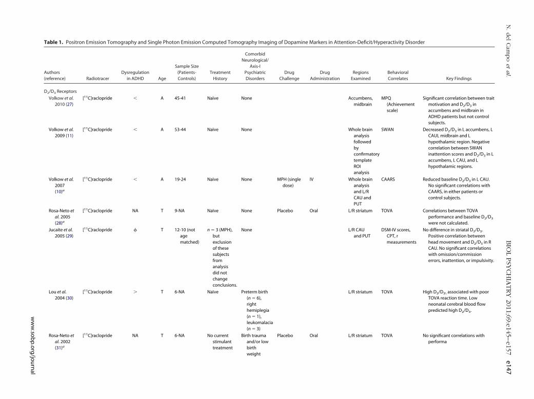
Table 1. Positron Emission Tomography and Single Photon Emission Computed Tomography Imaging of Dopamine Markers in Attention-Deficit/Hyperactivity Disorder
Authors(reference) Radiotracer
Dysregulationin ADHD Age
Sample Size(Patients-Controls)
TreatmentHistory
ComorbidNeurological/
Axis-IPsychiatricDisorders
DrugChallenge
DrugAdministration
RegionsExamined
BehavioralCorrelates Key Findings
D2/D3 Receptors
Volkow et al.2010 (27)
[11C]raclopride � A 45-41 Naïve None Accumbens,midbrain
MPQ(Achievementscale)
Significant correlation between traitmotivation and D2/D3 inaccumbens and midbrain inADHD patients but not controlsubjects.
Volkow et al.2009 (11)
[11C]raclopride � A 53-44 Naïve None Whole brainanalysisfollowedbyconfirmatorytemplateROIanalysis
SWAN Decreased D2/D3 in L accumbens, LCAUL midbrain and Lhypothalamic region. Negativecorrelation between SWANinattention scores and D2/D3 in Laccumbens, L CAU, and Lhypothalamic regions.
Volkow et al.2007(10)a
[11C]raclopride � A 19-24 Naïve None MPH (singledose)
IV Whole brainanalysisand L/RCAU andPUT
CAARS Reduced baseline D2/D3 in L CAU.No significant correlations withCAARS, in either patients orcontrol subjects.
Rosa-Neto etal. 2005(28)a
[11C]raclopride NA T 9-NA Naïve None Placebo Oral L/R striatum TOVA Correlations between TOVAperformance and baseline D2/D3
were not calculated.
Jucaite et al.2005 (29)
[11C]raclopride � T 12-10 (notagematched)
n � 3 (MPH),butexclusionof thesesubjectsfromanalysisdid notchangeconclusions.
None L/R CAUand PUT
DSM-IV scores,CPT, rmeasurements
No difference in striatal D2/D3.Positive correlation betweenhead movement and D2/D3 in RCAU. No significant correlationswith omission/commissionerrors, inattention, or impulsivity.
Lou et al.2004 (30)
[11C]raclopride � T 6-NA Naïve Preterm birth(n � 6),righthemiplegia(n � 1),leukomalacia(n � 3)
L/R striatum TOVA High D2/D3\ associated with poorTOVA reaction time. Lowneonatal cerebral blood flowpredicted high D2/D3.
Rosa-Neto etal. 2002(31)a
[11C]raclopride NA T 6-NA No currentstimulanttreatment
Birth traumaand/or lowbirthweight
Placebo Oral L/R striatum TOVA No significant correlations withperforma
N.del
Cam
po
eta
l.BIO
LPSY
CH
IATRY
2011;69:e145–e157
e147
ww
w.so
bp
.org
/jou
rnal
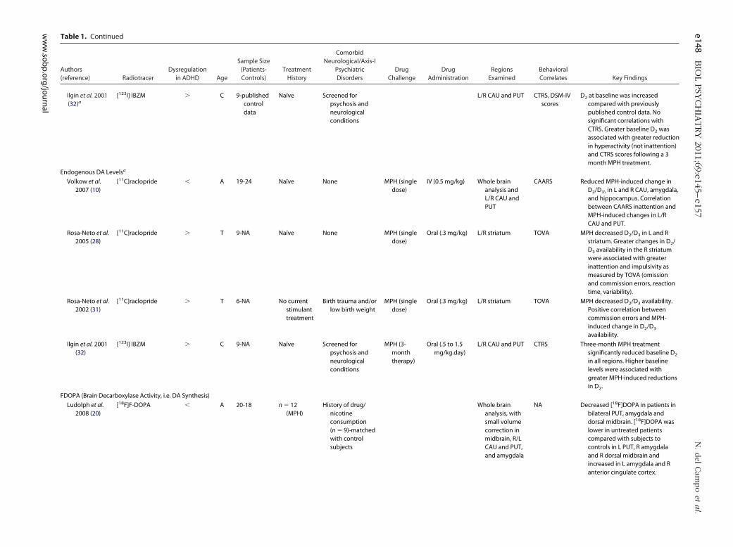
Table 1. Continued
Authors(reference) Radiotracer
Dysregulationin ADHD Age
Sample Size(Patients-Controls)
TreatmentHistory
ComorbidNeurological/Axis-I
PsychiatricDisorders
DrugChallenge
DrugAdministration
RegionsExamined
BehavioralCorrelates Key Findings
Ilgin et al. 2001(32)a
[123I] IBZM � C 9-publishedcontroldata
Naïve Screened forpsychosis andneurologicalconditions
L/R CAU and PUT CTRS, DSM-IVscores
D2 at baseline was increasedcompared with previouslypublished control data. Nosignificant correlations withCTRS. Greater baseline D2 wasassociated with greater reductionin hyperactivity (not inattention)and CTRS scores following a 3month MPH treatment.
Endogenous DA Levelsa
Volkow et al.2007 (10)
[11C]raclopride � A 19-24 Naïve None MPH (singledose)
IV (0.5 mg/kg) Whole brainanalysis andL/R CAU andPUT
CAARS Reduced MPH-induced change inD2/D3\ in L and R CAU, amygdala,and hippocampus. Correlationbetween CAARS inattention andMPH-induced changes in L/RCAU and PUT.
Rosa-Neto et al.2005 (28)
[11C]raclopride � T 9-NA Naïve None MPH (singledose)
Oral (.3 mg/kg) L/R striatum TOVA MPH decreased D2/D3 in L and Rstriatum. Greater changes in D2/D3 availability in the R striatumwere associated with greaterinattention and impulsivity asmeasured by TOVA (omissionand commission errors, reactiontime, variability).
Rosa-Neto et al.2002 (31)
[11C]raclopride � T 6-NA No currentstimulanttreatment
Birth trauma and/orlow birth weight
MPH (singledose)
Oral (.3 mg/kg) L/R striatum TOVA MPH decreased D2/D3 availability.Positive correlation betweencommission errors and MPH-induced change in D2/D3
availability.
Ilgin et al. 2001(32)
[123I] IBZM � C 9-NA Naïve Screened forpsychosis andneurologicalconditions
MPH (3-monththerapy)
Oral (.5 to 1.5mg/kg.day)
L/R CAU and PUT CTRS Three-month MPH treatmentsignificantly reduced baseline D2
in all regions. Higher baselinelevels were associated withgreater MPH-induced reductionsin D2.
FDOPA (Brain Decarboxylase Activity, i.e. DA Synthesis)
Ludolph et al.2008 (20)
[18F]F-DOPA � A 20-18 n � 12(MPH)
History of drug/nicotineconsumption(n � 9)-matchedwith controlsubjects
Whole brainanalysis, withsmall volumecorrection inmidbrain, R/LCAU and PUT,and amygdala
NA Decreased [18F]DOPA in patients inbilateral PUT, amygdala anddorsal midbrain. [18F]DOPA waslower in untreated patientscompared with subjects tocontrols in L PUT, R amygdalaand R dorsal midbrain andincreased in L amygdala and Ranterior cingulate cortex.
e148B
IOL
PSY
CH
IATRY
2011;69:e145–e157
N.del
Cam
po
eta
l.
ww
w.so
bp
.org
/jou
rnal
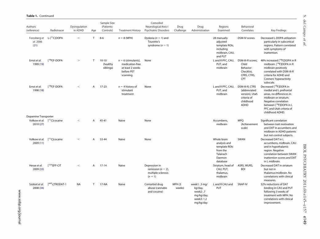
Table 1. Continued
Authors(reference) Radiotracer
Dysregulationin ADHD Age
Sample Size(Patients-Controls) Treatment History
ComorbidNeurological/Axis-I
Psychiatric DisordersDrug
ChallengeDrug
AdministrationRegions
ExaminedBehavioralCorrelates Key Findings
Forssberg etal. 2006(21)
L-[11C]DOPA � T 8-6 n � 8 (MPH) Dyslexia (n � 1) andTourette’ssyndrome (n � 1)
28 manuallyadjustedtemplate ROIs,includingmidbrain, CAU,and PUT
DSM-IV scores Decreased L-DOPA utilizationparticularly in subcorticalregions. Pattern correlatedwith symptoms ofinattention.
Ernst et al.1999 (19)
[18F]F-DOPA � T 10-10(healthysiblings)
n � 6 (stimulants),medication-freeat least 2 weeksbefore PETscanning.
None L and R PFC, CAU,PUT, andmidbrain
DSM-III-R scores,ChildBehaviorChecklist,CPRS, CTRS,CPT
48% increased [18F]DOPA in Rmidbrain. [18F]DOPA in Rmidbrain positivelycorrelated with DSM-III-Rcriteria for ADHD andConners’ hyperactivitysubscale.
Ernst et al.1998 (18)
[18F]F-DOPA � A 17-23 n � 4 history ofstimulanttreatment
None L and R PFC, CAU,PUT, andmidbrain
DSM-III-R, CTRS(abbreviatedversion), Utahcriteria ofchildhoodADHD
Decreased [18F]DOPA inmedial and L prefrontalareas, no differences inmidbrain or striatum.Negative correlationbetween [18F]DOPA in LPFC and Utah criteria ofchildhood ADHD.
Dopamine Transporter
Volkow et al.2010 (27)
[11C]cocaine � A 45-41 Naïve None Accumbens,midbrain
MPQ(Achievementscale)
Significant correlationbetween trait motivationand DAT in accumbens andmidbrain in ADHD patientsbut not control subjects.
Volkow et al.2009 (11)
[11C]cocaine � A 53-44 Naïve None Whole brainanalysis andtemplate ROIsfrom theTalairachDaemondatabase
SWAN Decreased DAT in Laccumbens, midbrain, CAUand in hypothalamicregion. Negativecorrelation between SWANinattention scores and DATin L midbrain.
Hesse et al.2009 (33)
[123I]FP-CIT � A 17-14 Naïve Depression inremission (n � 2),multiple sclerosis(n � 1)
Striatum, head ofCAU, PUT,thalamus,midbrain
ASRS, WURS,BDI
Decreased DAT in striatumbut not inthalamus/midbrain. Nocorrelations with clinicalmeasures.
Szobot et al.2008 (34)
[99Tc]TRODAT-1 NA T 17-NA Naïve Comorbid drugabuse (cannabisand cocaine)
MPH (3weeks)
week1: .3 mg/kg/day,week2: .7mg/kg/day,week3: 1.2mg/kg/day
L and R CAU andPUT
SNAP-IV 52% reductions of DATbinding in CAU and PUTfollowing 3 weeks oftreatment with MPH. Nocorrelations with clinicalimprovement.
N.del
Cam
po
eta
l.BIO
LPSY
CH
IATRY
2011;69:e145–e157
e149
ww
w.so
bp
.org
/jou
rnal
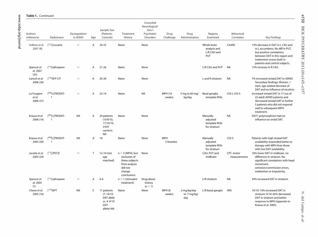
Table 1. Continued
Authors(reference) Radiotracer
Dysregulationin ADHD Age
Sample Size(Patients-Controls)
TreatmentHistory
ComorbidNeurological/
Axis-IPsychiatricDisorders
DrugChallenge
DrugAdministration
RegionsExamined
BehavioralCorrelates Key Findings
Volkow et al.2007 (9)
[11C]cocaine � A 20-25 Naïve None Whole brainanalysis andL/R CAU andPUT
CAARS 13% decrease in DAT in L CAU andin L accumbens. No diff in PUT,but positive correlationsbetween DAT in this region andinattention scores both inpatients and control subjects.
Spencer etal. 2007(35)
[11C]altropane � A 21-26 Naïve None L/R CAU and PUT NA 15% increase in R CAU.
Larisch et al.2006 (36)
[123I]FP-CIT � A 20-20 Naïve None L and R striatum NA 7% increased striatal DAT in ADHD.Secondary findings: Women �men, age-related decrease ofDAT and no influence of nicotine.
La Fougereet al.2006 (37)
[99Tc]TRODAT-1
� A 22-14 Naïve NA MPH (10weeks)
5 mg to 60 mg/kg/day
Basal gangliatemplate ROIs
CGI-I, CGI-S Increased striatal DAT in 17 out of22 adult ADHD patients anddecreased striatal DAT in further5 patients who did not respondwell to subsequent MPHtreatment.
Krause et al.2006 (14)
[99Tc]TRODAT-1
NA A 29 patients(10:9/10,17:10/10,2:9/9carriers).NA
Naïve None Manuallyadjustedtemplate ROIsfor striatum
NA DAT1 polymorphism had noinfluence on strital DAT.
Krause et al.2005 (38)
[99Tc]TRODAT-1
NA A 18 Naïve None MPH(10weeks)
Manuallyadjustedtemplate ROIsfor striatum
CGI-S Patients with high striatal DATavailability responded better totherapy with MPH than thosewith low DAT availability.
Jucaite et al.2005 (29)
[11C]PET2I � T 12-10 (notagematched)
n � 3 (MPH), butexclusion ofthese subjectsfrom analysisdid notchangeconclusions.
None CAU, PUT andmidbrain
CPT, motormeasurements
16% lower DAT in midbrain, nodifference in striatum. Nosignificant correlations with headmovement,omission/commission errors,inattention or impulsivity.
Spencer etal. 2005(7)
[11C]altropane � A 6-6 n � 1 (stimulanttreatment)
Drug abusehistory(n � 1)
L/R striatum NA 34% increased DAT in striatum.
Cheon et al.2005 (16)
[123I]IPT NA C 11 patients(7: 10/10DAT allelevs. 4: 9/10DATallele)-NA
Naïve None MPH (8weeks)
.3 mg/kg/dayto .7 mg/kg/day
L/R basal ganglia ARS 10/10: 14% increased DAT instriatum; 9/10: 65% decreasedDAT in striatum and betterresponse to MPH (opposite toKrause et al. 2005)
e150B
IOL
PSY
CH
IATRY
2011;69:e145–e157
N.del
Cam
po
eta
l.
ww
w.so
bp
.org
/jou
rnal
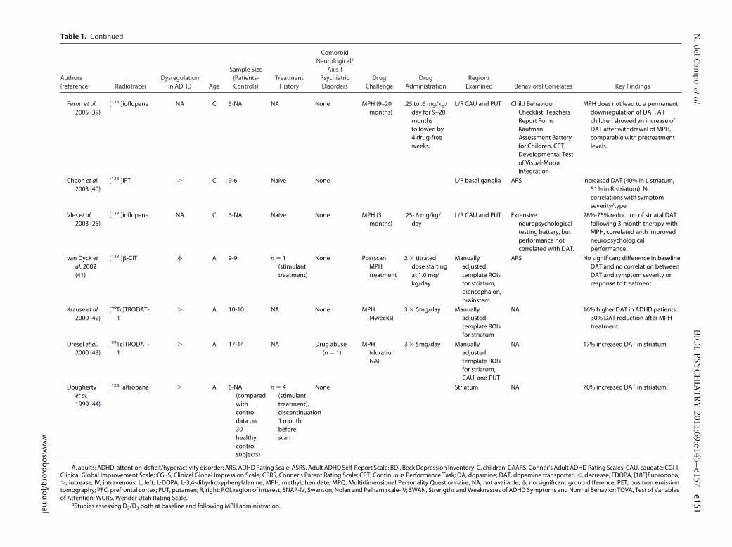
Table 1. Continued
Authors(reference) Radiotracer
Dysregulationin ADHD Age
Sample Size(Patients-Controls)
TreatmentHistory
ComorbidNeurological/
Axis-IPsychiatricDisorders
DrugChallenge
DrugAdministration
RegionsExamined Behavioral Correlates Key Findings
Feron et al.2005 (39)
[123I]ioflupane NA C 5-NA NA None MPH (9–20months)
.25 to .6 mg/kg/day for 9–20monthsfollowed by4 drug-freeweeks.
L/R CAU and PUT Child BehaviourChecklist, TeachersReport Form,KaufmanAssessment Batteryfor Children, CPT,Developmental Testof Visual-MotorIntegration
MPH does not lead to a permanentdownregulation of DAT. Allchildren showed an increase ofDAT after withdrawal of MPH,comparable with pretreatmentlevels.
Cheon et al.2003 (40)
[123I]IPT � C 9-6 Naïve None L/R basal ganglia ARS Increased DAT (40% in L striatum,51% in R striatum). Nocorrelations with symptomseverity/type.
Vles et al.2003 (25)
[123I]ioflupane NA C 6-NA Naïve None MPH (3months)
.25-.6 mg/kg/day
L/R CAU and PUT Extensiveneuropsychologicaltesting battery, butperformance notcorrelated with DAT.
28%-75% reduction of striatal DATfollowing 3-month therapy withMPH, correlated with improvedneuropsychologicalperformance.
van Dyck etal. 2002(41)
[123I]�-CIT � A 9-9 n � 1(stimulanttreatment)
None PostscanMPHtreatment
2 � titrateddose startingat 1.0 mg/kg/day
Manuallyadjustedtemplate ROIsfor striatum,diencephalon,brainstem
ARS No significant difference in baselineDAT and no correlation betweenDAT and symptom severity orresponse to treatment.
Krause et al.2000 (42)
[99Tc]TRODAT-1
� A 10-10 NA None MPH(4weeks)
3 � 5mg/day Manuallyadjustedtemplate ROIsfor striatum
NA 16% higher DAT in ADHD patients.30% DAT reduction after MPHtreatment.
Dresel et al.2000 (43)
[99Tc]TRODAT-1
� A 17-14 NA Drug abuse(n � 1)
MPH(durationNA)
3 � 5mg/day Manuallyadjustedtemplate ROIsfor striatum,CAU, and PUT
NA 17% increased DAT in striatum.
Doughertyet al.1999 (44)
[123I]altropane � A 6-NA(comparedwithcontroldata on30healthycontrolsubjects)
n � 4(stimulanttreatment),discontinuation1 monthbeforescan
None Striatum NA 70% increased DAT in striatum.
A, adults; ADHD, attention-deficit/hyperactivity disorder; ARS, ADHD Rating Scale; ASRS, Adult ADHD Self-Report Scale; BDI, Beck Depression Inventory; C, children; CAARS, Conner’s Adult ADHD Rating Scales; CAU, caudate; CGI-I,Clinical Global Improvement Scale; CGI-S, Clinical Global Impression Scale; CPRS, Conner’s Parent Rating Scale; CPT, Continuous Performance Task; DA, dopamine; DAT, dopamine transporter; �, decrease; FDOPA, [18F]fluorodopa;�, increase; IV, intravenous; L, left; L-DOPA, L-3,4-dihydroxyphenylalanine; MPH, methylphenidate; MPQ, Multidimensional Personality Questionnaire; NA, not available; �, no significant group difference; PET, positron emissiontomography; PFC, prefrontal cortex; PUT, putamen; R, right; ROI, region of interest; SNAP-IV, Swanson, Nolan and Pelham scale-IV; SWAN, Strengths and Weaknesses of ADHD Symptoms and Normal Behavior; TOVA, Test of Variablesof Attention; WURS, Wender Utah Rating Scale.
aStudies assessing D2/D3 both at baseline and following MPH administration.
N.del
Cam
po
eta
l.BIO
LPSY
CH
IATRY
2011;69:e145–e157
e151
ww
w.so
bp
.org
/jou
rnal
oest
tmadttit
g(hcDlt
I
acdOsmdpitsn
oifri
pagdpdr(
ID
psttuiA
fttuiealc
ir
e152 BIOL PSYCHIATRY 2011;69:e145–e157 N. del Campo et al.
date, hippocampus, and left amygdala. These findings contrastwith those previously reported in adolescent ADHD patientssuggesting that greater oral MPH-induced increases in DA con-centrations in the right striatum are associated with greatersymptom severity (28) (Table 1). A potential factor explainingthis discrepancy is the difference in age of the patients assessed.Whether stimulant effects in the cortex or in other low D2/D3
receptor regions differ in ADHD patients and control subjectsneeds to be explored further.
Understanding the Therapeutic Effects ofPsychostimulants
Implication of Frontostriatal NetworksThe frontal cortex and the basal ganglia operate together to
execute goal-directed behaviors via functionally segregated neuralnetworks. Based on the characterization of neuroanatomical pro-jections in nonhuman primates, inputs to the striatum from limbic,associative, and motor areas of the prefrontal cortex are known tobe organized topographically along a ventromedial to dorsolateralgradient, with activity along this gradient modulating limbic, cog-nitive, and motor processing (75). This framework has served to
rganize the human striatum into ventral striatum, implicated inmotion, motivation, and reward-guided behaviors; associativetriatum, involved in cognition; and sensorimotor striatum, relatedo motor function (76).
A number of neuroimaging studies have recently attemptedo map striatal subregions and associated neural circuits in hu-
ans. Accumulating evidence from probabilistic tractographynalyses on magnetic resonance diffusion-weighted imagingata have confirmed the topographic segregation of corticos-
riatal projections (77–79). Moreover, using functional connec-ivity analyses of resting state functional magnetic resonancemaging data, which allow for the mapping of large-scale func-ional networks, Di Martino et al. (80) demonstrated differential
patterns of connectivity in striatal subregions along an affective/cognitive/motor axis predicted by the above model of basalganglia function.
In vivo evidence for the dopaminergic modulation of distinctcorticostriatal networks comes from a study investigating the ef-fects of a DA manipulation on seed-based resting-state functionalconnectivity in healthy control subjects (81). Acute administrationof L-3,4-dihydroxyphenylalanine, a DA precursor, was observed toalter resting state functional connectivity in pathways implicated inmotor and cognitive function. Although the above methodologiesenabling the mapping of frontostriatal connectivity are yet to beused in conjunction with psychostimulant challenges, abnormalfrontostriatal connectivity in unmedicated, but not medicated, chil-dren with ADHD was demonstrated (82).
Recent PET evidence has highlighted the utility of guiding theanalysis and interpretation of DA receptor imaging studies by em-ploying a model of functional rather than anatomical subdivisionsof the striatum. A well-replicated region-based approach to inves-tigate the distribution of D2/D3 receptors within the human stria-tum across ventral, associative, and sensorimotor striatum is thatdescribed by Martinez et al. (26). The wide application of this meth-odology has proved extremely valuable to our understanding ofcorticostriatal DA neurotransmission, both in health (26,83) anddisease (84,85). An important shortcoming of the extant PET litera-ture in ADHD is that studies often averaged data across the entirestriatum or used different landmarks to define caudate and puta-
men. mwww.sobp.org/journal
The differential effects of D-AMPH on the above striatal subre-ions constitute a well-replicated finding in healthy volunteers
26,68,69). Using [11C]raclopride, oral MPH administered to youngealthy subjects was also reported to result in different sized in-reases in endogenous DA in these subregions. The increment inA levels in specific subregions predicted performance on particu-
ar cognitive tasks (56). Importantly, some of these effects appearedo be modulated by trait impulsivity.
mplication of Nigrostriatal NetworksThe midbrain, via its connections with the striatum, provides
continuous feedforward mechanism of information flow acrossorticostriatal circuits, thereby operating as an interface for theynamic processing of functionally distinct information (76).ne critical aspect that remains particularly unexplored is how
timulants interact with the afferent control of midbrain dopa-ine neurons to orchestrate their effects on cognition. Somato-
endritic D2/D3 autoreceptors located on midbrain DA neuronslay an important role regulating DA synthesis and release, act-
ng as potent inhibitors in the presence of high DA concentra-ions (86,87). Mediated by this negative feedback mechanism,timulant-induced increases in endogenous DA levels inhibit DAeuron firing (88,89).
How this regulatory mechanism modulates therapeutic effectsf psychostimulant medication in ADHD remains to be character-
zed. Volkow et al. (66) suggested that increased endogenous DAollowing DAT blockade by MPH attenuates background firingates, increasing the signal-to-noise ratio of striatal cells, therebymproving attention and reducing distractibility.
The implication of nigrostriatal dysregulations in the thera-eutic effects of stimulants is of particular interest because 1)nimal models of ADHD provide evidence for a hypodopaminer-ic nigrostriatal system in the disorder (90,91), and 2) PET studiesocument abnormal midbrain dopaminergic markers in ADHDatients. Although initially evidence pointed toward increasedopamine synthesis in ADHD (19), more recent investigations
eported reductions in both synthesis (20,21) and midbrain DAT11,29) (Table 1).
nteractions Between Prefrontal Cortex and SubcorticalA Systems
Alterations in endogenous DA levels are likely to trigger pre- andost-synaptic compensatory changes to restore the balance in theystem, including changes in DA synthesis, release, receptor sensi-ivity, and neuronal responsiveness. To specify better DA dysfunc-ion underlying ADHD, abnormal dopaminergic markers need to benderstood in the presence of powerful counteracting regulatory
nfluences. This critical aspect is often overlooked in models ofDHD, primarily because of methodological limitations.
Multiple lines of evidence suggest that D2/D3 receptors are dif-erentially expressed across distinct DA-modulated circuits. Whilehere is evidence indicating that different inverted U-shaped func-ions exist for different forms of behavior (56), it remains to benderstood how DA activity in each of the brain regions implicated
n the aforementioned functional circuits is associated with differ-nt forms of behavior. Microdialysis studies demonstrate that lownd clinically relevant MPH doses preferentially increase extracellu-
ar catecholamines within the prefrontal cortex relative to subcorti-al and other cortical regions (92).
So far, progress in our understanding of cortical DA function-ng based on PET has been hampered by the lack of D2/D3
eceptor ligands suited to image receptors in regions with dra-
atically different receptor densities (as is the case with the
aev
bdleecsrmiiihitvtNbtor
M
bthficNi(
smcs
C
atsAophctmomTeat
dtu
N. del Campo et al. BIOL PSYCHIATRY 2011;69:e145–e157 e153
striatum and the cortex). As a consequence, striatal D2/D3 bind-ing has been widely treated as a proxy for D2/D3 functioningthroughout the brain. This extrapolation is based on a simplisticassumption that individual differences in striatal binding arepredictive of binding elsewhere in the brain. Inasmuch as[18F]fallypride allows simultaneous quantification of receptor
vailability (and changes in endogenous DA) in both striatal andxtrastriatal regions from the same scan, this tracer represents aaluable tool to investigate ADHD and its treatment.
Role of Noradrenaline in ADHD
Though DA dysregulation is central to understanding the neu-robiology of ADHD and its pharmacological treatment, the poten-tial contribution of NA has also long been implicated in the patho-physiology of ADHD (93,94).
Noradrenaline projections originate primarily from neuronsin the locus coeruleus and send projections to multiple regions,including the prefrontal cortices, which play a critical role inhigh-level cognitive functions that are often impaired in ADHD,such as working memory and inhibitory response control(51,95). However, there is only very sparse innervation of thestriatum by NE, and so it is much less implicated in any striatalchanges in ADHD or in effects of stimulant effects that are stria-tally dependent.
Reuptake of NA by NAT is the principal mechanism for terminat-ing NA neurotransmission in the central nervous system (96), withrapid termination of NA actions before diffusion of NA moleculesaway from the synapse. Of the pharmacological treatment optionsavailable for ADHD, it is noteworthy that the overwhelming major-ity of drugs shown to be effective have important effects on NAtransmission, including MPH and D-AMPH. By contrast, selectiveserotonin reuptake inhibitors are generally regarded as ineffectivein treating the cardinal symptoms of ADHD and its cognitive se-quelae (97). The indirect DA/NA agonist buproprion has beenshown to be effective in ADHD (98), as have tricyclic drugs withpotent noradrenergic properties, such as desipramine (99), andspecific alpha-2 receptor agonists such as guanfacine (100).
Modafinil, though not currently approved by the Food and DrugAdministration for ADHD, appears to be effective in its treatment(101). Certain of the behavioral and cognitive effects of modafinilare contingent on the integrity of NA transmission (102). The al-pha-1 receptor antagonist prazosin antagonizes the prolocomotoreffects of modafinil seen in mice (103), while co-administration ofprazosin in healthy volunteers blocks the beneficial effects ofmodafinil seen on the more difficult levels of the Tower of Londontest of frontal lobe function (104). Furthermore, functional mag-netic resonance imaging data suggest that modafinil may modu-late the noradrenergic locus coeruleus system that potentially af-fects prefrontal cortical functioning (105,106).
Another line of evidence implicating brain NA pathways in thetreatment of ADHD symptoms is the development of the relativelyselective NA reuptake inhibitor atomoxetine and its approval forthe treatment of ADHD by the Food and Drug Administration (107).In animals, atomoxetine has been shown to increase cortical NAand DA levels several-fold when given systemically, without puta-tive effects on the subcortical DA system (108). It is these subcorticalDA effects that are thought to be responsible for the abuse poten-tial of psychostimulant treatments (109). As such, NA targetingagents such as atomoxetine may offer clinical advantages by virtueof their limited effects on subcortical DA.
The effects of NA-modulating pharmacotherapies on ADHD
symptomatology are thought to stem, in part, from intermediary teneficial effects on cognition (110). Translational studies haveissociated cognitive effects of NA at subreceptors: moderate
evels of NA improve cognition (including impulse control) viaffects at alpha-2a receptors, while higher levels, such as duringxtreme stress, impair cognition by engaging alpha-1-adreno-eptors (17). The stop-signal task (SST), which measures impul-ivity and is sensitive to ADHD, depends on the integrity of theight inferior frontal gyrus (111–114). On this test, participants
ake speeded motor responses to go stimuli and attempt tonhibit responses when stop stimuli occur. Impaired responsenhibition on the SST is one of the most robust cognitive findingsn ADHD (115). Single doses of MPH, modafinil, and atomoxetineave been found to improve response inhibition in humans and
n rats (116 –118). By contrast, serotonin manipulations appearo have no behavioral effects on the SST (119). In a healthyolunteer study, atomoxetine was found to augment right fron-al lobe activation during SST inhibitory control, linking togetherA reuptake blockade with cognition and prefrontal cortexlood oxygenation level-dependent response (120). It remains
o be seen in humans whether the benefits of ADHD medicationsn response inhibition can be convincingly dissociated with
espect to cortical NA as opposed to DA.
olecular Imaging of the NA System
For many years, there has been a paucity of suitable NA tracers,ecause of nonspecific binding of putative ligands and other fac-
ors (121). The recent successful deployment of NAT radioligands inumans (122–124) represents an important step forward in theeld. Using (S,S)-[11C]methylreboxetine PET in healthy volunteers,linically relevant doses of MPH were found to significantly reduceAT availability in a dose-dependent manner in NAT-rich regions,
ncluding locus coeruleus, raphé, hypothalamus, and thalamus122).
The questions of whether there is a difference in NAT density intimulant-naive ADHD patients and whether current first-line phar-
acotherapies such as MPH and atomoxetine induce similarhanges in NAT availability in ADHD patients and healthy controlubjects remain to be addressed.
onclusions
Overall, the previously discussed findings are consistent withdual role of DA and NA in the pathophysiology of ADHD and its
reatment. It is via the close interplay of both the DA and NAystems in corticostriatal circuitry that pharmacotherapies forDHD operate on different cognitive processes. A growing bodyf evidence suggests that psychostimulants exert their thera-eutic effects in a baseline-dependent manner, according to aypothesized inverted U-shaped function (48,50). The neuro-hemical mechanisms underlying this functional effect remaino be fully specified, although they presumably depend on a
ixture of dopaminergic and noradrenergic actions at the levelf the cortex (especially the prefrontal cortex [95]) and of dopa-inergic effects subcortically, e.g., within the basal ganglia.
hese actions may well be responsible for different therapeuticffects of methylphenidate, whereas more selective agents suchs atomoxetine, which is presumably devoid of significant stria-al activity, may not exert such a range of effects.
Recent PET findings suggest that DA activity in adult ADHD isepressed (9 –11,18,20,21), confirming the catecholamine-agonist
heory of stimulant drugs. However, these recent findings are notniversally accepted, and references to the old and long-accepted
heories regarding ADHD (e.g., increased DAT) still permeate the
www.sobp.org/journal
e154 BIOL PSYCHIATRY 2011;69:e145–e157 N. del Campo et al.
literature. There is a need to replicate and expand the molecularneuroimaging literature in ADHD, controlling for potential con-founding variables, including examination of DA markers in striatalsubregions implicated differentially in limbic, associative, and mo-tor circuits governed by cortical influences.
NdC was funded by the Gates Cambridge Trust. The Behaviouraland Clinical Neuroscience Institute is co-funded by a joint award fromthe Medical Research Council and the Wellcome Trust.
We thank Tim Fryer for help with the manuscript preparation andDr. Ulrich Müller and Javier Bernácer for fruitful discussions.
NdC consults for Cambridge Cognition. SRC consults for CambridgeCognition, P1Vital, and Shire Pharmaceuticals. BJS consults for Cam-bridge Cognition and holds shares in CeNeS. She has consulted forNovartis, Shire, GlaxoSmithKline, Eli Lilly, and Boehringer-Ingelheim.She also receives an honorarium from the Journal of PsychologicalMedicine. TWR is a consultant for Cambridge Cognition, Pfizer, Eli Lilly,GlaxoSmithKline, Allon Therapeutics, Lundbeck, and Pangenics andreceives royalties from Cambridge Cognition and Springer-Verlag. Hehas received research grants from Pfizer, Eli Lilly, and GlaxoSmithKline.
1. Biederman J (2005): Attention-deficit/hyperactivity disorder: A selec-tive overview. Biol Psychiatry 57:1215–1220.
2. Solanto MV (2001): Attention-Deficit/Hyperactivity Disorder New York:Oxford University Press.
3. Molina BS, Hinshaw SP, Swanson JM, Arnold LE, Vitiello B, Jensen PS, etal. (2009): The MTA at 8 years: Prospective follow-up of children treatedfor combined-type ADHD in a multisite study. J Am Acad Child AdolescPsychiatry 48:484 –500.
4. Biederman J, Monuteaux MC, Mick E, Spencer T, Wilens TE, Silva JM, etal. (2006): Young adult outcome of attention deficit hyperactivity dis-order: A controlled 10-year follow-up study. Psychol Med 36:167–179.
5. Arnsten AF (2006): Fundamentals of attention-deficit/hyperactivitydisorder: Circuits and pathways. J Clin Psychiatry 67(suppl 8):7–12.
6. Axelrod J (1974): Regulation of the Neurotransmitter Norepinephrine.Cambridge, MA: MIT Press.
7. Spencer TJ, Biederman J, Madras BK, Faraone SV, Dougherty DD, BonabAA, et al. (2005): In vivo neuroreceptor imaging in attention-deficit/hyperactivity disorder: A focus on the dopamine transporter. Biol Psy-chiatry 57:1293–1300.
8. Madras BK, Fischman AJ, Meltzer PC (2006): Methods for diagnosingand monitoring treatment ADHD by assessing the dopamine trans-porter level. In: Office UP, editor.
9. Volkow ND, Wang GJ, Newcorn J, Fowler JS, Telang F, Solanto MV, et al.(2007): Brain dopamine transporter levels in treatment and drug naïveadults with ADHD. Neuroimage 34:1182–1190.
10. Volkow ND, Wang GJ, Newcorn J, Telang F, Solanto MV, Fowler JS, et al.(2007): Depressed dopamine activity in caudate and preliminary evi-dence of limbic involvement in adults with attention-deficit/hyperac-tivity disorder. Arch Gen Psychiatry 64:932–940.
11. Volkow ND, Wang GJ, Kollins SH, Wigal TL, Newcorn JH, Telang F, et al.(2009): Evaluating dopamine reward pathway in ADHD: Clinical impli-cations. JAMA 302:1084 –1091.
12. Krause J (2008): SPECT and PET of the dopamine transporter in atten-tion-deficit/hyperactivity disorder. Expert Rev Neurother 8:611– 625.
13. Bowton E, Saunders C, Erreger K, Sakrikar D, Matthies HJ, Sen N, et al.(2010): Dysregulation of dopamine transporters via dopamine D2 au-toreceptors triggers anomalous dopamine efflux associated with at-tention-deficit hyperactivity disorder. J Neurosci 30:6048 – 6057.
14. Krause J, Dresel SH, Krause KH, La Fougère C, Zill P, Ackenheil M, et al.(2006): Striatal dopamine transporter availability and DAT-1 gene inadults with ADHD: No higher DAT availability in patients with homozy-gosity for the 10-repeat allele. World J Biol Psychiatry 7:152–157.
15. van Dyck CH, Malison RT, Jacobsen LK, Seibyl JP, Staley JK, Laruelle M, etal. (2005): Increased dopamine transporter availability associated withthe 9-repeat allele of the SLC6A3 gene. J Nucl Med 46:745–751.
16. Cheon KA, Ryu YH, Kim JW, Cho DY (2005): The homozygosity for
10-repeat allele at dopamine transporter gene and dopamine trans-porter density in Korean children with attention deficit hyperactivitywww.sobp.org/journal
disorder: Relating to treatment response to methylphenidate. EurNeuropsychopharmacol 15:95–101.
17. Arnsten AF, Li BM (2005): Neurobiology of executive functions: Cate-cholamine influences on prefrontal cortical functions. Biol Psychiatry57:1377–1384.
18. Ernst M, Zametkin AJ, Matochik JA, Jons PH, Cohen RM (1998): DOPAdecarboxylase activity in attention deficit hyperactivity disorderadults. A [fluorine-18]fluorodopa positron emission tomographicstudy. J Neurosci 18:5901–5907.
19. Ernst M, Zametkin AJ, Matochik JA, Pascualvaca D, Jons PH, Cohen RM(1999): High midbrain [18F]DOPA accumulation in children with atten-tion deficit hyperactivity disorder. Am J Psychiatry 156:1209 –1215.
20. Ludolph AG, Kassubek J, Schmeck K, Glaser C, Wunderlich A, Buck AK,et al. (2008): Dopaminergic dysfunction in attention deficit hyperactiv-ity disorder (ADHD), differences between pharmacologically treatedand never treated young adults: A 3,4-dihydroxy-6-[18F]fluorophenyl-l-alanine PET study. Neuroimage 41:718 –727.
21. Forssberg H, Fernell E, Waters S, Waters N, Tedroff J (2006): Alteredpattern of brain dopamine synthesis in male adolescents with atten-tion deficit hyperactivity disorder. Behav Brain Funct 2:40.
22. van Dyck CH, Seibyl JP, Malison RT, Laruelle M, Zoghbi SS, Baldwin RM,et al. (2002): Age-related decline in dopamine transporters: Analysis ofstriatal subregions, nonlinear effects, and hemispheric asymmetries.Am J Geriatr Psychiatry 10:36 – 43.
23. Takahashi H, Fujimura Y, Hayashi M, Takano H, Kato M, Okubo Y, et al.(2008): Enhanced dopamine release by nicotine in cigarette smokers: Adouble-blind, randomized, placebo-controlled pilot study. Int J Neuro-psychopharmacol 11:413– 417.
24. Salokangas RK, Vilkman H, Ilonen T, Taiminen T, Bergman J,Haaparanta M, et al. (2000): High levels of dopamine activity in thebasal ganglia of cigarette smokers. Am J Psychiatry 157:632– 634.
25. Vles JS, Feron FJ, Hendriksen JG, Jolles J, van Kroonenburgh MJ, WeberWE (2003): Methylphenidate down-regulates the dopamine receptorand transporter system in children with attention deficit hyperkineticdisorder (ADHD). Neuropediatrics 34:77– 80.
26. Martinez D, Slifstein M, Broft A, Mawlawi O, Hwang DR, Huang Y, et al.(2003): Imaging human mesolimbic dopamine transmission with pos-itron emission tomography. Part II: Amphetamine-induced dopaminerelease in the functional subdivisions of the striatum. J Cereb BloodFlow Metab 23:285–300.
27. Volkow ND, Wang GJ, Newcorn JH, Kollins SH, Wigal TL, Telang F, et al.(2010): Motivation deficit in ADHD is associated with dysfunction ofthe dopamine reward pathway [published online ahead of print Sep-tember 21]. Mol Psychiatry.
28. Rosa-Neto P, Lou HC, Cumming P, Pryds O, Karrebaek H, Lunding J, etal. (2005): Methylphenidate-evoked changes in striatal dopamine cor-relate with inattention and impulsivity in adolescents with attentiondeficit hyperactivity disorder. Neuroimage 25:868 – 876.
29. Jucaite A, Fernell E, Halldin C, Forssberg H, Farde L (2005): Reducedmidbrain dopamine transporter binding in male adolescents with at-tention-deficit/hyperactivity disorder: Association between striataldopamine markers and motor hyperactivity. Biol Psychiatry 57:229 –238.
30. Lou HC, Rosa P, Pryds O, Karrebaek H, Lunding J, Cumming P, et al.(2004): ADHD: Increased dopamine receptor availability linked to at-tention deficit and low neonatal cerebral blood flow. Dev Med ChildNeurol 46:179 –183.
31. Rosa Neto P, Lou H, Cumming P, Pryds O, Gjedde A (2002): Methyl-phenidate-evoked potentiation of extracellular dopamine in the brainof adolescents with premature birth: Correlation with attentional def-icit. Ann N Y Acad Sci 965:434 – 439.
32. Ilgin N, Senol S, Gucuyener K, Gokcora N, Sener S (2001): Is increased D2receptor availability associated with response to stimulant medicationin ADHD. Dev Med Child Neurol 43:755–760.
33. Hesse S, Ballaschke O, Barthel H, Sabri O (2009): Dopamine transporterimaging in adult patients with attention-deficit/hyperactivity disorder.Psychiatry Res 171:120 –128.
34. Szobot CM, Shih MC, Schaefer T, Júnior N, Hoexter MQ, Fu YK, et al. (2008):Methylphenidate DAT binding in adolescents with attention-deficit/hy-peractivity disorder comorbid with substance use disorder–a single pho-
ton emission computed tomography with [Tc(99m)]TRODAT-1 study.Neuroimage 40:1195–1201.
N. del Campo et al. BIOL PSYCHIATRY 2011;69:e145–e157 e155
35. Spencer TJ, Biederman J, Madras BK, Dougherty DD, Bonab AA, Livni E,et al. (2007): Further evidence of dopamine transporter dysregulationin ADHD: A controlled PET imaging study using altropane. Biol Psychi-atry 62:1059 –1061.
36. Larisch R, Sitte W, Antke C, Nikolaus S, Franz M, Tress W, et al. (2006):Striatal dopamine transporter density in drug naive patients with at-tention-deficit/hyperactivity disorder. Nucl Med Commun 27:267–270.
37. la Fougère C, Krause J, Krause KH, Josef Gildehaus F, Hacker M, Koch W,et al. (2006): Value of 99mTc-TRODAT-1 SPECT to predict clinical re-sponse to methylphenidate treatment in adults with attention deficithyperactivity disorder. Nucl Med Commun 27:733–737.
38. Krause J, la Fougere C, Krause KH, Ackenheil M, Dresel SH (2005):Influence of striatal dopamine transporter availability on the responseto methylphenidate in adult patients with ADHD. Eur Arch PsychiatryClin Neurosci 255:428 – 431.
39. Feron FJ, Hendriksen JG, van Kroonenburgh MJ, Blom-Coenjaerts C,Kessels AG, Jolles J, et al. (2005): Dopamine transporter in attention-deficit hyperactivity disorder normalizes after cessation of methyl-phenidate. Pediatr Neurol 33:179 –183.
40. Cheon KA, Ryu YH, Kim YK, Namkoong K, Kim CH, Lee JD (2003):Dopamine transporter density in the basal ganglia assessed with[123I]IPT SPET in children with attention deficit hyperactivity disorder.Eur J Nucl Med Mol Imaging 30:306 –311.
41. van Dyck CH, Quinlan DM, Cretella LM, Staley JK, Malison RT, BaldwinRM, et al. (2002): Unaltered dopamine transporter availability in adultattention deficit hyperactivity disorder. Am J Psychiatry 159:309 –312.
42. Krause KH, Dresel SH, Krause J, Kung HF, Tatsch K (2000): Increasedstriatal dopamine transporter in adult patients with attention deficithyperactivity disorder: Effects of methylphenidate as measured bysingle photon emission computed tomography. Neurosci Lett 285:107–110.
43. Dresel S, Krause J, Krause KH, LaFougere C, Brinkbäumer K, Kung HF, etal. (2000): Attention deficit hyperactivity disorder: Binding of [99mTc-]TRODAT-1 to the dopamine transporter before and after methyl-phenidate treatment. Eur J Nucl Med 27:1518 –1524.
44. Dougherty DD, Bonab AA, Spencer TJ, Rauch SL, Madras BK, FischmanAJ (1999): Dopamine transporter density in patients with attentiondeficit hyperactivity disorder. Lancet 354:2132–2133.
45. Wilens TE (2008): Effects of methylphenidate on the catecholaminergicsystem in attention-deficit/hyperactivity disorder. J Clin Psychophar-macol 28:S46 –S53.
46. Zetterström T, Sharp T, Collin AK, Ungerstedt U (1988): In vivo measure-ment of extracellular dopamine and DOPAC in rat striatum after vari-ous dopamine-releasing drugs; implications for the origin of extracel-lular DOPAC. Eur J Pharmacol 148:327–334.
47. Kuczenski R, Segal DS (1975): Differential effects of D- and L-amphet-amine and methylphenidate on rat striatal dopamine biosynthesis. EurJ Pharmacol 30:244 –251.
48. Robbins TW, Sahakian BJ (1979): “Paradoxical” effects of psychomotorstimulant drugs in hyperactive children from the standpoint of behav-ioural pharmacology. Neuropharmacology 18:931–950.
49. Turner DC, Blackwell AD, Dowson JH, McLean A, Sahakian BJ (2005):Neurocognitive effects of methylphenidate in adult attention-deficit/hyperactivity disorder. Psychopharmacol (Berl) 178:286 –295.
50. Mehta MA, Goodyer IM, Sahakian BJ (2004): Methylphenidate im-proves working memory and set-shifting in AD/HD: Relationships tobaseline memory capacity. J Child Psychol Psychiatry 45:293–305.
51. Chamberlain SR, Robbins TW, Winder-Rhodes S, Müller U, Sahakian BJ,Blackwell AD, et al. (2010): Translational approaches to frontostriataldysfunction in attention-deficit/hyperactivity disorder using a com-puterized neuropsychological battery [published online ahead of printNovember 2]. Biol Psychiatry.
52. Elliott R, Sahakian BJ, Matthews K, Bannerjea A, Rimmer J, Robbins TW(1997): Effects of methylphenidate on spatial working memory andplanning in healthy young adults. Psychopharmacol (Berl) 131:196 –206.
53. Mehta MA, Owen AM, Sahakian BJ, Mavaddat N, Pickard JD, RobbinsTW (2000): Methylphenidate enhances working memory by modulat-ing discrete frontal and parietal lobe regions in the human brain.J Neurosci 20:RC65.
54. Koelega HS (1993): Stimulant drugs and vigilance performance: A re-view. Psychopharmacol (Berl) 111:1–16.
55. Turner DC, Robbins TW, Clark L, Aron AR, Dowson J, Sahakian BJ (2003):Relative lack of cognitive effects of methylphenidate in elderly malevolunteers. Psychopharmacol (Berl) 168:455– 464.
56. Clatworthy PL, Lewis SJ, Brichard L, Hong YT, Izquierdo D, Clark L, et al.(2009): Dopamine release in dissociable striatal subregions predictsthe different effects of oral methylphenidate on reversal learning andspatial working memory. J Neurosci 29:4690 – 4696.
57. Naylor H, Halliday R, Callaway E (1985): The effect of methylphenidateon information processing. Psychopharmacol (Berl) 86:90 –95.
58. Rhodes SM, Coghill DR, Matthews K (2006): Acute neuropsychologicaleffects of methylphenidate in stimulant drug-naive boys with ADHDII--broader executive and non-executive domains. J Child Psychol Psy-chiatry 47:1184 –1194.
59. Rapoport JL, Buchsbaum MS, Weingartner H, Zahn TP, Ludlow C, Mik-kelsen EJ (1980): Dextroamphetamine. Its cognitive and behavioraleffects in normal and hyperactive boys and normal men. Arch GenPsychiatry 37:933–943.
60. Mattay VS, Callicott JH, Bertolino A, Heaton I, Frank JA, Coppola R, et al.(2000): Effects of dextroamphetamine on cognitive performance andcortical activation. Neuroimage 12:268 –275.
61. Arnsten AF, Goldman-Rakic PS (1998): Noise stress impairs prefrontalcortical cognitive function in monkeys: Evidence for a hyperdopamin-ergic mechanism. Arch Gen Psychiatry 55:362–368.
62. Mattay VS, Goldberg TE, Fera F, Hariri AR, Tessitore A, Egan MF, et al.(2003): Catechol O-methyltransferase val158 –met genotype and indi-vidual variation in the brain response to amphetamine. Proc Natl AcadSci U S A 100:6186 – 6191.
63. Williams GV, Goldman-Rakic PS (1995): Modulation of memory fieldsby dopamine D1 receptors in prefrontal cortex. Nature 376:572–575.
64. Robbins TW (2010): From behavior to cognition: Functions of mesos-triatal, mesolimbic and mesocortical dopamine systems. In: Iversen LL,Iversen SD, Dunnett SB, Bjorklund A, editors. Dopamine Handbook.New York: Oxford University Press, 203–214.
65. Volkow ND, Wang GJ, Fowler JS, Gatley SJ, Logan J, Ding YS, et al.(1998): Dopamine transporter occupancies in the human brain in-duced by therapeutic doses of oral methylphenidate. Am J Psychiatry155:1325–1331.
66. Volkow ND, Wang G, Fowler JS, Logan J, Gerasimov M, Maynard L, et al.(2001): Therapeutic doses of oral methylphenidate significantly in-crease extracellular dopamine in the human brain. J Neurosci 21:RC121.
67. Volkow ND, Fowler JS, Wang G, Ding Y, Gatley SJ (2002): Mechanism ofaction of methylphenidate: Insights from PET imaging studies. J AttenDisord 6(suppl 1):S31–S43.
68. Riccardi P, Li R, Ansari MS, Zald D, Park S, Dawant B, et al. (2006):Amphetamine-induced displacement of [18F] fallypride in striatumand extrastriatal regions in humans. Neuropsychopharmacology 31:1016 –1026.
69. Cropley VL, Innis RB, Nathan PJ, Brown AK, Sangare JL, Lerner A, et al.(2008): Small effect of dopamine release and no effect of dopaminedepletion on [18F]fallypride binding in healthy humans. Synapse 62:399 – 408.
70. Slifstein M, Kegeles LS, Xu X, Thompson JL, Urban N, Castrillon J, et al.(2010): Striatal and extrastriatal dopamine release measured with PETand [(18)F] fallypride. Synapse 64:350 –362.
71. Moghaddam B, Berridge CW, Goldman-Rakic PS, Bunney BS, Roth RH(1993): In vivo assessment of basal and drug-induced dopamine re-lease in cortical and subcortical regions of the anesthetized primate.Synapse 13:215–222.
72. Sesack SR, Hawrylak VA, Matus C, Guido MA, Levey AI (1998): Dopa-mine axon varicosities in the prelimbic division of the rat prefrontalcortex exhibit sparse immunoreactivity for the dopamine transporter.J Neurosci 18:2697–2708.
73. Morón JA, Brockington A, Wise RA, Rocha BA, Hope BT (2002): Dopa-mine uptake through the norepinephrine transporter in brain regionswith low levels of the dopamine transporter: Evidence from knock-outmouse lines. J Neurosci 22:389 –395.
74. Montgomery AJ, Asselin MC, Farde L, Grasby PM (2007): Measurementof methylphenidate-induced change in extrastriatal dopamine con-
centration using [11C]FLB 457 PET. J Cereb Blood Flow Metab 27:369 –377.www.sobp.org/journal
1
1
1
1
1
1
1
1
1
1
1
1
1
1
1
1
1
1
e156 BIOL PSYCHIATRY 2011;69:e145–e157 N. del Campo et al.
75. Haber SN, Fudge JL, McFarland NR (2000): Striatonigrostriatal path-ways in primates form an ascending spiral from the shell to the dorso-lateral striatum. J Neurosci 20:2369 –2382.
76. Haber SN (2003): The primate basal ganglia: Parallel and integrativenetworks. J Chem Neuroanat 26:317–330.
77. Lehéricy S, Ducros M, Van de Moortele PF, Francois C, Thivard L, Pou-pon C, et al. (2004): Diffusion tensor fiber tracking shows distinct corti-costriatal circuits in humans. Ann Neurol 55:522–529.
78. Draganski B, Kherif F, Klöppel S, Cook PA, Alexander DC, Parker GJ, et al.(2008): Evidence for segregated and integrative connectivity patternsin the human basal ganglia. J Neurosci 28:7143–7152.
79. Cohen MX, Schoene-Bake JC, Elger CE, Weber B (2009): Connectivity-based segregation of the human striatum predicts personality charac-teristics. Nat Neurosci 12:32–34.
80. Di Martino A, Scheres A, Margulies DS, Kelly AM, Uddin LQ, Shehzad Z,et al. (2008): Functional connectivity of human striatum: A resting stateFMRI study. Cereb Cortex 18:2735–2747.
81. Kelly C, de Zubicaray G, Di Martino A, Copland DA, Reiss PT, Klein DF, etal. (2009): L-dopa modulates functional connectivity in striatal cogni-tive and motor networks: A double-blind placebo-controlled study.J Neurosci 29:7364 –7378.
82. Rubia K, Halari R, Cubillo A, Mohammad AM, Brammer M, Taylor E(2009): Methylphenidate normalises activation and functional connec-tivity deficits in attention and motivation networks in medication-naïve children with ADHD during a rewarded continuous performancetask. Neuropharmacology 57:640 – 652.
83. Cervenka S, Bäckman L, Cselényi Z, Halldin C, Farde L (2008): Associa-tions between dopamine D2-receptor binding and cognitive perfor-mance indicate functional compartmentalization of the human stria-tum. Neuroimage 40:1287–1295.
84. Kegeles LS, Abi-Dargham A, Frankle WG, Gil R, Cooper TB, Slifstein M, etal. (2010): Increased synaptic dopamine function in associative regionsof the striatum in schizophrenia. Arch Gen Psychiatry 67:231–239.
85. Martinez D, Narendran R, Foltin RW, Slifstein M, Hwang DR, Broft A, etal. (2007): Amphetamine-induced dopamine release: Markedlyblunted in cocaine dependence and predictive of the choice to self-administer cocaine. Am J Psychiatry 164:622– 629.
86. Sesack SR, Aoki C, Pickel VM (1994): Ultrastructural localization of D2receptor-like immunoreactivity in midbrain dopamine neurons andtheir striatal targets. J Neurosci 14:88 –106.
87. Mercuri NB, Saiardi A, Bonci A, Picetti R, Calabresi P, Bernardi G, et al.(1997): Loss of autoreceptor function in dopaminergic neurons fromdopamine D2 receptor deficient mice. Neuroscience 79:323–327.
88. Bunney BS, Achajanian GK (1976): d-amphetamine-induced inhibitionof central dopaminergic neurons: Mediation by a striato-nigral feed-back pathway. Science 192:391–393.
89. Shi WX, Pun CL, Zhou Y (2004): Psychostimulants induce low-fre-quency oscillations in the firing activity of dopamine neurons. Neuro-psychopharmacology 29:2160 –2167.
90. Leo D, Sorrentino E, Volpicelli F, Eyman M, Greco D, Viggiano D, et al.(2003): Altered midbrain dopaminergic neurotransmission during de-velopment in an animal model of ADHD. Neurosci Biobehav Rev 27:661– 669.
91. Viggiano D, Vallone D, Sadile A (2004): Dysfunctions in dopaminesystems and ADHD: Evidence from animals and modeling. Neural Plast11:97–114.
92. Berridge CW, Devilbiss DM (2010): Psychostimulants as cognitive en-hancers: The prefrontal cortex, catecholamines, and attention-deficit/hyperactivity disorder [published online ahead of print September 25].Biol Psychiatry.
93. Zametkin AJ, Rapoport JL (1987): Neurobiology of attention deficitdisorder with hyperactivity: Where have we come in 50 years? J AmAcad Child Adolesc Psychiatry 26:676 – 686.
94. Arnsten AF, Steere JC, Hunt RD (1996): The contribution of alpha 2-nor-adrenergic mechanisms of prefrontal cortical cognitive function. Po-tential significance for attention-deficit hyperactivity disorder. ArchGen Psychiatry 53:448 – 455.
95. Robbins TW, Arnsten AF (2009): The neuropsychopharmacology offronto-executive function: Monoaminergic modulation. Annu Rev Neu-rosci 32:267–287.
96. Seneca N, Gulyas B, Varrone A, Schou M, Airaksinen A, Tauscher J, et al.(2006): Atomoxetine occupies the norepinephrine transporter in a
www.sobp.org/journal
dose-dependent fashion: A PET study in nonhuman primate brainusing (S,S)-[18F]FMeNER-D2. Psychopharmacol (Berl) 188:119 –127.
97. Biederman J, Spencer T (1999): Attention-deficit/hyperactivity disor-der (ADHD) as a noradrenergic disorder. Biol Psychiatry 46:1234 –1242.
98. Casat CD, Pleasants DZ, Van Wyck Fleet J (1987): A double-blind trial ofbupropion in children with attention deficit disorder. Psychopharma-col Bull 23:120 –122.
99. Wilens TE, Biederman J, Prince J, Spencer TJ, Faraone SV, Warburton R,et al. (1996): Six-week, double-blind, placebo-controlled study of de-sipramine for adult attention deficit hyperactivity disorder. Am J Psy-chiatry 153:1147–1153.
00. Sallee FR, McGough J, Wigal T, Donahue J, Lyne A, Biederman J, SPD503Study Group (2009): Guanfacine extended release in children and ad-olescents with attention-deficit/hyperactivity disorder: A placebo-controlled trial. J Am Acad Child Adolesc Psychiatry 48:155–165.
01. Greenhill LL, Biederman J, Boellner SW, Rugino TA, Sangal RB, Earl CQ,et al. (2006): A randomized, double-blind, placebo-controlled study ofmodafinil film-coated tablets in children and adolescents with atten-tion-deficit/hyperactivity disorder. J Am Acad Child Adolesc Psychiatry45:503–511.
02. Minzenberg MJ, Carter CS (2008): Modafinil: A review of neurochemicalactions and effects on cognition. Neuropsychopharmacology 33:1477–1502.
03. Duteil J, Rambert FA, Pessonnier J, Hermant JF, Gombert R, Assous E(1990): Central alpha 1-adrenergic stimulation in relation to the behav-iour stimulating effect of modafinil; studies with experimental animals.Eur J Pharmacol 180:49 –58.
04. Winder-Rhodes SE, Chamberlain SR, Idris MI, Robbins TW, Sahakian BJ,Müller U (2010): Effects of modafinil and prazosin on cognitive andphysiological functions in healthy volunteers. J Psychopharmacol (Ox-ford) 24:1649 –1657.
05. Astafiev SV, Snyder AZ, Shulman GL, Corbetta M (2010): Comment on“Modafinil shifts human locus coeruleus to low-tonic, high-phasic ac-tivity during functional MRI” and “Homeostatic sleep pressure andresponses to sustained attention in the suprachiasmatic area.” Science328:309.
06. Minzenberg MJ, Watrous AJ, Yoon JH, Ursu S, Carter CS (2008):Modafinil shifts human locus coeruleus to low-tonic, high-phasic ac-tivity during functional MRI. Science 322:1700 –1702.
07. Faraone SV, Biederman J, Spencer T, Michelson D, Adler L, Reimherr F,Glatt SJ (2005): Efficacy of atomoxetine in adult attention-deficit/hy-peractivity disorder: A drug-placebo response curve analysis. BehavBrain Funct 1:16.
08. Bymaster FP, Katner JS, Nelson DL, Hemrick-Luecke SK, Threlkeld PG,Heiligenstein JH, et al. (2002): Atomoxetine increases extracellular lev-els of norepinephrine and dopamine in prefrontal cortex of rat: Apotential mechanism for efficacy in attention deficit/hyperactivity dis-order. Neuropsychopharmacology 27:699 –711.
09. Volkow ND (2006): Stimulant medications: How to minimize their rein-forcing effects? Am J Psychiatry 163:359 –361.
10. Chamberlain SR, Robbins TW, Sahakian BJ (2007): The neurobiology ofattention-deficit/hyperactivity disorder. Biol Psychiatry 61:1317–1319.
11. Aron AR, Fletcher PC, Bullmore ET, Sahakian BJ, Robbins TW (2003):Stop-signal inhibition disrupted by damage to right inferior frontalgyrus in humans. Nat Neurosci 6:115–116.
12. Hampshire A, Chamberlain SR, Monti MM, Duncan J, Owen AM (2010):The role of the right inferior frontal gyrus: Inhibition and attentionalcontrol. Neuroimage 50:1313–1319.
13. Logan GD, Cowan WB, Davis KA (1984): On the ability to inhibit simpleand choice reaction time responses: A model and a method. J ExpPsychol Hum Percept Perform 10:276 –291.
14. Aron AR, Robbins TW, Poldrack RA (2004): Inhibition and the rightinferior frontal cortex. Trends Cogn Sci (Regul Ed) 8:170 –177.
15. Lijffijt M, Kenemans JL, Verbaten MN, van Engeland H (2005): A meta-analytic review of stopping performance in attention-deficit/hyperac-tivity disorder: Deficient inhibitory motor control? J Abnorm Psychol114:216 –222.
16. Chamberlain SR, Sahakian BJ (2007): The neuropsychiatry of impulsiv-ity. Curr Opin Psychiatry 20:255–261.
17. Eagle DM, Bari A, Robbins TW (2008): The neuropsychopharmacology
of action inhibition: Cross-species translation of the stop-signal andgo/no-go tasks. Psychopharmacol (Berl) 199:439 – 456.
1
1
1
N. del Campo et al. BIOL PSYCHIATRY 2011;69:e145–e157 e157
118. Bari A, Eagle DM, Mar AC, Robinson ES, Robbins TW (2009): Dissociableeffects of noradrenaline, dopamine, and serotonin uptake blockade onstop task performance in rats. Psychopharmacol (Berl) 205:273–283.
119. Chamberlain SR, Müller U, Deakin JB, Corlett PR, Dowson J, Cardinal RN,et al. (2007): Lack of deleterious effects of buspirone on cognition inhealthy male volunteers. J Psychopharmacol (Oxford) 21:210 –215.
120. Chamberlain SR, Hampshire A, Müller U, Rubia K, Del Campo N, Craig K,et al. (2009): Atomoxetine modulates right inferior frontal activationduring inhibitory control: A pharmacological functional magnetic res-onance imaging study. Biol Psychiatry 65:550 –555.
121. Ding YS, Fowler J (2005): New-generation radiotracers for nAChR and
NET. Nucl Med Biol 32:707–718.22. Hannestad J, Gallezot JD, Planeta-Wilson B, Lin SF, Williams WA, vanDyck CH, et al. (2010): Clinically relevant doses of methylphenidatesignificantly occupy norepinephrine transporters in humans in vivo.Biol Psychiatry 68:854 – 860.
23. Ding YS, Singhal T, Planeta-Wilson B, Gallezot JD, Nabulsi N, Labaree D,et al. (2010): PET imaging of the effects of age and cocaine on thenorepinephrine transporter in the human brain using (S,S)-[(11)C]O-methylreboxetine and HRRT. Synapse 64:30 –38.
24. Takano A, Varrone A, Gulyás B, Karlsson P, Tauscher J, Halldin C(2008): Mapping of the norepinephrine transporter in the humanbrain using PET with (S,S)-[18F]FMeNER-D2. Neuroimage 42:474 –
482.www.sobp.org/journal
Related Documents