INVITED MANUSCRIPT The role of whole brain radiation therapy in the management of newly diagnosed brain metastases: a systematic review and evidence-based clinical practice guideline Laurie E. Gaspar • Minesh P. Mehta • Roy A. Patchell • Stuart H. Burri • Paula D. Robinson • Rachel E. Morris • Mario Ammirati • David W. Andrews • Anthony L. Asher • Charles S. Cobbs • Douglas Kondziolka • Mark E. Linskey • Jay S. Loeffler • Michael McDermott • Tom Mikkelsen • Jeffrey J. Olson • Nina A. Paleologos • Timothy C. Ryken • Steven N. Kalkanis Received: 7 September 2009 / Accepted: 8 November 2009 / Published online: 4 December 2009 Ó The Author(s) 2009. This article is published with open access at Springerlink.com Abstract Should whole brain radiation therapy (WBRT) be used as the sole therapy in patients with newly-diagnosed, surgi- cally accessible, single brain metastases, compared with WBRT plus surgical resection, and in what clinical set- tings? Target population This recommendation applies to adults with newly diag- nosed single brain metastases amenable to surgical resec- tion; however, the recommendation does not apply to relatively radiosensitive tumors histologies (i.e., small cell lung cancer, leukemia, lymphoma, germ cell tumors and multiple myeloma). Recommendation Surgical resection plus WBRT versus WBRT alone Level 1 Class I evidence supports the use of surgical resection plus post-operative WBRT, as compared to WBRT alone, in patients with good performance status (functionally indepen- dent and spending less than 50% of time in bed) and limited extra-cranial disease. There is insufficient evidence to make a recommendation for patients with poor performance scores, advanced systemic disease, or multiple brain metastases. If WBRT is used, is there an optimal dosing/fractionation schedule? Target population This recommendation applies to adults with newly diag- nosed brain metastases. Recommendation Level 1 Class I evidence suggests that altered dose/frac- tionation schedules of WBRT do not result in significant differences in median survival, local control or neurocognitive outcomes when compared with ‘‘standard’’ WBRT dose/fractionation. (i.e., 30 Gy in 10 fractions or a biologically effective dose (BED) of 39 Gy10). If WBRT is used, what impact does tumor histopathology have on treatment outcomes? Target population This recommendation applies to adults with newly diag- nosed brain metastases. Recommendation Given the extremely limited data available, there is insuf- ficient evidence to support the choice of any particular dose/fractionation regimen based on histopathology. The following question is fully addressed in the surgery guideline paper within this series by Kalkanis et al. Given that the recommendation resulting from the systematic review of the literature on this topic is also highly relevant to the dis- cussion of the role of WBRT in the management of brain metastases, this recommendation has been included below. Does the addition of WBRT after surgical resection improve outcomes when compared with surgical resection alone? Target population This recommendation applies to adults with newly diagnosed single brain metastases amenable to surgical resection. Recommendation Surgical resection plus WBRT versus surgical resection alone Level 1 Surgical resection followed by WBRT represents a superior treatment modality, in terms of improving tumor control at the original site of the metastasis and in the brain overall, when compared to surgical resection alone. L. E. Gaspar Department of Radiation Oncology, University of Colorado- Denver, Denver, CO, USA M. P. Mehta Department of Human Oncology, Universtity of Wisconsin School of Public Health and Medicine, Madison, WI, USA 123 J Neurooncol (2010) 96:17–32 DOI 10.1007/s11060-009-0060-9

Welcome message from author
This document is posted to help you gain knowledge. Please leave a comment to let me know what you think about it! Share it to your friends and learn new things together.
Transcript
INVITED MANUSCRIPT
The role of whole brain radiation therapy in the managementof newly diagnosed brain metastases: a systematic reviewand evidence-based clinical practice guideline
Laurie E. Gaspar • Minesh P. Mehta • Roy A. Patchell • Stuart H. Burri •
Paula D. Robinson • Rachel E. Morris • Mario Ammirati • David W. Andrews •
Anthony L. Asher • Charles S. Cobbs • Douglas Kondziolka • Mark E. Linskey •
Jay S. Loeffler • Michael McDermott • Tom Mikkelsen • Jeffrey J. Olson •
Nina A. Paleologos • Timothy C. Ryken • Steven N. Kalkanis
Received: 7 September 2009 / Accepted: 8 November 2009 / Published online: 4 December 2009
� The Author(s) 2009. This article is published with open access at Springerlink.com
Abstract
Should whole brain radiation therapy (WBRT) be used as
the sole therapy in patients with newly-diagnosed, surgi-
cally accessible, single brain metastases, compared with
WBRT plus surgical resection, and in what clinical set-
tings?
Target population
This recommendation applies to adults with newly diag-
nosed single brain metastases amenable to surgical resec-
tion; however, the recommendation does not apply to
relatively radiosensitive tumors histologies (i.e., small cell
lung cancer, leukemia, lymphoma, germ cell tumors and
multiple myeloma).
Recommendation
Surgical resection plus WBRT versus WBRT alone
Level 1 Class I evidence supports the use of surgical resection
plus post-operative WBRT, as compared to WBRT alone, in
patients with good performance status (functionally indepen-
dent and spending less than 50% of time in bed) and limited
extra-cranial disease. There is insufficient evidence to make a
recommendation for patients with poor performance scores,
advanced systemic disease, or multiple brain metastases.
If WBRT is used, is there an optimal dosing/fractionation
schedule?
Target population
This recommendation applies to adults with newly diag-
nosed brain metastases.
Recommendation
Level 1 Class I evidence suggests that altered dose/frac-
tionation schedules of WBRT do not result in significant
differences in median survival, local control or
neurocognitive outcomes when compared with ‘‘standard’’
WBRT dose/fractionation. (i.e., 30 Gy in 10 fractions or a
biologically effective dose (BED) of 39 Gy10).
If WBRT is used, what impact does tumor histopathology
have on treatment outcomes?
Target population
This recommendation applies to adults with newly diag-
nosed brain metastases.
Recommendation
Given the extremely limited data available, there is insuf-
ficient evidence to support the choice of any particular
dose/fractionation regimen based on histopathology.
The following question is fully addressed in the surgery
guideline paper within this series by Kalkanis et al. Given that
the recommendation resulting from the systematic review of
the literature on this topic is also highly relevant to the dis-
cussion of the role of WBRT in the management of brain
metastases, this recommendation has been included below.
Does the addition of WBRT after surgical resection
improve outcomes when compared with surgical resection
alone?
Target population
This recommendation applies to adults with newly diagnosed
single brain metastases amenable to surgical resection.
Recommendation
Surgical resection plus WBRT versus surgical resection
alone
Level 1 Surgical resection followed by WBRT represents a
superior treatment modality, in terms of improving tumor
control at the original site of the metastasis and in the brain
overall, when compared to surgical resection alone.
L. E. Gaspar
Department of Radiation Oncology, University of Colorado-
Denver, Denver, CO, USA
M. P. Mehta
Department of Human Oncology, Universtity of Wisconsin
School of Public Health and Medicine, Madison, WI, USA
123
J Neurooncol (2010) 96:17–32
DOI 10.1007/s11060-009-0060-9
Keywords Brain metastases �Whole brain radiation therapy � Radiotherapy �Surgical resection � Fractionation � Histopathology �Systematic review � Practice guideline
Rationale
Whole-brain radiation therapy (WBRT) has long been a
standard treatment for patients with brain metastases.
Based on preclinical and observational data, some physi-
cians alter dose fractionation or withhold WBRT based
upon tumor histology.
This paper will systematically review the evidence
available for altered WBRT dose fractionation and the
impact of tumor histopathology on treatment outcomes
when WBRT is used. In addition, this paper will also
systematically review the evidence for the use of surgical
resection plus WBRT compared with WBRT alone in
patients with newly diagnosed, surgically accessible, single
brain metastases. The studies identified through this process
will be used to make evidence-based recommendations for
the role of WBRT in the management of patients with
newly diagnosed brain metastases.
As WBRT has been a mainstay of the treatment
approach for patients with brain metastases, several other
papers in this guideline series also include comparisons and
recommendations regarding the use of WBRT for this
patient population. Of particular note are the papers by
Kalkanis et al. [1], (surgical resection) and Linskey et al.
[2], (stereotactic radiosurgery) for patients with newly
diagnosed brain metastases.
Methods
Search strategy
The following electronic databases were searched from
1990 to September 2008: MEDLINE�, Embase�, Coch-
rane Database of Systematic Reviews, Cochrane Con-
trolled Trials Registry, and Cochrane Database of Abstracts
of Reviews of Effects. A broad search strategy using a
combination of subheadings and text words was employed.
The search strategy is documented in the methodology
R. A. Patchell
Department of Neurology, Barrow Neurological Institute,
Phoenix, AZ, USA
S. H. Burri
Department of Radiation Oncology, Carolinas Medical Center,
Charlotte, NC, USA
P. D. Robinson � R. E. Morris
McMaster University Evidence-Based Practice Center,
Hamilton, ON, Canada
M. Ammirati
Department of Neurosurgery, Ohio State University Medical
Center, Columbus, OH, USA
D. W. Andrews
Department of Neurosurgery, Thomas Jefferson University,
Philadelphia, PA, USA
A. L. Asher
Department of Neurosurgery, Carolina Neurosurgery and Spine
Associates, Charlotte, NC, USA
C. S. Cobbs
Department of Neurosciences, California Pacific Medical Center,
San Francisco, CA, USA
D. Kondziolka
Department of Neurological Surgery, University of Pittsburgh
Medical Center, Pittsburgh, PA, USA
M. E. Linskey
Department of Neurosurgery, University of California-Irvine
Medical Center, Orange, CA, USA
J. S. Loeffler
Department of Radiation Oncology, Massachusetts General
Hospital, Boston, MA, USA
M. McDermott
Department of Neurosurgery, University of California San
Francisco, San Francisco, CA, USA
T. Mikkelsen
Department of Neurology, Henry Ford Health System, Detroit,
MI, USA
J. J. Olson
Department of Neurosurgery, Emory University School of
Medicine, Atlanta, GA, USA
N. A. Paleologos
Department of Neurology, Northshore University Health
System, Evanston, IL, USA
T. C. Ryken
Department of Neurosurgery, Iowa Spine and Brain Institute,
Iowa City, IA, USA
S. N. Kalkanis (&)
Department of Neurosurgery, Henry Ford Health System,
Hermelin Brain Tumor Center, 2799 West Grand Blvd, K-11,
Detroit, MI 48202, USA
e-mail: [email protected]; [email protected]
18 J Neurooncol (2010) 96:17–32
123
paper for this guideline series by Robinson et al. [3] Ref-
erence lists of included studies were also reviewed.
Eligibility criteria
(a) For WBRT versus surgical resection plus WBRT
question:
• Published in English with a publication date of
1990 forward.
• Patients with newly diagnosed single brain
metastases.
• Fully published peer-reviewed primary compara-
tive studies (all comparative study designs for
primary data collection included; e.g., randomized
controlled trials (RCTs), non-randomized trials,
cohort studies or case–control studies)
• Study comparisons include: WBRT versus
surgery ? WBRT
• Number of participants with newly diagnosed
brain metastases [5 per study arm
• Baseline information on study participants is
provided by treatment group in studies evaluating
interventions exclusively in patients with newly
diagnosed brain metastases. For studies with
mixed populations (i.e., includes participants with
conditions other than newly diagnosed brain
metastases), baseline information is provided for
the intervention sub-groups of participants with
newly diagnosed brain metastases.
(b) For optimal dosing/fractionation schedule for WBRT
question:
• Published in English.
• Patients with newly diagnosed brain metastases.
• Fully published peer-reviewed primary compara-
tive studies (all comparative study designs for
primary data collection included; e.g., RCT, non-
randomized trials, cohort studies or case–control
studies) for studies published 1990 forward; RCTs
published 1970 forward.
• Study comparisons include: WBRT dose/fraction-
ation schedule 1 versus WBRT dose/fractionation
schedule 2
• Number of participants with newly diagnosed
brain metastases [5 per study arm.
• Baseline information on study participants is
provided by treatment group in studies evaluating
interventions exclusively in patients with newly
diagnosed brain metastases. For studies with
mixed populations (i.e., includes participants with
conditions other than newly diagnosed brain
metastases), baseline information is provided for
the intervention sub-groups of participants with
newly diagnosed brain metastases.
(c) For whether tumor histopathology has an impact on
WBRT treatment outcomes?
• Published in English with a publication date of
1990 forward.
• Patients with newly diagnosed brain metastases.
• Fully published peer-reviewed primary studies (all
study designs for primary data collection
included; e.g., RCT, non-randomized trials, cohort
studies, case–control studies or case series).
• Any study evaluating the outcome(s) of WBRT by
tumor histopathology (or primary tumor type).
• Number of participants with newly diagnosed brain
metastases [5 per study arm for comparative
studies and[5 overall for non-comparative studies.
• For studies evaluating the outcome(s) of WBRT
by histopathology (or primary tumor type) exclu-
sively in patients with newly diagnosed brain
metastases, baseline characteristics are presented
and stratified by histologic/primary tumor group.
For studies with mixed populations (i.e., includes
participants with conditions other than newly
diagnosed brain metastases), baseline characteris-
tics are presented and stratified by histologic/
primary tumor group for the sub-group of partic-
ipants with newly diagnosed brain metastases.
Study selection and quality assessment
Two independent reviewers evaluated citations using a pri-
ori criteria for relevance and documented decisions in
standardized forms. Cases of disagreement were resolved
by a third reviewer. The same methodology was used for
full text screening of potentially relevant papers. Studies
which met the eligibility criteria were data extracted by one
reviewer and the extracted information was checked by a
second reviewer. The PEDro scale [4, 5] was used to rate the
quality of randomized trials. The quality of comparative
studies using non-randomized designs was evaluated using
eight items selected and modified from existing scales.
Meta-analyses
Meta-analyses of RCTs were undertaken when sufficient
data for pooling was available for the outcomes of interest.
For the following outcomes, 6 month mortality, overall
survival and neurologic function, the altered WBRT dose/
fractionation schedules were compared to conventional
scheduling. The pooled relative risk (RR) was estimated
using a random-effects model and each RCT was weighted
J Neurooncol (2010) 96:17–32 19
123

by the inverse of its variance. Chi-square heterogeneity tests
were used to test for statistical heterogeneity amongst the
RCTs. I2 was calculated in order to quantify inconsistency
across trials and assess the impact of heterogeneity on the
meta-analysis. Publication bias was evaluated graphically
with funnel plots. All statistical analyses were carried out
using Revman 5.
Evidence classification and recommendation levels
Both the quality of the evidence and the strength of the
recommendations were graded according to the American
Association of Neurological Surgeons (AANS)/Congress
of Neurological Surgeons (CNS) criteria. These criteria are
provided in the methodology paper for this guideline series.
Guideline development process
The AANS/CNS convened a multi-disciplinary panel of
clinical experts to develop a series of practice guidelines on
the management of brain metastases based on a systematic
review of the literature conducted in collaboration with
methodologists at the McMaster University Evidence-
based Practice Center.
Scientific foundation
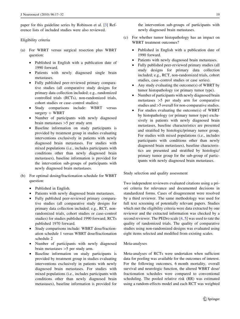
Overall, 24 primary studies [6–29] and seven companion
papers [30–36] met the eligibility criteria for this system-
atic review (Fig. 1).
Title and Abstract Screening n=16,966
Full Text Screening n=65
Excluded at Title and Abstract n=16,901
Eligible Studies n=31
34 Excluded No extractable data…………………………………………........1 No baseline patient data by treatment/ histology group . ……...24 No treatment comparison of interest………………….........……4 ≤5 patients with brain metastases /group..................................... 3 Non-comparative study…………………………………….........1 Unclear treatment interventions….………………………...........1
31 Included WBRT vs. WBRT + Surgery…………………………………7 [6 unique studies, 1 companion study] Different dose/fractionation schedules for WBRT…………..23 [17 unique studies, 6 companion studies] WBRT by histology…………………………………………..1
Fig. 1 Flowchart of studies to
final number of eligible studies
20 J Neurooncol (2010) 96:17–32
123
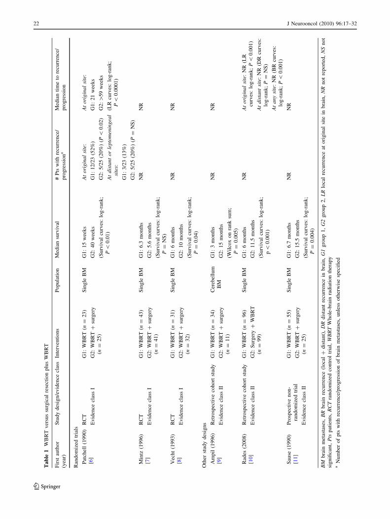
Surgical Resection plus WBRT versus WBRT alone
Seven studies met the eligibility criteria for this treatment
comparison, and of these six were unique and one was a
companion study [6–11, 30]. Three of these studies were
prospective randomized trials [6–8] (Table 1). Given that
the treatment modalities being compared included surgical
resection in only one arm of each trial, all of the RCTs
were non-blinded. In a randomized trial performed at the
University of Kentucky [6], 48 patients with known sys-
temic cancer were treated with either biopsy of the sus-
pected brain metastasis plus WBRT or complete surgical
resection of the metastasis plus WBRT. The radiation doses
were the same in both groups and consisted of a total dose
of 36 Gy given in 12 daily fractions of 3 Gy each. Patients
had to be capable of caring for themselves independently,
with a Karnofsky performance score (KPS) of at least 70.
Patients were ineligible if they had a need for immediate
treatment to prevent acute neurologic deterioration, or if
they had tumors considered to be relatively radiosensitive
[small cell lung cancer (SCLC), germ-cell tumors, lym-
phoma, leukemia, and multiple myeloma]. Patients were
not excluded based on the extent of systemic disease.
Randomization was performed by computer-generated
random numbers. Information on allocation concealment
was not reported. All the patients in the surgical group
were considered to have had complete resection as assessed
by postoperative computerized tomography (CT) scanning.
Follow up brain CT or magnetic resonance imaging (MRI)
scans were required every 3 months. There was a statisti-
cally significant increase in survival in the surgical group
(40 vs. 15 weeks). In addition, the time to recurrence of
brain metastases, freedom from death due to neurologic
causes, and duration of functional independence were
significantly longer in the surgical resection group. The
1 month mortality was 4% in each group, indicating that
there was no extra mortality from surgery. Although sur-
gical resection was the only variable positively associated
with maintaining performance status, the extent of systemic
disease and increased age were associated with poor per-
formance status post-treatment.
A second randomized study [8], conducted as a multi-
institutional trial in the Netherlands, contained 63 evalu-
able patients. Patients with single brain metastases were
randomized to complete surgical resection plus WBRT or
WBRT alone. Randomization was performed centrally by
telephone. The WBRT schedules were the same for both
treatment arms and consisted of 40 Gy given in a non-
standard fractionation scheme of 2 Gy twice per day for
2 weeks (10 treatment days). Patients had to have a rea-
sonable quality of life and neurological status, defined as
spending no more than 50% of their time in bed and not
requiring continuous nursing care or hospitalization.
Excluded histologies were SCLC and lymphoma. Infor-
mation is not given regarding the extent of resection in the
surgical group or the use and frequency of imaging in
follow up. Survival was significantly longer in the surgical
group (10 vs. 6 months). There was also a non-significant
trend toward longer duration of functional independence in
the surgically treated patients. No data concerning recur-
rence of brain metastases were provided. The 1 month
mortality rates were 9% in the surgery group and 0% in the
WBRT alone group, a statistically insignificant difference.
The authors concluded that the addition of surgery to
WBRT provided a survival benefit except to those patients
who were 60 years of age or older, or those patients with
progressive systemic disease in the 3 months prior to the
diagnosis of the brain metastasis.
A third randomized trial, conducted as a multi-center
trial in Canada by Mintz et al. [7], failed to find a benefit
from surgical treatment. In that study, 84 patients with a
single brain metastasis were randomized to receive radio-
therapy alone (30 Gy given in 10 daily fractions of 3 Gy)
or surgery plus radiotherapy. Randomization was per-
formed centrally by telephone. Eligible patients had to be
less than 80 years of age, and they had to have a KPS of at
least 50, i.e., they could be spending more than 50% of
their time in bed but had to be able to care for some per-
sonal needs. Patients were not eligible if they had leuke-
mia, lymphoma, or SCLC. A CT scan was done in the first
postoperative week to assess the extent of tumor removed.
Follow up CT scans were performed monthly for 6 months
and every 3 months after that. A gross total resection was
achieved in 38 of the 40 patients in the surgical group. No
difference was found in overall survival; the median sur-
vival time was 6.3 months in the radiotherapy alone group
and 5.6 months for the surgical group. There was also no
difference in causes of death or quality of life.
It is unclear why the Canadian study was not in agree-
ment with the other two trials. In all three studies, the
control arms (the radiation alone arms) had median lengths
of survival in the 3–6 months range—within the expected
range for patients treated with radiotherapy alone. The
major difference in the studies was the poor results
obtained in the surgical arm of the Canadian trial. That
study contained a higher proportion of patients with
extensive systemic disease and lower performance scores.
It is possible that these factors resulted in more patients
dying of their systemic cancer before a long term benefit of
surgery was seen. Additionally, MRI was not mandatory in
the Canadian study, and it is theoretically possible that
patients with additional lesions not detected by CT may
have been included in the study.
All three of the evidence class II studies [9–11] dem-
onstrated a survival benefit for patients who underwent
surgical resection followed by WBRT as compared to
J Neurooncol (2010) 96:17–32 21
123
Ta
ble
1W
BR
Tv
ersu
ssu
rgic
alre
sect
ion
plu
sW
BR
T
Fir
stau
tho
r
(yea
r)
Stu
dy
des
ign
/ev
iden
cecl
ass
Inte
rven
tio
ns
Po
pu
lati
on
Med
ian
surv
ival
#P
tsw
ith
recu
rren
ce/
pro
gre
ssio
na
Med
ian
tim
eto
recu
rren
ce/
pro
gre
ssio
n
Ran
do
miz
edtr
ials
Pat
chel
l(1
99
0)
[6]
RC
T
Ev
iden
cecl
ass
I
G1
:W
BR
T(n
=2
3)
G2
:W
BR
T?
surg
ery
(n=
25
)
Sin
gle
BM
G1
:1
5w
eek
s
G2
:4
0w
eek
s
(Su
rviv
alcu
rves
:lo
g-r
ank
;
P\
0.0
1)
At
ori
gin
al
site
:
G1
:1
2/2
3(5
2%
)
G2
:5
/25
(20
%)
(P\
0.0
2)
At
ori
gin
al
site
:
G1
:2
1w
eek
s
G2
:[5
9w
eek
s
(LR
curv
es:
log
-ran
k;
P\
0.0
00
1)
At
dis
tan
to
rle
pto
men
ing
eal
site
s:
G1
:3
/23
(13
%)
G2
:5
/25
(20
%)
(P=
NS
)
Min
tz(1
99
6)
[7]
RC
T
Ev
iden
cecl
ass
I
G1
:W
BR
T(n
=4
3)
G2
:W
BR
T?
surg
ery
(n=
41
)
Sin
gle
BM
G1
:6
.3m
on
ths
G2
:5
.6m
on
ths
(Su
rviv
alcu
rves
:lo
g-r
ank
;
P=
NS
)
NR
NR
Vec
ht
(19
93
)
[8]
RC
T
Ev
iden
cecl
ass
I
G1
:W
BR
T(n
=3
1)
G2
:W
BR
T?
surg
ery
(n=
32
)
Sin
gle
BM
G1
:6
mo
nth
s
G2
:1
0m
on
ths
(Su
rviv
alcu
rves
:lo
g-r
ank
;
P=
0.0
4)
NR
NR
Oth
erst
ud
yd
esig
ns
Am
pil
(19
96
)
[9]
Ret
rosp
ecti
ve
coh
ort
stu
dy
Ev
iden
cecl
ass
II
G1
:W
BR
T( n
=3
4)
G2
:W
BR
T?
surg
ery
(n=
11
)
Cer
ebel
lum
BM
G1
:3
mo
nth
s
G2
:1
5m
on
ths
(Wil
cox
on
ran
ksu
m;
P=
0.0
05
)
NR
NR
Rad
es(2
00
8)
[10
]
Ret
rosp
ecti
ve
coh
ort
stu
dy
Ev
iden
cecl
ass
II
G1
:W
BR
T(n
=9
6)
G2
:S
urg
ery
?W
BR
T
(n=
99
)
Sin
gle
BM
G1
:6
mo
nth
s
G2
:1
1.5
mo
nth
s
(Su
rviv
alcu
rves
:lo
g-r
ank
;
p\
0.0
01
)
NR
At
ori
gin
al
site
:N
R(L
R
curv
es:
log
-ran
k;
P\
0.0
01
)
At
dis
tan
tsi
te:
NR
(DR
curv
es:
log
-ran
k;
P=
NS
)
At
an
ysi
te:
NR
(BR
curv
es:
log
-ran
k;
P\
0.0
01
)
Sau
se(1
99
0)
[11
]
Pro
spec
tiv
en
on
-
ran
do
miz
edtr
ial
Ev
iden
cecl
ass
II
G1
:W
BR
T(n
=5
5)
G2
:W
BR
T?
surg
ery
(n=
25
)
Sin
gle
BM
G1
:6
.7m
on
ths
G2
:1
5.5
mo
nth
s
(Su
rviv
alcu
rves
:lo
g-r
ank
;
P=
0.0
04
)
NR
NR
BM
bra
inm
etas
tase
s,B
Rb
rain
recu
rren
ce(l
oca
l?
dis
tan
t),
DR
dis
tan
tre
curr
ence
inb
rain
,G
1g
rou
p1
,G
2g
rou
p2
,L
Rlo
cal
recu
rren
ceat
ori
gin
alsi
tein
bra
in,
NR
no
tre
po
rted
,N
Sn
ot
sig
nifi
can
t,P
tsp
atie
nts
,R
CT
ran
do
miz
edco
ntr
ol
tria
l,W
BR
TW
ho
le-b
rain
rad
iati
on
ther
apy
aN
um
ber
of
pts
wit
hre
curr
ence
/pro
gre
ssio
no
fb
rain
met
asta
ses,
un
less
oth
erw
ise
spec
ified
22 J Neurooncol (2010) 96:17–32
123
WBRT alone. Ampil et al., published a single institution
experience with either surgery plus WBRT (11 patients)
versus WBRT alone (34 patients) in 45 patients who had a
cerebellar metastasis. The majority of the patients in the
WBRT alone arm had additional supratentorial brain
metastases. Although there was a significant difference in
survival noted between the two groups, the authors con-
cluded that the outcome of patients with a brain metastasis
within the cerebellum was improved with surgical resec-
tion if the primary was not from the lung.
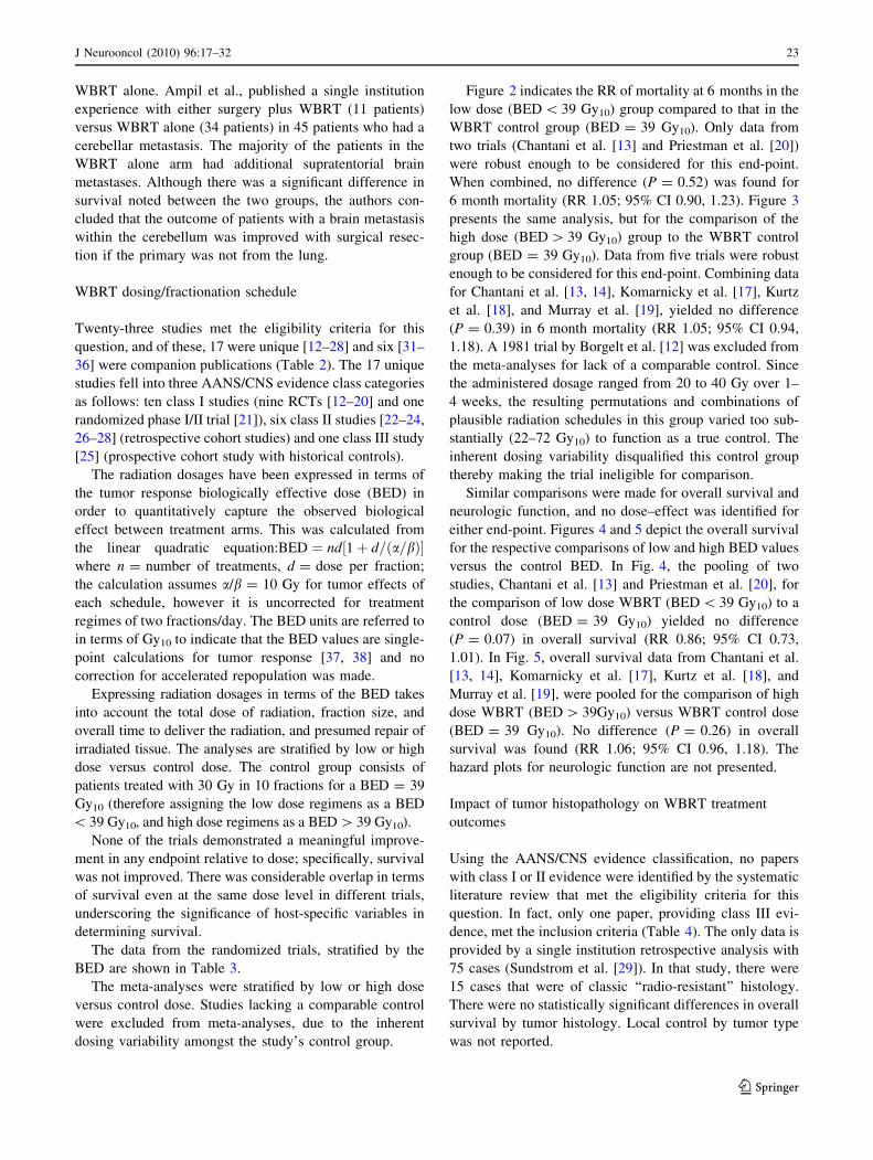
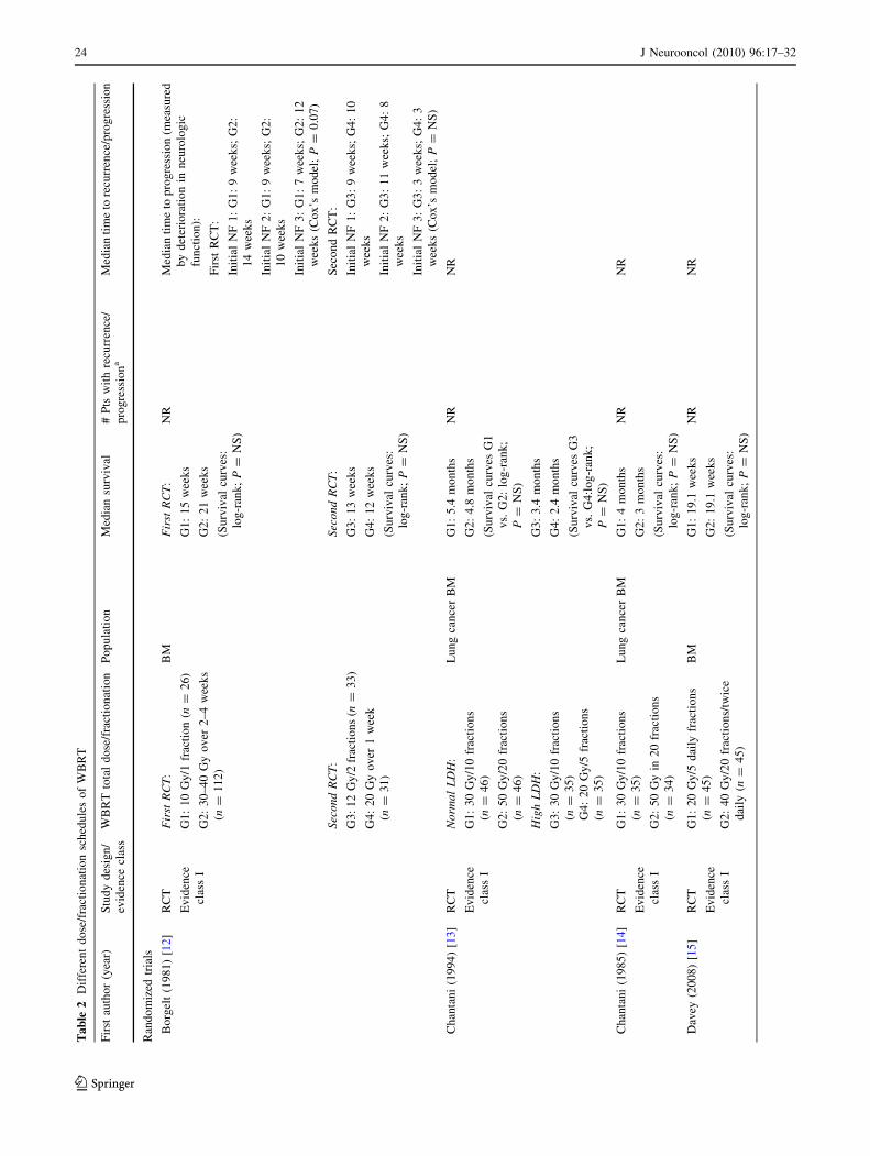
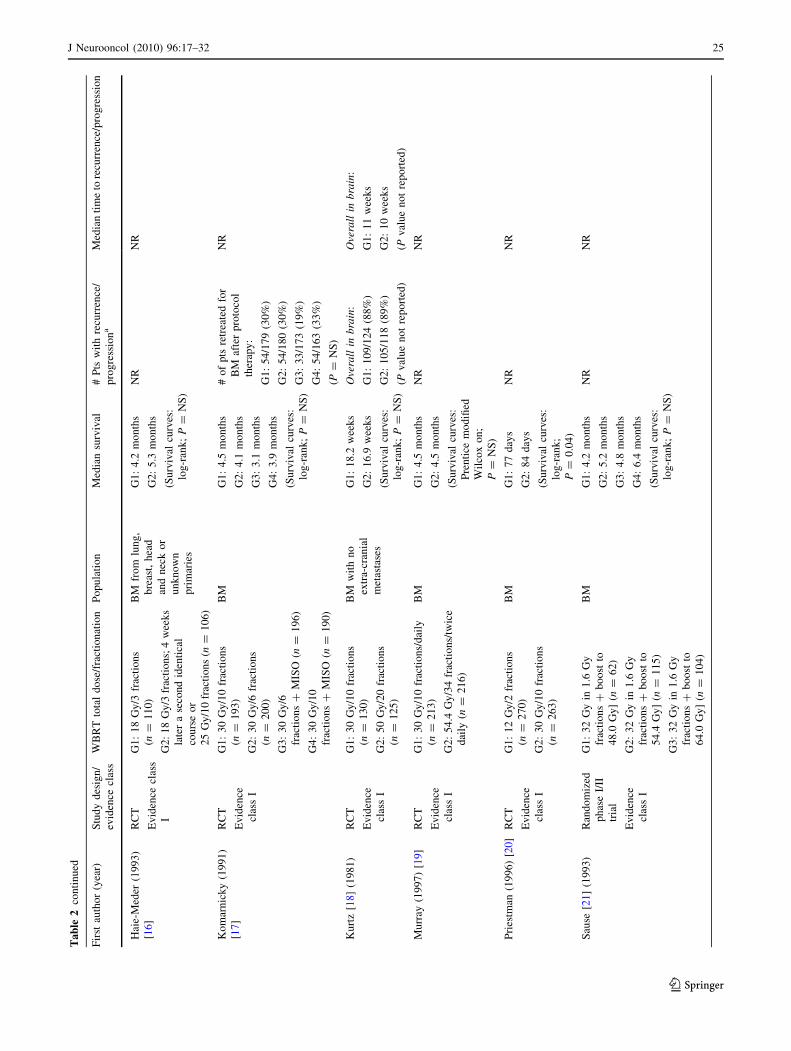
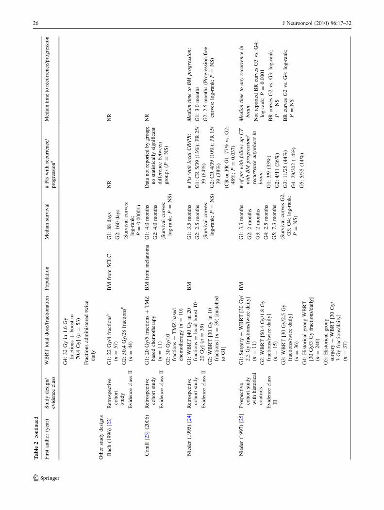
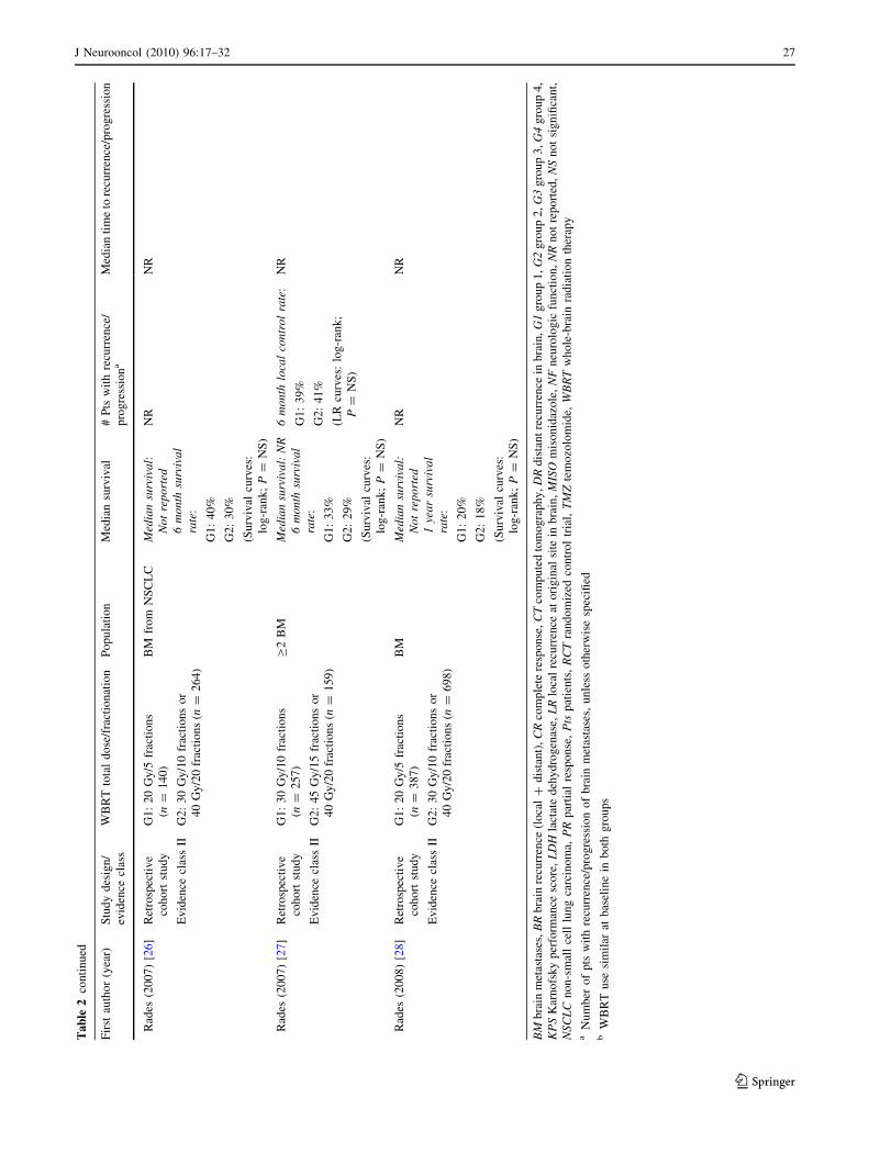
WBRT dosing/fractionation schedule
Twenty-three studies met the eligibility criteria for this
question, and of these, 17 were unique [12–28] and six [31–
36] were companion publications (Table 2). The 17 unique
studies fell into three AANS/CNS evidence class categories
as follows: ten class I studies (nine RCTs [12–20] and one
randomized phase I/II trial [21]), six class II studies [22–24,
26–28] (retrospective cohort studies) and one class III study
[25] (prospective cohort study with historical controls).
The radiation dosages have been expressed in terms of
the tumor response biologically effective dose (BED) in
order to quantitatively capture the observed biological
effect between treatment arms. This was calculated from
the linear quadratic equation:BED ¼ nd 1þ d=ða=bÞ½ �where n = number of treatments, d = dose per fraction;
the calculation assumes a/b = 10 Gy for tumor effects of
each schedule, however it is uncorrected for treatment
regimes of two fractions/day. The BED units are referred to
in terms of Gy10 to indicate that the BED values are single-
point calculations for tumor response [37, 38] and no
correction for accelerated repopulation was made.
Expressing radiation dosages in terms of the BED takes
into account the total dose of radiation, fraction size, and
overall time to deliver the radiation, and presumed repair of
irradiated tissue. The analyses are stratified by low or high
dose versus control dose. The control group consists of
patients treated with 30 Gy in 10 fractions for a BED = 39
Gy10 (therefore assigning the low dose regimens as a BED
\ 39 Gy10, and high dose regimens as a BED [ 39 Gy10).
None of the trials demonstrated a meaningful improve-
ment in any endpoint relative to dose; specifically, survival
was not improved. There was considerable overlap in terms
of survival even at the same dose level in different trials,
underscoring the significance of host-specific variables in
determining survival.
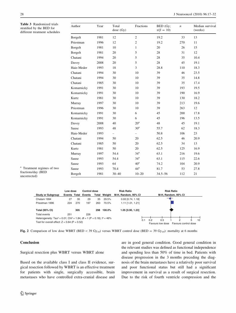
The data from the randomized trials, stratified by the
BED are shown in Table 3.
The meta-analyses were stratified by low or high dose
versus control dose. Studies lacking a comparable control
were excluded from meta-analyses, due to the inherent
dosing variability amongst the study’s control group.
Figure 2 indicates the RR of mortality at 6 months in the
low dose (BED \ 39 Gy10) group compared to that in the
WBRT control group (BED = 39 Gy10). Only data from
two trials (Chantani et al. [13] and Priestman et al. [20])
were robust enough to be considered for this end-point.
When combined, no difference (P = 0.52) was found for
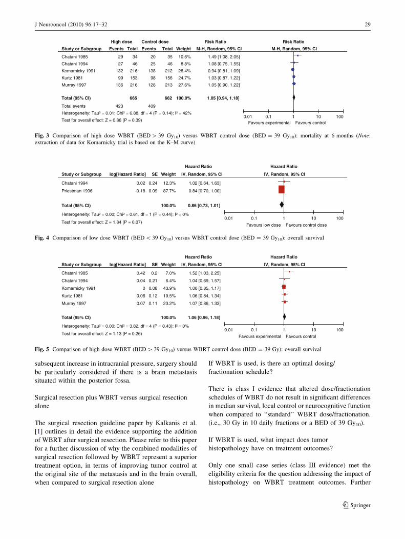
6 month mortality (RR 1.05; 95% CI 0.90, 1.23). Figure 3
presents the same analysis, but for the comparison of the
high dose (BED [ 39 Gy10) group to the WBRT control
group (BED = 39 Gy10). Data from five trials were robust
enough to be considered for this end-point. Combining data
for Chantani et al. [13, 14], Komarnicky et al. [17], Kurtz
et al. [18], and Murray et al. [19], yielded no difference
(P = 0.39) in 6 month mortality (RR 1.05; 95% CI 0.94,
1.18). A 1981 trial by Borgelt et al. [12] was excluded from
the meta-analyses for lack of a comparable control. Since
the administered dosage ranged from 20 to 40 Gy over 1–
4 weeks, the resulting permutations and combinations of
plausible radiation schedules in this group varied too sub-
stantially (22–72 Gy10) to function as a true control. The
inherent dosing variability disqualified this control group
thereby making the trial ineligible for comparison.
Similar comparisons were made for overall survival and
neurologic function, and no dose–effect was identified for
either end-point. Figures 4 and 5 depict the overall survival
for the respective comparisons of low and high BED values
versus the control BED. In Fig. 4, the pooling of two
studies, Chantani et al. [13] and Priestman et al. [20], for
the comparison of low dose WBRT (BED \ 39 Gy10) to a
control dose (BED = 39 Gy10) yielded no difference
(P = 0.07) in overall survival (RR 0.86; 95% CI 0.73,
1.01). In Fig. 5, overall survival data from Chantani et al.
[13, 14], Komarnicky et al. [17], Kurtz et al. [18], and
Murray et al. [19], were pooled for the comparison of high
dose WBRT (BED [ 39Gy10) versus WBRT control dose
(BED = 39 Gy10). No difference (P = 0.26) in overall
survival was found (RR 1.06; 95% CI 0.96, 1.18). The
hazard plots for neurologic function are not presented.
Impact of tumor histopathology on WBRT treatment
outcomes
Using the AANS/CNS evidence classification, no papers
with class I or II evidence were identified by the systematic
literature review that met the eligibility criteria for this
question. In fact, only one paper, providing class III evi-
dence, met the inclusion criteria (Table 4). The only data is
provided by a single institution retrospective analysis with
75 cases (Sundstrom et al. [29]). In that study, there were
15 cases that were of classic ‘‘radio-resistant’’ histology.
There were no statistically significant differences in overall
survival by tumor histology. Local control by tumor type
was not reported.
J Neurooncol (2010) 96:17–32 23
123
Ta
ble
2D
iffe
ren
td
ose
/fra
ctio
nat
ion
sch
edu
les
of
WB
RT
Fir
stau
tho
r(y
ear)
Stu
dy
des
ign
/
evid
ence
clas
s
WB
RT
tota
ld
ose
/fra
ctio
nat
ion
Po
pu
lati
on
Med
ian
surv
ival
#P
tsw
ith
recu
rren
ce/
pro
gre
ssio
na
Med
ian
tim
eto
recu
rren
ce/p
rog
ress
ion
Ran
do
miz
edtr
ials
Bo
rgel
t(1
98
1)
[12
]R
CT
Ev
iden
ce
clas
sI
Fir
stR
CT
:
G1
:1
0G
y/1
frac
tio
n(n
=2
6)
G2
:3
0–
40
Gy
ov
er2
–4
wee
ks
(n=
11
2)
BM
Fir
stR
CT
:
G1
:1
5w
eek
s
G2
:2
1w
eek
s
(Su
rviv
alcu
rves
:
log
-ran
k;
P=
NS
)
NR
Med
ian
tim
eto
pro
gre
ssio
n(m
easu
red
by
det
erio
rati
on
inn
euro
log
ic
fun
ctio
n):
Fir
stR
CT
:
Init
ial
NF
1:
G1
:9
wee
ks;
G2
:
14
wee
ks
Init
ial
NF
2:
G1
:9
wee
ks;
G2
:
10
wee
ks
Init
ial
NF
3:
G1
:7
wee
ks;
G2
:1
2
wee
ks
(Co
x’s
mo
del
;P
=0
.07
)
Sec
on
dR
CT
:
G3
:1
2G
y/2
frac
tio
ns
(n=
33
)
G4
:2
0G
yo
ver
1w
eek
(n=
31
)
Sec
on
dR
CT
:
G3
:1
3w
eek
s
G4
:1
2w
eek
s
(Su
rviv
alcu
rves
:
log
-ran
k;
P=
NS
)
Sec
on
dR
CT
:
Init
ial
NF
1:
G3
:9
wee
ks;
G4
:1
0
wee
ks
Init
ial
NF
2:
G3
:1
1w
eek
s;G
4:
8
wee
ks
Init
ial
NF
3:
G3
:3
wee
ks;
G4
:3
wee
ks
(Co
x’s
mo
del
;P
=N
S)
Ch
anta
ni
(19
94
)[1
3]
RC
T
Ev
iden
ce
clas
sI
No
rma
lL
DH
:
G1
:3
0G
y/1
0fr
acti
on
s
(n=
46
)
G2
:5
0G
y/2
0fr
acti
on
s
(n=
46
)
Lu
ng
can
cer
BM
G1
:5
.4m
on
ths
G2
:4
.8m
on
ths
(Su
rviv
alcu
rves
G1
vs.
G2
:lo
g-r
ank
;
P=
NS
)
G3
:3
.4m
on
ths
G4
:2
.4m
on
ths
(Su
rviv
alcu
rves
G3
vs.
G4
:lo
g-r
ank
;
P=
NS
)
NR
NR
Hig
hL
DH
:
G3
:3
0G
y/1
0fr
acti
on
s
(n=
35
)
G4
:2
0G
y/5
frac
tio
ns
(n=
35
)
Ch
anta
ni
(19
85
)[1
4]
RC
T
Ev
iden
ce
clas
sI
G1
:3
0G
y/1
0fr
acti
on
s
(n=
35
)
G2
:5
0G
yin
20
frac
tio
ns
(n=
34
)
Lu
ng
can
cer
BM
G1
:4
mo
nth
s
G2
:3
mo
nth
s
(Su
rviv
alcu
rves
:
log
-ran
k;
P=
NS
)
NR
NR
Dav
ey(2
00
8)
[15]
RC
T
Ev
iden
ce
clas
sI
G1
:2
0G
y/5
dai
lyfr
acti
on
s
(n=
45
)
G2
:4
0G
y/2
0fr
acti
on
s/tw
ice
dai
ly(n
=4
5)
BM
G1
:1
9.1
wee
ks
G2
:1
9.1
wee
ks
(Su
rviv
alcu
rves
:
log
-ran
k;
P=
NS
)
NR
NR
24 J Neurooncol (2010) 96:17–32
123
Ta
ble
2co
nti
nu
ed
Fir
stau
tho
r(y
ear)
Stu
dy
des
ign
/
evid
ence
clas
s
WB
RT
tota
ld
ose
/fra
ctio
nat
ion
Po
pu
lati
on
Med
ian
surv
ival
#P
tsw
ith
recu
rren
ce/
pro
gre
ssio
na
Med
ian
tim
eto
recu
rren
ce/p
rog
ress
ion
Hai
e-M
eder
(19
93
)
[16]
RC
T
Ev
iden
cecl
ass
I
G1
:1
8G
y/3
frac
tio
ns
(n=
11
0)
G2
:1
8G
y/3
frac
tio
ns;
4w
eek
s
late
ra
seco
nd
iden
tica
l
cou
rse
or
25
Gy
/10
frac
tio
ns
(n=
10
6)
BM
fro
mlu
ng
,
bre
ast,
hea
d
and
nec
ko
r
un
kn
ow
n
pri
mar
ies
G1
:4
.2m
on
ths
G2
:5
.3m
on
ths
(Su
rviv
alcu
rves
:
log
-ran
k;
P=
NS
)
NR
NR
Ko
mar
nic
ky
(19
91
)
[17]
RC
T
Ev
iden
ce
clas
sI
G1
:3
0G
y/1
0fr
acti
on
s
(n=
19
3)
G2
:3
0G
y/6
frac
tio
ns
(n=
20
0)
G3
:3
0G
y/6
frac
tio
ns
?M
ISO
(n=
19
6)
G4
:3
0G
y/1
0
frac
tio
ns
?M
ISO
(n=
19
0)
BM
G1
:4
.5m
on
ths
G2
:4
.1m
on
ths
G3
:3
.1m
on
ths
G4
:3
.9m
on
ths
(Su
rviv
alcu
rves
:
log
-ran
k;
P=
NS
)
#o
fp
tsre
trea
ted
for
BM
afte
rp
roto
col
ther
apy
:
G1
:5
4/1
79
(30
%)
G2
:5
4/1
80
(30
%)
G3
:3
3/1
73
(19
%)
G4
:5
4/1
63
(33
%)
(P=
NS
)
NR
Ku
rtz
[18]
(19
81
)R
CT
Ev
iden
ce
clas
sI
G1
:3
0G
y/1
0fr
acti
on
s
(n=
13
0)
G2
:5
0G
y/2
0fr
acti
on
s
(n=
12
5)
BM
wit
hn
o
extr
a-cr
ania
l
met
asta
ses
G1
:1
8.2
wee
ks
G2
:1
6.9
wee
ks
(Su
rviv
alcu
rves
:
log
-ran
k;
P=
NS
)
Ove
rall
inb
rain
:
G1
:1
09
/12
4(8
8%
)
G2
:1
05
/11
8(8
9%
)
(Pv
alu
en
ot
rep
ort
ed)
Ove
rall
inb
rain
:
G1
:1
1w
eek
s
G2
:1
0w
eek
s
(Pv
alu
en
ot
rep
ort
ed)
Mu
rray
(19
97
)[1
9]
RC
T
Ev
iden
ce
clas
sI
G1
:3
0G
y/1
0fr
acti
on
s/d
aily
(n=
21
3)
G2
:5
4.4
Gy
/34
frac
tio
ns/
twic
e
dai
ly(n
=2
16
)
BM
G1
:4
.5m
on
ths
G2
:4
.5m
on
ths
(Su
rviv
alcu
rves
:
Pre
nti
cem
od
ified
Wil
cox
on
;
P=
NS
)
NR
NR
Pri
estm
an(1
99
6)
[20]
RC
T
Ev
iden
ce
clas
sI
G1
:1
2G
y/2
frac
tio
ns
(n=
27
0)
G2
:3
0G
y/1
0fr
acti
on
s
(n=
26
3)
BM
G1
:7
7d
ays
G2
:8
4d
ays
(Su
rviv
alcu
rves
:
log
-ran
k;
P=
0.0
4)
NR
NR
Sau
se[2
1]
(19
93
)R
and
om
ized
ph
ase
I/II
tria
l
Ev
iden
ce
clas
sI
G1
:3
2G
yin
1.6
Gy
frac
tio
ns
?b
oo
stto
48
.0G
y]
(n=
62
)
G2
:3
2G
yin
1.6
Gy
frac
tio
ns
?b
oo
stto
54
.4G
y]
(n=
11
5)
G3
:3
2G
yin
1.6
Gy
frac
tio
ns
?b
oo
stto
64
.0G
y]
(n=
10
4)
BM
G1
:4
.2m
on
ths
G2
:5
.2m
on
ths
G3
:4
.8m
on
ths
G4
:6
.4m
on
ths
(Su
rviv
alcu
rves
:
log
-ran
k;
P=
NS
)
NR
NR
J Neurooncol (2010) 96:17–32 25
123
Ta
ble
2co
nti
nu
ed
Fir
stau
tho
r(y
ear)
Stu
dy
des
ign
/
evid
ence
clas
s
WB
RT
tota
ld
ose
/fra
ctio
nat
ion
Po
pu
lati
on
Med
ian
surv
ival
#P
tsw
ith
recu
rren
ce/
pro
gre
ssio
na
Med
ian
tim
eto
recu
rren
ce/p
rog
ress
ion
G4
:3
2G
yin
1.6
Gy
frac
tio
ns
?b
oo
stto
70
.4G
y]
(n=
53
)
Fra
ctio
ns
adm
inis
tere
dtw
ice
dai
ly
Oth
erst
ud
yd
esig
ns
Bac
h(1
99
6)
[22]
Ret
rosp
ecti
ve
coh
ort
stu
dy
Ev
iden
cecl
ass
II
G1
:2
2G
y/4
frac
tio
nsb
(n=
57
)
G2
:5
0.4
Gy
/28
frac
tio
nsb
(n=
44
)
BM
fro
mS
CL
CG
1:
88
day
s
G2
:1
60
day
s
(Su
rviv
alcu
rves
:
log
-ran
k;
P=
0.0
00
01
)
NR
NR
Co
nil
l[2
3]
(20
06
)R
etro
spec
tiv
e
coh
ort
stu
dy
Ev
iden
cecl
ass
II
G1
:2
0G
y/5
frac
tio
ns
?T
MZ
bas
edch
emo
ther
apy
(n=
11
)
G2
:3
0G
y/1
0
frac
tio
ns
?T
MZ
bas
ed
chem
oth
erap
y(n
=1
0)
BM
fro
mm
elan
om
aG
1:
4.0
mo
nth
s
G2
:4
.0m
on
ths
(Su
rviv
alcu
rves
:
log
-ran
k;
P=
NS
)
Dat
an
ot
rep
ort
edb
yg
rou
p;
no
stat
isti
call
ysi
gn
ifica
nt
dif
fere
nce
bet
wee
n
gro
up
s(P
=N
S)
NR
Nie
der
(19
95
)[2
4]
Ret
rosp
ecti
ve
coh
ort
stu
dy
Ev
iden
cecl
ass
II
G1
:W
BR
T[4
0G
yin
20
frac
tio
ns
±lo
cal
bo
ost
10
–
20
Gy
](n
=3
9)
G2
:W
BR
T[3
0G
yin
10
frac
tio
ns]
(n=
39
)[m
atch
ed
toG
1]
BM
G1
:3
.5m
on
ths
G2
:2
.5m
on
ths
(Su
rviv
alcu
rves
:
log
-ran
k;
P=
NS
)
#P
tsw
ith
loca
lC
R/P
R:
G1
:C
R5
/39
(13
%);
PR
25
/
39
(64
%)
G2
:C
R4
/39
(10
%);
PR
15
/
39
(38
%)
(CR
or
PR
G1
:7
7%
vs.
G2
:
48
%;
P=
0.0
37
)
Med
ian
tim
eto
BM
pro
gre
ssio
n:
G1
:3
.0m
on
ths
G2
:2
.5m
on
ths
(Pro
gre
ssio
n-f
ree
curv
es:
log
-ran
k;
P=
NS
)
Nie
der
(19
97
)[2
5]
Pro
spec
tiv
e
coh
ort
stu
dy
wit
hh
isto
rica
l
con
tro
ls
Ev
iden
cecl
ass
III
G1
:S
urg
ery
?W
BR
T[3
0G
y/
2.5
Gy
frac
tio
ns/
twic
ed
aily
]
(n=
11
)
G2
:W
BR
T[5
0.4
Gy
/1.8
Gy
frac
tio
ns/
twic
ed
aily
]
(n=
15
)
G3
:W
BR
T[3
0G
y/2
.5G
y
frac
tio
ns/
twic
ed
aily
]
(n=
36
)
G4
:H
isto
rica
lg
rou
pW
BR
T
[30
Gy
/3G
yfr
acti
on
s/d
aily
]
(n=
24
6)
G5
:H
isto
rica
lg
rou
p
surg
ery
?W
BR
T[3
0G
y/
3G
yfr
acti
on
s/d
aily
]
(n=
37
)
BM
G1
:3
.3m
on
ths
G2
:2
mo
nth
s
G3
:2
mo
nth
s
G4
:2
.5m
on
ths
G5
:7
.3m
on
ths
(Su
rviv
alcu
rves
G2
,
G3
,G
4:
log
-ran
k;
P=
NS
)
#o
fp
tsw
ith
foll
ow
up
CT
wit
hB
Mp
rog
ress
ion
/re
curr
ence
an
ywh
ere
inb
rain
:
G1
:3
/9(3
3%
)
G2
:4
/11
(36
%)
G3
:1
1/2
5(4
4%
)
G4
:2
9/2
02
(14
%)
G5
:5
/35
(14
%)
Med
ian
tim
eto
an
yre
curr
ence
inb
rain
:
No
tre
po
rted
BR
curv
esG
3v
s.G
4:
log
-ran
k;
P=
0.0
00
1
BR
curv
esG
2v
s.G
3:
log
-ran
k;
P=
NS
BR
curv
esG
2v
s.G
4:
log
-ran
k;
P=
NS
26 J Neurooncol (2010) 96:17–32
123
Ta
ble
2co
nti
nu
ed
Fir
stau
tho
r(y
ear)
Stu
dy
des
ign
/
evid
ence
clas
s
WB
RT
tota
ld
ose
/fra
ctio
nat
ion
Po
pu
lati
on
Med
ian
surv
ival
#P
tsw
ith
recu
rren
ce/
pro
gre
ssio
na
Med
ian
tim
eto
recu
rren
ce/p
rog
ress
ion
Rad
es(2
00
7)
[26]
Ret
rosp
ecti
ve
coh
ort
stu
dy
Ev
iden
cecl
ass
II
G1
:2
0G
y/5
frac
tio
ns
(n=
14
0)
G2
:3
0G
y/1
0fr
acti
on
so
r
40
Gy
/20
frac
tio
ns
(n=
26
4)
BM
fro
mN
SC
LC
Med
ian
surv
iva
l:N
ot
rep
ort
ed6
mo
nth
surv
iva
lra
te:
G1
:4
0%
G2
:3
0%
(Su
rviv
alcu
rves
:
log
-ran
k;
P=
NS
)
NR
NR
Rad
es(2
00
7)
[27]
Ret
rosp
ecti
ve
coh
ort
stu
dy
Ev
iden
cecl
ass
II
G1
:3
0G
y/1
0fr
acti
on
s
(n=
25
7)
G2
:4
5G
y/1
5fr
acti
on
so
r
40
Gy
/20
frac
tio
ns
(n=
15
9)
C2
BM
Med
ian
surv
iva
l:N
R6
mo
nth
surv
iva
lra
te:
G1
:3
3%
G2
:2
9%
(Su
rviv
alcu
rves
:
log
-ran
k;
P=
NS
)
6m
on
thlo
cal
con
tro
lra
te:
G1
:3
9%
G2
:4
1%
(LR
curv
es:
log
-ran
k;
P=
NS
)
NR
Rad
es(2
00
8)
[28]
Ret
rosp
ecti
ve
coh
ort
stu
dy
Ev
iden
cecl
ass
II
G1
:2
0G
y/5
frac
tio
ns
(n=
38
7)
G2
:3
0G
y/1
0fr
acti
on
so
r
40
Gy
/20
frac
tio
ns
(n=
69
8)
BM
Med
ian
surv
iva
l:N
ot
rep
ort
ed1
yea
rsu
rviv
al
rate
:
G1
:2
0%
G2
:1
8%
(Su
rviv
alcu
rves
:
log
-ran
k;
P=
NS
)
NR
NR
BM
bra
inm
etas
tase
s,B
Rb
rain
recu
rren
ce(l
oca
l?
dis
tan
t),
CR
com
ple
tere
spo
nse
,C
Tco
mp
ute
dto
mo
gra
ph
y,
DR
dis
tan
tre
curr
ence
inb
rain
,G
1g
rou
p1
,G
2g
rou
p2
,G
3g
rou
p3
,G
4g
rou
p4
,
KP
SK
arn
ofs
ky
per
form
ance
sco
re,
LD
Hla
ctat
ed
ehy
dro
gen
ase,
LR
loca
lre
curr
ence
ato
rig
inal
site
inb
rain
,M
ISO
mis
on
idaz
ole
,N
Fn
euro
log
icfu
nct
ion
,N
Rn
ot
rep
ort
ed,
NS
no
tsi
gn
ifica
nt,
NS
CL
Cn
on
-sm
all
cell
lun
gca
rcin
om
a,P
Rp
arti
alre
spo
nse
,P
tsp
atie
nts
,R
CT
ran
do
miz
edco
ntr
ol
tria
l,T
MZ
tem
ozo
lom
ide,
WB
RT
wh
ole
-bra
inra
dia
tio
nth
erap
ya
Nu
mb
ero
fp
tsw
ith
recu
rren
ce/p
rog
ress
ion
of
bra
inm
etas
tase
s,u
nle
sso
ther
wis
esp
ecifi
edb
WB
RT
use
sim
ilar
atb
asel
ine
inb
oth
gro
up
s
J Neurooncol (2010) 96:17–32 27
123
Conclusion
Surgical resection plus WBRT versus WBRT alone
Based on the available class I and class II evidence, sur-
gical resection followed by WBRT is an effective treatment
for patients with single, surgically accessible, brain
metastases who have controlled extra-cranial disease and
are in good general condition. Good general condition in
the relevant studies was defined as functional independence
and spending less than 50% of time in bed. Patients with
disease progression in the 3 months preceding the diag-
nosis of the brain metastases have a relatively poor survival
and poor functional status but still had a significant
improvement in survival as a result of surgical resection.
Due to the risk of fourth ventricle compression and the
Table 3 Randomized trials
stratified by the BED for
different treatment schedules
a Treatment regimes of two
fractions/day (BED
uncorrected)
Author Year Total
dose (Gy)
Fractions BED (Gy;
a/b = 10)
n Median survival
(weeks)
Borgelt 1981 12 2 19.2 33 13
Priestman 1996 12 2 19.2 270 11
Borgelt 1981 10 1 20 26 15
Borgelt 1981 20 5 28 31 12
Chatani 1994 20 5 28 35 10.4
Davey 2008 20 5 28 45 19.1
Haie-Meder 1993 18 3 28.8 110 18.3
Chatani 1994 30 10 39 46 23.5
Chatani 1994 30 10 39 35 14.8
Chatani 1985 30 10 39 35 17.4
Komarnicky 1991 30 10 39 193 19.5
Komarnicky 1991 30 10 39 190 16.9
Kurtz 1981 30 10 39 130 18.2
Murray 1997 30 10 39 213 19.6
Priestman 1996 30 10 39 263 12
Komarnicky 1991 30 6 45 200 17.8
Komarnicky 1991 30 6 45 196 13.5
Davey 2008 40 20a 48 45 19.1
Sause 1993 48 30a 55.7 62 18.3
Haie-Meder 1993 – – 50.8 106 23
Chatani 1994 50 20 62.5 46 20.9
Chatani 1985 50 20 62.5 34 13
Kurtz 1981 50 20 62.5 125 16.9
Murray 1997 54.4 34a 63.1 216 19.6
Sause 1993 54.4 34a 63.1 115 22.6
Sause 1993 64 40a 74.2 104 20.9
Sause 1993 70.4 44a 81.7 53 27.8
Borgelt 1981 30–40 10–20 34.5–56 112 21
Study or Subgroup
Chatani 1994
Priestman 1996
Total (95% CI)
Total events
Heterogeneity: Tau² = 0.01; Chi² = 1.84, df = 1 (P = 0.18); I² = 46%
Test for overall effect: Z = 0.64 (P = 0.52)
Events
27
224
251
Total
35
270
305
Events
29
197
226
Total
35
263
298
Weight
29.5%
70.5%
100.0%
M-H, Random, 95% CI
0.93 [0.74, 1.18]
1.11 [1.01, 1.21]
1.05 [0.90, 1.23]
oitaR ksiRoitaR ksiResod lortnoCesod woLM-H, Random, 95% CI
0.1 0.2 0.5 1 2 5 10Favours low dose Favours control dose
Fig. 2 Comparison of low dose WBRT (BED \ 39 Gy10) versus WBRT control dose (BED = 39 Gy10): mortality at 6 months
28 J Neurooncol (2010) 96:17–32
123
subsequent increase in intracranial pressure, surgery should
be particularly considered if there is a brain metastasis
situated within the posterior fossa.
Surgical resection plus WBRT versus surgical resection
alone
The surgical resection guideline paper by Kalkanis et al.
[1] outlines in detail the evidence supporting the addition
of WBRT after surgical resection. Please refer to this paper
for a further discussion of why the combined modalities of
surgical resection followed by WBRT represent a superior
treatment option, in terms of improving tumor control at
the original site of the metastasis and in the brain overall,
when compared to surgical resection alone
If WBRT is used, is there an optimal dosing/
fractionation schedule?
There is class I evidence that altered dose/fractionation
schedules of WBRT do not result in significant differences
in median survival, local control or neurocognitive function
when compared to ‘‘standard’’ WBRT dose/fractionation.
(i.e., 30 Gy in 10 daily fractions or a BED of 39 Gy10).
If WBRT is used, what impact does tumor
histopathology have on treatment outcomes?
Only one small case series (class III evidence) met the
eligibility criteria for the question addressing the impact of
histopathology on WBRT treatment outcomes. Further
Study or Subgroup
Chatani 1985
Chatani 1994
Komarnicky 1991
Kurtz 1981
Murray 1997
Total (95% CI)
Total events
Heterogeneity: Tau² = 0.01; Chi² = 6.88, df = 4 (P = 0.14); I² = 42%
Test for overall effect: Z = 0.86 (P = 0.39)
Events
29
27
132
99
136
423
Total
34
46
216
153
216
665
Events
20
25
138
98
128
409
Total
35
46
212
156
213
662
Weight
10.6%
8.8%
28.4%
24.7%
27.6%
100.0%
M-H, Random, 95% CI
1.49 [1.08, 2.05]
1.08 [0.75, 1.55]
0.94 [0.81, 1.09]
1.03 [0.87, 1.22]
1.05 [0.90, 1.22]
1.05 [0.94, 1.18]
oitaR ksiRoitaR ksiResod lortnoCesod hgiH
M-H, Random, 95% CI
0.01 0.1 1 10 100Favours experimental Favours control
Fig. 3 Comparison of high dose WBRT (BED [ 39 Gy10) versus WBRT control dose (BED = 39 Gy10): mortality at 6 months (Note:
extraction of data for Komarnicky trial is based on the K–M curve)
Study or Subgroup
Chatani 1985
Chatani 1994
Komarnicky 1991
Kurtz 1981
Murray 1997
Total (95% CI)
Heterogeneity: Tau² = 0.00; Chi² = 3.82, df = 4 (P = 0.43); I² = 0%
Test for overall effect: Z = 1.13 (P = 0.26)
log[Hazard Ratio]
0.42
0.04
0
0.06
0.07
SE
0.2
0.21
0.08
0.12
0.11
Weight
7.0%
6.4%
43.9%
19.5%
23.2%
100.0%
IV, Random, 95% CI
1.52 [1.03, 2.25]
1.04 [0.69, 1.57]
1.00 [0.85, 1.17]
1.06 [0.84, 1.34]
1.07 [0.86, 1.33]
1.06 [0.96, 1.18]
oitaR drazaHoitaR drazaH
IV, Random, 95% CI
0.01 0.1 1 10 100
Favours experimental Favours control
Fig. 5 Comparison of high dose WBRT (BED [ 39 Gy10) versus WBRT control dose (BED = 39 Gy): overall survival
Study or Subgroup
Chatani 1994
Priestman 1996
Total (95% CI)
Heterogeneity: Tau² = 0.00; Chi² = 0.61, df = 1 (P = 0.44); I² = 0%
Test for overall effect: Z = 1.84 (P = 0.07)
log[Hazard Ratio]
0.02
-0.18
SE
0.24
0.09
Weight
12.3%
87.7%
100.0%
IV, Random, 95% CI
1.02 [0.64, 1.63]
0.84 [0.70, 1.00]
0.86 [0.73, 1.01]
oitaR drazaHoitaR drazaH
IV, Random, 95% CI
0.01 0.1 1 10 100
Favours low dose Favours control dose
Fig. 4 Comparison of low dose WBRT (BED \ 39 Gy10) versus WBRT control dose (BED = 39 Gy10): overall survival
J Neurooncol (2010) 96:17–32 29
123

Table 4 Study evaluating WBRT treatment outcome by histopathology
First author (year): Sundstrom (1998) [29]
Study characteristics Study outcomes Study quality
Study design: case series Primary outcome: survival Quality assessment: N/A as non-
comparative study
Inclusion criteria:
Pts treated with WBRT for BM diagnosed by CT or
MRI
Minimum midline dose to the whole brain of at least
25 Gy
Median survival by primary tumor type:
Breast: 7 months (range 1–62 months)
Lung: 4 months (range 1–21 months)
Renal cell: 4 months (range 2–34 months)
Melanoma: 3 months (range 1–6 months)
Other primaries: 4 months (range 1–9 months)
(Survival curves: P-value not reported)
AANS/CNS evidence classification:
class III
Interventions:
WBRT [mean dose 30 Gy (range 25–40 Gy) in 1.8–
3 Gy fractions]
Tumor control: not reported
Median follow-up: not reported
# Male:
Breast: 0/19, lung: 26/35, renal cell: 4/9, melanoma:
6/6, other: 6/6
Median time to recurrence of brain metastases:
not reported
Median age (range):
Breast: 53 years (39–77 years)
Lung: 64 years (43–78 years)
Renal cell: 61 years (41–69 years)
Melanoma: 61 years (39–62 years)
Other: 61 years (50–71 years)
Functional performance: not reported by
histology
Cause of death: not reported
Adverse events: not reported by tumor type
# Of brain metastases:
Breast: 1 BM 9/19, 2 BM 4/19, [2 BM 6/19
Lung: 1 BM 15/35, 2 BM 5/35, [2 BM 15/35
Renal cell: 1 BM 3/9, 2 BM 2/9, [2 BM 4/9
Melanoma: 1 BM 3/6, 2 BM 0/6, [2 BM 3/6
Other: 1 BM 3/6, 2 BM 2/6, [2 BM 1/6
Extra-cranial disease: Extra-cranial metastases:
Breast: 17/19
Lung: 6/35
Renal cell: 5/9
Melanoma: 4/6
Other: 5/6
Baseline functional performance:
WHO classification (0 (best) to 4 (worst)
Performance status (PS):
Breast: PS 0–1 9/19, PS 2 5/19, PS 3 5/19
Lung: PS 0-1 10/35, PS 2 16/35, PS 3 9/35
Renal cell: PS 0–1 2/9, PS 2 5/9, PS 3 2/9
Melanoma: PS 0–1 6/6
Other: PS 0–1 3/6, PS 2 1/6, PS 3 2/6
AANS American Association of Neurological Surgeons, BM brain metastases, CNS Congress of Neurological Surgeons, CT computed tomog-
raphy, MRI magnetic resonance imaging, NA not available, Pts patients, WBRT whole brain radiation therapy, WHO World Health Organization
30 J Neurooncol (2010) 96:17–32
123
studies in this area are needed before any recommendations
can be made.
Key issues for future investigation
There have been numerous studies comparing various dose/
fractionation schemes for WBRT. Unless future studies
incorporate more sophisticated measures of neurocognitive
outcome there is little need to repeat these studies. There
are, however, very few studies evaluating the effectiveness
of WBRT for one histopathological type versus another.
Future studies of WBRT explicitly addressing treatment
outcomes for well-defined patient groups by different
tumor types are needed.
The following is a list of major ongoing or recently
closed randomized trials pertaining to the use of whole
brain radiation therapy that evaluate treatment comparisons
addressed by this guideline paper for the management of
newly diagnosed brain metastases.
1. Brain metastases study: radiotherapy schemes in the
treatment of brain metastases
Official Title: To determine which of two radiotherapy
brain fractionation schemes is superior in the treatment
of brain metastases
Status: Completed
Clinicaltrials.gov Identifier: NCT00138788
Principal Investigator: Associate Professor, Peter H
Graham, Cancer Care Centre, St George Hospital
Location: Australia
Sponsors and Collaborators: St George Hospital,
Australia
Acknowledgments We would like to acknowledge the contribu-
tions of the McMaster Evidence-based Practice Center (EPC), Dr.
Parminder Raina (Director). Dr. Lina Santaguida (Co-Associate
Director, Senior Scientist) led the EPC staff, which was respon-
sible for managing the systematic review process, searching for
and retrieving, reviewing, data abstraction of all articles, prepara-
tion of the tables and the formatting and editing of the final
manuscripts.
Disclaimer of liability The information in these guidelines reflects
the current state of knowledge at the time of completion. The pre-
sentations are designed to provide an accurate review of the subject
matter covered. These guidelines are disseminated with the under-
standing that the recommendations by the authors and consultants
who have collaborated in their development are not meant to replace
the individualized care and treatment advice from a patient’s physi-
cian(s). If medical advice or assistance is required, the services of a
competent physician should be sought. The proposals contained in
these guidelines may not be suitable for use in all circumstances. The
choice to implement any particular recommendation contained in
these guidelines must be made by a managing physician in light of
the situation in each particular patient and on the basis of existing
resources.
Disclosures All panel members provided full disclosure of conflicts
of interest, if any, prior to establishing the recommendations con-
tained within these guidelines.
Open Access This article is distributed under the terms of the
Creative Commons Attribution Noncommercial License which per-
mits any noncommercial use, distribution, and reproduction in any
medium, provided the original author(s) and source are credited.
References
1. Kalkanis SN, Kondziolka D, Gaspar LE, Burri SH, Asher AL,
Cobbs CS et al (2009) The role of surgical resection in the
management of newly diagnosed brain metastases: a systematic
review and evidence-based clinical practice guideline. J Neu-
rooncol. doi:10.1007/s11060-009-0061-8
2. Linskey ME, Andrews DW, Asher AL, Burri SH, Kondziolka
DS, Robinson PD et al (2009) The role of stereotactic radiosur-
gery in the management of newly diagnosed brain metastases: a
systematic review and evidence-based clinical practice guideline.
J Neurooncol. doi:10.1007/s11060-009-0073-4
3. Robinson PD, Kalkanis SN, Linskey ME, Santaguida PL (2009)
Methodology used to develop the AANS/CNS management of
brain metastases evidence-based clinical practice parameter
guidelines. J Neurooncol. doi:10.1007/s11060-009-0059-2
4. Centre for Evidence-Based Physiotherapy (2009) Physiotherapy
Evidence Database (PEDro). http://www.pedro.org.au/. Accessed
January 2009
5. Maher CG, Sherrington C, Herbert RD, Moseley AM, Elkins M
(2003) Reliability of the PEDro scale for rating quality of ran-
domized controlled trials. Phys Ther 83(8):713–721
6. Patchell RA, Tibbs PA, Walsh JW, Dempsey RJ, Maruyama Y,
Kryscio RJ et al (1990) A randomized trial of surgery in the
treatment of single metastases to the brain. N Engl J Med
322(8):494–500
7. Mintz AH, Kestle J, Rathbone MP, Gaspar L, Hugenholtz H,
Fisher B et al (1996) A randomized trial to assess the efficacy of
surgery in addition to radiotherapy in patients with a single
cerebral metastasis. Cancer 78(7):1470–1476
8. Vecht CJ, Haaxma-Reiche H, Noordijk EM, Padberg GW,
Voormolen JH, Hoekstra FH et al (1993) Treatment of single
brain metastasis: radiotherapy alone or combined with neuro-
surgery? Ann Neurol 33(6):583–590
9. Ampil FL, Nanda A, Willis BK, Nandy I, Meehan R (1996)
Metastatic disease in the cerebellum The LSU experience in
1981–1993. Am J Clin Oncol 19(5):509–511
10. Rades D, Kieckebusch S, Haatanen T, Lohynska R, Dunst J,
Schild SE (2008) Surgical resection followed by whole brain
radiotherapy versus whole brain radiotherapy alone for single
brain metastasis. Int J Radiat Oncol Biol Phys 70(5):1319–1324
11. Sause WT, Crowley JJ, Morantz R, Rotman M, Mowry PA,
Bouzaglou A et al (1990) Solitary brain metastasis: results of an
RTOG/SWOG protocol evaluation surgery ? RT versus RT
alone. Am J Clin Oncol 13(5):427–432
12. Borgelt B, Gelber R, Larson M, Hendrickson F, Griffin T, Roth R
(1981) Ultra-rapid high dose irradiation schedules for the palli-
ation of brain metastases: final results of the first two studies by
the Radiation Therapy Oncology Group. Int J Radiat Oncol Biol
Phys 7(12):1633–1638
13. Chatani M, Matayoshi Y, Masaki N, Inoue T (1994) Radiation
therapy for brain metastases from lung carcinoma. Prospective
J Neurooncol (2010) 96:17–32 31
123
randomized trial according to the level of lactate dehydrogenase.
Strahlenther Onkol 170(3):155–161
14. Chatani M, Teshima T, Hata K, Inoue T, Suzuki T (1985) Whole
brain irradiation for metastases from lung carcinoma. A clinical
investigation. Acta Radiol Oncol 24(4):311–314
15. Davey P, Hoegler D, Ennis M, Smith J (2008) A phase III study
of accelerated versus conventional hypofractionated whole brain
irradiation in patients of good performance status with brain
metastases not suitable for surgical excision. Radiother Oncol
88(2):173–176 Update Sept 24 2008
16. Haie-Meder C, Pellae-Cosset B, Laplanche A, Lagrange JL,
Tuchais C, Nogues C et al (1993) Results of a randomized
clinical trial comparing two radiation schedules in the palliative
treatment of brain metastases. Radiother Oncol 26(2):111–116
17. Komarnicky LT, Phillips TL, Martz K, Asbell S, Isaacson S,
Urtasun R (1991) A randomized phase III protocol for the eval-
uation of misonidazole combined with radiation in the treatment
of patients with brain metastases (RTOG-7916). Int J Radiat
Oncol Biol Phys 20(1):53–58
18. Kurtz JM, Gelber R, Brady LW, Carella RJ, Cooper JS (1981)
The palliation of brain metastases in a favorable patient popula-
tion: a randomized clinical trial by the Radiation Therapy
Oncology Group. Int J Radiat Oncol Biol Phys 7(7):891–895
19. Murray KJ, Scott C, Greenberg HM, Emami B, Seider M, Vora
NL et al (1997) A randomized phase III study of accelerated
hyperfractionation versus standard in patients with unresected
brain metastases: a report of the Radiation Therapy Oncology
Group (RTOG) 9104. Int J Radiat Oncol Biol Phys 39(3):571–
574
20. Priestman TJ, Dunn J, Brada M, Rampling R, Baker PG (1996)
Final results of the Royal College of Radiologists’ trial com-
paring two different radiotherapy schedules in the treatment of
cerebral metastases. Clin Oncol (R Coll Radiol) 8(5):308–315
21. Sause WT, Scott C, Krisch R, Rotman M, Sneed PK, Janjan N
et al (1993) Phase I/II trial of accelerated fractionation in brain
metastases RTOG 85–28. Int J Radiat Oncol Biol Phys
26(4):653–657
22. Bach F, Sorensen JB, Adrian L, Larsen H, Langer SW, Nelausen
KM et al (1996) Brain relapses in chemotherapy-treated small cell
lung cancer: a retrospective review of two time-dose regimens of
therapeutic brain irradiation. Lung Cancer 15(2):171–181
23. Conill C, Jorcano S, Domingo-Domenech J, Gallego R, Malvehy
J, Puig S et al (2006) Whole brain irradiation and temozolomide
based chemotherapy in melanoma brain metastases. Clin Transl
Oncol 8(4):266–270
24. Nieder C, Berberich W, Nestle U, Niewald M, Walter K, Schn-
abel K (1995) Relation between local result and total dose of
radiotherapy for brain metastases. Int J Radiat Oncol Biol Phys
33(2):349–355
25. Nieder C, Nestle U, Niewald M, Schnabel K (1997) Accelerated
radiotherapy for brain metastases. Radiother Oncol 45(1):17–22
26. Rades D, Schild SE, Lohynska R, Veninga T, Stalpers LJ, Dunst J
(2007) Two radiation regimens and prognostic factors for brain
metastases in nonsmall cell lung cancer patients. Cancer
110(5):1077–1082
27. Rades D, Haatanen T, Schild SE, Dunst J (2007) Dose escalation
beyond 30 grays in 10 fractions for patients with multiple brain
metastases. Cancer 110(6):1345–1350
28. Rades D, Bohlen G, Dunst J, Lohynska R, Veninga T, Stalpers L
et al (2008) Comparison of short-course versus long-course
whole-brain radiotherapy in the treatment of brain metastases.
Strahlenther Onkol 184(1):30–35
29. Sundstrom JT, Minn H, Lertola KK, Nordman E (1998) Prog-
nosis of patients treated for intracranial metastases with whole-
brain irradiation. Ann Med 30(3):296–299
30. Noordijk EM, Vecht CJ, Haaxma-Reiche H, Padberg GW,
Voormolen JH, Hoekstra FH et al (1994) The choice of treatment
of single brain metastasis should be based on extracranial tumor
activity and age. Int J Radiat Oncol Biol Phys 29(4):711–717
31. Chatani M, Teshima T, Hata K, Inoue T (1986) Prognostic factors
in patients with brain metastases from lung carcinoma. Strah-
lenther Onkol 162(3):157–161
32. Regine WF, Scott C, Murray K, Curran W (2001) Neurocognitive
outcome in brain metastases patients treated with accelerated-
fractionation vs. accelerated-hyperfractionated radiotherapy: an
analysis from Radiation Therapy Oncology Group Study 91–04.
Int J Radiat Oncol Biol Phys 51(3):711–717
33. Epstein BE, Scott CB, Sause WT, Rotman M, Sneed PK, Janjan
NA et al (1993) Improved survival duration in patients with un-
resected solitary brain metastasis using accelerated hyperfrac-
tionated radiation therapy at total doses of 54.4 gray and greater.
Results of Radiation Therapy Oncology Group 85–28. Cancer
71(4):1362–1367
34. Rades D, Bohlen G, Lohynska R, Veninga T, Stalpers LJ, Schild
SE et al (2007) Whole-brain radiotherapy with 20 Gy in 5 frac-
tions for brain metastases in patients with cancer of unknown
primary (CUP). Strahlenther Onkol 183(11):631–636
35. Rades D, Kieckebusch S, Lohynska R, Veninga T, Stalpers LJ,
Dunst J et al (2007) Reduction of overall treatment time in
patients irradiated for more than three brain metastases. Int J
Radiat Oncol Biol Phys 69(5):1509–1513 (update Sept 24 2008)
36. Rades D, Lohynska R, Veninga T, Stalpers LJ, Schild SE (2007)
Evaluation of 2 whole-brain radiotherapy schedules and prog-
nostic factors for brain metastases in breast cancer patients.
Cancer 110(11):2587–2592
37. Fowler JF (2006) Practical time-dose evaluations, or how to stop
worrying and learn to love linear quadratics. In: Levitt SH, Purdy
JA, Perez CA, Vijayakumar S (eds) Technical basis of radiation
therapy: practical clinical applications, 4th edn. Springer, Berlin,
Heidelberg, pp 3–31
38. Jones B, Dale RG, Deehan C, Hopkins KI, Morgan DA (2001)
The role of biologically effective dose (BED) in clinical oncol-
ogy. Clin Oncol (R Coll Radiol) 13(2):71–81
32 J Neurooncol (2010) 96:17–32
123
Related Documents


