Research Paper The role of vitamin D in regulating the iron-hepcidin-ferroportin axis in monocytes Susu M. Zughaier a, d, * , Jessica A. Alvarez b , John H. Sloan c , Robert J. Konrad c , Vin Tangpricha b, d a Department of Microbiology and Immunology, Emory University School of Medicine, Atlanta, GA, USA b Division of Endocrinology, Metabolism and Lipids, Department of Medicine, Emory University School of Medicine, Atlanta, GA, USA c Lilly Research Laboratories, Eli Lilly and Company, Indianapolis, IN, USA d Atlanta Veterans Affairs Medical Center, Atlanta, GA, USA article info Article history: Received 13 December 2013 Received in revised form 21 January 2014 Accepted 23 January 2014 Keywords: Vitamin D Hepcidin Ferroportin NRAMP1 IL-6 IL-1b Macrophage Chronic kidney disease Anemia of inflammation abstract Chronic kidney disease affects 40% of adults aged 65 and older. Anemia of CKD is present in 30% of patients with CKD and is associated with increased cardiovascular risk, decreased quality of life, and increased mortality. Hepcidin-25 (hepcidin), the key iron regulating hormone, prevents iron egress from macrophages and thus prevents normal recycling of the iron needed to support erythropoiesis. Hepcidin levels are increased in adults and children with CKD. Vitamin D insufficiency is highly prevalent in CKD and is associated with erythropoietin hyporesponsiveness. Recently, hepcidin levels were found to be inversely correlated with vitamin D status in CKD. The aim of this study was to investigate the role of vitamin D in the regulation of hepcidin expression in vitro and in vivo. This study reports that 1,25- dihydroxyvitamin D 3 (1,25(OH) 2 D 3 ), the hormonally active form of vitamin D, is associated with decreased hepcidin and increased ferroportin expression in lipopolysaccharide (LPS) stimulated THP- 1 cells. 1,25(OH) 2 D 3 also resulted in a dose-dependent decrease in pro-hepcidin cytokines, IL-6 and IL-1b, release in vitro. Further, we show that high-dose vitamin D therapy impacts systemic hepcidin levels in subjects with early stage CKD. These data suggest that improvement in vitamin D status is associated with lower systemic concentrations of hepcidin in subjects with CKD. In conclusion, vitamin D regulates the hepcidin-ferroportin axis in macrophages which may facilitate iron egress. Improvement in vitamin D status in patients with CKD may reduce systemic hepcidin levels and may ameliorate anemia of CKD. Ó 2014 Elsevier Inc. All rights reserved. Introduction Vitamin D insufficiency is common in patients with chronic kidney disease (CKD) [1], with a prevalence rate of up to 80% of all patients with CKD stage 3 or worse [2]. Optimal vitamin D status is important in patients with CKD to regulate parathyroid hormone (PTH) con- centrations [3e5] for optimal bone health and prevention of osteo- malacia and for potential cardioprotective effects [3,6,7]. Recent reports have established an association between vitamin D insuffi- ciency and anemia in patients with CKD [8e10]; however, the role for vitamin D in the regulation of anemia has not been fully explained. Iron is one of the key nutrients involved in the pathophysiology of anemia of CKD. The absorption and recycling of iron is under control of the hepcidin-ferroportin axis in humans [11,12]. Elevated hepcidin level inhibits iron uptake from the gut and se- questers iron in the reticuloendothelial system [13]. Macrophages engulf senescent red blood cells and, therefore, play a central role in iron recycling. Hepcidin retains iron in macrophages by binding to its receptor ferroportin, the only iron exporter, causing its internalization and degradation, consequently preventing iron egress from macrophages to circulation [14]. In inflammatory states such as CKD, hepcidin antimicrobial peptide (referred to as hepcidin) is elevated [15,16]. Two cytokines (IL-1b and IL-6) are commonly elevated in CKD and stimulate hepcidin production from the liver and macrophages [17e20]. Hepcidin prevents iron egress from macrophages and thus prevents normal recycling of the iron needed to support erythropoiesis [21e23]. Additionally, reduced kidney function likely prevents efficient hepcidin clear- ance from the plasma [8,24]. Recent investigations show that vitamin D concentrations [assessed by serum 25-hydroxyvitamin D (25(OH)D)] are inversely associated with hepcidin concentra- tions and positively associated with hemoglobin and iron con- centrations [8e10,24,25]. The authors declare no conflict of interest. * Corresponding author. Department of Microbiology and Immunology, Emory University School of Medicine, VAMC (I-151), 1670 Clairmont Road, Atlanta, GA 30033, USA. Tel.: þ1 404 321 6111x12461; fax: þ1 404 329 2210. E-mail address: [email protected] (S.M. Zughaier). Contents lists available at ScienceDirect Journal of Clinical & Translational Endocrinology journal homepage: www.elsevier.com/locate/jcte 2214-6237/$ e see front matter Ó 2014 Elsevier Inc. All rights reserved. http://dx.doi.org/10.1016/j.jcte.2014.01.003 Journal of Clinical & Translational Endocrinology 1 (2014) e19ee25

Welcome message from author
This document is posted to help you gain knowledge. Please leave a comment to let me know what you think about it! Share it to your friends and learn new things together.
Transcript

Contents lists available at ScienceDirect
Journal of Clinical & Translational Endocrinology
journal homepage: www.elsevier .com/locate/ jcte
Journal of Clinical & Translational Endocrinology 1 (2014) e19ee25
Research Paper
The role of vitamin D in regulating the iron-hepcidin-ferroportin axisin monocytes
SusuM. Zughaier a,d,*, Jessica A. Alvarez b, John H. Sloan c, Robert J. Konrad c, Vin Tangpricha b,d
aDepartment of Microbiology and Immunology, Emory University School of Medicine, Atlanta, GA, USAbDivision of Endocrinology, Metabolism and Lipids, Department of Medicine, Emory University School of Medicine, Atlanta, GA, USAc Lilly Research Laboratories, Eli Lilly and Company, Indianapolis, IN, USAdAtlanta Veterans Affairs Medical Center, Atlanta, GA, USA
a r t i c l e i n f o
Article history:Received 13 December 2013Received in revised form21 January 2014Accepted 23 January 2014
Keywords:Vitamin DHepcidinFerroportinNRAMP1IL-6IL-1bMacrophageChronic kidney diseaseAnemia of inflammation
The authors declare no conflict of interest.* Corresponding author. Department of Microbiolo
University School of Medicine, VAMC (I-151), 1670 C30033, USA. Tel.: þ1 404 321 6111x12461; fax: þ1 404
E-mail address: [email protected] (S.M. Zughai
2214-6237/$ e see front matter � 2014 Elsevier Inc. Ahttp://dx.doi.org/10.1016/j.jcte.2014.01.003
a b s t r a c t
Chronic kidney disease affects 40% of adults aged 65 and older. Anemia of CKD is present in 30% ofpatients with CKD and is associated with increased cardiovascular risk, decreased quality of life, andincreased mortality. Hepcidin-25 (hepcidin), the key iron regulating hormone, prevents iron egress frommacrophages and thus prevents normal recycling of the iron needed to support erythropoiesis. Hepcidinlevels are increased in adults and children with CKD. Vitamin D insufficiency is highly prevalent in CKDand is associated with erythropoietin hyporesponsiveness. Recently, hepcidin levels were found to beinversely correlated with vitamin D status in CKD. The aim of this study was to investigate the role ofvitamin D in the regulation of hepcidin expression in vitro and in vivo. This study reports that 1,25-dihydroxyvitamin D3 (1,25(OH)2D3), the hormonally active form of vitamin D, is associated withdecreased hepcidin and increased ferroportin expression in lipopolysaccharide (LPS) stimulated THP-1 cells. 1,25(OH)2D3 also resulted in a dose-dependent decrease in pro-hepcidin cytokines, IL-6 and IL-1b,release in vitro. Further, we show that high-dose vitamin D therapy impacts systemic hepcidin levels insubjects with early stage CKD. These data suggest that improvement in vitamin D status is associatedwith lower systemic concentrations of hepcidin in subjects with CKD. In conclusion, vitamin D regulatesthe hepcidin-ferroportin axis in macrophages which may facilitate iron egress. Improvement in vitaminD status in patients with CKD may reduce systemic hepcidin levels and may ameliorate anemia of CKD.
� 2014 Elsevier Inc. All rights reserved.
Introduction
VitaminD insufficiency is common inpatientswith chronickidneydisease (CKD) [1], with a prevalence rate of up to 80% of all patientswith CKD stage 3 or worse [2]. Optimal vitamin D status is importantin patients with CKD to regulate parathyroid hormone (PTH) con-centrations [3e5] for optimal bone health and prevention of osteo-malacia and for potential cardioprotective effects [3,6,7]. Recentreports have established an association between vitamin D insuffi-ciency and anemia inpatientswith CKD [8e10]; however, the role forvitamin D in the regulation of anemia has not been fully explained.
Iron is one of the key nutrients involved in the pathophysiologyof anemia of CKD. The absorption and recycling of iron is under
gy and Immunology, Emorylairmont Road, Atlanta, GA329 2210.
er).
ll rights reserved.
control of the hepcidin-ferroportin axis in humans [11,12].Elevated hepcidin level inhibits iron uptake from the gut and se-questers iron in the reticuloendothelial system [13]. Macrophagesengulf senescent red blood cells and, therefore, play a central rolein iron recycling. Hepcidin retains iron in macrophages by bindingto its receptor ferroportin, the only iron exporter, causing itsinternalization and degradation, consequently preventing ironegress from macrophages to circulation [14]. In inflammatorystates such as CKD, hepcidin antimicrobial peptide (referred to ashepcidin) is elevated [15,16]. Two cytokines (IL-1b and IL-6) arecommonly elevated in CKD and stimulate hepcidin productionfrom the liver and macrophages [17e20]. Hepcidin prevents ironegress from macrophages and thus prevents normal recycling ofthe iron needed to support erythropoiesis [21e23]. Additionally,reduced kidney function likely prevents efficient hepcidin clear-ance from the plasma [8,24]. Recent investigations show thatvitamin D concentrations [assessed by serum 25-hydroxyvitaminD (25(OH)D)] are inversely associated with hepcidin concentra-tions and positively associated with hemoglobin and iron con-centrations [8e10,24,25].
S.M. Zughaier et al. / Journal of Clinical & Translational Endocrinology 1 (2014) e19ee25e20
Given the high prevalence of vitamin D insufficiency in patientswith CKD and the potential link between vitamin D and anemia,iron and hepcidin concentrations, we hypothesized that vitamin Dtherapy could improve expression of iron regulating proteins inmacrophages in vitro which would translate into improved circu-lating hepcidin concentrations in humans. We examined the threekey iron regulating proteins, hepcidin, NRAMP1 (the endosomaliron transporter that transfers recycled iron from the late endosometo the cytosol) [26,27], and ferroportin, the only known cellular ironexporter [28,29], in addition to other pro-hepcidin cytokines inmonocytic cell cultures in vitro. In order to translate these findingsto humans, we conducted a pilot study to examine the impact ofhigh-dose vitamin D on circulating hepcidin concentrations.
Materials and methods
Macrophage cell culture and stimulation
THP-1 macrophage-like monocytic cells obtained from ATCC(Manassas, VA) were grown in RPMI 1640 medium supplementedwith 10% fetal bovine serum (FBS), 50 mg/ml penicillin and 50 IU/mlof streptomycin. Freshly grown cells were harvested and adjusted to1 million cells/ml and transferred into 12-well tissue culture platesat 2 ml/well. THP-1 cells were cultured with 1,25(OH)2D3 (SigmaAldrich, St. Louis, MO) doses ranging from 5 nM to 40 nM andincubated overnight. THP-1 monocytic cells differentiate intomacrophage phenotype upon vitamin D exposure [30]. To induceinflammation, cells were exposed to lipopolysaccharide (LPS)(20 ng/ml) and further incubated for 6 h at 37 �C. LPS from Neisseriameningitidis serogroup B was purified and quantified as previousdescribed [31]. Cell suspensions were centrifuged and supernatantswere removed and saved at �20 �C for cytokine measurements.Harvested THP-1 cells were washed with phosphate buffered saline(PBS) then placed in RLT buffer (Qiagen; Hilden, Germany) con-taining 1% b-mercaptoethanol, passed over QiaShredder columns,and the resulting lysates were saved at�80 �C for mRNA extraction.
RNA isolation, quantitative real-time PCR and gene expressionanalysis
RNAwas isolated using RNeasy Mini kits (Qiagen) following themanufacturer’s instructions, as previously described [32]. Briefly,cell lysates saved in RLT buffer were mixed in 70% ethanol thenpassed over RNeasy columns. Columns were washed and treatedwith 10 ml of RNase-free DNase (Qiagen) for 15 min at roomtemperature prior to RNA extraction, followed by additionalwashing and centrifugation. RNAwas eluted in 35 ml of RNase-freewater, then was reverse transcribed to cDNA using QuantiTect�
Reverse Transcription kit (Qiagen) following the manufacturer’sinstructions. Relative gene expression was determined by quan-titative RT-PCR performed on resulting cDNA using SYBR Green(Promega; Madison, WI) following the manufacturer’s in-structions. The mRNA level was calculated in reference to b-actin,and fold change gene expression was calculated in reference tovehicle treated controls using the DDCT method. Results werenormalized to vehicle-treated cells which were used as controlsfor basal gene expression level. The following primers wereused for qRT-PCR reactions: human hepcidin 50-GACCAGTGGCTCTGTTTTCC-30 and 50-CACATCCCACACTTTGATCG-30; humanNRAMP1 50-GCGAGGTCTGCCATCTCTAC-30 and 50-GTGTCCAC-GATGGTGATGAG-30; human LL-37 50-CACAGCAGTCACCAGAGGATTG-30 and 50-GGCCTGGTTGAGGGTCACT-30; human b-actin 50-TCTTCCAGCCTTCCTTCCT-30 and 50-AGCACTGTGTTGGCGTACAG-30.Ferroportin QuantiTect primers (Hs_SLC40A1_1_SG) were pur-chased from Qiagen.
Cytokine release quantification
Cytokines IL-6 and IL-1b released from THP-1 cells were quan-tified by DuoSet ELISA (R&D Systems, Minneapolis, MN) as previ-ously described [31,33].
Hepcidin-25 measurements
Antibody labeling: Anti-hepcidin monoclonal antibodies wereadjusted to an approximate concentration of 2 mg/ml and wereBiotin- and MSD-SulfoTag (Meso Scale Discovery (MSD), Gaithers-burg, MD, USA) labeled according to manufacturer’s protocols.Capture antibody was biotin-labeled with Thermo no-weigh EZLink Sulfo-NHS-LC Biotin with a 20-fold molar excess of biotin.Conjugate antibody was labeled with MSD Sulfotag NHS Ester witha 12-fold molar excess of ruthenium. Following the labeling re-actions, antibodies were extensively dialyzed to remove unboundlabel.
Hepcidin electrochemiluminescence [34] immunoassay: Thehepcidin sandwich assay [29] was performed on MSD Streptavidin96-well plates that were washed three times with TBST (Tris buff-ered saline containing 10mmol/l Tris pH 7.40,150mmol/l NaCl with1 ml Tween 20/l) and blocked with 1% Bovine serum albumin(Sigma, St. Louis, MO, USA) in TBS for 1 h at room temperature.Following washing of the plate, 25 ml of biotin-labeled captureantibody (4 mg/ml) was added and allowed to bind to the plate forone hour with gentle shaking. Afterward, the wells were washedthree times with TBST, and 100 ml of hepcidin standards consistingof varying concentrations of hepcidin protein in assay buffer con-sisting of 50 mmol/l HEPES, pH 7.40, 150 mmol/l NaCl, 1 ml/l TritonX-100, 5 mmol/l EDTA, and 5mmol/l EGTA and 0.1% BSA, which wassupplemented with 100 mg/ml Heterophilic Blocking Reagent(Scantibodies, Santee, CA, USA) were added to the wells to generatea calibration curve. Plasma samples were diluted 1:50 in the sameassay buffer, added to their respective wells, and incubated for 1 hat room temperature with gentle rocking. Following aspiration,wells were washed 3 times with TBST, and 25 ml of 0.1 mg/mlruthenium-labeled conjugate hepcidin-specific detection antibodywere added to the wells, which were incubated for 1 h at roomtemperature. The plate was again washed three times with TBST,and 150 ml of 2X-MSD Read Buffer T was added to the wells. Theplate was then read on an MSD SECTOR Imager 6000 reader, whichrecorded ruthenium electrochemiluminescence. Concentrations ofhepcidin in samples were interpolated against a standard curvemade up of reference standard hepcidin (Eli Lilly and Company,Indianapolis, IN, USA) using a 4 PL fit (Meso Scale DiscoveryWorkbench).
Pilot clinical study design
We obtained serum from subjects with early stage CKD (stages2/3) who completed an IRB-approved, double-blind, randomized,placebo-controlled trial of oral vitamin D3 (cholecalciferol,50,000 IU weekly for 12 weeks, followed by 50,000 IU every otherweek for 40 weeks) or matching placebo for one year. CKD stagingwas defined by an estimated glomerular filtration rate (eGFR) of60e89 ml/min/1.73 m2 and 30e59 ml/min/1.73 m2 for stages 2 and3, respectively, calculated using the Modification of Diet in RenalDisease Study equation [35]. The primary endpoint of this studywas serum 25(OH)D and PTH, the results of which have beenpublished [3,36]. All subjects provided informed consent for eval-uation of their blood samples for future sub-studies to explore theimpact of high-dose vitamin D on a variety of health outcomes. Theclinicaltrials.gov registration number was NCT00781417. This sub-study includes only subjects with available paired serum
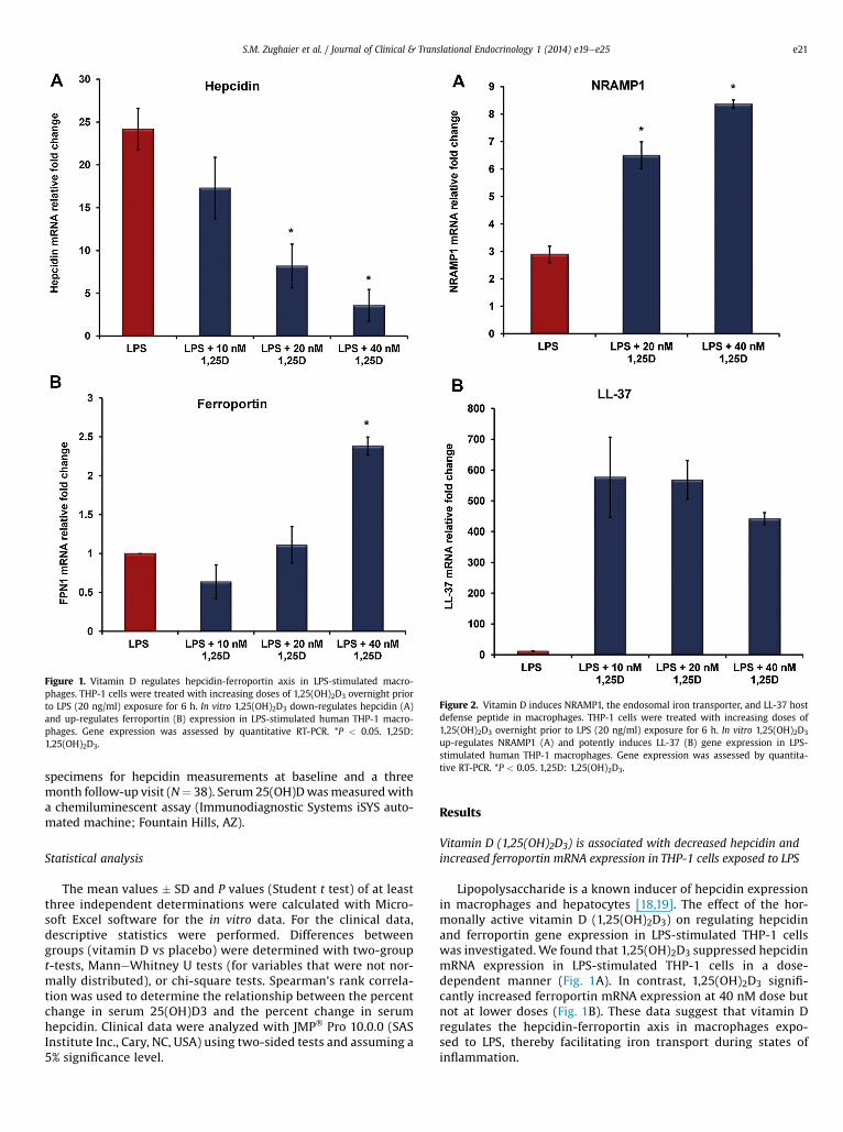
Figure 1. Vitamin D regulates hepcidin-ferroportin axis in LPS-stimulated macro-phages. THP-1 cells were treated with increasing doses of 1,25(OH)2D3 overnight priorto LPS (20 ng/ml) exposure for 6 h. In vitro 1,25(OH)2D3 down-regulates hepcidin (A)and up-regulates ferroportin (B) expression in LPS-stimulated human THP-1 macro-phages. Gene expression was assessed by quantitative RT-PCR. *P < 0.05. 1,25D:1,25(OH)2D3.
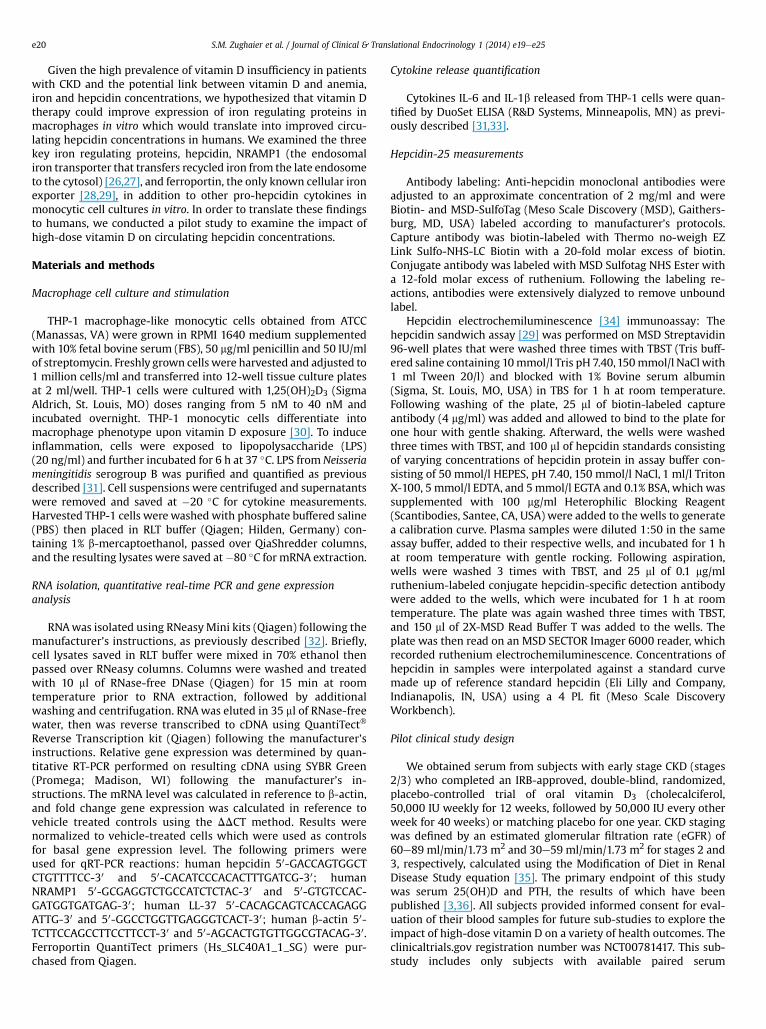
Figure 2. Vitamin D induces NRAMP1, the endosomal iron transporter, and LL-37 hostdefense peptide in macrophages. THP-1 cells were treated with increasing doses of1,25(OH)2D3 overnight prior to LPS (20 ng/ml) exposure for 6 h. In vitro 1,25(OH)2D3
up-regulates NRAMP1 (A) and potently induces LL-37 (B) gene expression in LPS-stimulated human THP-1 macrophages. Gene expression was assessed by quantita-tive RT-PCR. *P < 0.05. 1,25D: 1,25(OH)2D3.
S.M. Zughaier et al. / Journal of Clinical & Translational Endocrinology 1 (2014) e19ee25 e21
specimens for hepcidin measurements at baseline and a threemonth follow-up visit (N¼ 38). Serum 25(OH)Dwasmeasuredwitha chemiluminescent assay (Immunodiagnostic Systems iSYS auto-mated machine; Fountain Hills, AZ).
Statistical analysis
The mean values � SD and P values (Student t test) of at leastthree independent determinations were calculated with Micro-soft Excel software for the in vitro data. For the clinical data,descriptive statistics were performed. Differences betweengroups (vitamin D vs placebo) were determined with two-groupt-tests, ManneWhitney U tests (for variables that were not nor-mally distributed), or chi-square tests. Spearman’s rank correla-tion was used to determine the relationship between the percentchange in serum 25(OH)D3 and the percent change in serumhepcidin. Clinical data were analyzed with JMP� Pro 10.0.0 (SASInstitute Inc., Cary, NC, USA) using two-sided tests and assuming a5% significance level.
Results
Vitamin D (1,25(OH)2D3) is associated with decreased hepcidin andincreased ferroportin mRNA expression in THP-1 cells exposed to LPS
Lipopolysaccharide is a known inducer of hepcidin expressionin macrophages and hepatocytes [18,19]. The effect of the hor-monally active vitamin D (1,25(OH)2D3) on regulating hepcidinand ferroportin gene expression in LPS-stimulated THP-1 cellswas investigated. We found that 1,25(OH)2D3 suppressed hepcidinmRNA expression in LPS-stimulated THP-1 cells in a dose-dependent manner (Fig. 1A). In contrast, 1,25(OH)2D3 signifi-cantly increased ferroportin mRNA expression at 40 nM dose butnot at lower doses (Fig. 1B). These data suggest that vitamin Dregulates the hepcidin-ferroportin axis in macrophages expo-sed to LPS, thereby facilitating iron transport during states ofinflammation.
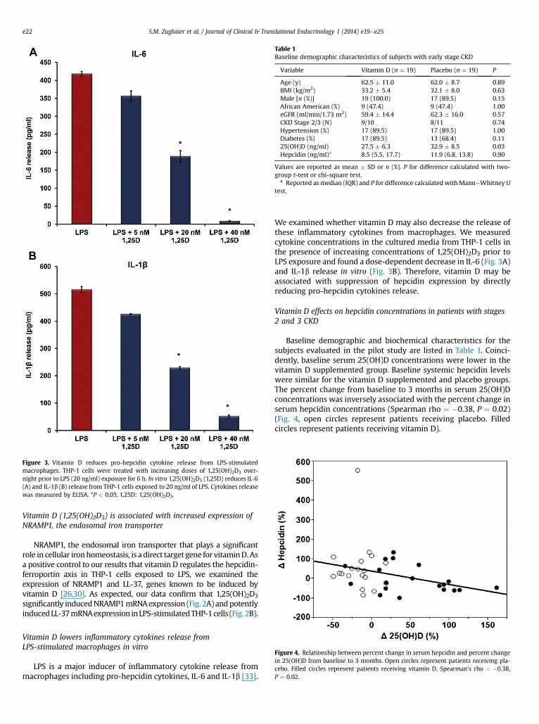
Figure 3. Vitamin D reduces pro-hepcidin cytokine release from LPS-stimulatedmacrophages. THP-1 cells were treated with increasing doses of 1,25(OH)2D3 over-night prior to LPS (20 ng/ml) exposure for 6 h. In vitro 1,25(OH)2D3 (1,25D) reduces IL-6(A) and IL-1b (B) release from THP-1 cells exposed to 20 ng/ml of LPS. Cytokines releasewas measured by ELISA. *P < 0.05. 1,25D: 1,25(OH)2D3.
Table 1Baseline demographic characteristics of subjects with early stage CKD
Variable Vitamin D (n ¼ 19) Placebo (n ¼ 19) P
Age (y) 62.5 � 11.0 62.0 � 8.7 0.89BMI (kg/m2) 33.2 � 5.4 32.1 � 8.0 0.63Male [n (%)] 19 (100.0) 17 (89.5) 0.15African American (%) 9 (47.4) 9 (47.4) 1.00eGFR (ml/min/1.73 m2) 59.4 � 14.4 62.3 � 16.0 0.57CKD Stage 2/3 (N) 9/10 8/11 0.74Hypertension (%) 17 (89.5) 17 (89.5) 1.00Diabetes (%) 17 (89.5) 13 (68.4) 0.1125(OH)D (ng/ml) 27.5 � 6.3 32.9 � 8.5 0.03Hepcidin (ng/ml)a 8.5 (5.5, 17.7) 11.9 (6.8, 13.8) 0.90
Values are reported as mean � SD or n (%). P for difference calculated with two-group t-test or chi-square test.
a Reported as median (IQR) and P for difference calculated with ManneWhitney Utest.
S.M. Zughaier et al. / Journal of Clinical & Translational Endocrinology 1 (2014) e19ee25e22
Vitamin D (1,25(OH)2D3) is associated with increased expression ofNRAMP1, the endosomal iron transporter
NRAMP1, the endosomal iron transporter that plays a significantrole in cellular ironhomeostasis, is a direct target gene for vitaminD.Asa positive control to our results that vitamin D regulates the hepcidin-ferroportin axis in THP-1 cells exposed to LPS, we examined theexpression of NRAMP1 and LL-37, genes known to be induced byvitamin D [26,30]. As expected, our data confirm that 1,25(OH)2D3significantly inducedNRAMP1mRNAexpression (Fig. 2A) andpotentlyinducedLL-37mRNAexpression inLPS-stimulatedTHP-1cells (Fig.2B).
Figure 4. Relationship between percent change in serum hepcidin and percent changein 25(OH)D from baseline to 3 months. Open circles represent patients receiving pla-cebo. Filled circles represent patients receiving vitamin D. Spearman’s rho ¼ �0.38,P ¼ 0.02.
Vitamin D lowers inflammatory cytokines release fromLPS-stimulated macrophages in vitro
LPS is a major inducer of inflammatory cytokine release frommacrophages including pro-hepcidin cytokines, IL-6 and IL-1b [33].
We examined whether vitamin D may also decrease the release ofthese inflammatory cytokines from macrophages. We measuredcytokine concentrations in the cultured media from THP-1 cells inthe presence of increasing concentrations of 1,25(OH)2D3 prior toLPS exposure and found a dose-dependent decrease in IL-6 (Fig. 3A)and IL-1b release in vitro (Fig. 3B). Therefore, vitamin D may beassociated with suppression of hepcidin expression by directlyreducing pro-hepcidin cytokines release.
Vitamin D effects on hepcidin concentrations in patients with stages2 and 3 CKD
Baseline demographic and biochemical characteristics for thesubjects evaluated in the pilot study are listed in Table 1. Coinci-dently, baseline serum 25(OH)D concentrations were lower in thevitamin D supplemented group. Baseline systemic hepcidin levelswere similar for the vitamin D supplemented and placebo groups.The percent change from baseline to 3 months in serum 25(OH)Dconcentrations was inversely associated with the percent change inserum hepcidin concentrations (Spearman rho ¼ �0.38, P ¼ 0.02)(Fig. 4, open circles represent patients receiving placebo. Filledcircles represent patients receiving vitamin D).
S.M. Zughaier et al. / Journal of Clinical & Translational Endocrinology 1 (2014) e19ee25 e23
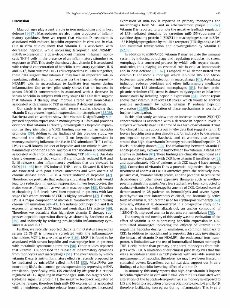
Discussion
Macrophages play a central role in iron metabolism and in hostdefense [14,37]. Macrophages are also major producers of inflam-matory cytokines. Here we report that vitamin D treatment isassociated with reduced hepcidin expression in vitro and in vivo.Our in vitro studies show that vitamin D is associated withdecreased hepcidin while increasing ferroportin and NRAMP1mRNA expression in a dose-dependent manner in human mono-cytic THP-1 cells in the presence of an inflammatory stimulus (i.e.exposure to LPS). This study also shows that vitamin D is associatedwith reduced concentration of hepcidin stimulatory cytokines, IL-6and IL-1b, from cultured THP-1 cells exposed to LPS. Taken together,these data suggest that vitamin D may have an important role inregulating cellular iron homeostasis via the hepcidin-ferroportin-NRAMP1 axis in macrophages to facilitate iron egress duringinflammation. Our in vivo pilot study shows that an increase inserum 25(OH)D concentration is associated with a decrease inserum hepcidin in subjects with early stage CKD. Our data suggestthat vitamin D therapy may improve altered iron homeostasisassociated with anemia of CKD in vitamin D deficient patients.
Our study is in agreement with recent studies showing thatvitamin D suppresses hepcidin expression in macrophages [38,39].Bacchetta and co-workers show that vitamin D significantly sup-pressed hepcidin expression in monocytes by 0.5-fold and providesevidence that vitamin D directly downregulates hepcidin expres-sion as they identified a VDRE binding site on human hepcidinpromoter [39]. Adding to the findings of this previous study, weexamined the effect of vitamin D on hepcidin expression inmonocytes during inflammation (i.e. in LPS-stimulated THP-1 cells).LPS is a well-known inducer of hepcidin and can mimic in vivo in-flammatory conditions since microbial translocation is commonlyassociated with chronic diseases including CKD [40e43]. Our dataclearly demonstrate that vitamin D significantly reduced IL-6 andIL-1b release (major inflammatory cytokines that are elevated inCKD) [44e46] from LPS-induced THP-1 cells. Elevated IL-6 levelsare associated with poor clinical outcomes and with anemia ofchronic disease since IL-6 is a direct inducer of hepcidin [47].Therefore, we postulate that reducing circulating IL-6 levels wouldlead to a reduction in hepcidin expression in liver hepatocytes, themajor source of hepcidin, as well as in macrophages [48]. Elevationin circulating IL-6 levels have been reported in patients with latestage CKD where anemia of CKD is highly prevalent [47]. Further,LPS is a major component of microbial translocation seen duringchronic inflammation [40e43]. LPS induces both hepcidin and IL-6expression whereas LL-37 binds and neutralizes LPS activity [49].Therefore, we postulate that high-dose vitamin D therapy sup-presses hepcidin expression directly, as shown by Bacchetta et al.[39], and indirectly by reducing pro-hepcidin inflammatory cyto-kines IL-6 and IL-1b.
Further, we recently reported that vitamin D status assessed asserum 25(OH)D is inversely correlated with the inflammatorychemokine, MCP-1, in vivo and in vitro [3,36]. MCP-1 is found to beassociated with serum hepcidin and macrophage iron in patientswith metabolic syndrome alterations [50]. Other studies reportedthat vitamin D suppressed the release of inflammatory cytokinesfrom monocytes and macrophages [51]. The mechanism by whichvitamin D exerts anti-inflammatory effects is recently proposed tobe mediated by microRNA called miR-155 [52]. MicroRNAs arenoncoding RNAs that control genes expression by repressing mRNAtranslation. Specifically, miR-155 encoded by bic gene is a criticalregulator of TLR signaling in macrophages. miR-155 targets SOCS1(cytokine signaling protein 1), the negative feedback regulator ofcytokine release, therefore high miR-155 expression is associatedwith a heightened cytokine release from macrophages. Increased
expression of miR-155 is reported in primary monocytes andmacrophages from SLE and in atherosclerotic plaque [53e57].Vitamin D is reported to promote the negative feedback regulationof LPS-mediated signaling by targeting miR-155-suppressor ofcytokine signaling protein 1 (SOCS1) in macrophages since miRNA-155 is highly upregulated by toll-like receptors (TLR) ligands, i.e. LPSand microbial translocation and downregulated by vitamin D[52,58].
In addition to miRNA-155, vitamin D may regulate the immunesystem by inducing autophagy and regulating endoplasmic stress.Autophagy is a conserved process by which cells recycle macro-molecules, thus playing an essential role in cellular homeostasisand in host defense [59e62]. Campbell et al. demonstrated thatvitamin D enhanced autophagy, which inhibited HIV and Myco-bacterium tuberculosis infection in macrophages [61]. Autophagyinduction reduces cytokines and other inflammatory mediatorsrelease from LPS-stimulated macrophages [63]. Further, endo-plasmic reticulum (ER) stress is shown to dysregulate cellular ironhomeostasis by inducing hepcidin expression [64]. Recent reportshows that vitamin D relieves ER stress, which would be anotherpossible mechanism by which vitamin D reduces hepcidinexpression [65,66]. Elucidation of these mechanisms is worthy offurther investigation.
In this pilot study we show that an increase in serum 25(OH)Dconcentrations is associated with a decrease in hepcidin levels insubjectswith early stage CKD enrolled in a high-dose vitaminD trial.Our clinical finding supports our in vitro data that suggest vitamin Dlowers hepcidin expression directly and/or indirectly by decreasingpro-hepcidin cytokines. Bacchetta and co-workers also demon-strated that high-dose vitaminD therapy reduced bloodhepcidin-25levels in healthy donors [39]. The relationship between vitamin Dand hepcidinmayexplain the link between low vitaminD status andanemia in children [67]. These findings are clinically significant as alarge majority of patients with CKD have vitamin D insufficiency [2],and approximately 40% of patients with CKD stage 4 have anemia[68]. Correction of vitamin D as a potential adjunctive therapy intreatment of anemia of CKD is attractive given the relatively inex-pensive cost, favorable safety profile, and the potential to reduce thedependence on other more expensive therapies such as erythro-poietin stimulating agents. Very few studies have been conducted toevaluate vitamin D as a therapy for anemia of CKD. Goicoechea et al.demonstrated in 28 patients on hemodialysis and severe hyper-parathyroidism that intravenous calcitriol, the hormonally activeform of vitamin D, reduced the need for erythropoietin therapy [69].Similarly, Albitar et al. demonstrated in a prospective study of 12patients, treatment with alfacalcidol, an analog compound of1,25(OH)2D, improved anemia in patients on hemodialysis [70].
The strength and novelty of this study was the evaluation of theeffect of vitamin D on suppressing hepcidin expression in LPS-stimulated monocytes indicating the efficacy of vitamin D onregulating hepcidin during inflammation, a common hallmark ofCKD. In addition to hepcidin and ferroportin, this study investigatedthe impact of vitamin D on NRAMP1, the endosomal iron trans-porter. A limitation was the use of immortalized human monocyticTHP-1 cells rather than primary peripheral monocytes from sub-jects with CKD. A limitation of our clinical pilot study was that thiswas a secondary analysis in CKD patients with available serum formeasurement of hepcidin; therefore, we may have been limited instatistical power. Regardless, our clinical data support our in vitrofindings of a hepcidin-lowering effect of vitamin D.
In summary, this study reports that high-dose vitamin D impactshepcidin expression in vitro and in vivo. Vitamin D is associated withalterations of the hepcidin-ferroportin axis in monocytes exposed toLPS and leads to a reduction of pro-hepcidin cytokine, IL-6 and IL-1b,therefore facilitating iron egress during inflammation. This in vitro
S.M. Zughaier et al. / Journal of Clinical & Translational Endocrinology 1 (2014) e19ee25e24
observation appears to be supported by our early translational pilotstudy in subjects with early CKD where changes in vitamin D statusinduced by high-dose oral vitamin D therapy impacted changes insystemic hepcidin levels.
Acknowledgment
This work is supported in part by grants from Emory-EglestonChildren’s Research Center and Center for Pediatric Nanomedicineof Emory þ Children’s Pediatrics Research Center to S.M.Z, and byT32 DK007298-32S1 to J.A.A. S.M.Z. especially acknowledges thesupport of N. McCarty (Center for Cystic Fibrosis Research, EmoryUniversity School of Medicine) for providing the research facilityand laboratory space where part of this work was conducted. Thecontent is solely the responsibility of the authors and does notnecessarily represent the official views of the National Institutes ofHealth.
References
[1] Mallbris L, O’Brien KP, Hulthen A, Sandstedt B, Cowland JB, Borregaard N, et al.Neutrophil gelatinase-associated lipocalin is a marker for dysregulated kera-tinocyte differentiation in human skin. Exp Dermatol 2002;11(6):584e91.
[2] LaClair RE, Hellman RN, Karp SL, Kraus M, Ofner S, Li Q, et al. Prevalence ofcalcidiol deficiency in CKD: a cross-sectional study across latitudes in theUnited States. Am J Kidney Dis 2005;45(6):1026e33.
[3] Alvarez JA, Law J, Coakley KE, Zughaier SM, Hao L, Shahid Salles K, et al. High-dose cholecalciferol reduces parathyroid hormone in patients with earlychronic kidney disease: a pilot, randomized, double-blind, placebo-controlledtrial. Am J Clin Nutr 2012;96(3):672e9. PMCID: 3417221.
[4] Wasse H, Huang R, Long Q, Zhao Y, Singapuri S, McKinnon W, et al. Very high-dose cholecalciferol and arteriovenous fistula maturation in ESRD: a ran-domized, double-blind, placebo-controlled pilot study. J Vasc Access2013;0(0):0.
[5] Chandra P, Binongo JN, Ziegler TR, Schlanger LE, Wang W, Someren JT, et al.Cholecalciferol (vitamin D3) therapy and vitamin D insufficiency in patientswith chronic kidney disease: a randomized controlled pilot study. EndocrPract 2008;14(1):10e7. PMCID: 2654595.
[6] Judd SE, Tangpricha V. Vitamin D therapy and cardiovascular health. CurrHypertens Rep 2011;13(3):187e91.
[7] Ullah MI, Uwaifo GI, Nicholas WC, Koch CA. Does Vitamin D deficiency causehypertension? Current evidence from clinical studies and potential mecha-nisms. Int J Endocrinol 2010;2010:1e11.
[8] Carvalho C, Isakova T, Collerone G, Olbina G, Wolf M, Westerman M, et al.Hepcidin and disordered mineral metabolism in chronic kidney disease. ClinNephrol 2011;76(2):90e8.
[9] Perlstein TS, Pande R, Berliner N, Vanasse GJ. Prevalence of 25-hydroxyvitaminD deficiency in subgroups of elderly persons with anemia: association withanemia of inflammation. Blood 2011;117(10):2800e6.
[10] Icardi A, Paoletti E, De Nicola L, Mazzaferro S, Russo R, Cozzolino M. Renalanaemia and EPO hyporesponsiveness associated with vitamin D deficiency:the potential role of inflammation. Nephrol Dial Transplant 2013;28(7):1672e9.
[11] Ganz T. Hepcidin and iron regulation, 10 years later. Blood 2011;117(17):4425e33. PMCID: 3099567.
[12] Sun CC, Vaja V, Babitt JL, Lin HY. Targeting the hepcidin-ferroportin axis todevelop new treatment strategies for anemia of chronic disease and anemia ofinflammation. Am J Hematol 2012;87(4):392e400. PMCID: 3653431.
[13] Ganz T, Nemeth E. Hepcidin and disorders of iron metabolism. Annu Rev Med2011;62:347e60.
[14] Ganz T. Macrophages and systemic iron homeostasis. J Innate Immun2012;4(5e6):446e53.
[15] Zaritsky J, Young B, Gales B, Wang HJ, Rastogi A, Westerman M, et al.Reduction of serum hepcidin by hemodialysis in pediatric and adult patients.Clin J Am Soc Nephrol 2010;5(6):1010e4. PMCID: 2879302.
[16] Babitt JL, Lin HY. Molecular mechanisms of hepcidin regulation: implicationsfor the anemia of CKD. Am J Kidney Dis 2010;55(4):726e41. PMCID: 2905036.
[17] Ganz T, Olbina G, Girelli D, Nemeth E, Westerman M. Immunoassay for humanserum hepcidin. Blood 2008;112(10):4292e7.
[18] Nemeth E, Rivera S, Gabayan V, Keller C, Taudorf S, Pedersen BK, et al. IL-6mediates hypoferremia of inflammation by inducing the synthesis of the ironregulatory hormone hepcidin. J Clin Invest 2004;113(9):1271e6. PMCID:398432.
[19] Lee P, Peng H, Gelbart T, Wang L, Beutler E. Regulation of hepcidin tran-scription by interleukin-1 and interleukin-6. Proc Natl Acad Sci U S A2005;102(6):1906e10. PMCID: 548537.
[20] Wrighting DM, Andrews NC. Interleukin-6 induces hepcidin expressionthrough STAT3. Blood 2006;108(9):3204e9. PMCID: 1895528.
[21] Pigeon C, Ilyin G, Courselaud B, Leroyer P, Turlin B, Brissot P, et al. A newmouse liver-specific gene, encoding a protein homologous to human antimi-crobial peptide hepcidin, is overexpressed during iron overload. J Biol Chem2001;276(11):7811e9.
[22] Park CH, Valore EV, Waring AJ, Ganz T. Hepcidin, a urinary antimicrobialpeptide synthesized in the liver. J Biol Chem 2001;276(11):7806e10.
[23] Krause A, Neitz S, Magert HJ, Schulz A, Forssmann WG, Schulz-Knappe P, et al.LEAP-1, a novel highly disulfide-bonded human peptide, exhibits antimicro-bial activity. FEBS Lett 2000;480(2e3):147e50.
[24] Zaritsky J, Young B, Wang HJ, Westerman M, Olbina G, Nemeth E, et al.Hepcidinea potential novel biomarker for iron status in chronic kidney dis-ease. Clin J Am Soc Nephrol 2009;4(6):1051e6. PMCID: 2689881.
[25] Cabrera AR, Cuneo KC, Desjardins A, Sampson JH, McSherry F, Herndon 2nd JE,et al. Concurrent stereotactic radiosurgery and bevacizumab in recurrentmalignant gliomas: a prospective trial. Int J Radiat Oncol Biol Phys 2013;86(5):873e9.
[26] Roig EA, Richer E, Canonne-Hergaux F, Gros P, Cellier MF. Regulation ofNRAMP1 gene expression by 1alpha,25-dihydroxy-vitamin D(3) in HL-60phagocytes. J Leukoc Biol 2002;71(5):890e904.
[27] Rodrigues PN, Gomes SS, Neves JV, Gomes-Pereira S, Correia-Neves M, Nunes-Alves C, et al. Mycobacteria-induced anaemia revisited: a molecular approachreveals the involvement of NRAMP1 and lipocalin-2, but not of hepcidin.Immunobiology 2011;216(10):1127e34.
[28] Nemeth E, Tuttle MS, Powelson J, Vaughn MB, Donovan A, Ward DM, et al.Hepcidin regulates cellular iron efflux by binding to ferroportin and inducingits internalization. Science 2004;306(5704):2090e3.
[29] De Domenico I, Ward DM, Langelier C, Vaughn MB, Nemeth E, Sundquist WI,et al. The molecular mechanism of hepcidin-mediated ferroportin down-regulation. Mol Biol Cell 2007;18(7):2569e78. PMCID: 1924807.
[30] Cellier M, Shustik C, Dalton W, Rich E, Hu J, Malo D, et al. Expression of thehuman NRAMP1 gene in professional primary phagocytes: studies in bloodcells and in HL-60 promyelocytic leukemia. J Leukoc Biol 1997;61(1):96e105.
[31] Zughaier SM, Tzeng YL, Zimmer SM, Datta A, Carlson RW, Stephens DS.Neisseria meningitidis lipooligosaccharide structure-dependent activation ofthe macrophage CD14/Toll-like receptor 4 pathway. Infect Immun 2004;72(1):371e80.
[32] Zughaier SM. Neisseria meningitidis capsular polysaccharides induce inflam-matory responses via TLR2 and TLR4-MD-2. J Leukoc Biol 2011;89(3):469e80.PMCID: 3040465.
[33] Zughaier SM, Zimmer SM, Datta A, Carlson RW, Stephens DS. Differential in-duction of the toll-like receptor 4-MyD88-dependent and -independentsignaling pathways by endotoxins. Infect Immun 2005;73(5):2940e50.
[34] Leclercq A, Houard X, Philippe M, Ollivier V, Sebbag U, Meilhac O, et al.Involvement of intraplaque hemorrhage in atherothrombosis evolution vianeutrophil protease enrichment. J Leukoc Biol 2007;82(6):1420e9.
[35] Levey AS, Coresh J, Greene T, Stevens LA, Zhang YL, Hendriksen S, et al. Usingstandardized serum creatinine values in the modification of diet in renaldisease study equation for estimating glomerular filtration rate. Ann InternMed 2006;145(4):247e54.
[36] Alvarez JA, Zughaier SM, Law J, Hao L, Wasse H, Ziegler TR, et al. Effects ofhigh-dose cholecalciferol on serum markers of inflammation and immunity inpatients with early chronic kidney disease. Eur J Clin Nutr 2013;67(3):264e9.
[37] Wang J, Pantopoulos K. Regulation of cellular iron metabolism. Biochem J2011;434(3):365e81. PMCID: 3048577.
[38] Bell SJ. The effect of VitaminD3 on hepcidin and IL-8 expression in monocytes.J Hematol 2013;2(1):1e7.
[39] Bacchetta J, Zaritsky JJ, Sea JL, Chun RF, Lisse TS, Zavala K, et al. Suppression ofiron-regulatory hepcidin by vitamin D. J Am Soc Nephrol 2014;25:564e72.
[40] Layoun A, Santos MM. Bacterial cell wall constituents induce hepcidinexpression in macrophages through MyD88 signaling. Inflammation2012;35(4):1500e6.
[41] Wang L, Harrington L, Trebicka E, Shi HN, Kagan JC, Hong CC, et al. Selectivemodulation of TLR4-activated inflammatory responses by altered iron ho-meostasis in mice. J Clin Invest 2009;119(11):3322e8. PMCID: 2769199.
[42] Theurl I, Theurl M, Seifert M, Mair S, Nairz M, Rumpold H, et al. Autocrineformation of hepcidin induces iron retention in human monocytes. Blood2008;111(4):2392e9.
[43] Ramezani A, Raj DS. The gut microbiome, kidney disease, and targeted in-terventions. J Am Soc Nephrol 2013. PMCID: 24231662.
[44] McCarley P. The KDOQI clinical practice guidelines and clinical practice rec-ommendations for treating anemia in patients with chronic kidney disease:implications for nurses. Nephrol Nurs J 2006;33(4):423e6. 45; quiz 7e8.
[45] Malyszko J, Malyszko JS, Bachorzewska-Gajewska H, Poniatowski B,Dobrzycki S, Mysliwiec M. Neutrophil gelatinase-associated lipocalin is a newand sensitive marker of kidney function in chronic kidney disease patients andrenal allograft recipients. Transplant Proc 2009;41(1):158e61.
[46] Malyszko J, Bachorzewska-Gajewska H, Sitniewska E, Malyszko JS,Poniatowski B, Dobrzycki S. Serum neutrophil gelatinase-associated lipocalinas a marker of renal function in non-diabetic patients with stage 2-4 chronickidney disease. Ren Fail 2008;30(6):625e8.
[47] Pecoits-Filho R, Barany P, Lindholm B, Heimburger O, Stenvinkel P. Inter-leukin-6 is an independent predictor of mortality in patients starting dialysistreatment. Nephrol Dial Transplant 2002;17(9):1684e8.
[48] Wang FD, Zhou DB, Li SL, Wang X, Zhang JP, Duan MH, et al. Effect of re-combinant human erythropoietin on hepcidin mRNA expression in patients
S.M. Zughaier et al. / Journal of Clinical & Translational Endocrinology 1 (2014) e19ee25 e25
with multiple myeloma. Zhongguo Shi Yan Xue Ye Xue Za Zhi 2011;19(2):390e4.
[49] Zughaier SM, Shafer WM, Stephens DS. Antimicrobial peptides and endotoxininhibit cytokine and nitric oxide release but amplify respiratory burst responsein human and murine macrophages. Cell Microbiol 2005;7(9):1251e62.
[50] Valenti L, Dongiovanni P, Motta BM, Swinkels DW, Bonara P, Rametta R, et al.Serum hepcidin and macrophage iron correlate with MCP-1 release andvascular damage in patients with metabolic syndrome alterations. ArteriosclerThromb Vasc Biol 2011;31(3):683e90.
[51] Zhang Y, Leung DY, Richers BN, Liu Y, Remigio LK, Riches DW, et al. Vitamin Dinhibits monocyte/macrophage proinflammatory cytokine production by tar-getingMAPK phosphatase-1. J Immunol 2012;188(5):2127e35. PMCID: 3368346.
[52] Chen Y, Liu W, Sun T, Huang Y, Wang Y, Deb DK, et al. 1,25-dihydroxyvitamin Dpromotes negative feedback regulation of TLR signaling via targeting microRNA-155-SOCS1 in macrophages. J Immunol 2013;190(7):3687e95. PMCID: 3608760.
[53] Katsushima F, Takahashi A, Sakamoto N, Kanno Y, Abe K, Ohira H. Expressionof micro-RNAs in peripheral blood mononuclear cells from primary biliarycirrhosis patients. Hepatol Res; 2013. http://dx.doi.org/10.1111/hepr.12198.
[54] Wang H, Peng W, Ouyang X, Li W, Dai Y. Circulating microRNAs as candidatebiomarkers in patients with systemic lupus erythematosus. Transl Res2012;160(3):198e206.
[55] Leng RX, Pan HF, Qin WZ, Chen GM, Ye DQ. Role of microRNA-155 in auto-immunity. Cytokine Growth Factor Rev 2011;22(3):141e7.
[56] Wang G, Tam LS, Li EK, Kwan BC, Chow KM, Luk CC, et al. Serum and urinarycell-free MiR-146a and MiR-155 in patients with systemic lupus erythema-tosus. J Rheumatol 2010;37(12):2516e22.
[57] Li YC, Chen Y, Liu W, Thadhani R. MicroRNA-mediated mechanism of vitaminD regulation of innate immune response. J Steroid Biochem Mol Biol 2013.http://dx.doi.org/10.1016/j.jsbmb.2013.09.014.
[58] Chen NK, Chong TW, Loh HL, Lim KH, Gan VH, Wang M, et al. Negative reg-ulatory responses to metabolically triggered inflammation impair renalepithelial immunity in diabetes mellitus. J Mol Med (Berl) 2013;91(5):587e98.PMCID: 3644409.
[59] Jo EK. Innate immunity to mycobacteria: vitamin D and autophagy. CellMicrobiol 2010;12(8):1026e35.
[60] Yuk JM, Shin DM, Lee HM, Yang CS, Jin HS, Kim KK, et al. Vitamin D3 inducesautophagy in human monocytes/macrophages via cathelicidin. Cell HostMicrobe 2009;6(3):231e43.
[61] Campbell GR, Spector SA. Vitamin D inhibits human immunodeficiency vi-rus type 1 and Mycobacterium tuberculosis infection in macrophagesthrough the induction of autophagy. PLoS Pathog 2012;8(5):e1002689.PMCID: 3349755.
[62] Campbell GR, Spector SA. Toll-like receptor 8 ligands activate a vitamin Dmediated autophagic response that inhibits human immunodeficiency virustype 1. PLoS Pathog 2012;8(11):e1003017. PMCID: 3499571.
[63] Zughaier S, Karna P, Stephens D, Aneja R. Potent anti-inflammatory activity ofnovel microtubule-modulating brominated noscapine analogs. PLoS One2010;5(2):e9165. PMCID: 2820095.
[64] Vecchi C, Montosi G, Zhang K, Lamberti I, Duncan SA, Kaufman RJ, et al. ERstress controls iron metabolism through induction of hepcidin. Science2009;325(5942):877e80. PMCID: 2923557.
[65] Riek AE, Oh J, Bernal-Mizrachi C. 1,25(OH)(2) vitamin D suppresses macro-phage migration and reverses atherogenic cholesterol metabolism in type 2diabetic patients. J Steroid Biochem Mol Biol 2013;136:309e12.
[66] Riek AE, Oh J, Sprague JE, Timpson A, de las Fuentes L, Bernal-Mizrachi L, et al.Vitamin D suppression of endoplasmic reticulum stress promotes an anti-atherogenic monocyte/macrophage phenotype in type 2 diabetic patients.J Biol Chem 2012;287(46):38482e94. PMCID: 3493893.
[67] Atkinson MA, Melamed ML, Kumar J, Roy CN, Miller 3rd ER, Furth SL, et al.Vitamin D, race, and risk for anemia in children. J Pediatr 2014;164:153e8. e1.
[68] Astor BC, Muntner P, Levin A, Eustace JA, Coresh J. Association of kidneyfunction with anemia: the Third National Health and Nutrition ExaminationSurvey (1988-1994). Arch Intern Med 2002;162(12):1401e8.
[69] Goicoechea M, Vazquez MI, Ruiz MA, Gomez-Campdera F, Perez-Garcia R,Valderrabano F. Intravenous calcitriol improves anaemia and reduces theneed for erythropoietin in haemodialysis patients. Nephron 1998;78(1):23e7.
[70] Albitar S, Genin R, Fen-Chong M, Serveaux MO, Schohn D, Chuet C. High-dosealfacalcidol improves anaemia in patients on haemodialysis. Nephrol DialTransplant 1997;12(3):514e8.
Related Documents