diagnostics Review The Role of Ultrasound Guided Sampling Procedures in the Diagnosis of Pelvic Masses: A Narrative Review of the Literature Francesca Arezzo 1, *, Vera Loizzi 2 , Daniele La Forgia 3 , Adam Abdulwakil Kawosha 4 , Erica Silvestris 5 , Viviana Cataldo 1 , Claudio Lombardi 1 , Gerardo Cazzato 6 , Giuseppe Ingravallo 6 , Leonardo Resta 6 and Gennaro Cormio 1 Citation: Arezzo, F.; Loizzi, V.; La Forgia, D.; Abdulwakil Kawosha, A.; Silvestris, E.; Cataldo, V.; Lombardi, C.; Cazzato, G.; Ingravallo, G.; Resta, L.; et al. The Role of Ultrasound Guided Sampling Procedures in the Diagnosis of Pelvic Masses: A Narrative Review of the Literature. Diagnostics 2021, 11, 2204. https:// doi.org/10.3390/diagnostics11122204 Academic Editor: Manuela Ludovisi Received: 20 October 2021 Accepted: 24 November 2021 Published: 26 November 2021 Publisher’s Note: MDPI stays neutral with regard to jurisdictional claims in published maps and institutional affil- iations. Copyright: © 2021 by the authors. Licensee MDPI, Basel, Switzerland. This article is an open access article distributed under the terms and conditions of the Creative Commons Attribution (CC BY) license (https:// creativecommons.org/licenses/by/ 4.0/). 1 Obstetrics and Gynecology Unit, Department of Biomedical Sciences and Human Oncology, University of Bari “Aldo Moro”, Piazza Giulio Cesare 11, 70124 Bari, Italy; [email protected] (V.C.); [email protected] (C.L.); [email protected] (G.C.) 2 Obstetrics and Gynecology Unit, Interdisciplinar Department of Medicine, University of Bari “Aldo Moro”, Piazza Giulio Cesare 11, 70124 Bari, Italy; [email protected] 3 SSD Radiodiagnostica Senologica, IRCCS Istituto Tumori Giovanni Paolo II”, Via Orazio Flacco 65, 70124 Bari, Italy; [email protected] 4 Department of General Medicine, Universitatea Medicina si Farmacie Grigore T Popa, Strada Universitatii 16, 700115 Iasi, Romania; [email protected] 5 Gynecologic Oncology Unit, IRCCS Istituto Tumori “Giovanni Paolo II”, Via Orazio Flacco 65, 70124 Bari, Italy; [email protected] 6 Department of Emergency and Organ Transplantation, Pathology Section, University of Bari “Aldo Moro”, Piazza Giulio Cesare 11, 70124 Bari, Italy; [email protected] (G.C.); [email protected] (G.I.); [email protected] (L.R.) * Correspondence: [email protected]; Tel.: +39-3274961788 Abstract: Ultrasound-guided sampling methods are usually minimally invasive techniques applied to obtain cytological specimens or tissue samples, mainly used for the diagnosis of different types of tumors. The main benefits of ultrasound guidance is its availability. It offers high flexibility in the choice of sampling approach (transabdominal, transvaginal, and transrectal) and short duration of procedure. Ultrasound guided sampling of pelvic masses represents the diagnostic method of choice in selected patients. We carried out a narrative review of literatures regarding the ultrasound-guided methods of cytological and histological evaluation of pelvic masses as well as the positive and negative predictors for the achievement of an adequate sample. Keywords: ultrasound-guided sampling procedures; fine-needle aspiration cytology; fine-needle aspiration biopsy; tru-cut biopsy; pelvic masses 1. Introduction Ultrasound-guided sampling methods are usually minimally invasive techniques applied to obtain cytological specimens or tissue samples, mainly used for the diagnosis of different types of tumors, such as breast [1] and prostate cancers [2]. Currently, the available minimally invasive technique for carrying-out cytological examination is fine-needle aspiration cytology (FNAC), and the techniques for obtaining tissue samples for histological examination includes the fine-needle aspiration biopsy (FNAB) and tru-cut biopsy [3]. Other procedures that can be used to guide sampling procedures include computer to- mography (CT), magnetic resonance imaging (MRI), and the digitally directed approach [4]. CT-guided percutaneous biopsy is regarded as an efficient and safe procedure in the evalua- tion of retroperitoneal and abdominal masses, but it has some limitations when the masses are deep within the pelvis due to limited transabdominal accessibility or limited imaging in Diagnostics 2021, 11, 2204. https://doi.org/10.3390/diagnostics11122204 https://www.mdpi.com/journal/diagnostics

Welcome message from author
This document is posted to help you gain knowledge. Please leave a comment to let me know what you think about it! Share it to your friends and learn new things together.
Transcript
diagnostics
Review
The Role of Ultrasound Guided Sampling Procedures in theDiagnosis of Pelvic Masses: A Narrative Review ofthe Literature
Francesca Arezzo 1,*, Vera Loizzi 2 , Daniele La Forgia 3 , Adam Abdulwakil Kawosha 4, Erica Silvestris 5,Viviana Cataldo 1, Claudio Lombardi 1, Gerardo Cazzato 6 , Giuseppe Ingravallo 6 , Leonardo Resta 6
and Gennaro Cormio 1
�����������������
Citation: Arezzo, F.; Loizzi, V.; La
Forgia, D.; Abdulwakil Kawosha, A.;
Silvestris, E.; Cataldo, V.; Lombardi,
C.; Cazzato, G.; Ingravallo, G.; Resta,
L.; et al. The Role of Ultrasound
Guided Sampling Procedures in the
Diagnosis of Pelvic Masses: A
Narrative Review of the Literature.
Diagnostics 2021, 11, 2204. https://
doi.org/10.3390/diagnostics11122204
Academic Editor: Manuela Ludovisi
Received: 20 October 2021
Accepted: 24 November 2021
Published: 26 November 2021
Publisher’s Note: MDPI stays neutral
with regard to jurisdictional claims in
published maps and institutional affil-
iations.
Copyright: © 2021 by the authors.
Licensee MDPI, Basel, Switzerland.
This article is an open access article
distributed under the terms and
conditions of the Creative Commons
Attribution (CC BY) license (https://
creativecommons.org/licenses/by/
4.0/).
1 Obstetrics and Gynecology Unit, Department of Biomedical Sciences and Human Oncology, University ofBari “Aldo Moro”, Piazza Giulio Cesare 11, 70124 Bari, Italy; [email protected] (V.C.);[email protected] (C.L.); [email protected] (G.C.)
2 Obstetrics and Gynecology Unit, Interdisciplinar Department of Medicine, University of Bari “Aldo Moro”,Piazza Giulio Cesare 11, 70124 Bari, Italy; [email protected]
3 SSD Radiodiagnostica Senologica, IRCCS Istituto Tumori Giovanni Paolo II”, Via Orazio Flacco 65,70124 Bari, Italy; [email protected]
4 Department of General Medicine, Universitatea Medicina si Farmacie Grigore T Popa, Strada Universitatii 16,700115 Iasi, Romania; [email protected]
5 Gynecologic Oncology Unit, IRCCS Istituto Tumori “Giovanni Paolo II”, Via Orazio Flacco 65,70124 Bari, Italy; [email protected]
6 Department of Emergency and Organ Transplantation, Pathology Section, University of Bari “Aldo Moro”,Piazza Giulio Cesare 11, 70124 Bari, Italy; [email protected] (G.C.); [email protected] (G.I.);[email protected] (L.R.)
* Correspondence: [email protected]; Tel.: +39-3274961788
Abstract: Ultrasound-guided sampling methods are usually minimally invasive techniques appliedto obtain cytological specimens or tissue samples, mainly used for the diagnosis of different types oftumors. The main benefits of ultrasound guidance is its availability. It offers high flexibility in thechoice of sampling approach (transabdominal, transvaginal, and transrectal) and short duration ofprocedure. Ultrasound guided sampling of pelvic masses represents the diagnostic method of choicein selected patients. We carried out a narrative review of literatures regarding the ultrasound-guidedmethods of cytological and histological evaluation of pelvic masses as well as the positive andnegative predictors for the achievement of an adequate sample.
Keywords: ultrasound-guided sampling procedures; fine-needle aspiration cytology; fine-needleaspiration biopsy; tru-cut biopsy; pelvic masses
1. Introduction
Ultrasound-guided sampling methods are usually minimally invasive techniquesapplied to obtain cytological specimens or tissue samples, mainly used for the diagnosis ofdifferent types of tumors, such as breast [1] and prostate cancers [2].
Currently, the available minimally invasive technique for carrying-out cytologicalexamination is fine-needle aspiration cytology (FNAC), and the techniques for obtainingtissue samples for histological examination includes the fine-needle aspiration biopsy(FNAB) and tru-cut biopsy [3].
Other procedures that can be used to guide sampling procedures include computer to-mography (CT), magnetic resonance imaging (MRI), and the digitally directed approach [4].CT-guided percutaneous biopsy is regarded as an efficient and safe procedure in the evalua-tion of retroperitoneal and abdominal masses, but it has some limitations when the massesare deep within the pelvis due to limited transabdominal accessibility or limited imaging in
Diagnostics 2021, 11, 2204. https://doi.org/10.3390/diagnostics11122204 https://www.mdpi.com/journal/diagnostics
Diagnostics 2021, 11, 2204 2 of 15
the osseous pelvic space. MRI is rarely used, regardless of its imaging benefits, because it re-quires special non-magnetic equipment and experience [5]. The main benefits of ultrasoundguidance are its availability. It offers high flexibility in the choice of sampling approach(transabdominal, transvaginal, and transrectal) and short duration of procedure [6]. Thelack of radiation is also a benefit when compared with CT guidance. Ultrasound guidedtru-cut biopsy also allows precise real-time tissue collection with complete control of theneedle tip during the entire procedure. When combined with color doppler imaging, it alsofacilitates the choice of the part of the tumor that is most suitable for biopsy. An accurateguidance of the needle tip during the entire procedure also reduces complication risks [7].
2. Methods
In September 2021, we searched MEDLINE and Scopus for randomized controlledtrials; narrative and systematic reviews; meta-analyses; observational studies, either lon-gitudinal or historical; and case series published in English in the last 25 years usingkeywords ultrasound-guided sampling procedures, fine-needle aspiration cytology, fine-needle aspiration biopsy, tru-cut biopsy, and pelvic masses. For this narrative review,abstracts from 127 manuscripts found in the literature were assessed by two independentauthors; of these, 42 (six fine-needle aspiration cytology, 11 fine-needle aspiration biopsyand tru-cut biopsy, 25 ultrasound evaluation of pelvic masses) were included, based on theimpact of the latter studies on current patient management.
3. Procedures
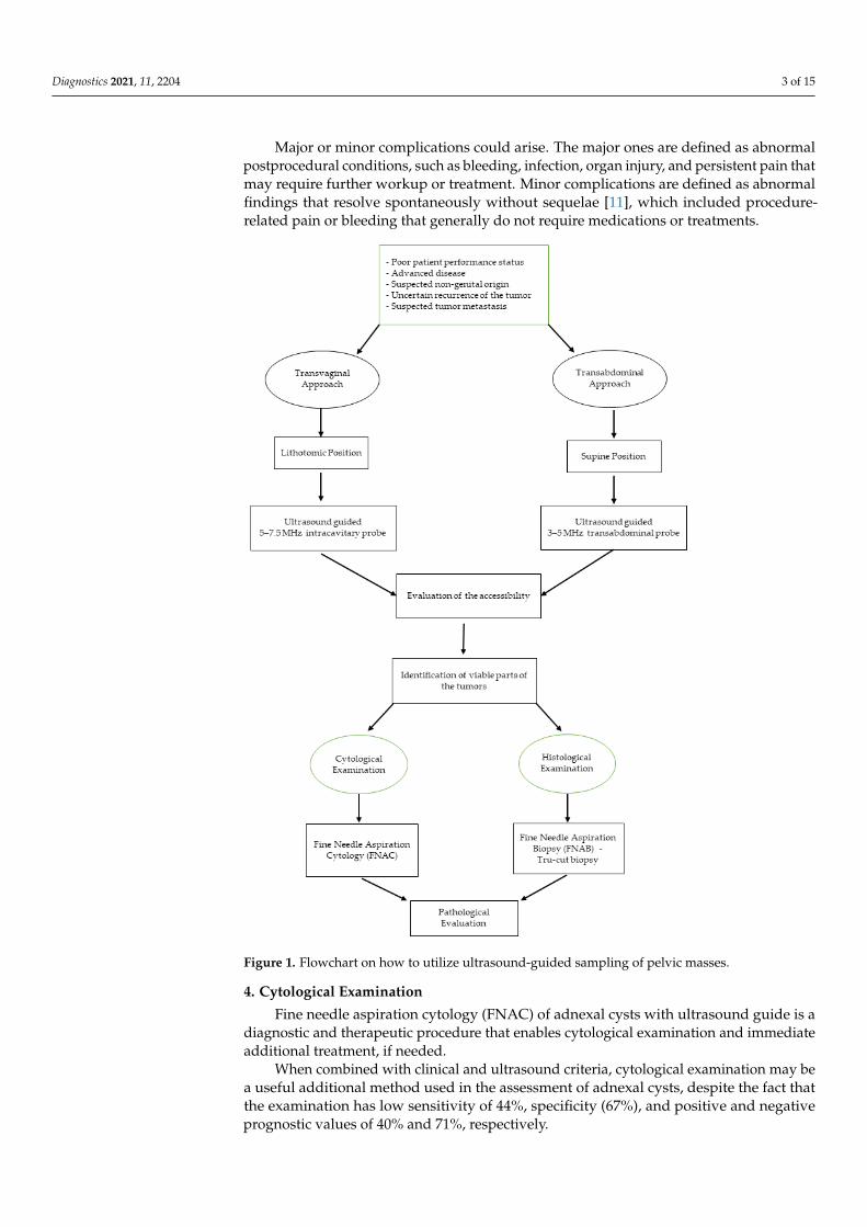
Normally, the first step in performing an ultrasound-guided sampling is to evalu-ate the accessibility of the pelvic tumor and then the feasibility of the procedure thatcould be carried-out through different approaches (transvaginally, transrectally, and/ortransabdominally) (Figure 1) [7].
3.1. Transvaginal
This procedure is performed in the lithotomy position guided by ultrasound with a5–7.5 MHz intracavitary transducer with the needle directed to the lesion using a specialneedle guide attached to an endovaginal ultrasound probe.
The mass location in relation to the vaginal cuff is important in evaluating accessibilityvia a transvaginal approach. To ensure a good exploration, the mass should be withinsome centimeters off the vaginal cuff in any single plane of view, and there should be nointerposing structures, such as the bowel and bladder [8].
Attention should be paid to identifying viable parts of the tumors detected by bloodperfusion on the Doppler imaging, and the tissue samples should be obtained using thesingle use disposable automatic bioptic gun. The cut penetration should be controlled bysetting the stopper to a depth of 10–20 mm.
3.2. Transabdominal
This procedure is performed guided by ultrasound using a 3–5 MHz transabdominal probe.Depending on the gynecological pathology, a higher accuracy of transvaginal biopsies
is likely attributable to the proximity of the lesion to the probe and ensures a better capacityfor guiding the probe more precisely into the vital parts of the tumor [9].
One to three tissue cores of 10–20 mm length and 1.6–2 mm width should be collectedand submitted for histologic analysis, after which they would be fixed and stained appro-priately. Samples would be examined for the presence of tumors and classified based onsuitability for immunohistochemical staining and tumor characteristics such as the origin,subtype, and grade [10].
At the conclusion of the procedure, it is imperative to obtain post-biopsy imageswith grayscale and color doppler imaging to assess for post biopsy hemorrhage or otherlikely complications.
Diagnostics 2021, 11, 2204 3 of 15
Major or minor complications could arise. The major ones are defined as abnormalpostprocedural conditions, such as bleeding, infection, organ injury, and persistent pain thatmay require further workup or treatment. Minor complications are defined as abnormalfindings that resolve spontaneously without sequelae [11], which included procedure-related pain or bleeding that generally do not require medications or treatments.
Diagnostics 2021, 11, x FOR PEER REVIEW 3 of 16
Figure 1. Flowchart on how to utilize ultrasound-guided sampling of pelvic masses.
3.1. Transvaginal This procedure is performed in the lithotomy position guided by ultrasound with a
5–7.5 MHz intracavitary transducer with the needle directed to the lesion using a special needle guide attached to an endovaginal ultrasound probe.
The mass location in relation to the vaginal cuff is important in evaluating accessibil-ity via a transvaginal approach. To ensure a good exploration, the mass should be within some centimeters off the vaginal cuff in any single plane of view, and there should be no interposing structures, such as the bowel and bladder [8].
Attention should be paid to identifying viable parts of the tumors detected by blood perfusion on the Doppler imaging, and the tissue samples should be obtained using the single use disposable automatic bioptic gun. The cut penetration should be controlled by setting the stopper to a depth of 10–20 mm.
Figure 1. Flowchart on how to utilize ultrasound-guided sampling of pelvic masses.
4. Cytological Examination
Fine needle aspiration cytology (FNAC) of adnexal cysts with ultrasound guide is adiagnostic and therapeutic procedure that enables cytological examination and immediateadditional treatment, if needed.
When combined with clinical and ultrasound criteria, cytological examination may bea useful additional method used in the assessment of adnexal cysts, despite the fact thatthe examination has low sensitivity of 44%, specificity (67%), and positive and negativeprognostic values of 40% and 71%, respectively.

Diagnostics 2021, 11, 2204 4 of 15
There is also the risk of malignant cells spreading in an unrecognized way. Therefore,this method is yet to be widely accepted as a diagnostic and therapeutic procedure inpathologic adnexal cases. As a therapeutic procedure, Zanetta et al. reported no significantdifference in the recurrence rate of aspirated simple cysts over expectant management (46%and 45%, respectively) [8].
This procedure is not suitable for all cysts that are presumed to be benign. Forexample, dermoid cyst aspiration is not advised because of the risk of chemical peritonealgranulomatosis and adhesion formation.
The first study about FNAC under ultrasound control of ovarian cysts was publishedin 1990 (Table 1). Khaw et al. reported about 22 patients with premenopausal unilocularovarian cysts. No complications were reported. It was the first study that concluded thatFNAC is a simple, safe, and useful procedure, and the authors also stated that the surgerycan be reserved for cysts with ultrasound suspicion of malignancy, cysts that recur afteraspiration, or cystic masses in post-menopause [9].
In 1997, Brunner et al. analyzed FNAC as a simple outpatient strategy for immediatepain relief as well as effective treatment of 26 sterilized patients with ovarian cysts. Similarly,in this case, no complication was observed, and neither was there evidence of malignantcells on cytological examination. Only in a single case, dyskaryotic cells were diagnosedbut, after histological examination of the excision of the ovarian cyst was performed, itrevealed a benign tumor. In nine patients, the ovarian cysts recurred, and in two patients,surgery were performed [10].
Petrovic et al. in 2001 reported 72 cases of transvaginal ultrasound guided aspiration ofadnexal cystic mass. Based on cyst type, 62 were unilocular and 10 multilocular. Fifty-fiveof these were premenopausal, and 17 were postmenopausal. Disease recurrence wasobserved in 32 cases (44%) and was more common with larger cysts, while patient’s agedid not influence the recurrence rate.
There were complications in one case (1.4%). After aspiration of an endometrioticcyst, an inflammation developed and was treated with antibiotics and aspiration of puscontent [11].
Díaz de la Noval et al. performed a study evaluating the effectiveness of ultrasound-guided aspiration for the management of 156 low-risk adnexal cysts. Post treatmentfollow-up protocol included transvaginal US at 3 and 12 months. The study elicited acomplication rate of the procedure of 2.6% (n = 4), with three cases of a major complicationattributed to a pelvic abscess and one case of a minor complication of self-limited vaginalspotting. In the study, more than one-third of patients had high-risk comorbidity, although69.2% (n = 108) avoided surgery, but the overall success rate of US-guided aspiration(resolution rate of the cys) was quite low: only 36.5% [12].
Another transvaginal US-guided aspiration of ovarian cysts was reported byKostrzewa et al. in 2019. Eighty-four women with simple cysts were analyzed accordingto the International Ovarian Tumor Analysis (IOTA). They had 100% compatibility withultrasound diagnosis and cytological examination of aspirated fluid, although, in the study,the cumulative rate of cyst recurrence was high (17/84, 20.02%). The higher percentagewas in the premenopausal group, 27% (10/38) vs. 15.2% (7/46) in the postmenopausalgroup, but the difference was not statistically significant (hazard ratio (HR) = 1.89, 95%confidence interval (95% CI) = 0.72–4.97; p = 0.19) [13].
Therefore, it could be ascertained that ultrasound-guided aspiration of adnexal cystsis a diagnostic and therapeutic procedure that enables a cytologic examination. As atreatment option, ultrasound-guided aspiration could be useful in selected cases, such aselderly patients or patients at high risk, with a low rate of resolution, with even 63.5% ofrecurrence reported by Díaz de la Noval et al. [14,15].
Its usefulness could be applied as a diagnostic approach in certain cases in which thewomen are high risk for surgery, and it is of interest to avoid it; surgery can be reserved forwhen it is desirable to obtain a therapeutic result.
Diagnostics 2021, 11, 2204 5 of 15
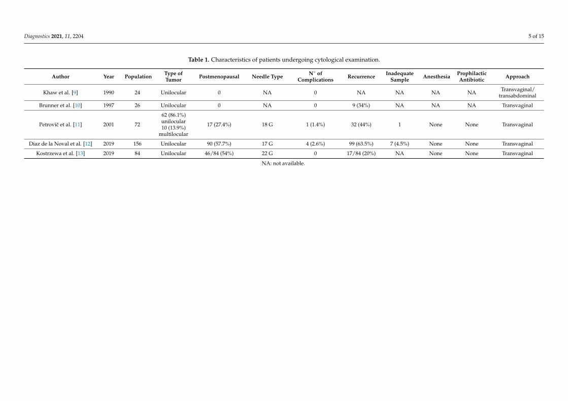
Table 1. Characteristics of patients undergoing cytological examination.
Author Year Population Type ofTumor Postmenopausal Needle Type N◦ of
Complications Recurrence InadequateSample Anesthesia Prophilactic
Antibiotic Approach
Khaw et al. [9] 1990 24 Unilocular 0 NA 0 NA NA NA NA Transvaginal/transabdominal
Brunner et al. [10] 1997 26 Unilocular 0 NA 0 9 (34%) NA NA NA Transvaginal
Petrovic et al. [11] 2001 72
62 (86.1%)unilocular10 (13.9%)
multilocular
17 (27.4%) 18 G 1 (1.4%) 32 (44%) 1 None None Transvaginal
Díaz de la Noval et al. [12] 2019 156 Unilocular 90 (57.7%) 17 G 4 (2.6%) 99 (63.5%) 7 (4.5%) None None Transvaginal
Kostrzewa et al. [13] 2019 84 Unilocular 46/84 (54%) 22 G 0 17/84 (20%) NA None None Transvaginal
NA: not available.

Diagnostics 2021, 11, 2204 6 of 15
5. Histological Examination
Fine needle aspiration biopsy (FNAB) is a simple less-invasive biopsy technique, firstdescribed by Guthrie in 1921. It is feasible with both 16–18 G needles for the transvaginalapproach and 14–16 G needles for the transabdominal procedure [7]. For many years, thishas been the standard method for histological verification of tumors in various locations.This method has some limitations, however; the main limitations are the small sample sizeand the limited amount and integrity of sample tissue collected, often with disrupted tissuearchitecture, resulting in a high rate of inadequate samples for histological assessment [16].
Moreover, low sample quality may prevent proper immunohistochemistry staining,which could play a crucial role in the differential diagnosis, especially when it involvesovarian or colon cancer [17].
On the other hand, tru-cut biopsy provides a sample with preserved tissue architecturethat allows comprehensive histological evaluation as well as permitting the collectionof larger tissue samples, thus permitting other vital histological examinations, such asimmunohistochemistry [18]. Obviously, this technique would require a larger-sized cuttingneedle in comparison to FNAB.
Several papers have compared FNAB and tru-cut biopsy. The largest study in-cluded 1300 consecutive CT-guided biopsies from chest, abdomen, retroperitoneum, andhead/neck regions that evaluated the adequacy and specific diagnosis rates of FNAB(22 G needle) and tru-cut biopsy (16–18 G needle) [19]. Adequate samples were obtainedin 72–92% of cases using FNAB and 93–100% using tru-cut biopsy; the specific diagnosisrates were 54–67% and 82–100%, respectively.
In gynecology, in comparison to other diagnostic techniques, this procedure could bea valid alternative method to laparoscopy or laparotomy to obtain histological samples,especially in patients where there are doubts concerning the benefit of a primary surgicalintervention (poor patient performance status due to co-morbidities or advanced disease)or the nature of the lesion (uncertain recurrence of the tumor, suspected tumor metastasis,or cases of advanced tumors of probably non-genital origin).
As reported in Table 2, the first study about FNAB in gynecology was reported in1991 by Volpi et al. They performed 18 ultrasound-guided biopsies on pelvic masses using23 cm long 16 or 18 G biopsy needles. One false-negative histological evaluation and oneinadequate sample was reported, and no complications were observed [20]. Hence, fromthe preliminary data, the authors supported that this was a promising technique that iseasy to perform and well-accepted by the patients.
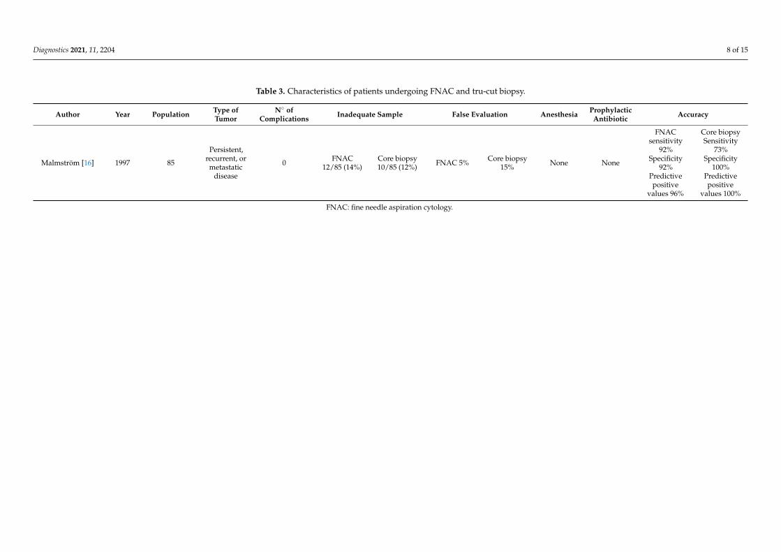
In 1997, the accuracy of FNAC and core biopsy in the diagnosis of gynecologic lesionswas compared by Malmström, making it the first report about the use of core biopsyin gynecology. A total of 85 patients with persistent, recurrent, or metastatic diseasewere analyzed using an automatic biopsy instrument (Biopty) simultaneously performingFNAC and core biopsy. Three hundred and thirty-nine FNAC and 141 biopsies using thebiopsy core instrument were obtained. A correct diagnosis was made with FNAC in 67/85(79%) and with core biopsy in 62/85 (73%) of the cases (p = 0.08). Insufficient materialfor evaluation was recorded for FNAC in 12/85 (14%) compared to 10/85 (12%) for thecore biopsy (p = 0.29). False-negative diagnoses occurred in 5% of the cases with FNACcompared to 15% with core biopsy (NS). The sensitivity of FNAC was 92% and that of corebiopsy 73% (p = 0.01) and the specificities 92% and 100% (NS), respectively. The predictivevalues of positive results for the two methods were 96% and 100%, respectively. Thecomplication rate was negligible. In conclusion, FNAC in combination with core biopsyin gynecologic lesions is a simple and safe operation using needle guides. As reported inTable 3, in comparison with FNAC the sensitivity for core biopsy is low but the specificityis high. There were no significant differences in accuracy between the two methods [16].

Diagnostics 2021, 11, 2204 7 of 15
Table 2. Characteristics of patients undergoing histological examination.
Author Year Population Type of Tumor Postmenopausal Tecnique Needle Type N◦ ofComplications Inadequate Sample False
Evaluation Anesthesia ProphylacticAntibiotic Approach Accuracy
Volpiet al. [20] 1991 18 Pelvic masses NA FNAB 16 G or 18 G 0 1 (5.5%) 1 (5.5%) NA NA transvaginal NA
Fischerovaet al. [21] 2008 86
Advancedprimary orrecurrent
pelvic and/orabdominal
tumors
NA Tru-cut
Transvaginal16 G, transab-
dominal14 G
1 (1.2%) 6 (7%) 2 (3.3%)
Transvaginal:no anesthesia
Transabdominal:local
anesthesia
NA
Transvaginal46 (53.5%)
Transabdominal40 (46.5%)
97.7% (95% CI91.85–99.72%)
Zikanet al. [22] 2010 190
Disseminatedmalignancy of
unknownprimary origin
NA Tru-cut 18 G 2 (1%) 12 (6%)
2/118 (1.7%)patients whounderwentsubsequent
surgery
None noneTransvaginaltransabdomi-
nal98.3%
Epsteinet al. [23] 2016 143
Disseminatedmalignancy of
unknownprimary origin
NA Tru-cut
Transvaginal18 G, Transab-
dominal16 G
2 (1.4%) 5 (3.5%) NA None none
Transvaginal,transrectal,
transabdomi-nal
NA
Parket al. [24] 2016 55 Pelvic lesions NA Tru-cut 18 G 12 (22%) 4 (7%) NA
Localanesthesia
with 1%lidocaine
none Transvaginal 93%
Eitanet al. [25] 2017 59 Pelvic lesion NA
33 FNAB26
FNAC17 G 0 7 (11.9%) NA None none transvaginal 85%
Linet al. [26] 2017 200
Disseminatedmalignancy of
unknownprimary origin
NA FNAB 18 G 0 8 (4%) 2 (1%) None none Transvaginal NA
Masciliniet al. [27] 2019 62
Disseminatedmalignancy of
unknownprimary origin
50 (80.6%) FNAB 18 G 0 0
2/10 (20%)who
underwentsurgery
none none Transvaginal NA
Gaoet al. [28] 2019 40 Pelvic masses NA Tru-cut 18 G 0 0 0 None none Transvaginal/
transrectal 90%
Verschuereet al. [29] 2021 155
Disseminatedmalignancy of
unknownprimary origin
NA FNAB 18 G Minor 7 (4.5%)Major 0
Single-tissue
cylinder24 (15.7%)
At least twocylinders8 (<5%)
NA none none Transvaginal 97.2%
FNAB: Fine Needle Aspiration Biopsy; FNAC: Fine Needle Aspiration Cytology.
Diagnostics 2021, 11, 2204 8 of 15
Table 3. Characteristics of patients undergoing FNAC and tru-cut biopsy.
Author Year Population Type ofTumor
N◦ ofComplications Inadequate Sample False Evaluation Anesthesia Prophylactic
Antibiotic Accuracy
Malmström [16] 1997 85
Persistent,recurrent, ormetastatic
disease
0 FNAC12/85 (14%)
Core biopsy10/85 (12%) FNAC 5% Core biopsy
15% None None
FNACsensitivity
92%Specificity
92%Predictivepositive
values 96%
Core biopsySensitivity
73%Specificity
100%Predictivepositive
values 100%
FNAC: fine needle aspiration cytology.

Diagnostics 2021, 11, 2204 9 of 15
In 2008, Fiscerova et al. published the first study evaluating the accuracy and safety ofultrasound-guided transvaginal or transabdominal tru-cut biopsy in 86 cases of advancedprimary or recurrent pelvic and/or abdominal tumors. Biopsy indications were primarilyinoperable pelvic tumor, poor performance status, and recurrent disease requiring his-tologic verification. The samples collected were from pelvic tumor in 47 cases (54.6%),omental cake in 12 cases (14%), and parietal or visceral carcinomatosis in 27 cases (31.4%).On average, two samples were taken per case (range 1–3). The time taken to obtain twosamples of tissue ranged between 10 and 15 min in each patient. Six were inadequate forhistologic evaluation. The only complication encountered was bleeding from a tumor in apatient with mild thrombocytopenia, requiring laparotomy. The procedure reached highdiagnostic accuracy of 97.7% (95% CI 91.85–99.72%) [21].
Zikan et al. in 2010 analyzed 190 patients with advanced abdominal and pelvic tumorswho would not benefit from a primary oncogynecological surgery, patients with historyof non-gynecological tumor and who were suspected of having a secondary ovarian orperitoneal tumor. Statistical analysis reported that the single dependent variable was theadequacy of the histological sample from the tru-cut biopsy, while the other variables(age, BMI, CA 125 level, ascites, histology, indication, biopsy site, and biopsy approach)represented the independent factors. Age and BMI did not influence the adequacy of thebiopsy sample. Ascites, elevated CA 125, primary suboptimal operable tumor, serousepithelial ovarian cancer histology, carcinomatosis, and vaginal approach were significantpositive predictors for the achievement of an adequate sample, while recurrence as anindication, non-serous and non-ovarian histotypes, and a transabdominal approach werenegative predictors. Ascites and elevated CA 125 levels were positive predictors for theachievement of an adequate sample for the large tumor load in these cases. Carcinomatosis,as a biopsy site that is often easily accessible transvaginally in the pouch of Douglas andtransabdominally by a peritoneal approach through the anterior abdominal wall, alsocorrelated positively with adequacy. The serous histotype of ovarian cancer was a strongpositive predictor of adequacy because this histotype is often present as solid nodulesof carcinomatosis and a large tumor load in advanced stages. Nonovarian tumors were,on the other hand, a strong negative predictor. This finding cannot be easily explained,whereas inoperable/sub-optimally operable tumors were a favorable indication witha strong positive correlation with adequacy. This finding is in concordance with theabovementioned explanation. A large tumor mass as a biopsy site is usually accessible inthese patients, while in patients with recurrent tumor or with tumor of atypical morphology,only small, necrotic, or cystic lesions are often present.
Obesity is also considered a factor impeding the accuracy of ultrasound, thus also po-tentially affecting the performance of tru-cut biopsy (especially by the abdominal approach).
Two cases of bleeding after tru-cut biopsy (1%) were reported. In the first case, bleed-ing from the biopsy site on the surface of a Krukenberg tumor caused hemoperitoneum andneeded adnexectomy. This patient had a bone marrow infiltration caused by an advanceddisseminated tumor that caused thrombocytopenia. We currently consider thrombocytope-nia to be a significant contraindication for tru-cut biopsy. In the second patient, ultrasoundexamination revealed bleeding into the ascitic fluid from the site of the biopsy of pelviccarcinomatosis. A laparoscopy was performed immediately, but, in the meantime, thebleeding stopped spontaneously [22].
Epstein et al. compared subjective ultrasound assessment and the ADNEX (Assess-ment of Different NEoplasias in the adneXa) model with ultrasound-guided tru-cut biopsyto differentiate disseminated primary ovarian cancer from metastatic non-ovarian cancer.They undertook a prospective study including 143 women with disseminated malignancyof unknown primary origin, with a pelvic tumor/carcinosis. The ultrasound examinerassessed tumor morphology, spread in the pelvis and abdomen, and predicted tumor originas primary ovarian or metastatic using both subjective assessment and the ADNEX model.Histology from tru-cut biopsy served as the gold standard for assessment of diagnosticaccuracy. Subjective ultrasound evaluation had a sensitivity of 82% (73/89) and a specificity
Diagnostics 2021, 11, 2204 10 of 15
of 70% (26/37) in predicting primary ovarian cancer. The ADNEX model had an area underthe receiver–operating characteristics curve of 0.891 (95% CI, 0.794–0.946) (in women withan ovarian lesion, n = 104). Subjective ultrasound assessment and the ADNEX model canboth be used to predict whether a pelvic tumor is metastatic and of non-ovarian origin,indicating the need for tru-cut biopsy.
They reported two admissions for inpatient care related to biopsy: one patient had anabdominal wall hematoma after transabdominal percutaneous biopsy, and another patienthad a suspected pelvic infection after tru-cut biopsy with a transvaginal approach [23].
Park et al. analyzed 55 women with pelvic masses who underwent US-guidedtransvaginal core biopsy.
The overall diagnostic accuracy of US-guided transvaginal core biopsy was 93%(51/55). Of the 55 lesions, 46 (84%) were confirmed to be either benign or malignanttumors, and five (9%) were diagnosed as active or chronic inflammatory lesions. Fourlesions (7%) were not histopathologically diagnosed after biopsy.
In terms of minor complications, vaginal bleeding occurred in 10 patients (18%), andgross hematuria occurred in two patients (4%). These complications resolved spontaneouslyin all patients without further workup or treatment. They reported that the reliable andaccurate diagnostic performance of the procedure might be explained by the ability toadvance the needle parallel to the longitudinal axis of the transvaginal probe using anattached guide similar to that used in transrectal prostate biopsy.
The mean gap between the probe and the targeted lesion was only 1.6 cm. The safetyand accuracy of the procedure may be explained by the short distance of the biopsy route,although a significant difference between diagnostic and non-diagnostic biopsies was notfound [24].
Eitan et al. in 2017 reported 55 patients with undiagnosed pelvic lesions. When it couldbe measured (n = 48), the median size of the lesions biopsied was 43 mm (range 16–130 mm).
Thirty-three lesions were evaluated by FNAB of the solid structure and 26 by aspirationof fluid for cytology. Pathologic feasibility rate was 88% (52/59). Considering the seveninconclusive results, the procedure had sensitivity of 88% (29/33) and specificity of 88%(23/26). Overall accuracy of TVUS-FNA for this patient cohort was 85%. No patientcharacteristics were found to distinguish between accurate and inaccurate or inconclusiveTVUS-FNA result [15].
Also in 2017, Lin et al. analyzed 200 patients affected by primary inoperable tumors,suspicion of metastases to the ovaries or peritoneum, recurrence, or other solid lesions inthe pelvis.
A large number of transvaginal biopsies was carried out and reported in this study.Adequate samples were obtained in 96% of biopsies without any complications.With a univariate analysis, they evaluated factors that may affect transvaginal
ultrasound-guided FNAB success.The biopsy site had a significant effect on biopsy adequacy with a significantly lower
probability of obtaining satisfactory specimens for histologic verification from the peri-toneal cake compared to pelvic tumors (83.9% versus 98.5%) (p < 0.05) and from theperitoneal cake compared to vaginal cuff masses (83.9% versus 97.1%), (p < 0.01). Adequacywas also affected by tumor size (p < 0.05) but not by vascularization, ascites, or tumortype [26].
In 2019, Mascilini et al. analyzed imaging of abdominal or pelvic tumors in 62 patientsconsidered not-ideal candidates for primary gynecological surgery (poor performancestatus, not suitable for surgery, or patients with multiple or non-resectable metastases) orwhere the origin and/or nature of the tumor was unclear. Of this, ultrasound characteristicswere a solid tumor for 61 (98.4%) and a multilocular-solid for 1 (1.6%) (Table 4).
The ultrasound-guided biopsy was mostly performed through the vaginal cuff (25/62,40.3%). Other sites of biopsy were the cervix (16/62, 25.8%), vaginal wall (11/62, 17.7%),pelvic mass (7/62, 11.3%), and peri-urethral region (3/62, 4.8%).

Diagnostics 2021, 11, 2204 11 of 15
An adequate sample for histological analysis was obtained in all cases. No majorcomplications were registered. Histopathological examinations showed 24 (38.7%) benignlesions and 38 (61.3%) malignant tumors. Ten patients eventually underwent surgery.Final histology was not in concordance with the results from transvaginal ultrasound-guided biopsy in 2 out of 10 patients (20%); in particular, benign disease at transvaginalultrasound-guided biopsy was malignant at final histology (two cases of recurrence ofcervical cancer) [27].
Gao et al. in 2019 [28] conducted a retrospective analysis on 40 patients with pelvicmasses who underwent transvaginal or transrectal ultrasound-guided aspiration biopsy.No complications were identified. The median lesion size was 5.5 cm (range, 1–15 cm).Thirty-four of the lesions were solid while six were cystic. The median number of biopsycores obtained from each patient was 4.0 (range, 2–7 cores). The specimens were obtainedfrom pelvic cavity and pelvic floor in 18 cases (45%), the vaginal stump in six cases (15%),the cervix in two cases (5%), and the vaginal fornix in 13 cases (32.5%). All the specimenswere adequate for histologic evaluation and diagnosis. The study showed detection inalmost all the suspected clinical cases and was consistent with a more than 90% adequacyand accuracy.
In 2021, Verschuere et al. analyzed the safety and efficiency of performing transvaginalultrasound-guided tru-cut biopsy for pelvic masses. The main indication for tru-cut biopsywas suspected disseminated disease or recurrence of malignant conditions.
They reported 155 patients. No major complications occurred in the study. Procedure-related events were limited to moderate blood loss (<50 mL) without the need for treatmentin 4.5%. Biopsies were deemed adequate for histological evaluation in 84.3% in whicha single tissue cylinder was available for diagnosis. When at least two cylinders wereavailable, diagnostic adequacy increased to >95%.
Table 4. Ultrasound characteristics of patients who underwent histological examination.
Authors Type of Tumor Median Diameter of the Lesion Site of Biopsy
Mascilini et al. [27] Solid tumor 61 (98.4%), multilocular-solid 1 (1.6%). 31 (10–132) mm
Vaginal cuff (25/62, 40.3%).Cervix (16/62, 25.8%),
Vaginal wall (11/62, 17.7%),Pelvic mass (7/62, 11.3%),
Peri-uretral region (3/62, 4.8%)
Verschuere et al. [29]142 (80.7%) solid
8 (4.5%) unilocular-solid26 (14.8%) multilocular-solid
NA
Ovary 70 (39.8%)Cervix 10 (5.7%)Vagina 4 (2.3%)
Pelvic peritoneum 74 (42.0%)Other sites: 7 (4.0%) pararectal,bladder wall, or parametrium
Lin et al. [26] NA 4.80 ± 2.16 cm
pelvic cavity masses 134/200(67.0%),
vaginal cuff or the vaginal wall35/200 (17.5%)
peritoneal cake 31/200 (15.5%)
Zikan et al. [22] NA NA
Pelvic mass 125 (64.1%)Carcinomatosis 41 (21.0%)
Omental cake 12 (6.2%)Lymph node 11 (5.6%)
Other 6 (3.1%)
Fischerova et al. [21] NA NA
Pelvic mass (54.6%)peritoneal visceral or parietal
metastases (31.4%)Omental cake (14%).
Epstein et al. [23]
Primary ovarianSolid 48 (53.9%)
Multilocular solid 33(37.1%)
Unilocular solid 1 (1.1%)Only carcinomatosis 7
(7.9%)>10 locules12 (14.6%)
Metastatic non-ovarianSolid 21 (56.8%)
Multilocular solid13 (35.1%)
Unilocular solid0 (0)
Only carcinomatosis3 (8.1%)
>10 locules8 (23.5%)
Primary ovarian82 (50–110) mm
Metastaticnon-ovarian
80 (57–113) mm
Diagnostics 2021, 11, 2204 12 of 15
They also investigated the influence of obesity on the adequacy of transvaginal tru-cutbiopsies, but BMI did not seem to affect adequacy, according to the study of Zikan et al. Asopposed to the transabdominal approach, the close proximity of the transvaginal probe tothe lesions enables tru-cut biopsies even in extreme obesity.
Comparing final histology, the diagnostic accuracy of the tru-cut biopsies was 97.2%.Therefore, the authors concluded that transvaginal tru-cut biopsy of pelvic masses is asafe procedure to perform with high adequacy, but multiple biopsies need to be taken tooptimize the amount of tissue for histological examination [29].
6. Conclusions
Ultrasound-guided sampling of pelvic masses is a useful procedure with a lot of prosand some cons. Ad reported in Table 5. It represents a diagnostic method of choice inselected patients. In patients with severely advanced disease who are not deemed ideal forcytoreduction, ultrasound-guided biopsy represents a real-time, convenient, economical,and quick way of obtaining histological diagnosis, usually not requiring any recoverytime as after diagnostic laparoscopy or laparotomy and allowing for an early start ofneoadjuvant chemotherapy [30].
The other types of patients eligible for this method are patients with high comorbid-ity burden and those with suspected relapse or atypical tumor morphology with highprobability of nonovarian etiology [31–33].
Patients whose result of the tru-cut biopsy was non-gynecological malignancy can bedirected to the appropriate specialist assessment before performing a surgery.
Table 5. Pros and cons of each technique.
Pros Cons
Histological examination FNAB- simple- less invasive
- small sample size- limited integrity of sample
tissue collected- high rate of inadequate samples
Tru-cut biopsy
- larger tissue samples- preserved tissue architecture- possibility of
immunohistochemistryexaminations
- more adequate samples- more specific diagnosis rates
- larger-size cutting needle
Cytological examination FNAC - also therapeutic - possibility of recurrence- not suitable for all types of cyst
FNAB: fine needle aspiration biopsy; FNAC: fine needle aspiration cytology.
Complication rate is negligible; no cases were reported by Volpi et al. [20], Eitan et al. [25],Lin et al. [26]. Mascilini et al. [27], or Gao et al. [28]. Two studies reported only a fewcomplications (Fischerova et al. [21] 1/86 (1.2%) and Zikan et al. [22] 2/190 (1%)). Onlyone study (Park et al. [24]) reported a major complication rate in 12/55 (22%), all beingminor; vaginal bleeding in 10/55 (18%); and gross hematuria in 2/55 (4%).
The general clinical condition of the patient must always be taken into account. Oneof the two cases of complications reported by Zikan et al. [22] was a hemoperitoneum andrevealed bleeding from the biopsy site on the surface of the mass. This patient sufferedfrom thrombocytopenia as a consequence of bone marrow infiltration by an advanceddisseminated tumor. It would therefore be appropriate to consider thrombocytopenia as asignificant contraindication for tru-cut biopsy.
From the analysis of the studies, we also deduce that the risk of an inadequatesample must be considered (8% for Epstein et al. [23], 6% for Zikan et al. [22], 11.9% forEitan et al. [25], and 8% for Lin et al. [26]). Failure to obtain an adequate sample may be
Diagnostics 2021, 11, 2204 13 of 15
related to cystic or necrotic tumors, which are more common in patients with recurrenceor with tumors of atypical morphology at ultrasound than in patients with advancedinoperable tumors [27].
As reported by Lin et al., also the biopsy site had a significant effect on biopsy adequacywith a significantly lower probability of obtaining satisfactory specimens for histologicverification from the peritoneal cake compared to pelvic tumors or vaginal cuff masses [26].
Another risk may be the false evaluation or biopsy-failed diagnosis: 1% for Lin et al. [26],1.7% Zikan et al. [22], and 20% for Mascilini et al. [27] (but this high number is probablyuntrue due to the low number of patients who have undergone surgery). Probably, in thesepatients, the small size of the tumor or the cystic component of the tumor did not allow usto obtain the correct samples [27].
Additionally, the number of samples is related to the adequacy of the technique:Fischerova et al. reported that, with an average two samples, they found an accuracy of97.7% [21].
Tru-cut biopsy is a simple procedure, but it needs to be undertaken by an experiencedoperator who has undergone a specific training. This is also demonstrated by the study ofVerschuere et al. [29], who reported an increase of the biopsy adequacy over the years, from75% in 2014 to 88% in 2018. This could be attributed to the operators’ increasing experiencewith the procedure.
The need for anesthesia is not reported in most studies; therefore, this procedurecan be performed in a suitable clinic space, without anesthesia with minimal discomfort.Verschuere et al. [29], from their experience, suggested attention is to be paid duringgentle probe manipulation using a sufficient amount of gel to determine the mostminimal discomfort.
Therefore, ultrasound-guided sampling procedures, in the hands of an experiencedoperator, represent a simple and reliable method of extreme help to the gynecologistfor the correct diagnosis of gynecological diseases and more and for the correct clinicalmanagement of the patient.
Author Contributions: Conceptualization F.A. and V.L.; methodology, G.C. (Gennaro Cormio), C.L.and D.L.F.; resources F.A., G.C. (Gerardo Cazzato) and V.C.; data curation, G.I.; writing—original draftpreparation, F.A., G.C. (Gennaro Cormio) and A.A.K.; writing—review and editing, G.I. and A.A.K.;visualization C.L. and E.S.; supervision, G.C. (Gennaro Cormio) and L.R.; project administration,F.A. and V.L.; funding acquisition, G.C. (Gennaro Cormio). All authors have read and agreed to thepublished version of the manuscript.
Funding: This research received no external funding.
Institutional Review Board Statement: Not applicable.
Informed Consent Statement: Not applicable.
Data Availability Statement: Not applicable.
Acknowledgments: We thank the association “ACTO—alleanza contro il tumore ovarico” for sup-porting the research activity of Francesca Arezzo with the “Adele Leone” grant.
Conflicts of Interest: The authors declare no conflict of interest.
References1. Stucci, L.S.; Interno, V.; Tucci, M.; Perrone, M.; Mannavola, F.; Palmirotta, R.; Porta, C. The ATM Gene in Breast Cancer: Its
Relevance in Clinical Practice. Genes 2021, 12, 727. [CrossRef] [PubMed]2. Woodcock, N.P.; Glaves, I.; Morgan, D.R.; MacFie, J. Ultrasound-guided Tru-cut biopsy of the breast. Ann. R. Coll. Surg. Engl.
1998, 80, 253–256.3. Faulkner, R.L.; Mohiyiddeen, L.; McVey, R.; Kitchener, H.C. Transvaginal biopsy in the diagnosis of ovarian cancer. BJOG 2005,
112, 991–993. [CrossRef]4. Arezzo, F.; Loizzi, V.; La Forgia, D.; Moschetta, M.; Tagliafico, A.S.; Cataldo, V.; Kawosha, A.A.; Venerito, V.; Cazzato, G.;
Ingravallo, G.; et al. Radiomics Analysis in Ovarian Cancer: A Narrative Review. Appl. Sci. 2021, 11, 7833. [CrossRef]
Diagnostics 2021, 11, 2204 14 of 15
5. Gupta, S.; Nguyen, H.L.; Morello, F.A., Jr.; Ahrar, K.; Wallace, M.J.; Madoff, D.C.; Murthy, R.; Hicks, M.E. Various approaches forCT-guided percutaneous biopsy of deep pelvic lesions: Anatomic and technical considerations. Radiographics 2004, 24, 175–189.[CrossRef]
6. Arezzo, F.; Franchi, D.; Loizzi, V.; Cataldo, V.; Lombardi, C.; Cazzato, G.; Cormio, G. Blue mass in the pelvis: Serous cystadenofi-broma of the peritoneum. Ultrasound Obstet. Gynecol. 2021. [CrossRef]
7. Guo, Z.; Kurtycz, D.F.; De Las Casas, L.E.; Hoerl, H.D. Radiologically guided percutaneous fine-needle aspiration biopsy of pelvicand retroperitoneal masses: A retrospective study of 68 cases. Diagn. Cytopathol. 2001, 25, 43–49. [CrossRef] [PubMed]
8. Zanetta, G.; Lissoni, A.; Torri, V.; Dalla Valle, C.; Trio, D.; Rangoni, G.; Mangioni, C. Role of puncture and aspiration in expectantmanagement of simple ovarian cysts: A randomised study. BMJ 1996, 313, 1110–1113. [CrossRef]
9. Khaw, K.T.; Walker, W.J. Ultrasound guided fine needle aspiration of ovarian cysts: Diagnosis and treatment in pregnant andnon-pregnant women. Clin. Radiol. 1990, 41, 105–108. [CrossRef]
10. Brunner, M.; Krampl, E.; Feichtinger, W. Ultrasound-guided transvaginal puncture of ovarian cysts. Zentralbl. Gynakol. 1997,119, 225–228. [PubMed]
11. Petrovic, N.; Arko, D.; Lovrec, V.G.; Takac, I. Ultrasound guided aspiration in pathological adnexal processes. Eur. J. Obstet.Gynecol. Reprod. Biol. 2002, 104, 52–57. [CrossRef]
12. Diaz de la Noval, B.; Rodriguez Suarez, M.J.; Fernandez Ferrera, C.B.; Valdes Lafuente, D.; Arias Cailleau, R.; Perez Arias, H.;Torrejon Becerra, J.C.; Suarez Gil, P.; Lucio Gonzalez, L.R. Transvaginal Ultrasound-Guided Fine-Needle Aspiration of AdnexalCysts With a Low Risk of Malignancy: Our Experience and Recommendations. J. Ultrasound Med. 2020, 39, 1787–1797. [CrossRef][PubMed]
13. Kostrzewa, M.; Zajac, A.; Wilczynski, J.R.; Stachowiak, G. Retrospective analysis of transvaginal ultrasound-guided aspiration ofsimple ovarian cysts. Adv. Clin. Exp. Med. 2019, 28, 1531–1535. [CrossRef] [PubMed]
14. Pal, S.; Chakrabarti, S.; Deuoghuria, D.; Phukan, J.P.; Sinha, A.; Mondal, P.K. Evaluation of Ultrasound-Guided Fine-NeedleAspiration Cytology of Ovarian Masses with Histopathological Correlation. Acta Cytol. 2015, 59, 149–155. [CrossRef] [PubMed]
15. Dordoni, D.; Zaglio, S.; Zucca, S.; Favalli, G. The role of sonographically guided aspiration in the clinical management of ovariancysts. J. Ultrasound Med. 1993, 12, 27–31. [CrossRef] [PubMed]
16. Malmstrom, H. Fine-needle aspiration cytology versus core biopsies in the evaluation of recurrent gynecologic malignancies.Gynecol. Oncol. 1997, 65, 69–73. [CrossRef] [PubMed]
17. Interno, V.; Tucci, M.; Pezzicoli, G.; Silvestris, F.; Porta, C.; Mannavola, F. Liquid Biopsy as a Tool Exploring in Real-Time BothGenomic Perturbation and Resistance to EGFR Antagonists in Colorectal Cancer. Front. Oncol. 2020, 10, 581130. [CrossRef]
18. Pellerino, A.; Interno, V.; Mo, F.; Franchino, F.; Soffietti, R.; Ruda, R. Management of Brain and Leptomeningeal Metastases fromBreast Cancer. Int. J. Mol. Sci. 2020, 21, 8534. [CrossRef]
19. Chojniak, R.; Isberner, R.K.; Viana, L.M.; Yu, L.S.; Aita, A.A.; Soares, F.A. Computed tomography guided needle biopsy:Experience from 1300 procedures. Sao Paulo Med. J. 2006, 124, 10–14. [CrossRef]
20. Volpi, E.; Zola, P.; de Grandis, T.; Danese, S.; Rumore, A.; Sismondi, P. Pelvic mass biopsy by operative transvaginal sonography(OTVS). Eur. J. Gynaecol. Oncol. 1991, 12, 477–480.
21. Fischerova, D.; Cibula, D.; Dundr, P.; Zikan, M.; Calda, P.; Freitag, P.; Slama, J. Ultrasound-guided tru-cut biopsy in themanagement of advanced abdomino-pelvic tumors. Int. J. Gynecol. Cancer 2008, 18, 833–837. [CrossRef]
22. Zikan, M.; Fischerova, D.; Pinkavova, I.; Dundr, P.; Cibula, D. Ultrasound-guided tru-cut biopsy of abdominal and pelvic tumorsin gynecology. Ultrasound Obstet. Gynecol. 2010, 36, 767–772. [CrossRef]
23. Epstein, E.; Van Calster, B.; Timmerman, D.; Nikman, S. Subjective ultrasound assessment, the ADNEX model and ultrasound-guided tru-cut biopsy to differentiate disseminated primary ovarian cancer from metastatic non-ovarian cancer. Ultrasound Obstet.Gynecol. 2016, 47, 110–116. [CrossRef] [PubMed]
24. Park, J.J.; Kim, C.K.; Park, B.K. Ultrasound-Guided Transvaginal Core Biopsy of Pelvic Masses: Feasibility, Safety, and Short-TermFollow-Up. AJR Am. J. Roentgenol. 2016, 206, 877–882. [CrossRef] [PubMed]
25. Eitan, R.; Peled, Y.; Sabah, G.; Krissi, H.; Haroush, A.B.; Meizner, I.; Danon, D.; Bardin, R.; Jakobson-Setton, A.; Salzer, L.; et al.Diagnosis of deep pelvic masses on a gynaecology service: Trans-vaginal ultrasound-guided needle aspiration of pelvic solid andcystic lesions. Aust. N. Z. J. Obstet. Gynaecol. 2017, 57, 197–200. [CrossRef] [PubMed]
26. Lin, S.Y.; Xiong, Y.H.; Yun, M.; Liu, L.Z.; Zheng, W.; Lin, X.; Pei, X.Q.; Li, A.H. Transvaginal Ultrasound-Guided Core NeedleBiopsy of Pelvic Masses. J. Ultrasound Med. 2018, 37, 453–461. [CrossRef] [PubMed]
27. Mascilini, F.; Quagliozzi, L.; Moro, F.; Moruzzi, M.C.; De Blasis, I.; Paris, V.; Scambia, G.; Fagotti, A.; Testa, A.C. Role oftransvaginal ultrasound-guided biopsy in gynecology. Int. J. Gynecol. Cancer 2020, 30, 128–132. [CrossRef] [PubMed]
28. Gao, C.; Wang, L.; Zhang, C.; Li, X. Transvaginal/transrectal ultrasound-guided aspiration biopsy for diagnosis of pelvic/pelvicfloor tumors in females: A retrospective analysis. Exp. Ther. Med. 2019, 18, 352–357. [CrossRef]
29. Verschuere, H.; Froyman, W.; Van den Bosch, T.; Van Hoefs, M.; Kaijser, J.; Van Schoubroeck, D.; Van Rompuy, A.S.;Vergote, I.; Timmerman, D. Safety and efficiency of performing transvaginal ultrasound-guided tru-cut biopsy for pelvic masses.Gynecol. Oncol. 2021, 161, 845–851. [CrossRef]
30. Arezzo, F.; La Forgia, D.; Venerito, V.; Moschetta, M.; Tagliafico, A.S.; Lombardi, C.; Loizzi, V.; Cicinelli, E.; Cormio, G. AMachine Learning Tool to Predict the Response to Neoadjuvant Chemotherapy in Patients with Locally Advanced CervicalCancer. Appl. Sci. 2021, 11, 823. [CrossRef]

Diagnostics 2021, 11, 2204 15 of 15
31. Cazzato, G.; Colagrande, A.; Arezzo, F.; Resta, L.; Ingravallo, G. “Black Ovaries”: An Uncommon Case of First SystemicRecurrence of Melanoma. Reports 2021, 4, 13. [CrossRef]
32. Cazzato, G.; Colagrande, A.; Cimmino, A.; Caporusso, C.; Candance, P.M.V.; Trabucco, S.M.R.; Zingarelli, M.; Lorusso, A.;Marrone, M.; Stellacci, A.; et al. Urological Melanoma: A Comprehensive Review of a Rare Subclass of Mucosal Melanoma withEmphasis on Differential Diagnosis and Therapeutic Approaches. Cancers 2021, 13, 4424. [CrossRef] [PubMed]
33. Arezzo, F.; Cazzato, G.; Loizzi, V.; Ingravallo, G.; Resta, L.; Cormio, G. Peritoneal Tuberculosis Mimicking Ovarian Cancer:Gynecologic Ultrasound Evaluation with Histopathological Confirmation. Gastroenterol. Insights 2021, 12, 278–282. [CrossRef]
Related Documents


![Ultrasound Guided Vascular Access[2]](https://static.cupdf.com/doc/110x72/5420582a7bef0a06088b4679/ultrasound-guided-vascular-access2.jpg)

