The Role of Schlemm's Canal in Aqueous Outflow from the Human Eye M. C. Johnson and R. D. Kamm A mathematical model of Schlemm's canal is developed to simulate collapse of the canal and its effect on resistance to flow through the aqueous outflow network. Schlemm's canal is modeled as a porous, compliant channel that is held open by the trabecular meshwork. The trabecular meshwork is modeled as a series of linear springs that allow the inner wall of Schlemm's canal to deform in proportion to the local pressure drop across it. Based on comparisons between the model and results in the literature, the following tentative conclusions are reached: (1) Most of the resistance in the aqueous outflow network occurs in the inner wall of Schlemm's canal. (2) Glaucoma is not caused by a weakening of the trabecular meshwork and a resultant collapse of Schlemm's canal alone. Instead, glaucoma prob- ably results from an increased flow resistance of the inner wall of the canal. Invest Ophthalmol Vis Sci 24:320-325, 1983 Aqueous humorflowsthrough the outflow network at an extremely low flow rate (2.0 ^1/min), yet this flow produces a surprisingly large pressure drop over a short flow distance (less than 1 mm). The pressure drop is around 6 mmHg in normal eyes and can be as much as 40 mmHg in glaucomatous eyes. In order to achieve a better understanding of the outflow net- work, changes in which can lead to ocular hyperten- sion and glaucoma, we studied the role of Schlemm's canal in producing a pressure drop in the normal eye. Many studies have sought to determine the prin- cipal site of flow resistance in the aqueous outflow network. Toward this end, numerous perfusion stud- ies have been conducted, the results of which indicate that the relationship between pressure and flow is nearly linear with flow resistance increasing slowly with pressure. 1 A number of sites have been identified as possible sources of resistance: (1) corneoscleral meshwork, (2) juxtacanalicular meshwork, (3) endothelial lining of the inner wall of Schlemm's canal, (4) Schlemm's canal, and (5) collector channels and aqueous veins. Two considerations preclude the corneoscleral meshwork from being the principal site of flow resis- tance: (1) the size of the openings in the corneoscleral From the Department of Mechanical Engineering, Massachu- setts Institute of Technology, Cambridge, Massachusetts. Supported by a grant from the National Eye Institute (Grant #1 R01 Ey 03141-01). Presented in part at the annual meeting of the Association for Research in Vision and Ophthalmology, Sarasota, Florida, 1981. Submitted for publication November 23, 1981. Reprint requests: Prof. Roger Kamm, Room 3-258, Massachu- setts Institute of Technology, 77 Massachusetts Avenue, Cam- bridge, MA 02139. meshwork is too large to cause an appreciable resis- tance, 2 and (2) Johnstone and Grant 3 showed that as IOP increases, the spaces in the trabecular meshwork expand. As the spaces open, the hydrodynamic re- sistance would decrease. Thus, if the corneoscleral meshwork were the principal site of flow resistance, one would expect the resistance to decrease rather than increase with an increase in IOP. The aqueous veins are much too large to cause any appreciable resistance. Stresses in the sclera due to IOP could conceivably cause collapse of the aqueous veins (and thus generate significant flow resistance), but a recent study indicates that this does not occur until IOP is substantially elevated. 4 The juxtacanalicular meshwork and the endothelial lining of the inner wall ofSchlemm 's canal are jointly considered as the inner wall of Schlemm's canal for this study. We have grouped these two regions to- gether, in part, because there seems to be no general consensus concerning the mechanism of aqueous hu- mor transport through them, and in part because our primary intent is to examine the effect of Schlemm's canal collapse by itself. It appears likely that the prin- cipal site of flow resistance is the inner wall of Schlemm 's canal, or Schlemm 's canal, or both. In this study, therefore, we develop a theoretical model that includes the flow resistance of both, and accounts only for the variations in flow resistance associated with collapse of the canal. A Model of Schlemm's Canal as a Porous, Compliant Channel Theoretical Model Moses 5 modeled Schlemm's canal as a porous, rigid channel. From the predictions of his model, he con- 0146-0404/83/0300/320/$ 1.10 © Association for Research in Vision and Ophthalmology 320

Welcome message from author
This document is posted to help you gain knowledge. Please leave a comment to let me know what you think about it! Share it to your friends and learn new things together.
Transcript
The Role of Schlemm's Canal in AqueousOutflow from the Human Eye
M. C. Johnson and R. D. Kamm
A mathematical model of Schlemm's canal is developed to simulate collapse of the canal and its effecton resistance to flow through the aqueous outflow network. Schlemm's canal is modeled as a porous,compliant channel that is held open by the trabecular meshwork. The trabecular meshwork is modeledas a series of linear springs that allow the inner wall of Schlemm's canal to deform in proportion tothe local pressure drop across it. Based on comparisons between the model and results in the literature,the following tentative conclusions are reached: (1) Most of the resistance in the aqueous outflownetwork occurs in the inner wall of Schlemm's canal. (2) Glaucoma is not caused by a weakening ofthe trabecular meshwork and a resultant collapse of Schlemm's canal alone. Instead, glaucoma prob-ably results from an increased flow resistance of the inner wall of the canal. Invest Ophthalmol VisSci 24:320-325, 1983
Aqueous humor flows through the outflow networkat an extremely low flow rate (2.0 ^1/min), yet thisflow produces a surprisingly large pressure drop overa short flow distance (less than 1 mm). The pressuredrop is around 6 mmHg in normal eyes and can beas much as 40 mmHg in glaucomatous eyes. In orderto achieve a better understanding of the outflow net-work, changes in which can lead to ocular hyperten-sion and glaucoma, we studied the role of Schlemm'scanal in producing a pressure drop in the normal eye.
Many studies have sought to determine the prin-cipal site of flow resistance in the aqueous outflownetwork. Toward this end, numerous perfusion stud-ies have been conducted, the results of which indicatethat the relationship between pressure and flow isnearly linear with flow resistance increasing slowlywith pressure.1
A number of sites have been identified as possiblesources of resistance: (1) corneoscleral meshwork, (2)juxtacanalicular meshwork, (3) endothelial lining ofthe inner wall of Schlemm's canal, (4) Schlemm'scanal, and (5) collector channels and aqueous veins.
Two considerations preclude the corneoscleralmeshwork from being the principal site of flow resis-tance: (1) the size of the openings in the corneoscleral
From the Department of Mechanical Engineering, Massachu-setts Institute of Technology, Cambridge, Massachusetts.
Supported by a grant from the National Eye Institute (Grant #1R01 Ey 03141-01).
Presented in part at the annual meeting of the Association forResearch in Vision and Ophthalmology, Sarasota, Florida, 1981.
Submitted for publication November 23, 1981.Reprint requests: Prof. Roger Kamm, Room 3-258, Massachu-
setts Institute of Technology, 77 Massachusetts Avenue, Cam-bridge, MA 02139.
meshwork is too large to cause an appreciable resis-tance,2 and (2) Johnstone and Grant3 showed that asIOP increases, the spaces in the trabecular meshworkexpand. As the spaces open, the hydrodynamic re-sistance would decrease. Thus, if the corneoscleralmeshwork were the principal site of flow resistance,one would expect the resistance to decrease ratherthan increase with an increase in IOP.
The aqueous veins are much too large to cause anyappreciable resistance. Stresses in the sclera due toIOP could conceivably cause collapse of the aqueousveins (and thus generate significant flow resistance),but a recent study indicates that this does not occuruntil IOP is substantially elevated.4
The juxtacanalicular meshwork and the endotheliallining of the inner wall ofSchlemm 's canal are jointlyconsidered as the inner wall of Schlemm's canal forthis study. We have grouped these two regions to-gether, in part, because there seems to be no generalconsensus concerning the mechanism of aqueous hu-mor transport through them, and in part because ourprimary intent is to examine the effect of Schlemm'scanal collapse by itself. It appears likely that the prin-cipal site of flow resistance is the inner wall ofSchlemm 's canal, or Schlemm 's canal, or both. In thisstudy, therefore, we develop a theoretical model thatincludes the flow resistance of both, and accountsonly for the variations in flow resistance associatedwith collapse of the canal.
A Model of Schlemm's Canal as a Porous,Compliant Channel
Theoretical Model
Moses5 modeled Schlemm's canal as a porous, rigidchannel. From the predictions of his model, he con-
0146-0404/83/0300/320/$ 1.10 © Association for Research in Vision and Ophthalmology
320

No. 3 SCHLEMM'S CANAL IN AQUEOUS OUTFLOW / Johnson and Kornrn 321
eluded that Schlemm's canal contributes little to thetotal flow resistance until it is greatly collapsed. How-ever, his model did not allow canal dimensions tochange with IOP, and thus could neither determinethe state of collapse of the canal, nor demonstrate thechange in outflow resistance associated with this col-lapse as IOP is elevated.
In our flow model, depicted in Figure 1, aqueoushumor flows from the trabecular meshwork (TM)through the inner wall, into Schlemm's canal, alongthe canal in the circumferential direction, and exitsthrough one of 30 equally spaced collector channels(only the portion of the canal between two collectorchannels is shown in Figure 1). The trabecular mesh-work is represented by a series of linear springs thatallow the inner wall to deform in proportion to thelocal pressure drop across it; the inner wall is flexibleand is supported by the meshwork attachment whilethe outer wall is assumed rigid.
The fluid pressure is assumed constant (and equalto IOP) until the aqueous humor passes through theinner wall, which is assumed to have a constant con-ductance per unit length (1/RW) along the canal. Theincremental flow entering the canal at any locationis determined by the pressure drop across the innerwall at that point and by the flow resistance of theinner wall:
X=O X=S
COLLECTOR! '
dQ(x)/dx = [IOP - P(x)]/Rw. (1)
The distance along the canal (x) is measured fromthe midpoint between two adjacent collector chan-nels. P(x) and Q(x) represent, respectively, the pres-sure and flow rate at location x in the canal.
Assuming that the height* of the canal (h(x)) variesslowly with position and is typically much smallerthan the canal width (the meridional diameter ofSchlemm's canal), the flow can be modeled as quasi-rectilinear flow between two nearly parallel sheets (theinner and outer wall). Furthermore, recognizing thatthe Reynolds number of the flow is much less thanone and hence the flow is inertia-free, the followingrelationship can be obtained from a force balance6:
dP(x)/dx - 12/iQ(x)/wh3(x). (2)
is theHere w is the width of Schlemm's canal andviscosity of aqueous humor.
As suggested by Figure 1, the inner wall is heldaway from the outer wall by a network of linearsprings that allow the inner wall to deform in pro-portion to the local pressure drop across it. The dis-
* Moses5 uses "width" to denote the distance between the innerwall and the outer wall of the canal. We here use "height" to denotethis distance.
/ v SCHLEMM'S \>UTER WALL£ / f ^ \ CANAL 'INNER WALL
_t_l_-l4l__\_ ji_t*i_ _b__b_Ji4A_i4vi I,.} I l_t_INTRAOCULAR PRESSURE (IOP)
Fig. 1. Model of Schlemm's canal as a porous, compliant chan-nel. The height of the channel has been exaggerated.
placement of these springs is described as follows:
[h(x) - hol/ho = [P(x) - IOP]/E (3)
where E is the elastic modulus (or stiffness) of thesprings and h0 is the undeformed height of the canal(the height when IOP equals collector channel pres-sure).
Two boundary conditions are applied (Fig. 1): (a)by symmetry, there is no circumferential flow inSchlemm's canal at the point halfway between col-lector channels (x = 0). (b) The pressure at x = s (thecollector channel pressure—Pcc) is specified.
(a) Q(0) = 0
(b)P(s)= Pcc
It is convenient here to introduce several nondi-mensional variables. They serve to simplify the formof the final equation.
x' ss x/s: the nondimensional distancealong the canal.
H(x') = h(x)/h0 = 1 - [IOP - P(x)/E:the nondimensional canalheight. It varies between zero(totally collapsed) and one (theundeformed state).
P = (IOP — Pcc)/E: the nondimensional pressuredrop of the system. 1 — P isthe nondimensional height atthe collector channel.
12/iS/whp3
Rw/s
the ratio between the resistancein the undeformed canal andthe resistance of the inner wall.
Introducing these nondimensional variables andcombining Equations (l)-(3) yields a single governingdifferential equation:/?2[H(x') - 1] = H3(x') d2H(x')/dx'2
+ 3H2(x')[dH(x')/dxf (4)and boundary conditions:
dH(0)/dx' = 0 (4a)
H(l)= 1 - P . (4b)

322 INVESTIGATIVE OPHTHALMOLOGY & VISUAL SCIENCE / March 1983 Vol. 24
Table 1. Typical values of the parameters of the human aqueous outflow network
Parameter Description Typical value Reference
QTOTAL Flow through the conventional outflow pathwayIOP Intraocular pressurePec Collector channel pressure (assumed equal to episcleral
venous pressure)AP Pressure drop (IOP-PCC)n Number of collector channelsQ Flow through each side of the collector channel (2Q
enters each collector channel)M Viscosity of aqueous humor @ 37 C
2s Length of canal between two collector channelsw Width of the canalh0 Undeformed height of the canalRw/s Inner wall resistance on one side of a collector channel
( ~ AP/Q)E Modulus of the trabecular meshwork/ 3 2 The ratio between the resistance to flow in Schlemm's
canal in the undeformed state, and the resistance toflow through the inner wall of Schlemm's canal
P Pressure dropModulus of TM
2.415 mmHg9 mmHg
6 mmHg300.04 fi\/min
7.5 X 10"4 -r
1200 Mm300/mi25 nm
mmHg^1/min
20 mmHg9 X 10"4
kg
150
Moses5
Moses5
Moses5
Moses5
Johnstone & Grant3
See text
Johnstone & Grant3
0.3
Note that two parameters, /? and eP, completely de-termine the character of H(x'). Using numerical meth-ods,7 we have determined the solutions to this equa-tion for 0.01 < j8 < 0.5 and 0 < 9 < 1.
Results
Typical values of the various parameters are shownin Table 1. Rw/s is estimated by assuming all the flowresistance at normal IOP is due to inner wall resis-tance; thus, as shown in the table: Rw/s ~ AP/Q.E and h0 are estimated using micrographs of the out-flow network obtained at different IOPs.3
Each of the graphs in this paper has a double setof axes: the inner axis will show the nondimensionalcoordinates, while the outer axis will show dimen-sional coordinates that are found using the typicalvalues of the parameters shown in Table 1.
Figure 2 shows the height of the canal as a functionof distance along the canal for several different valuesof pressure drop. Note that at normal IOP (P = 0.3),the canal is nearly wide open. As IOP is increased,
25
20
E=Li5
0 L
1.0
08
-—0.6x
0.2
/3=OO3
Pz0~b (AP = 6mmHg)
P=07 (AP= 14 mm Hg)
P--QS (AP= 18mm Hg)
0 0.1 0.2 0.3 0.4 (0.5 0.6 Q7 0.8 0.9 1.0X = X/S
0 L 120 240 360 480 600x(/xm)
Fig. 2. Degree of collapse as a function of distance along thechannel.
the canal collapses with the greatest collapse occur-ring adjacent to the collector channel. Once the canalwalls come into contact, the solution can be carriedno further due to the restriction imposed by equation(2) that h(x) be greater than zero.
Figure 3 shows the flow rate entering a collectorchannel as a function of pressure drop for variousvalues of /3. Q is the nondimensional flow rate (Q= /zQ(s)s/who3 E); QTOTAL > shown on the outer axis,is the total dimensional flow entering all 30 collectorchannels. The curves indicate that at low IOP theresistance to flow is constant, but as IOP is increased,canal collapse becomes significant and flow resistanceincreases. It is because /? is small that the outflowresistance changes only slightly with IOP; most of theresistance is in the inner wall and, by assumption, isconstant; the resistance within the canal changes dra-matically as the canal collapses, but it is only a smallpart of the total resistance until the canal is almostcompletely collapsed.
Complete Collapse of the CanalPrevented by Septae
Theoretical Model
The model introduced in the previous section hasa problem when P = 1: this corresponds to total coUlapse of the canal at the collector channel and gen-erates an infinite flow resistance per unit length. How-ever, this is unlikely to occur due to the presence ofseptae (or endothelial tubules) in Schlemm's canal ashave been observed by many investigators.8'9 The sep-tae are concentrated near collector channels whereour model predicts collapse would first occur andwhere they would therefore be of greatest value. John-

No. 3 SCHLEMM'S CANAL IN AQUEOUS OUTFLOW / Johnson ond Komm 320
60r
'•20
6r
g- x
10
/3=o:io
0 4 8 12 16AP(mmHg)
Fig. 3. Flow rate vs pressure drop for the porous, compliantchannel.t
stone (personal communication) has estimated thatthere are 2000 endothelial tubules in the eye. Otherfactors may also prevent complete collapse of the ca-nal such as the existence or persistence of a pocketat the posterior portion of the canal near the scleralspur. In this paper we consider only the effects ofseptae in preventing complete collapse. We expectqualitatively similar results to those described in thissection if complete collapse is prevented by other fac-tors.
In this section, we model the septae as supportingmembers that inhibit Schlemm's canal from furthercollapse when the canal has sufficiently collapsed forthe supporting members to become effective (here-after the effective height of the supporting membersis referred to as the support height). The supportheight would probably be approximately equal to thethe septae diameter. For the purpose of this analysis,the supporting members are assumed to be distrib-uted uniformly along the canal.
Unlike the situation described in the previous sec-tion for which one dimensionless solution describedthe entire range of pressure, the addition of septaegives rise to three separate flow regimes (Fig. 4):
Regime I (V < "PJ: The canal height is everywheregreater than the support height, and thus the canalbehaves as the porous, compliant channel describedin the previous section.
Regime II (Pc < 9 < *PR): Within this range of*P the canal can be divided into two regions (Fig. 5):one, extending from x' = 0 to x' = xc where the canalhas a height greater than the support height and be-haves as the porous, compliant channel of the pre-vious section; and the other extending from x' = xc
to x' = 1 where the canal is collapsed to the supportheight and behaves as a porous, rigid channel. The
f The flow rates considered are extended well beyond the phys-iologic range but are necessary to clearly illustrate the nature offlow resistance at high degrees of canal collapse.
in
fir
Fig. 4. Schematic of typical pressure-flow curve for the rigidseptae model. Sketches show channel geometry for each of thethree regimes.
flow solution in these two regions can be linked tosolve for the overall pressure-flow relationship inRegime II.7
Regime HI (P > PR): The canal height is every-where collapsed to the support height, and thereforeconstrained from further collapse. The results in thisregime are similar to those obtained in the rigid chan-nel model of Moses.5 The only difference between thetwo models lies in the assumed cross-sectional shapeof the canal that has only a small influence on theresults.
Results
Pc, the dimensionless pressure at which the septaefirst become important, is determined by the supportheight. Pc = (h0 - hs)/h0 where hs is the support
x=o x=xcs x=sI I I
COLLECTOR
r SCHLEMM'S S ,<»
CANAL , y f l T T T
f 1 1 — -i-f I L _ if _ l I l | I
INTRAOCULAR PRESSURE (IOP)Fig. 5. Model of Schlemm's canal as a porous, compliant channel
with complete collapse prevented by septae.
324 INVESTIGATIVE OPHTHALMOLOGY 6 VISUAL SCIENCE / March 1983 Vol. 24
120
•p 80
40
OX
IO
|3=O1O
16 24AP(mmHg)
32
Fig. 6. Flow rate vs. pressure drop for the rigid septae model.f
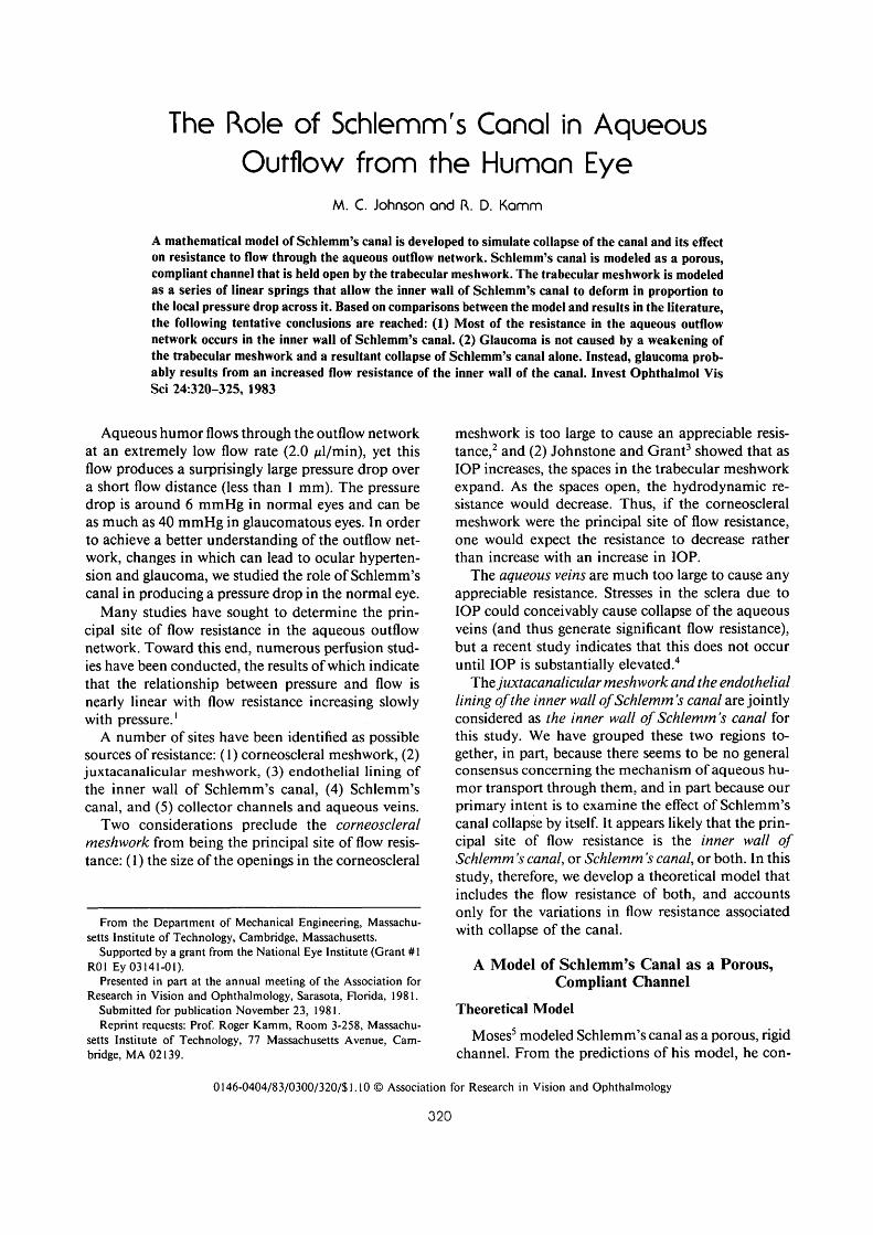
height. If we choose the support height to be slightlyless than the diameter of typical septae or about 5Aim, then the value of Pc would be 0.8. Figure 6 showsthe pressure-flow curves for Pc = 0.8. The outflowresistance is nearly constant until the canal is signif-icantly collapsed whereupon further increases in IOPcause the resistance to increase, until the canal is ev-erywhere collapsed to the support height. Above thispressure (PR) the canal is rigid and the resistanceremains constant.
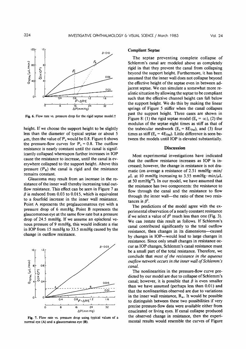
Glaucoma may result from an increase in the re-sistance of the inner wall thereby increasing total out-flow resistance. This effect can be seen in Figure 7 as/3 is reduced from 0.03 to 0.015, which is equivalentto a fourfold increase in the inner wall resistance.Point A represents the preglaucomatous eye with apressure drop of 6 mmHg; Point B represents theglaucomatous eye at the same flow rate but a pressuredrop of 24.5 mmHg. If we assume an episcleral ve-nous pressure of 9 mmHg, this would indicate a risein IOP from 15 mmHg to 33.5 mmHg caused by thechange in outflow resistance.
3 s
1.2
0.8
0.4
n
B .
P = 0.03/
0.015
^ ~0.01
0.4 1.6
16 24AP(mmHq)
32
Fig. 7. Flow rate vs. pressure drop using typical values of anormal eye (A) and a glaucomatous eye (B).
Compliant Septae
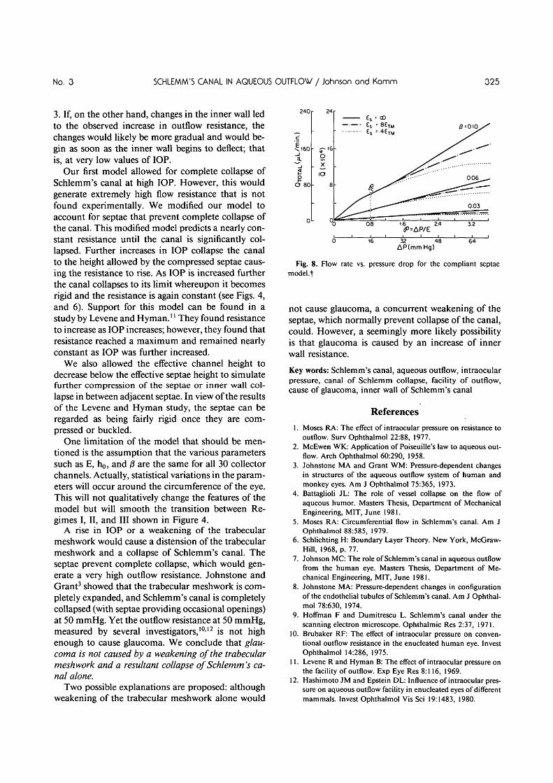
The septae preventing complete collapse ofSchlemm's canal are modeled above as completelyrigid in that they prevent the canal from collapsingbeyond the support height. Furthermore, it has beenassumed that the inner wall does not collapse beyondthe effective height of the septae even in between ad-jacent septae. We can simulate a somewhat more re-alistic situation by allowing the septae to be compliantsuch that the effective channel height can fall belowthe support height. We do this by making the linearsprings of Figure 5 stiffer when the canal collapsespast the support height. Three cases are shown inFigure 8: (1) the rigid septae model (Es = oo), (2) themodulus of the septae eight times as stiff as that ofthe trabecular meshwork (Es = 8ETM), and (3) fourtimes as stiff (Es = 4ETM). Little difference is seen be-tween the models until IOP is elevated substantially.
Discussion
Most experimental investigations have indicatedthat the outflow resistance increases as IOP is in-creased; however, the change in resistance is not dra-matic (on average a resistance of 2.51 mmHg* min/nL at 10 mmHg increasing to 3.55 mmHg • min/ViLat 50 mmHg10). In our model, we have assumed thatthe resistance has two components: the resistance toflow through the canal and the resistance to flowthrough the inner wall—the ratio of these two resis-tances is j82.
The predictions of the model agree with the ex-perimental observation of a nearly constant resistanceif we select a value of 01 much less than one (Fig. 3).We can restate this result as follows. If Schlemm'scanal contributed significantly to the total outflowresistance, then changes in its dimensions—causedby changes in IOP—would lead to large changes inresistance. Since only small changes in resistance oc-cur as IOP changes, Schlemm's canal resistance mustbe a small part of the total resistance. Therefore, weconclude that most of the resistance in the aqueousoutflow network occurs in the inner wall of Schlemm'scanal.
The nonlinearities in the pressure-flow curve pre-dicted by our model are due to collapse of Schlemm'scanal; however, it is possible that j8 is even smallerthan we have assumed (perhaps less than 0.01) andthat the nonlinearities observed are due to variationsin the inner wall resistance, Rw. It would be possibleto distinguish between these two possibilities if veryprecise pressure-flow data were available either fromenucleated or living eyes. If canal collapse producedthe observed change in resistance, then the experi-mental results would resemble the curves of Figure
No. 3 SCHLEMM'S CANAL IN AQUEOUS OUTFLOW / Johnson ond Komrn 325
3. If, on the other hand, changes in the inner wall ledto the observed increase in outflow resistance, thechanges would likely be more gradual and would be-gin as soon as the inner wall begins to deflect; thatis, at very low values of IOP.
Our first model allowed for complete collapse ofSchlemm's canal at high IOP. However, this wouldgenerate extremely high flow resistance that is notfound experimentally. We modified our model toaccount for septae that prevent complete collapse ofthe canal. This modified model predicts a nearly con-stant resistance until the canal is significantly col-lapsed. Further increases in IOP collapse the canalto the height allowed by the compressed septae caus-ing the resistance to rise. As IOP is increased furtherthe canal collapses to its limit whereupon it becomesrigid and the resistance is again constant (see Figs. 4,and 6). Support for this model can be found in astudy by Levene and Hyman.1' They found resistanceto increase as IOP increases; however, they found thatresistance reached a maximum and remained nearlyconstant as IOP was further increased.
We also allowed the effective channel height todecrease below the effective septae height to simulatefurther compression of the septae or inner wall col-lapse in between adjacent septae. In view of the resultsof the Levene and Hyman study, the septae can beregarded as being fairly rigid once they are com-pressed or buckled.
One limitation of the model that should be men-tioned is the assumption that the various parameterssuch as E, h0, and 0 are the same for all 30 collectorchannels. Actually, statistical variations in the param-eters will occur around the circumference of the eye.This will not qualitatively change the features of themodel but will smooth the transition between Re-gimes I, II, and III shown in Figure 4.
A rise in IOP or a weakening of the trabecularmesh work would cause a distension of the trabecularmeshwork and a collapse of Schlemm's canal. Theseptae prevent complete collapse, which would gen-erate a very high outflow resistance. Johnstone andGrant3 showed that the trabecular meshwork is com-pletely expanded, and Schlemm's canal is completelycollapsed (with septae providing occasional openings)at 50 mmHg. Yet the outflow resistance at 50 mmHg,measured by several investigators,1012 is not highenough to cause glaucoma. We conclude that glau-coma is not caused by a weakening of the trabecularmeshwork and a resultant collapse of Schlemm's ca-nal alone.
Two possible explanations are proposed: althoughweakening of the trabecular meshwork alone would
240
in.)
•^160
O 80
0
- -^
oX
IO
24
16
8
0.1.6 2.4
<P=AP/E3.2
32 48AP(mmHg)
64
Fig. 8. Flow rate vs. pressure drop for the compliant septaemodel.f
not cause glaucoma, a concurrent weakening of theseptae, which normally prevent collapse of the canal,could. However, a seemingly more likely possibilityis that glaucoma is caused by an increase of innerwall resistance.
Key words: Schlemm's canal, aqueous outflow, intraocularpressure, canal of Schlemm collapse, facility of outflow,cause of glaucoma, inner wall of Schlemm's canal
References
1. Moses RA: The effect of intraocular pressure on resistance tooutflow. Surv Ophthalmol 22:88, 1977.
2. McEwen WK: Application of Poiseuille's law to aqueous out-flow. Arch Ophthalmol 60:290, 1958.
3. Johnstone MA and Grant WM: Pressure-dependent changesin structures of the aqueous outflow system of human andmonkey eyes. Am J Ophthalmol 75:365, 1973.
4. Battaglioli JL: The role of vessel collapse on the flow ofaqueous humor. Masters Thesis, Department of MechanicalEngineering, MIT, June 1981.
5. Moses RA: Circumferential flow in Schlemm's canal. Am JOphthalmol 88:585, 1979.
6. Schlichting H: Boundary Layer Theory. New York, McGraw-Hill, 1968, p. 77.
7. Johnson MC: The role of Schlemm's canal in aqueous outflowfrom the human eye. Masters Thesis, Department of Me-chanical Engineering, MIT, June 1981.
8. Johnstone MA: Pressure-dependent changes in configurationof the endothelial tubules of Schlemm's canal. Am J Ophthal-mol 78:630, 1974.
9. Hoffman F and Dumitrescu L. Schlemm's canal under thescanning electron microscope. Ophthalmic Res 2:37, 1971.
10. Brubaker RF: The effect of intraocular pressure on conven-tional outflow resistance in the enucleated human eye. InvestOphthalmol 14:286, 1975.
11. Levene R and Hyman B: The effect of intraocular pressure onthe facility of outflow. Exp Eye Res 8:116, 1969.
12. Hashimoto JM and Epstein DL: Influence of intraocular pres-sure on aqueous outflow facility in enucleated eyes of differentmammals. Invest Ophthalmol Vis Sci 19:1483, 1980.
Related Documents


![Index [link.springer.com]978-0-387-76700-0/1.pdf · trabeculodescemet window (TDW), 801–802 Ab interno Schlemm's canal outflow devices trabectome handpiece advantage, 807 ... 1024](https://static.cupdf.com/doc/110x72/5acafb5f7f8b9aa1298e1944/index-link-978-0-387-76700-01pdftrabeculodescemet-window-tdw-801802.jpg)