MOLECULAR PLANT PATHOLOGY (2007) 8 (5), 677–700 DOI: 10.1111/J.1364-3703.2007.00419.X © 2007 BLACKWELL PUBLISHING LTD 677 Blackwell Publishing Ltd Review The role of plant defence proteins in fungal pathogenesis RICARDO B. FERREIRA 1,2 *, SARA MONTEIRO 1,2 , REGINA FREITAS 1,2 , CLÁUDIA N. SANTOS 2 , ZHENJIA CHEN 2 , LUÍS M. BATISTA 1 , JOÃO DUARTE 1 , ALEXANDRE BORGES 1 AND ARTUR R. TEIXEIRA 1 1 Departamento de Botânica e Engenharia Biológica, Instituto Superior de Agronomia, Universidade Técnica de Lisboa, 1349-017 Lisboa, Portugal 2 Disease and Stress Biology Laboratory, Instituto de Tecnologia Química e Biológica, Universidade Nova de Lisboa, Apartado 127, 2781-901 Oeiras, Portugal SUMMARY It is becoming increasingly evident that a plant–pathogen interac- tion may be compared to an open warfare, whose major weapons are proteins synthesized by both organisms. These weapons were gradually developed in what must have been a multimillion- year evolutionary game of ping-pong. The outcome of each battle results in the establishment of resistance or pathogenesis. The plethora of resistance mechanisms exhibited by plants may be grouped into constitutive and inducible, and range from morpho- logical to structural and chemical defences. Most of these mechanisms are defensive, exhibiting a passive role, but some are highly active against pathogens, using as major targets the fungal cell wall, the plasma membrane or intracellular targets. A considerable overlap exists between pathogenesis-related (PR) proteins and antifungal proteins. However, many of the now considered 17 families of PR proteins do not present any known role as antipathogen activity, whereas among the 13 classes of antifungal proteins, most are not PR proteins. Discovery of novel antifungal proteins and peptides continues at a rapid pace. In their long coevolution with plants, phytopathogens have evolved ways to avoid or circumvent the plant defence weaponry. These include protection of fungal structures from plant defence reactions, inhibition of elicitor-induced plant defence responses and suppression of plant defences. A detailed understanding of the molecular events that take place during a plant–pathogen interaction is an essential goal for disease control in the future. INTRODUCTION In their long association with pathogens, plants evolved an intricate and elaborate array of defensive tools. At the same time, those very same pathogens developed means to overcome plant resistance mechanisms in what must have been a multimillion- year evolutionary game of ping-pong (Keen, 1999b). As each defensive innovation was established in the host, new ways to circumvent it evolved in the pathogen. Over time, the coevolutionary struggles between would-be pathogens and their erstwhile hosts have generated some of the most complex and interesting interactions known to biology (Taylor, 1998). A plant–pathogen interaction may therefore be considered as an open warfare, whose major weapons are proteins synthesized by both organisms (Ferreira et al., 2006). The cuticle and the plant cell wall, for example, are pre-formed physical barriers often claimed to constitute the first line of plant defence by protecting against pathogen penetration. In addition, they are also a source of signals used by the invading pathogens to activate their responses or by plants to induce defence mech- anisms. Nevertheless, recent evidence challenged the traditional view of the plant cell wall as a passive structural barrier to pathogen invasion. Apparently, plants are able to sense perturba- tion of the cell wall by monitoring the integrity of its structure. For example, mutations in the Arabidopsis cellulose synthase gene CESA3 exhibit constitutive activation of jasmonate- and ethylene- mediated defence gene expression and enhanced resistance to powdery mildew pathogens (Cano-Delgado et al., 2003; Nishimura et al., 2003). The papilla is a local cell-wall fortification formed on the inner side of the plant cell walls at the site of pathogen penetra- tion that is regarded as an inducible structural barrier. In the case of powdery mildews, papillae have been reported to play an important role against fungal invasion (Thordal-Christensen et al., 2000; Zeyen et al., 2002). Fungal pathogens almost invariably trigger cell wall-associated defence responses, such as extracellular hydrogen peroxide generation and callose deposition, when they attempt to penetrate either resistant or susceptible plant cells. Expression of these defences involves communication between the plant cell wall and the cytosol across the plasma membrane. Indeed, peptides containing an Arg–Gly–Asp (RGD) motif which interfere with *Correspondence: Tel.: 351 213653416; Fax: 351 213653238; E-mail: [email protected]

Welcome message from author
This document is posted to help you gain knowledge. Please leave a comment to let me know what you think about it! Share it to your friends and learn new things together.
Transcript

MOLECULAR PLANT PATHOLOGY
(2007)
8
(5 ) , 677–700 DOI : 10 .1111/ J .1364-3703.2007.00419.X
© 2007 BLACKWELL PUBL ISH ING LTD
677
Blackwell Publishing Ltd
Review
The role of plant defence proteins in fungal pathogenesis
R ICARDO B. FERRE IRA
1,2
* , SARA MONTE IRO
1,2
, REG INA FRE I TAS
1,2
, CLÁUD IA N. SANTOS
2
, ZHENJ IA CHEN
2
, LU Í S M . BAT I STA
1
, JOÃO DUARTE
1
, ALEXANDRE BORGES
1
AND ARTUR R . TE IXE IRA
1
1
Departamento de Botânica e Engenharia Biológica, Instituto Superior de Agronomia, Universidade Técnica de Lisboa, 1349-017 Lisboa, Portugal
2
Disease and Stress Biology Laboratory, Instituto de Tecnologia Química e Biológica, Universidade Nova de Lisboa, Apartado 127, 2781-901 Oeiras, Portugal
SUMMARY
It is becoming increasingly evident that a plant–pathogen interac-tion may be compared to an open warfare, whose major weaponsare proteins synthesized by both organisms. These weapons weregradually developed in what must have been a multimillion-year evolutionary game of ping-pong. The outcome of each battleresults in the establishment of resistance or pathogenesis. Theplethora of resistance mechanisms exhibited by plants may begrouped into constitutive and inducible, and range from morpho-logical to structural and chemical defences. Most of thesemechanisms are defensive, exhibiting a passive role, but someare highly active against pathogens, using as major targets thefungal cell wall, the plasma membrane or intracellular targets.A considerable overlap exists between pathogenesis-related(PR) proteins and antifungal proteins. However, many of the nowconsidered 17 families of PR proteins do not present any knownrole as antipathogen activity, whereas among the 13 classes ofantifungal proteins, most are not PR proteins. Discovery of novelantifungal proteins and peptides continues at a rapid pace. Intheir long coevolution with plants, phytopathogens have evolvedways to avoid or circumvent the plant defence weaponry. Theseinclude protection of fungal structures from plant defencereactions, inhibition of elicitor-induced plant defence responsesand suppression of plant defences. A detailed understanding ofthe molecular events that take place during a plant–pathogen
interaction is an essential goal for disease control in the future.
INTRODUCTION
In their long association with pathogens, plants evolved anintricate and elaborate array of defensive tools. At the same time,
those very same pathogens developed means to overcome plantresistance mechanisms in what must have been a multimillion-year evolutionary game of ping-pong (Keen, 1999b). As each defensiveinnovation was established in the host, new ways to circumventit evolved in the pathogen. Over time, the coevolutionary strugglesbetween would-be pathogens and their erstwhile hosts havegenerated some of the most complex and interesting interactionsknown to biology (Taylor, 1998). A plant–pathogen interactionmay therefore be considered as an open warfare, whose majorweapons are proteins synthesized by both organisms (Ferreira
et al
., 2006).The cuticle and the plant cell wall, for example, are pre-formed
physical barriers often claimed to constitute the first line of plantdefence by protecting against pathogen penetration. In addition,they are also a source of signals used by the invading pathogensto activate their responses or by plants to induce defence mech-anisms. Nevertheless, recent evidence challenged the traditionalview of the plant cell wall as a passive structural barrier topathogen invasion. Apparently, plants are able to sense perturba-tion of the cell wall by monitoring the integrity of its structure. Forexample, mutations in the Arabidopsis cellulose synthase gene
CESA3
exhibit constitutive activation of jasmonate- and ethylene-mediated defence gene expression and enhanced resistance topowdery mildew pathogens (Cano-Delgado
et al
., 2003; Nishimura
et al
., 2003). The papilla is a local cell-wall fortification formed onthe inner side of the plant cell walls at the site of pathogen penetra-tion that is regarded as an inducible structural barrier. In thecase of powdery mildews, papillae have been reported to play animportant role against fungal invasion (Thordal-Christensen
et al
.,2000; Zeyen
et al
., 2002).Fungal pathogens almost invariably trigger cell wall-associated
defence responses, such as extracellular hydrogen peroxidegeneration and callose deposition, when they attempt to penetrateeither resistant or susceptible plant cells. Expression of thesedefences involves communication between the plant cell walland the cytosol across the plasma membrane. Indeed, peptidescontaining an Arg–Gly–Asp (RGD) motif which interfere with
*
Correspondence
: Tel.: 351 213653416; Fax: 351 213653238; E-mail: [email protected]

678
R. B. FERREIRA
et al.
MOLECULAR PLANT PATHOLOGY
(2007)
8
(5 ) , 677–700 © 2007 BLACKWELL PUBL ISH ING LTD
plasma membrane–cell wall adhesion or disruption of plantmicrofilaments reduce the expression of cell wall-associateddefence responses during the penetration of non-host plants bybiotrophic fungal pathogens with the consequent increment infungal penetration efficiency (Mellersh and Heath, 2001).
The actin cytoskeleton has been shown to contribute to bothpre-invasion resistance and papillary callose formation in theinteractions between
Arabidopsis thaliana
and non-adapted
Colletotrichum
species (Shimada
et al
., 2006). This study showedan extensive reorganization of actin microfilaments leading topolar orientation of large actin bundles towards appressorialcontact sites. Non-adapted
Colletotrichum
species differentiatemelanized appressoria on Arabidopsis leaves but fail to formintracellular hyphae. Analyses of non-pathogenic
C. lagenarium
mutants indicated penetration-peg formation as the inductive cuefor papillary callose formation. However, the incidence of papillaformation at fungal entry sites is greatly reduced during thecompatible interaction of Arabidopsis with
C. higginsianum
,indicating that this adapted pathogen may suppress pre-invasionresistance at the cell periphery (Shimada
et al
., 2006). Indeed,biotrophic fungi may manipulate the plant cell wall surveillancesystem for the establishment of biotrophy, subverting the inter-connected plant defence signalling pathways or the underlyingresistance mechanisms (Jones and Takemoto, 2004; Schulze-Lefert, 2004).
Recent observations have revealed that molecular processesoccurring at and in plant cell walls may also function in fungalpathogenesis. The
β
-1,3-
D
-glucan callose is rapidly synthesizedand deposited at plant cell wall upon microbial attack. Arabidopsismutants in the gene encoding the single glucan synthase responsiblefor papillary callose synthesis exhibit broad-spectrum enhancedresistance to powdery mildew fungi, suggesting a role for thewild-type gene in fungal colonization of the host cells. Callose mayfacilitate penetration of pathogens into host cells by providing astructural collar for the intruder. It has been suggested that theglucan synthase participates in the containment of pathogen-derived elicitors at infection sites, thereby preventing theirperception by the plant, or in the protection of the invadingpathogen against plant-derived antimicrobial compounds (Gomez-Gomez and Boller, 2002; Jacobs
et al
., 2003; Nishimura
et al
.,2003).
Therefore, emerging evidence suggests that pathogens maytake over selected aspects of plant gene expression to their ownbenefit. In this way, they may induce the expression of somecomponents required for the infection or development processes,or they may repress components of the host defence system,such as, for example, proteins of the cytoskeleton. In
Arabidopsisthaliana
and barley (
Hordeum vulgare
), for example, the presenceof specific isoforms of the family of heptahelical plasma membrane-localized MLO proteins is required for successful host-cellinvasion by powdery mildew fungi (Panstruga, 2005). This study
concluded that powdery mildew fungi appear specifically tocorrupt MLO to modulate vesicle-associated processes at theplant cell periphery for successful pathogenesis.
TARGETS FOR THE PLANT DEFENCE MECHANISMS
The fungal cell wall
The molecular model of the cell wall of
Saccharomyces cerevisiae
that is generally accepted (Klis
et al
., 2006; Theis and Stahl, 2004)contains three major classes of carbohydrate polymers, chitin,
β
-1,3-glucan and
β
-1,6-glucan, and two main classes of glycosylatedcell wall proteins (CWPs). There is no complete model of the cellwall of filamentous fungi available yet, but many similarities areexpected to occur between the cell walls of these groups of fungi.
Chitin, a natural homopolymer of
β
-1,4-linked
N
-acetyl-
D
-glucosamine residues, is the major cell wall component infilamentous fungi (BeMiller, 1965). The concentration of chitinin the cell walls of these organisms (~10%) is significantly higherthan in yeasts (2%) (Theis and Stahl, 2004). In Oomycetes, chitinis only a minor component of their cell walls (Barkai-Golan
et al
., 1978; Schoffelmeer
et al
., 1999). Therefore, it is not surpris-ing that plants synthesize a large number of defence proteinscapable of binding to chitin and chitin oligosaccharides. Most butnot all of these proteins bind to chitin through a conserved aminoacid sequence known as the chitin-binding domain (Chrispeelsand Raikhel, 1991). Many chitin-binding proteins have beenisolated from numerous plant species, especially from their seeds.Examples are class I chitinases (Gomes
et al
., 1996; Leah
et al
., 1991),lectins such as wheat (
Triticum aestivum
) germ agglutinin (WGA)(Broekaert
et al
., 1987; Wright
et al
., 1991), and antimicrobialpeptides such as hevein (Broekaert
et al
., 1992).One in six aminosaccharide chitin residues can be devoid of an
acetyl group (Blackwell, 1988). Deacetylation is a common processinvolved in chitin–protein interaction and leads to chitosan forma-tion (Blackwell, 1988), another structural polysaccharide foundin fungal cell walls. Plant antifungal proteins have been describedexhibiting chitosanase activity.
Glucans are the second major component of the fungal cellwall. Apart from
β
-1,3-glucan, which is the primary glucan,several other linkages such as
α
-1,3,
α
-1,4,
β
-1,4 and
β
-1,6 havealso been detected in fungal cell walls.
The fungal membrane
The fungal plasma membrane is the target for the largest groupof antifungal and antimicrobial proteins (Theis and Stahl, 2004).Over 500 naturally occurring proteins have been reported thatare believed to interact with the fungal membrane, leading topore formation, efflux of cellular components and changes in the

Plant defence proteins in fungal pathogenesis
679
© 2007 BLACKWELL PUBL ISH ING LTD
MOLECULAR PLANT PATHOLOGY
(2007)
8
(5 ) , 677–700
membrane potential (Tossi
et al
., 2000). They exhibit an enormousdiversity of structures but share at least two common features(Tossi
et al
., 2000): a positive net charge under physiologicalconditions, which promotes interaction with negatively chargedmicrobial surfaces; and an amphipathic structure (i.e. a structurewith two faces, one being positively charged and the otherneutral or hydrophobic) that allows incorporation into pathogenmembranes. A striking difference between higher eukaryotesand fungal membranes concerns the embedded sterols. Theplasma membrane of higher eukaryotes contains cholesterol,sitosterol and/or campesterol, whereas fungal membraneergosterol comprises ~2% of the fungal dry weight (Brennan
et al
., 1974; Rattray
et al
., 1975). It is noteworthy to mention thatergosterol is a non-specific fungal elicitor that induces expressionof a specific set of plant defence-related genes (Lochman andMikes, 2006).
Intracellular targets
To enter fungal cells, plant antifungal proteins must pass throughthe fungal cell wall and membrane. This may explain the low numberof reports on plant defence proteins interacting with the plethoraof potential fungal intracellular targets.
PREFORMED VERSUS INDUCIBLE DEFENCE MECHANISMS
Plants have evolved a network of intricate and elaborate defencesystems that mediate their interaction with the environment. Inwhat concerns the challenge by fungal pathogens, their resistanceis determined by an impressive combination of both constitutiveand inducible defence mechanisms that involve a vast array ofproteins and other organic molecules produced prior to infectionor during pathogen attack (Dixon and Harrison, 1990). For example,the completion of the Arabidopsis genome sequence showed thatthis plant species has a few hundred open reading frames thatencode potential surveillance proteins (Dangl and Jones, 2001).
Preformed or passive defences are the first obstacle aninvading pathogen has to overcome before disease is established.The typical preformed, constitutive defences are morphological,structural and chemical barriers. A recognized example of amorphological barrier is the height of lips of stomatal guard cells(Keen, 1999a). As shown by Hoch
et al
. (1987), certain fungal rustpathogens possess exquisite detection mechanisms that sensethe height of stomatal guard cell lips encountered on susceptibleplants. Thus, when the hyphae find a lip of the proper height, theyare programmed to undergo a developmental process resultingin the formation of invasive structures that enter the stomata andbegin colonization of the leaf interior.
Structural barriers, such as waxes, cutin, suberin, lignin, cellulose,callose and cell wall proteins, are often rapidly reinforced upon
the infection process. In addition, plants constitutively produce aplethora of secondary metabolites and antifungal proteins, manyof which can act as antimicrobial compounds during defenceagainst microorganisms. These include phenolics of varyingstructural sophistication, saponins, terpenoids and steroids. Somepreformed compounds are directly toxic, whereas others occur asconjugates such as glycosides that are not toxic
per se
, but becomepoisonous following disruption of the conjugate. Plant glycosides,for instance, are often hydrolysed by vacuolar glycosidases follow-ing pathogen invasion, releasing aglycones that may be quite toxic,not only to the invader but also to neighbouring plant cells (Keen,1999a). By contrast, some plant preformed compounds are toxicas glycosides, but lose toxicity when deglycosylated. The growthof the wheat root-infecting fungus
Gaeumannomyces graminis
var.
tritici
is inhibited by avenacin. Therefore, oat (
Avena sativa
)plants producing this preformed triterpene saponin glycosideexhibit resistance to the pathogen. However, the related oat rootpathogen
G. graminis
var.
avenae
produces a glycosidase thatremoves the sugar residue from avenacin, effectively detoxifyingit. Therefore, this strain is not inhibited by avenacin and oat plantsare susceptible to it (Osbourn
et al
., 1994). A mutation in theglycosidase gene rendered
G. graminis
var.
avenae
sensitiveto avenacin and incapable of infecting oat plants (Bowyer
et al
.,1995). By contrast, engineered oat plants that lack or have onlytrace amounts of avenacin are compromised for disease resistanceagainst the non-host fungal pathogens
G. graminis
var.
tritici
and
Fusarium culmorum
(Papadopoulou
et al
., 1999).Besides constitutive defences, an important plant strategy is
to initiate defences in response to pathogen attack (Karbanand Baldwin, 1997). Inducible resistance mechanisms are active,energy-requiring systems typified by specific recognition of aninvader that ultimately leads to the production of proteins ormetabolites that are antagonistic to the invader (Keen, 1999a).They are considered the second obstacle an invading pathogenhas to face when attempting infection. Inducible defences mayhave several advantages such as reducing biosynthetic costs ofdefence or avoiding the fact that other organisms may exploit thedefence to their own benefit (Cipollini
et al
., 2003; Karban andBaldwin, 1997; Zangerl, 2003). In addition, the variation in plantgenotype that is caused by inducible defences may reduce thechances that attackers adapt to plant defences (Agrawal andKarban, 1999). Inducible or active defence mechanisms mainlyinvolve the oxidative burst, localized cell death, accumulation ofphytoalexins, synthesis of pathogenesis-related (PR) proteinsand cell wall strengthening proteins such as hydroxyproline-richglycoproteins, and enhanced transcription of genes encodingenzymes involved in the flow of carbon from the primary to thesecondary metabolism of plants, such as peroxidases, lipooxy-genases, superoxide dismutases and phenylalanine ammonia lyase(PAL), a key enzyme in the biosynthesis of phenolic compounds withantimicrobial activity (Montesinos, 2000).

680
R. B. FERREIRA
et al.
MOLECULAR PLANT PATHOLOGY
(2007)
8
(5 ) , 677–700 © 2007 BLACKWELL PUBL ISH ING LTD
Besides chemical, structural inductive mechanisms are mostlyrelated to the plant cell wall. Modification of the plant cell wallwas recognized as a potential resistance mechanism more than80 years ago (Young, 1926). Lignification and other chemicalmodifications of plant cells around the sites of infection lead towall thickening and the formation of local additions or callositiesin the paramural space (i.e. the space between the cell walland the plasma membrane). Nevertheless, formation of the cellwall apposition (CWAs) or papillae is usually accompanied bythe induction of co-localized chemical responses (Bestwick
et al
.,1998; Matern
et al
., 1995; McLusky
et al
., 1999; Nicholson andHammerschmidt, 1992; Schmelzer, 2002; Thordal-Christensen
et al
., 1997).Plant cytoskeleton also plays a significant role in inducible
disease resistance. Plant actin microfilaments have beenimplicated in defence against fungal penetration and theirdisruption leads to the loss of non-host resistance againstseveral non-host pathogens (Kobayashi
et al
., 1992). Treatmentof several non-host plants (e.g. barley and wheat) with cytocha-lasins, specific inhibitors of actin polymerization, allows severalnon-host fungi to penetrate the cells of these plants (Kobayashi
et al
., 1997).The inducible responses are turned on systematically in the
plant in response to attempted infection. First, in the localizedresponse, a spatially confined necrosis is frequently induced thatresults either from cell death caused by the action of the pathogenin a compatible interaction or from an endogenous plant celldeath response following recognition of the pathogen by thehost in an incompatible interaction—the hypersensitive response(HR) (Maleck and Dietrich, 1999). The HR is a pathogen-induced,rapid and localized cell suicide at the spot of infection in whichthe plant cells react to the invading pathogen by a kind ofprogrammed cell death consisting of electrolyte leakage from thecytoplasm and oxidative burst (Montesinos, 2000). As a result,the pathogen remains confined to necrotic lesions near the siteof infection. Thus, as described by Chester (1933), HR is a type ofblocking necrosis often developed by non-host plants againstmany plant pathogens that invade their tissues. The knowledgeon signal transduction in the HR is still rather incomplete, butseveral interesting genes have already been identified, includingprotein kinases and phosphatases, calmodulin genes and othersof unknown biochemical function that ultimately activate transcrip-tion of defence response genes. Localized acquired resistance ina ring of cells surrounding necrotic lesions ensure that they becomefully refractory to subsequent infection (Bonas and Lahaye, 2002;Fritig
et al
., 1998). Experiments performed in Tetep, a rice (
Oryzasativa
) cultivar resistant to both
Cochliobolus miyabeanus
and
Magnaporthe grisea
, showed that inoculation with either pathogentriggered the HR. However, in rice cv. Nakdong, susceptible to bothfungi,
M. grisea
did not cause HR whereas
C. miyabeanus
causedrapid cell death (Ahn
et al
., 2005).
Within a few hours of the localized necrosis, the plant beginsto express a set of defence genes both locally, at the point ofinfection, and systemically, throughout the rest of the plant (Antoniw
et al
., 1980a; van Loon, 1985). Thus, local HR often triggers asystemic signal that transduces non-specific resistance throughoutthe plant, leading to systemic acquired resistance (SAR), whichconfers long-lasting, enhanced resistance against subsequentinfections by a broad spectrum of pathogens (Durrant and Dong,2004; Ryals
et al
., 1996; Somssich, 2003; Sticher
et al
., 1997).In contrast to HR, the development of SAR is slow and gradual(Scheel, 1998). In this systemic defence, the signal spreads fromthe place of plant–pathogen interaction and is mediated by aninteracting set of endogenous defence signalling molecules thathave been identified as messengers in plants, including salicylicacid (SA), jasmonic acid (JA), ethylene (ET), nitric oxide (NO) orreactive oxygen species (ROS) (Baker
et al
., 1997; Beckers andSpoel, 2006; Montesano
et al
., 2003). These messengers interactwith specific binding proteins, which are involved in the transcrip-tional activation of pathogen-responsive genes. These SAR genesare thought to be responsible for the increased resistance of thenon-infected, secondary plant tissues to subsequent infections bythe same or even unrelated pathogens (Maleck and Dietrich,1999; Montesano
et al
., 2003).Increasing evidence suggests the existence of cross-talk among
the induced defence mechanisms (Beckers and Spoel, 2006).Apparently, these are not controlled by independent linearsignalling cascades, but components of one pathway may affectthe signalling through other pathways (Maleck and Dietrich,1999). Several examples have been reported on antagonistic orsynergistic interactions between defence responses. Current know-ledge suggests that plants do not activate a stereotypic defenceresponse against all pathogen attacks, but rather appear torecognize a multitude of components from a particular pathogento fine-tune a specific response. Experiments performed underlaboratory conditions have shown that elicitors, avirulencefactors and mechanical stress each only induce a subset of theplant response to pathogens (Gus-Mayer
et al
., 1998).
PLANT DEFENSIVE WEAPONARY AGAINST FUNGAL PATHOGENESIS
A plant–pathogen interaction may be regarded as an openwarfare, whose weapons are proteins and low-molecular-masscompounds synthesized by both organisms. The outcome of eachbattle results in the establishment of resistance or pathogenesis.This is readily illustrated by the following example.
Plant cell walls, essentially composed of polysaccharides andproteins, are attacked by a range of degrading enzymes liberatedby many pathogenic fungi. These hydrolases fragment the plantcell wall polymers releasing oligosaccharides and facilitatingcolonization of the host cells. The oligosaccharides not only

Plant defence proteins in fungal pathogenesis
681
© 2007 BLACKWELL PUBL ISH ING LTD
MOLECULAR PLANT PATHOLOGY
(2007)
8
(5 ) , 677–700
provide the fungus with a carbon source but are also perceivedby and elicit the plant defensive mechanisms. Thus, for example,pectin, a major component of the cell walls in many plants, iscleaved by fungal endopolygalacturonases (EPGs) with the tran-sient formation of elicitor-active oligogalacturonides (OGAs) withdegrees of polymerization between 9 and 15. For this reason,OGAs are rapidly converted to smaller, biologically inactive frag-ments by the EPGs.
To increase the lifetime of the biologically active oligosaccha-rides, plants release inhibitors of fungal glycanases. These includeinhibitors of pectin-degrading enzymes such as polygalacturo-nases, pectin methyl esterases and pectin lyases, and cross-linkingglycan (known earlier as hemicelluloses)-degrading enzymessuch as endoxylanases and xyloglucan endoglucanases (Juge,2006). For example, plant polygalacturonase-inhibiting proteins(PGIPs) are glycoproteins present in the apoplast of many plantsthat form reversible high-affinity complexes with fungal EPGs,reducing their catalytic activity by one or two orders of magni-tude. By limiting EPG activity, the lifetime and concentration ofOGAs are increased, prolonging or enhancing plant defenceresponses (Desiderio
et al
., 1997; Powell
et al
., 1995).Conversely, fragmentation of the fungal cell wall by plant-
derived chitinases and
β
-1,3-glucanases also generates oligosac-charides that induce plant defence responses. In return, fungiproduce glucanase inhibitor proteins (GIPs) that prevent degra-dation of their own cell wall, thus limiting their perception by theplants (Albersheim and Valent, 1974). Major groups of proteinsinvolved in plant defence are the pathogenesis-related proteinsand the antifungal proteins, both of which are treated in the nextsections of this article. However, plants also produce lowermolecular mass defensive compounds. Selected examples, con-sidered below, are ROS and phytoalexins.
ROS, such as superoxide anions, hydroxyl radicals and hydro-gen peroxide, play an important role in plant defence duringplant–pathogen interactions (Wu
et al
., 1997). ROS are directlytoxic to microbial invaders, catalyse oxidative cross-linking of thecell wall at the site of attempted infection and participate in sig-nalling the onset of other defence responses (Nurnberger
et al
.,2004). Thus, hydrogen peroxide is produced by plant cells inresponse to infection (Baker and Orlandi, 1995), triggering theHR (Tenhaken
et al
., 1995), strengthening cell walls (Brisson
et al
., 1994) and enhancing lignin formation (Wu
et al
., 1995).In addition, H
2
O
2
directly inhibits pathogen growth (Wu
et al
.,1995) and induces the synthesis of PR proteins, phytoalexins,SA and ethylene (Chamnongpol
et al
., 1998; Mehdy, 1994; Wu
et al
., 1997).Phytoalexins are low-molecular-mass secondary metabolites
of a non-proteinaceous nature which are produced by a broadrange of plant species. They display an enormous chemical diver-sity, exhibit antimicrobial and antifungal activities, are inducedby pathogen infection and elicitors (Grayer and Kokubun, 2001;
Hammerschmidt, 1999), and are synthesized through complexbiochemical pathways such as the shikimic acid pathway (Dixon
et al
., 1996).As for chitinases and glucanases, it has been difficult to demon-
strate a direct role for phytoalexins in plant resistance to pathogenattack (Punja, 2001). Wild-type
Arabidopsis thaliana
is resistantto
Alternaria brassicicola
, exhibiting a typical HR in responseto inoculation with this fungus (Thomma
et al
., 1998).
pad3-1
, anArabidopsis phytoalexin-deficient (pad) mutant, is compromisedfor non-host resistance against
A. brassicicola
(Thomma
et al
.,1999). pad3-1 is required for the biosynthesis of the phytoalexincamalexin and encodes a putative cytochrome P450 monooxygenase(Zhou et al., 1999).
Phytoalexins from Vitis species, for example, belong to thestilbene family of phenolic compounds and derive from trans-resveratrol (3,5,4′-trihydroxy stilbene) (Jeandet et al., 2002). Trans-resveratrol has been shown to be excreted from grapevinecell cultures. Among the viniferins, considered as oligomers ofresveratrol, ε-viniferin (a cyclic resveratrol dehydrodimer) andα-viniferin (a cyclic resveratrol dehydrotrimer), have been reportedto accumulate in grapevine in response to pathogen attack orstress (Jeandet et al., 1997; Langcake and Pryce, 1977; Pryce andLangcake, 1977). ε-Viniferin, for example, increases intracellularlyin response to endopolygalacturonase I (elicitor) from Botrytiscinerea and to UV-light irradiation. Other stilbenes detected ingrapevine include trans-pterostilbene, a dimethylated resveratrolderivative (3,5-dimethoxy-4′-hydroxystilbene), trans- and cis-piceid,a 3-O-β-D-glucoside of resveratrol, trans- and cis-astringin, a 3-O-β-D-glucoside of 3′-hydroxyresveratrol, and trans- and cis-resveratrol-oside, a 4′-O-β-D-glucoside of resveratrol (Jeandet et al., 2002).
PATHOGENESIS-RELATED PROTEINS
The concept of pathogenesis-related protein (abbreviation: PR)was introduced in 1980 to designate any protein coded for by thehost plant but induced only in pathological or related situations(Antoniw et al., 1980b), including viral, fungal or bacterial infections,parasitic attack by nematodes, phytophagous insects and otherhigher forms of animals such as herbivores. Abiotic stresses anddisorders were not considered inducers of PR proteins, althoughcertain non-infectious physiological conditions (e.g. toxin-inducedchlorosis or necrosis) often trigger induction of certain PR proteins(Jayaraj et al., 2004). More recent reports have shown the induc-tion of PR proteins as a result of colonization by non-pathogenic/beneficial fungi and bacteria (Blilou et al., 2000; Coventry andDubery, 2001; Yedidia et al., 2000; Zehnder et al., 2001). The majorcriterion for inclusion among the PR is that the protein (or proteinisoform) concerned is newly expressed upon infection, althoughnot necessarily in all pathological conditions (van Loon, 1999).According to this definition, proteins that are constitutivelypresent in low but detectable amounts in healthy tissues but which

682 R. B. FERREIRA et al.
MOLECULAR PLANT PATHOLOGY (2007) 8 (5 ) , 677–700 © 2007 BLACKWELL PUBL ISH ING LTD
are induced under pathological conditions are not consideredPR proteins. These concepts were initially based on experimentsperformed in the 1970s on tobacco (Nicotiana tabacum) leavesreacting hypersensitively to tobacco mosaic virus (van Loon andvan Kammen, 1970).
PR proteins were initially found to be typically acidic, of lowmolecular mass, highly resistant to proteolytic degradation andto low pH values, and localized predominantly extracellularly in theintercellular space of leaves. Following infection, they accumulatein leaves and other organs, where they may comprise more than10% of the total soluble protein.
The term PR-like protein was proposed to accommodate proteinsthat are present in healthy plants, being induced essentially ina developmentally controlled, tissue-specific manner. Theseproteins, which are not synthesized in response to pathogeninfection or related stresses, are predominantly basic andlocalized intracellularly in the vacuole (van Loon et al., 1994).As new proteins are discovered, a clear distinction between PRand PR-like proteins is sometimes difficult to establish. This is thecase, for example, of basic chitinase and glucanase from matureleaves, which can be expressed in a developmentally controlledmanner or induced in response to infection in the same organs;and also for acidic and basic glucanase and chitinase, whichmay be expressed constitutively in floral organs and inducible bypathogens in leaves (van Loon, 1999).
The distinction between PR proteins and PR-like proteinsbecame blurred by the discovery of specific PR proteins in healthytissues and the induction of PR-like proteins upon pathogenattack. Contrary to the initial definition of PR protein, manyauthors subsequently considered as PR proteins many proteinswhose synthesis is induced in response to biotic stress. Recently,van Loon et al. (2006) introduced the general term ‘inducibledefence-related proteins’ to include proteins that are mostlynon-detectable in healthy tissues and for which induction at theprotein level has been demonstrated after pathogen infection.Therefore, this general term encompasses both known PR proteinfamilies and non-classified proteins meeting the above criteriabut fails to include many proteins that are present in healthytissues and are induced upon microbial infection.
The induction of some PR proteins under pathological conditionssuggests, but does not prove, a role for these proteins in plantdefence (van Loon, 1990). Therefore, these proteins have beengenerally considered as defence proteins, functioning in preventingor limiting pathogen invasion and spread. However, their contribu-tion to resistance against the initial infection is usually poor.Nevertheless, if they are already present in a tissue, or if theyhave been induced in non-infected, distant tissues as a result ofprimary infection in the vicinity, then they confer an enhancedlevel of protection.
PR proteins are produced by plants during normal developmentor as part of an induced defence from fungal pathogens, against
which they exert biocontrol. Therefore, their biosynthesis andaccumulation is considered a major defence mechanism of plantsagainst fungal pathogens (Odjakova and Hadjiivanova, 2001;Somssich and Hahlbrock, 1998). Not only do some of theseproteins exhibit antifungal properties in vitro, but also they havebeen shown to be induced in vivo in a very large number ofplants in response to fungal attack. A large group of PR proteinshas been shown to be rapidly and massively induced both locallyaround infection sites and systemically (Kombrink and Somssich,1997). PR proteins are also induced in response to various environ-mental stress factors, such as drought, salinity, wounding, heavymetals, endogenous and exogenous elicitor treatment, andplant growth regulators (Derckel et al., 1996; Xie et al., 1999; Yuet al., 2001).
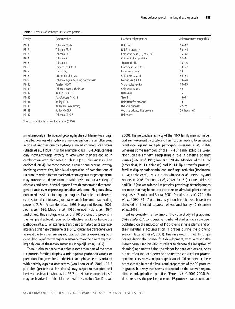
The PR proteins encompass several different groups ofstructurally and functionally unrelated proteins, which have beengrouped into protein families according to coding sequencesimilarities, serological relationships, and/or enzymatic or biologicalactivities, although additional pathogen-induced proteins withpotential antipathogenic action are consistently being described(Fritig et al., 1998; Somssich and Hahlbrock, 1998). Initially, fourprotein components were detected in hypersensitive tobacco plants,which were designated I, II, III and IV based on their increasingorder of electrophoretic mobility (van Loon and van Kammen, 1970).Subsequently, these proteins were classified into five groups, PR-1to PR-5. Each of these five classical groups of PR proteins com-prised two subclasses: an acidic subclass, usually encountered inthe extracellular space, whose members are induced by salicylicacid, and a basic subclass, found in the plant cell vacuole, whosemembers are induced by ethylene or jasmonic acid (Boller et al., 1983;Hamel and Bellemare, 1995; Samac et al., 1990; Selitrennikoff,2001; Thomma et al., 1998). Seventeen classes are now con-sidered, numbered in the order in which they were discovered,from PR-1 to PR-17 (Table 1). The families are numbered and thedifferent members of the same family are assigned letters accordingto the order in which they were described. The function of manyPR proteins remains a mystery. However, members of several ofthese families were demonstrated to have damaging actions onthe structures of the parasite, thus exhibiting antifungal activity,in in vitro bioassays and supporting a possible role for theseproteins in plant defence (Kombrink and Somssich, 1997; Odjakovaand Hadjiivanova, 2001). These include PR-1 and PR-5 (thaumatin-like proteins and osmotins), which are thought to create transmem-brane pores and have therefore been termed permatins; PR-2(β-1,3-glucanases) and PR-3, 4, 8 and 11 (chitinases), which attackβ-1,3-glucans and chitin, respectively, components of the cell wallsin most higher fungi (Honée, 1999). In most cases, an assortmentof PR proteins belonging to diverse subclasses are induced, ratherthan a single member of a single family of PR proteins (Dattaet al., 1999). It is also common for some PR proteins to displaysynergism. Because chitin and β-1,3-glucan are synthesized
Plant defence proteins in fungal pathogenesis 683
© 2007 BLACKWELL PUBL ISH ING LTD MOLECULAR PLANT PATHOLOGY (2007) 8 (5 ) , 677–700
simultaneously in the apex of growing hyphae of filamentous fungi,the effectiveness of a hydrolase may depend on the simultaneousaction of another one to hydrolyse mixed chitin–glucan fibres(Stintzi et al., 1993). Thus, for example, class II β-1,3-glucanasesonly show antifungal activity in vitro when they are applied incombination with chitinases or class I β-1,3-glucanases (Theisand Stahl, 2004). For these reasons, a genetic engineering strategyinvolving constitutive, high-level expression of combinations ofPR proteins with different modes of action against target organismsmay provide broad-spectrum, durable resistance to a variety ofdiseases and pests. Several reports have demonstrated that trans-genic plants over-expressing constitutively some PR genes showenhanced resistance to fungal pathogens. Examples include over-expression of chitinases, glucanases and ribosome-inactivatingproteins (RIPs) (Alexander et al., 1993; Hong and Hwang, 2006;Jach et al., 1995; Mauch et al., 1988), osmotin (Liu et al., 1994)and others. This strategy ensures that PR proteins are present inthe host plant at levels required for effective resistance before thepathogen attack. For example, transgenic tomato plants express-ing only a chitinase transgene or a β-1,3-glucanase transgene weresusceptible to Fusarium oxysporum, but plants expressing bothgenes had significantly higher resistance than the plants express-ing only one of these two enzymes (Jongedijk et al., 1955).
There is also evidence that at least some members of the otherPR protein families display a role against pathogen attack orpredation. Thus, members of the PR-1 family have been associatedwith activity against oomycetes (van Loon et al., 2006). PR-6proteins (proteinase inhibitors) may target nematodes andherbivorous insects, whereas the PR-7 protein (an endoproteinase)may be involved in microbial cell wall dissolution (Jordá et al.,
2000). The peroxidase activity of the PR-9 family may act in cellwall reinforcement by catalysing lignification, leading to enhancedresistance against multiple pathogens (Passardi et al., 2004),whereas some members of the PR-10 family exhibit a weakribonuclease activity, suggesting a role in defence againstviruses (Bufe et al., 1996; Park et al., 2004a). Members of the PR-12(defensins), PR-13 (thionins) and PR-14 (lipid transfer proteins)families display antibacterial and antifungal activities (Bohlmann,1994; Epple et al., 1997; Garcia-Olmedo et al., 1995; Lay andAnderson, 2005; Thomma et al., 2002). PR-15 (oxalate oxidases)and PR-16 (oxalate oxidase-like proteins) proteins generate hydrogenperoxide that may be toxic to attackers or stimulate plant defenceresponses (Bernier and Berna, 2001; Donaldson et al., 2001; Huet al., 2003). PR-17 proteins, as yet uncharacterized, have beendetected in infected tobacco, wheat and barley (Christensenet al., 2002).
Let us consider, for example, the case study of grapevine(Vitis vinifera). A considerable number of studies have now beenpublished on the induction of PR proteins in vine plants and ontheir inevitable accumulation in grapes during the growingseason (Tattersall et al., 2001). This may occur in healthy grapeberries during the normal fruit development, with véraison (theFrench term used by viticulturalists to denote the inception ofripening) apparently being the trigger for gene expression, or asa part of an induced defence against the classical PR proteingene inducers, stress and pathogenic attack. Taken together, theseprocesses modulate the levels and proportions of the PR proteinsin grapes, in a way that seems to depend on the cultivar, region,climate and agricultural practices (Ferreira et al., 2001, 2004). Forthese reasons, the precise pattern of PR proteins that accumulate
Table 1 Families of pathogenesis-related proteins.
Family Type member Biochemical properties Molecular mass range (kDa)
PR-1 Tobacco PR-1a Unknown 15–17PR-2 Tobacco PR-2 β-1,3-glucanase 30–41PR-3 Tobacco P,Q Chitinase class I, II, IV, VI, VII 35–46PR-4 Tobacco R Chitin-binding proteins 13–14PR-5 Tobacco S Thaumatin-like 16–26PR-6 Tomato inhibitor I Proteinase inhibitor 8–22PR-7 Tomato P69 Endoproteinase 69PR-8 Cucumber chitinase Chitinase class III 30–35PR-9 Tobacco ‘lignin forming peroxidase’ Peroxidase (POC) 50–70PR-10 Parsley ‘PR-1’ ‘Ribonuclease-like’ 18–19PR-11 Tobacco class V chitinase Chitinase class V 40PR-12 Radish Rs-AFP3 Defensins 5PR-13 Arabidopsis THI-2.1 Thionins 5–7PR-14 Barley LTP4 Lipid transfer proteins 9PR-15 Barley OxOa (germin) Oxalate oxidases 22–25PR-16 Barley OxOLP Oxalate oxidase-like protein 100 (hexamer)PR-17 Tobacco PRp27 Unknown ?
Source: modified from van Loon et al. (2006).
684 R. B. FERREIRA et al.
MOLECULAR PLANT PATHOLOGY (2007) 8 (5 ) , 677–700 © 2007 BLACKWELL PUBL ISH ING LTD
in mature berries is determined by the precise environmental andpathological conditions that prevail during vegetative growth(Monteiro et al., 2003b).
ANTIFUNGAL PROTEINS
Introduction
Antifungal proteins, as their name implies, serve a protectivefunction against fungal invasion. They are involved in constitutiveand induced resistance to fungal attack and are producedby a multitude of organisms including flowering plants, gymno-sperms, fungi, bacteria, insects, molluscs and mammals (Ng,2004; Selitrennikoff, 2001). Plant seeds are especially rich inantimicrobial proteins, with levels that are several fold higherthan those present in leaves or flowers (Wang et al., 2001).
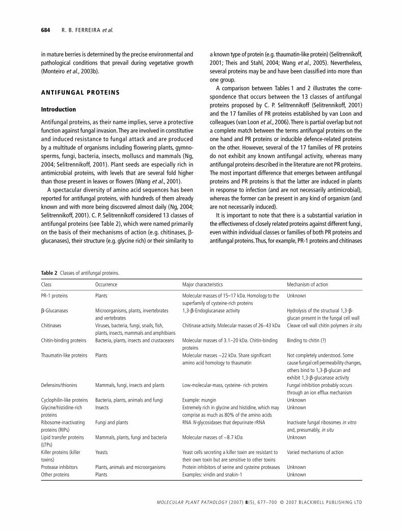
A spectacular diversity of amino acid sequences has beenreported for antifungal proteins, with hundreds of them alreadyknown and with more being discovered almost daily (Ng, 2004;Selitrennikoff, 2001). C. P. Selitrennikoff considered 13 classes ofantifungal proteins (see Table 2), which were named primarilyon the basis of their mechanisms of action (e.g. chitinases, β-glucanases), their structure (e.g. glycine rich) or their similarity to
a known type of protein (e.g. thaumatin-like protein) (Selitrennikoff,2001; Theis and Stahl, 2004; Wang et al., 2005). Nevertheless,several proteins may be and have been classified into more thanone group.
A comparison between Tables 1 and 2 illustrates the corre-spondence that occurs between the 13 classes of antifungalproteins proposed by C. P. Selitrennikoff (Selitrennikoff, 2001)and the 17 families of PR proteins established by van Loon andcolleagues (van Loon et al., 2006). There is partial overlap but nota complete match between the terms antifungal proteins on theone hand and PR proteins or inducible defence-related proteinson the other. However, several of the 17 families of PR proteinsdo not exhibit any known antifungal activity, whereas manyantifungal proteins described in the literature are not PR proteins.The most important difference that emerges between antifungalproteins and PR proteins is that the latter are induced in plantsin response to infection (and are not necessarily antimicrobial),whereas the former can be present in any kind of organism (andare not necessarily induced).
It is important to note that there is a substantial variation inthe effectiveness of closely related proteins against different fungi,even within individual classes or families of both PR proteins andantifungal proteins. Thus, for example, PR-1 proteins and chitinases
Table 2 Classes of antifungal proteins.
Class Occurrence Major characteristics Mechanism of action
PR-1 proteins Plants Molecular masses of 15–17 kDa. Homology to the superfamily of cysteine-rich proteins
Unknown
β-Glucanases Microorganisms, plants, invertebrates and vertebrates
1,3-β-Endoglucanase activity Hydrolysis of the structural 1,3-β-glucan present in the fungal cell wall
Chitinases Viruses, bacteria, fungi, snails, fish, plants, insects, mammals and amphibians
Chitinase activity. Molecular masses of 26–43 kDa Cleave cell wall chitin polymers in situ
Chitin-binding proteins Bacteria, plants, insects and crustaceans Molecular masses of 3.1–20 kDa. Chitin-binding proteins
Binding to chitin (?)
Thaumatin-like proteins Plants Molecular masses ~22 kDa. Share significant amino acid homology to thaumatin
Not completely understood. Some cause fungal cell permeability changes, others bind to 1,3-β-glucan and exhibit 1,3-β-glucanase activity
Defensins/thionins Mammals, fungi, insects and plants Low-molecular-mass, cysteine- rich proteins Fungal inhibition probably occurs through an ion efflux mechanism
Cyclophilin-like proteins Bacteria, plants, animals and fungi Example: mungin UnknownGlycine/histidine-rich proteins
Insects Extremely rich in glycine and histidine, which may comprise as much as 80% of the amino acids
Unknown
Ribosome-inactivating proteins (RIPs)
Fungi and plants RNA N-glycosidases that depurinate rRNA Inactivate fungal ribosomes in vitro and, presumably, in situ
Lipid transfer proteins (LTPs)
Mammals, plants, fungi and bacteria Molecular masses of ~8.7 kDa Unknown
Killer proteins (killer toxins)
Yeasts Yeast cells secreting a killer toxin are resistant to their own toxin but are sensitive to other toxins
Varied mechanisms of action
Protease inhibitors Plants, animals and microorganisms Protein inhibitors of serine and cysteine proteases UnknownOther proteins Plants Examples: viridin and snakin-1 Unknown

Plant defence proteins in fungal pathogenesis 685
© 2007 BLACKWELL PUBL ISH ING LTD MOLECULAR PLANT PATHOLOGY (2007) 8 (5 ) , 677–700
have been described which are totally devoid of antifungal activity.Moreover, more than a 100-fold difference in anti-oomycete activityhas been reported for various PR-1 proteins.
The target structures of the antifungal proteins range fromthe outermost part of the fungal cell, the cell wall, to the plasmamembrane and finally to several intracellular targets (Theis andStahl, 2004). Therefore, these proteins exhibit a very wide diversityof action mechanisms, including, for example, inhibition of thesynthesis of the fungal cell wall or disruption of its structureand/or function, membrane channel and pore formation, damageto cellular ribosomes, inhibition of DNA synthesis and inhibitionof the cell cycle. Nevertheless, the mode of action of most of theseproteins remains to be elucidated (Ng, 2004; Selitrennikoff, 2001).
PR-1 proteins
The PR-1 family is often the most abundant group of proteins andis induced to very high levels upon infection, reaching up to 1–2%of the total leaf protein (Jayaraj et al., 2004). This family is stronglyconserved and has been detected in every plant species examinedto date. Homologues have been encountered in fungi, insects andvertebrates, including humans (van Loon and van Strien, 1999).
Although the biological function of PR-1 proteins has not yetbeen established and their mechanism of action is not understood,plant PR-1 proteins exhibit antifungal activity both in vitro and inplanta (e.g. in transgenic plants over-expressing tobacco PR-1)(Niderman et al., 1995; Tahiri-Alaoui et al., 1993). An associationbetween PR-1 proteins and enhanced resistance against oomyceteshas been suggested. However, not enough data have been reportedin the available literature to rule out a direct role of PR-1against non-oomycete pathogens (van Loon et al., 2006).Notably, the prominent PR-1 proteins are often used as markersof the enhanced defensive state conferred by pathogen-inducedSAR (van Loon et al., 2006).
PR-1 proteins may be divided into two groups, one being acidicand the other basic (sequence similarity between the two groupsis about 65%) (Jayaraj et al., 2004). In tobacco, at least 16 PR-1-type genes were detected (Cornelissen et al., 1987). Three acidicproteins (1a, 1b and 1c) and one basic (1g) protein were foundto be induced upon tobacco mosaic virus infection (van Loon et al.,1994). The fully sequenced genomes of Arabidopsis and riceinclude 22 and 39 PR-1-type genes, respectively (van Loon et al.,2006). In Arabidopsis, a single PR-1 gene is activated uponinfection, insect attack or chemical treatment, but ten and eightdifferent PR-1-type genes are constitutively expressed in rootsand pollen, respectively (van Loon et al., 2006).
ββββ-Glucanases
Laminarinases (β-1,3-endoglucanases; EC 3.2.1.6) are presentin a wide variety of plants (including vegetative parts and seeds),
animals (vertebrates and invertebrates) and microorganisms(Jwanny et al., 2001).
Plant β-1,3-glucanases are referred to as PR-2 proteins andare subdivided into three classes. Class I glucanases are basicproteins of about 33 kDa and are localized in the plant vacuole(Bulcke et al., 1989). Classes II and III include acidic, extracellularproteins of about 36 kDa (Theis and Stahl, 2004). They participatein several physiological and developmental plant processes. Inaddition, class I β-1,3-glucanases exhibit antifungal activity bothin vitro and in planta, using transgenic plants over-expressinga PR-2 protein (Joshi et al., 1998; Mauch et al., 1988). Class IIβ-1,3-glucanases exhibit in vitro antifungal activity only if appliedin combination with chitinases or class I β-1,3-glucanases (Theisand Stahl, 2004).
Chitinases
Chitinases (EC 3.2.1.14) constitute the second largest group ofantifungal proteins. They catalyse the hydrolytic cleavage of theβ-1,4-glycoside bond present in biopolymers of N-acetyl-D-glucosamine, mainly in chitin. Chitinases can be grouped in twocategories: exochitinases, acting on non-reducing ends of thechitin chain, and endochitinases, which hydrolyse internal bonds(Kasprzewska, 2003). In general, these enzymes catalyse chitindegradation, acting most often as endochitinases and producingchito-oligosaccharides of 2–6 N-acetyl-D-glucosamine residues inlength (Stintzi et al., 1993).
Chitinases are classified in families 18 and 19 of the 57 familiesin which O-glycoside hydrolases are presently subdivided(Henrissat and Bairoch, 1996). Higher plants synthesize sevendifferent classes of chitinases which differ in protein structure,substrate specificity, mechanism of catalysis and sensitivity toinhibitors (Brunner et al., 1998). For example, unlike classe IIchitinases, class I chitinases contain a chitin-binding, hevein-likedomain identical to that of chitin binding proteins (Theis andStahl, 2004). These classes are grouped into three families of PRproteins (Neuhaus et al., 1996; Table 1): chitinases of classes Ia,Ib, II, IV, VI and VII belong to the PR-3 family, whereas those ofclasses III and V are included in the PR-8 and PR-11 families,respectively. Additionally, some proteins with low endochitinaseactivity occur in the PR-4 family (chitin-binding proteins) (Melcherset al., 1994). Acidic chitinases belonging to classes Ib, II, III, IVand VI are secreted to the apoplast, whereas basic chitinasesincluded in classes Ia, III and VI are located in vacuoles (Arieet al., 2000).
Chitinases have been found in a very wide range of organisms,containing or not containing chitin, such as viruses, bacteria, fungi,plants (gymnosperms and angiosperms) and animals (insects, snails,fish, amphibians and mammals) (Goormachtig et al., 1998). Forexample, a 30.8-kDa chitinase with antifungal activity has beenisolated from mung bean (Phaseolus mungo) seeds (Wang et al.,

686 R. B. FERREIRA et al.
MOLECULAR PLANT PATHOLOGY (2007) 8 (5 ) , 677–700 © 2007 BLACKWELL PUBL ISH ING LTD
2005), whereas two 28-kDa chitinases designated chitinase Aand chitinase B also exhibiting antifungal activity were characterizedin maize (Zea mays) seeds (Huynh et al., 1992). Interestingly, somechitinases such as dolichin (28 kDa; Graham and Sticklen, 1994;Ye et al., 2000a), delandin (28 kDa; Ye and Ng, 2002a) andpananotin (35 kDa; Lam and Ng, 2002), present in field bean(Dolichos lablab) and ricebean (Delandia umbellata) seeds andsanchi ginseng (Panax notoginseng) roots, respectively, exhibitantifungal activity and also cell-free translating inhibitingactivity and inhibitory activity against HIV-1 reverse transcriptase(Ng, 2004).
Chitinases, as with many other PR proteins, may be synthesizedin both a constitutive and an inducible manner. Some chitinaseforms, both apoplastic and vacuolar, are synthesized constitutivelyin healthy plants in a developmentally and tissue- and organ-specific mode. In addition, chitinases, again as with many otherPR proteins, are also up-regulated by biotic and abiotic stresses,such as fungal challenge, wounding, drought, cold, ozone, heavymetals, excessive salinity and UV-light, and treatment withphytohormones such as ethylene, jasmonic acid and salicylic acid(Kasprzewska, 2003).
Chitinases are apparently involved in numerous physiologicalprocesses, including development and growth. In those organismsthat contain chitin, they are presumably required for morphogenesisof cell walls and exoskeleton (Gooday, 1971). For instance, inyeast and various fungi they take part in remodelling cell wallstructure and daughter cell separation (Cohen-Kupiec and Chet,1998; Patil et al., 2000; Shimono et al., 2002). A role in nutritionis fulfilled in some bacteria species that secrete chitinases andare able to grow on chitin as their only carbon source (Wang andChang, 1997; Wang et al., 2002; Watanabe et al., 1999). Even inplants, chitinases have been reported to play a role in growth anddevelopment, during the nodulation process or in programmedcell death (Collinge et al., 1993; Cullimore et al., 2001; De Jonget al., 1992; Goormachtig et al., 1998; Helleboid et al., 2000; vanHengel et al., 1998; van der Holst et al., 2001; Passarinho et al.,2001; Regalado et al., 2000). A chitinase from Musa spp.behaves as a fruit-specific vegetative storage protein (Peumanset al., 2002), whereas chitinases from monocotyledonous plantshave been reported to display antifreeze activity, suggesting arole in plant frost resistance (Yeh et al., 2000). Chitinasesexhibiting aspartic protease inhibitor activity (in Solanum tuberosumtubers; Guevara et al., 1999) or α-amylase inhibitor activity (Coixlochrymajobi; Ary et al., 1989) have also been reported.
Plant chitinases that hydrolyse chitin inhibit the growth of fungiand generate chitin oligosaccharides that act as elicitors. Inaddition, many chitinases are induced by pathogen attack andsome isoforms exhibit in vitro antifungal properties. For thesereasons, chitinases are believed to play a major role in planthost defence against pathogens. Nevertheless, their precise rolein plant disease resistance has been difficult to establish in non-
transgenic plants because chitinases are often present in bothresistant and susceptible tissues and their expression is triggeredby many inducers other than pathogen attack (Punja and Zhang, 1993).Based on early work on the characterization of plant chitinasesmade by Thomas Boller and Fred Meins (Neuhaus et al., 1991),Collinge et al. (1993) and Kasprzewska (2003) suggested thatchitinases fulfil a double function in the protection againstfungal colonization. Apparently, apoplastic chitinases function inthe early stages of pathogenesis in the signalling process thatinforms plants about the attack. Indeed, partial digestion ofchitin releases oligosaccharides that are perceived by the plantcells as elicitors which, in turn, switch on the active plant defencemechanisms. During the subsequent phases of pathogenesis thatfollow fungal penetration, vacuolar chitinases released by hyphae-induced, protoplast burst directly repress fungal growth by degradingthe newly synthesized chitin chains. This hypothesis is supportedby the substrate specificity of cell wall and vacuolar enzymes(Collinge et al., 1993).
It should be noted, however, that many chitinases do not showany antifungal activity in vitro. For example, of the two chitinasespresent in chickpea (Cicer arietinum) cell-suspension cultures, onlythe basic form possessed antifungal activity (Vogelsang and Barz,1993). In addition, bacterial family 18 chitinases do not haveantifungal activity (Theis and Stahl, 2004).
The antifungal activity displayed by many chitinases was initiallyassumed to derive from their ability to digest chitin, leading to aweakened fungal cell wall and subsequent cell lysis. However,recent evidence indicates that the mechanisms by which chitinasesinhibit fungal growth seem to be more dependent on thepresence of a chitin-binding domain than on chitinolytic activity.Thus, the antifungal activity of a tobacco class I chitinase is threetimes higher when a chitin-binding domain is present (Iseli et al.,1993), whereas a mutant class I chitinase from chestnut (Castaneasativa) seeds displaying no chitinolytic activity exhibits as muchantifungal activity as the wild-type chitinase (Garcia-Casado et al.,1998). By contrast, a mutant class II chitinase from barley show-ing no chitinolytic activity possesses only 15% of the antifungalactivity displayed by the wild-type chitinase (Andersen et al., 1997).Also, a class I chitinase from rye (Secale cereale) contains a chitin-binding domain devoid of antifungal activity and a catalyticdomain capable of inhibiting fungal growth (Taira et al., 2001).
Chitin and β-1,3-glucan are synthesized simultaneously in theapex of growing hyphae of filamentous fungi (Theis and Stahl,2004). Therefore, it is not surprising the number of studies thathave reported a synergistic action between chitinases and β-1,3-glucanases in the hydrolysis of mixed chitin-glucan fibres, both invitro and in vivo (Jach et al., 1995; Stintzi et al., 1993). In fact, thedouble function proposed by Kasprzewska (2003) for the antifungalactivity of chitinases may well be extended to β-1,3-glucanases,with an indirect antifungal activity resulting from partial digestionof chitin and glucans and the corresponding release of elicitors,

Plant defence proteins in fungal pathogenesis 687
© 2007 BLACKWELL PUBL ISH ING LTD MOLECULAR PLANT PATHOLOGY (2007) 8 (5 ) , 677–700
and a direct antifungal activity derived from digestion of mixedchitin–glucan fibres and the resulting weakening of fungal cellwalls (Ryan and Farmer, 1991).
Chitin-binding proteins
Plant chitin-binding proteins have been classified as PR-4 proteins(Theis and Stahl, 2004; Tables 1 and 2) and are usually subdividedinto two classes: class I PR-4 proteins contain a chitin-bindingdomain similar to a domain present in hevein (a protein fromrubber latex; Parijs et al., 1991) and belong to the superfamily ofchitin-binding lectins; class II PR-4 proteins lack the chitin-binding hevein domain (Selitrennikoff, 2001; Theis and Stahl,2004). A chitin-binding protein, inducible by ethylene, has beenpurified from the leaves of guilder rose (Hydrangea macrophyla)(Yang and Gong, 2002).
The antifungal activity of chitin-binding proteins is mainly dueto their ability to bind fungal cell wall chitin, which results indisruption of cell polarity and consequent inhibition of growth bymechanisms that have not been elucidated (Bormann et al., 1999).It is possible that the antifungal activity of at least some chitin-binding proteins is just a side-effect (Theis and Stahl, 2004).
Chitin-binding proteins that exhibit antifungal activity but thatare not PR-4 proteins have been isolated from a number of sourcesincluding bacteria, plants, insects and crustaceans (Selitrennikoff,2001). In addition, chitin-binding peptides (hevein- and knottin-type, 36–40 residues in length) have been found in several plantseeds (Punja, 2001).
One particular case of chitin-binding proteins that deservesspecial mention are the vicilins. Indeed, a considerable number ofreports indicate that vicilins may be considered as class II chitin-binding proteins, but not class II PR-4 proteins.
The most abundant proteins in legume seeds are the globulins,which comprise the legumins (11S) and the vicilins (7S) andusually account for approximately 80% of the total protein intheir mature seeds (Derbyshire et al., 1976). Therefore, vicilinsare seed storage proteins of the 7S globulin family, which arepresent in the seeds of leguminous and other plants (Casey et al.,1986; Derbyshire et al., 1976). They are oligomeric proteins (150–170 kDa) with variable degrees of glycosylation, composed ofthree similar subunits of ~40–70 kDa, with no disulphide linkagesand stabilized by non-covalent forces (Casey et al., 1986; Shutovet al., 1995). The combination of multiple structural genes andextensive post-translation processing (proteolysis and glycosyla-tion) results in a high degree of subunit polymorphism for theseproteins (Higgins, 1984). Nevertheless, vicilins from differentlegume seeds exhibit a considerable amount of sequence homologyand have similar three-dimensional protein structures (Argos et al.,1985; Ko et al., 1993; Lawrence et al., 1990, 1994).
Vicilins isolated from the seeds of the legumes cowpea (Vignaunguiculata), adzuki bean (Vigna angularis), jack bean (Canavalia
ensiformis), soybean (Glycine max), common bean (Phaseolusvulgaris) and lima bean (Phaseolus lunatus) were shown to beimmunologically related and to bind strongly to chitin, chitosanand fully acetylated chitin (Firmino et al., 1996; Gomes et al.,1998a; Sales et al., 1996). Association of vicilin to chitin has beenshown to be dependent on tryptophan residues in the molecule(Miranda et al., 1998).
Vicilins from different legume seeds have detrimental effects ondevelopment of the cowpea weevil (Callosobruchus maculatus),a bruchid insect which is a pest of cowpea seeds (Macedo et al.,1993; Yunes et al., 1998). Although the mechanism of actionof vicilins upon bruchids is not yet completely understood, theseeffects have been attributed to the binding of vicilins to thechitinous structures present in the mid-gut of insects (Firminoet al., 1996; Sales et al., 2001). Interestingly, vicilins from all non-host seeds, including those of the C. maculatus-resistant cowpealine, strongly inhibit larval development. However, vicilins fromC. maculatus-susceptible cowpea line and adzuki bean seeds arethe exception.
In addition, vicilins from V. unguiculata and other legume seedsinterfere with the germination of spores or conidia of phytopath-ogenic fungi and bind to fungal structures, possibly chitin-containing structures of the cell wall (Gomes et al., 1997). Forexample, V. unguiculata vicilins affect growth and inhibit sporegermination of the pathogens Fusarium solani, Fusarium oxysporum,Collectotricum musae, Phytophthora caprici, Neurospora crassaand Ustilago maydissporidia, bind to chitin-like structures ofSaccharomyces cerevisiae and lead to abnormal development(sporulation) of yeast cells (Gomes et al., 1998a,b). Vicilin-relatedbasic proteins isolated from cotton (Gossypium hirsutum) seedshave also been shown to inhibit the growth of various filamentousfungi (Chung et al., 1997).
A common property of seed storage proteins is that they aresynthesized in high levels in certain developmental stages andaccumulate in discrete vesicles called protein storage vacuoles.Therefore, they act as a reserve for surplus organic carbon, nitrogenand sulphur (Pernollet, 1978). Some storage proteins had alreadybeen reported to contribute to plant defence mechanisms(Shewry et al., 1995). Nevertheless, it was unexpected to find thevicilins as a group of defensive proteins. Vicilins may therefore beconsidered multifunctional proteins, functioning as a source ofamino acids for the plant during germination and subsequentgrowth and at the same time being toxic to fungi and insects(Macedo et al., 1993; Sales et al., 2000; Shutov et al., 1995).
Thaumatin-like proteins
Osmotin and thaumatin-like (TL) proteins are basic, 24-kDa proteinsbelonging to the PR-5 family and sharing significant sequencehomology to thaumatin, a sweet-tasting (to humans) proteinfrom the South African Ketemfe berry bush (Thaumatococcus

688 R. B. FERREIRA et al.
MOLECULAR PLANT PATHOLOGY (2007) 8 (5 ) , 677–700 © 2007 BLACKWELL PUBL ISH ING LTD
danielli) (van der Wel and Loeve, 1972). TL proteins have beendetected in a vast number of plants. For example, a 24-kDa TLprotein is abundantly expressed in grapevine fruits not only in aberry- and ripening-specific manner (Tattersall et al., 1997) butalso in response to Erysiphe necator infection (Monteiro et al.,2003a). TL proteins have been isolated from the intercellular fluidof lupin (Lupinus albus) leaf, stem and root tissues (Regalado andRicardo, 1996) and from the intercellular fluid of chickpea leaves(Hanselle et al., 2001). TL proteins are also produced in plantsunder different stress conditions (Zhu et al., 1995).
Osmotin and TL proteins induce fungal cell leakiness presumablythrough a specific interaction with the plasma membrane thatresults in the formation of transmembrane pores (Kitajimaand Sato, 1999; Roberts and Selitrennikoff, 1990). These proteinshave also been reported to possess β-1,3-glucanase activity(Grenier et al., 1999) or bind to actin (Takemoto et al., 1997).Tobacco osmotin stimulates a mitogen-activated protein kinase,subverting a signal transduction pathway to enhance fungal cellsusceptibility (Grenier et al., 1999; Yun et al., 1998). The proteinsexhibit antifungal activity in vitro (Liu et al., 1994; Melcherset al., 1993; Woloshuk et al., 1991) and show enhanced lyticactivity when tested in combination with chitinases and/or β-1,3-glucanases (Lorito et al., 1996). Furthermore, the simultaneouspresence of both osmotin and TL protein from grapevine displaysa synergistic antifungal effect (Monteiro et al., 2003a).
Defensins/thionins
Defensins and thionins are families of low-molecular-mass (about5 kDa), cysteine-rich peptides (45–54 amino acid residues inlength) that form a prominent group of membrane-acting proteinsfound in mammals, insects, plants and fungi (Theis and Stahl,2004). Plant defensins (PR-12 proteins) and thionins (PR-13proteins), present in both monocotyledonous and dicotyledonousplants, are toxic to fungi (Bohlmann, 1994; Broekaert et al., 1995;Evans and Greenland, 1998). Broekaert et al. (1997) suggestedthat these peptides play a role in protecting seeds from infectionby pathogens.
The mode of action of plant defensins has not yet beenproperly elucidated. These peptides induce a prompt potassiumefflux, calcium uptake, alkalinization of the medium and membranepotential changes in Neurospora crassa (Theis and Stahl, 2004).In contrast to other membrane-acting proteins, plant defensinsdo not form pores on artificial membranes (Thevissen et al., 1996).Indeed, selective calcium uptake through activated ion channels,but not membrane permeabilization, is thought to be a majorcomponent of plant defensin antifungal action (Theis and Stahl,2004). A specific, high-affinity binding site for a plant defensin onN. crassa has been found (Thevissen et al., 1997). The defensinDm-AMP1 from dalhia (Dalhia merckii ) has been shown tointeract specifically with a sphingolipid from Saccharomyces
cerevisiae. Yeast mutants lacking the sphingolipid are highlyresistant to the defensin (Thevissen et al., 2000).
Differential antifungal activity has been detected amongstructurally related plant defensins. MsDef1, a seed defensinfrom alfalfa (Medicago sativa), inhibits the growth of Fusariumgraminearium in vitro. However, MtDef2 from Medicago trunculata,which shares 65% amino acid sequence identity with MsDef1, lacksantifungal activity towards F. graminiarum (Spelbrink et al., 2004).
Cyclophilin-like proteins
Cyclophilins are a highly conserved group of proteins that functionas intracellular receptors for cyclosporin. Mungin, for example, isan 18-kDa protein present in mung bean which shows a signifi-cant homology to cyclophilins and inhibits α- and β-glycosidasesin vitro (Ye and Ng, 2000). Unguilin, another example, is an 18-kDacyclophilin-like protein isolated from the seeds of the black-eyedpea (Vigna unguiculata) that exhibits antimitogenic, antiviral andantifungal activities towards fungi, including Coprinus comatus,Mycosphaerella arachidicola and Botrytis cinerea.
Glycine/histidine-rich proteins
These are antifungal insect proteins whose mechanism of actionremains to be elucidated. Glycine and histidine may comprise upto 80% of their amino acid residues.
Ribosome-inactivating proteins
A prominent intracellular target for antifungal proteins is ribosomes.RIPs are a group of cytotoxic N-glycosidases that specificallycleave nucleotide N–C glycosidic bonds (Park et al., 2004b). Theyare enzymes with RNA N-glycosidase activity, which dependingon their specificity, can inactivate non-specific or foreign ribosomes,thereby shutting down protein synthesis (Punja, 2001). Regardlessof the activity type, ribosome damage (i.e. depurination) occursat the sarcin/ricin loop, a highly conserved sequence of the 28SrRNA gene (Endo and Wool, 1982). Some RIPs consist of a singlepolypeptide chain (type I), whereas others are dimeric proteinswith one catalytic polypeptide chain and another responsible fortranslocation into cells via recognition of protein receptors(type II). In type III RIPs, both domains are contained in a singlepolypeptide (Peumans et al., 2001; Stirpe et al., 1992).
Plant RIPs inhibit mammalian, plant, fungal and bacterialprotein syntheses, either in vivo or in vitro (Iglesias et al., 1993). Howplants protect themselves from their own RIPs has been underinvestigation. For example, type I RIP from the endosperm of cerealgrains does not act on plant ribosomes but affects foreign ribosomessuch as those of fungi (Hartley et al., 1996; Stirpe et al., 1992).
Although RIPs were first identified more than 100 years ago,their biological function(s) still remains open to speculation (Park

Plant defence proteins in fungal pathogenesis 689
© 2007 BLACKWELL PUBL ISH ING LTD MOLECULAR PLANT PATHOLOGY (2007) 8 (5 ) , 677–700
et al., 2004b). Several independent studies suggest that theirantimicrobial activity and their inhibitory mechanism against HIVreplication are separate from their host-ribosome-inactivatingactivity. It has also been reported that their ribosome-inactivatingactivity does not account for their cytotoxicity (Park et al., 2002b).Increasing evidence suggests that RIPs can target non-ribosomalsubstrates. Thus, for example, RIPs may regulate protein expressionby targeting mRNA instead of ribosomes (Park et al., 2004b).
Examples of plant RIPs are ricin from castor bean (Ricinuscommunis) (Endo et al., 1987), ME1+2 from Mirabilis expansa(Vivanco et al., 1999), RIP1 from maize (Nielsen et al., 2001), PAP-Hfrom pokeweed (Phytolacca americana) (Park et al., 2002a),α- and β-pisavins from pea (Pisum sativum) seeds (Lam et al.,1998) and ebulin1, a type II RIP from Sambucus ebulus (Girbeset al., 1993). Hispin is a 21-kDa RIP from hairy melon seeds (Ngand Parkash, 2002), whereas luffacylin is an arginine- andglutamate-rich RIP from loofah (Luffa cylindrical) (Parkash et al.,2002). A 30-kDa RIP was isolated from dehursked barley grains(Roberts and Selitrennikoff, 1986).
Lipid transfer proteins
Plant lipid transfer proteins (LTPs; PR-14) are small, basic proteins,stabilized by four disulphide bonds, which transfer phospholipidsbetween membranes. LTPs contain typically an internal, tunnel-like hydrophobic cavity that runs through the molecule (Chenget al., 2004; Selitrennikoff, 2001). The mechanism responsible fortheir antifungal activity remains unknown, although it wassuggested that these proteins insert themselves into the fungalcell membrane with their central hydrophobic cavity forminga pore, allowing efflux of intracellular ions and leading to fungalcell death (Selitrennikoff, 2001). Unlike the non-specific LTPs(nsLTPs) from raddish (Raphanus sativus) and maize seeds,Ace-AMP1, a 10-kDa LTP from onion (Allium cepa) seeds, isincapable of phospholipid transfer from liposomes to mitochondria(Ng, 2004). Expression of Ace-AMP1 in transgenic wheat has beenshown to enhance antifungal activity and defence responses (Roy-Barman et al., 2006). A putative LTP from Arabidopsis has beenimplicated in the transport of the systemic signal in SAR, leadingto enhanced resistance to subsequent attack by a broad rangeof normally virulent pathogens (Maldonado et al., 2002). OtherLTPs have recently been characterized in the seeds of cowpea andmotherwort (Leonurus japonicus) (Carvalho et al., 2006; Yanget al., 2006).
Killer proteins
Killer proteins or killer toxins are secreted by several yeasts andbind to specific surface receptors in sensitive fungal cells. Theyare subsequently internalized and can disrupt cell wall synthesis,DNA synthesis and K+ channel activity, inhibit β-1,3-glucan
synthesis and arrest the cell cycle, leading to inhibition of fungalgrowth and fungal cell death (Ahmed et al., 1999; Eisfeld et al.,2000; Kimura et al., 1997, 1999; Suzuki and Shimma, 1999). AKluyveromyces yeast killer toxin has been shown to sharehomology with chitinases from plant, yeast and bacterial sources(Bradshaw, 1990; Kuranda and Robbins, 1991; Stark et al., 1984).Perhaps, the secreted Saccharomyces chitinase exhibiting homologywith the killer toxin plays a dual role by selectively modifying theyeast cell wall and also suppressing the growth of other micro-organisms (Kuranda and Robbins, 1991).
Protease inhibitors
One of the major classes of proteins present in some plant tissues,with correspondence to the PR-6 family, include inhibitors ofmetal, aspartic, serine and cysteine proteases (Kassel, 1970).Inhibitors of serine proteases, such as trypsin and chymotrypsin,are sometimes considered bifunctional proteins because theyalso inhibit other enzymes such as α-amylase and fungal growth(Selitrennikoff, 2001). A 7.5-kDa antifungal, Bowman–Birk-typetrypsin–chymotrypsin inhibitor was isolated from broad bean(Vicia faba) seeds (Ye et al., 2001a), whereas an antifungal,sporamin-type trypsin inhibitor was purified from wampee (Clausenalansium) seeds (Ng et al., 2003). Plant antifungal cysteine proteaseinhibitors, termed phytocystatins, have been isolated from manyplants (Joshi et al., 1998; Park et al., 2000; Soares-Costa et al.,2002).
Other antifungal proteins
As new antifungal proteins continue to be discovered, an increas-ing number of them do not fall clearly into any of the previous12 classes. Among them are well-known proteins such as lectins,ribonucleases, deoxyribonucleases and peroxidases. Selectedexamples are viridin, present in the culture medium of Trichodermaviride, and snakin-1 isolated from potato (Solanum tuberosum).
Lectins with antifungal activity have been reported in the redkidney bean (Phaseolus vulgaris) (Ye et al., 2001b), stingingnettle (Urtica dioica) (Broekaert et al., 1989), potato tuber(Gozia et al., 1995) and slender amaranth (Amaranthus viridis)(Kaur et al., 2006). A role in plant defence against fungi wasproposed for the cotyledonary Lutzelburgia auriculata agglutinin(LAA). This lectin is located in the periphery of the cotyledon andis released during germination into the surrounding medium.Inclusion of LAA in the culture medium inhibits growth ofColletrotichum lindemuthianum, Fusarium solani, Aspergillus nigerand Saccharomyces cerevisiae. LAA was found to bind reversiblyto yeast cells (Melo et al., 2005).
Quinqueginsin and panaxagin are two antifungal proteinsexhibiting ribonuclease activity that have been purified from theroots of the American ginseng (Panax quinquefolium) and the

690 R. B. FERREIRA et al.
MOLECULAR PLANT PATHOLOGY (2007) 8 (5 ) , 677–700 © 2007 BLACKWELL PUBL ISH ING LTD
Chinese ginseng (Panax ginseng), respectively. Quinqueginsinpossesses specific ribonucleolytic activity towards poly C (Ng andWang, 2001; Wang and Ng, 2000b). In addition, a deoxyribonu-clease with antifungal activity was extracted from asparagus(Asparagus officinalis) seeds (Wang and Ng, 2001), whereas anantifungal peroxidase was reported in french bean (Phaseolusvulgaris) legumes (Ye and Ng, 2002b).
The storage 2S albumin proteins have also been reported todisplay antifungal activity in passion fruit (Passiflora edulis),radish (Raphanus sativus) and oilseed rape (Brassica napus)(Agizzio et al., 2003; Pelegrini et al., 2006; Terras et al., 1993). Inaddition, the puroindolines, endosperm-specific proteins involvedin wheat seed hardness or texture (Morris, 2002), have beenshown to play a role in plant defence (Giroux et al., 2003). Indeed,rice plants, which normally lack puroindolines, were transformedto express constitutively puroindoline genes. The transgenic riceshowed significantly increased tolerance to Magnaporthe grisea,with a 29–54% reduction on symptoms, and to Rhizoctonia solani,with an 11–22% reduction in symptoms (Krishnamurthy et al., 2001).
Ginkbilobin is a 13-kDa antifungal protein from maidenhairtree (Ginkgo biloba) seeds with sequence similarity to embryo-abundant protein (Wang and Ng, 2000a). Hypogin is an allergen-like, antifungal protein from peanut (Arachis hypogaea) withsequence similarity to peanut allergen (Ye and Ng, 2000).Pisumin, an antifungal protein with a novel N-terminal sequence,and sativin, a miraculin-like antifungal protein, have beencharacterized in legumes of the sugar snap pea (Pisum sativumvar. macrocarpon) (Ye et al., 2000b).
Germin-like oxalate oxidases (proposed PR-15 and PR-16families) are stable glycoproteins that were initially discovered incereals. They are present during seed germination and are inducedin response to fungal infection (Dumas et al., 1995; Zhang et al.,1995). The hydrogen peroxide produced as a result of theiractivity upon the substrate oxalic acid induces plant defenceresponses and enhances cell-wall strengthening (Brisson et al.,1994; Mehdy, 1994).
Antifungal peptides
Although there is not a generalized consensus concerning wherea peptide ends and a protein begins, it may be arbitrarily assumedthat proteins possess molecular masses greater than 5 kDa orabout 50 amino acid residues in length. Therefore, some groupsof antifungal compounds, such as plant defensins, include bothproteins and peptides.
An enormous variety of peptides with antifungal activity areproduced by mammals (e.g. defensis, protegrins, gallinacins,tritrpticin, lactoferricin, and BPI protein domain III analogues),insects (e.g. cecropins, drosomycin, antifungal peptide, holotricin3 and thanatin), amphibians (e.g. magainins and dermaseptin),bacteria and fungi (e.g. iturins, syringomycins and related
peptides, nikkomycins, polyoxins, echinocandins and echinocandinanalogues, pneumocandins and pneumocandin analogues,aculeacins, mulundocandins, aureobasidins, Bacillus licheniformispeptides, schizotrin A, cepacidines, leucinostatin-trichopolyn group,and helioferins) and plants (e.g. plant defensins, lipid transfer proteins,zeamatin and cyclopeptides) (De Lucca and Walsh, 1999).
Antifungal peptides are classified according to their mode ofaction. One large group, including many amphipathic lyticpeptides, act by cell lysis, which operate via a number of differentmechanisms (Shai, 1995). Another group interferes with cell wallsynthesis or with the biosynthesis of essential cell components,such as glucan or chitin (Debono and Gordee, 1994).
Antifungal peptides are continuously being discovered in plants.Thus, for example, cicerin and arietin were detected in the seedsof chickpea (Ye et al., 2002). The 8-kDa angularin was purifiedfrom red beans (Ye and Ng, 2002c). A 30-amino acid residuepeptide containing six cysteine and seven glycine residues andexhibiting sequence homology to the chitin-binding domain ofchitin-binding chitinases and a higher affinity to chitin thanchitin-binding chitinases has been isolated from the intercellularwashing fluid from sugar beet (Beta vulgaris) leaves (Kristensenet al., 2001; Nielsen et al., 1997). A 1.244-kDa chitin-bindingpeptide with an amino acid sequence and a cystein/glycine-richchitin-binding domain typical of many chitin-binding proteinswas characterized in Ginkgo biloba leaves (Huang et al., 2000).
Plant defensins that are not related to either mammalian orinsect defensins have been characterized in plant tissues. This isthe case, for example, of Ib-AMP3, a highly basic, icosapeptideproduced by garden balsam (Impatiens balsamina) (Tailor et al.,1997). Zeamatin is a 27-amino-acid residue peptide produced bymaize seeds. However, peptides from the zeamatin family arealso present in oat, sorghum and wheat seeds (De Lucca andWalsh, 1999). Examples of plant cyclopeptides, recently reviewedby Tan and Zhou (2006), are frangufoline, amphibine H, rugosaninesA and B, and nummularines B, K, R and S (Panday and Devi, 1990).
STRATEGIES EMPLOYED BY FUNGI TO AVOID PLANT DETECTION OR DEFENCE
Emerging evidence demonstrates that the molecular interac-tion between plant and pathogen is far more elaborate than thestraightforward production of attack molecules by the pathogenand the corresponding response of defensive molecules by thehost. Plant pathogens use several strategies to avoid detection bythe host plant or to escape the plant defence responses.
One of the strategies involves protection of fungal structuresfrom plant defence mechanisms. Thus, for example, the attack offungal cell walls by plant chitinases is an important plant defenceto fungal infection because it liberates elicitor-active chitinoligomers and weakens the fungal cell wall (van den Burg et al.,2003). The antifungal activity of most plant chitinases derives

Plant defence proteins in fungal pathogenesis 691
© 2007 BLACKWELL PUBL ISH ING LTD MOLECULAR PLANT PATHOLOGY (2007) 8 (5 ) , 677–700
from the presence of a non-catalytic, plant-specific chitin-bindingdomain (ChBD). The race-specific elicitor avr4, an extracellular avrprotein produced by the leaf mould fungus Cladosporium fulvumand detected by the tomato Cf-4 LRR-RLP protein, protects thefungus against degradation by tomato chitinases because it con-tains a novel type of ChBD (Jones and Takemoto, 2004). However,the binding site of avr4 is larger than that of a plant ChBD. Inother words, avr4 interacts only with chitotriose whereas a plantChBD also interacts with the monomer N-acetyl-glucosamine.Binding additional avr4 molecules to chitin occurs through posi-tive cooperative protein–protein interactions. These observationssuggest that avr4 shields fungal cell wall chitin from the actionof plant chitinases (van den Burg et al., 2004).
During invasive growth of biotrophic rust fungi, chitin is exclu-sively present in the cell walls of exterior infection structures, i.e.germ tubes and appressoria. Instead of this elicitor-activemolecule, the surface hyphae that grow within host leavescontain chitosan, a deacetylation product of chitin possiblygenerated by the enzymatic activity of a differentiation-inducedfungal-chitin deacetylase (Deising and Siegrist, 1995; El Gueddariet al., 2002). The observation that chitosan lacks elicitor activityled Schulze-Lefert and Panstruga (2003) to speculate that the‘wolf intrudes in sheep’s clothing’.
The tobacco pathogen Alternaria alternata synthesizes andsecretes the reactive oxygen quencher mannitol as a means ofsuppressing reactive oxygen-mediated plant defences (Jenningset al., 2002). Interestingly, the non-mannitol-containing hosttobacco plants respond by expressing a pathogen-inducedmannitol dehydrogenase that catabolizes mannitol of fungalorigin. Indeed, constitutive expression of a celery (Apium graveolens)mannitol dehydrogenase cDNA in tobacco plants conferredenhanced resistance to A. alternata, but not to the non-mannitol-producing fungal pathogen Cercospora nicotianae (Jenningset al., 2002).
Another strategy is associated with inhibition of elicitor-inducedplant defence responses. Oligomers of galacturonic acid releasedfrom plant cell walls during pathogenesis may function as plant-derived suppressors of defence responses. In fact, the simultaneousaddition of oligogalacturonides and the glycoproteogalactanelicitor isolated from germ tubes of the wheat stem rust fungusPuccinia graminis f. sp. tritici suppresses the elicitor-induceddisease resistance reactions in wheat leaves (Moerschbacheret al., 1999).
Yet another strategy concerns the suppression of plant defences.An increasing body of evidence suggests that infecting fungalpathogens may modulate host gene expression to their ownbenefit, either by suppressing inducible plant defence responsesin physical proximity to infection sites and/or by inducing specifichost genes required for infection. An example of fungal-inducedplant defence suppression is provided by the infection of barleycoleoptile cells by the grass powdery mildew fungus Blumeria
graminis f. sp. hordei. Barley cells penetrated by the fungus or inclose proximity to haustoria exhibit induced susceptibility tosubsequent attack by the non-host pathogen Erysiphe pisi or bya second challenge with B. graminis (Kunoh et al., 1985, 1991).These observations suggest that B. graminis suppresses defencegene activation of barley cells by an as yet unknown mechanismthat is effective against race-specific, race-non-specific and non-host resistance (Schulze-Lefert and Panstruga, 2003).
CONCLUDING REMARKS
The continuous increase in the human population and in publicconcern over the generalized use of chemical fungicides, associatedwith the increasing number of obsolete fungicides that derivefrom the development of fungal resistance, demand alternativeways for disease control. These may include the development ofnew, effective and environmentally friendly fungicides. However,emerging evidence suggests that during a plant–fungus interac-tion, the pathogen may take over selected aspects of plant geneexpression to its own benefit. Therefore, it is expected that thefungus may induce the expression of some components requiredfor the infection or development processes or repress componentsof the host defence system. In this context, a detailed under-standing of the molecular events that take place during a plant–pathogen interaction is a prerequisite for boosting the naturalinherent defences of plants, and to transfer defensive traits intothe genome of economically important crops.
REFERENCES
Agizzio, A.P., Carvalho, A.O., Ribeiro Sde, F., Machado, O.L.,Alves, E.W., Okorokov, L.A., Samarao, S.S., Bloch, C. Jr, Prates,M.V. and Gomes, V.M. (2003) A 2S albumin-homologous protein frompassion fruit seeds inhibits the fungal growth and acidification of themedium by Fusarium oxysporum. Arch. Biochem. Biophys. 416, 188–195.
Agrawal, A.A. and Karban, R. (1999) Why induced defenses may be.favored over constitutive strategies in plants. In: The Ecology andEvolution of Inducible Defenses (Tollrian, R. and Harvell, C.D., eds),pp. 45–61. Princeton, NJ: Princeton University Press.
Ahmed, A., Sesti, F., Ilan, N., Shih, T.M., Sturley, S.L. and Goldstein, S.A.(1999) A molecular target for viral killer toxin: TOK1 potassium channels.Cell, 99, 283–291.
Ahn, I.-P., Kim, S., Kang, S., Suh, S.-C. and Lee, Y.-H. (2005) Rice defensemechanisms against Cochliobolus miyabeanus and Magnaporthe griseaare distinct. Phytopathology, 95, 1248–1255.
Albersheim, P. and Valent, B.S. (1974) Host–pathogen interactions. VII.Plant pathogens secrete proteins which inhibit enzymes of the hostcapable of attacking the pathogen. Plant Physiol. 53, 684–687.
Alexander, D., Goodman, R.M., Gut-Rella, M., Glascock, C., Weymann, K.,Friedrich, L., Maddox, D., Ahl-Goy, P., Luntz, T., Ward, E. and Ryals, J.(1993) Increased tolerance to two oomycete pathogens in transgenictobacco expressing pathogenesis-related protein 1a. Proc. Natl Acad.Sci. USA, 90, 7327–7331.
692 R. B. FERREIRA et al.
MOLECULAR PLANT PATHOLOGY (2007) 8 (5 ) , 677–700 © 2007 BLACKWELL PUBL ISH ING LTD
Andersen, M.D., Jensen, A., Robertus, J.D., Leah, R. and Skriver, K.(1997) Heterologous expression and characterization of wild-type andmutant forms of a 26 kDa endochitinase from barley (Hordeum vulgareL.). Biochem. J. 322, 815–822.
Antoniw, J., Ooms, G., White, R.F. and Wullems, G.J. (1980a) Thepresence of pathogenesis-related proteins in callus of Xanthi-nc tobacco.Phytophathol. Z. 101, 179–184.
Antoniw, J.F., Ritter, C.E., Pierpoint, W.S. and van Loon, L.C. (1980b)Comparison of three pathogenesis-related proteins from plants of twocultivars of tobacco infected with TMV. J. Gen. Virol. 47, 79–87.
Argos, P., Narayana, S.V. and Nielsen, N.C. (1985) Structural similaritybetween legumin and vicilin storage proteins from legumes. EMBO J. 4,1111–1117.
Arie, M., Hikichi, K., Takahashi, K. and Esaka, M. (2000) Characteriza-tion of a basic chitinase which is secreted by cultured pumpkin cells.Physiologia Plantarum, 110, 232–239.
Ary, M.B., Richardson, M. and Shewry, P.R. (1989) Purification andcharacterization of an insect alpha-amylase inhibitor/endochitinase from seedsof Job’s Tears (Coix lachryma-jobi ). Biochim. Biophys. Acta, 999, 260–266.
Baker, C.J. and Orlandi, E.W. (1995) Active oxygen in plant pathogenesis.Annu. Rev. Phytopathol. 33, 299–322.
Baker, B., Zambryski, P., Staskawicz, B. and Dinesh-Kumar, S.P. (1997)Signaling in plant–microbe interactions. Science, 276, 726–733.
Barkai-Golan, R., Mirelman, D. and Sharon, N. (1978) Studies ongrowth inhibition by lectins of Penicillia and Aspergilli. Arch. Microbiol.116, 119–121.
Beckers, G.J. and Spoel, S.H. (2006) Fine-tuning plant defence signalling:salicylate versus jasmonate. Plant Biol. (Stuttg), 8, 1–10.
BeMiller, J.N. (1965) Chitin. In: Methods in Carbohydrate Chemistry,Vol. 5 (Whistler, R., ed.), pp. 103–106. New York: Academic Press.
Bernier, F. and Berna, A. (2001) Germins and germin-like proteins: plantdo-all proteins. But what do they do exactly? Plant Physiol. Biochem. 39,545–554.
Bestwick, C.S., Brown, I.R. and Mansfield, J.W. (1998) Localizedchanges in peroxidase activity accompany hydrogen peroxide generationduring the development of a nonhost hypersensitive reaction in Lettuce.Plant Physiol. 118, 1067–1078.
Blackwell, J. (1988) Physical methods for the determination of chitin.structure and conformation. Methods Enzymol. 161, 435–442.
Blilou, I., Ocampo, J.A. and Garcia-Garrido, J.M. (2000) Induction of Ltp(lipid transfer protein) and Pal (phenylalanine ammonia-lyase) geneexpression in rice roots colonized by the arbuscular mycorrhizal fungusGlomus mosseae. J. Exp. Bot. 51, 1969–1977.
Bohlmann, H. (1994) The role of thionins in plant protection. Crit. Rev.Plant Sci. 13, 1–16.
Boller, T., Gehri, A., Mauch, F. and Vögeli, U. (1983) Chitinase in beanleaves: induction by ethylene, purification, properties, and possible function.Planta, 157, 22–31.
Bonas, U. and Lahaye, T. (2002) Plant disease resistance triggered bypathogen-derived molecules: refined models of specific recognition.Curr. Opin. Microbiol. 5, 44–50.
Bormann, C., Baier, D., Horr, I., Raps, C., Berger, J., Jung, G. and Schwarz, H.(1999) Characterization of a novel, antifungal, chitin-binding proteinfrom Streptomyces tendae Tu901 that interferes with growth polarity.J. Bacteriol. 181, 7421–7429.
Bowyer, P., Clarke, B.R., Lunness, P., Daniels, M.J. and Osbourn, A.E.(1995) Host range of a plant pathogenic fungus determined by a saponindetoxifying enzyme. Science, 267, 371–374.
Bradshaw, H.D. Jr (1990) Killer toxins. Nature, 345, 299.Brennan, P.J., Griffin, P.F., Losel, D.M. and Tyrrell, D. (1974) The lipids
of fungi. Prog. Chem. Fats Other Lipids, 14, 49–89.Brisson, L.F., Tenhaken, R. and Lamb, C. (1994) Function of oxidative
cross-linking of cell wall structural proteins in plant disease resistance.Plant Cell, 6, 1703–1712.
Broekaert, W.F., Allen, A.K. and Peumans, W.J. (1987) Separation andpartial characterization of isolectins with different subunit compositionsfrom Datura stramonium seeds. FEBS Lett. 220, 116–120.
Broekaert, W.F., Cammue, B.P.A., De Bolle, M.F.C., Thevissen, K.,De Samblanx, G.W. and Osborn, R.W. (1997) Antimicrobial peptidesfrom plants. Crit. Rev. Plant Sci. 16, 297–323.
Broekaert, W.F., Marien, W., Terras, F.R., De Bolle, M.F., Proost, P.,Van Damme, J., Dillen, L., Claeys, M., Rees, S.B., Vanderleyden, J.and Cammue, B. (1992) Antimicrobial peptides from Amaranthuscaudatus seeds with sequence homology to the cysteine/glycine-richdomain of chitin-binding proteins. Biochemistry, 31, 4308–4314.
Broekaert, W.F., Parijs, J.V., Leyns, F., Joos, H. and Peumans, W.J. (1989)A chitin-binding lectin from stinging nettle rhizomes with antifungalproperties. Science, 245, 1100–1102.
Broekaert, W.F., Terras, F.R., Cammue, B.P. and Osborn, R.W. (1995)Plant defensins: novel antimicrobial peptides as components of the hostdefense system. Plant Physiol. 108, 1353–1358.
Brunner, F., Stintzi, A., Fritig, B. and Legrand, M. (1998) Substratespecificities of tobacco chitinases. Plant J. 14, 225–234.
Bufe, A., Spangfort, M., Kahlert, H., Schlaak, M. and Becker, W.-M.(1996) The major birch pollen allergen, Bet v, 1, shows ribonucleaseactivity. Planta, 199, 413–415.
Bulcke, M.V., Bauw, G., Castresana, C., Van Montagu, M. andVandekerckhove, J. (1989) Characterization of vacuolar and extracellularbeta (1,3)-glucanases of tobacco: Evidence for a strictly compartmentalizedplant defense system. Proc. Natl Acad. Sci. USA, 86, 2673–2677.
van den Burg, H.A., Spronk, C.A., Boeren, S., Kennedy, M.A., Vissers, J.P.,Vuister, G.W., de Wit, P.J. and Vervoort, J. (2004) Binding of the AVR4elicitor of Cladosporium fulvum to chitotriose units is facilitated bypositive allosteric protein–protein interactions: the chitin-binding site ofAVR4 represents a novel binding site on the folding scaffold sharedbetween the invertebrate and the plant chitin-binding domain. J. Biol.Chem. 279, 16786–16796.
van den Burg, H.A., Westerink, N., Francoijs, K.-J., Roth, R.,Woestenenk, E., Boeren, S., de Wit, P.J.G.M., Joosten, M.H.A.J. andVervoort, J. (2003) Natural disulfide bond-disrupted mutants of AVR4 ofthe tomato pathogen Cladosporium fulvum are sensitive to proteolysis,circumvent Cf-4-mediated resistance, but retain their chitin bindingability. J. Biol. Chem. 278, 27340–27346.
Cano-Delgado, A., Penfield, S., Smith, C., Catley, M. and Bevan, M.(2003) Reduced cellulose synthesis invokes lignification and defenseresponses in Arabidopsis thaliana. Plant J. 34, 351–362.
Carvalho, A.O., Souza-Filho, G.A., Ferreira, B.S., Branco, A.T., Araujo, I.S.,Fernandes, K.V., Retamal, C.A. and Gomes, V.M. (2006) Cloning andcharacterization of a cowpea seed lipid transfer protein cDNA: expres-sion analysis during seed development and under fungal and coldstresses in seedlings’ tissues. Plant. Physiol. Biochem. 44, 732–742.
Casey, R., Domoney, C. and Ellis, N. (1986) Legume storage proteins.Oxford Surv. Plant Mol. Cell Biol. 3, 1–95.
Chamnongpol, S., Willekens, H., Moeder, W., Langebartels, C.,Sandermann, H., Van Montagu, M., Inze, D. and Van Camp, W.(1998) Defense activation and enhanced pathogen tolerance induced by

Plant defence proteins in fungal pathogenesis 693
© 2007 BLACKWELL PUBL ISH ING LTD MOLECULAR PLANT PATHOLOGY (2007) 8 (5 ) , 677–700
H2O2 in transgenic tobacco. Proc. Natl Acad. Sci. USA, 95, 5818–5823.
Cheng, C.S., Samuel, D., Liu, Y.J., Shyu, J.C., Lai, S.M., Lin, K.F. andLyu, P.C. (2004) Binding mechanism of nonspecific lipid transfer proteinsand their role in plant defense. Biochemistry, 43, 13628–13636.
Chester, K.S. (1933) The problem of acquired physiological immunity inplants. Q. Rev. Biol. 8, 129–154.
Chrispeels, M.J. and Raikhel, N.V. (1991) Lectins, lectin genes, and theirrole in plant defense. Plant Cell, 3, 1–9.
Christensen, A., Ho Cho, B., Naesby, M., Gregersen, P., Brandt, J.,Madriz-Ordeñana, K., Collinge, D. and Thordal-Christensen, H.(2002) The molecular characterization of two barley proteins establishesthe novel PR-17 family of pathogenesis-related proteins. Mol. PlantPathol. 3, 135–144.
Chung, R.P.-T., Neumann, G.M. and Polya, G.M. (1997) Purification andcharacterization of basic proteins with in vitro antifungal activity fromseeds of cotton, Gossypium hirsutum. Plant Sci. 127, 1–16.
Cipollini, D., Purrington, C.B. and Bergelson, J. (2003) Costs of inducedresponses in plants. Basic Appl. Ecol. 4, 79–89.
Cohen-Kupiec, R. and Chet, I. (1998) The molecular biology of chitindigestion. Curr. Opin. Biotechnol. 9, 270–277.
Collinge, D.B., Kragh, K.M., Mikkelsen, J.D., Nielsen, K.K., Rasmussen, U.and Vad, K. (1993) Plant chitinases. Plant J. 3, 31–40.
Cornelissen, B.J., Horowitz, J., van Kan, J.A., Goldberg, R.B. andBol, J.F. (1987) Structure of tobacco genes encoding pathogenesis-related proteins from the PR-1 group. Nucleic Acids Res. 15, 6799–6811.
Coventry, H.S. and Dubery, I.A. (2001) Lipopolysaccharides from Burkholderiacepacia contribute to an enhanced defensive capacity and the inductionof pathogenesis-related proteins in Nicotiana tabacum. Physiol. Mol.Plant Pathol. 58, 149–158.
Cullimore, J.V., Ranjeva, R. and Bono, J.-J. (2001) Perception of lipo-chitooligosaccharidic Nod factors in legumes. Trends Plant Sci. 6, 24–30.
Dangl, J.L. and Jones, J.D.G. (2001) Plant pathogens and integrateddefence responses to infection. Nature, 411, 826–833.
Datta, K.N., Muthukrishnan, S. and Datta, S.K. (1999) Expression andfunction of PR-protein genes in transgenic plants. In: Pathogenesis-RelatedProteins in Plants (Datta, S.K. and Muthukrishnan, S., eds), pp. 261–277.New York: CRC Press.
De Jong, A.J., Cordewener, J., Lo Schiavo, F., Terzi, M., Vandekerckhove, J.,Van Kammen, A. and De Vries, S.C. (1992) A carrot somatic embryomutant is rescued by chitinase. Plant Cell, 4, 425–433.
De Lucca, A.J. and Walsh, T.J. (1999) Antifungal peptides: novel therapeuticcompounds against emerging pathogens. Antimicrob. Agents Chemother.43, 1–11.
Debono, M. and Gordee, R.S. (1994) Antibiotics that inhibit fungal cellwall development. Ann. Rev. Microbiol. 48, 471–497.
Deising, H. and Siegrist, J. (1995) Chitin deacetylase activity of therust Uromyces viciae fabae is controlled by fungal morphogenesis. FEMSMicrobiol. Lett. 127, 207–211.
Derbyshire, E., Wright, D.J. and Boulter, D. (1976) Legumin and vicilin,storage proteins of legume seeds. Phytochemistry, 15, 3–24.
Derckel, J.-P., Legendre, L., Audran, J.-C., Haye, B. and Lambert, B.(1996) Chitinases of the grapevine (Vitis vinefera L.): five isoformsinduced in leaves by salicylic acid are constitutively expressed in othertissues. Plant Sci. 119, 31–37.
Desiderio, A., Aracri, B., Leckie, F., Mattei, B., Salvi, G., Tigelaar, H.,Van Roekel, J.S., Baulcombe, D.C., Melchers, L.S., De Lorenzo, G.and Cervone, F. (1997) Polygalacturonase-inhibiting proteins (PGIPs)
with different specificities are expressed in Phaseolus vulgaris. Mol.Plant–Microbe Interact. 10, 852–860.
Dixon, R.A. and Harrison, M.J. (1990) Activation, structure, and organizationof genes involved in microbial defense in plants. Adv. Genet. 28, 165–234.
Dixon, R.A., Lamb, C.J., Masoud, S., Sewalt, V.J. and Paiva, N.L. (1996)Metabolic engineering: prospects for crop improvement through thegenetic manipulation of phenylpropanoid biosynthesis and defenseresponses—a review. Gene, 179, 61–71.
Donaldson, P., Anderson, T., Lane, B., Davidson, A. and Simmonds, D.(2001) Soybean plants expressing an active oligomeric oxalate oxidasefrom the wheat gf-2.8 (germin) gene are resistant to the oxalate-secretingpathogen Sclerotina sclerotiorum. Physiol. Mol. Plant Pathol. 59, 297–307.
Dumas, B., Freyssinet, G. and Pallett, K.E. (1995) Tissue-specificexpression of germin-like oxalate oxidase during development andfungal infection of barley seedlings. Plant Physiol. 107, 1091–1096.
Durrant, W. and Dong, X. (2004) Systemic acquired resistance. Ann. Rev.Phytopathol. 42, 185–209.
Eisfeld, K., Riffer, F., Mentges, J. and Schmitt, M.J. (2000) Endocytoticuptake and retrograde transport of a virally encoded killer toxin in yeast.Mol. Microbiol. 37, 926–940.
El Gueddari, N.E., Rauchhaus, U., Moerschbacher, B.M. and Deising, H.B.(2002) Developmentally regulated conversion of surface-exposed chitinto chitosan in cell walls of plant pathogenic fungi. New Phytologist, 156,103–112.
Endo, Y., Mitsui, K., Motizuki, M. and Tsurugi, K. (1987) The mechanismof action of ricin and related toxic lectins on eukaryotic ribosomes. Thesite and the characteristics of the modification in 28 S ribosomal RNAcaused by the toxins. J. Biol. Chem. 262, 5908–5912.
Endo, Y. and Wool, I.G. (1982) The site of action of alpha-sarcin on eukary-otic ribosomes. The sequence at the alpha-sarcin cleavage site in 28Sribosomal ribonucleic acid. J. Biol. Chem. 257, 9054–9060.
Epple, P., Apel, K. and Bohlmann, H. (1997) Overexpression of anendogenous thionin enhances resistance of Arabidopsis againstFusarium oxysporum. Plant Cell, 9, 509–520.
Evans, I.J. and Greenland, A.J. (1998) Transgenic approaches to diseaseprotection: applications of antifungal proteins. Pestic. Sci. 54, 353–359.
Ferreira, R., Monteiro, S., Freitas, R., Santos, C., Chen, Z., Batista, L.,Duarte, J., Borges, A. and Teixeira, A. (2006) Fungal pathogens: thebattle for plant infection. Crit Rev. Plant Sci. 25, 505–524.
Ferreira, R.B., Monteiro, S.S., Picarra-Pereira, M.A. and Teixeira, A.R.(2004) Engineering grapevine for increased resistance to fungal pathogenswithout compromising wine stability. Trends Biotechnol. 22, 168–173.
Ferreira, R.B., Picarra-Pereira, M.A., Monteiro, S., Loureiro, V.B. andTeixeira, A.R. (2001) The wine proteins. Trends Food Sci. Technol. 12,230–239.
Firmino, F., Fernandes, K., Sales, M., Gomes, V., Miranda, M., Domingues, S.and Xavier-Filho, J. (1996) Cowpea (Vigna unguiculata) vicilins associatewith putative chitinous structures in midgut and feces of the bruchid beetlesCallosobruchus maculatus and Zabrotes subfasciatus. Braz. J. Med. Biol.Res. 29, 749–756.
Fritig, B., Heitz, T. and Legrand, M. (1998) Antimicrobial proteins ininduced plant defense. Curr. Opin. Immunol. 10, 16–22.
Garcia-Casado, G., Collada, C., Allona, I., Casado, R., Pacios, L.F.,Aragoncillo, C. and Gomez, L. (1998) Site-directed mutagenesis ofactive site residues in a class I endochitinase from chestnut seeds.Glycobiology, 8, 1021–1028.

694 R. B. FERREIRA et al.
MOLECULAR PLANT PATHOLOGY (2007) 8 (5 ) , 677–700 © 2007 BLACKWELL PUBL ISH ING LTD
Garcia-Olmedo, F., Molina, A., Segura, A. and Moreno, M. (1995)The defensive role of nonspecific lipid-transfer proteins in plants. TrendsMicrobiol. 3, 72–74.
Girbes, T., Citores, L., Iglesias, R., Ferreras, J.M., Munoz, R., Rojo, M.A.,Arias, F.J., Garcia, J.R., Mendez, E. and Calonge, M. (1993) Ebulin 1,a nontoxic novel type 2 ribosome-inactivating protein from Sambucusebulus L. leaves. J. Biol. Chem. 268, 18195–18199.
Giroux, M.J., Sripo, T., Gerhardt, S. and Sherwood, J. (2003) Puroindo-lines: their role in grain hardness and plant defence. Biotechnol. Genet.Eng. Rev. 20, 277–290.
Gomes, V., Da Cunha, M., Miguens, F., Fernandes, K., Rose, T. andXavier-Filho, J. (1998a) Ultrastructure and immunolabelling of fungicells treated with Vigna unguiculata vicilins (7S storage proteins). PlantSci. 138, 81–89.
Gomes, V., Mosqueda, M.-I., Blanco-Labra, A., Sales, M., Fernandes, K.,Cordeiro, R. and Xavier-Filho, J. (1997) Vicilin storage proteins fromVigna unguiculata (legume) seeds inhibit fungal growth. J. Agric. FoodChem. 45, 4110–4115.
Gomes, V.M., Okorokov, L.A., Rose, T.L., Fernandes, K.V. andXavier-Filho, J. (1998b) Legume vicilins (7S storage globulins) inhibityeast growth and glucose stimulated acidification of the medium byyeast cells. Biochim. Biophys. Acta, 1379, 207–216.
Gomes, V.M., Oliveira, A.E.A. and Xavier-Filho, J. (1996) A chitinaseand b-1.3-glucanase isolated from seeds of cowpea (Vigna unguiculataL. Walp) inhibit the growth of fungi and insect pest of the seed. J. Sci.Food Agric. 72, 86–90.
Gomez-Gomez, L. and Boller, T. (2002) Flagellin perception: a paradigmfor innate immunity. Trends Plant Sci. 7, 251–256.
Gooday, G.W. (1971) An audioradiographic study of hyphal growth ofsome fungi. J. Gen. Microbiol. 67, 125–133.
Goormachtig, S., Lievens, S., Van de Velde, W., Van Montagu, M.and Holsters, M. (1998) Srchi13, a novel early nodulin from Sesbaniarostrata, is related to acidic class III chitinases. Plant Cell, 10, 905–915.
Gozia, O., Ciopraga, J., Bentia, T., Lungu, M., Zamfirescu, I., Tudor, R.,Roseanu, A. and Nitu, F. (1995) Antifungal properties of lectin and newchitinases from potato tuber. FEBS Lett. 370, 245–249.
Graham, L.S. and Sticklen, M.B. (1994) Plant chitinases. Can. J. Bot. 72,1057–1083.
Grayer, R.J. and Kokubun, T. (2001) Plant–fungal interactions: the searchfor phytoalexins and other antifungal compounds from higher plants.Phytochemistry, 56, 253–263.
Grenier, J., Potvin, C., Trudel, J. and Asselin, A. (1999) Some thaumatin-like proteins hydrolyse polymeric beta-1,3-glucans. Plant J. 19, 473–480.
Guevara, M.G., Oliva, C.R., Machinandiarena, M. and Daleo, G.R.(1999) Purification and properties of an aspartic protease from potatotuber that is inhibited by a basic chitinase. Physiologia Plantarum, 106,164–169.
Gus-Mayer, S., Naton, B., Hahlbrock, K. and Schmelzer, E. (1998)Local mechanical stimulation induces components of the pathogendefense response in parsley. Proc. Natl Acad. Sci. USA, 95, 8398–8403.
Hamel, F. and Bellemare, G. (1995) Characterization of a class I chitinasegene and of wound-inducible, root and flower-specific chitinase expressionin Brassica napus. Biochim. Biophys. Acta. 1263, 212–220.
Hammerschmidt, R. (1999) Phytoalexins: what have we learned after 60years? Annu. Rev. Phytopathol. 37, 285–306.
Hanselle, T., Ichinoseb, Y. and Barz, W. (2001) Biochemical and molecularbiological studies on infection (Ascochyta rabiei)-induced thaumatin-like
proteins from chickpea plants (Cicer arietinum L.). Z. Naturforsch. [C],56, 1095–1107.
Hartley, M.R., Chaddock, J.A. and Bonness, M.S. (1996) The structureand function of ribosome inactivating proteins. Trends Plant Sci. 1, 254–260.
Helleboid, S., Hendriks, T., Bauw, G., Inze, D., Vasseur, J. and Hilbert, J.L.(2000) Three major somatic embryogenesis related proteins in Cichoriumidentified as PR proteins. J. Exp. Bot. 51, 1189–1200.
van Hengel, A.J., Guzzo, F., van Kammen, A. and de Vries, S.C. (1998)Expression pattern of the carrot EP3 endochitinase genes in suspensioncultures and in developing seeds. Plant Physiol. 117, 43–53.
Henrissat, B. and Bairoch, A. (1996) Updating the sequence-basedclassification of glycosyl hydrolases. Biochem. J. 316, 695–696.
Higgins, T.J.V. (1984) Synthesis and regulation of the major proteins inseeds. Annu. Rev. Plant Physiol. 35, 191–221.
Hoch, H., Staples, R., Whitehead, B., Comeau, J. and Wolf, E. (1987)Signaling for growth orientation and cell differentiation by surfacetopography in Uromyces. Science, 235, 1659–1662.
van der Holst, P.P., Schlaman, H.R. and Spaink, H.P. (2001) Proteinsinvolved in the production and perception of oligosaccharides in relationto plant and animal development. Curr. Opin. Struct. Biol. 11, 608–616.
Honée, G. (1999) Engineered resistance against fungal plant pathogens.Eur. J. Plant Pathol. V105, 319–326.
Hong, J.K. and Hwang, B.K. (2006) Promoter activation of pepper class IIbasic chitinase gene, CAChi2, and enhanced bacterial disease resistanceand osmotic stress tolerance in the CAChi2-overexpressing Arabidopsis.Planta, 223, 433–448.
Hu, X., Bidney, D.L., Yalpani, N., Duvick, J.P., Crasta, O., Folkerts, O.and Lu, G. (2003) Overexpression of a gene encoding hydrogen peroxide-generating oxalate oxidase evokes defense responses in sunflower. PlantPhysiol. 133, 170–181.
Huang, X., Xie, W.-j. and Gong, Z.-Z. (2000) Characteristics and antifungalactivity of a chitin binding protein from Ginkgo biloba. FEBS Lett. 478,123–126.
Huynh, Q.K., Hironaka, C.M., Levine, E.B., Smith, C.E., Borgmeyer, J.R.and Shah, D.M. (1992) Antifungal proteins from plants. Purification,molecular cloning, and antifungal properties of chitinases from maizeseed. J. Biol. Chem. 267, 6635–6640.
Iglesias, R., Arias, F.J., Rojo, M.A., Escarmis, C., Ferreras, J.M. andGirbes, T. (1993) Molecular action of the type 1 ribosome-inactivatingprotein saporin 5 on Vicia sativa ribosomes. FEBS Lett. 325, 291–294.
Iseli, B., Boller, T. and Neuhaus, J.M. (1993) The N-terminal cysteine-richdomain of tobacco class I chitinase is essential for chitin binding but notfor catalytic or antifungal activity. Plant Physiol. 103, 221–226.
Jach, G., Gornhardt, B., Mundy, J., Logemann, J., Pinsdorf, E., Leah, R.,Schell, J. and Maas, C. (1995) Enhanced quantitative resistance againstfungal disease by combinatorial expression of different barley antifungalproteins in transgenic tobacco. Plant J. 8, 97–109.
Jacobs, A.K., Lipka, V., Burton, R.A., Panstruga, R., Strizhov, N.,Schulze-Lefert, P. and Fincher, G.B. (2003) An Arabidopsis callosesynthase, GSL5, is required for wound and papillary callose formation.Plant Cell, 15, 2503–2513.
Jayaraj, J., Anand, A. and Muthukrishnan, S. (2004) Pathogenesis-related proteins and their roles in resistance to fungal pathogens. In:Fungal Disease Resistance in Plants. Biochemistry, Molecular Biology,and Genetic Engineering (Punja, Z.K., ed.), pp. 139–177. New York:Haworth Press.

Plant defence proteins in fungal pathogenesis 695
© 2007 BLACKWELL PUBL ISH ING LTD MOLECULAR PLANT PATHOLOGY (2007) 8 (5 ) , 677–700
Jeandet, P., Breuil, A.C., Adrian, M., Weston, L.A., Debord, S.,Meunier, P., Maume, G. and Bessis, R. (1997) HPLC analysis of grapevinephytoalexins coupling photodiode array detection and fluorometry. Anal.Chem. 69, 5172–5177.
Jeandet, P., Douillet-Breuil, A.C., Bessis, R., Debord, S., Sbaghi, M.and Adrian, M. (2002) Phytoalexins from the Vitaceae: biosynthesis,phytoalexin gene expression in transgenic plants, antifungal activity, andmetabolism. J. Agric. Food Chem. 50, 2731–2741.
Jennings, D.B., Daub, M.E., Pharr, D.M. and Williamson, J.D. (2002)Constitutive expression of a celery mannitol dehydrogenase in tobaccoenhances resistance to the mannitol-secreting fungal pathogen Alternariaalternata. Plant J. 32, 41–49.
Jones, D.A. and Takemoto, D. (2004) Plant innate immunity—direct andindirect recognition of general and specific pathogen-associated molecules.Curr. Opin. Immunol. 16, 48–62.
Jongedijk, E., Tigelaar, H., Roekel, J.S.C., Bres-Vloemans, S.A.,Dekker, I., Elzen, P.J.M., Cornelissen, B.J.C. and Melchers, L.S.(1955) Synergistic activity of chitinases and beta-1,3-glucanases enhancesfungal resistance in transgenic tomato plants. Euphytica, 85, 173–180.
Jordá, L., Conejero, V. and Vera, P. (2000) Characterization of P69E andP69F, two differentially regulated genes encoding new members of thesubtilisin-like proteinase family from tomato plants. Plant Physiol. 122,67–74.
Joshi, B.N., Sainani, M.N., Bastawade, K.B., Gupta, V.S. and Ranjekar, P.K.(1998) Cysteine protease inhibitor from pearl millet: a new class ofantifungal protein. Biochem. Biophys. Res. Commun. 246, 382–387.
Juge, N. (2006) Plant protein inhibitors of cell wall degrading enzymes.Trends Plant Sci. 11, 359–367.
Jwanny, E.W., El-Sayed, S., Salem, A.M. and Shehata, A.N. (2001)Characterization and antifungal evaluation of chitinase and laminarinasesfrom sugar beet leaves. Pakistan J. Biol. Sci. 4, 271–276.
Karban, R. and Baldwin, I.T. (1997) Induced Responses to Herbivory.Chicago: University of Chicago Press.
Kasprzewska, A. (2003) Plant chitinases-regulation and function. CellMol. Biol. Lett. 8, 809–824.
Kassel, B. (1970) Naturally occuring activators and inhibitors. MethodsEnzymol. 19, 839–932.
Kaur, N., Dhuna, V., Kamboj, S.S., Agrewala, J.N. and Singh, J. (2006)A novel antiproliferative and antifungal lectin from Amaranthus viridisLinn seeds. Protein Pept. Lett. 13, 897–905.
Keen, N. (1999a) Mechanisms of Pest Resistance in Plants. Bethesda, MD:Workshop on Ecological Effects of Pest Resistance Genes in ManagedEcosystems.
Keen, N.T. (1999b) Plant disease resistance: Progress in basic understandingand practical application. Adv. Bot. Res. Incorp. Adv. Plant Pathol. 30,291–328.
Kimura, T., Kitamoto, N., Kito, Y., Iimura, Y., Shirai, T., Komiyama, T.,Furuichi, Y., Sakka, K. and Ohmiya, K. (1997) A novel yeast gene,RHK1, is involved in the synthesis of the cell wall receptor for the HM-1killer toxin that inhibits beta-1,3-glucan synthesis. Mol. Gen. Genet. 254,139–147.
Kimura, T., Komiyama, T., Furuichi, Y., Iimura, Y., Karita, S., Sakka, K.and Ohmiya, K. (1999) N-glycosylation is involved in the sensitivity ofSaccharomyces cerevisiae to HM-1 killer toxin secreted from Hansenulamrakii IFO 0895. Appl. Microbiol. Biotechnol. 51, 176–184.
Kitajima, S. and Sato, F. (1999) Plant pathogenesis-related proteins:molecular mechanisms of gene expression and protein function. J. Biochem.(Tokyo), 125, 1–8.
Klis, F.M., Boorsma, A. and De Groot, P.W. (2006) Cell wall constructionin Saccharomyces cerevisiae. Yeast, 23, 185–202.
Ko, T.P., Ng, J.D. and McPherson, A. (1993) The three-dimensional structureof canavalin from jack bean (Canavalia ensiformis). Plant Physiol. 101,729–744.
Kobayashi, I., Kobayashi, Y., Yamaoka, N. and Kunoh, H. (1992)Recognition of a pathogen and a nonpathogen by barley coleoptile cells.III. Responses of microtubules and actin filaments in barley coleoptilecells to penetration attempts. Can. J. Bot. 70, 1815–1823.
Kobayashi, Y., Yamada, M., Kobayashi, I. and Kunoh, H. (1997) Actinmicrofilaments are required for the expression of nonhost resistance inhigher plants. Plant Cell Physiol. 38, 725–733.
Kombrink, E. and Somssich, I.E. (1997) Pathogenesis-related proteinsand plant defense. In: The Mycota (Plant Relationships) (Carrol, G. andTudzynski, P., eds), pp. 107–128. Berlin: Springer-Verlag.
Krishnamurthy, K., Balconi, C., Sherwood, J.E. and Giroux, M.J. (2001)Wheat puroindolines enhance fungal disease resistance in transgenicrice. Mol. Plant–Microbe Interact. 14, 1255–1260.
Kristensen, A.K., Burnstedt, J., Nielsen, J.E., Kreiberg, J.D., Mikkelsen, J.D.,Roepstorff, P. and Nielsen, K.K. (2001) Partial characterization andlocalization of a novel type of antifungal protein (IWF6) isolated fromsugar beet leaves. Plant Sci. 159, 29–38.
Kunoh, H., Hayashimoto, A., Harui, M. and Ishizaki, H. (1985) Inducedsusceptibility and enhanced resistance at the cellular level in barleycoleoptiles. 1. The significance of timing of fungal invasion. Physiol. PlantPathol. 27, 43–54.
Kunoh, H., Kuroda, K., Toyoda, K. and Yamaoka, N. (1991) Inducedaccessibility and enhanced inaccessibility at the cellular level inbarley coleoptiles. IX. Accessibility induced by Erysiphe graminis whichpromotes the subsequent infection of challenging E. graminis. Ann.Phytopathol. Soc. Jpn. 57, 47–60.
Kuranda, M.J. and Robbins, P.W. (1991) Chitinase is required for cellseparation during growth of Saccharomyces cerevisiae. J. Biol. Chem.266, 19758–19767.
Lam, S.K. and Ng, T.B. (2002) Pananotin, a potent antifungal protein fromroots of the traditional chinese medicinal herb Panax notoginseng.Planta Med. 68, 1024–1028.
Lam, S.S., Wang, H. and Ng, T.B. (1998) Purification and characterizationof novel ribosome inactivating proteins, alpha- and beta-pisavins,from seeds of the garden pea Pisum sativum. Biochem. Biophys. Res.Commun. 253, 135–142.
Langcake, P. and Pryce, R.J. (1977) A new class of phytoalexins fromgrapevines. Experientia, 33, 151–152.
Lawrence, M.C., Izard, T., Beuchat, M., Blagrove, R.J. and Colman, P.M.(1994) Structure of phaseolin at 2.2 A resolution. Implications for acommon vicilin/legumin structure and the genetic engineering of seedstorage proteins. J. Mol. Biol. 238, 748–776.
Lawrence, M.C., Suzuki, E., Varghese, J.N., Davis, P.C., Van Donkelaar, A.,Tulloch, P.A. and Colman, P.M. (1990) The three-dimensional structureof the seed storage protein phaseolin at 3 A resolution. EMBO J. 9, 9–15.
Lay, F.T. and Anderson, M.A. (2005) Defensins-components of the innateimmune system in plants. Curr. Protein Pept. Sci. 6, 85–101.
Leah, R., Tommerup, H., Svendsen, I. and Mundy, J. (1991) Biochemicaland molecular characterization of three barley seed proteins withantifungal properties. J. Biol. Chem. 266, 1564–1573.
Liu, D., Raghothama, K.G., Hasegawa, P.M. and Bressan, R.A. (1994)Osmotin overexpression in potato delays development of diseasesymptoms. Proc. Natl Acad. Sci. USA, 91, 1888–1892.

696 R. B. FERREIRA et al.
MOLECULAR PLANT PATHOLOGY (2007) 8 (5 ) , 677–700 © 2007 BLACKWELL PUBL ISH ING LTD
Lochman, J. and Mikes, V. (2006) Ergosterol treatment leads to theexpression of a specific set of defence-related genes in tobacco. PlantMol. Biol. 62, 43–51.
van Loon, L.C. (1985) Pathogenesis-related proteins. Plant. Mol. Biol. 4,111–116.
van Loon, L. (1990) The nomenclature of pathogenesis-related proteins.Physiol. Mol. Plant Pathol. 37, 229–230.
van Loon, L.C. (1999) Occurrence and properties of plant pathogenesis-related proteins. In: Pathogenesis-Related Proteins in Plants (Datta, S.K.and Muthukrishnan, S., eds), pp. 1–19. New York: CRC Press.
van Loon, L.C. and van Kammen, A. (1970) Polyacrylamide gel disc elec-trophoresis of the soluble leaf proteins from Nicotiana tabacum var.‘Sansum’ and ‘Sansum NN’. Changes in protein constitution after infec-tion with tobacco mosaic virus. Virology, 4, 199–211.
van Loon, L.C., Pierpoint, W.S., Boller, T. and Conejero, V. (1994)Recommendations for naming plant pathogenesis-related proteins. PlantMol. Biol. Reporter, 12, 245–264.
van Loon, L.C., Rep, M. and Pieterse, C.M. (2006) Significance of inducibledefense-related proteins in infected plants. Annu. Rev. Phytopathol. 44,135–162.
van Loon, L. and van Strien, E. (1999) The families of pathogenesis-relatedproteins, their activities, and comparative analysis of PR-1 type proteins.Physiol. Mol. Plant Pathol. 55, 85–97.
Lorito, M., Woo, S.L., D’Ambrosio, M., Harman, G.E., Hayes, C.K.,Kubicek, C.P. and Scala, F. (1996) Synergistic interaction between cellwall degrading enzymes and membrane affecting compounds. Mol.Plant–Microbe Interact. 9, 206–213.
Macedo, M.L.R., Da Andrade, S.L.B., Moraes, R.A. and Xavier-Filho, J.(1993) Vicilin variants and the resistance of cowpea (Vigna unguiculata)seeds to the cowpea weevil (Callosobruchus maculatus). Comp. Biochem.Physiol. 105, 89–94.
Maldonado, A.M., Doerner, P., Dixon, R.A., Lamb, C.J. and Cameron, R.K.(2002) A putative lipid transfer protein involved in systemic resistancesignalling in Arabidopsis. Nature, 419, 399–403.
Maleck, K. and Dietrich, R.A. (1999) Defense on multiple fronts: how doplants cope with diverse enemies? Trends Plant Sci. 4, 215–219.
Matern, U., Grimmig, B. and Kneusel, R.E. (1995) Plant cell wallreinforcement in the disease resistance response—molecular com-position and regulation. Can. J. Bot. 73, S511–S517.
Mauch, F., Mauch-Mani, B. and Boller, T. (1988) Antifungal hydrolasesin pea tissue. II. Inhibition of fungal growth by combinations of chitinaseand beta-1,3-glucanase. Plant Physiol. 88, 936–942.
McLusky, S.R., Bennett, M.H., Beale, M.H., Lewis, M.J., Gaskin, P. andMansfield, J.W. (1999) Cell wall alterations and localized accumulationof feruloyl-3′-methoxytyramine in onion epidermis at sites of attemptedpenetration by Botrytis allii are associated with actin polarisation,peroxidase activity and suppression of flavonoid biosynthesis. Plant J.17, 523–534.
Mehdy, M.C. (1994) Active oxygen species in plant defense against pathogens.Plant Physiol. 105, 467–472.
Melchers, L.S., Apotheker-de Groot, M., van der Knaap, J.A.,Ponstein, A.S., Sela-Buurlage, M.B., Bol, J.F., Cornelissen, B.J.,van den Elzen, P.J. and Linthorst, H.J. (1994) A new class of tobaccochitinases homologous to bacterial exo-chitinases displays antifungalactivity. Plant J. 5, 469–480.
Melchers, L.S., Sela-Buurlage, M.B., Vloemans, S.A., Woloshuk, C.P.,Van Roekel, J.S., Pen, J., van den Elzen, P.J. and Cornelissen, B.J.(1993) Extracellular targeting of the vacuolar tobacco proteins AP24,
chitinase and beta-1,3-glucanase in transgenic plants. Plant Mol. Biol.21, 583–593.
Mellersh, D.G. and Heath, M.C. (2001) Plasma membrane-cell walladhesion is required for expression of plant defense responses duringfungal penetration. Plant Cell, 13, 413–424.
Melo, V., Vasconcelos, I., Gomes, V., Cunha, M. and Oliveira, J. (2005)A cotyledonary agglutinin from Luetzelburgia auriculata inhibits thefungal growth of Colletotrichum lindemuthianum, Fusarium solaniand Aspergillus niger and impairs glucose-stimulated acidification of theincubation medium by Saccharomyces cerevisiae cells. Plant Sci. 169,629–639.
Miranda, M.R.A., Oliveira, A.E.A., Fernandes, K.V.S. and Xavier-Filho, J.(1998) Chemical modifications of vicilins from Vigna unguiculata seeds.XXVII Meeting of the Brazilian Biochemistry and Molecular Biology Society,Caxambu (MG) Brazil.
Moerschbacher, B.M., Mierau, M., Graessner, B., Noll, U. andMort, A.J. (1999) Small oligomers of galacturonic acid are endogenoussuppressors of disease resistance reactions in wheat leaves. J. Exp. Bot.50, 605–612.
Monteiro, S., Barakat, M., Piçarra-Pereira, M.A., Teixeira, A.R. andFerreira, R.B. (2003a) Osmotin and thaumatin from grape: a putativegeneral defense mechanism against pathogenic fungi. Phytopathology,93, 1505–1512.
Monteiro, S., Piçarra-Pereira, M., Teixeira, A., Loureiro, V. andFerreira, R. (2003b) Environmental conditions during vegetative growthdetermine the major proteins that accumulate in mature grapes. J. Agric.Food Chem. 51, 4046–4053.
Montesano, M., Brader, G. and Palva, E.T. (2003) Pathogen derivedelicitors: searching for receptors in plants. Mol. Plant Pathol. 4, 73–79.
Montesinos, E. (2000) Pathogenic plant–microbe interactions. What weknow and how we benefit. Int. Microbiol. 3, 69–70.
Morris, C.F. (2002) Puroindolines: the molecular genetic basis of wheatgrain hardness. Plant Mol. Biol. 48, 633–647.
Neuhaus, J.-M., Fritig, B., Linthorst, H.J.M., Meins, F., Mikkelsen, J.D.and Ryals, J. (1996) A revised nomenclature for chitinase genes. PlantMol. Biol. Report, 14, 102–104.
Neuhaus, J.M., Sticher, L., Meins, F. Jr and Boller, T. (1991) A short C-terminal sequence is necessary and sufficient for the targeting of chiti-nases to the plant vacuole. Proc. Natl Acad. Sci. USA, 88, 10362–10366.
Ng, T.B. (2004) Antifungal proteins and peptides of leguminous and non-leguminous origins. Peptides, 25, 1215–1222.
Ng, T.B., Lam, S.K. and Fong, W.P. (2003) A homodimeric sporamin-typetrypsin inhibitor with antiproliferative, HIV reverse transcriptase-inhibitoryand antifungal activities from wampee (Clausena lansium) seeds. Biol.Chem. 384, 289–293.
Ng, T.B. and Parkash, A. (2002) Hispin, a novel ribosome inactivatingprotein with antifungal activity from hairy melon seeds. Protein Expr.Purif. 26, 211–217.
Ng, T.B. and Wang, H. (2001) Panaxagin, a new protein from Chineseginseng possesses anti-fungal, anti-viral, translation-inhibiting andribonuclease activities. Life Sci. 68, 739–749.
Nicholson, R.L. and Hammerschmidt, R. (1992) Phenolic compoundsand their role in disease resistance. Annu. Rev. Phytopathol. 30, 369–389.
Niderman, T., Genetet, I., Bruyere, T., Gees, R., Stintzi, A., Legrand, M.,Fritig, B. and Mosinger, E. (1995) Pathogenesis-related PR-1 proteinsare antifungal. Isolation and characterization of three 14-kilodalton proteinsof tomato and of a basic PR-1 of tobacco with inhibitory activity againstPhytophthora infestans. Plant Physiol. 108, 17–27.
Plant defence proteins in fungal pathogenesis 697
© 2007 BLACKWELL PUBL ISH ING LTD MOLECULAR PLANT PATHOLOGY (2007) 8 (5 ) , 677–700
Nielsen, K.K., Nielsen, J.E., Madrid, S.M. and Mikkelsen, J.D. (1997)Characterization of a new antifungal chitin-binding peptide from sugarbeet leaves. Plant Physiol. 113, 83–91.
Nielsen, K., Payne, G.A. and Boston, R.S. (2001) Maize ribosome-inactivating protein inhibits normal development of Aspergillus nidulansand Aspergillus flavus. Mol. Plant–Microbe Interact. 14, 164–172.
Nishimura, M.T., Stein, M., Hou, B.H., Vogel, J.P., Edwards, H. andSomerville, S.C. (2003) Loss of a callose synthase results in salicylicacid-dependent disease resistance. Science, 301, 969–972.
Nurnberger, T., Brunner, F., Kemmerling, B. and Piater, L. (2004)Innate immunity in plants and animals: striking similarities and obviousdifferences. Immunol. Rev. 198, 249–266.
Odjakova, M. and Hadjiivanova, C. (2001) The complexity of pathogendefence in plants. Bulg. J. Plant Physiol. 27, 101–109.
Osbourn, A.E., Clarke, B.R., Lunness, P., Scott, P.R. and Daniels, M.J.(1994) An oat species lacking avenacin is susceptible to infection byGaeumannomyces graminis var. tritici. Physiol. Mol. Plant Pathol. 45,457–467.
Panday, V.B. and Devi, S. (1990) Biologically active cyclopeptide alkaloidsfrom Rhamnaceae plants. Planta Med. 56, 649–650.
Panstruga, R. (2005) Serpentine plant MLO proteins as entry portals forpowdery mildew fungi. Biochem. Soc. Trans. 33, 389–392.
Papadopoulou, K., Melton, R., Leggett, M., Daniels, M. and Osbourn,A. (1999) Compromised disease resistance in saponin-deficient plants.Proc. Natl Acad. Sci. USA, 96, 12923–12928.
Parijs, J., Broekaert, W.F., Goldstein, I.J. and Peumans, W.J. (1991)Hevein: an antifungal protein from rubber-tree (Hevea brasiliensis) latex.Planta, V183, 258–264.
Park, K.S., Cheong, J.J., Lee, S.J., Suh, M.C. and Choi, D. (2000) A novelproteinase inhibitor gene transiently induced by tobacco mosaic virusinfection. Biochim. Biophys. Acta, 1492, 509–512.
Park, C.J., Kim, K.J., Shin, R., Park, J.M., Shin, Y.C. and Paek, K.H.(2004a) Pathogenesis-related protein 10 isolated from hot pepperfunctions as a ribonuclease in an antiviral pathway. Plant J. 37, 186–198.
Park, S.W., Lawrence, C.B., Linden, J.C. and Vivanco, J.M. (2002a)Isolation and characterization of a novel ribosome-inactivating proteinfrom root cultures of pokeweed and its mechanism of secretion fromroots. Plant Physiol. 130, 164–178.
Park, S.W., Stevens, N.M. and Vivanco, J.M. (2002b) Enzymatic specificityof three ribosome-inactivating proteins against fungal ribosomes, andcorrelation with antifungal activity. Planta, 216, 227–234.
Park, S.W., Vepachedu, R., Sharma, N. and Vivanco, J.M. (2004b)Ribosome-inactivating proteins in plant biology. Planta, 219, 1093–1096.
Parkash, A., Ng, T.B. and Tso, W.W. (2002) Isolation and characterizationof luffacylin, a ribosome inactivating peptide with anti-fungal activityfrom sponge gourd (Luffa cylindrica) seeds. Peptides, 23, 1019–1024.
Passardi, F., Penel, C. and Dunand, C. (2004) Performing the paradoxical:How plant peroxidases modify the cell wall. Trends Plant Sci. 9, 534–540.
Passarinho, P.A., Van Hengel, A.J., Fransz, P.F. and de Vries, S.C. (2001)Expression pattern of the Arabidopsis thaliana AtEP3/AtchitIV endochitinasegene. Planta, 212, 556–567.
Patil, R.S., Ghormade, V.V. and Deshpande, M.V. (2000) Chitinolyticenzymes: an exploration. Enzyme Microb. Technol. 26, 473–483.
Pelegrini, P.B., Noronha, E.F., Muniz, M.A., Vasconcelos, I.M.,Chiarello, M.D., Oliveira, J.T. and Franco, O.L. (2006) An antifungalpeptide from passion fruit (Passiflora edulis) seeds with similarities to 2Salbumin proteins. Biochim. Biophys. Acta, 1764, 1141–1146.
Pernollet, J.C. (1978) Protein bodies of seeds: Ultrastructure, biochemistryand degradation. Phytochemistry, 17, 1473–1480.
Peumans, W.J., Hao, Q. and Van Damme, E.J. (2001) Ribosome-inactivatingproteins from plants: more than RNA N-glycosidases? FASEB J. 15,1493–1506.
Peumans, W.J., Proost, P., Swennen, R.L. and Van Damme, E.J.(2002) The abundant class III chitinase homolog in young developingbanana fruits behaves as a transient vegetative storage protein andmost probably serves as an important supply of amino acids for thesynthesis of ripening-associated proteins. Plant Physiol. 130, 1063–1072.
Powell, A., Stotz, H., Labavitch, J. and Bennett, A. (1995) GlycoproteinInhibitors of Fungal Polygalacturonases. In: Advances in MolecularGenetics of Plant–Microbe Interactions, Vol. III (Daniels, M., Downie, J.and Osborne, A., eds), pp. 399–402. Boston, MA: Kluwer AcademicPublishers.
Pryce, R.J. and Langcake, P. (1977) [alpha]-Viniferin: An antifungalresveratrol trimer from grapevines. Phytochemistry, 16, 1452–1454.
Punja, Z. (2001) Genetic engineering of plants to enhance resistance tofungal pathogens—a review of progress and future prospects. Can. J.Plant Pathol. 23, 216–235.
Punja, Z.K. and Zhang, Y.-Y. (1993) Plant chitinases and their roles inresistance to fungal diseases. J. Nematol. 25, 526–540.
Rattray, J.B., Schibeci, A. and Kidby, D.K. (1975) Lipids of yeasts.Bacteriol. Rev. 39, 197–231.
Regalado, A.P., Pinheiro, C., Vidal, S., Chaves, I., Ricardo, C.P. andRodrigues-Pousada, C. (2000) The Lupinus albus class-III chitinase gene,IF3, is constitutively expressed in vegetative organs and developingseeds. Planta, 210, 543–550.
Regalado, A.P. and Ricardo, C.P. (1996) Study of the intercellular fluid ofhealthy Lupinus albus organs. Presence of a chitinase and a thaumatin-likeprotein. Plant Physiol. 110, 227–232.
Roberts, W.K. and Selitrennikoff, C.P. (1986) Isolation and partialcharacterization of two antifungal proteins from barley. Biochim. Biophys.Acta, 880, 161–170.
Roberts, W. and Selitrennikoff, C.P. (1990) Zeamatin, an antifungalprotein from maize with membrane-permeabilizing activity. J. Gen.Microbiol. 136, 1771–1778.
Roy-Barman, S., Sautter, C. and Chattoo, B.B. (2006) Expression of thelipid transfer protein Ace-AMP1 in transgenic wheat enhances antifungalactivity and defense responses. Transgenic Res. 15, 435–446.
Ryals, J.A., Neuenschwander, U.H., Willits, M.G., Molina, A., Steiner, H.Y.and Hunt, M.D. (1996) Systemic acquired resistance. Plant Cell, 8,1809–1819.
Ryan, C.A. and Farmer, E.E. (1991) Oligosaccharide signals in plants: acurrent assessment. Annu. Rev. Plant Physiol. Plant Mol. Biol. 42, 651–674.
Sales, M.P., Gerhardt, I.R., Grossi-De-Sa, M.F. and Xavier-Filho, J.(2000) Do legume storage proteins play a role in defending seeds againstbruchids? Plant Physiol. 124, 515–522.
Sales, M.P., Gomes, V.M., Fernandes, K.V. and Xavier-Filho, J. (1996)Chitin-binding proteins from cowpea (Vigna unguiculata) seeds. Braz. J.Med. Biol. Res. 29, 319–326.
Sales, M.P., Pimenta, P.P., Paes, N.S., Grossi-de-Sa, M.F. andXavier-Filho, J. (2001) Vicilins (7S storage globulins) of cowpea (Vignaunguiculata) seeds bind to chitinous structures of the midgut of Calloso-bruchus maculatus (Coleoptera: Bruchidae) larvae. Braz. J. Med. Biol.Res. 34, 27–34.

698 R. B. FERREIRA et al.
MOLECULAR PLANT PATHOLOGY (2007) 8 (5 ) , 677–700 © 2007 BLACKWELL PUBL ISH ING LTD
Samac, D.A., Hironaka, C.M., Yallaly, P.E. and Shah, D.M. (1990)Isolation and characterization of the genes encoding basic and acidicchitinase in Arabidopsis thaliana. Plant Physiol. 93, 907–914.
Scheel, D. (1998) Resistance response physiology and signal transduction.Curr. Opin. Plant Biol. 1, 305–310.
Schmelzer, E. (2002) Cell polarization, a crucial process in fungal defence.Trends Plant Sci. 7, 411–415.
Schoffelmeer, E.A., Klis, F.M., Sietsma, J.H. and Cornelissen, B.J.(1999) The cell wall of Fusarium oxysporum. Fungal Genet. Biol. 27, 275–282.
Schulze-Lefert, P. (2004) Knocking on the heaven’s wall: pathogenesis ofand resistance to biotrophic fungi at the cell wall. Curr. Opin. Plant Biol.7, 377–383.
Schulze-Lefert, P. and Panstruga, R. (2003) Establishment of biotrophyby parasitic fungi and reprogramming of host cells for disease resistance.Annu. Rev. Phytopathol. 41, 641–667.
Selitrennikoff, C.P. (2001) Antifungal proteins. Appl. Environ. Microbiol.67, 2883–2894.
Shai, Y. (1995) Molecular recognition between membrane-spanning polypeptides.Trends Biochem. Sci. 20, 460–464.
Shewry, P.R., Napier, J.A. and Tatham, A.S. (1995) Seed storage proteins:structures and biosynthesis. Plant Cell, 7, 945–956.
Shimada, C., Lipka, V., O’Connell, R., Okuno, T., Schulze-Lefert, P. andTakano, Y. (2006) Nonhost resistance in Arabidopsis–Colletotrichuminteractions acts at the cell periphery and requires actin filament function.Mol. Plant–Microbe Interact. 19, 270–279.
Shimono, K., Matsuda, H. and Kawamukai, M. (2002) Functionalexpression of chitinase and chitosanase, and their effects on morphologiesin the yeast Schizosaccharomyces pombe. Biosci. Biotechnol. Biochem.66, 1143–1147.
Shutov, A.D., Kakhovskaya, I.A., Braun, H., Baumlein, H. and Muntz, K.(1995) Legumin-like and vicilin-like seed storage proteins: evidencefor a common single-domain ancestral gene. J. Mol. Evol. 41, 1057–1069.
Soares-Costa, A., Beltramini, L.M., Thiemann, O.H. and Henrique-Silva, F.(2002) A sugarcane cystatin: recombinant expression, purification, andantifungal activity. Biochem. Biophys. Res. Commun. 296, 1194–1199.
Somssich, I.E. (2003) Closing another gap in the plant SAR puzzle. Cell,113, 815–816.
Somssich, I.E. and Hahlbrock, K. (1998) Pathogen defence in plants—Aparadigm of biological complexity. Trends Plant Sci. 3, 86–90.
Spelbrink, R.G., Dilmac, N., Allen, A., Smith, T.J., Shah, D.M. andHockerman, G.H. (2004) Differential antifungal and calcium channel-blocking activity among structurally related plant defensins. PlantPhysiol. 135, 2055–2067.
Stark, M.J., Mileham, A.J., Romanos, M.A. and Boyd, A. (1984) Nucleotidesequence and transcription analysis of a linear DNA plasmid associatedwith the killer character of the yeast Kluyveromyces lactis. Nucleic AcidsRes. 12, 6011–6030.
Sticher, L., Mauch-Mani, B. and Metraux, J.P. (1997) Systemic acquiredresistance. Annu. Rev. Phytopathol. 35, 235–270.
Stintzi, A., Heitz, T., Prasad, V., Wiedemann-Merdinoglu, S.,Kauffmann, S., Geoffroy, P., Legrand, M. and Fritig, B. (1993) Plant‘pathogenesis-related’ proteins and their role in defense againstpathogens. Biochimie, 75, 687–706.
Stirpe, F., Barbieri, L., Battelli, M.G., Soria, M. and Lappi, D.A. (1992)Ribosome-inactivating proteins from plants: present status and futureprospects. Biotechnology, 10, 405–412.
Suzuki, C. and Shimma, Y.I. (1999) P-type ATPase spf1 mutants show anovel resistance mechanism for the killer toxin SMKT. Mol. Microbiol. 32,813–823.
Tahiri-Alaoui, A., Dumas-Gaudot, E. and Gianinnazzi, S. (1993)Immunocytochemical localization of pathogenesis-related PR-1 proteinsin tobacco root tissues infected in vitro by the black root rot fungusChalara elegans. Physiol. Mol. Plant Pathol. 42, 69–82.
Tailor, R.H., Acland, D.P., Attenborough, S., Cammue, B.P.A., Evans, I.J.,Osborn, R.W., Ray, J.A., Rees, S.B. and Broekaert, W.F. (1997) Anovel family of small cysteine-rich antimicrobial peptides from seed ofImpatiens balsamina is derived from a single precursor protein. J. Biol.Chem. 272, 24480–24487.
Taira, T., Yamagami, T., Aso, Y., Ishiguro, M. and Ishihara, M. (2001)Localization, accumulation, and antifungal activity of chitinases in rye(Secale cereale) seed. Biosci. Biotechnol. Biochem. 65, 2710–2718.
Takemoto, D., Furuse, K., Doke, N. and Kawakita, K. (1997) Identifica-tion of chitinase and osmotin-like protein as actin-binding proteins insuspension-cultured potato cells. Plant Cell Physiol. 38, 441–448.
Tan, N.H. and Zhou, J. (2006) Plant cyclopeptides. Chem. Rev. 106, 840–895.
Tattersall, D.B., Pocock, K.F., Hayasaka, Y., Adams, K.S., van Heeswijck, R.,Waters, E.J. and Høj, P.B. (2001) Pathogenesis related proteins—Theiraccumulation in grapes during berry growth and their involvement inwhite wine heat instability. Current knowledge and future perspectives inrelation to winemaking practices. In: Molecular Biology and Biotechnologyof the Grapevine (Roubelakis-Angelakis, K.A., ed.), pp. 183–201. Dordrecht:Kluwer Academic Publishers.
Tattersall, D.B., van Heeswijck, R. and Hoj, P.B. (1997) Identification andcharacterization of a fruit-specific, thaumatin-like protein that accumulatesat very high levels in conjunction with the onset of sugar accumulationand berry softening in grapes. Plant Physiol. 114, 759–769.
Taylor, C.B. (1998) Defense responses in plants and animals-more of thesame. Plant Cell, 10, 873–876.
Tenhaken, R., Levine, A., Brisson, L.F., Dixon, R.A. and Lamb, C. (1995)Function of the oxidative burst in hypersensitive disease resistance. Proc.Natl Acad. Sci. USA, 92, 4158–4163.
Terras, F., Schoofs, H., Thevissen, K., Osborn, R.W., Vanderleyden, J.,Cammue, B. and Broekaert, W.F. (1993) Synergistic enhancement ofthe antifungal activity of wheat and barley thionins by radish and oilseedrape 2S albumins and by barley trypsin inhibitors. Plant Physiol. 103,1311–1319.
Theis, T. and Stahl, U. (2004) Antifungal proteins: targets, mechanismsand prospective applications. Cell Mol. Life Sci. 61, 437–455.
Thevissen, K., Cammue, B.P., Lemaire, K., Winderickx, J., Dickson, R.C.,Lester, R.L., Ferket, K.K., Van Even, F., Parret, A.H. and Broekaert, W.F.(2000) A gene encoding a sphingolipid biosynthesis enzyme determinesthe sensitivity of Saccharomyces cerevisiae to an antifungal plantdefensin from dahlia (Dahlia merckii). Proc. Natl Acad. Sci. USA, 97,9531–9536.
Thevissen, K., Ghazi, A., De Samblanx, G.W., Brownlee, C., Osborn, R.W.and Broekaert, W.F. (1996) Fungal membrane responses induced byplant defensins and thionins. J. Biol. Chem. 271, 15018–15025.
Thevissen, K., Osborn, R.W., Acland, D.P. and Broekaert, W.F. (1997)Specific, high affinity binding sites for an antifungal plant defensin onNeurospora crassa hyphae and microsomal membranes. J. Biol. Chem.272, 32176–32181.
Thomma, B.P., Cammue, B.P. and Thevissen, K. (2002) Plant defensins.Planta, 216, 193–202.
Plant defence proteins in fungal pathogenesis 699
© 2007 BLACKWELL PUBL ISH ING LTD MOLECULAR PLANT PATHOLOGY (2007) 8 (5 ) , 677–700
Thomma, B., Eggermont, K., Penninckx, I., Mauch-Mani, B.,Vogelsang, R., Cammue, B.P.A. and Broekaert, W.F. (1998) Separatejasmonate-dependent and salicylate-dependent defense-responsepathways in Arabidopsis are essential for resistance to distinct microbialpathogens. Proc. Natl Acad. Sci. USA, 95, 15107–15111.
Thomma, B.P., Nelissen, I., Eggermont, K. and Broekaert, W.F. (1999)Deficiency in phytoalexin production causes enhanced susceptibility ofArabidopsis thaliana to the fungus Alternaria brassicicola. Plant J. 19,163–171.
Thordal-Christensen, H., Gregersen, P. and Collinge, D. (2000) Thebarley/Blumeria (syn. Erysiphe) graminis interaction. In: Mechanisms ofResistance to Plant Diseases (Slusarenko, A.F.R. and van Loon, L.C., eds),pp. 77–100. Dordrecht: Kluwer Academic Publishers.
Thordal-Christensen, H., Zhang, Z., Wei, Y. and Collinge, D.B. (1997)Subcellular localization of H2O2 in plants. H2O2 accumulation in papillaeand hypersensitive response during the barley–powdery mildew interac-tion. Plant J. 11, 1187–1194.
Tossi, A., Sandri, L. and Giangaspero, A. (2000) Amphipathic, alpha-helical antimicrobial peptides. Biopolymers, 55, 4–30.
Vivanco, J.M., Savary, B.J. and Flores, H.E. (1999) Characterization oftwo novel type I ribosome-inactivating proteins from the storage roots ofthe andean crop Mirabilis expansa. Plant Physiol. 119, 1447–1456.
Vogelsang, R. and Barz, W. (1993) Purification, characterization anddifferential hormonal regulation of a beta-1,3-glucanase and two chitinasesfrom chickpea (Cicer arietinum L.). Planta, 189, 60–69.
Wang, S.L. and Chang, W.T. (1997) Purification and characterization oftwo bifunctional chitinases/lysozymes extracellularly produced by Pseu-domonas aeruginosa K-187 in a shrimp and crab shell powder medium.Appl. Environ. Microbiol. 63, 380–386.
Wang, X., Bunkers, G.J., Walters, M.R. and Thoma, R.S. (2001) Purificationand characterization of three antifungal proteins from cheeseweed (Malvaparviflora). Biochem. Biophys. Res. Commun. 282, 1224–1228.
Wang, H. and Ng, T.B. (2000a) Ginkbilobin, a novel antifungal proteinfrom Ginkgo biloba seeds with sequence similarity to embryo-abundantprotein. Biochem. Biophys. Res. Commun. 279, 407–411.
Wang, H.X. and Ng, T.B. (2000b) Quinqueginsin, a novel protein with anti-human immunodeficiency virus, antifungal, ribonuclease and cell-freetranslation-inhibitory activities from american ginseng roots. Biochem.Biophys. Res. Commun. 269, 203–208.
Wang, H. and Ng, T.B. (2001) Isolation of a novel deoxyribonuclease withantifungal activity from Asparagus officinalis seeds. Biochem. Biophys.Res. Commun. 289, 120–124.
Wang, S.L., Shih, I.L., Liang, T.W. and Wang, C.H. (2002) Purification andcharacterization of two antifungal chitinases extracellularly produced byBacillus amyloliquefaciens V656 in a shrimp and crab shell powdermedium. J. Agric. Food Chem. 50, 2241–2248.
Wang, S., Wu, J., Rao, P., Ng, T.B. and X. (2005) A chitinase with antifun-gal activity from the mung bean. Protein Expr. Purif. 40, 230–236.
Watanabe, T., Kanai, R., Kawase, T., Tanabe, T., Mitsutomi, M.,Sakuda, S. and Miyashita, K. (1999) Family 19 chitinases of Strepto-myces species: characterization and distribution. Microbiology, 145,3353–3363.
van der Wel, H. and Loeve, K. (1972) Isolation and characterization ofthaumatin I and II, the sweet-tasting proteins from Thaumatococcusdaniellii Benth. Eur. J. Biochem. 31, 221–225.
Woloshuk, C.P., Meulenhoff, J.S., Sela-Buurlage, M., van den Elzen, P.J.and Cornelissen, B.J. (1991) Pathogen-induced proteins with inhibitoryactivity toward Phytophthora infestans. Plant Cell, 3, 619–628.
Wright, H.T., Sandrasegaram, G. and Wright, C.S. (1991) Evolution of afamily of N-acetylglucosamine binding proteins containing the disulfide-rich domain of wheat germ agglutinin. J. Mol. Evol. 33, 283–294.
Wu, G., Shortt, B.J., Lawrence, E.B., Leon, J., Fitzsimmons, K.C.,Levine, E.B., Raskin, I. and Shah, D.M. (1997) Activation of hostdefense mechanisms by elevated production of H2O2 in transgenic plants.Plant Physiol. 115, 427–435.
Wu, G., Shortt, B.J., Lawrence, E.B., Levine, E.B., Fitzsimmons, K.C.and Shah, D.M. (1995) Disease resistance conferred by expression of agene encoding H2O2-generating glucose oxidase in transgenic potatoplants. Plant Cell, 7, 1357–1368.
Xie, Z., Staehelin, C., Wiemken, A., Broughton, W., Muller, J. andBoller, T. (1999) Symbiosis-stimulated chitinase isoenzymes of soybean(Glycine max (L.) Merr.). J. Exp. Bot. 50, 327–333.
Yang, Q. and Gong, Z.-Z. (2002) Purification and characterization of anethylene-induced antifungal protein from leaves of guilder rose(Hydrangea macrophylla). Protein Expr. Purif. 24, 76–82.
Yang, X., Li, J., Li, X., She, R. and Pei, Y. (2006) Isolation and character-ization of a novel thermostable non-specific lipid transfer protein-likeantimicrobial protein from motherwort (Leonurus japonicus Houtt)seeds. Peptides, 27, 3122–3128.
Ye, X.Y. and Ng, T.B. (2000) Mungin, a novel cyclophilin-like antifungalprotein from the mung bean. Biochem. Biophys. Res. Commun. 273,1111–1115.
Ye, X.Y. and Ng, T.B. (2002b) Isolation of a novel peroxidase from Frenchbean legumes and first demonstration of antifungal activity of a non-milkperoxidase. Life Sci. 71, 1667–1680.
Ye, X.Y. and Ng, T.B. (2002c) Purification of angularin, a novel antifungalpeptide from adzuki beans. J. Pep. Sci. 8, 101–106.
Ye, X.Y., Ng, T.B. and Rao, P.F. (2001a) A Bowman-Birk-Type trypsin-chymotrypsin inhibitor from broad beans. Biochem. Biophys. Res.Commun. 289, 91–96.
Ye, X.Y., Ng, T.B. and Rao, P.F. (2002) Cicerin and arietin, novel chickpeapeptides with different antifungal potencies. Peptides, 23, 817–822.
Ye, X.Y., Ng, T.B., Tsang, P.W. and Wang, J. (2001b) Isolation of ahomodimeric lectin with antifungal and antiviral activities from redkidney bean (Phaseolus vulgaris) seeds. J. Prot. Chem. 20, 367–375.
Ye, X.Y., Wang, H.X. and Ng, T.B. (2000a) Dolichin, a new chitinase-likeantifungal protein isolated from field beans (Dolichos lablab). Biochem.Biophys. Res. Comm. 269, 155–159.
Ye, X.Y., Wang, H.X. and Ng, T.B. (2000b) Sativin: a novel antifungalmiraculin-like protein isolated from legumes of the sugar snap Pisumsativum var. macrocarpon. Life Sci. 67, 775–781.
Yedidia, I., Benhamou, N., Kapulnik, Y. and Chet, I. (2000) Inductionand accumulation of PR protein activity during early stages of root colo-nization by the mycoparasite Trichoderma harzianum strain T-203. PlantPhysiol. Biochem. 38, 863–873.
Yeh, S., Moffatt, B.A., Griffith, M., Xiong, F., Yang, D.S., Wiseman, S.B.,Sarhan, F., Danyluk, J., Xue, Y.Q., Hew, C.L., Doherty-Kirby, A. andLajoie, G. (2000) Chitinase genes responsive to cold encode antifreezeproteins in winter cereals. Plant Physiol. 124, 1251–1264.
Young, P.A. (1926) Penetration phenomena and facultative parasitism inAlternaria, Diplodia and other fungi. Bot. Gaz. 81, 258–279.
Yu, X.M., Griffith, M. and Wiseman, S.B. (2001) Ethylene inducesantifreeze activity in winter rye leaves. Plant Physiol. 126, 1232–1240.
Yun, D.-J., Ibeas, J.I., Lee, H., Coca, M.A., Narasimhan, M.L., Uesono, Y.,
700 R. B. FERREIRA et al.
MOLECULAR PLANT PATHOLOGY (2007) 8 (5 ) , 677–700 © 2007 BLACKWELL PUBL ISH ING LTD
Hasegawa, P.M., Pardo, J.M. and Bressan, R.A. (1998) Osmotin, aplant antifungal protein, subverts signal transduction to enhance fungalcell susceptibility. Mol. Cell, 1, 807–817.
Yunes, A., Andrade, M., Sales, M., Morais, R., Fernandes, K., Gomes, V.and Xavier-Filho, J. (1998) Legume seed vicilins (7S storage proteins)interfere with the development of the cowpea weevil [Callosobruchusmaculatus (F.)]. J. Sci. Food Agric. 76, 111–116.
Zangerl, A.R. (2003) Evolution of induced plant responses to herbivores.Bas. Appl. Ecol. 4, 91–103.
Zehnder, G.W., Murphy, J.F., Sikora, E.J. and Kloepper, J.W. (2001)Application of Rhizobacteria for induced resistance. Eur. J. Plant Pathol.107, 39–50.
Zeyen, R., Carver, T. and Lyngkjaer, M. (2002) Epidermal cell papillae.In: The Powdery Mildews: a Comprehensive Treatise (Belanger, R. andBushnell, W., eds), pp. 107–125. St. Paul, MN: APS Press.
Zhang, Z., Collinge, D.B. and Thordal-Christensen, H. (1995) Germin-like oxalate oxidase, a H2O2-producing enzyme, accumulates in barleyattacked by the powdery mildew fungus. Plant J. 8, 139–145.
Zhou, N., Tootle, T.L. and Glazebrook, J. (1999) Arabidopsis PAD3, a generequired for camalexin biosynthesis, encodes a putative cytochromeP450 monooxygenase. Plant Cell, 11, 2419–2428.
Zhu, B., Chen, T.H. and Li, P.H. (1995) Activation of two osmotin-likeprotein genes by abiotic stimuli and fungal pathogen in transgenic potatoplants. Plant Physiol. 108, 929–937.
Related Documents


![Pathogenesis of eosinophilic chronic rhinosinusitis · 2017. 8. 25. · sinusitis [19], 3) nonallergic fungal ECRS [20], and 4) aspirin-exacerbated ECRS. Within each subcategory,](https://static.cupdf.com/doc/110x72/61198275387af96a314aa9d0/pathogenesis-of-eosinophilic-chronic-rhinosinusitis-2017-8-25-sinusitis-19.jpg)