The role of keratin intermediate filaments in the colon epithelial cells Julia Oliwia Misiorek 2016

Welcome message from author
This document is posted to help you gain knowledge. Please leave a comment to let me know what you think about it! Share it to your friends and learn new things together.
Transcript

The role of keratin intermediate �lamentsin the colon epithelial cells
Julia Oliwia Misiorek
2016
ISBN 978-952-12-3353-1 (Print)ISBN 978-952-12-3354-8 (PDF)
Painosalama oy – Turku, Finland 2016
Julia Oliw
ia Misiorek | The role of keratin interm
ediate filaments in the colon epithelial cells | 2016
The role of keratin intermediate filaments in the colon epithelial cells
Julia Oliwia Misiorek
Cell Biology - Department of BiosciencesFaculty of Science and Engineering, Åbo Akademi University
Turku Doctoral Programme of Biomedical SciencesTurku Doctoral Network in Molecular Biosciences
2016

From the Department of Biosciences, Faculty of Science and Engineering, Åbo Akademi University, Turku Doctoral Programme of Biomedical Sciences, Turku Doctoral Network in Molecular Biosciences
Supervised byDocent Diana M. Toivola, Ph.D.Department of Biosciences, Åbo Akademi UniversityTurku, Finland
Reviewed byProfessor Jyrki Heino, M.D., Ph.D.Department of Biochemistry, University of Turku Turku, Finland
Associate Professor Johannes Haybaeck, M.D., Ph.D.Institute of Pathology, Medical University of Graz Graz, Austria
OpponentAssociate Professor Pavel Strnad, M.D., (Priv.-Doz.)University Hospital AachenAachen, Germany
ISBN 978-952-12-3353-1 (Print)ISBN 978-952-12-3354-8 (PDF)Painosalama oy – Turku, Finland 2016

“If you put your mind to it, you can accomplish anything.”
Marty McFly
Dziadkowi
4 Abstract
ABSTRACT
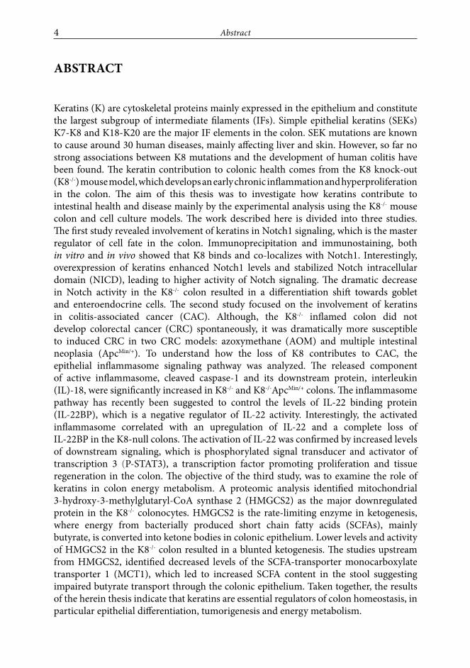
Keratins (K) are cytoskeletal proteins mainly expressed in the epithelium and constitute the largest subgroup of intermediate filaments (IFs). Simple epithelial keratins (SEKs) K7-K8 and K18-K20 are the major IF elements in the colon. SEK mutations are known to cause around 30 human diseases, mainly affecting liver and skin. However, so far no strong associations between K8 mutations and the development of human colitis have been found. The keratin contribution to colonic health comes from the K8 knock-out (K8-/-) mouse model, which develops an early chronic inflammation and hyperproliferation in the colon. The aim of this thesis was to investigate how keratins contribute to intestinal health and disease mainly by the experimental analysis using the K8-/- mouse colon and cell culture models. The work described here is divided into three studies. The first study revealed involvement of keratins in Notch1 signaling, which is the master regulator of cell fate in the colon. Immunoprecipitation and immunostaining, both in vitro and in vivo showed that K8 binds and co-localizes with Notch1. Interestingly, overexpression of keratins enhanced Notch1 levels and stabilized Notch intracellular domain (NICD), leading to higher activity of Notch signaling. The dramatic decrease in Notch activity in the K8-/- colon resulted in a differentiation shift towards goblet and enteroendocrine cells. The second study focused on the involvement of keratins in colitis-associated cancer (CAC). Although, the K8-/- inflamed colon did not develop colorectal cancer (CRC) spontaneously, it was dramatically more susceptible to induced CRC in two CRC models: azoxymethane (AOM) and multiple intestinal neoplasia (ApcMin/+). To understand how the loss of K8 contributes to CAC, the epithelial inflammasome signaling pathway was analyzed. The released component of active inflammasome, cleaved caspase-1 and its downstream protein, interleukin (IL)-18, were significantly increased in K8-/- and K8-/-ApcMin/+ colons. The inflammasome pathway has recently been suggested to control the levels of IL-22 binding protein (IL-22BP), which is a negative regulator of IL-22 activity. Interestingly, the activated inflammasome correlated with an upregulation of IL-22 and a complete loss of IL-22BP in the K8-null colons. The activation of IL-22 was confirmed by increased levels of downstream signaling, which is phosphorylated signal transducer and activator of transcription 3 (P-STAT3), a transcription factor promoting proliferation and tissue regeneration in the colon. The objective of the third study, was to examine the role of keratins in colon energy metabolism. A proteomic analysis identified mitochondrial 3-hydroxy-3-methylglutaryl-CoA synthase 2 (HMGCS2) as the major downregulated protein in the K8-/- colonocytes. HMGCS2 is the rate-limiting enzyme in ketogenesis, where energy from bacterially produced short chain fatty acids (SCFAs), mainly butyrate, is converted into ketone bodies in colonic epithelium. Lower levels and activity of HMGCS2 in the K8-/- colon resulted in a blunted ketogenesis. The studies upstream from HMGCS2, identified decreased levels of the SCFA-transporter monocarboxylate transporter 1 (MCT1), which led to increased SCFA content in the stool suggesting impaired butyrate transport through the colonic epithelium. Taken together, the results of the herein thesis indicate that keratins are essential regulators of colon homeostasis, in particular epithelial differentiation, tumorigenesis and energy metabolism.
Sammanfattning 5
SAMMANFATTNING
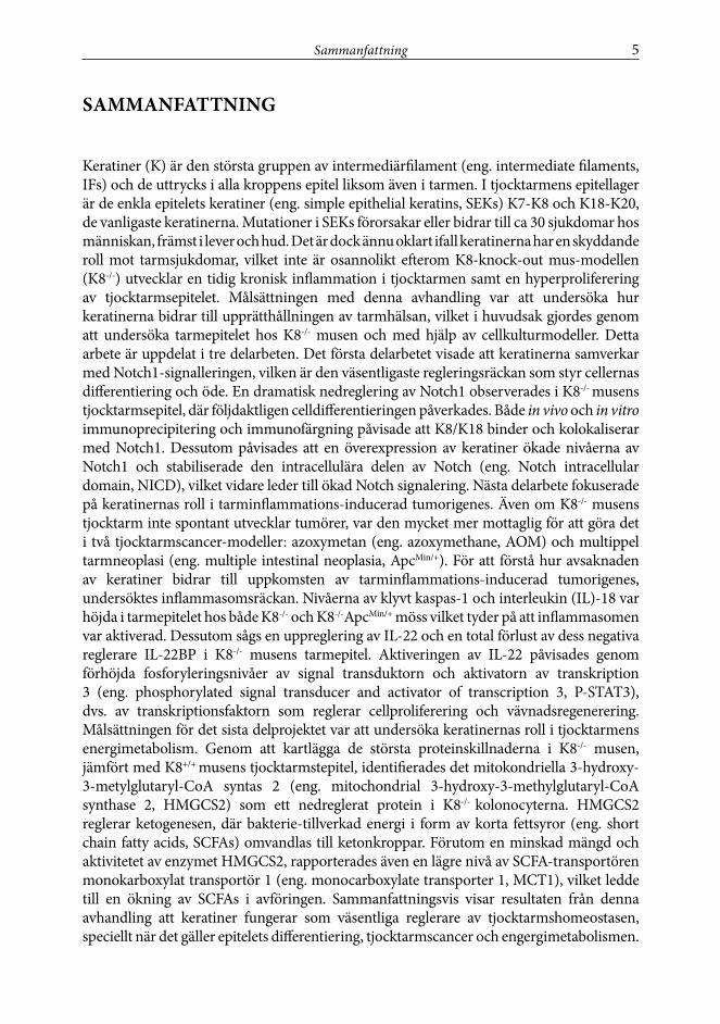
Keratiner (K) är den största gruppen av intermediärfilament (eng. intermediate filaments, IFs) och de uttrycks i alla kroppens epitel liksom även i tarmen. I tjocktarmens epitellager är de enkla epitelets keratiner (eng. simple epithelial keratins, SEKs) K7-K8 och K18-K20, de vanligaste keratinerna. Mutationer i SEKs förorsakar eller bidrar till ca 30 sjukdomar hos människan, främst i lever och hud. Det är dock ännu oklart ifall keratinerna har en skyddande roll mot tarmsjukdomar, vilket inte är osannolikt efterom K8-knock-out mus-modellen (K8-/-) utvecklar en tidig kronisk inflammation i tjocktarmen samt en hyperproliferering av tjocktarmsepitelet. Målsättningen med denna avhandling var att undersöka hur keratinerna bidrar till upprätthållningen av tarmhälsan, vilket i huvudsak gjordes genom att undersöka tarmepitelet hos K8-/- musen och med hjälp av cellkulturmodeller. Detta arbete är uppdelat i tre delarbeten. Det första delarbetet visade att keratinerna samverkar med Notch1-signalleringen, vilken är den väsentligaste regleringsräckan som styr cellernas differentiering och öde. En dramatisk nedreglering av Notch1 observerades i K8-/- musens tjocktarmsepitel, där följdaktligen celldifferentieringen påverkades. Både in vivo och in vitro immunoprecipitering och immunofärgning påvisade att K8/K18 binder och kolokaliserar med Notch1. Dessutom påvisades att en överexpression av keratiner ökade nivåerna av Notch1 och stabiliserade den intracellulära delen av Notch (eng. Notch intracellular domain, NICD), vilket vidare leder till ökad Notch signalering. Nästa delarbete fokuserade på keratinernas roll i tarminflammations-inducerad tumorigenes. Även om K8-/- musens tjocktarm inte spontant utvecklar tumörer, var den mycket mer mottaglig för att göra det i två tjocktarmscancer-modeller: azoxymetan (eng. azoxymethane, AOM) och multippel tarmneoplasi (eng. multiple intestinal neoplasia, ApcMin/+). För att förstå hur avsaknaden av keratiner bidrar till uppkomsten av tarminflammations-inducerad tumorigenes, undersöktes inflammasomsräckan. Nivåerna av klyvt kaspas-1 och interleukin (IL)-18 var höjda i tarmepitelet hos både K8-/- och K8-/-ApcMin/+ möss vilket tyder på att inflammasomen var aktiverad. Dessutom sågs en uppreglering av IL-22 och en total förlust av dess negativa reglerare IL-22BP i K8-/- musens tarmepitel. Aktiveringen av IL-22 påvisades genom förhöjda fosforyleringsnivåer av signal transduktorn och aktivatorn av transkription 3 (eng. phosphorylated signal transducer and activator of transcription 3, P-STAT3), dvs. av transkriptionsfaktorn som reglerar cellproliferering och vävnadsregenerering. Målsättningen för det sista delprojektet var att undersöka keratinernas roll i tjocktarmens energimetabolism. Genom att kartlägga de största proteinskillnaderna i K8-/- musen, jämfört med K8+/+ musens tjocktarmstepitel, identifierades det mitokondriella 3-hydroxy-3-metylglutaryl-CoA syntas 2 (eng. mitochondrial 3-hydroxy-3-methylglutaryl-CoA synthase 2, HMGCS2) som ett nedreglerat protein i K8-/- kolonocyterna. HMGCS2 reglerar ketogenesen, där bakterie-tillverkad energi i form av korta fettsyror (eng. short chain fatty acids, SCFAs) omvandlas till ketonkroppar. Förutom en minskad mängd och aktivitetet av enzymet HMGCS2, rapporterades även en lägre nivå av SCFA-transportören monokarboxylat transportör 1 (eng. monocarboxylate transporter 1, MCT1), vilket ledde till en ökning av SCFAs i avföringen. Sammanfattningsvis visar resultaten från denna avhandling att keratiner fungerar som väsentliga reglerare av tjocktarmshomeostasen, speciellt när det gäller epitelets differentiering, tjocktarmscancer och engergimetabolismen.

6 List of Original Publications
LIST OF ORIGINAL PUBLICATIONS
This thesis is based on the following original publication and manuscripts, to which the text refers by Roman numerals (study I.-III.). The original publication has been reproduced with the permission of the copyright owners.
I. Lähdeniemi I.A.K., Misiorek J.O.*, Antila C.J.M. *, Nyström J.H., Fortelius L.E., Sahlgren C.#, Toivola D.M.# Keratins regulate colonic epithelial cell differentiation through the Notch1 signaling pathway. Manuscript.
II. Misiorek J.O., Lähdeniemi I.A.K., Nyström J.H., Gullmets J.A., Saarento H., Husøy T., Taimen P., Toivola D.M. Keratin 8-deletion induced colitis predisposes to murine colorectal cancer enforced by the inflammasome and IL-22 pathway. Submitted manuscript, under revision.
III. Helenius T.O.*, Misiorek J.O.*, Nyström J.H.*, Fortelius L.E., Habtezion A. Liao J., Asghar M.N., Zhang H., Azhar S., Omary M.B., Toivola D.M. (2015). Keratin 8 absence downregulates colonocyte HMGCS2 and modulates colonic ketogenesis and energy metabolism. Mol Biol Cell., 26(12):2298-310. doi:10.1091/mbc.E14-02-0736.
* # Equal author contribution
In addition, some unpublished data are presented in this thesis.

Abbreviations 7
ABBREVIATIONS
ADAM A disintegrin and metalloproteinaseAOM AzoxymethaneApc Adenomatous polyposis coliARP-1 Apo A1 regulatory protein-1 ATP Adenosine triphosphate BMPs Bone morphogenic proteins CBC Crypt base columnar (cells)CD Crohn’s diseaseCK1 Casein kinase I CoA CoactivatorCRC Colorectal cancerCREB Cyclic AMP-responsive element-binding protein CSL CBF1, Suppressor of Hairless, Lag-1, also known as RBP-JDC Distal colonDhh Desert hedgehogDsh Dishevelled DSS Dextran sulphate sodiumEMT Epithelial to mesenchymal transitionEph Ephrin receptorER Endoplasmatic reticulum FAP Familiar adenomatous polyposisFLN Full length NotchFz FrizzledGPI Glycosylphosphatidylinositol GSK3 Glycogen synthase kinase 3βHDAC Histone deacetylase Hes Hairy enhancer of splitHey Hairy/E(spl)-related with YRPW motifHMGCS2 3-hydroxy-3-methylglutaryl-CoA synthase, also known as HMG-CoA
synthaseHNF-4 Hepatocyte nuclear factor 4 IBD Inflammatory bowel disease IFs Intermediate filamentsIhh Indian hedgehog

8 Abbreviations
IL InterleukinIP ImmunoprecipitationK KeratinLRP Lipoprotein-receptor-related protein MAML Mastermind-likeMCT1 (slc16a1) Monocarboxylate transporter 1MDBs Mallory-Denk bodiesNHE2, NHE3 Sodium/hydrogen exchanger 2, 3NICD Notch intracellular domainNLRP3 NACHT, LRR and PYD domains-containing protein 3NSP Nonstarch polysaccharidesP PhosphorylationPC Proximal colonPls1 Plastin 1, also known as fimbrinPPARα Peroxisome proliferator-activated receptor α Ptc Patched (receptor)PTMs Post-translational modificationsROS Reactive Oxygen SpeciesRXR Retinoid X receptor S Serine SCFAs Short chain fatty acidsSEKs Simple epithelial keratinsShh Sonic hedgehog SMCT (slc5a8) Sodium-coupled monocarboxylate transporterSp1 Specificity protein 1 T Threonine TA Transient-amplifying (cells)TGF-β Transforming growth factor-betaTLRs Toll-like receptors TNF Tumor necrosis factorUb UbiquitinationUC Ulcerative colitisWB Western blottingY Tyrosine ZO-1 Zonula occludens-1 β-cat β-catenin
Contents 9
CONTENTS
ABSTRACT ...................................................................................................................4
SAMMANFATTNING ..................................................................................................5
LIST OF ORIGINAL PUBLICATIONS .......................................................................6
ABBREVIATIONS ........................................................................................................7
INTRODUCTION ......................................................................................................11
REVIEW OF THE LITERATURE ..............................................................................12
1. Keratin intermediate filaments .........................................................................121.1. Keratins and their regulation ............................................................................... 131.2. Keratin associated diseases .................................................................................. 141.3. Keratin expression and role in the colon ........................................................... 15
2. Colon .................................................................................................................172.1. Colon epithelial cells ............................................................................................. 172.2. Maintenance of colon cell homeostasis: proliferation and differentiation .... 19
2.2.1. Wnt/β-catenin pathway ............................................................................. 192.2.2. Notch pathway ............................................................................................ 212.2.3. TGF-β/BMP, Hedgehog, Hippo and Eph/Ephrin pathways ................. 23
2.3. Energy metabolism of the colon ......................................................................... 232.3.1. SCFA production and absorption ............................................................ 232.3.2. Ketogenesis ................................................................................................. 252.3.3. The role of butyrate for colonic health .................................................... 27
2.4. Diseases affecting colon ........................................................................................ 282.4.1. Inflammatory Bowel Disease (IBD) ........................................................ 28
2.4.1.1. Risk factors for developing IBD ................................................. 292.4.1.2. Animal models of IBD ................................................................. 302.4.1.3. The role of epithelial inflammasomes and IL-22 pathway
in IBD ............................................................................................. 322.4.1.4. IBD treatment ............................................................................... 33
2.4.2. Colorectal Cancer (CRC) .......................................................................... 342.4.2.1. Epidemiology and mortality of CRC ......................................... 342.4.2.2. Risk factors and screening for CRC ........................................... 342.4.2.3. Molecular pathways leading to CRC ......................................... 352.4.2.4. IBD as a risk factor for developing CRC ................................... 382.4.2.5. Mouse models of CRC ................................................................. 392.4.2.6. CRC treatment .............................................................................. 41

10 Contents
OUTLINE AND AIMS OF THE THESIS ..................................................................42
EXPERIMENTAL PROCEDURES .............................................................................43
RESULTS AND DISCUSSION ...................................................................................46
1. K8 regulates differentiation in the colon through Notch1 signaling (Study I.) ............................................................................................................461.1. K8 binds and colocalizes with Notch 1 in vitro and in vivo ............................. 461.2. Notch1 levels and activity are modified by K8/K18 in in vitro cell
culture models ....................................................................................................... 471.3. Decreased Notch1 in the K8-/- mouse colon results in a cell
differentiation shift ................................................................................................ 48
2. K8-deletion induced colitis is a risk factor for CRC development (Study II.) ..502.1. Increased susceptibility of K8-/- colon to induced tumorigenesis
indicates keratin involvement in colonic homeostasis ..................................... 502.2. IL-22 pathway is involved in the K8-/- colon tumorigenesis ............................ 512.3. K8 binds the inflammasome - a potentially new cytoskeletal
contributor to the IL-22 pathway ........................................................................ 54
3. K8 influences colon energy metabolism (Study III.) ........................................553.1. HMGCS2 downregulation is K8-/- colon specific and leads to blunted
ketogenesis ............................................................................................................. 553.2. Colon mitochondria and energy intermediates are largely unaffected
after K8 inactivation.............................................................................................. 573.3. K8-/- colon has increased levels of luminal SCFAs but decreased levels
of their transporter MCT1 ................................................................................... 58
4. Future prospects in the colon keratin field .......................................................59
SUMMARY ..................................................................................................................61
ACKNOWLEDGEMENTS .........................................................................................62
REFERENCES .............................................................................................................64
ORIGINAL PUBLICATION AND MANUSCRIPTS ................................................77
Introduction 11
INTRODUCTION
The cytoskeleton is a system of filaments present principally in the cytoplasm of eukaryotic cells. It consists of three groups: microtubules, microfilaments and intermediate filaments. The main role of the cytoskeleton is to support the shape and motility of cells.
Keratins (K) constitute the largest subgroup of intermediate filaments. They are expressed in a cell- and tissue-dependent manner and as highly dynamic structures they reorganize during cellular events like mitosis and apoptosis. Keratin ability to reorganize is regulated by post-translational modifications (PTMs) and keratin-binding proteins. The cell and tissue-specific expression pattern of keratins has been used for epithelial tumor diagnosis. This feature of keratins also nominates them to serve as a prognostic marker. For example, decreased levels of K8 and K20 follow epithelial-to-mesenchymal transition (EMT), which facilitates tumor progression in the colon. However, the molecular details on how keratins are involved in tumorigenesis remain unknown.
Mutations in simple epithelial keratins (SEKs) have been associated with liver and skin diseases. Although, SEKs are abundantly expressed in the colon, their role remains ambiguous. Few studies reported keratin mutations in the patients with inflammatory bowel disease (IBD), but further investigation is needed to confirm this link. So far, the strongest evidence for keratin involvement in the etiology of IBD comes from the K8 knock-out (K8-/-) mouse. Apart from the development of chronic colitis, K8-/- colonocytes hyperproliferate, are resistant to apoptosis and have mistargeted sodium and chloride ion transporters, which results in diarrhea. Antibiotic treatment attenuates the inflammation developed by K8-/- colon, which suggests bacteria involvement in the reported phenotypes.
Bacteria are an integral component of the colon. Bacterial fermentation of carbohydrates results in the production of short chain fatty acids (SCFAs), which are used as a source of energy by colon epithelial cells and e.g. prevent cancer development.
Colonic epithelium originates from the stem cells, which proliferate and differentiate into highly specialized cells. Notch signaling regulates the proliferation of stem cells and progenitor cells as well as defines the fate of colonic epithelial cells by orchestrating their differentiation. However, the regulators of Notch signaling in the colon remain to be defined.
The aim of this thesis was to investigate the role of keratin intermediate filaments in colon homeostasis and disease. The specific areas of the studies included: differentiation and proliferation of colonic epithelium, colitis-induced tumorigenesis and colon energy metabolism.

12 Review of the Literature
REVIEW OF THE LITERATURE
1. Keratin intermediate filaments
Intermediate filaments (IFs) together with microfilaments and microtubules form the cytoskeleton of a cell, providing it with mechanical support and spatial organization (Fletcher and Mullins, 2010). IFs constitute a diverse family of proteins encoded by over 70 genes and are expressed in a cell and tissue specific manner. Each IF protein consists of an α-helical rod domain flanked by an N-terminal head and a C-terminal tail (Fig. 1) (Pan et al., 2013). Based on the amino acid content in the α-helical rod domain, IFs can be grouped into six types presented in Table 1 (Omary, 2009).
1A 2A1B 2BL1 L12 L2
N-terminalhead
C-terminaltailHighly conserved α-helical rod domain
Figure 1. Tripartite domain structure of IFs. Generally, the IF protein structure is composed of an α-helical rod flanked by an amino terminal head and a carboxyl terminal tail. The rod is formed by coils 1A, 1B and 2A, 2B separated from each other by linkers L1, L12 and L2. Based on this structure IF proteins can form either homopolymers (vimentin) or obligate heteropolymers (keratins type I with type II). Based on (Toivola et al., 2005; Eriksson et al., 2009).
Table 1. Classification of IFs.
IF class Components ExpressionType I Acidic keratins EpitheliaType II Neutral to basic keratins Epithelia
Type III
DesminSyncolin Muscles
PeripherinGFAP
Vimentin
NeuronsGlial cells
Mesenchyma
Type IV
α-InternexinNeurofilaments (NF-L,-M,-H)
Neurons
NestinSynemin
Pluripotent cellsMuscle
Type V Lamins(A-, B1-2, C1-, C2) All metazoan cell types
Type VI Bfsp 1Bfsp 2 Lens cell
Created based on The Human Intermediate Filament Database (Szeverenyi et al., 2008).

Review of the Literature 13
1.1. Keratins and their regulationKeratins (Ks) are classified as type I (acidic) and type II (neutral to basic) IFs, which form heteropolymers with each other in 1:1 ratio. Keratins, as all IFs, are expressed in a cell and tissue specific manner, Table 2 (Moll et al., 1982).
Table 2. Classification of keratin IFs.
Keratin types Type I Type II Example of expression
Epithelial keratins
K9 – K20K23 – K24
K1 – K8K76 – K80
Basal keratinocytes (K5/K14)
Simple epithelium of liver (K8/K18)
Hair follicle -specific epithelial keratins K25 – K28 K71 – K75,
K80.1Root sheath(K25/K71)
Hair/ nail keratins K31 – K40 K79 – K86 Hair fiber
K31/K86Created based on (Moll et al., 2008; Langbein et al., 2010)
The keratins expressed in single layer epithelia are termed simple epithelial keratins (SEKs) and are composed of K7-K8, K18-K20 and K23, while keratins expressed in the stratified epithelium are known as epidermal keratins or keratinocyte-type keratins and consists mainly of K5-K6, K14 and K16 (Omary et al., 2009).
The main role of keratins is cytoprotection from different types of stresses and the maintenance of tissue integrity. Apart from this, keratins are involved in processes such as protein synthesis and targeting, cell polarity and attenuation of tumor cell migration. However, the exact molecular roles of keratins need to be unravel (Omary et al., 2009; Pan et al., 2013).
Keratins are functionally regulated by post-translational modifications (PTMs) including: phosphorylation, glycosylation, prenylation, acetylation, sumoylation, transamidation and caspase cleavage. The PTMs mostly affect the non-conserved head and tail domains of keratins regulating their organization, integrity and solubility (Snider and Omary, 2014) (Fig. 1). The most common keratin PTM is phosphorylation at the amino acid serine (S), threonine (T) and tyrosine (Y). The most abundant is keratin phosphorylation at S: K8 pS74 (a substrate for p38 and c-Jun N-terminal kinases) (He et al., 2002; Ku et al., 2002a), K18 pS34 (phosphorylated by cyclin dependent kinase 2, Cdk2) (Ku et al., 1998) and K18 pS53 (phosphorylated by protein kinase 2) (Liao et al., 1995). Phosphorylation of S and T amino acids makes keratin filaments more soluble, which also affects their dynamics. Keratin phosphorylation is believed to act as a phosphate sponge preventing activation of pro-apoptotic proteins in liver injury (Ku and Omary, 2006). Moreover, K5, K17 and K18 phosphorylation enables these filaments to bind to 14-3-3 protein and be involved in cell growth (Liao and Omary, 1996; Kim et al., 2006). Phosphorylation of K8 pS432 is also known to influence the migration of cells by decreasing migration of oral squamous

14 Review of the Literature
carcinoma cells (Alam et al., 2011a) and increasing migration of gastric and pancreatic cells (Busch et al., 2012). The other PTMs affecting keratins is acetylation, e.g. at lysine 207 of K8 which is increased upon hyperglycaemic conditions when deacetylase SIRT2 activity is inhibited (Snider et al., 2013). Acetylation at this site is known to decrease filament solubility and promote filament formation (Snider et al., 2013; Snider and Omary, 2014). Acetylation of keratins has been suggested to be promoted by short chain fatty acids (SCFAs) in colon cancer cell lines studies (Leech et al., 2008) since SCFAs are inhibitors of deacetylases (Gibson et al., 1999).
Another regulatory mechanism of SEK functions is via their interaction with various keratin-associated proteins. Keratin filaments bind to the desmoplakin, a protein present in desmosomes in the lateral parts of the cell (Alberts et al., 2015) and thus provide tissues with mechanical strength (Delva et al., 2009). In the apical part of the cells and also in the region of desmosomes, keratins bind trichoplein protein (Nishizawa et al., 2005). The best studied keratin interaction so far, is the binding of keratin to the 14-3-3 scaffold protein. K18 pS33 binds 14-3-3 during mitosis (Ku et al., 1998) and K17 T9/S44 during wound healing (Kim et al., 2006). Proteins binding keratins also connect them to the other cytoskeletal components. A protein which links keratins with microfilaments is plastin 1 (Pls1), a major actin bundling protein, which interacts with K19 (Grimm-Gunter et al., 2009). Keratins are also connected to microtubules via binding to phosphorylated γ-tubulin complex protein GCP6 (Oriolo et al., 2007). In addition, there is an evidence for keratin-nuclear protein interaction, which comes from keratin binding to plectin (Suozzi et al., 2012). Plectin also connects with the nesprin-3, which is an outer nuclear envelope protein. Deletion of nesprin-3 in Zebra fish has demonstrated diminished amount of keratins around the nucleus (Postel et al.; Postel et al., 2011). Recent studies also show K17 colocalization with autoimmune transcription factor Aire in the nucleus of epidermoid carcinoma cells (Hobbs et al., 2015). Many of the described keratin-associated proteins were found in the colon, which suggests the regulatory role of keratins in this organ.
1.2. Keratin associated diseasesKeratin mutations are associated with certain diseases (Toivola et al., 2015b). Mutations in stratified epithelium keratins lead to numerous skin diseases, e.g.: epidermolysis bullosa simplex (mutations in K5 and K14) (Bonifas et al., 1991; Coulombe et al., 1991; Lane et al., 1992) and hyperkeratosis (mutations in K1 or K10) (Syder et al., 1994) .
Mutations in SEKs have been reported to predispose to liver diseases. Around 12% of patients with liver diseases carry K8/K18 mutations (Omary, 2009). The main diseases, to which K8 and K18 mutations predispose, are: acute liver failure and chronic liver disease (Strnad et al., 2010). Moreover, mutations in K8 and K19 can lead to primary biliary cirrhosis (Omary, 2009; Omary et al., 2009). Although, SEKs are expressed in both the endocrine and exocrine pancreas, there are no clear evidences for keratin mutation causing diseases in this organ (Cavestro et al., 2003). Nevertheless, the loss of K8 in mice leads to decreased insulin production and increased susceptibility to develop diabetes (Alam et al. 2013).

Review of the Literature 15
So far mutations in K7, K20 or K23 in human diseases have not been reported (Omary et al., 2009). K7 knock-out (K7-/-) mice display only minor changes in the proliferation of bladder urothelial cells and altered expression of K7 partners: K18 downregulation and K20 upregulation (Sandilands et al., 2013). K7 and K20 are often used as markers of colorectal cancers, in which they exhibit decreased and increased expression, respectively (Karantza, 2011). Moreover, increased levels of phosphorylated K23 form are detected in colon adenocarcinomas (Birkenkamp-Demtroder et al., 2007). Keratin expression is also known to decrease in the process of epithelial to mesenchymal transition (EMT), which occurs e.g. during cancer invasion and metastasis (Kalluri and Weinberg, 2009). K8 and K20 show decreased expression during EMT of colorectal tumors (Knosel et al., 2006).
1.3. Keratin expression and role in the colonK8 and its partners K18 and K19 are the main SEKs expressed in colonic epithelium (Fig. 2). There are also expressed K7 and K20 in smaller amounts (Zhou et al., 2003). Although, increased levels of K23 were reported in colon adenocarcinomas (Birkenkamp-Demtroder et al., 2007) it remains unknown whether it is expressed under the basal conditions in colonic epithelium.
K7 K8 K18 K19 K20
X
X
X
K23*
?
Legend: goblet cells X enteroendocrine cells
Crypt tip
Crypt base
X
X
X
COLONIC KERATIN IFs
Colonic crypts
Type II Type I
Figure 2. Schematic distribution of keratins in the colonic crypts. K8 displays the highest expression pattern and its partners K18 and K19. The other type II keratin, K7 is expressed at a very low level, including in the goblet cells. The levels of K18 and K19 in the colon are similar. K20 is distributed in the upper portion of crypts and in enteroendocrine cells. Note, most Ks are expressed in goblet cells but not specified in the figure. *The expression and distribution of K23 in the colon remains to be studied. Adapted from (Zhou et al., 2003).
The main role of keratins in the colon is to maintain tissue integrity and protect from different types of stresses (Ameen et al., 2001; Haines and Lane, 2012; Pan et al., 2013).

16 Review of the Literature
However, the exact role of keratins in colon health remains unclear. The key evidence for keratin involvement in the colon health comes from the K8 knock-out (K8-/-) mouse model (Baribault et al., 1994). K8-/- mouse colon displays T helper cell 2 (Th2)-type early chronic inflammation resembling human ulcerative colitis, crypt hyperproliferation and resistance to apoptosis (Habtezion et al. 2005; Habtezion et al., 2011). Moreover, mistargeted sodium and chloride ion transporters at the apical membrane of K8-/- ileum and colon lead to diarrhea in these mice and highlights the role of keratins in protein targeting (Toivola et al., 2004). The decreased amount of keratins increases the susceptibility to experimental colitis, as shown on K8 heterozygote (K8+/-) mice (Asghar et al., 2015a)
Apart from the K8-/- and K8+/- model, few studies showing human keratin mutations in inflammatory bowel disease (IBD) have been published so far (Buning et al., 2004; Owens et al., 2004; Tao et al., 2007), although they do not indicate a strong link between these two. However, in human colitis the decreased levels of K8, K18-19 and changes in K8 phosphorylation have been observed (Corfe et al., 2015a). An interesting study revealed that interleukin (IL)-6 can induce the expression of colonic K8 and K18, which contributes to the maintenance of the colonic barrier (Wang et al., 2007). As mentioned earlier, keratin filaments are posttranslationally modified. K8 was found to be a substrate for Ubc9 enzyme, which regulates the sumoylation of filaments in the small intestine (Demarque et al., 2011) and contributes to the mechanical stability. Proteomic analysis revealed K8 to be highly acetylated in colon cancer cell lines (Leech et al., 2008), which is likely linked to the levels of the deacetylase inhibitor - butyrate. The study from the same research group suggested that butyrate can decrease the levels of K8 expressed in colon tumors (Khan et al., 2011). However, the mechanism and importance of these changes remain unclear. Increased levels of phosphorylated K23 have been observed in microsatellite-stable colon tumors, which distinguishes them from the microsatellite-instable tumors (Birkenkamp-Demtroder et al., 2007).
Several studies indicated binding partners of keratins in the colon or small intestine. One of the keratin-binding protein is the previously mentioned trichoplein, which by binding to K8/K18 may regulate filament organization at the apical part of the cell (Nishizawa et al., 2005). The binding of Albatross protein to the K8/K18 is essential for Caco-2 cell polarity. The knockdown of the Albatross results in keratin filament reorganization and loss of apical junctional complex proteins (Sugimoto et al., 2008). K19 is bound by plastin 1 (fimbrin) protein in the small intestine, which help in the organization of the terminal web. Interestingly, the same study showed that the loss of plastin 1 in mice makes them more susceptible to induced-colitis (Grimm-Gunter et al., 2009). A crucial role for establishing an apico-basal polarity has atypical protein kinase C (aPKC) (Suzuki and Ohno, 2006). Its levels are strongly decreased in K8-/- colon (Mashukova et al., 2009) as well as in IBD patients (Wald et al., 2011). Recent studies have shown that keratin-Hsp70 chaperoning of aPKC is regulated by BAG protein during inflammation (Mashukova et al., 2014).
Review of the Literature 17
2. Colon
Colon (large intestine) together with small intestine constitute the lower parts of the gastrointestinal tract. Anatomically the human colon is subdivided into four parts: ascending, transverse, descending and sigmoid. The ascending and transverse colon form the proximal colon (PC), while the descending and sigmoid form the distal colon (DC) (Ross, 2011). The main role of the colon is to pass undigested food and reabsorb water, sodium ions and water-soluble vitamins. The large intestine is also a place inhabited by microbiota which stimulate the immune system and produce SCFAs essential for colon homeostasis (Tan et al., 2014).
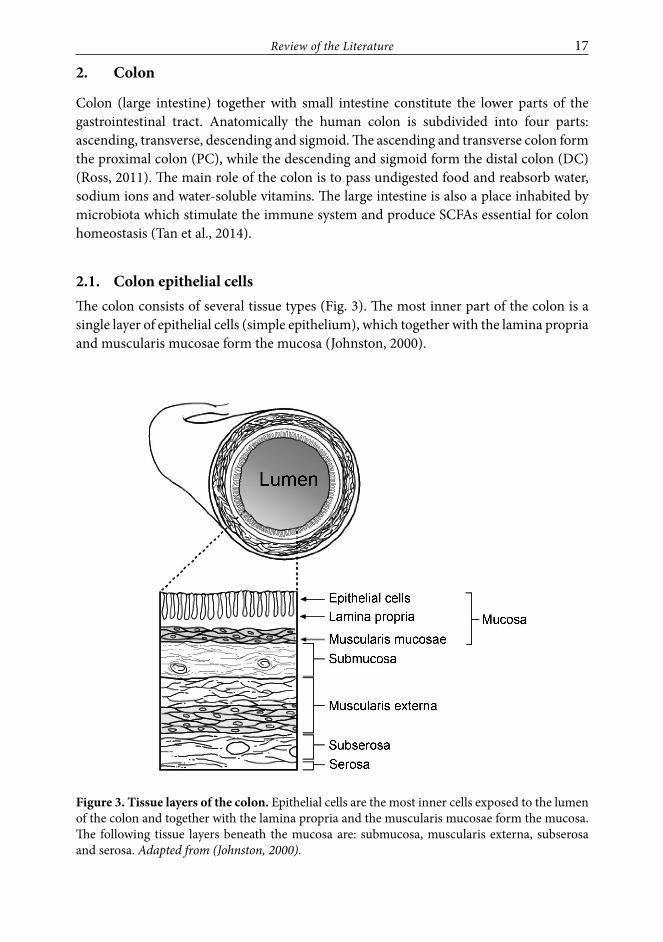
2.1. Colon epithelial cellsThe colon consists of several tissue types (Fig. 3). The most inner part of the colon is a single layer of epithelial cells (simple epithelium), which together with the lamina propria and muscularis mucosae form the mucosa (Johnston, 2000).
Figure 3. Tissue layers of the colon. Epithelial cells are the most inner cells exposed to the lumen of the colon and together with the lamina propria and the muscularis mucosae form the mucosa. The following tissue layers beneath the mucosa are: submucosa, muscularis externa, subserosa and serosa. Adapted from (Johnston, 2000).

18 Review of the Literature
Epithelial cells form invaginations in the colonic mucosa called crypts (Fig. 2-4) and are the fastest renewing cells in the whole body (Schepers and Clevers, 2012). The process of renewal takes 3-5 days and is stimulated by multipotent stem cells crypt base columnar (CBC) and +4 stem cells located in the bottom of the crypts (Fig. 4) (Medema and Vermeulen, 2011; Krausova and Korinek, 2014). Stem cells self-renew and give rise to proliferative progenitor cells called transient-amplifying (TA) cells, which can next differentiate into absorptive enterocytes, enteroendocrine cells, goblet cells and tuft cells. Enterocyte cells are mainly responsible for absorption of ions, water and vitamin B12 in the colon. Enteroendocrine cells are secretory cells releasing different types of hormones mainly serotonine and somatostatin, which regulate colonic motility and peristalsis (Gunawardene et al., 2011). Goblet cells are a very abundant cell type in the distal part of the colon, where they produce mucus, a lubricant protecting from bacterial adhesion and epithelium abrasion caused by passing stool (Corazziari, 2009). Tuft cells constitute the smallest population of epithelial cells in the colon and their exact role remains ambiguous. Nevertheless, some data suggests the involvement of tuft cells in intestinal smooth muscle contraction and absorption of fatty acids (Gerbe et al., 2012). The processes of colon epithelial cell proliferation and differentiation are tightly controlled by several signaling pathways, mainly Wnt and Notch signaling. Differentiated epithelial cells die by the apoptosis occurring on the top of the crypt.
CBC
Progenitor cell
+ 4 cell
TA cell
Goblet cell
Enterocyte
Enteroendocrine cell
Tuft cell
APOPTOTIC CELLS
DIFFERENTIATING CELLS
PROLIFERATING CELLS
Figure 4. Colon crypt with epithelial cells. Stem cells crypt base columnar (CBC) and +4 cells give rise to transient amplifying (TA) cells, which proliferate and then differentiate into absorptive enterocytes or secretory enteroendocrine cells, goblet cells and tuft cells. Differentiated cells die of apoptosis on the top of the crypt. Adapted from (Varedi et al., 2001; Sancho et al., 2015).

Review of the Literature 19
2.2. Maintenance of colon cell homeostasis: proliferation and differentiation
Epithelial cell proliferation, differentiation, migration and apoptosis are processes involved in colon homeostasis. The Wnt/β-catenin signaling pathway together with Hedgehog, transforming growth factor beta (TGF-β) and bone morphogenetic protein (BMP) pathways regulate proliferation. The Notch signaling pathway controls both proliferation and differentiation of the colonic cells, while the role of Eph/Ephrin pathway is to maintain cell contact and facilitate cell migration within the colonic crypt.
2.2.1. Wnt/β-catenin pathway
Wnt/β-catenin pathway (the canonical Wnt pathway) is a contact-dependent signaling, in which the proteolysis of β-catenin (β-cat) is regulated. In the colon, the Wnt/β-cat pathway is mostly active in the bottom of the crypt, where transmembrane receptor Frizzled (Fz) and low-density lipoprotein-receptor-related protein (LRP) are bound to the Wnt ligand proteins. Receptor activation leads to the binding of Dishevelled (Dsh) and Axin proteins to Fz and LRP, respectively. Axin binding to LRP is regulated by phosphorylation through casein kinase I (CK1) and glycogen synthase kinase 3β (GSK3). Axin and Dsh are in the complex with these kinases as well as tumor suppressor protein adenomatous polyposis coli (Apc) and β-cat in the cytoplasm. The complex they form is also called the destruction complex. When there is no Wnt ligand, CK1 and GSK3 kinases phosphorylate β-cat, which is next ubiquitinated by β-TrCP and degraded in the proteasome. Proteasomal degradation of β-cat prevents its nuclear translocation and activation of target genes (Fig. 5A). β-cat degradation is inhibited when Wnt ligands are present. Next, β-cat translocates to the nucleus and binds to LEF1/TCF transcription factors, which results in Wnt target gene transcription, mainly Axin2, c-Myc and Lgr5 (Fig. 5B) (Clevers and Nusse, 2012; Alberts et al., 2015).
The Lgr5 gene has been discovered as a marker of stem cells in both small and large intestine (Barker et al., 2007). Since Wnt signaling is important for colonic stem cell amplification, mutations in this pathway leads to the colorectal cancer. Most common mutations in the Wnt pathway occur in the APC gene. Germline mutations in APC lead to hereditary colorectal cancer called familiar adenomatous polyposis (FAP) (Nishisho et al., 1991), (Kinzler et al., 1991). A total loss of Apc alleles is observed in many sporadic cases of colorectal cancer. In both cases of colorectal cancer, β-cat cannot be stabilized and constitutively binds to the TCF activator of transcription in the colon, Tcf4 (Korinek et al., 1997). Constitutively active Axin2 upregulates Snail1, which results in EMT in the colon tissue (Wu et al., 2012), while Myc overexpression stimulates hyperproliferation and uncontrolled cell growth (Alberts et al., 2015).

20 Review of the Literature
Axin2, Myc, Lgr5
Frizzled FrizzledWNT
LRP LRP
DshAxin
GSK3CK1
APC-cat
-cat
-cat
-cat
-cat
-cat
DshAxin
APCGSK3
CK1
TrCP Proteasome
PP P P
PP P P
Ub
Ub
Ub
LEF-1/TCF
P P
Proliferation &
Cell growth
LEF-1/TCF
Groucho
A. B.
Figure 5. The Wnt/β-catenin pathway. A. When the Wnt ligand (WNT) is absent, β-catenin (β-cat) is constantly phosphorylated by the destruction complex and ubiquitinated by β-TrCP, which marks it for the degradation by the proteasome. B. In the presence of Wnt ligand, the destruction complex binds phosphorylated LRP. Phosphorylation of β-cat is maintained; however, its ubiquitination is blocked. This leads to the accumulation of β-cat in the cytoplasm and its translocation to the nucleus, where β-cat replaces Groucho (repressor of transcription) and causes transcription of genes responsible for cell proliferation and growth. P = phosphorylation, Ub = ubiquitination. Adapted from (Clevers and Nusse, 2012; Alberts et al., 2015).
There are also non-canonical Wnt pathways: the planar polarity pathway and the Wnt/calcium pathway. The planar polarity pathway is dependent on the GTPases Rho and defines the polarization of epithelial cells during development (Alberts et al., 2015), while the Wnt/calcium pathway relays on PLC or PDE proteins, which by binding Dsh, can either cause calcium release from endoplasmatic reticulum (ER) (binding to PLC) or calcium release inhibition (binding to PDE), (Komiya and Habas, 2008).
β-cat, apart from being a part of Wnt signaling pathway, is a binding partner of E-cadherin in adherens junctions of the simple epithelia (Peifer et al., 1992), where it helps to bind to the actin filaments and is, thus, involved in the response to tensions in the cell (Alberts et al., 2015).

Review of the Literature 21
2.2.2. Notch pathway
The Notch pathway regulates the proliferation of colonic stem cells and progenitor cells and defines cell fate by influencing the differentiation of transient amplifying cells (Fre et al., 2005). Notch signaling is a contact-dependent pathway. Notch heterodimeric transmembrane receptor (isoforms Notch 1-4) is located on the cell membrane of one cell (signal-receiving cell) and the Notch ligand (Jagged 1–2 or Delta-like proteins 1, 3 and 4) is present on the cell membrane of other cell (signal-sending cell) (Kopan and Ilagan, 2009). Both Notch ligands and Notch receptors are expressed in the colonic epithelial cells (Reichrath J. and S., 2012). The interaction between Notch and Delta leads to a lateral inhibition, a phenomenon in which one differentiating cell inhibits the simultaneous differentiation of a neighboring cell (Alberts et al., 2015).
In the canonical Notch pathway, ligand binding to the FLN (Full Length Notch) causes a cleavage of Notch extracellular domain by ADAM (A Disintegrin And Metalloproteinase) and next the intracellular domain is cleaved by γ-secretase (Fig. 6). A single cleavage by ADAM leads to a generation of ΔE Notch, while the subsequent γ-secretase creates a truncated form of Notch called NICD (Notch Intracellular Domain). NICD stability is regulated by E3 ubiquitin ligases (Kopan and Ilagan, 2009). NICD translocates to the nucleus, where it binds to the transcription factor CSL (CBF1, Suppressor of Hairless, Lag-1) together with MAML (mastermind-like) and other coactivators, and acts as a transcription factor inducing the transcription of Hes (hairy enhancer of split) and Hey (Hairy/E(spl)-related with YRPW motif) genes (Sancho et al., 2015). These genes initiate a genetic program, which determines the proliferation of stem cells and progenitor cells as well as differentiation of TA cells into absorptive enterocyte cells. Hes blocks the transcription factor Atoh1 (Math1) which in the lack of NICD determine a secretory fate of the cells in the intestine (High and Epstein, 2008; Sancho et al., 2015) (Fig. 6).
The Notch pathway is also regulated via non-canonical mechanism, which might be independent from the ligand and CSL. The best studied Notch non-canonical pathway is the regulation of Wnt/β-cat, in which Notch can directly or indirectly bind to β-cat in stem cells and progenitor cells leading to lysosomal β-cat degradation (Kwon et al., 2011).
Notch can either act as a tumor suppressor or an oncogene depending on the context (Reedijk et al., 2008; Lobry et al., 2011; Sonoshita et al., 2015). In colon cancer, Notch is mostly known as an oncogene (Ghaleb et al., 2008; Miyaki et al., 2009; Rodilla et al., 2009), although one study shows that deletion of Notch1 leads to colon tumorigenesis in mice (Liu et al. 2011).

22 Review of the Literature
Jagged / Delta
ADAM-secretaseγ
NICD
CSL
MAM
LCoANICD
Hes & Hey
Atoh1
FLN
Differentiation Stem cells
Progenitor cells
Absorptive Enterocytes
Differentiation TA cells
Secretory cells
EnteroendocrineGoblet
cellsTuft cells
Proliferation TA cells
+
Signal-sending cell
Signal-receiving cell
Figure 6. The Notch signaling pathway determines cell fate in the colonic crypt. Notch signaling is active when one of the ligands (Jagged or Delta) on the signal-sending cell binds to the FLN (Full Length Notch) present on the signal-receiving cell. This interaction causes FLN cleavage into NICD (Notch Intracellular Domain) by ADAM and γ-secretase. NICD translocates to the nucleus and binds to the CSL protein, other coactivators (CoA) and MAML, which together induce transcription of Hes and Hey genes. Hes and Hey stimulate the proliferation of stem cells and progenitor cells as well as differentiation of TA cells into absorptive enterocytes. Because Hes and Hey block the Atoh1 transcriptional factor, no differentiation of TA cells into secretory lineage occurs. The scheme does not present the first cleavage of Notch receptor into a mature heterodimeric form of FLN, which happens in the Golgi lumen prior Notch transport to plasma membrane. Based on (High and Epstein, 2008; Guruharsha et al., 2012; Sancho et al., 2015).

Review of the Literature 23
2.2.3. TGF-β/BMP, Hedgehog, Hippo and Eph/Ephrin pathways
BMPs are expressed by the mesenchymal cells in the colon. TGF-β/BMP pathway inhibits Wnt signaling and stimulates differentiation in the intestine (Medema and Vermeulen, 2011). This stimulation is mediated via SMAD transcriptional activator (Brazil et al., 2015).
Hedgehog signaling involves the hedgehog protein (Hh) and its receptor Patched (Ptc), which activates the transcription factors GLI1-3. Hh is encoded by three different genes in the mammalians: Sonic (Shh), Desert (Dhh) and Indian (Ihh) (Alberts et al., 2015). Ihh is the main type of Hh in the colon, which regulates the differentiation of enterocytes. On the other hand, the Ptc receptor is expressed by the mesenchymal cells, which suggest the Hedgehog pathway to signal from the epithelium to the mesenchyme (van den Brink, 2007).
Hippo signaling regulates the growth of the cells by inhibiting apoptosis and proliferation thus it is an antagonist of Wnt pathway. The main effectors of the Hippo pathway are the Yap/Taz proteins, which bind Tead1-4 transcription factor in the nucleus. Yap/Taz proteins can also bind proteins of tight and adherens junctions (Jeon et al., 2013; Yu et al., 2015). In the dextran sulfate sodium (DSS)-treated colon, Hippo signaling is known to stimulate regeneration of the cells by upregulating Yap levels (Cai et al., 2010).
Eph/Ephrins belong to the receptor tyrosine kinase (RTK) family. Eph receptor is a transmembrane protein, which is activated upon binding to its ligand - ephrins. There are two types of ephrins: ephrins A, attached to the membrane by a glycosylphosphatidylinositol (GPI) and ephrins B, which are transmembrane proteins. EphA receptors bind ephrins A, while EphB receptors interact with ephrins B. The role of the Eph/Ephrin signaling in the colon, is to maintain cell-cell contact and cell migration (Park and Lee, 2015). It is known that EphB/EphrinB expression is regulated by the Wnt pathway (Batlle et al., 2002).
2.3. Energy metabolism of the colonThe human colon is colonised by an enormous amount of bacteria. They protect from pathogen invasion and regulate immune responses. Due to bacterial ability to ferment undigested carbohydrates, they also contribute to the energy metabolism of epithelial cells.
2.3.1. SCFA production and absorption
Short chain fatty acids (SCFAs) are a group of saturated, either branched or unbranched monocarboxylic acids. Apart from one carboxyl group (-COOH), they consist of 2-6 carbon atoms in their chains (Table 3).

24 Review of the Literature
Table 3. Classification and chemical formulas of main SCFAs present in the colon.
Name Chemical Formula Type / % in the colon Acetate CH3-COOH
Unbranched90-95%Propionate CH3-CH2-COOH
Butyrate CH3-(CH2)2-COOH
IsobutyrateCH3-CH-COOH
CH3 Branched5-10%
IsovalerateCH3-CH-CH2-COOH
CH3
SCFAs, mainly acetate, propionate and butyrate, are produced in the process of bacterial fermentation. Microbiota residing in the colon ferment mostly dietary fiber in the proximal colon (Fig. 7), which can be described by a chemical reaction as: 59 C6H12O6 + 38 H2O → 60 CH3COOH + 22 CH3CH2COOH + 18 CH3CH2CH2COOH + 96 CO2 + 268 H+ + heat + additional bacteria (Topping and Clifton, 2001).
COMMINUTED FOOD
SMALL INTESTINE
LARGE INTESTINE
FECESfat
protein
CH
O
other nutrients
fat
amino
acids
mono-
saccharides
other nutrients
FERMENTATIONhigh low
oligosaccharidesproteins
fatSCFAs
SCFA absorption gradient
undigested carbohydratelignin
biomassunabsorbed nutrients
Figure 7. Transit, digestion and fermentation of the food by the gastrointestinal tract. Comminuted food is digested in the small intestine by the intrinsic enzymes. Undigested and unabsorbed elements like starch, nonstarch polysaccharides (NSP, mainly dietary fiber) are moved to the large intestine. The fermentation is primarily conducted by bacteria in the proximal colon, thus produced and absorbed SCFA amounts are the greatest there. Adapted from (Topping and Clifton, 2001).
SCFAs are absorbed and used as a source of energy by colon epithelial cells (Havenaar, 2011). There are two ways by which SCFA are transported through the membrane of epithelial cells. The protonated pool of SCFAs (Cx-COOH, 0.1 – 5% of the whole SCFA pool), which is lipid soluble, is transported through the cell membrane by diffusion. The diffusion can be supported by sodium/hydrogen exchangers NHE2 and NHE3

Review of the Literature 25
expressed in the apical membrane of the cells (Fig. 8) (Krishnan et al., 2003; Guan et al., 2006). However, most of the luminal SCFAs is in the ionized form (Cx-COO-) and requires active transport via special transporters. The most common SCFA transporters expressed by epithelial cells are: monocarboxylate transporter 1 (MCT1, slc16a1) and sodium-coupled monocarboxylate transporter (SMCT, slc5a8) (Fig. 8). MCT1 and SMCT apart from SCFA, also transport pyruvate and lactate (Ritzhaupt et al., 1998).
MCT1 is a membrane protein bound in a complex with a glycoprotein basigin (CD147) (Kirk et al., 2000). Both MCT1 and basigin levels can be upregulated by somatostatin in the colon resulting in a higher uptake of butyrate (Saksena et al., 2009). Recent studies have showed that G protein coupled receptor GPR109A is activated by SCFAs (Thangaraju et al., 2009) and mediates the MCT1 targeting on the apical membrane of the colonocytes (Borthakur et al., 2012). The levels of MCT1 mRNA can also be increased by butyrate (Cuff et al., 2002) and transcription factor peroxisome proliferator-activated receptor alpha (PPARα) (Konig et al., 2008).
SMCT is also a plasma membrane transporter of SCFA and it is expressed on the apical membrane of the colonocytes (Gopal et al., 2007). SMCT is known as a tumor suppressor due to its function to transport butyrate, which acts as a histone deacetylase (HDAC) inhibitor and leads to apoptosis of cancer cells (Thangaraju et al., 2008).
2.3.2. Ketogenesis
Butyrate is the most essential SCFA absorbed since it is used as a fuel by colonocytes. After the uptake, butyrate is transported into the mitochondria of the colon epithelial cells, while acetate and propionate are transported to the liver. Colonocytes spend 70% of their oxygen resources into the butyrate oxidation, in which acetyl-CoA is produced. Acetyl-coA is next used in the Krebs cycle or in ketogenesis (Fig. 8). Although the main organ of ketogenesis is the liver, ketone body production also occurs in the colon and kidneys. (Hegardt, 1999)
Ketogenesis consists of four enzymatic reactions, during which acetyl-coA is converted into ketone bodies: acetoacetate, acetate, and β-hydroxybutyrate. In the first step, two molecules of acetyl-CoA are condensed into acetoacetyl-CoA, to which in the next step, another acetyl-CoA is added by 3-hydroxy-3-methylglutaryl-CoA synthase (HMG-CoA synthase, HMGCS2). The intermediate HMG-CoA is produced, which is then converted by HMG-CoA lyase into acetoacetate. Acetoacetate can be transformed either into acetone in the non-enzymatic reaction or into β-hydroxybutyrate by β-hydroxybutyrate dehydrogenase (Fig. 8).

26 Review of the Literature
Cx-COOH
Cx-COOH Cx-COO-
Cx-COO-
Cx-COO-
Cx-COO- Cx-COO-
Cx-COO-
Cx-COOHβ-oxidation
acetyl-CoA
Krebs cycle
Ketogenesis
diffusion active transport
NHE2/3 SMCT MCT1
acetyl-CoA + acetyl-CoA
acetoacetyl-CoA + acetyl-CoA
Cx-COOH Cx-COO- Cx-COO-
3-HMG-CoA
acetoacetate + NADH + H+
acetoacetyl-CoA thiolase
HMGCS2
HMG-CoA lyase
hydroxybutyrate β -
β-oxidation
dehydrogenase
acetoacetyl-CoA + CoA
HMG-CoA + CoA
acetoacetate + acetyl-CoA
β-hydroxybutyrate + NAD+
acetone
Figure 8. Transport of SCFA and their utilization during ketogenesis to produce ketone bodies. SCFA are absorbed into epithelial cells by diffusion (protonated form Cx-COOH) using NHE2/3 exchangers or mostly by active transport (the ionized form Cx-COO-) using SMCT and MCT1 transporters. After absorption, SCFA are transported to the mitochondria where they are oxidized into acetyl-CoA. Acetyl-CoA is either used for the production of ATP in the Krebs cycle or enter the ketogenic pathway. Ketogenesis consists of 4 steps in which ketone bodies acetoacetate, acetone and β-hydroxybutyrate, are produced. The rate-limiting enzyme of ketogenesis is HMGCS2 (marked in red). Based on (Hegardt, 1999).
HMGCS2 is the rate-limiting enzyme of the ketogenic pathway, since only this enzyme activity is lower when the ketogenesis level is decreased. The HMGCS2 enzyme activity corresponds with its transcription and protein expression (Hegardt, 1999). HMGCS2 transcription is regulated by physiological conditions such as starvation, diabetes or prolonged exercise. To the molecules which are responsible for activation of HMGCS2 transcription belong: cyclic AMP-responsive element-binding protein (CREB), specificity

Review of the Literature 27
protein 1 (Sp1) and the most prominent – the transcription factor peroxisome proliferator-activated receptor α (PPARα). PPARα mediates the transcription of HMGCS2 in response to SCFA availability, thus the lack of bacteria or dietary fiber correlates with decreased expression of HMGCS2. PPARα upon SCFA activation binds to the promotor sequence PPRE and as an obligatory heterodimer with retinoid X receptor (RXR) activates the transcription of HMGCS2. Interestingly, HMGCS2 itself serves as a ligand for PPARα, which enables HMGCS2 to regulate its own expression. Studies with PPARα wild-type and knock-out mice have shown that PPARα can also regulate the transcription of MCT1 in liver, kidney and small intestine (Konig et al., 2008). HMGCS2 activity is repressed by c-myc, hepatocyte nuclear factor 4 (HNF-4), apo A1 regulatory protein-1 (ARP-1) and insulin (Hegardt, 1999).
Ketone bodies are used as a source of energy in colon or are transported to the organs requiring energy, like brain or muscles. The uptake of ketone bodies is possible by monocarboxylate transporters (MCTs) expressed in the cell membranes. Next, ketone bodies are transported to the mitochondria, where they are transformed to acetyl-CoA, which enters Krebs cycle and the energy in a form of adenosine triphosphate (ATP) is produced. (Hegardt, 1999)
2.3.3. The role of butyrate for colonic health
Butyrate not only serves as a fuel for colonocytes, but extensive studies have revealed an inhibitory role of butyrate to colitis and colorectal cancer. The most common effects of butyrate observed in tumor cell lines are the inhibition of proliferation and induction of apoptosis (Hodin et al., 1996; Comalada et al., 2006). Apart from this, butyrate is believed to shift the differentiation pattern of the colonic epithelium cells from secretory to absorptive type by Muc2 repression (Augenlicht et al., 2003). The molecular mechanism behind butyrate action is the ability to regulate gene expression. Butyrate acts as an inhibitor of enzyme histone deacetylase (HDAC), which prevents chromatin condensation and makes DNA more accessible for transcriptional factors (Hinnebusch et al., 2002; Sekhavat et al., 2007). Butyrate can affect the defense barrier and permeability of the intestine by increasing the expression of glycoproteins and trefoil factors (Hamer et al., 2008). The anti-inflammatory effects of butyrate to colonic mucosa are mainly mediated by the inhibition of nuclear factor kappa B (NF-κB), which is often dysregulated in colitis (Inan et al., 2000; Yin et al., 2001). Moreover, butyrate regulates the differentiation of immune cells (Furusawa et al., 2013) and their function (Chang et al., 2014). The anti-carcinogenic properties of butyrate result from its ability to decrease plasmin activation (a protease involved in cancer invasion and metastasis), inhibit angiogenesis related proteins (VEGF and HIF-1α) and activate glutathione-S-transferases (GSTs, enzymes detoxifying carcinogens) (Antalis and Reeder, 1995; Ebert et al., 2001; Zgouras et al., 2003; Yu et al., 2010). The effect of butyrate on K8 expression in the colorectal cancer has also been studied. A proteomic analysis of human colorectal cancer cells (HCT116) revealed high acetylation of K8 upon butyrate treatment, which led to decrease in filament solubility (Leech et al.,

28 Review of the Literature
2008). However, two in vivo studies on human CRC, showed contradictory results on how butyrate affects the K8 expression in tumor tissue (Khan et al., 2011; Evans et al., 2015).
2.4. Diseases affecting colon
2.4.1. Inflammatory Bowel Disease (IBD)
Inflammation is a protective response of vascularized tissues to infection and tissue damage. The main components of inflammatory responses are blood vessels and leukocytes. They respond to the inflammatory stimuli such as microbial infections or tissue necrosis. The process of inflammatory response is multistep and can be roughly divided into five phases: recognition of the inflammatory stimulus, leukocyte recruitment, injurious agent removal, response control and repair. Cells are able to recognize invading microbiota by receptors expressed in the plasma membrane, cytoplasm or endosomes with the best characterized Toll-like receptors (TLRs). Moreover, cells express receptors in their cytoplasm, which can sense cell damage, e.g. DNA damage (uric acid release), mitochondria damage (ATP release) or cell membrane damage (potassium ion decrease). These receptors, upon cell damage, activate inflammasomes (see more detailed information in paragraph 2.4.1.3), which are multiprotein complexes present in the cytoplasm. Inflammasomes trigger the immune response by activating interleukins (IL)-1 and IL-18, which in turn recruit leukocytes. Leukocytes also express receptors, which recognize Fc parts of antibodies present on the microbes, and lead to the destruction of the invader. Leukocytes remove the microbes and dead cells by phagocytosis, a process in which microbes are engulfed into phagolysosomes and destroyed by reactive oxygen species (ROS), nitric oxide (NO) and lysosomal enzymes. There are several mediators of inflammation, which initiate and control the inflammatory response. Foremost mediators are: cytokines, vasoactive amines and lipid products. The anti-inflammatory mediators including lipoxins and the cytokines: IL-10 and TGF-β inhibit the inflammatory response when not needed anymore. However, when the cause of inflammation cannot be eliminated, it might lead to a chronic inflammation such as inflammatory bowel disease (IBD). (Vinay Kumar, 2015)
IBD is a chronic inflammation of multifactorial etiology including genetic susceptibility, microbial flora, dysregulated immune system and environmental factors. Diseases considered as IBDs are Crohn’s disease (CD) and ulcerative colitis (UC), which together affect around 5 million people worldwide (Chrohn’s and Colitis Fundation of America). The major difference between CD and UC relates to the affected region of bowel and inflammation range. CD can affect both ileum and colon, while UC is present only in the colon region. The range of inflammation is also wider in the CD, since it can be transmural compared to the exclusively affected mucosal area in UC. To the most common symptoms of IBD belong diarrhea, abdominal pain and fever (Vinay Kumar, 2015).

Review of the Literature 29
2.4.1.1. Risk factors for developing IBDGenetic factors. Up to year 2012, genetic studies have identified 163 risk-conferring loci for IBD, from which 110 loci overlap for CD and UC risk, 30 loci are exclusive for CD and 23 for UC incidence (Jostins et al., 2012). Most of the loci common for UC and CD are associated with the immune system and examples of genetic risk factors are presented in Table 4.
Table 4. Examples of genetic risk factors for developing IBD.
Gene Protein function; putative changes in IBD
Susceptibility toReferences
CD UC
NOD2 (CARD15)
intracellular receptor, microbial peptidoglycan detection and NF-κB signaling activation;
dysfunction of intestinal barrier
(Hugot et al., 2001)(Ogura et al., 2001)
(Philpott et al., 2014)
CARD9scaffold protein, MAPK and NF-κB signaling activation; dysfunction of
intestinal barrier
(Rivas et al., 2011)(Beaudoin et al., 2013)
NLRP3scaffold protein, IL-1β and
IL-18 maturation; deregulation of IL-1β and IL-18 synthesis
(Villani et al., 2009)
IL10 and IL10R
inhibition of innate and adaptive immunity; impaired inflammatory
response to bacteria
(Kucharzik et al., 1995) (Glocker et al., 2009)(Kotlarz et al., 2012)
JAK2/STAT3signal transmission from cytokines to nucleus; dysfunction of intestinal
barrier, alterations in T cell activation
(Barrett et al., 2008)(Polgar et al., 2012)(Basso et al., 2014)
IRGM GTP-binding protein, autophagy induction; dysfunction of autophagy
(Palomino-Morales et al., 2009)
ATG16L1
protein involved in preautophagosomal complex
formation; impaired autophagosome formation
(Palomino-Morales
et al., 2009)
ITLN1microbial galactofuranosyl
recognition and binding; dysfunction of intestinal barrier
(Barrett et al., 2008)
susceptible, unsusceptible. Abbreviations: nucleotide-binding oligomerization domain 2 (NOD2), caspase recruitment domain-containing protein 15 (CARD15), nuclear factor Kappa-B (NF-κB), mitogen-activated protein kinases (MAPK), interleukin 10 (IL-10), interleukin 10 receptor (IL-10R), Janus kinase 2 (JAK2), signal transducer and activator of transcription 3 (STAT3), immunity-related GTPase family M protein (IRGM), autophagy related 16-Like 1 (ATG16L1), intelectin 1 (ITLN1).
Interestingly, missense mutations in K8 (KRT8) and a polymorphism of K19 gene (KRT19) have been detected in IBD patients (Owens et al., 2004). In vitro studies revealed impaired filament assembly as well as disruptions in intestinal barrier in disease-associated keratin mutations (Zupancic et al., 2014). Although different variants of KRT8 and KRT19 are

30 Review of the Literature
present in spontaneous cases of IBD, so far they have not been linked to the familial IBD (Tao et al., 2007).
Microbial flora. Microbial flora is another factor essential for the pathogenesis of IBD. Most studies profiling microbiota in the IBD gut show depletion of Firmicutes and increase of the Proteobacteria phyla, although the exact contribution of this dysbiosis to the inflammatory process remains unclear (Matsuoka and Kanai, 2015). So far the only clear evidence of bacterial impact on the etiology of inflammation comes from TNFΔARE mice, which develop tumor necrosis factor (TNF)-dependent CD. Transplantation of dysbiotic microbiota from these mice to their germ-free siblings causes CD-development (Schaubeck et al., 2015). There are also several bacteria associated with IBD: Mycobacterium avium paratuberculosis (MAP) (Feller et al., 2007), adhesive-invasive E. coli (AIEC) (Barnich and Darfeuille-Michaud, 2007), Fusobacterium varium (Ohkusa et al., 2002) and enterotoxigenic Bacteroides fragilis (ETBF) (Prindiville et al., 2000), which invade the intestinal epithelial cells.
Immune system. Microbial flora is the main stimulant of the immune system the response of which is dysregulated in IBD. During inflammation, many of the activated immune cells translocate to the mucosa where they express cytokines, chemokines and integrins. CD+ T cells produce increased levels of T helper cell (Th)1/(Th)17-type cytokines in CD, while Th2/Th17-type cytokines, produced by CD+ T cells and NK T cells are elevated in UC (Xu et al., 2014). Dysregulation of immune system is very often a result of the changes in genes linked with immunity, e.g. NOD polymorphism, as described in Table 4.
Environmental factors. Smoking has been one of the major environmental factors, which influences IBD development. Interestingly, smoking seems to protect from UC development while it has a deleterious effect on CD, probably due to microbe changes in the intestine (Lakatos et al., 2013). Another contributor to IBD development is the type of diet. IBD patients have lower levels of vitamin D, which suggests its protective role in IBD development (Joseph et al., 2009). This would explain a higher incidence of IBD among Nordic populations (Shivananda et al., 1996), which are exposed to less sunlight, a main promotor of vitamin D synthesis. Studies also show a promising inhibitory effect of vitamin D on TNF, which brings into consideration supplementation of vitamin D in IBD treatment (Zhu et al., 2005; Zator et al., 2014). Studies of macronutrients revealed that the most essential nutrient, which reduces the risk of IBD development is fiber (Galvez et al., 2005; Ananthakrishnan et al., 2013). There are several mechanisms explaining the role of fiber in protection from IBD. Fiber is metabolized by intestinal microbiota into SCFAs, which have an inhibitory role on the transcription of inflammatory mediators like TNF or IL-1 (Galvez et al., 2005). Moreover, fiber supports the maintenance of the intestinal barrier and helps in preventing the translocation of E. coli through the colonic epithelium (Roberts et al., 2010).
2.4.1.2. Animal models of IBDSince IBD is an idiopathic chronic disease, there have been several animal models created to study the possible mechanisms behind its development.
Review of the Literature 31
Chemically induced IBD-models. The most commonly used chemical agent, which induces murine colitis is dextran sulfate sodium (DSS). DSS is a sulfated polysaccharide salt of a molecular weight between 5 – 50 kDa. DSS given in the drinking water to rodents causes colitis, which resembles human UC. There are many factors, which influence the activity of DSS including: molecular weight (the higher, the more severe colitis is), duration (acute: 1-2 weeks vs. chronic: 3-5 1-week cycles with 1-2 weeks rest in between), dosage (1-5%), animal sex (higher susceptibility in males) and background (the most susceptible are Balb/c and C3H/HeJ strains) (Goyal et al., 2014). The exact mechanism of DSS transport through intestinal epithelial cells remains unclear; however, some data suggest DSS binding into complexes with medium chain fatty acids (MCFA) and in this form passing through the colon cells (Laroui et al., 2012). DSS might also be absorbed together with water and electrolyte from the lumen colonized by bacteria (Chassaing et al., 2014). DSS has a toxic effect on epithelial cells leading to their erosion and colon barrier break (Chassaing et al., 2014), which results in bloody diarrhea, weight loss, or eventually death (Goyal et al., 2014). Since the first day of the application, DSS induces the upregulation of Th1 inflammatory mediators like IL-1, IL-12, TNF and IFN-γ and causes the loss of zonula occludens-1 (ZO-1) in the tight junctions (Poritz et al., 2007; Yan et al., 2009). Interestingly, the Th1 pattern is shifted in the chronic DSS-colitis into the Th2-type inflammatory response with the increased levels of IL-4, IL-6 and IL-10 (Alex et al., 2009). There are also histological changes observed in the colonic mucosa upon DSS treatment. Acute DSS treatment usually causes mucin depletion and necrosis leading to the loss of epithelium, while chronic DSS treatment results in mononuclear leukocyte infiltration (Goyal et al., 2014). Other chemically induced models of IBD are 2,4-dinitrobenzene sulfonic acid (DNBS) and 2,4,6-trinitrobenzene sulfonic acid (TNBS) (Goyal et al., 2014). TNBS is more effective at lower concentration than DNBS due to an extra reactive nitro group. Both of the compounds are haptens, which by binding to the proteins turn into antigens that elicit an immune response. DNBS and TNBS are applied rectally dissolved in 45-50% ethanol, which breaks the colonic epithelial barrier and, thus, enables haptens to act and induce predominantly CD-resembling inflammation (Motavallian-Naeini et al., 2012). The mice subjected to DNBS and TNBS suffer from weight loss, bloody diarrhea and prolapse. The increase in Th1-type cytokines, prostaglandins and myeloperoxidase (MPO) is observed at the molecular level, while transmural necrosis with neutrophil infiltration and edemas are seen at the histological level (Goyal et al., 2014).
Genetically induced IBD-models. IL-10 knock-out (IL-10-/-) mice lack one of the most important anti-inflammatory cytokine IL-10 and develop chronic enterocolitis (Kuhn et al., 1993). IL-10 is expressed by a broad range of cells of both innate (Th1, Th2 and Th17, TReg, CD8+ T and B cells) and adaptive (DC, NK and mast cells, macrophages, eosinophils and neutrophils) immunity (Saraiva and O'Garra, 2010). The role of IL-10 is to inhibit activated macrophages and dendritic cells, which leads to the resting state of the immune system. In case of IL-10 loss, macrophages are constantly active, which results in inflammation resembling human CD (Abbas Abdul K., 2010; Goyal et al., 2014). Another
32 Review of the Literature
example of genetically induced IBD model is the K8-knock-out (K8-/-) mouse which in the FVB/N strain develops early chronic colitis resembling human UC (Baribault et al., 1994). It has been observed that colitis in K8-/- is amenable to antibiotic treatment (Habtezion et al., 2005), suggesting the involvement of bacteria in this phenotype.
2.4.1.3. The role of epithelial inflammasomes and IL-22 pathway in IBDInflammasomes are multiprotein complexes expressed in the colon mainly by macrophages and intestinal epithelial cells (IECs). The inflammasome complex consists of NOD-like receptor (NLR) sensor molecule, which is bound to pro-caspase via the adaptor protein ASC (Guo et al., 2015). NLR senses different types of ligands including microbial products (Mariathasan et al., 2006) and ROS (Martinon, 2010), upon which inflammasomes assemble. This leads to pro-caspase self-cleavage and release from the inflammasome complex. In a canonical inflammasome pathway, activated caspase-1 cleaves IL-1β and IL-18 to their mature forms. So far, two studies showed the involvement of IFs in the regulation of NLRP3 inflammasome activity: keratins in the skin (Roth et al., 2012) and vimentin in the lungs (dos Santos et al., 2015). Most of the knowledge about inflammasomes in the colon comes from the studies on macrophages. However, the inflammasomes have also been detected in the IECs. The role of inflammasomes in epithelium is still an object of extensive studies. The NAIP/NLRC4 epithelial inflammasomes were found to protect from bacterial colitis models (Nordlander et al., 2014; Sellin et al., 2014) as well as colitis-induced tumorigenesis (Hu et al., 2010; Allam et al., 2015), but the underlying mechanisms of these observations are missing. Studies with Nlrp6-/- mice also showed increased susceptibility to DSS-colitis and AOM/DSS-induced CRC (Chen et al., 2011; Elinav et al., 2011). At the molecular level, NLRP6 was shown to influence mucus production by regulating the autophagy of goblet cells (Wlodarska et al., 2014). The most controversial role among epithelial inflammasomes has NLRP3, since animal studies present its both protective and contributory role to colitis and CRC development (Allen et al., 2010; Huber et al., 2012). In the study, (Huber et al., 2012) also showed active inflammasome-IL-18 to decrease the expression of interleukin-22 binding protein IL-22BP, an inhibitor of IL-22 activity (Fig. 9B). As mentioned earlier (see Table 4), NLRP3 polymorphism may predispose humans to Crohn’s disease (Villani et al., 2009).
IL-22 is a Th17-type cytokine produced by various immune cells, mainly activated T cells, natural killer (NK) cells and natural killer T (NKT) cells (Witte et al., 2010). The role of IL-22 is to stimulate proliferation, production of antimicrobial peptides and tissue protecting proteins in epithelial cells by activation of signal transducer and activator of transcription 3 (STAT3). This transcription factor is activated during the JAK/STAT pathway, which IL-22 induces by binding to IL-22RA1/IL-10R2 receptors placed on the epithelial cell membrane (Fig. 9A) (Bleicher et al., 2008). Increased levels of IL-22 are often present in psoriasis and IBD patients (Sonnenberg et al., 2011). Interestingly, IL-22 activation through P-STAT3 can stimulate both wound healing and tumorigenesis in the colon (Pickert et al., 2009; Jiang et al., 2013). As mentioned earlier, the loss of IL-22BP in

Review of the Literature 33
the colon, leads to prolonged activity of IL-22 and increased tumorigenesis (Huber et al., 2012). However, apart from the NLRP3 inflammasome (Huber et al., 2012) and retinoic acid (Martin et al., 2014), (Fig. 9B), the regulators of IL-22BP are unknown.
Dendritic cell IL-22BP
TYKP
STAT
3 STAT3
P
P
P
Antimicrobial peptides (RegIIIγ, S100)
Tissue protection (Mucins)
JAK
IL-22RA1 IL-10R2
IL-22
STAT
3 STAT3
P
P
ACTIVE IL-22
INACTIVE IL-22
LUM
ENC
OLO
N E
PITH
ELIU
MLA
MIN
A PR
OPR
IA
Epithelial cell proliferation
(Cyclin D1)
IL-18 transcription
mature IL-18
IL-22BP
?
Dendritic cell
assembled inflammasome
caspase-1
retinoic acid
A. B.
Figure 9. The IL-22 pathway and the IL-22BP regulation by active NLRP3 inflammasome in the colon. A. IL-22 is produced by immune cells in lamina propria of the colon. IL-22 in its active state binds IL-22RA1 and IL-10R2 receptors expressed at the membrane of epithelial cells. This binding leads to the activation of JAK/STAT pathway, upon which STAT3 is phosphorylated, dimerizes and translocates to the nucleus to serve as a transcriptional factor for genes involved in antimicrobial peptide production, tissue protection and epithelial cell proliferation. IL-22 remains inactive when bound to IL-22BP, which is produced by dendritic cells. B. The regulation of IL-22BP levels is barely known. The current studies suggest that active inflammasome/IL-18 pathway decreases the levels of IL-22BP, while retinoic acid increases it. Based on (Huber et al., 2012; Martin et al., 2014; Sabat et al., 2014).
2.4.1.4. IBD treatmentAs mentioned earlier, IBD is a complex disease including different origin, severity and site of action; thus, there are various IBD therapies available: medical, biological, nutritional and surgical. In medical therapy drugs from numerous classes are used: aminosalicylates, antibiotics, corticosteroids and immunomodulators (The Crohn's & Colitis Foundation of America). Aminosalicylates contain 5-aminosalicylic acid (5-ASA) as an active compound, which inhibits the transcription factor PPARγ

34 Review of the Literature
(Rousseaux et al., 2005). Antibiotics aim to manipulate the microbiome by removing certain bacteria, which may contribute to IBD pathology and, thus, reduce the immune response. Corticosteroids have both anti-inflammatory and immunosuppressive effects, however, due to the side effects they are recommended to be used in low doses for short periods of time. Immunomodulators modulate the response of the immune system and very often are used with corticosteroid to increase their efficiency (The Crohn's & Colitis Foundation of America). Biological therapy is based on monoclonal antibodies targeted against TNF-α, α4 and α4β7 integrins and interleukins, e.g. IL-12/23 (Moss, 2015). Moreover, there are attempts to cure IBD with fecal microbiota transplantation (FMT). The aim is to let bacteria from the healthy donor colonize the gut of the IBD patient, which might overpopulate the harmful microbiota. So far the efficacy of this method varies depending on the case (Colman and Rubin, 2014). Nutrition therapies are often a must in IBD due to the malnutrition and, thus, increased risk for osteoporosis and anemia development. IBD patients are mostly supplemented with vitamin D, calcium and iron (Hwang et al., 2012). Some studies also suggest a diet reduced in gluten, high fat, emulsifiers and maltodextrin (Sarbagili-Shabat et al., 2015). Surgery is the last option for UC when other methods fail. The most common surgery performed is total proctocolectomy, in which colon and rectum are removed. Proctocolectomy is often assisted with ileoanal pouch anastomosis – the intestinal tract reconnection using small intestine. In case of CD, surgery is a temporary solution, since CD reemission rate is high in this disease and the small intestine cannot be totally removed. This surgery is also a combination of resection and anastomosis (The Crohn's & Colitis Foundation of America).
2.4.2. Colorectal Cancer (CRC)
2.4.2.1. Epidemiology and mortality of CRCColorectal cancer (CRC) is the third most commonly developed type of cancer worldwide (Jemal et al., 2011). The highest CRC incidence is observed in developed countries and its increase seems to follow the economic growth. So far the highest CRC incidence rates are in Australia and New Zealand, in which CRC development is 10-fold higher than in African countries with the lowest CRC incidence worldwide (Ferlay J, 2013).
CRC is the fourth most deadly cancer type worldwide. The highest mortality rates are in Central and Eastern Europe – 20.3 per 100,000 cases for men, 11.7 per 100,000 for women and the lowest in Western Africa – 3.5 and 3.0 per 100,000 cases, for men and women respectively (Ferlay J, 2013). The prediction of mortality rates in the USA is 37%, estimated as 49,700 deaths out of 132,700 new CRC cases in 2015 (American Cancer Society).
2.4.2.2. Risk factors and screening for CRCAge is the most crucial factor for CRC development - 90% of the people suffering from CRC are at age 50 and older. This is due to the accumulation of mutations during lifespan.

Review of the Literature 35
A big role in CRC development is played by genetics. The chance of developing CRC increases 80% when having a first-degree relative suffering from this disease. To the most common hereditary cases of CRC belong familial adenomatous polyposis (FAP) and hereditary nonpolyposis colon cancer (HNPCC; Lynch Syndrome). Lifestyle is also known to contribute to the development of CRC remarkably. Diet rich in red and processed meat increases CRC incidence probably due to the high content of iron, which stimulates cell growth. Alcohol consumption and smoking promote carcinogenesis of colon and rectum. Obesity also has a strong contribution to the CRC incidence. According to some studies, obese people can have even a 33% higher risk of developing CRC compared to healthy non-obese individuals (Ma et al., 2013).
There are different methods available to screen for CRC. The most reliable so far is colonoscopy (Lieberman et al., 2000), although its invasiveness can cause perforation and bleeding of the colon and is less effective in detecting changes in the proximal colon (Imperiale et al., 2000). A less invasive alternative for colonoscopy is sigmoidoscopy; however, it screens only the most distal part of the colon. There are also several stool tests available on the market, which detect blood in the feces: fecal occult blood test (FOBT), fecal immunochemical test (FIT) and Cologuard test. FOBT detects hem using a chemical called guaiac with an effectiveness varying between 50-79%, while FIT applies an antibody recognizing hemoglobin and shows an effectiveness at 55-100% (Willyard, 2015). Cologuard also screens for KRAS gene mutations and gene modifications from colon epithelial cells present in the stool and its effectiveness was valued as 92% (Imperiale et al., 2014). Although the effectiveness rates of stool tests is relatively high, the colonoscopy examination is still recommended to confirm their results.
2.4.2.3. Molecular pathways leading to CRCThere are three major molecular pathways leading to CRC development: chromosomal instability (CIN), microsatellite instability (MSI) and CpG island methylation phenotype (CIMP) (Bogaert and Prenen, 2014). CRC is believed to arise from a sequence of mutations. In 1990, Fearon and Vogelstein presented the model of adenoma-carcinoma sequence followed by CIN (Fig. 10). Nevertheless, tumors can also arise from another pathway called serrated pathway like many tumors of MSI and CIMP-origin (Fig. 11).
Chromosomal instability (CIN). CIN is a cause of 70% CRC cases and is defined as an accumulation of numerical and structural abnormalities in the chromosomes (Bogaert and Prenen, 2014). Among mechanisms causing CIN are defects in chromosome segregation, centromere and telomere dysfunction and deficiency of the DNA damage response. The consequences of CIN are usually loss of heterozygosity (LOH), aneuploidy and subchromosomal genomic amplifications (Hung, 2013). CIN very often affects genes from the Wnt pathway (genes encoding for Apc and β-catenin proteins), Ras pathways and TP53 (Table 5).
Mutations in the APC tumor suppressor gene are the most common in CRC and mainly lead to protein truncation. As mentioned earlier (see section 2.2.1), Apc is involved in the

36 Review of the Literature
Wnt-pathway and forms a complex with β-catenin. Truncated Apc disrupts the complex formation causing β-catenin accumulation in the cytoplasm and translocation to the nucleus, where it activates genes of tumor growth and invasion (Hung, 2013). Recent data show that Apc restoration leads to homeostasis in the colon even in the presence of other CRC-inducing mutations, a phenomenon which highlights the importance of Apc in tumorigenesis (Dow et al., 2015). The β-catenin oncogene CTNNB1 can be mutated itself, an event observed in 50% of tumors without APC mutations. KRAS is another oncogene often mutated in CRC. Single nucleotide point mutations in this gene lock the protein in its active state with bound guanosine triphosphate (GTP), which activates Ras pathway and thus affects multiple cellular functions like differentiation and proliferation. One of the Ras effector proteins is phosphatidylinositol 3-kinase (PIK3, encoded by PIK3CA), of which mutant forms also are found in the CRC. Nearly half of colorectal tumors have a mutation in the TP53 gene encoding for p53- tumor suppressor, which as a transcription factor regulates cellular responses to stress. Loss of DCC, SMAD2 and SMAD4 alleles at 18q chromosome has also been reported to lead to increased tumorigenesis in the colon (Pino and Chung, 2010).
Table 5. Colorectal cancer mutations arising from the chromosomal instability pathway.
Gene Chromosomal location
Prevalence of mutations (in %)
Function of gene product
Tumor suppressor genesAPC 5q21 30-70 Wnt signaling inhibitionTP53 17p13 40-50 Cell cycle arrest, apoptosis
inductionSMAD2, SMAD4 18q21 10-20 Intracellular mediators of the
TGF-β pathwayDCC 18q21 6 Cell surface receptor for netrin-1OncogenesKRAS 12p12 30-50 Cell proliferation, survival, and
transformationCTNNB1 3p22 4-15 (50*) Regulation of Wnt pathway target
genes promoting tumor growth and invasion
PIK3CA 3q26 20 Cell proliferation and survival*Identified in 50% tumors without APC mutations. Copied from (Pino and Chung, 2010).
Microsatellite instability (MSI). Nearly 15% of CRC cases arise from microsatellite instability (MSI), from which 12% are identified as sporadic cases and 3% as heritable cases of Lynch syndrome (Boland and Goel, 2010; Hung, 2013). Microsatellites are repeatable 1-6 base pair long nucleotide sequences spread through the genome. They are prone to accumulate mutations because DNA polymerase cannot bind them efficiently, which results in nucleotide insertions and deletions. Mutations in microsatellites are
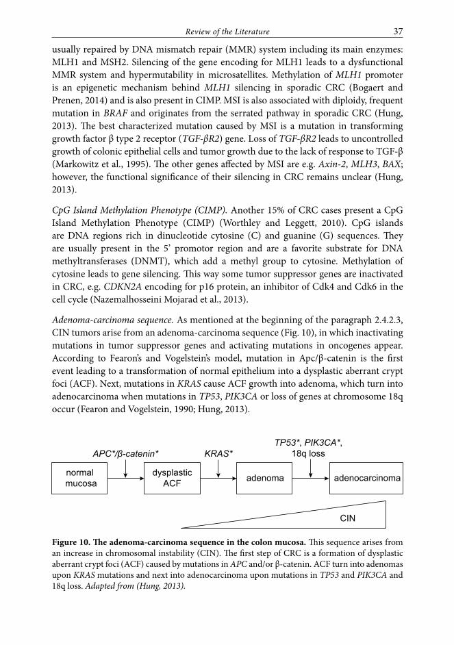
Review of the Literature 37
usually repaired by DNA mismatch repair (MMR) system including its main enzymes: MLH1 and MSH2. Silencing of the gene encoding for MLH1 leads to a dysfunctional MMR system and hypermutability in microsatellites. Methylation of MLH1 promoter is an epigenetic mechanism behind MLH1 silencing in sporadic CRC (Bogaert and Prenen, 2014) and is also present in CIMP. MSI is also associated with diploidy, frequent mutation in BRAF and originates from the serrated pathway in sporadic CRC (Hung, 2013). The best characterized mutation caused by MSI is a mutation in transforming growth factor β type 2 receptor (TGF-βR2) gene. Loss of TGF-βR2 leads to uncontrolled growth of colonic epithelial cells and tumor growth due to the lack of response to TGF-β (Markowitz et al., 1995). The other genes affected by MSI are e.g. Axin-2, MLH3, BAX; however, the functional significance of their silencing in CRC remains unclear (Hung, 2013).
CpG Island Methylation Phenotype (CIMP). Another 15% of CRC cases present a CpG Island Methylation Phenotype (CIMP) (Worthley and Leggett, 2010). CpG islands are DNA regions rich in dinucleotide cytosine (C) and guanine (G) sequences. They are usually present in the 5’ promotor region and are a favorite substrate for DNA methyltransferases (DNMT), which add a methyl group to cytosine. Methylation of cytosine leads to gene silencing. This way some tumor suppressor genes are inactivated in CRC, e.g. CDKN2A encoding for p16 protein, an inhibitor of Cdk4 and Cdk6 in the cell cycle (Nazemalhosseini Mojarad et al., 2013).
Adenoma-carcinoma sequence. As mentioned at the beginning of the paragraph 2.4.2.3, CIN tumors arise from an adenoma-carcinoma sequence (Fig. 10), in which inactivating mutations in tumor suppressor genes and activating mutations in oncogenes appear. According to Fearon’s and Vogelstein’s model, mutation in Apc/β-catenin is the first event leading to a transformation of normal epithelium into a dysplastic aberrant crypt foci (ACF). Next, mutations in KRAS cause ACF growth into adenoma, which turn into adenocarcinoma when mutations in TP53, PIK3CA or loss of genes at chromosome 18q occur (Fearon and Vogelstein, 1990; Hung, 2013).
normal mucosa
dysplasticACF adenoma adenocarcinoma
APC*/β-catenin* KRAS*TP53*, PIK3CA*,
18q loss
CIN
Figure 10. The adenoma-carcinoma sequence in the colon mucosa. This sequence arises from an increase in chromosomal instability (CIN). The first step of CRC is a formation of dysplastic aberrant crypt foci (ACF) caused by mutations in APC and/or β-catenin. ACF turn into adenomas upon KRAS mutations and next into adenocarcinoma upon mutations in TP53 and PIK3CA and 18q loss. Adapted from (Hung, 2013).

38 Review of the Literature
Serrated pathway. Nearly 30% of CRC are developed via the serrated pathway, which is followed by many tumors of CIMP- origin (Fig. 11) (Bettington et al., 2013). The name of this pathway originates from a serrated looking like epithelium formed at low levels of apoptosis. Decreased levels of apoptosis result from activated MAPK-ERK pathway. Based on the mechanism activating MAPK-ERK pathway, there are two kinds of pathways leading to serrated adenocarcinoma: traditional and sessile serrated pathway. Traditional serrated pathway originates from KRAS mutation that activates MAPK-ERK pathway and is more common in the distal colon.
normal mucosa
non-serratedhyperplastic
ACF
serratedhyperplastic
ACF
goblet cell-richhyperplastic
polyp
microvesicularhyperplastic
polyp
sessileserratedadenoma
traditionalserratedadenoma
serratedadenocarcinoma
MGMT methylation
KRAS*
CIMP-high
CIMP-low
MLH1 methylation
BRAF*
p16↑, IGFBP7↑
p16, IGFBP7
methylation
Figure 11. The sessile and traditional serrated pathway in the colon mucosa. This sequence arises from a high (sessile pathway) or low (traditional pathway) increase in CpG Island Methylation Phenotype (CIMP). The first step is an activation of MAPK-ERK pathway by KRAS (traditional pathway) or BRAF mutation (serrated pathway) which leads to the formation of hyperplastic aberrant crypt focus (ACF). The final methylation of MGMT or MLH1 in traditional and sessile pathway, respectively, cause a development of serrated adenocarcinoma. Adapted from (Patai et al., 2013).
Sessile serrated pathway originates from a BRAF mutation that activates the MAPK-ERK pathway and is more common in the proximal colon. This pathway gives rise to serrated hyperplastic ACF, which turn into polyps when the expression of two tumor suppressor genes: p16 and IGFBP7 is increased. When these genes become methylated, polyps grow into sessile serrated adenomas – the adenomas that grow directly from the colon wall without any stem. Serrated adenocarcinoma arises from sessile adenoma when MLH1 is methylated. In this pathway CIMP profiles are usually high (Bettington et al., 2013; Patai et al., 2013).
2.4.2.4. IBD as a risk factor for developing CRCIBD is a well-known risk factor for developing CRC. Colitis-associated CRC incidences constitute 1% of all CRC cases and is low due to colonoscopic surveillance, colectomy and chemopreventive effects of 5-ASA (Beaugerie and Itzkowitz, 2015). The risk of

Review of the Literature 39
CRC development increases with the duration of IBD. The other factors that influence CRC development in IBD patients are: area affected by colitis, severity of inflammation, heritable genetic factors and presence of primary sclerosing cholangitis (Feagins et al., 2009). Inflammation is also caused by oxidative DNA damage, which promotes the mutations. The molecular pathogenesis of colitis-associated cancer is similar to sporadic cases of CRC. However, the same mutations occur in different timing and frequency in the dysplasia-carcinoma sequence (Fig. 12). TP53 mutations usually initiate the transformation of normal mucosa into dysplasia, while mutations in APC cause the dysplastic tissue to transform into adenocarcinomas. Dysplastic lesions that arise in IBD-associated CRC are usually flat and of less distinct borders.
normal mucosa
indefinitedysplasia
low-gradedysplasia
adenocarcinoma
APC*KRAS*TP53*
high-gradedysplasia
Figure 12. The IBD-CRC associated pathway. TP53 mutations occur early and lead to the transformation of normal epithelium to the dysplastic one. The grade of dysplasia is increasing with KRAS mutations. APC mutations occur at the late stage of CRC formation. Adapted from (Beaugerie and Itzkowitz, 2015).
Interestingly, studies on IBD-animal models reveal that mice do not develop cancer under germ-free conditions or upon antibiotic treatment, which highlights the importance of microflora contribution to the CRC phenotype (Itzkowitz and Yio, 2004; Beaugerie and Itzkowitz, 2015).
2.4.2.5. Mouse models of CRCMouse models of CRC are useful to study the mechanism of pathogenesis and validate new therapeutic agents.
Chemically induced CRC-models. Mice very rarely develop spontaneous CRC. Thus, several different carcinogens are used for the CRC studies, for example: dimethylhydrazine (DMH) and its metabolite azoxymethane (AOM) often in a combination with inflammatory agent DSS, 2-amino-1-methyl-6-phenyl-imidazol [4,5-b] pyridine (PhIP), N-methyl-N’-nitro-N-nitrosoguanidine (MNNG), 3,2’-dimethyl-4-aminobiphenyl (DMBA) and 3,2’-dimethyl-4-aminobiphenyl (DMBGA). The most commonly used agent inducing CRC is AOM due to its high stability and potency (Neufert et al., 2007). AOM, in order to react with DNA, needs to be activated by several enzymatic reactions (Fig. 13A), which start in the liver. A final metabolic product of AOM, methyl cation reacts with DNA in the colon and adds a methyl group at the O6 and N7 position of the guanine. Formation of O6-methyl-deoxyguanosine is mutagenic (Fig. 13B). This molecule acquires different hydrogen bonding properties and instead of binding cytosine it binds thymidine. The thymidine binding causes a substitution of guanine into adenine during the DNA replication, which results in mutations (Brown

40 Review of the Literature
and Brown; Rosenberg et al., 2009). The mutations caused by AOM are usually in KRAS, β-catenin and TGFβ genes (Chen 2009). The number of tumors induced in the AOM model is dose, duration and strain dependent, e.g. in FVB/N strain the average tumor number per mouse is 3.6 upon 10 mg/kg AOM dose once per week for 4 weeks and sacrifice after 20 weeks (Nambiar et al., 2003).
NH2
N-H
ON
N
dR
CH3 N N CH3 H H
DMH
CH3 N N CH3
O
AOM
CH3 N N CH2OH
O
MAM
CH3 N N OH CH3 ++
CH3 +
-
Methyl diazonium ion
Methyl cationMethyl cation
CH3 + +
NH2
N-H
ON
N
dR
NH2
N-H
ON
N
dR
+
CH3
CH3
N
N
N
Methyl cation
2’-deoxyguanosine
O6-methyl-deoxyguanosine
N7-methyl-deoxyguanosine
O6-methyl-deoxyguanosine : tyminemispair to
adenine : tymine MUTATIONS
A.
B.
Figure 13. The AOM metabolic pathway. A. Dimethylhydrazine (DMH) is a precursor of azoxymethane (AOM), which undergoes metabolic activation by hepatic enzyme cytochrome P450 2E1 (CYP2E1) into methylazoxymethanol (MAM). Next, MAM is metabolized into methyl diazonium ion, which gives rise to methyl cation in the colon. B. Methyl cation reacts with DNA in the colon and leads to the methylation of 2’-deoxyguanosine into mutagenic O6-methyl-deoxyguanosine and non-mutagenic N7-methyl-deoxyguanosine. -dR deoxyribose. Adapted from (Brown and Brown; Rosenberg et al., 2009).
Genetically induced CRC-models. There are numerous genetically engineered mouse models of CRC available. The most popular is a model for FAP, the ApcMin/+ mice, which carry a germline mutation in APC leading to multiple intestinal neoplasia (Min). This model, however, develops adenomas mostly in small intestine, while in humans with FAP they are localized in colon. The ApcMin/+ adenomas usually do not progress into adenocarcinomas due to a short lifespan of the animals. However, a new model of ApcMin/+ mice - FabplCre, Apc15lox/+ has been created, which has a longer lifespan and develops adenocarcinomas in the colon (Robanus-Maandag et al., 2010). Adenoma multiplicity in ApcMin/+ is dependent on the mouse strain and environmental factors including type of bacteria or diet (Karim and Huso, 2013). Moreover, the number of tumors in different ApcMin/+ strains are affected by the modifiers of Min called Mom. Apart from ApcMin/+ models, conditional knock-out mouse models of MSH2 and MLH1 develop lymphomas in the intestinal tract, which mimic HNPCC (Kucherlapati et al., 2010; Reiss et al., 2010).

Review of the Literature 41
2.4.2.6. CRC treatmentSurgery is used at an early stage of CRC and is successful in 90% of the cases. Metastatic CRC (mCRC) is usually treated with chemotherapy combined with molecular therapy. This combination is used due to tumor heterogeneity and ability to compensate for loss by generating mutations in other pathways. There are several drugs available in the market for mCRC treatment (Table 6) (Sridharan et al., 2014; Scudellari, 2015). The oldest CRC drug available is 5-fluorouracil, a DNA synthesis inhibitor used in chemotherapy. The newer chemotherapeutics used in a combination with 5-fluorouracil are capecitabine, irinotecan, and oxaliplatin. The first medicine of a targeted therapy was bevacizumab, a monoclonal antibody, which by binding to vascular endothelial growth factor A (VEGF-A) blocks the vasculogenesis. The other drug from this class is aflibercept, which not only blocks VEGF but also platelet-derived growth factor (PDGF). Among drugs against mCRC are also epidermal growth factor receptor (EGFR) antibodies: cetuximab and panitumumab. EGFR is a receptor tyrosine kinase, the phosphorylation of which leads to the activation of pathways like MAPK, Akt and JNK resulting in DNA synthesis and proliferation. The disadvantage of EGFR antibodies is their inefficiency if KRAS, a protein downstream from EGFR, is mutated. The newest mCRC drug in the market is regorafenib, which inhibits many kinases (Sridharan et al., 2014). A big problem in the field is the use of more than two drugs for mCRC due to high toxicity in the patients. In order to solve this problem scientists and clinicians try to find biological markers: characteristic genetic mutations or specific patterns of gene expression. So far, three negative biological markers have been found: KRAS, NRAS and BRAF genes, which show resistance to cetuximab and panitumumab therapies. In order to find more markers and establish precise molecular profiles of the tumors, the next generation sequencing and bank of organoids are used. The other issue that needs an urgent solution is finding drugs for the most common CRC mutations in RAS genes and Wnt pathway. Attempts to develop immunotherapy against CRC have been unsuccessful so far (Scudellari, 2015).
Table 6. FDA approved agents for metastatic CRC treatment.
Class Agent CharacteristicsCytotoxic chemotherapy 5-fluorouracil Fluoropyrimidine,
DNA synthesis inhibitor
Capecitabine Fluoropyrimidine, DNA synthesis inhibitor
Irinotecan Topoisomerase I inhibitor, DNA synthesis inhibitorOxaliplatin Platinum-based compound, DNA synthesis inhibitor
VEGF inhibitor Bevacizumab Humanized monoclonal antibody, binds VEGF-A
AfliberceptRecombinant fusion molecule, contains extracellular domain of VEGFR-1 and -2 fused to IgG-1 Fc part, binds VEGF-A,VEGF-B, PGF-1 and PGF-2
EGFR antibody Cetuximab Chimeric IgG1 antibody, binds EGFRPanitumumab Human igG2 antibody, binds EGFR
Kinase inhibitor Regorafenib Binds RTK, inhibits VEGF and EGFRBased on (Sridharan et al., 2014; Scudellari, 2015)
42 Outline and Aims of the Thesis
OUTLINE AND AIMS OF THE THESIS
Although K8 is the main IF expressed in the colon, its exact role remains ambiguous. The general aim of this thesis was to investigate the role of K8 in colon cell fate, colitis-induced carcinogenesis and metabolism. Most studies were performed using K8-/- mouse model, which spontaneously develops early chronic colitis resembling human IBD.
The specific aims of the thesis were:
Aim 1: Study the effect of keratins on proliferation and differentiation (Study I.).
Aim 2: Analyze if K8-/- mice are susceptible to developing colon cancer basally and in genetic or carcinogen colorectal cancer models. Find mechanism for increased tumorigenesis (Study II.).
Aim 3: Determine whether keratins are involved in the regulation of colon energy metabolism (Study III.).

Experimental Procedures 43
EXPERIMENTAL PROCEDURES
Detailed information on experimental procedures used in the thesis are described in the manuscripts (I.-II.) and original publication (III.).
Animals
Name (background) StudyApcMin/+ (FVB/n) II.BALB/c III.K8+/+, K8+/-, K8-/- (FVB/n) I.-III.K8+/+ApcMin/+, K8+/-ApcMin/+, K8-/-ApcMin/+ (FVB/n) II.
Cell lines
Name StudyCaco-2, human colorectal adenocarcinoma III.HEK293, HEK293-FLN, human embryonic kidney cells, expressing full length Notch (FLN) I.HT-29, human colorectal adenocarcinoma III.MEFvim-/-, mouse embryonic fibroblasts from vimentin knock-out mice I.
Antibodies
Target, name (manufacturer) Application StudyAlexa Fluor 488 (ThermoFisher Scientific) staining I., III.Alexa Fluor 546 (ThermoFisher Scientific) staining I., III.Cytochrome c (Cell signaling) WB III.FLN, c-20 (Santa Cruz Biotechnology) WB, staining I.GFP (Clontech) WB III.H3 (Abcam) WB I.HMGCS2 (AVIVA and Geneway) WB, staining III.Hsc70 (Enzo Life Sciences) WB I.-III.IL-18 (R&D) WB II.IL-22BP (R&D) WB II.K8, Troma I (Developmental Hybridoma Bank, University of Iowa) WB, staining I.-III.K8, 273 (kind gift of John Eriksson’s laboratory) IP I.-II.K8/K18, 275 (kind gift of John Eriksson’s laboratory) IP I.-II.MCT1 (Chemicon and Santa Cruz Biotechnology) WB, staining III.Caspase-1 (AdipoGen) WB II.PHH3 (Cell signaling) WB, staining I.PPARα (Santa Cruz Biotechnology) WB III.P-STAT3 (Cell Signaling Technology) WB II.Prohibitin (Abcam) WB III.Synaptophysin (Abcam) WB I.STAT3 (Cell Signaling Technology) WB II.Tubulin (Sigma-Aldrich) WB III.WB = western blotting, IP = immunoprecipitation

44 Experimental Procedures
Methods
Name Study2D DIGE III.Antibiotic treatment I.-III.AOM- and AOM/DSS- induced colorectal carcinogenesis II.BrdU staining I.Cell culture I., III.Colon permeability assay, FD4 II.DSS-induced colitis II.-III.Image analysis I.-III.Immunofluorescence I., III.Immunohistochemistry I.Immunoprecipitation (IP) I.-II.Mitochondria isolation and HMGCS2 enzyme activity assay III.PCR as a method for mouse genotypying I.-III.Periodic acid-schiff (PAS) staining I.Quantitative reverse transcription PCR I.-III.SDS-PAGE and immunoblotting I.-III.Statistical analysis I.-III.Transmission electron microscopy III.
Selected reagents
Name (manufacturer) Application Study5-Bromo-2’-deoxyuridine, BrdU (BD Biosciences)
BrdU staining I.
Azoxymethane, AOM (Sigma-Aldrich)
Colorectal carcinogenesis II.
Cycloheximide, CHX (Sigma-Aldrich) Inhibition of translation I.DAPI (Life Technologies) Immunofluorescence I., III.Dextran sulphate sodium, DSS (TdB) Colitis induction II.-III.Fluorescein isothiocyanate–dextran, FD4 (TdB) Colon permeability assay II.Imipenem (Hospira) Antibiotic treatment I.-II.Lipofectamine 2000 (Invitrogen) Transfection III.MG132 (Santa Cruz) Inhibition of proteasome I.Myers hemalum (Merck) PAS staining I.Periodic acid (Sigma-Aldrich) PAS staining I.Protein G sepharose (GE Healthcare) Immunoprecipitation I.-II.Shiff reagent (Merck) PAS staining I.Vancomycin (Hospira) Antibiotic treatment I.-II.
siRNA
Name (Manufacturer) StudyKRT8 Pre-design Chimera RNAi (Abnova) III.KRT18 Pre-design Chimera RNAi (Abnova) III.Naito1 scrambled negative control Pre-design Chimera RNAi (Abnova) III.KRT8 siMAX siRNA (Eurofins Genomics) III.Nontarget scrambled negative control siMAX siRNA (Eurofins Genomics) III.

Experimental Procedures 45
Plasmids
Name (Manufacturer) StudyFLN (kind gift of Cecilia Sahlgren’s and Urban Lendahl’s laboratory) I.K18 (kind gift of Bishr Omary’s laboratory) I.K19 (kind gift of Bishr Omary’s laboratory) I.K8 (kind gift of Bishr Omary’s laboratory) I.K8 S74A (kind gift of Bishr Omary’s laboratory) I.MCT1-GFP (kind gift of Pradeep Dudeja’s laboratory) III.NICD-GFP-Flag (kind gift of Cecilia Sahlgren’s and Urban Lendahl’s laboratory) I.PCDNA3.1(AddGene) I.ΔE Notch1 (kind gift of Cecilia Sahlgren’s and Urban Lendahl’s laboratory) I.

46 Results and Discussion
RESULTS AND DISCUSSION
1. K8 regulates differentiation in the colon through Notch1 signaling (Study I.)
K8 deletion in mice leads to a broad colonic phenotype including colonocyte hyperproliferation (Baribault et al., 1994). The colon epithelium of K8+/- mice express 50% less keratins compared to K8+/+ and display increased proliferation (Asghar et al., 2015). These data suggest the involvement of keratins in colon epithelial proliferation. Colon epithelial tissue originates from stem cells, which differentiate into various cell types. As mentioned in section 2.2.2, the master regulator of proliferation and differentiation in the colon epithelium is the Notch1 signaling, the regulators of which are largely unknown. The aim was to study whether any link exists between keratins and Notch1 in the colon and to characterize the population of epithelial cells in the K8-/- colon.
1.1. K8 binds and colocalizes with Notch 1 in vitro and in vivoKeratins are known to serve as scaffolds for many proteins (Pan et al., 2013). To find out whether keratins are the binding partners of Notch1, a K8/K18 immunoprecipitation on K8+/+ colonic epithelium was performed. Subsequent Western blot analysis showed that NICD is pulled down together with K8/K18 from both proximal (PC) and distal colon (DC) epithelium (I.: Fig. 1A, SFig. 1). To confirm this result and specify which part of Notch1 binds to K8/K18, a Notch1 immunoprecipitation was done in mouse embryonic fibroblasts lacking vimentin and other IF (MEFvim-/-) and overexpressing FLN, ΔE Notch1 or NICD with and without the presence of K8/K18 or K8 S74A/K18. Based on the Western blot results, we observed that K8 is co-immunoprecipitated with all Notch forms, while K18 was not detected in this complex (I.: Fig. 1B, SFig. 2A). K8 S74A was also pulled down with NICD from K8 S74A/K18 transfected cells as detected by Western blot (I.: Fig 1B) indicating that K8 binding to Notch1 is not phosphorylation dependent. Immunostaining of K8 and Notch1 in K8+/+ mouse colon and human embryonic kidney cells stably overexpressing FLN (HEK293-FLN) showed that K8 colocalize with Notch1 at the plasma membrane and cytoplasm both in vivo and in vitro, which was confirmed by image analysis using Manders’ co-localization. The coefficients for in vivo were: 0.65 for Notch1 and 0.69 for K8 in the epithelium (p = 1) and for in vitro: 0.68 for Notch1 and 0.75 for K8 in the HEK293-FLN cells (p = 1), (I.: Fig. 1C-D).
Taken together, our results indicate that K8 and Notch1 bind and colocalize both in vivo and in vitro. (I.: Fig. 1C, SFig. 1 and SFig. 2A). The colocalization of Notch1 and K8 at the plasma membrane and cytoplasm indicates them as sites of potential protein-protein interaction. A similar colocalization between K8 and Notch2 is seen at the plasma

Results and Discussion 47
membrane in the uretic bud of E17.5 murine kidney (Liu et al., 2013). Interestingly, the impairment of Notch2 signaling is also observed in the biliary epithelial cells of K19-/- mice treated with the 3,5-diethoxycarbonyl-1,4-dihydrocollidine (DDC) further supports the keratin - Notch interactions (Chen et al., 2015).
1.2. Notch1 levels and activity are modified by K8/K18 in in vitro cell culture models
Since K8 and Notch1 can bind together, it was tested whether keratins can regulate the Notch signaling pathway. First, MEFvim-/- cells were transfected with K8/K18 and NICD levels were evaluated. The NICD levels were increased 2-3 fold upon K8/K18 overexpression (I.: Fig. 2A). Also, overexpression of K8 phosphorylation-deficient mutant – K8 S74A resulted in the increase of NICD levels (I.: Fig. 2A). The increase in NICD expression was in a keratin-dose dependent manner (I.: Fig. 2B). Furthermore, the overexpression of K8/K19 dimer led to the increase in NICD levels (I.: Fig. 2C) indicating that the NICD induction is not dependent on the K8 dimeric partner. To investigate whether K8/K18 stabilize Notch1, cycloheximide (CHX) was used to block the translation of proteins in MEFvim-/- cells overexpressing NICD alone or together with K8/K18 or K8 S74A/K18. The K8/K18-induced NICD levels decreased gradually with a similar turnover as that of NICD alone, although the actual levels of NICD were 2-3 fold higher in the presence of K8/K18 or K8 S74A/K18 filaments (I.: Fig. 2D-E, SFig. 2B). Blocking the proteasome with MG132 for 12h in MEFvim-/- cells led to a significant increase in NICD levels only in the absence of keratins (I: Fig. 2F, SFig. 2C) compared to the slight increase observed when NICD was co-expressed with K8/K18 or K8 S74A/K18 (I.: Fig. 2F). The influence of keratins on the activity of Notch1 signaling was analyzed by evaluating the mRNA levels of the Notch target gene Hey1. The mRNA levels of Hey1 were significantly increased when NICD was overexpressed together with K8/K18 compared to NICD overexpression alone, while K8S74A/K18 overexpression did not have an influence on Hey1 mRNA levels compared to control MEFvim-/- cells (I: Fig. 2G). HEK293 and HEK293-FLN cell lysates were also analyzed for keratin expression. The Western blot clearly showed that stable FLN overexpression led to significant increase in K8 and K18 protein levels compared to control HEK293 cells (I.: Fig. 2H), further demonstrating a reciprocal regulatory relationship between K8 and Notch1.
Altogether, these data show that stable overexpression of FLN can increase the expression of both K8 and K18. This observation is in line with a previous study (Hamidi et al., 2011) where KRT18 has been found as a target of CSL- a Notch transcriptional factor, which is activated by NICD during hematopoiesis. We also show that K8 is able to increase the expression of NICD in a dose-dependent manner. This indicates that K8 – Notch1 interaction is reciprocal. Moreover, the data with MG132 proteasome inhibitor shows that K8/K18 protect Notch1 from proteasomal degradation. Mutation in K8 at S74 seems not to have a negative effect on the increase of NICD and its degradation stays at the same level regardless of Alanine substitution. Moreover, the overexpression of K8

48 Results and Discussion
S74A site with NICD does not increase the mRNA levels of Hey1, which suggests that K8 S74 phosphorylation may have a regulatory role in Notch signaling activity (I.: Fig. 2G). How keratins exactly affect Notch1 activity needs further studies. Since keratins are known to target proteins at the cell membranes (Toivola et al., 2004; Vijayaraj et al., 2009), it can be speculated that keratins facilitate Notch1 receptor cleavage – a membrane-proximal site event. Moreover, keratins may prolong NICD activity by stabilizing and retaining its pool in the cytoplasm. This might be accomplished through 14-3-3 protein - a binding partner of K8/K18 (Liao and Omary, 1996; Ku et al., 2002b), as recent studies have shown that it facilitates the Notch4 nuclear translocation (Ramakrishnan et al., 2015).
1.3. Decreased Notch1 in the K8-/- mouse colon results in a cell differentiation shift
The levels and activation of Notch signaling were also analyzed in vivo in the K8-/- colon epithelium. Both FLN and NICD were significantly decreased in the K8-/- colon on protein level (I.: Fig. 3A-C), while mRNA Notch1 levels remained unaltered (I.: SFig. 2D). The NICD downregulation was confirmed by immunohistochemistry staining, which showed less NICD throughout the whole colonic crypt in K8-/- colon compared to K8+/+ and K8+/- (I.: Fig. 3D). The decreased mRNA levels of Hey1 and Hey2 were also seen in K8-/- colon epithelium, which confirms lower Notch activity also in vivo (I.: Fig. 3E).
Since Notch1 target genes can be directly activated by colonic bacteria (Becker et al., 2013), we wanted to evaluate whether microbiota and inflammation affect the observed Notch phenotype in the K8-/- colon. After broad-spectrum antibiotic treatment for 8 weeks, a substantial decrease in FLN in the K8-/- colon was still observed (I.: SFig. 2E). This observation suggests that the Notch phenotype in the K8-/- colon is caused rather due to K8 loss than bacteria-induced inflammation. However, the evaluation of NICD as well as Hey1 and 2 levels are needed to further confirm this result.
Active Notch signaling is known to induce enterocyte differentiation and inhibit goblet cell and EEC differentiation (Sancho et al., 2015). To test whether decrease in Notch activity has a further impact on cell differentiation in the K8-/- colon, the pattern and composition of enteroendocrine cells (EEC), enterocytes and goblet cells was evaluated in the colonic epithelium. The following markers have been used: villin for enterocytes (Robine et al., 1997), mucus/mucins for goblet cells (Garg et al., 2007) and synaptophysin for EEC (Thanasupawat et al., 2013).
Villin protein levels were significantly decreased in K8-/- colon epithelium suggesting a reduced number of enterocytes (I.: Fig. 4A-B). This result was further confirmed by decreased mRNA levels of carbonic anhydrase 2 (CA2) in K8-/- colon, which is produced by enterocytes (I.: Fig. 4C).

Results and Discussion 49
Next, the amount of goblet cells was analyzed by Periodic acid-Schiff (PAS) staining, specific for goblet cells. Quantification of mucus positive cells revealed more goblet cells in the K8-/- colon in both proximal (PC) and distal (DC) parts compared to K8+/-. The amount of goblet cells in K8+/- colon was also increased in the distal part compared to K8+/+ (I.: Fig 4D, SFig. S3). However, the RT-PCR analysis of mucus components showed a significant increase in Mucin1 and Mucin2 (Kim and Ho, 2010) in K8-/- colon compared to K8+/+ (I.: Fig. 4E).
The immunoblotting against synaptophysin was done to analyze the amount of EEC cells. Synaptophysin levels in total colon lysate were significantly higher in the K8-/- compared to K8+/+ (I.: Fig. 4F-G). To confirm this result and visualize the position of EEC in the colon crypts, the immunofluorescent staining was performed. The synaptophysin was mostly stained in the crypt bottom in K8+/- and K8-/- colons. Interestingly, an increased number of EEC cells was observed in the K8-/- crypt bottom (I.: Fig. 4H b) compared to K8+/+ (I.: Fig. 4H a). Together, the data showed a decreased number of enterocytes and an increased number of goblet cells and EEC in K8-/- colon.
The next aim was to characterize and compare the amount and localization of TA cells which differentiate into absorptive enterocytes and secretory cells in the colon, (Schepers and Clevers, 2012). Phosphohistone H3 (PHH3) was used as a marker of TA cells. Western blot analysis showed higher levels of PHH3 in the K8-/- and K8+/- epithelium compared to K8+/+ (I.: Fig. 5A-B), which was confirmed by the immunofluorescent staining (I.: Fig. 5C). Moreover, the 5-bromo-2’-deoxyuridine (BrdU) labeling, 4 h after injection, also showed an increase in the number of proliferative cells and wider proliferative zone in K8-/- colon crypts compared to K8+/+ (I.: SFig. 4 a-c). Interestingly, 72 h after injection, only the K8+/+ cells clearly migrated to the top of the crypts compared to K8-/- and K8+/- crypts (I.: SFig. 4 d-f). Supportive for the impaired cell migration in K8-/- was a 60% decrease in the mRNA levels of its cell position regulator Ephrin type-B receptor 2 (EphB2) compared to K8+/+. The EphB2 levels were not significantly reduced in the K8+/- colon (I.: Fig. 5D).
Taken together, the analysis of colonic cell types in K8-/- mice shows a shift towards the secretory cell differentiation in the absence of K8 (I.: Fig. 6, STable 1). This observation supports the decreased Notch1 activity in the K8-/- colon. The study also indicates that keratin dose-dependent hyperproliferation (Asghar et al., 2015) is due to an increased number of TA cells and their widened proliferative zone results in longer crypts in the K8-/- colon (Toivola et al., 2004; Habtezion et al., 2011). The data presented here are supported by other studies involving Notch and IFs, e.g. Notch1 regulated nestin expression (Shih and Holland, 2006) or reduced Notch1 signaling in GFAP-/-vim-/- astrocytes (Wilhelmsson et al., 2012). On the other hand, K14 downregulation in skin has been reported to lead to Notch1 increase (Alam et al., 2011b), while Notch downregulation in skin cancer decreased K13 and K15, but increased K17 levels (Sakamoto et al., 2012).
50 Results and Discussion
2. K8-deletion induced colitis is a risk factor for CRC development (Study II.)
Mutations in mainly K8 are reported in IBD patients (Buning et al., 2004; Owens et al., 2004; Tao et al., 2007; Zupancic et al., 2014; Corfe et al., 2015a) and K8 deletion in mice leads to chronic colitis resembling human IBD. Since IBD is a well-known factor for developing CRC (Beaugerie and Itzkowitz, 2015), the aim was to investigate whether the K8-/- colon is prone to develop cancer. Moreover, epithelial cells hyperproliferate in the K8-/- colon (Toivola et al., 2004), which also might contribute to the CRC development.
2.1. Increased susceptibility of K8-/- colon to induced tumorigenesis indicates keratin involvement in colonic homeostasis
The examination of colons from both young and aging of K8-/- mice clearly revealed that they do not develop CRC spontaneously. Methylene blue staining of 9- month-old K8-/-, K8+/+ and K8+/- colons did not show any difference in the number of ACF, potential precursors of CRC (II.: SFig. 1A). Next, the susceptibility of K8-/- mice to develop cancer using two well-studied CRC models: AOM and ApcMin/+ was tested. The study with a single low dose (1 x 10 mg/kg) AOM treatment showed a significantly higher number of ACF in K8-/- colon compared to K8+/+ colon (II.: SFig. 1A). Multiple-dose AOM treatment (4 x 5mg/kg) led to tumor development mainly in the K8-/- colon, which displayed on average 6 tumors per mouse. K8+/+ and K8+/- colons developed one or no tumors, respectively upon the same treatment (II.: Fig. 1A-B). The K8-/- tumors were classified as tubular adenomas of high grade dysplasia compared to low grade dysplasia in K8+/+ tumors (II.: Fig. 1C). Moreover, the K8-/- tumors were nearly on average larger than K8+/+ tumors (3.2 ± 0.7 mm2 in K8-/- versus 1.6 ± 1.2 mm2 in K8+/+; p = 0.07). To determine whether colitis-induced tumorigenesis was affected by keratin-levels, the AOM-DSS was administered to K8+/+ and K8+/- mice. The examination of the colons showed a 2-fold increase in the number of polyps in the K8+/- mice compared to K8+/+ (18 ± 12 polyps in K8+/- mice versus 9 ± 9 in K8+/+ mice), but this result did not reach statistical significance (p = 0.17), (II: SFig. 1B).
Next, the susceptibility of K8-/- colon to develop CRC in the ApcMin/+ model was tested. K8-/- mice on FVB/N background were crossed with ApcMin/+ mice also on FVB/N background due to the K8-/- embryo lethality in the C57BL/6J strain (Baribault et al., 1993). Although, ApcMin/+ mice on C57BL/6J background develop numerous tumors in the small intestine (Moser et al., 1990), the tumorigenesis in the FVB/N strain is insignificant in either small or large intestine (Svendsen et al., 2011). The crosses of 4-6 month old K8-/-ApcMin/+ mice developed 11 ± 1.6 tumors, which were exclusively located in the distal colon and were classified as well differentiated intramucosal adenocarcinomas. No tumors were present in the intestinal tract of K8+/+ApcMin/+ or K8+/-ApcMin/+ mice (II.: Fig. 2A-C). However, treating these control mice with DSS for one week and no further treatment for 4 weeks resulted in colon tumor development, which was comparable between the genotypes (II.: SFig. 1C).

Results and Discussion 51
Keratins are known as biomarkers in tumor diagnostics. K7 expression is often decreased, while K20 is increased in colorectal adenocarcinomas. Moreover, decreased levels of K8 and K20 are observed in the tumors of epithelial-to-mesenchymal transition, which is associated with higher metastatic potential and decreased survival in patients (Karantza, 2011). Aside from these, the role of keratins in the colon tumorigenesis remains ambiguous. Our results reveal that K8 deletion alone does not lead to spontaneous CRC development even at the old age of mice.
Nevertheless, the results show a much higher susceptibility of the K8-/- colon to develop CRC in two models: AOM and ApcMin/+ models, which indicates the role of keratins in colon homeostasis. Since, K8-/- mouse colon displays both hyperproliferation and inflammation, we investigated which of these two processes is a triggering point for tumorigenesis. The experiments with AOM-DSS and DSS alone on K8+/+, K8-/- and K8+/+ApcMin/+, K8-/-ApcMin/+, clearly indicated that inflammatory environment, not hyperproliferation alone, is needed for the tumor development in the K8-/- colon. This is consistent with the fact that inflammation is one of the hallmarks of cancer (Hanahan and Weinberg, 2011). Furthermore, certain studies acknowledge keratins as the regulators of skin immunity (Roth et al., 2012), and the K8+/- mice display a slightly higher susceptibility to experimentally-induced colitis (Asghar et al., 2015). Also a recent proteomic study reported decreased K8, K18 and K19 levels in some IBD patients with a potential correlation to the development of CRC (Corfe et al., 2015), which supports the K8-/- colon phenotype. However, it remains to be determined whether humans or mouse models with disease-predisposing keratin mutations are more susceptible to CRC.
2.2. IL-22 pathway is involved in the K8-/- colon tumorigenesisIL-22 is involved in tissue repair, inflammation and inflammation-induced carcinogenesis in many organs including colon. IL-22 levels are often elevated in IBD patients (Schmechel et al., 2008; Sonnenberg et al., 2011). Since the K8-/- colon hyperproliferates and develops colitis and is susceptible to CRC, we examined the levels of IL-22. RT-PCR results showed an over 6-fold increase in the IL-22 mRNA level of the K8-/- colon compared to K8+/+ and K8+/- colons (II.: Fig. 3A). IL-22 activity is negatively regulated by IL-22BP; thus its mRNA levels were also analyzed. The IL-22BP mRNA levels were increased 2-fold compared to K8+/+ (II.: Fig. 3B). On the contrary, western blot analysis revealed nearly a total loss of IL-22BP protein basally in both the total colon lysates and epithelium enriched fractions of K8-/- colons as well as in the tumor tissue of K8-/-ApcMin/+ colons. Interestingly, IL-22BP levels were intermediate in K8+/- and K8+/-ApcMin/+ colon (II.: Fig. 3C-E). The protein levels of IL-22RA1, an epithelial receptor of IL-22, were variable and relatively unchanged in K8-/- colon epithelium compared to K8+/+ and K8+/- (Fig. 14A). The activated state of IL-22 was confirmed by the elevated levels of its downstream effector - phosphorylated STAT3 (P-STAT3) in both K8-/- colon epithelium and K8-/-ApcMin/+ tumor tissue (II.: Fig. 3D-E). Furthermore, the RT-PCR analysis showed increased mRNA levels
52 Results and Discussion
of STAT3 target genes RegIIIγ and S100A11 (7.5-fold and 2-fold, respectively) in the K8-/- colon compared to K8+/+ (II.: SFig. 2). It has earlier been shown that active IL-22 and STAT3 signaling can indicate tissue damage and therefore activated defense mechanisms; thus, the colonic barrier capacity was analyzed. Oral administration of fluorescent dextran (FD4) detected a break of the intestinal barrier in K8-/- mice compared to K8+/+ and K8+/- (II.: Fig. 4E). However, these results are somewhat contradictory to previous K8-/- colon permeability studies (Toivola et al., 2004); thus further work is needed to clarify the colonic barrier of K8-/- colon. In addition, the upstream of IL-22BP signaling was investigated which, according to the current literature, is barely known. Since one of the studies suggests IL-18 as an inhibitor of IL-22BP expression, IL-18 levels were examined by western blot. The IL-18 protein levels were elevated up to 20-fold in the colonic epithelium of K8-/- compared to K8+/+ and K8+/- (II.: Fig. 4C). Similar to IL-22BP, the levels of IL-18 were intermediate in K8+/- colon epithelium compared to other genotypes (5.7-fold increase compared to K8+/+, p = 0.05 in Student’s t-test), (II.: Fig. 4C).
Our results showed dramatically increased levels of IL-22 in the K8-/- mouse colon. Interestingly, no difference in the levels of colonic IL-22 was detected between K8+/+ and K8+/- mice, which likely explains why no tumors are developed in these animals. The IL-22 activity is neutralized by IL-22BP, a receptor known to be downregulated during tissue damage (Kotenko et al., 2001; Huber et al., 2012). IL-22BP-/- mice, apart from prolonged IL-22 signaling and epithelial tissue repair, develop an increased number of colonic tumors in AOM/DSS and ApcMin/+ models (Huber at al., 2012). Our data indicated a dramatically decreased or a total lack of IL-22BP protein in the K8-/- colon, both basally and in the tumor tissue of K8-/-ApcMin/+ mice (II.: Fig. 3C-E). Recently, two mouse models: STING (stimulator of interferon signaling)-knock-out mice and Rag-APC natural killer cell depleted mice have been reported to express decreased IL-22BP levels during induced tumorigenesis (Janakiram et al., 2014; Ahn et al., 2015). To our knowledge, K8-/- is the only mouse model so far, which presents such a strong downregulation/lack of IL-22BP protein under basal conditions. Supportive to the regulatory role of keratins in IL-22BP expression are the intermediate levels of IL-22BP between K8+/+ and K8+/- colon, which has 50% less keratin filaments. IL-22BP regulation likely occurs on a posttranslational level, since IL-22BP mRNA levels were not decreased in the K8-/- colon. Moreover, the treatment with broad-spectrum antibiotics, which alleviates colitis in K8-/- mice, has not influenced the levels of expressed IL-22BP (Fig. 14B) indicating likely no involvement of bacteria in the observed phenotype.
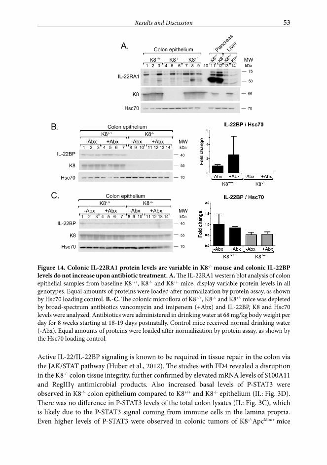
Results and Discussion 53
K8+/+ K8-/- K8+/- 1 2 3 4 5 6 7 8 9 10 11 12 13 14
Colon epithelium
IL-22RA1
Hsc70
kDa
70
MW
75
55K8
K8+/+
K8+/+
K8-/-
K8-/-
Pancre
as
Liver
50
K8+/+
1 2 3 4 5 6 7 8 9 10 11 12 13 14
Colon epithelium
IL-22BP
Hsc70
40
70
kDaMW
55K8
-Abx +AbxK8-/-
-Abx +Abx
K8+/+
1 2 3 4 5 6 7 8 9 10 11 12 13 14
Colon epithelium
40
70
kDaMW-Abx +Abx
K8+/-
-Abx +Abx
55
IL-22BP
Hsc70
K8
A.
B.
C.
Figure 14. Colonic IL-22RA1 protein levels are variable in K8-/- mouse and colonic IL-22BP levels do not increase upon antibiotic treatment. A. The IL-22RA1 western blot analysis of colon epithelial samples from baseline K8+/+, K8-/- and K8+/- mice, display variable protein levels in all genotypes. Equal amounts of proteins were loaded after normalization by protein assay, as shown by Hsc70 loading control. B.-C. The colonic microflora of K8+/+, K8-/- and K8+/- mice was depleted by broad-spectrum antibiotics vancomycin and imipenem (+Abx) and IL-22BP, K8 and Hsc70 levels were analyzed. Antibiotics were administered in drinking water at 68 mg/kg body weight per day for 8 weeks starting at 18-19 days postnatally. Control mice received normal drinking water (-Abx). Equal amounts of proteins were loaded after normalization by protein assay, as shown by the Hsc70 loading control.
Active IL-22/IL-22BP signaling is known to be required in tissue repair in the colon via the JAK/STAT pathway (Huber et al., 2012). The studies with FD4 revealed a disruption in the K8-/- colon tissue integrity, further confirmed by elevated mRNA levels of S100A11 and RegIIIγ antimicrobial products. Also increased basal levels of P-STAT3 were observed in K8-/- colon epithelium compared to K8+/+ and K8-/- epithelium (II.: Fig. 3D). There was no difference in P-STAT3 levels of the total colon lysates (II.: Fig. 3C), which is likely due to the P-STAT3 signal coming from immune cells in the lamina propria. Even higher levels of P-STAT3 were observed in colonic tumors of K8-/-ApcMin/+ mice
54 Results and Discussion
(II.: Fig. 3E), which is consistent with an oncogenic role of P-STAT3 (Levy and Inghirami, 2006). Recently, it has been shown that IL-22 is able to upregulate K17 in keratinocytes via STAT3 and Erk1/2 pathway, which could suggest a compensatory role of IL-22 in the lack of K8 in the colon. Our studies also focused on the role of IL-18, as it was proposed to negatively regulate the levels of IL-22BP. As detected by Western blotting, IL-18 expression in the K8-/- colon epithelium was highly upregulated, which provides support for IL-22BP regulation. Similarly, to IL-22BP, IL-18 expression was intermediate between K8+/+ and K8+/- colon epithelium, which demonstrates keratin involvement in the upstream processes from IL-18.
2.3. K8 binds the inflammasome - a potentially new cytoskeletal contributor to the IL-22 pathway
IL-18 is processed into mature form by an active NLRP3 inflammasome from which caspase-1 is released (Huber et al., 2012). In agreement with the IL-18 results, we observed elevated levels of active caspase-1 exclusively in K8-/- and K8-/-ApcMin/+ colons (II.: Fig. 4A-C). To address the involvement of keratins in the IL-22 pathway, K8/K18 were immunoprecipitated from the colonic epithelium of K8+/+ mice. Pro-caspase-1, a component of inflammasome co-immunoprecipitated with K8/K18 from both proximal and distal colon (II: Figure 4D), which indicates a potential interaction between these proteins. In summary, the results show the binding of intestinal keratins to the inflammasome and in the lack of K8 increased inflammasome activity. Supportive to these data is the keratin 1 knock-out (Krt1-/-) mouse model, in which inflammasome-dependent IL-18 upregulation together with increased production of antimicrobial ligands S100A8 and S100A9 are observed in keratinocytes with an inflammatory skin disease phenotype (Roth et al., 2012). Inflammasomes, similar to keratins, act as cytoplasmic sensors of cellular stress upon which they are activated (Schroder and Tschopp, 2010; Toivola et al., 2010). Common stress stimuli might shed more light on the keratin-inflammasome interactions. Interestingly, inflammasomes are also activated by toll-like receptors (TLR) (Fernandes-Alnemri et al., 2013) and TLR9 is upregulated in the K8-/- colon (Habtezion et al., 2011). Microarray data from experimental colitis of C57BL/6 mice identified keratins as major genes upregulated during the recovery phase (Breynaert et al., 2013), which confirms the stress-protective role of keratins essential for healing and repair of the colon. Recently, a mesenchymal IF, vimentin, was shown to bind to NLRP3 inflammasome and lead to its activation (dos Santos et al., 2015). In contrast to K8-/- mice, vimentin knock-out mice in that study were protected from lung inflammation and injury, which was associated with an active inflammasome. This discrepancy could be due to the different inflammasome localization- mesenchymal vs. epithelial and more tissue specific studies are needed to resolve this issue. Taken together, our data nominate keratins as novel ligands that bind inflammasome and elicit intestinal epithelial cell inflammasome activation and, that thus, can regulate the IL-22 pathway. Essential for further studies, is to analyze which inflammasome type is central in this process.

Results and Discussion 55
3. K8 influences colon energy metabolism (Study III.)
The main source of colonocyte energy is SCFA produced by bacterial fermentation of starch and carbohydrates. SCFAs are mainly transported to the colon epithelial cells via the monocarboxylate transporters, like MCT1. Butyrate, the major SCFA produced in the colon, regulates the levels of mitochondrial HMGCS2, which is a rate limiting enzyme of ketogenesis. Intriguingly, HMGCS2 turned out to be the major downregulated protein in K8-/- colonocytes. Thus the aim was to determine whether and how keratins are involved in the regulation of colon energy metabolism.
3.1. HMGCS2 downregulation is K8-/- colon specific and leads to blunted ketogenesis
2D DIGE and mass spectrometry were performed to identify differentially regulated proteins in K8+/+ and K8-/- colonocytes (III.: Fig.1A-B). Apart from the expected downregulation of K8 and K19, a group of mitochondrial, cytoplasmic and endoplasmic reticulum proteins was found most differentially expressed in K8-/- colonocytes (III: STable 1). Among these proteins was HMGCS2, a mitochondrial enzyme of ketogenesis, which displayed a 4-fold decrease in K8-/- colonocytes (III.: Fig. 1A-B, spots 1032, 1033, 1035 and 1044) compared with K8+/+. This finding was subsequently confirmed by HMGCS2 immunoblotting of K8+/+ and K8-/- colon crypts (III.: Fig. 1C). A slight downregulation of HMGCS2 was also observed in vitro in Caco-2 and HT-29 human colorectal cancer cells, when K8 levels were decreased by siRNA silencing (III.: SFigure 1). To examine if K8-/- loss also affected HMGCS2 enzyme activity, the evaluation of both HMGCS2 activity and quantity was performed on the same set of mitochondria isolated from colonic epithelium. The results clearly showed a parallel downregulation of HMGCS2 protein quantity and enzyme activity in K8-/- colonic epithelium compared to K8+/+ (III.: Fig. 1D-E). As revealed by HMGCS2 activity/quantity ratio, the decrease in enzyme quantity correlated with the decrease in enzyme activity in K8-/- colon (III.: Fig. 1F).
Next, we investigated whether HMGCS2 downregulation appears exclusively in the colon or also in other organs. The protein lysates from proximal and distal colon, small intestine and liver were analyzed by immunoblotting and the results showed that HMGCS2 downregulation occurs only in the colon (III.: Fig. 2A and C, SFig. 3). The blot also indicated higher levels of HMGCS2 expression in the proximal than in the distal colon, which is likely due to a higher number of SCFA-producing bacteria in the mouse proximal colon than in the distal colon (Hamer et al., 2008). The HMGCS2 downregulation in K8-/- colonocytes was also detected by the immunostaining and the remaining HMGCS2 was localized in the apical part of the K8-/- cells (III.: Fig. 2B). As previously reported, HMGCS2 is not expressed in the small intestine, but highly expressed in the liver, which was confirmed by our results. Interestingly, there were no major differences in HMGCS2 expression observed between K8+/+ and K8-/- livers (III.: Fig. 2C and SFig. 3). In addition to these results, the levels of the HMGCS2 transcription factor – PPARα were also found downregulated in K8-/- colon (III.: Fig. 2A). Since K8-/- mice spontaneously develop

56 Results and Discussion
colitis, it is possible that the inflammation leads to the downregulation of the HMGCS2 levels. However, the treatment of BALB/c mice with the colitis inducing reagent, DSS, resulted in unchanged or increased levels of HMGCS2 (III.: Fig. 2D).
Further, the response of K8-/- colonocytes to ketogenic conditions was analyzed. The mice were starved for 12 and 24 hours or fed with a ketogenic diet for 3 or 14 days, after which HMGCS2, blood glucose, and blood β-hydroxybutyrate levels were determined. Starvation for both 12 and 24 h induced ketogenic conditions, since blood glucose levels decreased and blood β-hydroxybutyrate levels increased at the same time (III.: Fig. 3A-B). Ketogenic diet did not influence glucose levels, but β-hydroxybutyrate levels were significantly upregulated after 14 days in both K8+/+ and K8-/- mice, which indicates induced ketogenic conditions (III.: Fig. 3C-D). Moreover, serum parameters including cholesterol, bicarbonate and triglycerides were assayed and no major differences between the genotypes and control versus ketogenic group were detected besides decreased cholesterol levels in K8-/- mice upon 7 days of ketogenic diet (III.: Fig. 3E). At the same time, the food ingestion and mouse weight were monitored and no differences were observed (III.: SFig. 4). These data provide further evidence for unaffected ketogenesis in K8-/- liver and show a suitability of these ketogenic conditions to study colon ketogenesis. The colonic HMGCS2 levels were upregulated in K8+/+ mice upon starvation and ketogenic diet for 3 days, in contrast to K8-/- mice, whose colonic HMGGCS2 levels were not significantly increased upon ketogenic conditions (III.: Fig. 4A-F). A similar trend of HMGCS2 mRNA increase was observed in K8+/+ under ketogenic conditions, while in K8-/-, the HMGCS2 mRNA remained almost unchanged (III.: Fig. 4G). The expression of PPARα followed HMGCS2 expression pattern and displayed a slight upregulation in K8+/+ and a slight downregulation in K8-/- colon under ketogenic conditions (III.: Fig. 4A, C, E). These results clearly indicate a blunted ketogenesis in K8-null colonocytes.
Altogether, the proteomic studies revealed HMGCS2 as a major downregulated protein in the K8-/- colonocytes. This observation was colon specific and no major HMGCS2 downregulation was detected in the liver of K8-null mice, although liver is the main ketogenic organ where keratins are highly expressed and have cytoprotective roles (Hegardt, 1999; Ku et al., 2007). Interestingly, HMGCS2 downregulation in the mouse liver has recently been reported to contribute to the inflammatory liver disease, steatohepatitis (Cotter et al., 2014). On the other hand, it is very probable that microbiota contribute to colonic HMGCS2 downregulation. Supportive for this hypothesis is the regulatory role of bacterially produced SCFA on HMGCS2 expression (Cherbuy et al., 2004) and the presence of fewer luminal bacteria in the K8-/- colon (Habtezion et al., 2011). Another factor modulating HMGCS2, which should be taken into consideration, is the colitis developed by K8-/- mice. Our results with DSS-induced colitis in BALB/c mice showed no effect or an increase in HMGCS2 expression, which is in line with a previous study (Naito et al., 2010) and suggests that HMGCS2 downregulation is specifically caused by the lack of K8 but not inflammation per se. In addition, the results clearly showed that K8-/- colon was not able to increase HMGCS2 levels under the ketogenic conditions compared to K8+/+.

Results and Discussion 57
3.2. Colon mitochondria and energy intermediates are largely unaffected after K8 inactivation
Since ketogenesis occurs in the mitochondria and is linked to other metabolic pathways, we investigated their potential impact on HMGCS2 downregulation in the K8-/- colon. Electron microscopy showed that there was no difference between the size of K8-/- and K8+/+ colonic mitochondria, although fewer cristae per mitochondria were present in the K8-/- (III: SFig. 5B-C). On the contrary, hepatic mitochondria of K8-/- are smaller than K8+/+ (Tao et al., 2009). Moreover, the mitochondrial function of K8-/- and K8+/+ colons was comparable, since no changes in the protein levels of mitochondrial markers, prohibitin and cytochrome c, were detected (III.: SFig. 2B).
Next, we analyzed a group of protein involved in the colon energy metabolism, under both basal and ketogenic conditions. Acetyl-CoA carboxylase (ACC) converts acetyl-CoA into malonyl-CoA during fatty acid biosynthesis and inhibits β-oxidation. Ketogenic conditions stimulate the β-oxidation of fatty acids into acetyl-CoA to maintain the substrate for ketone bodies production, while ACC activity is low (Lopaschuk et al., 2010). The colonic levels of ACC were unaltered in both K8+/+ and K8-/- basally, while a 3-day ketogenic diet induced a significant 2.5-fold decrease in ACC expression in the K8+/+ colon and nonsignificant 1.4-fold decrease in the K8-/- colon (III.: SFig. 6 A-C). This observation supports that the ketogenic pathway is blunted in the K8-/- colonocytes. However, nonsignificant changes in the expression of ACC inhibitors: adenosine monophosphate-activated protein kinase α and β1 (AMPKα and AMPKβ1) either in K8+/+ or K8-/- colons were detected under either basal or ketogenic conditions. The inactive phosphorylated forms of AMPKs; P-AMPKα and P-AMPKβ1, were also unaltered. Interestingly, the expression levels of glucose transporter 4 (GLUT4) and cytochrome c oxidase (COX IV) were slightly decreased in the K8-/- colon regardless of the conditions, whereas the same analysis on the colon epithelial scrapings did not show any differences (not shown). The ratios of ADP/ATP and NAD/NADH produced in the K8+/+ and K8-/- colons were also comparable (not shown).
In summary, the data from K8-/- colon show blunted ketogenesis. Although fewer cristae are present in K8-/- colonic mitochondria, they do not affect the overall size of the organelles. Interestingly, the decreased number of cristae has previously been linked to the downregulation of prohibitin and subsequent impairment in proliferation and resistance to apoptosis (Merkwirth et al., 2008), the two latter once of which are the characteristics of K8-/- colon. On the other hand, comparable levels of prohibitin in the mitochondria between the K8+/+ and K8-/- colon (III.: SFig. 2) exclude its direct involvement in the K8-/- phenotype. In addition to this, protein levels of ACC in the K8-/- colon do not decrease upon ketogenic conditions, which suggests diminished β-oxidation. As mentioned previously, β-oxidation provides mitochondria with acetyl-CoA, which is a building block for ketone body production; thus, its inhibition during ketogenic conditions can contribute to the blunted ketogenesis of K8-/- colon. Previous studies showed that K8/K18 can inhibit the activity of hepatic carnitine palmitoyltransferase I (CPTI), the rate limiting

58 Results and Discussion
enzyme of β-oxidation (Velasco et al., 1998). Moreover, impaired expression of proteins involved in ketogenesis and β-oxidation have been reported in the cardiac muscles of desmin knock-out mice (Fountoulakis et al., 2005). Nevertheless, further studies are needed to determine whether and how colonic keratins regulate the β-oxidation pathway.
3.3. K8-/- colon has increased levels of luminal SCFAs but decreased levels of their transporter MCT1
The lack of butyrate-producing bacteria leads to reduced metabolism in germ-free mice (Donohoe et al., 2011). It is also known that the colonic SCFA concentration regulates the expression of HMGCS2 (Cherbuy et al., 2004). Since HMGCS2 levels are only altered in the K8-/- colon, which is populated by fewer microorganisms than K8+/+ colon (Habtezion et al., 2011), the SCFA levels and transport into colonic cells were examined. The main SCFA transporter, MCT1 was downregulated in the K8-/- total colon lysates as well as in its epithelial fractions (isolated from both starved and unstarved mice) compared to K8+/+ (III.: Fig. 5A-C). This observation was confirmed in vitro in the K8-silenced HT-29 and Caco-2 cell lines (III.: SFig. 1). The MCT1 immunostaining of K8-siRNA silenced Caco-2 cells and K8-null colon did not show any changes in protein localization, which was plasma membrane specific in the cells and mainly laterally distributed within the epithelium of K8-/- colon (III.: SFig. 7). As the increase in butyrate concentration upregulates MCT1 levels, the ratio of butyrate-producing bacteria to total Eubacteria in K8+/+ and K8-/- stool was also analyzed. No differences between the number of Clostridial clusters XIVa and IV in K8+/+ and K8-/- colons were detected (III.: Fig. 6A). However, the SCFA analysis revealed increased butyrate and isobutyrate levels in the K8-/- stool compared to K8+/+ (III.: Fig. 6B).
Taken together, the data show that K8 absence is accompanied with MCT1 downregulation and butyrate increase in the colonic lumen. Similar observations of K8 downregulation and butyrate increase were made before in human colorectal cancer tumors (Khan et al., 2011). Interestingly, increased levels of butyrate do not result from the changes in the ratios of butyrate-producing bacteria to the K8-/- microbiome. Simultaneous increase in the concentration of luminal butyrate points towards its impaired transport through the K8-/- epithelium. This hypothesis is supported by low levels of the butyrate transporter MCT1 detected in K8-/- colonic epithelium. Since there is less butyrate transported, which is a substrate for ketogenesis, the HMGCS2 expression levels remain low. Keratins could be involved in the SCFA transport by targeting MCT1 to the cell membrane. Previously, mislocalization of the ion transporter AE1/2 and ENACγ has been reported in the K8-/- colon (Toivola et al., 2004). Moreover, lack of epithelial keratins was linked to the mislocalization of GLUT transporters in embryo (Vijayaraj et al., 2009) and pancreas (Alam et al., 2013). However, apart from downregulated levels of MCT1, we did not note any mistargeting of MCT1 in either the K8-/- colon or K8-siRNA silenced Caco-2 cells (III. SFig. 7). This would suggest the involvement of keratin in the stability of MCT1. The analysis of MCT1 levels in K8 silenced Caco-2 cells with overexpression of MCT1
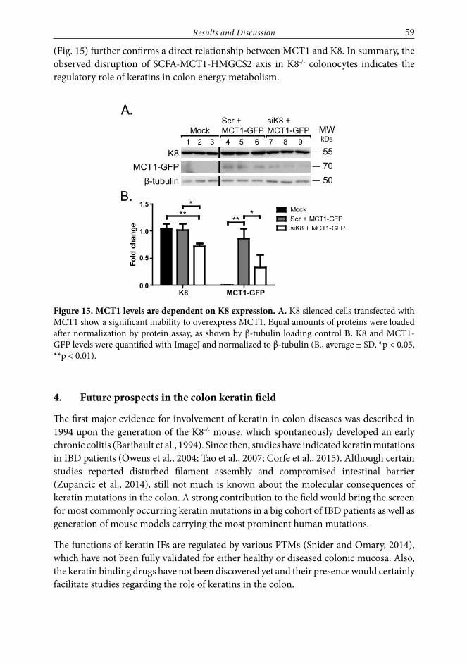
Results and Discussion 59
(Fig. 15) further confirms a direct relationship between MCT1 and K8. In summary, the observed disruption of SCFA-MCT1-HMGCS2 axis in K8-/- colonocytes indicates the regulatory role of keratins in colon energy metabolism.
A.
B.
K8MCT1-GFP
β-tubulin 50 7055
kDaMW
1 2 3 4 5 6 7 8 9
K8 MCT1-GFP0.0
0.5
1.0
1.5MockScr + MCT1-GFPsiK8 + MCT1-GFP
***
***
Fold
cha
nge
Scr +MCT1-GFPMock
siK8 +MCT1-GFP
Figure 15. MCT1 levels are dependent on K8 expression. A. K8 silenced cells transfected with MCT1 show a significant inability to overexpress MCT1. Equal amounts of proteins were loaded after normalization by protein assay, as shown by β-tubulin loading control B. K8 and MCT1-GFP levels were quantified with ImageJ and normalized to β-tubulin (B., average ± SD, *p < 0.05, **p < 0.01).
4. Future prospects in the colon keratin field
The first major evidence for involvement of keratin in colon diseases was described in 1994 upon the generation of the K8-/- mouse, which spontaneously developed an early chronic colitis (Baribault et al., 1994). Since then, studies have indicated keratin mutations in IBD patients (Owens et al., 2004; Tao et al., 2007; Corfe et al., 2015). Although certain studies reported disturbed filament assembly and compromised intestinal barrier (Zupancic et al., 2014), still not much is known about the molecular consequences of keratin mutations in the colon. A strong contribution to the field would bring the screen for most commonly occurring keratin mutations in a big cohort of IBD patients as well as generation of mouse models carrying the most prominent human mutations.
The functions of keratin IFs are regulated by various PTMs (Snider and Omary, 2014), which have not been fully validated for either healthy or diseased colonic mucosa. Also, the keratin binding drugs have not been discovered yet and their presence would certainly facilitate studies regarding the role of keratins in the colon.

60 Results and Discussion
Keratins have been commonly used as diagnostic tools in cancer (Karantza, 2011) and their significance as prognostic markers is rising. Decreased expression levels of K8 and K20 have been observed in EMT and are linked to more aggressive phenotypes of CRC tumors (Knosel et al., 2006). Also detection of caspase-cleaved K18 fragments in the sera of CRC patients with a tumor resection has been linked with disease recurrence (Ausch et al., 2009). Interestingly, recent studies have found a population of K19 positive intestinal stem cells, which are radioresistant and give rise to the CRC (Asfaha et al., 2015). However, how exactly keratins contribute to the development of CRC still remains an open question.
The novel genome editing tool CRISPR/Cas9 could be successfully used in modifying keratin expression in at least cancer colon cell lines. This would enable further analysis of cancer cell properties, like cell ability to metastasize depending on the keratin expression pattern. Promising is also an organoid biobank of colorectal cancer patients (van de Wetering et al., 2015), which might be screened from the keratin point of view. Essential in future studies is also the use of novel bioimaging techniques including stimulated emission depletion microscopy (STED) and fluorescence resonance energy transfer (FRET). The proximity ligation assay (PLA) and optogenetics also compromise potent tools in analyzing keratin dynamics and finding its novel binding partners (Zatloukal et al., 2014; Isogai et al., 2015).

Summary 61
SUMMARY
The aim of this thesis was to study the role of keratin IFs in the colon. In this work, new aspects of keratin involvement in Notch signaling, inflammation-induced colorectal cancer as well as energy metabolism were rised. The main findings of this study are summarized in Fig. 16 and are as follows:
1. By binding to Notch1 in vitro and in vivo, K8 enhances Notch1 signaling activity and target gene transcription. The loss of K8 in the colon leads to a dramatic decrease in Notch1, which alters epithelial cell differentiation.
2. Deletion of K8 results in spontaneous colitis, which increases the susceptibility to develop colorectal tumors in two cancer models: AOM and ApcMin/+. K8-/-ApcMin/+ mice, thus, could be used as a model for colitis-induced colorectal cancer. The tumorigenesis is enhanced by an activated inflammasome, which keratins can bind to in vivo, and a constantly active IL-22 signaling pathway due to the lack of its negative regulator IL-22BP protein.
3. The K8-/- epithelium displays reduced levels of the butyrate transporter, MCT1. This decreases the transport of SCFA through colonic epithelial membranes and leads to diminished HMGCS2 levels in colonocytes. Blunted ketogenesis in K8-/- colon may lead to alterations in energy metabolism.
The lack of K8in colonic epithelium
Notch1
Hey1, Hey2
Shifted differentiationtowards goblet and
enteroendocrine cells
caspase-1
Active IL-22 pathway
Increased susceptibility to induced CRC
MCT1
Blunted ketogenesis
HMGCS2
1. 2. 3.
Figure 16. The consequences of K8 absence to the colonic epithelium – the key findings of the thesis. Study 1. The lack of keratin 8 (K8) leads to a decrease in Notch1 and Hey1 and Hey2. The lower activity of Notch1 results in a shifted cell differentiation towards goblet and enteroendocrine cells. Study 2. The absence of K8 increases caspase-1 levels released from the inflammasome, which activates the IL-22 pathway. A constantly active IL-22 pathway contributes to increased susceptibility of K8-/- mice to induced colorectal cancer (CRC). Study 3. The levels of the SCFA transporter - MCT1 are decreased in the K8-/- colonic epithelium. Impaired SCFA transport decreases the levels of the ketogenic enzyme HMGCS2, which leads to blunted ketogenesis.
62 Acknowledgements
ACKNOWLEDGEMENTS
The work described in the thesis was carried out at the Department of Biosciences, at Åbo Akademi University. I am very grateful for the excellent expertise of Åbo Akademi University and University of Turku personnel. Your input in conducting my research was invaluable.
I would like to thank Professor John Eriksson, Professor Lea Sistonen and Professor Kid Törnquist for your constant efforts towards the development of Department of Biosciences, Cell Biology. I also highly value your encouragement to interact with other scientist. Thank you for all scientific meetings you organized and the opportunity to meet inspiring people from all over the world.
I am grateful to Professor Jyrki Heino and Professor Johannes Haybaeck, the reviewers of the thesis, for critical review and helpful comments.
My work would not be possible without the support from my graduate schools, Turku Doctoral Programme of Biomedical Sciences (TuBS) and Turku Doctoral Network in Molecular Biosciences. Thank you Professor Olli Lassila, Nina Wildberg and Annika Meinander for your support and encouragement through all these years.
My deepest gratitude goes to my supervisor Docent Diana Toivola. It has been a long journey since you introduced to me a mysterious world of keratin intermediate filaments. Thank you for giving me an opportunity to work in your laboratory and teaching the basics of cell biology as well as your time, effort and a big heart you always have to me.
I would like to thank Professor Johanna Ivaska and Professor Eriika Savontaus, my Ph.D. thesis committee advisors. I value a lot your critical thinking and the help I received from you throughout my Ph.D. studies.
I would not have derived so much joy from doing science if not meeting Prof. Adolfo Rivero-Müller. Thank you for always having time for scientific “small/big-talks” and spreading your amazing passion for science. Thank you also for introducing to me the world of CRISPR/Cas9, which brushed up my rusty skills in molecular biology and gave me so much self-confidence and fun.
I am also very grateful to current and former members of Diana Toivola’s laboratory. Thank you (in alphabetical order ;-)): Angeli, Anup, Basit, Calle, Catharina, Cecilia, Daniel, Dareen, Diti, Emine, Helena, Iris, Jacob, Joel, Jolanta, Jonas, Laura, Lijao, Lina, Linda, Matilda, Mueez, Nadeem, Rickard, Sina, Sofie and Terhi. It was a real pleasure to work with you. I have learned a lot from each of you. Thank you also for a good spirit inside and outside of the lab/office bench.

Acknowledgements 63
I also want to thank Professor Cecilia Sahlgren for passionate meetings related to Notch signaling and intermediate filaments as well as her scientific team in Turku. Thank you: Veronika, Marika, Valeriy, Sebastian, Rasmus, Christian and Daniel.
I have found the help of all my co-authors invaluable. I especially want to thank Terhi Helenius, Joel Nyström as well as Iris Lähdeniemi and Christian Antila for the tremendous job, you all did on our common projects, and your positive energy. Sincere thanks are extended to Docent Pekka Taimen, Doctor Fang “Rose” Cheng and Doctor Trine Husøy for scientific expertise in pathology, intermediate filament and colon cancer field. I am also very grateful to Jouko Sandholm and Markku Saari from the Cell Imaging Core; Terhi Hiltula-Maisala, Riina Valtonen, Anitta Niittymäki, Matilda Väntsi, Małgorzata Major, Beata Paziewska, Paulina Chruściel and Rafael Frias from the Central Animal Laboratory; Sinikka Kollanus and Erica Nyman from University of Turku; Helena Saarento, Gunilla Henriksson, Barbro Lindholm, Maija Liisa Hoffrén and Thomas Bymark from Åbo Akademi University for skillful technical support.
During all these years I had a pleasure to be “a guest” of very unique families. Thank You Beata, Adolfo, Paulina, Axel; Ola, Sami, Ida, Greta; Margo, Henry, Piotrek, Aleksander; Catharina, Parvez, Lilja and Shujon. Your cheerful company always means a lot to me!
Dearest Friends: Thank you for remembering about me and proving in certain cases that the distance between us is only on the map! Thank you/Dziękuję!
My debt to my family, is beyond words. Dziękuję za Waszą miłość i wsparcie. Kocham Was bardzo i wiem, że ta praca nigdy nie ujrzałaby światła dziennego gdyby nie Wasza zachęta i pozytywne nastawienie.
This work has been financially supported by Turku Doctoral Programme of Biomedical Sciences (TuBS), Oskar Öflunds Stiftelse, Stiftelsen för Åbo Akademi, K. Albin Johanssons Stiftelse and Rector of Åbo Akademi University.

64 References
REFERENCES
Crohn’s and Colitis Foundation of America. www.ccfa.org.
Abbas Abdul K., L.A.H., Pillai Shiv. 2010. Cellular and Molecular Immunology. Elsevier.
Ahn, J., H. Konno, and G.N. Barber. 2015. Diverse roles of STING-dependent signaling on the develop-ment of cancer. Oncogene. 34:5302-8.
Alam, C.M., J.S. Silvander, E.N. Daniel, G.Z. Tao, S.M. Kvarnstrom, P. Alam, M.B. Omary, A. Hanninen, and D.M. Toivola. 2013. Keratin 8 modulates be-ta-cell stress responses and normoglycaemia. J Cell Sci. 126:5635-44.
Alam, H., P. Gangadaran, A.V. Bhate, D.A. Chaukar, S.S. Sawant, R. Tiwari, J. Bobade, S. Kannan, K. D’Cruz A, S. Kane, and M.M. Vaidya. 2011a. Loss of keratin 8 phosphorylation leads to increased tu-mor progression and correlates with clinico-patho-logical parameters of OSCC patients. PLoS One. 6:e27767.
Alam, H., L. Sehgal, S.T. Kundu, S.N. Dalal, and M.M. Vaidya. 2011b. Novel function of keratins 5 and 14 in proliferation and differentiation of stratified epi-thelial cells. Mol Biol Cell. 22:4068-78.
Alberts, B., A. Johnson, J. Lewis, D. Morgan, M. Raff, K. Roberts, and P. Walter. 2015. Molecular Biology of the Cell Garland Science, New York.
Alex, P., N.C. Zachos, T. Nguyen, L. Gonzales, T.E. Chen, L.S. Conklin, M. Centola, and X. Li. 2009. Distinct cytokine patterns identified from mul-tiplex profiles of murine DSS and TNBS-induced colitis. Inflamm Bowel Dis. 15:341-52.
Allam, R., M.H. Maillard, A. Tardivel, V. Chennupati, H. Bega, C.W. Yu, D. Velin, P. Schneider, and K.M. Maslowski. 2015. Epithelial NAIPs protect against colonic tumorigenesis. J Exp Med. 212:369-83.
Allen, I.C., E.M. TeKippe, R.M. Woodford, J.M. Uronis, E.K. Holl, A.B. Rogers, H.H. Herfarth, C. Jobin, and J.P. Ting. 2010. The NLRP3 inflammasome functions as a negative regulator of tumorigen-esis during colitis-associated cancer. J Exp Med. 207:1045-56.
Ameen, N.A., Y. Figueroa, and P.J. Salas. 2001. Anom-alous apical plasma membrane phenotype in CK8-deficient mice indicates a novel role for in-termediate filaments in the polarization of simple epithelia. J Cell Sci. 114:563-75.
Ananthakrishnan, A.N., H. Khalili, G.G. Konijeti, L.M. Higuchi, P. de Silva, J.R. Korzenik, C.S. Fuchs, W.C. Willett, J.M. Richter, and A.T. Chan. 2013. A pro-spective study of long-term intake of dietary fiber
and risk of Crohn’s disease and ulcerative colitis. Gastroenterology. 145:970-7.
Antalis, T.M., and J.A. Reeder. 1995. Butyrate regulates gene expression of the plasminogen activating sys-tem in colon cancer cells. Int J Cancer. 62:619-26.
Asfaha, S., Y. Hayakawa, A. Muley, S. Stokes, T.A. Graham, R.E. Ericksen, C.B. Westphalen, J. von Burstin, T.L. Mastracci, D.L. Worthley, C. Guha, M. Quante, A.K. Rustgi, and T.C. Wang. 2015. Krt19(+)/Lgr5(-) Cells Are Radioresistant Can-cer-Initiating Stem Cells in the Colon and Intes-tine. Cell Stem Cell. 16:627-38.
Asghar, M.N., J.S. Silvander, T.O. Helenius, I.A. Lahde-niemi, C. Alam, L.E. Fortelius, R.O. Holmsten, and D.M. Toivola. 2015. The amount of keratins mat-ters for stress protection of the colonic epithelium. PLoS One. 10:e0127436.
Augenlicht, L., L. Shi, J. Mariadason, C. Laboisse, and A. Velcich. 2003. Repression of MUC2 gene ex-pression by butyrate, a physiological regulator of intestinal cell maturation. Oncogene. 22:4983-92.
Ausch, C., V. Buxhofer-Ausch, U. Olszewski, W. Hin-terberger, E. Ogris, R. Schiessel, and G. Hamilton. 2009. Caspase-cleaved cytokeratin 18 fragment (M30) as marker of postoperative residual tumor load in colon cancer patients. Eur J Surg Oncol. 35:1164-8.
Baribault, H., J. Penner, R.V. Iozzo, and M. Wil-son-Heiner. 1994. Colorectal hyperplasia and in-flammation in keratin 8-deficient FVB/N mice. Genes Dev. 8:2964-73.
Baribault, H., J. Price, K. Miyai, and R.G. Oshima. 1993. Mid-gestational lethality in mice lacking keratin 8. Genes Dev. 7:1191-202.
Barker, N., J.H. van Es, J. Kuipers, P. Kujala, M. van den Born, M. Cozijnsen, A. Haegebarth, J. Korving, H. Begthel, P.J. Peters, and H. Clevers. 2007. Identifi-cation of stem cells in small intestine and colon by marker gene Lgr5. Nature. 449:1003-7.
Barnich, N., and A. Darfeuille-Michaud. 2007. Adher-ent-invasive Escherichia coli and Crohn’s disease. Curr Opin Gastroenterol. 23:16-20.
Barrett, J.C., S. Hansoul, D.L. Nicolae, J.H. Cho, R.H. Duerr, J.D. Rioux, S.R. Brant, M.S. Silverberg, K.D. Taylor, M.M. Barmada, A. Bitton, T. Dassopoulos, L.W. Datta, T. Green, A.M. Griffiths, E.O. Kist-ner, M.T. Murtha, M.D. Regueiro, J.I. Rotter, L.P. Schumm, A.H. Steinhart, S.R. Targan, R.J. Xavier, C. Libioulle, C. Sandor, M. Lathrop, J. Belaiche, O. Dewit, I. Gut, S. Heath, D. Laukens, M. Mni, P. Rut-geerts, A. Van Gossum, D. Zelenika, D. Franchi-

References 65
mont, J.P. Hugot, M. de Vos, S. Vermeire, E. Lou-is, L.R. Cardon, C.A. Anderson, H. Drummond, E. Nimmo, T. Ahmad, N.J. Prescott, C.M. Onnie, S.A. Fisher, J. Marchini, J. Ghori, S. Bumpstead, R. Gwilliam, M. Tremelling, P. Deloukas, J. Mansfield, D. Jewell, J. Satsangi, C.G. Mathew, M. Parkes, M. Georges, and M.J. Daly. 2008. Genome-wide asso-ciation defines more than 30 distinct susceptibility loci for Crohn’s disease. Nat Genet. 40:955-62.
Basso, P.J., M.T. Fonseca, G. Bonfa, V.B. Alves, H. Sales-Campos, V. Nardini, and C.R. Cardoso. 2014. Association among genetic predisposition, gut mi-crobiota, and host immune response in the etio-pathogenesis of inflammatory bowel disease. Braz J Med Biol Res. 47:727-37.
Batlle, E., J.T. Henderson, H. Beghtel, M.M. van den Born, E. Sancho, G. Huls, J. Meeldijk, J. Robertson, M. van de Wetering, T. Pawson, and H. Clevers. 2002. Beta-catenin and TCF mediate cell position-ing in the intestinal epithelium by controlling the expression of EphB/ephrinB. Cell. 111:251-63.
Beaudoin, M., P. Goyette, G. Boucher, K.S. Lo, M.A. Rivas, C. Stevens, A. Alikashani, M. Ladouceur, D. Ellinghaus, L. Torkvist, G. Goel, C. Lagace, V. An-nese, A. Bitton, J. Begun, S.R. Brant, F. Bresso, J.H. Cho, R.H. Duerr, J. Halfvarson, D.P. McGovern, G. Radford-Smith, S. Schreiber, P.L. Schumm, Y. Sharma, M.S. Silverberg, R.K. Weersma, M. D’Am-ato, S. Vermeire, A. Franke, G. Lettre, R.J. Xavier, M.J. Daly, and J.D. Rioux. 2013. Deep resequencing of GWAS loci identifies rare variants in CARD9, IL23R and RNF186 that are associated with ulcer-ative colitis. PLoS Genet. 9:e1003723.
Beaugerie, L., and S.H. Itzkowitz. 2015. Cancers Com-plicating Inflammatory Bowel Disease. N Engl J Med. 373:195.
Becker, S., T.A. Oelschlaeger, A. Wullaert, K. Vlantis, M. Pasparakis, J. Wehkamp, E.F. Stange, and M. Gersemann. 2013. Bacteria regulate intestinal ep-ithelial cell differentiation factors both in vitro and in vivo. PLoS One. 8:e55620.
Bettington, M., N. Walker, A. Clouston, I. Brown, B. Leggett, and V. Whitehall. 2013. The serrated path-way to colorectal carcinoma: current concepts and challenges. Histopathology. 62:367-86.
Birkenkamp-Demtroder, K., F. Mansilla, F.B. Sorensen, M. Kruhoffer, T. Cabezon, L.L. Christensen, L.A. Aaltonen, H.W. Verspaget, and T.F. Orntoft. 2007. Phosphoprotein Keratin 23 accumulates in MSS but not MSI colon cancers in vivo and impacts vi-ability and proliferation in vitro. Mol Oncol. 1:181-95.
Bleicher, L., P.R. de Moura, L. Watanabe, D. Colau, L. Dumoutier, J.C. Renauld, and I. Polikarpov. 2008. Crystal structure of the IL-22/IL-22R1 complex and its implications for the IL-22 signaling mecha-nism. FEBS Lett. 582:2985-92.
Bogaert, J., and H. Prenen. 2014. Molecular genetics of colorectal cancer. Ann Gastroenterol. 27:9-14.
Boland, C.R., and A. Goel. 2010. Microsatellite in-stability in colorectal cancer. Gastroenterology. 138:2073-2087 e3.
Bonifas, J.M., A.L. Rothman, and E.H. Epstein, Jr. 1991. Epidermolysis bullosa simplex: evidence in two families for keratin gene abnormalities. Science. 254:1202-5.
Borthakur, A., S. Priyamvada, A. Kumar, A.A. Natara-jan, R.K. Gill, W.A. Alrefai, and P.K. Dudeja. 2012. A novel nutrient sensing mechanism underlies substrate-induced regulation of monocarboxyl-ate transporter-1. Am J Physiol Gastrointest Liver Physiol. 303:G1126-33.
Brazil, D.P., R.H. Church, S. Surae, C. Godson, and F. Martin. 2015. BMP signalling: agony and antagony in the family. Trends Cell Biol. 25:249-264.
Breynaert, C., T. Dresselaers, C. Perrier, I. Arijs, J. Cremer, L. Van Lommel, K. Van Steen, M. Ferrante, F. Schuit, S. Vermeire, P. Rutgeerts, U. Himmelreich, J.L. Ceuppens, K. Geboes, and G. Van Assche. 2013. Unique gene expression and MR T2 relaxometry patterns define chronic murine dextran sodium sul-phate colitis as a model for connective tissue chang-es in human Crohn’s disease. PLoS One. 8:e68876.
Brown, T., and T. Brown. Nucleic Acids Book (http://www.atdbio.com/nucleic-acids-book).
Buning, C., J. Halangk, A. Dignass, J. Ockenga, P. Deindl, R. Nickel, J. Genschel, O. Landt, H. Lochs, H. Schmidt, and H. Witt. 2004. Keratin 8 Y54H and G62C mutations are not associated with inflamma-tory bowel disease. Dig Liver Dis. 36:388-91.
Busch, T., M. Armacki, T. Eiseler, G. Joodi, C. Temme, J. Jansen, G. von Wichert, M.B. Omary, J. Spatz, and T. Seufferlein. 2012. Keratin 8 phosphorylation regulates keratin reorganization and migration of epithelial tumor cells. J Cell Sci. 125:2148-59.
Cai, J., N. Zhang, Y. Zheng, R.F. de Wilde, A. Maitra, and D. Pan. 2010. The Hippo signaling pathway restricts the oncogenic potential of an intestinal re-generation program. Genes Dev. 24:2383-8.
Cavestro, G.M., L. Frulloni, A. Nouvenne, T.M. Neri, B. Calore, B. Ferri, P. Bovo, L. Okolicsanyi, F. Di Ma-rio, and G. Cavallini. 2003. Association of keratin 8 gene mutation with chronic pancreatitis. Dig Liver Dis. 35:416-20.
Chang, P.V., L. Hao, S. Offermanns, and R. Medzhi-tov. 2014. The microbial metabolite butyrate reg-ulates intestinal macrophage function via histone deacetylase inhibition. Proc Natl Acad Sci U S A. 111:2247-52.
Chassaing, B., J.D. Aitken, M. Malleshappa, and M. Vijay-Kumar. 2014. Dextran sulfate sodium (DSS)-induced colitis in mice. Curr Protoc Immu-nol. 104:Unit 15 25.
66 References
Chen, G.Y., M. Liu, F. Wang, J. Bertin, and G. Nunez. 2011. A functional role for Nlrp6 in intestinal inflammation and tumorigenesis. J Immunol. 186:7187-94.
Chen, Y., N. Guldiken, M. Spurny, H.H. Mohammed, J. Haybaeck, M.J. Pollheimer, P. Fickert, N. Gassler, M.K. Jeon, C. Trautwein, and P. Strnad. 2015. Loss of keratin 19 favours the development of cholestat-ic liver disease through decreased ductular reac-tion. J Pathol. 237:343-54.
Cherbuy, C., C. Andrieux, E. Honvo-Houeto, M. Thomas, C. Ide, N. Druesne, C. Chaumontet, B. Darcy-Vrillon, and P.H. Duee. 2004. Expression of mitochondrial HMGCoA synthase and glutam-inase in the colonic mucosa is modulated by bacte-rial species. Eur J Biochem. 271:87-95.
Clevers, H., and R. Nusse. 2012. Wnt/beta-catenin sig-naling and disease. Cell. 149:1192-205.
Colman, R.J., and D.T. Rubin. 2014. Fecal microbiota transplantation as therapy for inflammatory bowel disease: a systematic review and meta-analysis. J Crohns Colitis. 8:1569-81.
Comalada, M., E. Bailon, O. de Haro, F. Lara-Villosla-da, J. Xaus, A. Zarzuelo, and J. Galvez. 2006. The effects of short-chain fatty acids on colon epithelial proliferation and survival depend on the cellular phenotype. J Cancer Res Clin Oncol. 132:487-97.
Corazziari, E.S. 2009. Intestinal mucus barrier in nor-mal and inflamed colon. J Pediatr Gastroenterol Nutr. 48 Suppl 2:S54-5.
Corfe, B.M., D. Majumdar, A. Assadsangabi, A.M. Marsh, S.S. Cross, J.B. Connolly, C.A. Evans, and A.J. Lobo. 2015. Inflammation decreases keratin level in ulcerative colitis; inadequate restoration associates with increased risk of colitis-associated cancer. BMJ Open Gastroenterol. 2:e000024.
Cotter, D.G., B. Ercal, X. Huang, J.M. Leid, D.A. d’Avi-gnon, M.J. Graham, D.J. Dietzen, E.M. Brunt, G.J. Patti, and P.A. Crawford. 2014. Ketogenesis pre-vents diet-induced fatty liver injury and hypergly-cemia. J Clin Invest. 124:5175-90.
Coulombe, P.A., M.E. Hutton, A. Letai, A. Hebert, A.S. Paller, and E. Fuchs. 1991. Point mutations in hu-man keratin 14 genes of epidermolysis bullosa sim-plex patients: genetic and functional analyses. Cell. 66:1301-11.
Cuff, M.A., D.W. Lambert, and S.P. Shirazi-Beechey. 2002. Substrate-induced regulation of the human colonic monocarboxylate transporter, MCT1. J Physiol. 539:361-71.
Delva, E., D.K. Tucker, and A.P. Kowalczyk. 2009. The desmosome. Cold Spring Harb Perspect Biol. 1:a002543.
Demarque, M.D., K. Nacerddine, H. Neyret-Kahn, A. Andrieux, E. Danenberg, G. Jouvion, P. Bomme, G. Hamard, B. Romagnolo, B. Terris, A. Cumano, N.
Barker, H. Clevers, and A. Dejean. 2011. Sumoyla-tion by Ubc9 regulates the stem cell compartment and structure and function of the intestinal epithe-lium in mice. Gastroenterology. 140:286-96.
Donohoe, D.R., N. Garge, X. Zhang, W. Sun, T.M. O’Connell, M.K. Bunger, and S.J. Bultman. 2011. The microbiome and butyrate regulate energy me-tabolism and autophagy in the mammalian colon. Cell Metab. 13:517-26.
dos Santos, G., M.R. Rogel, M.A. Baker, J.R. Troken, D. Urich, L. Morales-Nebreda, J.A. Sennello, M.A. Ku-tuzov, A. Sitikov, J.M. Davis, A.P. Lam, P. Cheresh, D. Kamp, D.K. Shumaker, G.R. Budinger, and K.M. Ridge. 2015. Vimentin regulates activation of the NLRP3 inflammasome. Nat Commun. 6:6574.
Dow, L.E., K.P. O’Rourke, J. Simon, D.F. Tschaharganeh, J.H. van Es, H. Clevers, and S.W. Lowe. 2015. Apc Restoration Promotes Cellular Differentiation and Reestablishes Crypt Homeostasis in Colorectal Cancer. Cell. 161:1539-52.
Ebert, M.N., G. Beyer-Sehlmeyer, U.M. Liegibel, T. Kautenburger, T.W. Becker, and B.L. Pool-Zobel. 2001. Butyrate induces glutathione S-transferase in human colon cells and protects from genetic dam-age by 4-hydroxy-2-nonenal. Nutr Cancer. 41:156-64.
Elinav, E., T. Strowig, A.L. Kau, J. Henao-Mejia, C.A. Thaiss, C.J. Booth, D.R. Peaper, J. Bertin, S.C. Eisenbarth, J.I. Gordon, and R.A. Flavell. 2011. NLRP6 inflammasome regulates colonic microbial ecology and risk for colitis. Cell. 145:745-57.
Eriksson, J.E., T. Dechat, B. Grin, B. Helfand, M. Men-dez, H.M. Pallari, and R.D. Goldman. 2009. Intro-ducing intermediate filaments: from discovery to disease. J Clin Invest. 119:1763-71.
Evans, C.A., R. Rosser, J.S. Waby, J. Noirel, D. Lai, P.C. Wright, E.A. Williams, S.A. Riley, J.P. Bury, and B.M. Corfe. 2015. Reduced keratin expression in colorectal neoplasia and associated fields is revers-ible by diet and resection. BMJ Open Gastroenterol. 2:e000022.
Feagins, L.A., R.F. Souza, and S.J. Spechler. 2009. Car-cinogenesis in IBD: potential targets for the pre-vention of colorectal cancer. Nat Rev Gastroenterol Hepatol. 6:297-305.
Fearon, E.R., and B. Vogelstein. 1990. A genetic model for colorectal tumorigenesis. Cell. 61:759-67.
Feller, M., K. Huwiler, R. Stephan, E. Altpeter, A. Shang, H. Furrer, G.E. Pfyffer, T. Jemmi, A. Baumgartner, and M. Egger. 2007. Mycobacterium avium sub-species paratuberculosis and Crohn’s disease: a systematic review and meta-analysis. Lancet Infect Dis. 7:607-13.
Ferlay J, S.I., Ervik M, Dikshit R, Eser S, Mathers C, Rebelo M, Parkin DM, Forman D, Bray, F. 2013. GLOBOCAN 2012 v1.0, Cancer Incidence and

References 67
Mortality Worldwide: IARC CancerBase No. 11 Lyon, France.
Fernandes-Alnemri, T., S. Kang, C. Anderson, J. Saga-ra, K.A. Fitzgerald, and E.S. Alnemri. 2013. Cutting edge: TLR signaling licenses IRAK1 for rapid ac-tivation of the NLRP3 inflammasome. J Immunol. 191:3995-9.
Fisher, S.A., M. Tremelling, C.A. Anderson, R. Gwil-liam, S. Bumpstead, N.J. Prescott, E.R. Nimmo, D. Massey, C. Berzuini, C. Johnson, J.C. Barrett, F.R. Cummings, H. Drummond, C.W. Lees, C.M. Onnie, C.E. Hanson, K. Blaszczyk, M. Inouye, P. Ewels, R. Ravindrarajah, A. Keniry, S. Hunt, M. Carter, N. Watkins, W. Ouwehand, C.M. Lewis, L. Cardon, A. Lobo, A. Forbes, J. Sanderson, D.P. Jew-ell, J.C. Mansfield, P. Deloukas, C.G. Mathew, M. Parkes, and J. Satsangi. 2008. Genetic determinants of ulcerative colitis include the ECM1 locus and five loci implicated in Crohn’s disease. Nat Genet. 40:710-2.
Fletcher, D.A., and R.D. Mullins. 2010. Cell mechanics and the cytoskeleton. Nature. 463:485-92.
Fountoulakis, M., E. Soumaka, K. Rapti, M. Mavroidis, G. Tsangaris, A. Maris, N. Weisleder, and Y. Capet-anaki. 2005. Alterations in the heart mitochondrial proteome in a desmin null heart failure model. J Mol Cell Cardiol. 38:461-74.
Franke, A., T. Balschun, T.H. Karlsen, J. Hedderich, S. May, T. Lu, D. Schuldt, S. Nikolaus, P. Rosenstiel, M. Krawczak, and S. Schreiber. 2008a. Replication of signals from recent studies of Crohn’s disease identifies previously unknown disease loci for ul-cerative colitis. Nat Genet. 40:713-5.
Franke, A., T. Balschun, T.H. Karlsen, J. Sventoraityte, S. Nikolaus, G. Mayr, F.S. Domingues, M. Albrecht, M. Nothnagel, D. Ellinghaus, C. Sina, C.M. Onnie, R.K. Weersma, P.C. Stokkers, C. Wijmenga, M. Gazouli, D. Strachan, W.L. McArdle, S. Vermeire, P. Rutgeerts, P. Rosenstiel, M. Krawczak, M.H. Vatn, C.G. Mathew, and S. Schreiber. 2008b. Sequence variants in IL10, ARPC2 and multiple other loci contribute to ulcerative colitis susceptibility. Nat Genet. 40:1319-23.
Fre, S., M. Huyghe, P. Mourikis, S. Robine, D. Louvard, and S. Artavanis-Tsakonas. 2005. Notch signals control the fate of immature progenitor cells in the intestine. Nature. 435:964-8.
Furusawa, Y., Y. Obata, S. Fukuda, T.A. Endo, G. Naka-to, D. Takahashi, Y. Nakanishi, C. Uetake, K. Kato, T. Kato, M. Takahashi, N.N. Fukuda, S. Murakami, E. Miyauchi, S. Hino, K. Atarashi, S. Onawa, Y. Fu-jimura, T. Lockett, J.M. Clarke, D.L. Topping, M. Tomita, S. Hori, O. Ohara, T. Morita, H. Koseki, J. Kikuchi, K. Honda, K. Hase, and H. Ohno. 2013. Commensal microbe-derived butyrate induces the differentiation of colonic regulatory T cells. Nature. 504:446-50.
Galvez, J., M.E. Rodriguez-Cabezas, and A. Zarzuelo. 2005. Effects of dietary fiber on inflammatory bow-el disease. Mol Nutr Food Res. 49:601-8.
Garg, P., A. Ravi, N.R. Patel, J. Roman, A.T. Gewirtz, D. Merlin, and S.V. Sitaraman. 2007. Matrix metallo-proteinase-9 regulates MUC-2 expression through its effect on goblet cell differentiation. Gastroenter-ology. 132:1877-89.
Gerbe, F., C. Legraverend, and P. Jay. 2012. The intes-tinal epithelium tuft cells: specification and func-tion. Cell Mol Life Sci. 69:2907-17.
Ghaleb, A.M., G. Aggarwal, A.B. Bialkowska, M.O. Nandan, and V.W. Yang. 2008. Notch inhibits ex-pression of the Kruppel-like factor 4 tumor sup-pressor in the intestinal epithelium. Mol Cancer Res. 6:1920-7.
Gibson, P.R., O. Rosella, A.J. Wilson, J.M. Mariadason, K. Rickard, K. Byron, and D.H. Barkla. 1999. Co-lonic epithelial cell activation and the paradoxical effects of butyrate. Carcinogenesis. 20:539-44.
Glocker, E.O., D. Kotlarz, K. Boztug, E.M. Gertz, A.A. Schaffer, F. Noyan, M. Perro, J. Diestelhorst, A. Allroth, D. Murugan, N. Hatscher, D. Pfeifer, K.W. Sykora, M. Sauer, H. Kreipe, M. Lacher, R. Nust-ede, C. Woellner, U. Baumann, U. Salzer, S. Koletz-ko, N. Shah, A.W. Segal, A. Sauerbrey, S. Buderus, S.B. Snapper, B. Grimbacher, and C. Klein. 2009. Inflammatory bowel disease and mutations af-fecting the interleukin-10 receptor. N Engl J Med. 361:2033-45.
Gopal, E., S. Miyauchi, P.M. Martin, S. Ananth, P. Roon, S.B. Smith, and V. Ganapathy. 2007. Transport of nicotinate and structurally related compounds by human SMCT1 (SLC5A8) and its relevance to drug transport in the mammalian intestinal tract. Pharm Res. 24:575-84.
Goyal, N., A. Rana, A. Ahlawat, K.R. Bijjem, and P. Ku-mar. 2014. Animal models of inflammatory bowel disease: a review. Inflammopharmacology. 22:219-33.
Grimm-Gunter, E.M., C. Revenu, S. Ramos, I. Hurbain, N. Smyth, E. Ferrary, D. Louvard, S. Robine, and F. Rivero. 2009. Plastin 1 binds to keratin and is re-quired for terminal web assembly in the intestinal epithelium. Mol Biol Cell. 20:2549-62.
Guan, Y., J. Dong, L. Tackett, J.W. Meyer, G.E. Shull, and M.H. Montrose. 2006. NHE2 is the main api-cal NHE in mouse colonic crypts but an alternative Na+-dependent acid extrusion mechanism is up-regulated in NHE2-null mice. Am J Physiol Gastro-intest Liver Physiol. 291:G689-99.
Gunawardene, A.R., B.M. Corfe, and C.A. Staton. 2011. Classification and functions of enteroendocrine cells of the lower gastrointestinal tract. Int J Exp Pathol. 92:219-31.

68 References
Guo, H., J.B. Callaway, and J.P. Ting. 2015. Inflam-masomes: mechanism of action, role in disease, and therapeutics. Nat Med. 21:677-87.
Guruharsha, K.G., M.W. Kankel, and S. Artavanis-Tsa-konas. 2012. The Notch signalling system: recent insights into the complexity of a conserved path-way. Nat Rev Genet. 13:654-66.
Habtezion, A., D.M. Toivola, M.N. Asghar, G.S. Kro-nmal, J.D. Brooks, E.C. Butcher, and M.B. Omary. 2011. Absence of keratin 8 confers a paradoxical microflora-dependent resistance to apoptosis in the colon. Proc Natl Acad Sci U S A. 108:1445-50.
Habtezion, A., D.M. Toivola, E.C. Butcher, and M.B. Omary. 2005. Keratin-8-deficient mice develop chronic spontaneous Th2 colitis amenable to anti-biotic treatment. J Cell Sci. 118:1971-80.
Haines, R.L., and E.B. Lane. 2012. Keratins and disease at a glance. J Cell Sci. 125:3923-8.
Hamer, H.M., D. Jonkers, K. Venema, S. Vanhoutvin, F.J. Troost, and R.J. Brummer. 2008. Review article: the role of butyrate on colonic function. Aliment Pharmacol Ther. 27:104-19.
Hamidi, H., D. Gustafason, M. Pellegrini, and J. Gasson. 2011. Identification of novel targets of CSL-depen-dent Notch signaling in hematopoiesis. PLoS One. 6:e20022.
Hanahan, D., and R.A. Weinberg. 2011. Hallmarks of cancer: the next generation. Cell. 144:646-74.
Havenaar, R. 2011. Intestinal health functions of colon-ic microbial metabolites: a review. Benef Microbes. 2:103-14.
He, T., A. Stepulak, T.H. Holmstrom, M.B. Omary, and J.E. Eriksson. 2002. The intermediate filament pro-tein keratin 8 is a novel cytoplasmic substrate for c-Jun N-terminal kinase. J Biol Chem. 277:10767-74.
Hegardt, F.G. 1999. Mitochondrial 3-hydroxy-3-meth-ylglutaryl-CoA synthase: a control enzyme in keto-genesis. Biochem J. 338 ( Pt 3):569-82.
High, F.A., and J.A. Epstein. 2008. The multifaceted role of Notch in cardiac development and disease. Nat Rev Genet. 9:49-61.
Hobbs, R.P., D.J. DePianto, J.T. Jacob, M.C. Han, B.M. Chung, A.S. Batazzi, B.G. Poll, Y. Guo, J. Han, S. Ong, W. Zheng, J.M. Taube, D. Cihakova, F. Wan, and P.A. Coulombe. 2015. Keratin-dependent reg-ulation of Aire and gene expression in skin tumor keratinocytes. Nat Genet. 47:933-8.
Hodin, R.A., S. Meng, S. Archer, and R. Tang. 1996. Cellular growth state differentially regulates en-terocyte gene expression in butyrate-treated HT-29 cells. Cell Growth Differ. 7:647-53.
Hu, B., E. Elinav, S. Huber, C.J. Booth, T. Strowig, C. Jin, S.C. Eisenbarth, and R.A. Flavell. 2010. Inflamma-tion-induced tumorigenesis in the colon is regulat-
ed by caspase-1 and NLRC4. Proc Natl Acad Sci U S A. 107:21635-40.
Huber, S., N. Gagliani, L.A. Zenewicz, F.J. Huber, L. Bo-surgi, B. Hu, M. Hedl, W. Zhang, W. O’Connor, Jr., A.J. Murphy, D.M. Valenzuela, G.D. Yancopoulos, C.J. Booth, J.H. Cho, W. Ouyang, C. Abraham, and R.A. Flavell. 2012. IL-22BP is regulated by the in-flammasome and modulates tumorigenesis in the intestine. Nature. 491:259-63.
Hugot, J.P., M. Chamaillard, H. Zouali, S. Lesage, J.P. Cezard, J. Belaiche, S. Almer, C. Tysk, C.A. O’Mo-rain, M. Gassull, V. Binder, Y. Finkel, A. Cortot, R. Modigliani, P. Laurent-Puig, C. Gower-Rousseau, J. Macry, J.F. Colombel, M. Sahbatou, and G. Thom-as. 2001. Association of NOD2 leucine-rich repeat variants with susceptibility to Crohn’s disease. Na-ture. 411:599-603.
Hung, J.R.a.K.E. 2013. Molecular Mechanisms of Col-orectal Carcinogenesis. In Molecular Pathogenesis of Colorectal Cancer. K.M. Haigis, editor. Springer, New York.
Hwang, C., V. Ross, and U. Mahadevan. 2012. Micronu-trient deficiencies in inflammatory bowel disease: from A to zinc. Inflamm Bowel Dis. 18:1961-81.
Imperiale, T.F., D.F. Ransohoff, S.H. Itzkowitz, T.R. Levin, P. Lavin, G.P. Lidgard, D.A. Ahlquist, and B.M. Berger. 2014. Multitarget stool DNA testing for colorectal-cancer screening. N Engl J Med. 370:1287-97.
Imperiale, T.F., D.R. Wagner, C.Y. Lin, G.N. Larkin, J.D. Rogge, and D.F. Ransohoff. 2000. Risk of advanced proximal neoplasms in asymptomatic adults ac-cording to the distal colorectal findings. N Engl J Med. 343:169-74.
Inan, M.S., R.J. Rasoulpour, L. Yin, A.K. Hubbard, D.W. Rosenberg, and C. Giardina. 2000. The luminal short-chain fatty acid butyrate modulates NF-kap-paB activity in a human colonic epithelial cell line. Gastroenterology. 118:724-34.
Isogai, T., R. van der Kammen, D. Leyton-Puig, K.M. Kedziora, K. Jalink, and M. Innocenti. 2015. Initi-ation of lamellipodia and ruffles involves cooper-ation between mDia1 and the Arp2/3 complex. J Cell Sci. 128:3796-810.
Itzkowitz, S.H., and X. Yio. 2004. Inflammation and cancer IV. Colorectal cancer in inflammatory bow-el disease: the role of inflammation. Am J Physiol Gastrointest Liver Physiol. 287:G7-17.
Janakiram, N.B., A. Mohammed, T. Bryant, M. Brewer, L. Biddick, S. Lightfoot, M.L. Lang, and C.V. Rao. 2014. Adoptive transfer of regulatory T cells pro-motes intestinal tumorigenesis and is associated with decreased NK cells and IL-22 binding protein. Mol Carcinog. 54:986-98.
Jemal, A., F. Bray, M.M. Center, J. Ferlay, E. Ward, and D. Forman. 2011. Global cancer statistics. CA Can-cer J Clin. 61:69-90.

References 69
Jeon, M.K., C. Klaus, E. Kaemmerer, and N. Gassler. 2013. Intestinal barrier: Molecular pathways and modifiers. World J Gastrointest Pathophysiol. 4:94-9.
Jiang, R., H. Wang, L. Deng, J. Hou, R. Shi, M. Yao, Y. Gao, A. Yao, X. Wang, L. Yu, and B. Sun. 2013. IL-22 is related to development of human colon can-cer by activation of STAT3. BMC Cancer. 13:59.
Johnston, L. 2000. Colon & Rectal Cancer A compre-hensive Guide for Patients & Families. O’Reilly Media / Patient Centered Guides.
Joseph, A.J., B. George, A.B. Pulimood, M.S. Seshadri, and A. Chacko. 2009. 25 (OH) vitamin D level in Crohn’s disease: association with sun exposure & disease activity. Indian J Med Res. 130:133-7.
Jostins, L., S. Ripke, R.K. Weersma, R.H. Duerr, D.P. Mc-Govern, K.Y. Hui, J.C. Lee, L.P. Schumm, Y. Shar-ma, C.A. Anderson, J. Essers, M. Mitrovic, K. Ning, I. Cleynen, E. Theatre, S.L. Spain, S. Raychaudhu-ri, P. Goyette, Z. Wei, C. Abraham, J.P. Achkar, T. Ahmad, L. Amininejad, A.N. Ananthakrishnan, V. Andersen, J.M. Andrews, L. Baidoo, T. Balschun, P.A. Bampton, A. Bitton, G. Boucher, S. Brand, C. Buning, A. Cohain, S. Cichon, M. D’Amato, D. De Jong, K.L. Devaney, M. Dubinsky, C. Edwards, D. Ellinghaus, L.R. Ferguson, D. Franchimont, K. Fransen, R. Gearry, M. Georges, C. Gieger, J. Glas, T. Haritunians, A. Hart, C. Hawkey, M. Hedl, X. Hu, T.H. Karlsen, L. Kupcinskas, S. Kugathasan, A. Latiano, D. Laukens, I.C. Lawrance, C.W. Lees, E. Louis, G. Mahy, J. Mansfield, A.R. Morgan, C. Mowat, W. Newman, O. Palmieri, C.Y. Ponsioen, U. Potocnik, N.J. Prescott, M. Regueiro, J.I. Rotter, R.K. Russell, J.D. Sanderson, M. Sans, J. Satsangi, S. Schreiber, L.A. Simms, J. Sventoraityte, S.R. Tar-gan, K.D. Taylor, M. Tremelling, H.W. Verspaget, M. De Vos, C. Wijmenga, D.C. Wilson, J. Winkel-mann, R.J. Xavier, S. Zeissig, B. Zhang, C.K. Zhang, H. Zhao, M.S. Silverberg, V. Annese, H. Hakonar-son, S.R. Brant, G. Radford-Smith, C.G. Mathew, J.D. Rioux, E.E. Schadt, et al. 2012. Host-microbe interactions have shaped the genetic architecture of inflammatory bowel disease. Nature. 491:119-24.
Kalluri, R., and R.A. Weinberg. 2009. The basics of epithelial-mesenchymal transition. J Clin Invest. 119:1420-8.
Karantza, V. 2011. Keratins in health and cancer: more than mere epithelial cell markers. Oncogene. 30:127-38.
Karim, B.O., and D.L. Huso. 2013. Mouse models for colorectal cancer. Am J Cancer Res. 3:240-50.
Kaser, A., S. Zeissig, and R.S. Blumberg. 2010. Inflam-matory bowel disease. Annu Rev Immunol. 28:573-621.
Khan, A.Q., J.P. Bury, S.R. Brown, S.A. Riley, and B.M. Corfe. 2011. Keratin 8 expression in colon cancer associates with low faecal butyrate levels. BMC Gastroenterol. 11:2.
Kim, S., P. Wong, and P.A. Coulombe. 2006. A keratin cytoskeletal protein regulates protein synthesis and epithelial cell growth. Nature. 441:362-5.
Kim, Y.S., and S.B. Ho. 2010. Intestinal goblet cells and mucins in health and disease: recent insights and progress. Curr Gastroenterol Rep. 12:319-30.
Kinzler, K.W., M.C. Nilbert, L.K. Su, B. Vogelstein, T.M. Bryan, D.B. Levy, K.J. Smith, A.C. Preisinger, P. Hedge, D. McKechnie, and et al. 1991. Identifica-tion of FAP locus genes from chromosome 5q21. Science. 253:661-5.
Kirk, P., M.C. Wilson, C. Heddle, M.H. Brown, A.N. Barclay, and A.P. Halestrap. 2000. CD147 is tight-ly associated with lactate transporters MCT1 and MCT4 and facilitates their cell surface expression. EMBO J. 19:3896-904.
Knosel, T., V. Emde, K. Schluns, P.M. Schlag, M. Dietel, and I. Petersen. 2006. Cytokeratin profiles identi-fy diagnostic signatures in colorectal cancer using multiplex analysis of tissue microarrays. Cell Oncol. 28:167-75.
Komiya, Y., and R. Habas. 2008. Wnt signal transduc-tion pathways. Organogenesis. 4:68-75.
Konig, B., A. Koch, K. Giggel, B. Dordschbal, K. Eder, and G.I. Stangl. 2008. Monocarboxylate transport-er (MCT)-1 is up-regulated by PPARalpha. Bio-chim Biophys Acta. 1780:899-904.
Kopan, R., and M.X. Ilagan. 2009. The canonical Notch signaling pathway: unfolding the activation mech-anism. Cell. 137:216-33.
Korinek, V., N. Barker, P.J. Morin, D. van Wichen, R. de Weger, K.W. Kinzler, B. Vogelstein, and H. Clevers. 1997. Constitutive transcriptional activation by a beta-catenin-Tcf complex in APC-/- colon carci-noma. Science. 275:1784-7.
Kotenko, S.V., L.S. Izotova, O.V. Mirochnitchenko, E. Esterova, H. Dickensheets, R.P. Donnelly, and S. Pestka. 2001. Identification, cloning, and charac-terization of a novel soluble receptor that binds IL-22 and neutralizes its activity. J Immunol. 166:7096-103.
Kotlarz, D., R. Beier, D. Murugan, J. Diestelhorst, O. Jensen, K. Boztug, D. Pfeifer, H. Kreipe, E.D. Pfis-ter, U. Baumann, J. Puchalka, J. Bohne, O. Egritas, B. Dalgic, K.L. Kolho, A. Sauerbrey, S. Buderus, T. Gungor, A. Enninger, Y.K. Koda, G. Guariso, B. Weiss, S. Corbacioglu, P. Socha, N. Uslu, A. Metin, G.T. Wahbeh, K. Husain, D. Ramadan, W. Al-Herz, B. Grimbacher, M. Sauer, K.W. Sykora, S. Koletzko, and C. Klein. 2012. Loss of interleukin-10 signaling and infantile inflammatory bowel disease: implica-tions for diagnosis and therapy. Gastroenterology. 143:347-55.
Krausova, M., and V. Korinek. 2014. Wnt signaling in adult intestinal stem cells and cancer. Cell Signal. 26:570-9.

70 References
Krishnan, S., V.M. Rajendran, and H.J. Binder. 2003. Apical NHE isoforms differentially regulate butyr-ate-stimulated Na absorption in rat distal colon. Am J Physiol Cell Physiol. 285:C1246-54.
Ku, N.O., S. Azhar, and M.B. Omary. 2002a. Keratin 8 phosphorylation by p38 kinase regulates cellular keratin filament reorganization: modulation by a keratin 1-like disease causing mutation. J Biol Chem. 277:10775-82.
Ku, N.O., J. Liao, and M.B. Omary. 1998. Phosphoryla-tion of human keratin 18 serine 33 regulates bind-ing to 14-3-3 proteins. EMBO J. 17:1892-906.
Ku, N.O., S. Michie, E.Z. Resurreccion, R.L. Broome, and M.B. Omary. 2002b. Keratin binding to 14-3-3 proteins modulates keratin filaments and hepato-cyte mitotic progression. Proc Natl Acad Sci U S A. 99:4373-8.
Ku, N.O., and M.B. Omary. 2006. A disease- and phos-phorylation-related nonmechanical function for keratin 8. J Cell Biol. 174:115-25.
Ku, N.O., P. Strnad, B.H. Zhong, G.Z. Tao, and M.B. Omary. 2007. Keratins let liver live: Mutations pre-dispose to liver disease and crosslinking generates Mallory-Denk bodies. Hepatology. 46:1639-49.
Kucharzik, T., R. Stoll, N. Lugering, and W. Domschke. 1995. Circulating antiinflammatory cytokine IL-10 in patients with inflammatory bowel disease (IBD). Clin Exp Immunol. 100:452-6.
Kucherlapati, M.H., K. Lee, A.A. Nguyen, A.B. Clark, H. Hou, Jr., A. Rosulek, H. Li, K. Yang, K. Fan, M. Lipkin, R.T. Bronson, L. Jelicks, T.A. Kunkel, R. Kucherlapati, and W. Edelmann. 2010. An Msh2 conditional knockout mouse for studying intesti-nal cancer and testing anticancer agents. Gastroen-terology. 138:993-1002 e1.
Kuhn, R., J. Lohler, D. Rennick, K. Rajewsky, and W. Muller. 1993. Interleukin-10-deficient mice devel-op chronic enterocolitis. Cell. 75:263-74.
Kwon, C., P. Cheng, I.N. King, P. Andersen, L. Shenje, V. Nigam, and D. Srivastava. 2011. Notch post-trans-lationally regulates beta-catenin protein in stem and progenitor cells. Nat Cell Biol. 13:1244-51.
Lakatos, P.L., Z. Vegh, B.D. Lovasz, G. David, T. Pandur, Z. Erdelyi, I. Szita, G. Mester, M. Balogh, I. Szipocs, C. Molnar, E. Komaromi, P.A. Golovics, M. Man-del, A. Horvath, M. Szathmari, L.S. Kiss, and L. Lakatos. 2013. Is current smoking still an import-ant environmental factor in inflammatory bowel diseases? Results from a population-based incident cohort. Inflamm Bowel Dis. 19:1010-7.
Lane, E.B., E.L. Rugg, H. Navsaria, I.M. Leigh, A.H. Heagerty, A. Ishida-Yamamoto, and R.A. Eady. 1992. A mutation in the conserved helix termina-tion peptide of keratin 5 in hereditary skin blister-ing. Nature. 356:244-6.
Langbein, L., L. Eckhart, M.A. Rogers, S. Praet-zel-Wunder, and J. Schweizer. 2010. Against the rules: human keratin K80: two functional alter-native splice variants, K80 and K80.1, with special cellular localization in a wide range of epithelia. J Biol Chem. 285:36909-21.
Laroui, H., S.A. Ingersoll, H.C. Liu, M.T. Baker, S. Ayyadurai, M.A. Charania, F. Laroui, Y. Yan, S.V. Sitaraman, and D. Merlin. 2012. Dextran sodium sulfate (DSS) induces colitis in mice by forming na-no-lipocomplexes with medium-chain-length fatty acids in the colon. PLoS One. 7:e32084.
Leech, S.H., C.A. Evans, L. Shaw, C.H. Wong, J. Con-nolly, J.R. Griffiths, A.D. Whetton, and B.M. Corfe. 2008. Proteomic analyses of intermediate filaments reveals cytokeratin8 is highly acetylated--impli-cations for colorectal epithelial homeostasis. Pro-teomics. 8:279-88.
Levy, D.E., and G. Inghirami. 2006. STAT3: a mul-tifaceted oncogene. Proc Natl Acad Sci U S A. 103:10151-2.
Liao, J., L.A. Lowthert, N.O. Ku, R. Fernandez, and M.B. Omary. 1995. Dynamics of human keratin 18 phosphorylation: polarized distribution of phos-phorylated keratins in simple epithelial tissues. J Cell Biol. 131:1291-301.
Liao, J., and M.B. Omary. 1996. 14-3-3 proteins associ-ate with phosphorylated simple epithelial keratins during cell cycle progression and act as a solubility cofactor. J Cell Biol. 133:345-57.
Lieberman, D.A., D.G. Weiss, J.H. Bond, D.J. Ahnen, H. Garewal, and G. Chejfec. 2000. Use of colonoscopy to screen asymptomatic adults for colorectal can-cer. Veterans Affairs Cooperative Study Group 380. N Engl J Med. 343:162-8.
Liu, Z., S. Chen, S. Boyle, Y. Zhu, A. Zhang, D.R. Pi-wnica-Worms, M.X. Ilagan, and R. Kopan. 2013. The extracellular domain of Notch2 increases its cell-surface abundance and ligand respon-siveness during kidney development. Dev Cell. 25:585-98.
Liu, Z., A. Turkoz, E.N. Jackson, J.C. Corbo, J.A. En-gelbach, J.R. Garbow, D.R. Piwnica-Worms, and R. Kopan. 2011. Notch1 loss of heterozygosity causes vascular tumors and lethal hemorrhage in mice. J Clin Invest. 121:800-8.
Lobry, C., P. Oh, and I. Aifantis. 2011. Oncogenic and tumor suppressor functions of Notch in cancer: it’s NOTCH what you think. J Exp Med. 208:1931-5.
Lopaschuk, G.D., J.R. Ussher, C.D. Folmes, J.S. Jaswal, and W.C. Stanley. 2010. Myocardial fatty acid me-tabolism in health and disease. Physiol Rev. 90:207-58.
Ma, Y., Y. Yang, F. Wang, P. Zhang, C. Shi, Y. Zou, and H. Qin. 2013. Obesity and risk of colorectal cancer: a systematic review of prospective studies. PLoS One. 8:e53916.

References 71
Mariathasan, S., D.S. Weiss, K. Newton, J. McBride, K. O’Rourke, M. Roose-Girma, W.P. Lee, Y. Weinrauch, D.M. Monack, and V.M. Dixit. 2006. Cryopyrin activates the inflammasome in response to toxins and ATP. Nature. 440:228-32.
Markowitz, S., J. Wang, L. Myeroff, R. Parsons, L. Sun, J. Lutterbaugh, R.S. Fan, E. Zborowska, K.W. Kinzler, B. Vogelstein, and et al. 1995. Inactiva-tion of the type II TGF-beta receptor in colon cancer cells with microsatellite instability. Science. 268:1336-8.
Martin, J.C., G. Beriou, M. Heslan, C. Chauvin, L. Utriainen, A. Aumeunier, C.L. Scott, A. Mowat, V. Cerovic, S.A. Houston, M. Leboeuf, F.X. Hubert, C. Hemont, M. Merad, S. Milling, and R. Josien. 2014. Interleukin-22 binding protein (IL-22BP) is consti-tutively expressed by a subset of conventional den-dritic cells and is strongly induced by retinoic acid. Mucosal Immunol. 7:101-13.
Martinon, F. 2010. Signaling by ROS drives inflam-masome activation. Eur J Immunol. 40:616-9.
Mashukova, A., Z. Kozhekbaeva, R. Forteza, V. Dulam, Y. Figueroa, R. Warren, and P.J. Salas. 2014. The BAG-1 isoform BAG-1M regulates keratin-associ-ated Hsp70 chaperoning of aPKC in intestinal cells during activation of inflammatory signaling. J Cell Sci. 127:3568-77.
Mashukova, A., A.S. Oriolo, F.A. Wald, M.L. Casanova, C. Kroger, T.M. Magin, M.B. Omary, and P.J. Salas. 2009. Rescue of atypical protein kinase C in epithe-lia by the cytoskeleton and Hsp70 family chaper-ones. J Cell Sci. 122:2491-503.
Matsuoka, K., and T. Kanai. 2015. The gut microbiota and inflammatory bowel disease. Semin Immuno-pathol. 37:47-55.
Medema, J.P., and L. Vermeulen. 2011. Microenviron-mental regulation of stem cells in intestinal ho-meostasis and cancer. Nature. 474:318-26.
Merkwirth, C., S. Dargazanli, T. Tatsuta, S. Geimer, B. Lower, F.T. Wunderlich, J.C. von Kleist-Ret-zow, A. Waisman, B. Westermann, and T. Langer. 2008. Prohibitins control cell proliferation and apoptosis by regulating OPA1-dependent cris-tae morphogenesis in mitochondria. Genes Dev. 22:476-88.
Miyaki, M., T. Yamaguchi, T. Iijima, K. Takahashi, H. Matsumoto, and T. Mori. 2009. Somatic mutations of the CDC4 (FBXW7) gene in hereditary colorec-tal tumors. Oncology. 76:430-4.
Moll, R., M. Divo, and L. Langbein. 2008. The human keratins: biology and pathology. Histochem Cell Biol. 129:705-33.
Moll, R., W.W. Franke, D.L. Schiller, B. Geiger, and R. Krepler. 1982. The catalog of human cytokeratins: patterns of expression in normal epithelia, tumors and cultured cells. Cell. 31:11-24.
Moser, A.R., H.C. Pitot, and W.F. Dove. 1990. A domi-nant mutation that predisposes to multiple intesti-nal neoplasia in the mouse. Science. 247:322-4.
Moss, A.C. 2015. Optimizing the use of biological ther-apy in patients with inflammatory bowel disease. Gastroenterol Rep (Oxf). 3:63-8.
Motavallian-Naeini, A., S. Andalib, M. Rabbani, P. Mahzouni, M. Afsharipour, and M. Minaiyan. 2012. Validation and optimization of experimental colitis induction in rats using 2, 4, 6-trinitroben-zene sulfonic acid. Res Pharm Sci. 7:159-69.
Naito, Y., T. Takagi, H. Okada, T. Omatsu, K. Mizushi-ma, O. Handa, S. Kokura, H. Ichikawa, H. Fuji-wake, and T. Yoshikawa. 2010. Identification of inflammation-related proteins in a murine colitis model by 2D fluorescence difference gel electro-phoresis and mass spectrometry. J Gastroenterol Hepatol. 25 Suppl 1:S144-8.
Nambiar, P.R., G. Girnun, N.A. Lillo, K. Guda, H.E. Whiteley, and D.W. Rosenberg. 2003. Preliminary analysis of azoxymethane induced colon tumors in inbred mice commonly used as transgenic/knock-out progenitors. Int J Oncol. 22:145-50.
Nazemalhosseini Mojarad, E., P.J. Kuppen, H.A. Agh-daei, and M.R. Zali. 2013. The CpG island methyl-ator phenotype (CIMP) in colorectal cancer. Gas-troenterol Hepatol Bed Bench. 6:120-8.
Neufert, C., C. Becker, and M.F. Neurath. 2007. An in-ducible mouse model of colon carcinogenesis for the analysis of sporadic and inflammation-driven tumor progression. Nat Protoc. 2:1998-2004.
Nishisho, I., Y. Nakamura, Y. Miyoshi, Y. Miki, H. Ando, A. Horii, K. Koyama, J. Utsunomiya, S. Baba, and P. Hedge. 1991. Mutations of chromosome 5q21 genes in FAP and colorectal cancer patients. Sci-ence. 253:665-9.
Nishizawa, M., I. Izawa, A. Inoko, Y. Hayashi, K. Naga-ta, T. Yokoyama, J. Usukura, and M. Inagaki. 2005. Identification of trichoplein, a novel keratin fila-ment-binding protein. J Cell Sci. 118:1081-90.
Nordlander, S., J. Pott, and K.J. Maloy. 2014. NLRC4 expression in intestinal epithelial cells mediates protection against an enteric pathogen. Mucosal Immunol. 7:775-85.
Ogura, Y., D.K. Bonen, N. Inohara, D.L. Nicolae, F.F. Chen, R. Ramos, H. Britton, T. Moran, R. Karalius-kas, R.H. Duerr, J.P. Achkar, S.R. Brant, T.M. Bay-less, B.S. Kirschner, S.B. Hanauer, G. Nunez, and J.H. Cho. 2001. A frameshift mutation in NOD2 associated with susceptibility to Crohn’s disease. Nature. 411:603-6.
Ohkusa, T., N. Sato, T. Ogihara, K. Morita, M. Ogawa, and I. Okayasu. 2002. Fusobacterium varium local-ized in the colonic mucosa of patients with ulcer-ative colitis stimulates species-specific antibody. J Gastroenterol Hepatol. 17:849-53.

72 References
Omary, M.B. 2009. “IF-pathies”: a broad spectrum of intermediate filament-associated diseases. J Clin Invest. 119:1756-62.
Omary, M.B., N.O. Ku, P. Strnad, and S. Hanada. 2009. Toward unraveling the complexity of simple ep-ithelial keratins in human disease. J Clin Invest. 119:1794-805.
Oriolo, A.S., F.A. Wald, G. Canessa, and P.J. Salas. 2007. GCP6 binds to intermediate filaments: a novel function of keratins in the organization of micro-tubules in epithelial cells. Mol Biol Cell. 18:781-94.
Owens, D.W., N.J. Wilson, A.J. Hill, E.L. Rugg, R.M. Porter, A.M. Hutcheson, R.A. Quinlan, D. van Heel, M. Parkes, D.P. Jewell, S.S. Campbell, S. Ghosh, J. Satsangi, and E.B. Lane. 2004. Human keratin 8 mutations that disturb filament assembly observed in inflammatory bowel disease patients. J Cell Sci. 117:1989-99.
Palomino-Morales, R.J., J. Oliver, M. Gomez-Garcia, M.A. Lopez-Nevot, L. Rodrigo, A. Nieto, B.Z. Al-izadeh, and J. Martin. 2009. Association of AT-G16L1 and IRGM genes polymorphisms with inflammatory bowel disease: a meta-analysis ap-proach. Genes Immun. 10:356-64.
Pan, X., R.P. Hobbs, and P.A. Coulombe. 2013. The ex-panding significance of keratin intermediate fila-ments in normal and diseased epithelia. Curr Opin Cell Biol. 25:47-56.
Park, I., and H.S. Lee. 2015. EphB/ephrinB signaling in cell adhesion and migration. Mol Cells. 38:14-9.
Patai, A.V., B. Molnar, Z. Tulassay, and F. Sipos. 2013. Serrated pathway: alternative route to colorectal cancer. World J Gastroenterol. 19:607-15.
Peifer, M., P.D. McCrea, K.J. Green, E. Wieschaus, and B.M. Gumbiner. 1992. The vertebrate adhesive junction proteins beta-catenin and plakoglobin and the Drosophila segment polarity gene armadil-lo form a multigene family with similar properties. J Cell Biol. 118:681-91.
Philpott, D.J., M.T. Sorbara, S.J. Robertson, K. Croitoru, and S.E. Girardin. 2014. NOD proteins: regulators of inflammation in health and disease. Nat Rev Im-munol. 14:9-23.
Pickert, G., C. Neufert, M. Leppkes, Y. Zheng, N. Wit-tkopf, M. Warntjen, H.A. Lehr, S. Hirth, B. Weig-mann, S. Wirtz, W. Ouyang, M.F. Neurath, and C. Becker. 2009. STAT3 links IL-22 signaling in intes-tinal epithelial cells to mucosal wound healing. J Exp Med. 206:1465-72.
Pino, M.S., and D.C. Chung. 2010. The chromosomal instability pathway in colon cancer. Gastroenterol-ogy. 138:2059-72.
Polgar, N., V. Csongei, M. Szabo, V. Zambo, B.I. Melegh, K. Sumegi, G. Nagy, Z. Tulassay, and B. Melegh. 2012. Investigation of JAK2, STAT3 and CCR6 polymorphisms and their gene-gene interactions
in inflammatory bowel disease. Int J Immunogenet. 39:247-52.
Poritz, L.S., K.I. Garver, C. Green, L. Fitzpatrick, F. Ruggiero, and W.A. Koltun. 2007. Loss of the tight junction protein ZO-1 in dextran sulfate sodium induced colitis. J Surg Res. 140:12-9.
Postel, R., M. Ketema, I. Kuikman, J.M. de Pereda, and A. Sonnenberg. 2011. Nesprin-3 augments periph-eral nuclear localization of intermediate filaments in zebrafish. J Cell Sci. 124:755-64.
Epidemiology of the IBD. Epidemiology of the IBD.
Prindiville, T.P., R.A. Sheikh, S.H. Cohen, Y.J. Tang, M.C. Cantrell, and J. Silva, Jr. 2000. Bacteroides fragilis enterotoxin gene sequences in patients with inflammatory bowel disease. Emerg Infect Dis. 6:171-4.
Ramakrishnan, G., G. Davaakhuu, W.C. Chung, H. Zhu, A. Rana, A. Filipovic, A.R. Green, A. Atfi, A. Pannuti, L. Miele, and G. Tzivion. 2015. AKT and 14-3-3 regulate Notch4 nuclear localization. Sci Rep. 5:8782.
Reedijk, M., S. Odorcic, H. Zhang, R. Chetty, C. Ten-nert, B.C. Dickson, G. Lockwood, S. Gallinger, and S.E. Egan. 2008. Activation of Notch signal-ing in human colon adenocarcinoma. Int J Oncol. 33:1223-9.
Reichrath J., and R. S. 2012. Notch Signaling in Embry-ology and Cancer. Springer Science, Landes Biosci-ence, New York.
Reiss, C., T. Haneke, H.U. Volker, M. Spahn, A. Ros-enwald, W. Edelmann, and B. Kneitz. 2010. Con-ditional inactivation of MLH1 in thymic and na-ive T-cells in mice leads to a limited incidence of lymphoblastic T-cell lymphomas. Leuk Lymphoma. 51:1875-86.
Ritzhaupt, A., I.S. Wood, A. Ellis, K.B. Hosie, and S.P. Shirazi-Beechey. 1998. Identification and char-acterization of a monocarboxylate transporter (MCT1) in pig and human colon: its potential to transport L-lactate as well as butyrate. J Physiol. 513 ( Pt 3):719-32.
Rivas, M.A., M. Beaudoin, A. Gardet, C. Stevens, Y. Sharma, C.K. Zhang, G. Boucher, S. Ripke, D. Ell-inghaus, N. Burtt, T. Fennell, A. Kirby, A. Latiano, P. Goyette, T. Green, J. Halfvarson, T. Haritunians, J.M. Korn, F. Kuruvilla, C. Lagace, B. Neale, K.S. Lo, P. Schumm, L. Torkvist, M.C. Dubinsky, S.R. Brant, M.S. Silverberg, R.H. Duerr, D. Altshuler, S. Gabriel, G. Lettre, A. Franke, M. D’Amato, D.P. McGovern, J.H. Cho, J.D. Rioux, R.J. Xavier, and M.J. Daly. 2011. Deep resequencing of GWAS loci identifies independent rare variants associat-ed with inflammatory bowel disease. Nat Genet. 43:1066-73.
Robanus-Maandag, E.C., P.J. Koelink, C. Breukel, D.C. Salvatori, S.C. Jagmohan-Changur, C.A. Bosch, H.W. Verspaget, P. Devilee, R. Fodde, and R. Smits.
References 73
2010. A new conditional Apc-mutant mouse mod-el for colorectal cancer. Carcinogenesis. 31:946-52.
Roberts, C.L., A.V. Keita, S.H. Duncan, N. O’Kennedy, J.D. Soderholm, J.M. Rhodes, and B.J. Campbell. 2010. Translocation of Crohn’s disease Escherich-ia coli across M-cells: contrasting effects of soluble plant fibres and emulsifiers. Gut. 59:1331-9.
Robine, S., F. Jaisser, and D. Louvard. 1997. Epithelial cell growth and differentiation. IV. Controlled spa-tiotemporal expression of transgenes: new tools to study normal and pathological states. Am J Physiol. 273:G759-62.
Rodilla, V., A. Villanueva, A. Obrador-Hevia, A. Rob-ert-Moreno, V. Fernandez-Majada, A. Grilli, N. Lopez-Bigas, N. Bellora, M.M. Alba, F. Torres, M. Dunach, X. Sanjuan, S. Gonzalez, T. Gridley, G. Capella, A. Bigas, and L. Espinosa. 2009. Jagged1 is the pathological link between Wnt and Notch pathways in colorectal cancer. Proc Natl Acad Sci U S A. 106:6315-20.
Rosenberg, D.W., C. Giardina, and T. Tanaka. 2009. Mouse models for the study of colon carcinogen-esis. Carcinogenesis. 30:183-96.
Ross, M.H.P.W. 2011. Histology: A Text and Atlas. Lip-pincott Williams and Wilkins, Philadelphia.
Roth, W., V. Kumar, H.D. Beer, M. Richter, C. Wohlen-berg, U. Reuter, S. Thiering, A. Staratschek-Jox, A. Hofmann, F. Kreusch, J.L. Schultze, T. Vogl, J. Roth, J. Reichelt, I. Hausser, and T.M. Magin. 2012. Ker-atin 1 maintains skin integrity and participates in an inflammatory network in skin through interleu-kin-18. J Cell Sci. 125:5269-79.
Rousseaux, C., B. Lefebvre, L. Dubuquoy, P. Lefebvre, O. Romano, J. Auwerx, D. Metzger, W. Wahli, B. Desvergne, G.C. Naccari, P. Chavatte, A. Farce, P. Bulois, A. Cortot, J.F. Colombel, and P. Desreu-maux. 2005. Intestinal antiinflammatory effect of 5-aminosalicylic acid is dependent on peroxisome proliferator-activated receptor-gamma. J Exp Med. 201:1205-15.
Rubin, D.C., A. Shaker, and M.S. Levin. 2012. Chronic intestinal inflammation: inflammatory bowel dis-ease and colitis-associated colon cancer. Front Im-munol. 3:107.
Sabat, R., W. Ouyang, and K. Wolk. 2014. Therapeu-tic opportunities of the IL-22-IL-22R1 system. Nat Rev Drug Discov. 13:21-38.
Sakamoto, K., T. Fujii, H. Kawachi, Y. Miki, K. Omura, K. Morita, K. Kayamori, K. Katsube, and A. Yama-guchi. 2012. Reduction of NOTCH1 expression pertains to maturation abnormalities of keratino-cytes in squamous neoplasms. Lab Invest. 92:688-702.
Saksena, S., S. Theegala, N. Bansal, R.K. Gill, S. Tyagi, W.A. Alrefai, K. Ramaswamy, and P.K. Dudeja. 2009. Mechanisms underlying modula-tion of monocarboxylate transporter 1 (MCT1)
by somatostatin in human intestinal epitheli-al cells. Am J Physiol Gastrointest Liver Physiol. 297:G878-85.
Sancho, R., C.A. Cremona, and A. Behrens. 2015. Stem cell and progenitor fate in the mammalian intes-tine: Notch and lateral inhibition in homeostasis and disease. EMBO Rep.
Sandilands, A., F.J. Smith, D.P. Lunny, L.E. Campbell, K.M. Davidson, S.F. MacCallum, L.D. Corden, L. Christie, S. Fleming, E.B. Lane, and W.H. McLean. 2013. Generation and characterisation of keratin 7 (K7) knockout mice. PLoS One. 8:e64404.
Saraiva, M., and A. O’Garra. 2010. The regulation of IL-10 production by immune cells. Nat Rev Immunol. 10:170-81.
Sarbagili-Shabat, C., R. Sigall-Boneh, and A. Levine. 2015. Nutritional therapy in inflammatory bowel disease. Curr Opin Gastroenterol. 31:303-8.
Schaubeck, M., T. Clavel, J. Calasan, I. Lagkou-vardos, S.B. Haange, N. Jehmlich, M. Basic, A. Dupont, M. Hornef, M.V. Bergen, A. Bleich, and D. Haller. 2015. Dysbiotic gut microbiota causes transmissible Crohn’s disease-like ileitis independent of failure in antimicrobial defence. Gut.
Schepers, A., and H. Clevers. 2012. Wnt signaling, stem cells, and cancer of the gastrointestinal tract. Cold Spring Harb Perspect Biol. 4:a007989.
Schmechel, S., A. Konrad, J. Diegelmann, J. Glas, M. Wetzke, E. Paschos, P. Lohse, B. Goke, and S. Brand. 2008. Linking genetic susceptibility to Crohn’s dis-ease with Th17 cell function: IL-22 serum levels are increased in Crohn’s disease and correlate with dis-ease activity and IL23R genotype status. Inflamm Bowel Dis. 14:204-12.
Schroder, K., and J. Tschopp. 2010. The inflammasomes. Cell. 140:821-32.
Scudellari, M. 2015. Drug development: Mix and match. Nature. 521:S12-4.
Sellin, M.E., A.A. Muller, B. Felmy, T. Dolowschiak, M. Diard, A. Tardivel, K.M. Maslowski, and W.D. Hardt. 2014. Epithelium-intrinsic NAIP/NLRC4 inflammasome drives infected enterocyte expul-sion to restrict Salmonella replication in the intes-tinal mucosa. Cell Host Microbe. 16:237-48.
Shih, A.H., and E.C. Holland. 2006. Notch signaling enhances nestin expression in gliomas. Neoplasia. 8:1072-82.
Shivananda, S., J. Lennard-Jones, R. Logan, N. Fear, A. Price, L. Carpenter, and M. van Blankenstein. 1996. Incidence of inflammatory bowel disease across Europe: is there a difference between north and south? Results of the European Collaborative Study on Inflammatory Bowel Disease (EC-IBD). Gut. 39:690-7.

74 References
Snider, N.T., J.M. Leonard, R. Kwan, N.W. Griggs, L. Rui, and M.B. Omary. Glucose and SIRT2 recipro-cally mediate the regulation of keratin 8 by lysine acetylation. J Cell Biol. 200:241-7.
Snider, N.T., and M.B. Omary. 2014. Post-translational modifications of intermediate filament proteins: mechanisms and functions. Nat Rev Mol Cell Biol. 15:163-77.
Sonnenberg, G.F., L.A. Fouser, and D. Artis. 2011. Bor-der patrol: regulation of immunity, inflammation and tissue homeostasis at barrier surfaces by IL-22. Nat Immunol. 12:383-90.
Sonoshita, M., Y. Itatani, F. Kakizaki, K. Sakimura, T. Terashima, Y. Katsuyama, Y. Sakai, and M.M. Take-to. 2015. Promotion of colorectal cancer invasion and metastasis through activation of NOTCH-DAB1-ABL-RHOGEF protein TRIO. Cancer Dis-cov. 5:198-211.
Sridharan, M., J.M. Hubbard, and A. Grothey. 2014. Colorectal cancer: how emerging molecular un-derstanding affects treatment decisions. Oncology (Williston Park). 28:110-8.
Strnad, P., Q. Zhou, S. Hanada, L.C. Lazzeroni, B.H. Zhong, P. So, T.J. Davern, W.M. Lee, and M.B. Om-ary. 2010. Keratin variants predispose to acute liver failure and adverse outcome: race and ethnic asso-ciations. Gastroenterology. 139:828-35, 835 e1-3.
Sugimoto, M., A. Inoko, T. Shiromizu, M. Nakayama, P. Zou, S. Yonemura, Y. Hayashi, I. Izawa, M. Sas-oh, Y. Uji, K. Kaibuchi, T. Kiyono, and M. Inagaki. 2008. The keratin-binding protein Albatross reg-ulates polarization of epithelial cells. J Cell Biol. 183:19-28.
Suozzi, K.C., X. Wu, and E. Fuchs. 2012. Spectraplak-ins: master orchestrators of cytoskeletal dynamics. J Cell Biol. 197:465-75.
Suzuki, A., and S. Ohno. 2006. The PAR-aPKC system: lessons in polarity. J Cell Sci. 119:979-87.
Svendsen, C., J. Alexander, H.K. Knutsen, and T. Hu-soy. 2011. The min mouse on FVB background: susceptibility to spontaneous and carcinogen-in-duced intestinal tumourigenesis. Anticancer Res. 31:785-8.
Syder, A.J., Q.C. Yu, A.S. Paller, G. Giudice, R. Pear-son, and E. Fuchs. 1994. Genetic mutations in the K1 and K10 genes of patients with epidermolytic hyperkeratosis. Correlation between location and disease severity. J Clin Invest. 93:1533-42.
Szeverenyi, I., A.J. Cassidy, C.W. Chung, B.T. Lee, J.E. Common, S.C. Ogg, H. Chen, S.Y. Sim, W.L. Goh, K.W. Ng, J.A. Simpson, L.L. Chee, G.H. Eng, B. Li, D.P. Lunny, D. Chuon, A. Venkatesh, K.H. Khoo, W.H. McLean, Y.P. Lim, and E.B. Lane. 2008. The Human Intermediate Filament Database: compre-hensive information on a gene family involved in many human diseases. Hum Mutat. 29:351-60.
Tan, J., C. McKenzie, M. Potamitis, A.N. Thorburn, C.R. Mackay, and L. Macia. 2014. The role of short-chain fatty acids in health and disease. Adv Immu-nol. 121:91-119.
Tao, G.Z., K.S. Looi, D.M. Toivola, P. Strnad, Q. Zhou, J. Liao, Y. Wei, A. Habtezion, and M.B. Omary. 2009. Keratins modulate the shape and function of he-patocyte mitochondria: a mechanism for protec-tion from apoptosis. J Cell Sci. 122:3851-5.
Tao, G.Z., P. Strnad, Q. Zhou, A. Kamal, L. Zhang, N.D. Madani, S. Kugathasan, S.R. Brant, J.H. Cho, M.B. Omary, and R.H. Duerr. 2007. Analysis of keratin polypeptides 8 and 19 variants in inflammatory bowel disease. Clin Gastroenterol Hepatol. 5:857-64.
Thanasupawat, T., K. Hammje, I. Adham, J.E. Ghia, M.R. Del Bigio, J. Krcek, C. Hoang-Vu, T. Klonisch, and S. Hombach-Klonisch. 2013. INSL5 is a nov-el marker for human enteroendocrine cells of the large intestine and neuroendocrine tumours. Oncol Rep. 29:149-54.
Thangaraju, M., G. Cresci, S. Itagaki, J. Mellinger, D.D. Browning, F.G. Berger, P.D. Prasad, and V. Ganap-athy. 2008. Sodium-coupled transport of the short chain fatty acid butyrate by SLC5A8 and its rele-vance to colon cancer. J Gastrointest Surg. 12:1773-81; discussion 1781-2.
Thangaraju, M., G.A. Cresci, K. Liu, S. Ananth, J.P. Gnanaprakasam, D.D. Browning, J.D. Mellinger, S.B. Smith, G.J. Digby, N.A. Lambert, P.D. Prasad, and V. Ganapathy. 2009. GPR109A is a G-pro-tein-coupled receptor for the bacterial fermen-tation product butyrate and functions as a tumor suppressor in colon. Cancer Res. 69:2826-32.
Toivola, D.M., P. Boor, C. Alam, and P. Strnad. 2015. Keratins in health and disease. Curr Opin Cell Biol. 32:73-81.
Toivola, D.M., S. Krishnan, H.J. Binder, S.K. Singh, and M.B. Omary. 2004. Keratins modulate colonocyte electrolyte transport via protein mistargeting. J Cell Biol. 164:911-21.
Toivola, D.M., P. Strnad, A. Habtezion, and M.B. Om-ary. 2010. Intermediate filaments take the heat as stress proteins. Trends Cell Biol. 20:79-91.
Toivola, D.M., G.Z. Tao, A. Habtezion, J. Liao, and M.B. Omary. 2005. Cellular integrity plus: organelle-re-lated and protein-targeting functions of intermedi-ate filaments. Trends Cell Biol. 15:608-17.
Topping, D.L., and P.M. Clifton. 2001. Short-chain fatty acids and human colonic function: roles of resis-tant starch and nonstarch polysaccharides. Physiol Rev. 81:1031-64.
van de Wetering, M., H.E. Francies, J.M. Francis, G. Bounova, F. Iorio, A. Pronk, W. van Houdt, J. van Gorp, A. Taylor-Weiner, L. Kester, A. McLar-en-Douglas, J. Blokker, S. Jaksani, S. Bartfeld, R. Volckman, P. van Sluis, V.S. Li, S. Seepo, C. Sekhar

References 75
Pedamallu, K. Cibulskis, S.L. Carter, A. McKen-na, M.S. Lawrence, L. Lichtenstein, C. Stewart, J. Koster, R. Versteeg, A. van Oudenaarden, J. Saez-Rodriguez, R.G. Vries, G. Getz, L. Wessels, M.R. Stratton, U. McDermott, M. Meyerson, M.J. Garnett, and H. Clevers. 2015. Prospective deriva-tion of a living organoid biobank of colorectal can-cer patients. Cell. 161:933-45.
van den Brink, G.R. 2007. Hedgehog signaling in de-velopment and homeostasis of the gastrointestinal tract. Physiol Rev. 87:1343-75.
Varedi, M., R. Chinery, G.H. Greeley, Jr., D.N. Hern-don, and E.W. Englander. 2001. Thermal injury ef-fects on intestinal crypt cell proliferation and death are cell position dependent. Am J Physiol Gastroin-test Liver Physiol. 280:G157-63.
Velasco, G., M.J. Geelen, T. Gomez del Pulgar, and M. Guzman. 1998. Malonyl-CoA-independent acute control of hepatic carnitine palmitoyltransferase I activity. Role of Ca2+/calmodulin-dependent pro-tein kinase II and cytoskeletal components. J Biol Chem. 273:21497-504.
Ventham, N.T., N.A. Kennedy, E.R. Nimmo, and J. Satsangi. 2013. Beyond gene discovery in inflam-matory bowel disease: the emerging role of epi-genetics. Gastroenterology. 145:293-308.
Vijayaraj, P., C. Kroger, U. Reuter, R. Windoffer, R.E. Leube, and T.M. Magin. 2009. Keratins regu-late protein biosynthesis through localization of GLUT1 and -3 upstream of AMP kinase and Rap-tor. J Cell Biol. 187:175-84.
Villani, A.C., M. Lemire, G. Fortin, E. Louis, M.S. Sil-verberg, C. Collette, N. Baba, C. Libioulle, J. Belaic-he, A. Bitton, D. Gaudet, A. Cohen, D. Langelier, P.R. Fortin, J.E. Wither, M. Sarfati, P. Rutgeerts, J.D. Rioux, S. Vermeire, T.J. Hudson, and D. Franchi-mont. 2009. Common variants in the NLRP3 re-gion contribute to Crohn’s disease susceptibility. Nat Genet. 41:71-6.
Vinay Kumar, A.K.A., Jon C. Aster. 2015. Robbins and Cotran Pathologic Basis of Disease. Elsevier.
Wald, F.A., R. Forteza, R. Diwadkar-Watkins, A. Mashukova, R. Duncan, M.T. Abreu, and P.J. Salas. 2011. Aberrant expression of the polarity complex atypical PKC and non-muscle myosin IIA in active and inactive inflammatory bowel disease. Virchows Arch. 459:331-8.
Wang, L., S. Srinivasan, A.L. Theiss, D. Merlin, and S.V. Sitaraman. 2007. Interleukin-6 induces keratin ex-pression in intestinal epithelial cells: potential role of keratin-8 in interleukin-6-induced barrier func-tion alterations. J Biol Chem. 282:8219-27.
Wilhelmsson, U., M. Faiz, Y. de Pablo, M. Sjoqvist, D. Andersson, A. Widestrand, M. Potokar, M. Stenovec, P.L. Smith, N. Shinjyo, T. Pekny, R. Zorec, A. Stahlberg, M. Pekna, C. Sahlgren, and M. Pekny. 2012. Astrocytes negatively regulate neurogenesis
through the Jagged1-mediated Notch pathway. Stem Cells. 30:2320-9.
Willyard, C. 2015. Screening: Early alert. Nature. 521:S4-5.
Witte, E., K. Witte, K. Warszawska, R. Sabat, and K. Wolk. 2010. Interleukin-22: a cytokine produced by T, NK and NKT cell subsets, with importance in the innate immune defense and tissue protection. Cytokine Growth Factor Rev. 21:365-79.
Wlodarska, M., C.A. Thaiss, R. Nowarski, J. Henao-Me-jia, J.P. Zhang, E.M. Brown, G. Frankel, M. Levy, M.N. Katz, W.M. Philbrick, E. Elinav, B.B. Finlay, and R.A. Flavell. 2014. NLRP6 inflammasome orchestrates the colonic host-microbial interface by regulating goblet cell mucus secretion. Cell. 156:1045-59.
Worthley, D.L., and B.A. Leggett. 2010. Colorectal can-cer: molecular features and clinical opportunities. Clin Biochem Rev. 31:31-8.
Wu, Z.Q., T. Brabletz, E. Fearon, A.L. Willis, C.Y. Hu, X.Y. Li, and S.J. Weiss. 2012. Canonical Wnt sup-pressor, Axin2, promotes colon carcinoma onco-genic activity. Proc Natl Acad Sci U S A. 109:11312-7.
Xu, X.R., C.Q. Liu, B.S. Feng, and Z.J. Liu. 2014. Dys-regulation of mucosal immune response in patho-genesis of inflammatory bowel disease. World J Gastroenterol. 20:3255-64.
Yan, Y., V. Kolachala, G. Dalmasso, H. Nguyen, H. Laroui, S.V. Sitaraman, and D. Merlin. 2009. Tem-poral and spatial analysis of clinical and molecular parameters in dextran sodium sulfate induced coli-tis. PLoS One. 4:e6073.
Yin, L., G. Laevsky, and C. Giardina. 2001. Butyrate suppression of colonocyte NF-kappa B activa-tion and cellular proteasome activity. J Biol Chem. 276:44641-6.
Yu, D.C., J.S. Waby, H. Chirakkal, C.A. Staton, and B.M. Corfe. 2010. Butyrate suppresses expression of neuropilin I in colorectal cell lines through in-hibition of Sp1 transactivation. Mol Cancer. 9:276.
Yu, F.X., Z. Meng, S.W. Plouffe, and K.L. Guan. 2015. Hippo pathway regulation of gastrointestinal tis-sues. Annu Rev Physiol. 77:201-27.
Zatloukal, B., I. Kufferath, A. Thueringer, U. Lande-gren, K. Zatloukal, and J. Haybaeck. 2014. Sensi-tivity and specificity of in situ proximity ligation for protein interaction analysis in a model of ste-atohepatitis with Mallory-Denk bodies. PLoS One. 9:e96690.
Zator, Z.A., S.M. Cantu, G.G. Konijeti, D.D. Nguyen, J. Sauk, V. Yajnik, and A.N. Ananthakrishnan. 2014. Pretreatment 25-hydroxyvitamin D levels and du-rability of anti-tumor necrosis factor-alpha therapy in inflammatory bowel diseases. JPEN J Parenter Enteral Nutr. 38:385-91.

76 References
Zgouras, D., A. Wachtershauser, D. Frings, and J. Stein. 2003. Butyrate impairs intestinal tumor cell-in-duced angiogenesis by inhibiting HIF-1alpha nu-clear translocation. Biochem Biophys Res Commun. 300:832-8.
Zhou, Q., D.M. Toivola, N. Feng, H.B. Greenberg, W.W. Franke, and M.B. Omary. 2003. Keratin 20 helps maintain intermediate filament organization in in-testinal epithelia. Mol Biol Cell. 14:2959-71.
Zhu, Y., B.D. Mahon, M. Froicu, and M.T. Cantorna. 2005. Calcium and 1 alpha,25-dihydroxyvitamin
D3 target the TNF-alpha pathway to suppress ex-perimental inflammatory bowel disease. Eur J Im-munol. 35:217-24.
Zupancic, T., J. Stojan, E.B. Lane, R. Komel, A. Bedi-na-Zavec, and M. Liovic. 2014. Intestinal cell bar-rier function in vitro is severely compromised by keratin 8 and 18 mutations identified in patients with inflammatory bowel disease. PLoS One. 9:e99398.

The role of keratin intermediate �lamentsin the colon epithelial cells
Julia Oliwia Misiorek
2016
ISBN 978-952-12-3353-1 (Print)ISBN 978-952-12-3354-8 (PDF)
Painosalama oy – Turku, Finland 2016
Julia Oliw
ia Misiorek | The role of keratin interm
ediate filaments in the colon epithelial cells | 2016
Related Documents