RESEARCH Open Access The role of high cell density in the promotion of neuroendocrine transdifferentiation of prostate cancer cells Zuzana Pernicová 1,2 , Eva Slabáková 1,2 , Radek Fedr 1 , Šárka Šimečková 1,2,3 , Josef Jaroš 4 , Tereza Suchánková 1 , Jan Bouchal 5 , Gvantsa Kharaishvili 5 , Milan Král 6 , Alois Kozubík 1,3 and Karel Souček 1,2* Abstract Background: Tumor heterogeneity and the plasticity of cancer cells present challenges for effective clinical diagnosis and therapy. Such challenges are epitomized by neuroendocrine transdifferentiation (NED) and the emergence of neuroendocrine-like cancer cells in prostate tumors. This phenomenon frequently arises from androgen-depleted prostate adenocarcinoma and is associated with the development of castration-resistant prostate cancer and poor prognosis. Results: In this study, we showed that NED was evoked in both androgen receptor (AR)-positive and AR-negative prostate epithelial cell lines by growing the cells to a high density. Androgen depletion and high-density cultivation were both associated with cell cycle arrest and deregulated expression of several cell cycle regulators, such as p27 Kip1 , members of the cyclin D protein family, and Cdk2. Dual inhibition of Cdk1 and Cdk2 using pharmacological inhibitor or RNAi led to modulation of the cell cycle and promotion of NED. We further demonstrated that the cyclic adenosine 3′,5′-monophosphate (cAMP)-mediated pathway is activated in the high-density conditions. Importantly, inhibition of cAMP signaling using a specific inhibitor of adenylate cyclase, MDL-12330A, abolished the promotion of NED by high cell density. Conclusions: Taken together, our results imply a new relationship between cell cycle attenuation and promotion of NED and suggest high cell density as a trigger for cAMP signaling that can mediate reversible NED in prostate cancer cells. Keywords: Neuroendocrine transdifferentiation, Androgen depletion, High cell density, Cell cycle arrest, cAMP signaling Background Prostate cancer is one of the leading causes of cancer in men worldwide. Although the growth of both normal prostate epithelial cells and cancer cells is dependent on the presence of androgens, chemical or surgical androgen depletion therapy is the mainstay of treatment for meta- static prostate disease. However, in many patients an androgen-independent (castration-resistant) form of pros- tate cancer develops within 18–24 months. Castration- resistant prostate cancer (CRPC) is currently an incurable stage with poor prognosis [1]. During attempts to find new treatment modalities for CRPC it has been hypothe- sized that one of the events contributing to the develop- ment of anti-androgen resistance is neuroendocrine transdifferentiation (NED) of prostate cancer cells (sum- marized in [2]). NED thus serves as an example of one of the many levels of tumor heterogeneity and cancer cell plasticity that in general represent a challenging issue for effective clinical diagnosis and therapy [3]. In prostate carcinoma there is an increase in the number of cells with neuroendocrine-like properties over time. Because normal NE cells are thought to be post-mitotic [4], it is proposed that new cells with NE-like properties * Correspondence: [email protected] 1 Department of Cytokinetics, Institute of Biophysics, Academy of Sciences of the Czech Republic, v.v.i, Královopolská 135, CZ-612 65 Brno, Czech Republic 2 Center of Biomolecular and Cellular Engineering, International Clinical Research Center, St. Anne’s University Hospital Brno, Brno, Czech Republic Full list of author information is available at the end of the article © 2014 Pernicová et al.; licensee BioMed Central Ltd. This is an Open Access article distributed under the terms of the Creative Commons Attribution License (http://creativecommons.org/licenses/by/2.0), which permits unrestricted use, distribution, and reproduction in any medium, provided the original work is properly credited. The Creative Commons Public Domain Dedication waiver (http://creativecommons.org/publicdomain/zero/1.0/) applies to the data made available in this article, unless otherwise stated. Pernicová et al. Molecular Cancer 2014, 13:113 http://www.molecular-cancer.com/content/13/1/113

Welcome message from author
This document is posted to help you gain knowledge. Please leave a comment to let me know what you think about it! Share it to your friends and learn new things together.
Transcript

Pernicová et al. Molecular Cancer 2014, 13:113http://www.molecular-cancer.com/content/13/1/113
RESEARCH Open Access
The role of high cell density in the promotion ofneuroendocrine transdifferentiation of prostatecancer cellsZuzana Pernicová1,2, Eva Slabáková1,2, Radek Fedr1, Šárka Šimečková1,2,3, Josef Jaroš4, Tereza Suchánková1,Jan Bouchal5, Gvantsa Kharaishvili5, Milan Král6, Alois Kozubík1,3 and Karel Souček1,2*
Abstract
Background: Tumor heterogeneity and the plasticity of cancer cells present challenges for effective clinicaldiagnosis and therapy. Such challenges are epitomized by neuroendocrine transdifferentiation (NED) and theemergence of neuroendocrine-like cancer cells in prostate tumors. This phenomenon frequently arises fromandrogen-depleted prostate adenocarcinoma and is associated with the development of castration-resistantprostate cancer and poor prognosis.
Results: In this study, we showed that NED was evoked in both androgen receptor (AR)-positive and AR-negativeprostate epithelial cell lines by growing the cells to a high density. Androgen depletion and high-density cultivationwere both associated with cell cycle arrest and deregulated expression of several cell cycle regulators, such asp27Kip1, members of the cyclin D protein family, and Cdk2. Dual inhibition of Cdk1 and Cdk2 using pharmacologicalinhibitor or RNAi led to modulation of the cell cycle and promotion of NED. We further demonstrated that the cyclicadenosine 3′, 5′-monophosphate (cAMP)-mediated pathway is activated in the high-density conditions. Importantly,inhibition of cAMP signaling using a specific inhibitor of adenylate cyclase, MDL-12330A, abolished the promotion ofNED by high cell density.
Conclusions: Taken together, our results imply a new relationship between cell cycle attenuation and promotionof NED and suggest high cell density as a trigger for cAMP signaling that can mediate reversible NED in prostatecancer cells.
Keywords: Neuroendocrine transdifferentiation, Androgen depletion, High cell density, Cell cycle arrest, cAMPsignaling
BackgroundProstate cancer is one of the leading causes of cancer inmen worldwide. Although the growth of both normalprostate epithelial cells and cancer cells is dependent onthe presence of androgens, chemical or surgical androgendepletion therapy is the mainstay of treatment for meta-static prostate disease. However, in many patients anandrogen-independent (castration-resistant) form of pros-tate cancer develops within 18–24 months. Castration-
* Correspondence: [email protected] of Cytokinetics, Institute of Biophysics, Academy of Sciences ofthe Czech Republic, v.v.i, Královopolská 135, CZ-612 65 Brno, Czech Republic2Center of Biomolecular and Cellular Engineering, International ClinicalResearch Center, St. Anne’s University Hospital Brno, Brno, Czech RepublicFull list of author information is available at the end of the article
© 2014 Pernicová et al.; licensee BioMed CentCommons Attribution License (http://creativecreproduction in any medium, provided the orDedication waiver (http://creativecommons.orunless otherwise stated.
resistant prostate cancer (CRPC) is currently an incurablestage with poor prognosis [1]. During attempts to findnew treatment modalities for CRPC it has been hypothe-sized that one of the events contributing to the develop-ment of anti-androgen resistance is neuroendocrinetransdifferentiation (NED) of prostate cancer cells (sum-marized in [2]). NED thus serves as an example of one ofthe many levels of tumor heterogeneity and cancer cellplasticity that in general represent a challenging issue foreffective clinical diagnosis and therapy [3].In prostate carcinoma there is an increase in the number
of cells with neuroendocrine-like properties over time.Because normal NE cells are thought to be post-mitotic[4], it is proposed that new cells with NE-like properties
ral Ltd. This is an Open Access article distributed under the terms of the Creativeommons.org/licenses/by/2.0), which permits unrestricted use, distribution, andiginal work is properly credited. The Creative Commons Public Domaing/publicdomain/zero/1.0/) applies to the data made available in this article,

Pernicová et al. Molecular Cancer 2014, 13:113 Page 2 of 16http://www.molecular-cancer.com/content/13/1/113
originate through the process of NED from pre-existingepithelial cancer cells [5]. Such cancer cells acquire a NE-like phenotype—they are able to secrete several neuropep-tides and are androgen-independent. It was shown thatNED can be induced in vitro by various stimuli, such asandrogen depletion [6,7], increased levels of interleukin-6(IL-6) [8], activation of Wnt [9] and EGF [10] signalingpathways, activation of the cyclic adenosine 3′, 5′-mono-phosphate (cAMP) signaling pathway [11-13], or ionizingradiation [14,15]. In addition, several genes and transcrip-tion factors were shown to be involved in NED, for ex-ample protocadherin-PC and the transcription factorsFoxa2 and NeuroD1 (summarized in [2]).Androgen depletion, which induces NED, is associated
with cell cycle arrest in G1 phase [16,17]. This cell cyclearrest is linked to modulation of well-known cell cycleregulators involved in G1 phase progression and the G1 toS phase transition [16,18]. Another mechanism that con-tributes to cell cycle arrest is the phenomenon of contactinhibition. High-density cultivation is associated witharrest in G1 phase that is accompanied by decreased Cdk2and Cdk4 activity, even in cancer cells that are refractoryto the typical contact inhibition exhibited by normal cells.Furthermore, cell density can also influence intracellularsignaling, as shown by density-dependent changes inintra- and extra-cellular distribution of cAMP [19].In the present study, we focused on the role of cell cycle
modulation in the regulation of NED in prostate cancercells. We showed that androgen depletion and cell cyclemodulation mediated by high cell density both promotedNED, which was demonstrated by increased expression ofcharacteristic markers both in AR-positive and AR-negative prostate epithelial cell lines of different origin.We identified an important role of Cdk1 and Cdk2 activityin promoting NED by cell cycle attenuation. Finally, ourresults suggest a role of cAMP signaling activation inNED promotion by high cell density in AR-positive pros-tate cancer cell lines. Taken together, our data identify anovel condition leading to the promotion of NED in pros-tate cancer cells and define specific molecular mecha-nisms that determine this process.
ResultsAndrogen depletion and high cell density promote NEDcharacteristics of prostate cancer cellsNED markers have diverse biologic functions: γ-enolase isone of the iso-enzymes of the glycolytic enzyme enolase,which catalyzes the conversion of 2-phospho-glycerate tophosphoenolpyruvate and is found in mature neurons(summarized in [20]); cytoskeletal protein tubulin β-III isan early marker of neuronal differentiation [21]; chromo-granin A is a prohormone expressed in endocrine cellsand peptidergic neurons that mediates granule formation(summarized in [22]); and L-dopa decarboxylase is an
enzyme involved in the synthesis of dopamine, serotonin,and tryptamine that was shown to interact with androgenreceptor (AR) [23]. We decided to assess several differentmarkers of NED at both the protein and mRNA level be-cause the expression of different NED markers may notcorrelate in every experimental set-up, as was shown forγ-enolase and chromogranin A in LNCaP cells undergoingNED [24].Androgen depletion in LNCaP cells increased protein
levels of the widely used NED markers γ-enolase, tubulinβ-III [25] (Figure 1A, right panel) and mRNA levels of theNED markers γ-enolase (ENO2) and aromatic L-amino-acid decarboxylase (DDC) [26] (Figure 1B). Surprisingly,NED markers were also up-regulated at both protein andmRNA levels in cells cultivated at high density in the pres-ence of androgens (FBS, day 8 and 16). Importantly, simi-lar effects of androgen depletion and high density inpromoting NED were observed in another prostate cancercell line, LAPC-4 (Figure 1A, B). Immunofluorescenceanalysis of tubulin β-III expression showed a positivesignal in LNCaP cells cultivated for 16 days underandrogen-depleted conditions in dextran/charcoal-strippedserum-containing medium (16d CS) or under high celldensity conditions after cultivation for 16 days in FBS (16dFBS), where the signals were detected mainly on the edgesof high-density areas (Figure 1C).Contradictory results have been published regarding the
reversibility of NED in prostate cancer cells [6,27]. There-fore, we investigated whether NED promoted by androgendepletion or high cell density is a reversible or irreversibleprocess. The expression of γ-enolase decreased in bothcell lines when cells cultivated for 16 days in the presenceof FBS were re-seeded into FBS at low density and culti-vated for another 4 to 8 days (Additional file 1: FigureS1A). However, splitting CS-cultivated cells (after 16 days)from CS to either FBS or CS at low density did notdecrease the androgen depletion-enhanced protein levelof γ-enolase during further cultivation for up to 8 daysafter re-seeding. This may imply that the NED phenotypeis reversible when evoked by high cell density (FBS), butirreversible when promoted by androgen depletion (CS).
High cell density promotes NED in both AR-positive andAR-negative prostate epithelial cell linesThe regulation of NED by high cell density in both LNCaP(androgen-sensitive) and LAPC-4 (androgen-dependent)cells led us to propose the hypothesis that cultivation at ahigh cell density may modulate the expression and activityof AR. In LNCaP cells, high cell density did not affect theprotein level of AR (Figure 1D, left panel). Androgendepletion initially induced a decrease in the protein levelof AR but after 16 days of cultivation AR expressionreturned to a level comparable to that of the control. Incontrast, AR expression was decreased in LAPC-4 cells in

Figure 1 Androgen depletion and high cell density both promote neuroendocrine transdifferentiation (NED) of LNCaP and LAPC-4 cellsindependent of AR activity. LNCaP and LAPC-4 cells were cultivated for 2 to 16 days in the presence (FBS) or absence (CS) of androgens in thecultivation medium. A, Western blot analysis of the expression of γ-enolase and tubulin β-III. B, qRT-PCR analysis of changes in mRNA levels of L-dopadecarboxylase (DDC), chromogranin A (CHGA), and γ-enolase (ENO2). The bars represent means ± standard deviation (SD) from three independentexperiments. C, Immunofluorescence detection of tubulin β-III expression in LNCaP cells. D, Western blot analysis of changes in expression of androgenreceptor (AR) in LNCaP and LAPC-4 cells. AR activity was assessed by (E) western blot analysis of protein levels of androgens-regulated protein tumornecrosis factor receptor superfamily member 10D (DcR2), and (F) qRT-PCR analysis of changes in mRNA levels of the prostate-specific antigen (KLK3).The data represent means ± SD of three independent experiments. “*”denotes statistical significance (P < 0.05) compared with control (2 days in FBS).
Pernicová et al. Molecular Cancer 2014, 13:113 Page 3 of 16http://www.molecular-cancer.com/content/13/1/113

Pernicová et al. Molecular Cancer 2014, 13:113 Page 4 of 16http://www.molecular-cancer.com/content/13/1/113
response to high cell density or androgen depletion(Figure 1D, right panel). Next, we analyzed changes in ex-pression levels of downstream targets of AR, namely thetumor necrosis factor receptor superfamily member 10D(decoy receptor 2, DcR2), expression of which is regulatedby androgens [28,29], and prostate-specific antigen (PSA,encoded by the KLK3 gene). In both cell lines tested, highcell density cultivation (FBS) was associated with anincrease in DcR2 protein expression and up-regulation ofKLK3 mRNA level (Figures 1E and F, respectively) imply-ing increasing activity of AR in this model. As expected,expression of both androgen-regulated genes was inhib-ited when cells were cultivated in the absence of andro-gens (CS), which is indicative of decreasing AR activity.Taken together, these results showed that high celldensity-induced NED promotion in AR-positive prostatecancer cell lines LNCaP and LAPC-4 in comparison toCS-induced NED is not associated with the inhibition ofAR activity.To further strengthen our results obtained from AR-
expressing prostate cancer cell lines, we performed high-density cultivation experiments with the AR-negativeprostate cancer cell lines PC3 and DU-145 (Additionalfile 1: Figure S1B-D). Cultivation of PC3 and DU-145 athigh density for 10 days led to up-regulation of the NEDmarker γ-enolase at both the protein and mRNA level(Additional file 1: Figure S1B, D). Moreover, we observeda similar increase in γ-enolase protein in PC3 cells stablytransfected with AR [30] (Additional file 1: Figure S1C)when cultivated at high density (Additional file 1: FigureS1B). Concurrently, the mRNA for AR-regulated gene forDcR2 (TNFRSF10D) was increased in response to high celldensity cultivation (Additional file 1: Figure S1E). Theseresults support our hypothesis that the promotion of NEDin response to high density may occur in both AR-positiveand AR-negative prostate epithelial cell lines.The effect of high density on NED promotion was also
assessed during growth under three-dimensional (3D)conditions using an Alvetex® scaffold (Additional file 2:Figure S2A). Western blot analysis showed that expres-sion of NED markers tubulin β-III and γ-enolase inLNCaP and LAPC-4 cells increased at both the proteinand mRNA level with increasing seeding density in 3Dconditions (Figure 2A). Confocal microscopy confirmedthat cells cultivated in a 3D scaffold expressed the NEDmarker tubulin β-III (Additional file 3: Figure S2B).Changes in AR activity, assessed by expression of its down-stream targets DcR2 (TNFRSF10D gene) and PSA (KLK3gene) at the protein and/or mRNA level (Figure 2A), weresimilar to those of high density-promoted NED in 2Dconditions (Figure 1E, F). Moreover, similar results wereobtained for C4-2 cells, an androgen-independent sublineof LNCaP, in both the presence (FBS) and absence (CS) ofandrogens (Figure 2B).
Again, we observed that high density in 3D conditionsevoked NED in both AR-positive and AR-negative celllines, as in 2D conditions (Figure 1 and Additional file 1:Figure S1B-D), because cultivation in 3D at high densityalso increased the expression of NED markers at bothprotein and mRNA level in the AR-negative prostate cellline BPH-1 and its tumorigenic clone CAFTD03, as wellas in the prostate cancer cell lines PC3 and DU-145(Figure 2C). Similar to results obtained for 2D culture,γ-enolase was up-regulated in PC3 cells stably express-ing AR in 3D conditions. Moreover, when comparingdifferent seeding densities on Alvetex scaffold, mRNAlevel for DcR2 gene TNFRSF10D increased in 3D condi-tions in PC3-AR cell line (Additional file 1: Figure S2C).To confirm the uniformity of AR activity in response
to high cell density at a single cell level, we used theStellaris® RNA FISH technique to assess KLK3 mRNAexpression in situ. As shown in Additional file 3: FigureS3A, the higher frequency of KLK3 transcript per cellwas detected in high-density cultivation (12 days FBS) incompare with low density cultivation (2 days FBS) espe-cially in LNCaP cells. Heterogeneity of AR activity inLAPC-4 cells in low high cell density condition is compar-able. On the other hand, the signal was totally absent aftercultivation in androgen-depleted conditions (12 days CS).These trends correlate with the KLK3 qRT-PCR analysis(Figure 1F). Next, we detected prostate specific membraneantigen (PSMA) using flow cytometry (Additional file 3:Figure S3B). Consistent with published data showing thatPSMA is repressed by androgens [31], we detecteddecreased expression of PSMA in both LNCaP and LAPC-4 cells after 8 days of cultivation at high density in completemedium (8 days FBS) compared with androgen-depletedcultivation (8 days CS). We did not detect any subpopula-tions or significant differences in the heterogeneity ofPSMA expression using flow cytometry. We can concludethat the activity of AR in high-density conditions is higheror comparable with low density condition and significantlyhigher in compare with androgen-depleted cultivation.In summary, these results show that high density and
androgen depletion both increase the expression ofNED markers in prostate cancer cell lines. Interestinglyour data demonstrated capability of high cell density topromote NED in both AR-positive and AR-negative celllines in both 2D and 3D condition. Moreover NED pro-motion in the high cell density condition was paralleledwith increased activity of AR in AR-positive cell lines.
Promotion of NED in response to high cell density orandrogen depletion is accompanied by cell cycle arrestLong-term androgen depletion led to arrest of LNCaP andLAPC-4 cells in the G0/G1 phase of the cell cycle(Figure 3A). Although induction of cell cycle arrest byhigh cell density was slower than that induced by

Figure 2 High cell density promotes NED of prostate cell lines cultured in 3D conditions independent of AR status. A, Analysis of NEDmarker expression in LNCaP and LAPC-4 cells seeded on Alvetex® scaffold at increasing density (0.5 × 10⁶, 1.0 × 10⁶, and 1.5 × 10⁶). Western blot(upper) and qRT-PCR (lower) analysis of the NED markers γ-enolase (ENO2) and tubulin β-III (TUBB3). Activity of AR was examined by detection ofexpression of the androgen-regulated protein DcR2 at protein and mRNA level (TNFRSF10D) and PSA at mRNA level (KLK3) in 3D conditions.B, Western blot (upper) and qRT-PCR (lower) analysis of NED markers in the C4-2 cell line (LNCaP androgen-independent clone) in 3D conditions.Activity of AR was confirmed by detection of DcR2 at both protein and mRNA level (TNFRSF10D) and PSA at mRNA level (KLK3). C, Changes in protein(upper) and mRNA (lower) levels of NED markers after 3D cultivation of AR-negative prostate cell lines BPH-1, CAFTD03, PC3, DU-145, and PC3 cellsstably transfected with AR (PC3-AR). qRT-PCR data are presented as mean ± S.D. of two independent experiments except for results for C4-2, which arefrom one experiment. Triangle represents increasing seeding density in 3D conditions on Alvetex (0.5 × 10⁶, 1.0 × 10⁶, and 1.5 × 10⁶, respectively).
Pernicová et al. Molecular Cancer 2014, 13:113 Page 5 of 16http://www.molecular-cancer.com/content/13/1/113

Figure 3 Androgen depletion and high cell density both induce cell cycle arrest in LNCaP and LAPC-4 cells. A, Analysis of changes in cellcycle distribution in response to high cell density (FBS) and androgen depletion (CS). The data represent means ± SD of three independentexperiments. “*”denotes statistical significance compared with control (2 days in FBS), “#” denotes statistical significance compared with 2 days inCS. B, Western blot analysis of the expression of selected cell cycle regulators in LNCaP and LAPC-4 cells. Graphs represent optical density (O.D.)of p-Cdk2 normalized to O.D. of total Cdk2.
Pernicová et al. Molecular Cancer 2014, 13:113 Page 6 of 16http://www.molecular-cancer.com/content/13/1/113

Pernicová et al. Molecular Cancer 2014, 13:113 Page 7 of 16http://www.molecular-cancer.com/content/13/1/113
androgen depletion, cells were significantly arrested in theG0/G1 phase in both the androgen-depleted and high-density models. Based on this observation, we furtherexamined expression levels of cyclin D family proteins,which are important factors in the transition through G1phase. As shown in Figure 3B, cyclin D1 was down-regulated in response to high density and androgen deple-tion in LNCaP cells but not in LAPC-4 cells. In contrast,cyclin D3 was down-regulated in both cell lines in bothmodels of NED promotion. We were unable to detect cyc-lin D2 in LNCaP or LAPC-4 cells (data not shown). Wealso analyzed expression of the cyclin-dependent kinaseinhibitors p27Kip1 and p21Cip1. As shown in Figure 3B,p27Kip1 levels increased in response to high cell density orandrogen depletion whereas p21Cip1 levels decreased inboth models of NED promotion (data not shown). In bothmodels, the cell cycle arrest observed in the G0/G1 phasecorrelated with decreased expression of phosphorylatedretinoblastoma protein (Rb) and decreased total level ofRb protein (Figure 3B). The Rb protein can be phos-phorylated on Ser 807/811 by different cyclin dependentkinases (Cdk) including Cdk4 and Cdk2 [32,33]. As Cdk2is an important regulator of G1/S transition, we next ex-amined the activity and expression of Cdk2 in our ex-perimental models. We detected phosphorylation ofCdk2 at Thr160, which is crucial for the activation ofCdk2, and showed that the levels of total and phosphory-lated Cdk2 decreased in response to androgen depletionand high cell density (Figure 3B).Taken together, these results showed that androgen
depletion and high cell density modulate cell cycle ma-chinery and induce cell cycle arrest in a similar fashion.To check if the correlation between decreased prolifera-
tion and promotion of NED is also observed in vivo inhuman cancer tissue, we examined the expression of NEDmarkers γ-enolase and chromogranin A and the prolifera-tion marker Ki-67 by immunohistochemical examinationof formalin-fixed paraffin-embedded tissue samples from18 patients with advanced prostate adenocarcinoma(patients information Additional file 4: Table S3; subset ofpatients with lymph node metastases was selected in orderto obtain a higher percentage of NED for evaluation).There was a slight trend towards an inverse correlationbetween Ki-67 score and γ-enolase expression (p = 0.146),suggesting that tumors with slower proliferation (Ki-67low) have higher expression of the NED marker γ-enolase(Additional file 5: Figure S4).
Deregulation of cell cycle by inhibition of Cdk1 and/orCdk2 activity leads to NED promotionNext, we focused on elucidating the mechanism under-lying the promotion of NED by high cell density. Giventhat both androgen depletion and high cell density evokescell cycle arrest, we hypothesized that modulation of the
cell cycle machinery is an important event in promotingNED in prostate cancer cells.First, we addressed the role of cyclin D1 and D3 in the
promotion of NED. Using RNA interference approach, wedown-regulated the expression of cyclin D1, cyclin D3, orboth, in LNCaP and LAPC-4 cells. A decrease in cyclinD1 and/or D3 protein levels led to significant modulationof the cell cycle; however, modulation of cell cycle pro-gression by cyclin D1 and D3 down-regulation was notsufficient for NED promotion in AR-positive LNCaP andLAPC-4 cells (Additional file 6: Figure S5).Because androgen depletion and high cell density both
decreased the expression and activity of Cdk2 andincreased expression of the Cdk2 inhibitor p27Kip1, wenext focused on the role of these molecules in deregula-tion of the cell cycle and the subsequent promotion ofNED. Using a p27Kip1 RNAi approach we were unable todetect any changes in the cell cycle in LAPC-4 cells,although p27Kip1 was effectively down-regulated in thesecells (data not shown). We therefore focused on Cdk2 andused a reversible ATP-competitive inhibitor, CVT-313, toinhibit Cdk2 activity. Efficient inhibition of Cdk2 activityby CVT-313 was demonstrated by de-phosphorylation ofSer 807/811 in the Rb protein, a downstream target ofCdk2 (Figure 4A). This was accompanied by deregulationof the cell cycle, as reflected by a decrease in the percent-age of cells in S-phase and a significant arrest of cells inG2/M phase (Figure 4B). Importantly, this down-regulation of Cdk2 activity and cell arrest in the G2/M phasewas followed by up-regulation of the NED marker γ-enolase at both protein and mRNA levels (Figures 4C, D).Next, we performed experiments using specific siRNA
to down-regulate expression of Cdk2. RNAi led to signifi-cant down-regulation of Cdk2 protein level but had nosignificant effects on the cell cycle in LNCaP and LAPC-4cells (data not shown). Because CVT-313 inhibitor wasshown to also inhibit Cdk1 activity in complex with cyclinB (IC50 4.2 μM) [34], we next performed experimentsusing specific siRNAs against Cdk1 or Cdk2 to elucidatewhether the effects observed with CVT-313 treatmentwere caused by decreased activity of both Cdk1 and Cdk2.Co-transfection of LAPC-4 cells with Cdk1- and Cdk2-specific siRNAs led to decreased levels of both proteins toa different degree (Figure 4E). This was associated withmodulation of the cell cycle, as documented by theincreased percentage of cells in G2/M phase after trans-fection with the siRNAs (Figure 4F). Next, we investigatedwhether this cell cycle deregulation affected NED promo-tion. Detection of mRNA levels of selected NED markersshowed a trend of increased expression of TUBB3 (118%)and CHGA (117%) (Figure 4G). To further investigate therole of Cdk1 inhibition in promoting NED we inhibitedCdk1 using two selective inhibitors, CGP 74514A andRO-3306 (Figure 4 H-J). Treatment with these inhibitors

Figure 4 (See legend on next page.)
Pernicová et al. Molecular Cancer 2014, 13:113 Page 8 of 16http://www.molecular-cancer.com/content/13/1/113
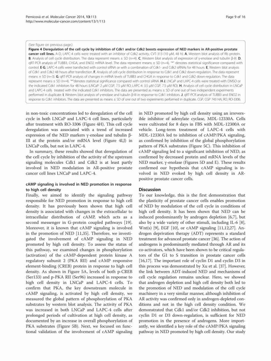
(See figure on previous page.)Figure 4 Deregulation of the cell cycle by inhibition of Cdk1 and/or Cdk2 boosts expression of NED markers in AR-positive prostatecancer cell lines. A-D, LAPC-4 cells were treated with an inhibitor of Cdk2 activity, CVT-313 (10 μM, 48 h). A, Western blot analysis of Rb protein.B, Analysis of cell cycle distribution. The data represent means ± SD (n=4). C, Western blot analysis of expression of γ-enolase and tubulin β-III. D,qRT-PCR analysis of TUBB3, CHGA, and ENO2 mRNA level. The data represent means ± SD (n=4). “*” denotes statistical significance compared withcontrol. E-G, LAPC-4 cells were transfected with control siRNA or with a combination of Cdk1- and Cdk2 siRNAs for 48 hours. E, Western blot analysisof Cdk1 and Cdk2 48 hours after transfection. F, Analysis of cell cycle distribution in response to Cdk1 and Cdk2 down-regulation. The data representmeans ± SD (n=3). G, qRT-PCR analysis of changes in mRNA levels of TUBB3 and CHGA in response to Cdk1 and Cdk2 down-regulation. The datarepresent means ± SD (n=4). “*”denotes statistical significance compared with control siRNA. H-J, LNCaP and LAPC-4 cells were treated with DMSO orthe indicated Cdk1 inhibitors for 48 hours (LNCaP: 2 μM CGP, 7.5 μM RO; LAPC-4: 3.5 μM CGP, 7.5 μM RO). H, Analysis of cell cycle distribution in LNCaPand LAPC-4 cells treated with the indicated Cdk1 inhibitors. The data are presented as means ± SD of one out of two independent experimentsperformed in duplicate. I, Western blot analysis of γ-enolase and tubulin β-III in response to Cdk1 inhibitors. J, qRT-PCR analysis of TUBB3 and ENO2 inresponse to Cdk1 inhibitors. The data are presented as means ± SD of one out of two experiments performed in duplicate. CGP, CGP 74514A; RO, RO-3306.
Pernicová et al. Molecular Cancer 2014, 13:113 Page 9 of 16http://www.molecular-cancer.com/content/13/1/113
in non-toxic concentrations led to deregulation of the cellcycle in both LNCaP and LAPC-4 cell lines, particularlyafter treatment with RO-3306 (Figure 4H). This cell cyclederegulation was associated with a trend of increasedexpression of the NED markers γ-enolase and tubulin β-III at the protein and/or mRNA level (Figure 4I,J) inLNCaP cells, but not in LAPC-4.In summary, these results showed that deregulation of
the cell cycle by inhibition of the activity of the upstreamsignaling molecules Cdk1 and Cdk2 is at least partlyinvolved in NED modulation in AR-positive prostatecancer cell lines LNCaP and LAPC-4.
cAMP signaling is involved in NED promotion in responseto high cell densityFinally, we aimed to identify the signaling pathwayresponsible for NED promotion in response to high celldensity. It has previously been shown that high celldensity is associated with changes in the extracellular tointracellular distribution of cAMP, which acts as asecond messenger in G-protein coupled pathways [19].Moreover, it is known that cAMP signaling is involvedin the promotion of NED [11,35]. Therefore, we investi-gated the involvement of cAMP signaling in NEDpromoted by high cell density. To assess the status ofthis pathway, we examined changes in phosphorylation(activation) of the cAMP-dependent protein kinase Aregulatory subunit 2 (PKA RII) and cAMP responsiveelement-binding (CREB) protein in response to high celldensity. As shown in Figure 5A, levels of both p-CREB(Ser133) and p-PKA RII (Ser96) increased in response tohigh cell density in LNCaP and LAPC-4 cells. Toconfirm that PKA, the key downstream molecule incAMP signaling, is activated by high cell density, wemeasured the global pattern of phosphorylation of PKAsubstrates by western blot analysis. The activity of PKAwas increased in both LNCaP and LAPC-4 cells afterprolonged periods of cultivation at high cell density, asdocumented by an increase in overall phosphorylation ofPKA substrates (Figure 5B). Next, we focused on func-tional validation of the involvement of cAMP signaling
in NED promoted by high cell density using an irrevers-ible inhibitor of adenylate cyclase, MDL-12330A. Cellswere cultivated for 8 days in FBS with MDL-12300A orvehicle. Long-term treatment of LAPC-4 cells withMDL-12330A led to inhibition of cAMP/PKA signaling,as confirmed by inhibition of the global phosphorylationpattern of PKA substrates (Figure 5C). This inhibition ofcAMP signaling led to a significant inhibition of NED, asconfirmed by decreased protein and mRNA levels of theNED marker, γ-enolase (Figures 5D and E). These resultsconfirmed our hypothesis that cAMP signaling is in-volved in NED evoked by high cell density in AR-positive prostate cancer cells.
DiscussionTo our knowledge, this is the first demonstration thatthe plasticity of prostate cancer cells enables promotionof NED by modulation of the cell cycle in conditions ofhigh cell density. It has been shown that NED can beinduced predominantly by androgen depletion [6,7], butalso by a wide variety of other stimuli, including IL-6 [8],Wnt(s) [9], EGF [10], or cAMP signaling [11,12,27]. An-drogen deprivation therapy (ADT) represents a standardtreatment for advanced prostate cancer [36]. The action ofandrogens is predominantly mediated through AR and itsco-activators, which have been shown to be critical regula-tors of the G1 to S transition in prostate cancer cells[16,17]. The important role of cyclin D1 and cyclin D3 inthis process was demonstrated by Xu et al. [37]. However,the link between ADT-induced NED and mechanisms ofcell cycle regulation remains unclear. Here, we showedthat androgen depletion and high cell density both led tothe promotion of NED and modulation of the cell cyclemachinery in a very similar manner, although inhibition ofAR activity was confirmed only in androgen-depleted con-ditions and not in the high cell density condition. Wedemonstrated that Cdk1 and/or Cdk2 inhibition, but notcyclin D1 or D3 down-regulation, is sufficient for NEDpromotion in the presence of androgens. More import-antly, we identified a key role of the cAMP/PKA signalingpathway in NED promoted by high cell density. Our study

Figure 5 Involvement of the cAMP signaling pathway in NED promoted by high cell density. Western blot analysis of changes inexpression of selected members of the cAMP signaling pathway in response to high cell density. A, Phosphorylation of PKA RII (Ser 96),phosphorylation of CREB (Ser 133), and total level of CREB were detected in cells cultivated in complete media with androgens (FBS) for 2 to16 days. B, Analysis of p-(Ser/Thr) PKA substrates in cells cultivated in the presence of androgens for 2 to 16 days. C, Analysis of the p-Ser/Thr PKAsubstrate in LAPC-4 cells in response to treatment with the MDL-12330A, an inhibitor of cAMP signaling. Cells were treated with 2 μM MDL-12330A for8 days. D, Analysis of the expression of the NED marker γ-enolase in response to treatment with MDL-12330A. E, qRT-PCR analysis of mRNA levels ofENO2 in response to inhibition of cAMP signaling. The data represent means ± SD of three experiments. “*” and “#” denote statistical significancecompared with control cells harvested on day 0 or cells cultivated for 8 days and treated with vehicle, respectively. F, Schematic illustration of theproposed mechanism for the promotion of NED by high cell density.
Pernicová et al. Molecular Cancer 2014, 13:113 Page 10 of 16http://www.molecular-cancer.com/content/13/1/113

Pernicová et al. Molecular Cancer 2014, 13:113 Page 11 of 16http://www.molecular-cancer.com/content/13/1/113
defines a novel mechanism highlighting how high tumordensity could modulate the plasticity of prostate cancercells and influence disease progression.In our previous study, we demonstrated that both andro-
gen depletion and high cell density cultivation led toincreased expression of cytokeratins, general markers ofepithelial differentiation [38]. Because androgen deprivationis a well-known promoter of NED [39], we compared theexpression of NED markers between prostate cancer cellscultured at high density and cells cultured in the absence ofandrogens. Surprisingly, androgen depletion and high celldensity both promoted increase in expression of severalNED markers (γ-enolase, tubulin β-III, and aromatic L-amino acid decarboxylase) in prostate cancer cells. Theeffect of high density on NED promotion is androgen-independent, since similar regulation of NED markers wasobserved in C4-2 cells, an androgen-independent sub-lineof LNCaP. Moreover, a similar high density-inducedincrease in the level of NED markers was detected usingthe AR-negative prostate cell line BPH-1 and its tumori-genic derivative CAFTD03, as well as the cancer cell linesDU-145 and PC3. Furthermore, re-introduction of AR intoAR-negative PC3 cells did not have any effect on high celldensity-induced NED. This phenotype was also confirmedin a 3D culture system in all above mentioned cell lines.Moreover, we observed a slight trend towards the correl-ation between decreased proliferation and NED promotionin a small cohort of patients with advanced prostate cancer.This is in contrast to previously published data showingthat a high Ki-67 labeling index is weakly associated withhigh chromogranin A expression (Spearman’s correlation0.164) [40]. However, the authors of that study showed nocorrelation between Ki-67 labeling index and expression ofAR or NeuroD1, another neuroendocrine marker. NeuroD1was previously shown to be expressed in aggressive prostatecancer cell lines and prostate cancer samples, although co-expression with chromogranin A was found only rarely[41]. Bubendorf et al. [42] did not found significant associ-ation between Ki-67 and neuroendocrine differentiationwhile Grobholz et al. [43] observed higher proliferationindex (assessed by Ki-67) in tumors with large clusters ofNE differentiation in comparison to negative tumors orwith solitary NE cells. With respect to our observations(both in-vitro and in our small patient cohort) and varyingresults in the literature, we may state that the neuroendo-crine transdifferentiation may be present in tumors withslower proliferation in some patients.Because the correlation of NED with cell cycle arrest
had been described in several experimental models[35,44], we next focused on modulation of the cell cyclemachinery. We showed that the induction of cell cyclearrest by androgen depletion is associated with down-regulation of cyclin D1 protein in LNCaP cells but notin LAPC-4 cells, and with down-regulation of cyclin D3
in both cell lines. Similar effects were observed whenNED was promoted by high cell density. These resultsare in accordance with previously published data showingthat androgen-induced proliferation of prostate cancercells is accompanied by increased levels of D-type cyclins[37] and, conversely, androgen deprivation causes down-regulation of protein levels of cyclin D1 and cyclin D3 inLNCaP cells [16,37]. RNAi-mediated down-regulation ofcyclin D1 in LNCaP cells and cyclin D3 in LAPC-4 cellsled to modulation of the cell cycle and an increased per-centage of cells in the G0/G1 phase; however, this was notassociated with the promotion of NED. Thus, we concludethat although the down-regulation of D-type cyclins leadsto cell cycle arrest in the G0/G1 phase, it is not sufficientfor promoting NED in AR-positive prostate cancer cells.Next, we focused on the role of the more general cell
cycle regulator Cdk2. Both the activity and expression ofCdk2 were down-regulated during NED promotion byandrogen depletion and high cell density, and this down-regulation correlated with up-regulation of the Cdk inhibi-tor p27Kip1 in both of our models. It was previously shownthat Cdk2 expression and activity together with Rb proteinphosphorylation, are regulated by androgens [16,45]. Gen-erally, inhibition of Cdk activity causes cell cycle arrestand inhibits proliferation of prostate cancer cells. Ourresults showed that inhibition of Cdk2 causes cell cycleattenuation, in particular accumulation in the G2/M phasein LAPC-4 cells; this arrest is associated with significantpromotion of NED. These results, which indicate a func-tional role of the inhibition of Cdk2 activity in the regula-tion of NED, are supported by the findings of otherinvestigators. For example, it has been reported thatsilibinin-induced NED in LNCaP cells is also associatedwith cell cycle arrest and decreased Cdk2 levels [46]. Wedid not observe a significant change in the expression ofNED markers when Cdk2 expression was reduced using asiRNA-mediated approach (data not shown). However,transfection of Cdk2 siRNA in combination with Cdk1siRNA resulted in a slight trend towards NED promotion.This observation is in agreement with results obtainedusing CVT-313 inhibitor, which at the applied dose prefer-entially inhibits Cdk2, but might also partially inhibitCdk1 [34]. To reveal the role of Cdk1 in NED promotionwe used selective inhibitors. Treatment with subtoxic con-centrations of the Cdk1-specific inhibitor CGP 74514 onlyslightly modulated the cell cycle, whereas RO-3306 causedaccumulation of cells in G2/M phase, in accordance withpreviously published data [47]. This was accompanied byincreased expression of NED markers at the protein andmRNA level in LNCaP cells but not in LAPC-4 cells,which only showed up-regulation of γ-enolase expressionat the protein level. Further studies of the mechanisms bywhich Cdk1 and Cdk2 are involved in the plasticity ofprostate cancer cells are necessary based on the fact that

Pernicová et al. Molecular Cancer 2014, 13:113 Page 12 of 16http://www.molecular-cancer.com/content/13/1/113
different approaches to the modulation of their expressionand activity were not uniformly reflected in terms of NEDpromotion. Since experiments for elucidating the involve-ment of Cdk1 and Cdk2 in promoting NED wereperformed only in AR-positive prostate cancer cell lines,investigating the AR-negative cell lines might shed morelight in proposed role of Cdk1 and Cdk2 deregulation inpromoting NED. Our observations suggest that the associ-ation between NED and the cell cycle, and the role of par-ticular regulators of cell cycle machinery is more complexand also cell type-dependent. The clinical potential ofpharmacological inhibition of Cdk activity in cancer ther-apy has been demonstrated in several studies (for reviewsee [48]). However, based on our observations, it is im-portant to consider the possible effects of Cdk1 and Cdk2inhibition in NED promotion in prostate cancer cells.It has previously been shown that NED can be induced
by physiological and pharmacological agents that elevateintracellular cAMP levels [27]. Treatment of prostate can-cer cells with cAMP leads to changes in the expression ofHox genes located at the HOXD locus [13] including theNeuro D1 transcription factor, which is expressed in ma-lignant NE cells [41]. Moreover, other downstream targetsof cAMP, PKA [11] and CREB [14], are directly involvedin NED. Furthermore, promotion of NED by cAMP-inducing agents is a reversible process. Interestingly, it hasbeen shown that the cAMP level can be modulated by celldensity [19]. Based on these facts, we hypothesized thatthe promotion of NED by high cell density can be medi-ated by the activation of cAMP signaling. Our resultsdemonstrated that cAMP signaling is indeed activated inresponse to high cell density, as demonstrated by in-creased levels of downstream target molecules of cAMP,such as phosphorylated PKA regulatory subunit II andphosphorylated CREB. Interestingly, cAMP inhibits prolif-eration of breast cancer cells via increased expression ofp27Kip1 and decreased activity of Cdk2 [49]. These obser-vations are indirectly in accordance with our observation,since in response to high cell density we observed in-creased activation of cAMP-mediated signaling, increasedp27Kip1, and decreased Cdk2 expression. Moreover, it wasshown that cAMP inhibits Cdk2 activity and Rb phos-phorylation in adipose stem cells [50]. More importantly,the functional involvement of cAMP was confirmed bythe demonstration that treatment of prostate cancer cellswith MDL-12330A, a potent inhibitor of adenylate cy-clase, abolished the promotion of NED by high cell dens-ity. In summary, these experiment support our hypothesisthat activation of cAMP signaling mediates NED promo-tion by high cell density in AR-positive prostate cancercell lines (Figure 5F). Based on our results we concludethat modulation of the cell cycle by high cell density canpromote reversible NED in prostate epithelial cancer cells.Our study also suggests that prostate cancer tissue
remodeling, in association with disease progression ortherapy, might contribute to tumor progression by modu-lating the plasticity of cancer cells and by promoting NED.
ConclusionsWe have demonstrated a new relationship between highcell density, cell cycle attenuation, and promotion ofNED and suggest high cell density as a trigger for cAMPsignaling that can mediate reversible NED in prostatecancer cells.
Materials and MethodsCell culture and treatmentLNCaP cells (DSMZ) [51] (androgen-sensitive cell linecarrying mutation in the gene encoding AR [52]), andLAPC-4 cells (androgen-dependent cell line carryingWT gene encoding AR [53]) were cultivated as describedpreviously [38]. Culture conditions for the androgen-independent subline C4-2 [54] were similar to those forthe parental LNCaP cell line. Under experimental condi-tions (high-density NED promotion, siRNA transfection,treatment with inhibitors), LNCaP cells were cultivatedwith 5% FBS or 5% dextran/charcoal-stripped FBS (CS, forandrogen depletion), and LAPC-4 cells were cultivatedwith 10% FBS and 1 nM R1881, or with 10% CS. BPH-1cells [55] and the BPH-1 tumorigenic clone CAFTD03[56] were cultivated as described previously [57]. PC3(ATCC) and PC3 cells stably expressing AR [30] werecultivated in F12 with 10% FBS and penicillin and strepto-mycin. DU-145 cells (ATCC) were cultivated in RPMI1640 with 10% FBS and penicillin and streptomycin.AmpFLSTR® Identifiler® PCR Amplification Kit (LifeTechnologies) was used to verify the origin of cell lines.To evoke NED, LNCaP and LAPC-4 cells were culti-
vated as follows: cells were seeded at a density of 20,000/cm2 in the appropriate complete medium with FBS(day -1). After 24 hours, the medium was exchanged formedium with FBS or CS (day 0). Cells were continuouslycultivated for 2 to 16 days without splitting, but withexchange of the medium for fresh medium twice a week.Cells were collected for further analysis on days 2, 4, 8,and 16 after the change of medium on day 0.For cultivation in 3D conditions, we used Alvetex® poly-
styrene scaffold inserts in 6-well plates (AVP004), 12-wellplates (AVP002), or 24-well plates (AVP006) containing200 μm thick Alvetex polystyrene scaffold (Reinnervate).Cells were seeded at a density of 0.5 × 10⁶, 1.0 × 10⁶, and1.5 × 106 cells per insert and cultivated for 72 to 96 hourswith regular media exchanges. Cells that were seeded onstandard Petri dishes in standard media and at standardseeding densities and cultivated for either 1 day or 4 dayswere used as a 2D control. Experiments in 3D were per-formed with two independent repetitions.

Pernicová et al. Molecular Cancer 2014, 13:113 Page 13 of 16http://www.molecular-cancer.com/content/13/1/113
For the inhibition of Cdk2 activity we used a selectiveATP-competitive Cdk2 inhibitor III [CVT-313, 2 (bis-(Hydroxyethyl) amino)-6-(4-methoxybenzylamino)-9-iso-propyl-purine)] (#238803 Merck). For inhibition of Cdk1activity we used the Cdk1 inhibitor CGP 74514A [N-(cis-2-Aminocyclohexyl) -N-(3-chlorophenyl)-9-ethyl-9H-purine-2,6-diamine, #217696, Calbiochem] and ATP-competitiveCdk1 Inhibitor IV RO-3306 [(5Z)-2-((Thiophen-2-yl)methylamino)-5-((quinolin-6-yl) methylene) thiazol-4(5H)-one, #217699, Calbiochem], both dissolved in DMSO. Forinhibition of cAMP signaling we used an adenylate cyclase-specific inhibitor MDL-12330A hydrochloride [N- (cis-2-phenyl-cyclopentyl) azacyclotridecan-2-imine-hydrochlor-ide, M-182, Sigma-Aldrich]. For all treatments, LNCaP andLAPC-4 cells were seeded at a density of 20,000 or 30,000cells/cm2 in appropriate media (IMDM+ 10% FBS + 1 nMR1881 + antibiotics for LAPC-4, RPMI + 5% FBS + antibi-otics for LNCaP cells). After 48 hours, cells were treatedwith the indicated concentrations of selected inhibitors andcontrol cells were treated with the equivalent concentrationof dMSO (not exceeding 0.1%). Cells were collected for fur-ther analysis 48 hours after treatment. All experiments wereperformed at least twice with technical duplicates.
Cell cycle analysisCells were fixed, stained, and analyzed by flow cytome-try using FACSCalibur™ or BD FACSVerse (BectonDickinson) as described previously [33]. At least twoindependent repetitions were performed for eachexperiment.
Cell transfection and RNA interferenceLNCaP and LAPC-4 cells were transfected with small in-hibitory RNA (siRNA) duplexes (Santa Cruz Biotechnology)directed against non-targeting control (sc-37007), Cdk1siRNA (sc-29252), and Cdk2 siRNA (sc-156139) using theNeon® Transfection System (Life Technologies). Transfec-tion was performed in a 10-μl tip according to the manufac-turer’s recommendations. Cells were harvested 48 hoursafter transfection for further analysis. Experiments wereperformed in three independent repetitions.
RNA isolation and real-time reverse transcriptionpolymerase chain reaction (qRT-PCR)Total RNA was isolated using High Pure RNA IsolationKit (Roche). PCR was performed using the One StepSYBR® PrimeScript™ RT-PCR Kit II (Perfect Real Time)according to the manufacturer’s recommendations on aRotorGene 6000 (Corbett Research) [33]. The sequencesof the primers used are listed in Additional file 7: TableS1-A. Changes in gene expression were calculated usingthe comparative threshold cycle method, with POLR2A asa normalizing gene [58]. Data from at least three experi-ments were normalized for each gene using the mean CT
value for the control sample (2 days of incubation withFBS, control siRNA, vehicle-treated cells, or cells har-vested at day 0). Alternatively, two-step qRT-PCR wasperformed. Up to 1 μg of isolated RNA was reverse tran-scribed to cDNA with the High Capacity RNA-to-cDNAKit (Applied Biosystems). qRT-PCR was performed on aLight Cycler 480 (Roche) using the Light Cycler 480 MasterMix in combination with Human Universal Probe Library(Roche). Primer and probe combinations used in assays arelisted in Additional file 7: Table S1B. Results for genesof interest were normalized to the housekeeping genePOLR2a assessed using Light Cycler 480 software andare presented as 2�ΔΔCT . experiments were performedin at least two independent repetitions or in technicalduplicates.
Electrophoresis and western blottingCollected cell pellets were lysed and the protein extractswere separated and blotted as described previously [33].The primary and secondary antibodies used are listedin Additional file 8: Table S2. Detection of α-tubulinand β-actin served as a control of equal loading. Allwestern blots are presented as typical results of atleast two independent repetitions. Densitometry ana-lyses were performed using ImageJ software (NIH).Values given below a particular band represent nor-malized results of densitometry analysis of the givenimage (integrated density for the particular band wasassessed and all values were normalized to the controlof equal loading).
Immunofluorescence microscopyCells were cultivated, fixed, permeabilized, and stainedas described previously [38]. The primary and secondaryantibodies used are listed in Additional file 8: Table S2.Fluorescence images of the cells were obtained using aconfocal microscope (TSC SP5X, Leica Microsystems).
Statistical analysisStatistical analysis was performed using STATISTICA forWindows software (StatSoft). When the data variance washomogenous, one-way analysis of variance followed by theFisher or Tukey range test was used. If the data variancewas non-homogenous, the Mann-Whitney U-test was performed.Supplementary Material and Methods can be found in
Additional file 9.
Additional files
Additional file 1: Figure S1. A, Induction of neuroendocrinetransdifferentiation by high cell density, but not by androgen depletion,is a reversible process. Cells were cultivated for 16 days to induce NED asdescribed in Material and Methods section. After 16 days the cells were

Pernicová et al. Molecular Cancer 2014, 13:113 Page 14 of 16http://www.molecular-cancer.com/content/13/1/113
re-seeded at a low density (10,000 cells/cm2) in the appropriate cultivationmedia. Cells grown in FBS were re-seeded into media with FBS; cells grownin CS were re-seeded into either CS or FBS. Cells were further cultivatedfor 2, 4, and 8 days (16+2, 16+4, and 16+8, respectively) without splittingand appropriate medium was exchanged with fresh one twice aweek. Expression of the NED marker γ-enolase in response to re-seedingwas assessed using western blot analysis. A typical result of three independentrepetitions is presented. B-D, NED is promoted by high density also inAR-negative prostate epithelial cell lines. B, PC-3, PC3-AR, and DU-145cells were cultivated as described in Supplementary Material andMethods. Expression of NED markers γ-enolase and tubulin β-III wasassessed by western blot analysis. C, Western blot analysis of AR expressionto confirm its presence in PC3-AR cells; LNCaP cells served as a positivecontrol. Results from one repetition out of two performed in technicalduplicate are presented. D, qRT-PCR analysis of the NED marker γ-enolase(ENO2) and tubulin β-III (TUBB3) in PC3, PC3-AR, and DU-145 cells cultivatedas described. Results from two repetitions performed in technical duplicateare presented (n=4). E, qRT-PCR analysis of DcR2 gene (TNFRSF10D) inPC3-AR cells cultivated as described in Additional file 9. Results fromtwo repetitions performed in technical duplicate are presented (n=4).
Additional file 2: Figure S2. Cultivation of prostate cancer cell lines in3D conditions using Alvetex scaffold. A, LNCaP and LAPC-4 cells werecultivated in 3D conditions using Alvetex® scaffold at the indicated seedingdensities per insert in complete media. After 72 hours, live cells werevisualized by staining with 0.5% neutral red solution (N6634, Sigma-Aldrich)according to the manufacturer’s protocol. Increased intensity of stainingindicates increased cell density. B, Immunofluorescence detection of tubulinβ-III expression in LNCaP and LAPC-4 cells after 3 days of cultivation onAlvetex® inserts. Staining was performed according to the manufacturer’sprotocol. Specifications of the antibodies used are provided in Table S2. C,qRT-PCR analysis of DcR2 gene (TNFRSF10D) in PC3-AR cells cultivated in 3Dconditions on Alvetex scaffold as described in Additional file 9. The trianglerepresents increasing seeding density in 3D conditions on Alvetex (0.5×106,1.0×106, and 1.5×10⁶, respectively). Results from twoindependent repetitions are presented (n=2).
Additional file 3: Figure S3. Assessment of AR activity at a single celllevel after high-density cultivation and prolonged androgen ablation. A,Activity of AR in response to androgen depletion (12d CS) and at highdensity (12d FBS) assessed by detection of KLK3 mRNA using a mRNAFISH technique and quantified (B) as described in Additional file 9. n,number of identified nuclei C, Flow cytometric analysis of prostate mem-brane specific antigen (PSMA) in LNCaP and LAPC-4 cells in response to an-drogen depletion (8d CS) or high density (8d FBS). Staining wasperformed as described in Additional file 9. Representative results from onerepetition out of two performed in replicate are presented. d, days.
Additional file 4: Table S3. Characteristics of human prostate tumorsamples. Subsets of patients with advanced CaP with lymph nodemetastases were selected to obtain a sufficient percentage of NED forstatistical evaluation. dg, diagnosis; GS, Gleason score; pT, pathologic Tstage; pN, positivity of lymph nodes; %, percentage of positive staining.
Additional file 5: Figure S4. Immunohistochemical staining offormalin-fixed paraffin-embedded patient samples. A, Patients 2 and 9display low Ki-67 expression (less than 30% nuclear positivity) andmultiple chromogranin A- and γ-enolase-positive NE and/or NE-like cells.Patients 13 and 15 display high Ki-67 expression (more than 30% nuclearpositivity) and single chromogranin A and γ-enolase-positive NE and/orNE-like cells (magnification 40×). B, Quantification of γ-enolase andchromogranin A expression in patient tumor samples. In total, 10 patientswith low Ki-67 expression and 8 patients with high Ki-67 expression wereexamined. Information on the patients is provided in Table S3.
Additional file 6: Figure S5. Modulation of the cell cycle bydown-regulation of cyclin D1 and/or cyclin D3 does not lead toinduction of NED in LNCaP and LAPC-4 cells. A, Western blot analysisof the efficiency of cyclin D1 and cyclin D3 down-regulation in LNCaP andLAPC-4 cells following transfection with control siRNA A or specific siRNA(20 or 40 nM). Experiments were performed as described in SupplementaryMaterials and Methods. B, Analysis of changes in cell cycle distribution inresponse to cyclin D1 and/or cyclin D3 down-regulation in LNCaP andLAPC-4 cells. Data represent means ± SD of three independent experiments.
C, Western blot analysis of changes in protein levels of NED markers inresponse to cyclin D1 and/or cyclin D3 siRNA. It should be noted that othersamples irrelevant to this study were analyzed on the same membranes.These samples were omitted from the pictures presented and the westernblots presented here are therefore cropped. D, qRT-PCR analysis of changesin mRNA level of the NED marker γ-enolase (ENO2) in response to cyclin D1and/or cyclin D3 siRNA. Data represent means ± SD of two independentexperiments performed in duplicate (n=4). “*” and “#” denote statisticalsignificance (P<0.05) compared with cells transfected with 20 nM or 40 nMcontrol siRNA A, respectively.
Additional file 7: Table S1. Sequences of primers used in quantitativeRT-PCR.
Additional file 8: Table S2. Specification of antibodies used forwestern blot analysis, immunofluorescence and flow cytometry.
Additional file 9: Supplementary Material and Methods.
AbbreviationsAR: Androgen receptor; cAMP: Cyclic adenosine 3′, 5′-monophosphate;CS: Dextran/charcoal-stripped FBS; Cdk1: Cyclin-dependent kinase 1;Cdk2: Cyclin-dependent kinase 2; CREB: CAMP responsive element-binding;CRPC: Castration-resistant prostate cancer; DcR2: Tumor necrosis factorreceptor superfamily member 10D (decoy receptor 2); NED: Neuroendocrinetransdifferentiation; PKA: Protein kinase A; PSA: Prostate-specific antigen;PSMA: Prostate-specific membrane antigen; Rb: Retinoblastoma protein.
Competing interestsThe authors declare that they have no competing interests.
Authors’ contributionsZP carried out experiments, analyzed results and wrote the manuscript. JBand GK provided IHC and data analysis. MK handled selection of patientsamples for IHC analysis. ES, RF, TS and ŠŠ carried out expression analysis andanalyzed data. JJ performed automatic image analysis of mRNA FISH. AKrevised the manuscript. KS carried out particular flow cytometry analyses,supervised the experimental work, participated in data analysis andinterpretation of results, and wrote the manuscript. All authors read andapproved the manuscript.
AcknowledgmentsThis work was supported by grants IGA MZD NT13573-4/2012, GA CR P301/12/P407, and by projects FNUSA-ICRC (CZ.1.05/1.1.00/02.0123), HistoPARK(CZ.1.07/2.3.00/20.0185), CELLBIOL (CZ.1.07/2.3.00/30.0030) and Biomedreg(no. CZ.1.05/2.1.00/01.0030) from the European Regional Development Fund.Institutional support was provided by the Academy of Sciences of the CzechRepublic. The authors thank Prof. Robert Reiter for his kind gift of LAPC-4cells; Prof. Simon W. Hayward, for his kind gift of BPH-1 and BPH-1 CAFTD03cells; Prof. Marián Hajdúch for the kind gift of the LNCaP C4-2 subline; Prof.Zoran Culig for the kind gift of the PC3-AR cell line; Dr. Milan Ešner for helpwith mRNA FISH microscopy; Eva Sedlmaierová, Monika Smějová, and JiřinaProcházková for help with sample preparation for western blotting and qRT-PCR; Iva Lišková, Martina Urbánková, Jaromíra Netíková, Miluše Andrysíková,and Kateřina Svobodová for excellent technical assistance.
Author details1Department of Cytokinetics, Institute of Biophysics, Academy of Sciences ofthe Czech Republic, v.v.i, Královopolská 135, CZ-612 65 Brno, Czech Republic.2Center of Biomolecular and Cellular Engineering, International ClinicalResearch Center, St. Anne’s University Hospital Brno, Brno, Czech Republic.3Department of Experimental Biology, Faculty of Sciences, Masaryk University,Brno, Czech Republic. 4Department of Histology and Embryology, Faculty ofMedicine, Masaryk University, Brno, Czech Republic. 5Laboratory of MolecularPathology and Institute of Molecular and Translational Medicine, Faculty ofMedicine and Dentistry, Palacky University Olomouc, Olomouc, CzechRepublic. 6Department of Urology, Faculty of Medicine and Dentistry, PalackyUniversity Olomouc, Olomouc, Czech Republic.
Received: 20 September 2013 Accepted: 13 May 2014Published: 20 May 2014

Pernicová et al. Molecular Cancer 2014, 13:113 Page 15 of 16http://www.molecular-cancer.com/content/13/1/113
References1. Nelson WG, De Marzo AM, Isaacs WB: Prostate cancer. N Engl J Med 2003,
349:366–381.2. Cindolo L, Cantile M, Vacherot F, Terry S, de la Taille A: Neuroendocrine
differentiation in prostate cancer: from lab to bedside. Urol Int 2007,79:287–296.
3. Bhatia S, Frangioni JV, Hoffman RM, Iafrate AJ, Polyak K: The challengesposed by cancer heterogeneity. Nat Biotech 2012, 30:604–610.
4. Abrahamsson PA: Neuroendocrine cells in tumour growth of theprostate. Endocr Relat Cancer 1999, 6:503–519.
5. Yuan TC, Veeramani S, Lin MF: Neuroendocrine-like prostate cancer cells:neuroendocrine transdifferentiation of prostate adenocarcinoma cells.Endocr Relat Cancer 2007, 14:531–547.
6. Yuan TC, Veeramani S, Lin FF, Kondrikou D, Zelivianski S, Igawa T, Karan D,Batra SK, Lin MF: Androgen deprivation induces human prostateepithelial neuroendocrine differentiation of androgen-sensitive LNCaPcells. Endocr Relat Cancer 2006, 13:151–167.
7. Ismail AH, Landry F, Aprikian AG, Chevalier S: Androgen ablation promotesneuroendocrine cell differentiation in dog and human prostate. Prostate2002, 51:117–125.
8. Deeble PD, Murphy DJ, Parsons SJ, Cox ME: Interleukin-6- and cyclic AMP-mediated signaling potentiates neuroendocrine differentiation of LNCaPprostate tumor cells. Mol Cell Biol 2001, 21:8471–8482.
9. Yang X, Chen MW, Terry S, Vacherot F, Chopin DK, Bemis DL, Kitajewski J,Benson MC, Guo Y, Buttyan R: A human- and male-specific protocadherinthat acts through the wnt signaling pathway to induce neuroendocrinetransdifferentiation of prostate cancer cells. Cancer Res 2005, 65:5263–5271.
10. Cortes MA, Cariaga-Martinez AE, Lobo MV, Martin Orozco RM, Motino O,Rodriguez-Ubreva FJ, Angulo J, Lopez-Ruiz P, Colas B: EGF promotesneuroendocrine-like differentiation of prostate cancer cells in the presenceof LY294002 through increased ErbB2 expression independent of thephosphatidylinositol 3-kinase-AKT pathway. Carcinogenesis 2012,33:1169–1177.
11. Cox ME, Deeble PD, Bissonette EA, Parsons SJ: Activated 3′,5′-cyclic AMP-dependent protein kinase is sufficient to induce neuroendocrine-likedifferentiation of the LNCaP prostate tumor cell line. J Biol Chem 2000,275:13812–13818.
12. Zelivianski S, Verni M, Moore C, Kondrikov D, Taylor R, Lin MF:Multipathways for transdifferentiation of human prostate cancer cellsinto neuroendocrine-like phenotype. Biochim Biophys Acta 2001,1539:28–43.
13. Cantile M, Kisslinger A, Cindolo L, Schiavo G, D’Anto V, Franco R, Altieri V,Gallo A, Villacci A, Tramontano D, Cillo C: cAMP induced modifications ofHOX D gene expression in prostate cells allow the identification of achromosomal area involved in vivo with neuroendocrinedifferentiation of human advanced prostate cancers. J Cell Physiol 2005,205:202–210.
14. Deng X, Liu H, Huang J, Cheng L, Keller ET, Parsons SJ, Hu CD: Ionizingradiation induces prostate cancer neuroendocrine differentiationthrough interplay of CREB and ATF2: implications for diseaseprogression. Cancer Res 2008, 68:9663–9670.
15. Deng X, Elzey BD, Poulson JM, Morrison WB, Ko SC, Hahn NM, Ratliff TL, HuCD: Ionizing radiation induces neuroendocrine differentiation of prostatecancer cells in vitro, in vivo and in prostate cancer patients. Am J CancerRes 2011, 1:834–844.
16. Knudsen KE, Arden KC, Cavenee WK: Multiple G1 regulatory elementscontrol the androgen-dependent proliferation of prostatic carcinomacells. J Biol Chem 1998, 273:20213–20222.
17. Balk SP, Knudsen KE: AR, the cell cycle, and prostate cancer. Nucl ReceptSignal 2008, 6:e001.
18. Fribourg AF, Knudsen KE, Strobeck MW, Lindhorst CM, Knudsen ES:Differential requirements for ras and the retinoblastoma tumorsuppressor protein in the androgen dependence of prostaticadenocarcinoma cells. Cell Growth Differ 2000, 11:361–372.
19. Orbo A, Jaeger R, Sager G: Cell density dependence of cAMP and cGMPlevels in four human cell lines derived from carcinomas of the uterinecervix. Gynecol Oncol 1994, 52:320–325.
20. Piast M, Kustrzeba-Wojcicka I, Matusiewicz M, Banas T: Molecular evolutionof enolase. Acta Biochim Pol 2005, 52:507–513.
21. Easter SS Jr, Ross LS, Frankfurter A: Initial tract formation in the mousebrain. J Neurosci 1993, 13:285–299.
22. Koshimizu H, Kim T, Cawley NX, Loh YP: Chromogranin A: a new proposalfor trafficking, processing and induction of granule biogenesis. RegulPept 2010, 160:153–159.
23. Wafa LA, Cheng H, Rao MA, Nelson CC, Cox M, Hirst M, Sadowski I, RenniePS: Isolation and identification of L-dopa decarboxylase as a protein thatbinds to and enhances transcriptional activity of the androgen receptorusing the repressed transactivator yeast two-hybrid system. Biochem J2003, 375:373–383.
24. Marchiani S, Tamburrino L, Nesi G, Paglierani M, Gelmini S, Orlando C,Maggi M, Forti G, Baldi E: Androgen-responsive and -unresponsive prostatecancer cell lines respond differently to stimuli inducing neuroendocrinedifferentiation. Int J Androl 2010, 33:784–793.
25. Terry S, Ploussard G, Allory Y, Nicolaiew N, Boissiere-Michot F, Maille P, KheuangL, Coppolani E, Ali A, Bibeau F, Culine S, Buttyan R, de la Taille A, Vacherot F:Increased expression of class III beta-tubulin in castration-resistant humanprostate cancer. Br J Cancer 2009, 101:951–956.
26. Wafa LA, Palmer J, Fazli L, Hurtado-Coll A, Bell RH, Nelson CC, Gleave ME,Cox ME, Rennie PS: Comprehensive expression analysis of L-dopadecarboxylase and established neuroendocrine markers in neoadjuvanthormone-treated versus varying Gleason grade prostate tumors. HumPathol 2007, 38:161–170.
27. Cox ME, Deeble PD, Lakhani S, Parsons SJ: Acquisition of neuroendocrinecharacteristics by prostate tumor cells is reversible: implications forprostate cancer progression. Cancer Res 1999, 59:3821–3830.
28. Vindrieux D, Reveiller M, Florin A, Blanchard C, Ruffion A, Devonec M,Benahmed M, Grataroli R: TNF-alpha-related apoptosis-inducing liganddecoy receptor DcR2 is targeted by androgen action in the rat ventralprostate. J Cell Physiol 2006, 206:709–717.
29. Vindrieux D, Reveiller M, Chantepie J, Yakoub S, Deschildre C, Ruffion A,Devonec M, Benahmed M, Grataroli R: Down-regulation of DcR2 sensitizesandrogen-dependent prostate cancer LNCaP cells to TRAIL-inducedapoptosis. Cancer Cell Int 2011, 11:42.
30. Neuwirt H, Puhr M, Cavarretta IT, Mitterberger M, Hobisch A, Culig Z:Suppressor of cytokine signalling-3 is up-regulated by androgen inprostate cancer cell lines and inhibits androgen-mediated proliferationand secretion. Endocr Relat Cancer 2007, 14:1007–1019.
31. Evans MJ, Smith-Jones PM, Wongvipat J, Navarro V, Kim S, Bander NH,Larson SM, Sawyers CL: Noninvasive measurement of androgen receptorsignaling with a positron-emitting radiopharmaceutical that targetsprostate-specific membrane antigen. Proc Natl Acad Sci U S A 2011,108:9578–9582.
32. Macdonald JI, Dick FA: Posttranslational modifications of theretinoblastoma tumor suppressor protein as determinants of function.Genes Cancer 2012, 3:619–633.
33. Lincova E, Hampl A, Pernicova Z, Starsichova A, Krcmar P, Machala M,Kozubik A, Soucek K: Multiple defects in negative regulation of the PKB/Akt pathway sensitise human cancer cells to the antiproliferative effectof non-steroidal anti-inflammatory drugs. Biochem Pharmacol 2009,78:561–572.
34. Brooks EE, Gray NS, Joly A, Kerwar SS, Lum R, Mackman RL, Norman TC,Rosete J, Rowe M, Schow SR, Schultz PG, Wang X, Wick MM, Shiffman D:CVT-313, a specific and potent inhibitor of CDK2 that preventsneointimal proliferation. J Biol Chem 1997, 272:29207–29211.
35. Bang YJ, Pirnia F, Fang WG, Kang WK, Sartor O, Whitesell L, Ha MJ, Tsokos M,Sheahan MD, Nguyen P, Niklinski WT, Myers CE, Trepel JB: Terminalneuroendocrine differentiation of human prostate carcinoma cells inresponse to increased intracellular cyclic AMP. Proc Natl Acad Sci U S A1994, 91:5330–5334.
36. Sharifi N, Gulley JL, Dahut WL: An update on androgen deprivationtherapy for prostate cancer. Endocr Relat Cancer 2010, 17:R305–R315.
37. Xu Y, Chen SY, Ross KN, Balk SP: Androgens induce prostate cancer cellproliferation through mammalian target of rapamycin activation and post-transcriptional increases in cyclin D proteins. Cancer Res 2006, 66:7783–7792.
38. Pernicova Z, Slabakova E, Kharaishvili G, Bouchal J, Kral M, Kunicka Z,Machala M, Kozubik A, Soucek K: Androgen depletion induces senescencein prostate cancer cells through down-regulation of Skp2. Neoplasia2011, 13:526–536.
39. Shariff AH, Ather MH: Neuroendocrine differentiation in prostate cancer.Urology 2006, 68:2–8.
40. Cindolo L, Cantile M, Franco R, Chiodini P, Schiavo G, Forte I, Zlobec I,Salzano L, Botti G, Gidaro S, Terracciano L, Cillo C: Parallel determination of

Pernicová et al. Molecular Cancer 2014, 13:113 Page 16 of 16http://www.molecular-cancer.com/content/13/1/113
NeuroD1, chromogranin-A, KI67 and androgen receptor expression insurgically treated prostate cancers. Int Braz J Urol 2011, 37:57–66.
41. Cindolo L, Franco R, Cantile M, Schiavo G, Liguori G, Chiodini P, Salzano L,Autorino R, Di Blasi A, Falsaperla M, Feudale E, Botti G, Gallo A, Cillo C: NeuroD1expression in human prostate cancer: can it contribute to neuroendocrinedifferentiation comprehension? Eur Urol 2007, 52:1365–1373.
42. Bubendorf L, Sauter G, Moch H, Schmid H-P, Gasser TC, Jordan P, Mihatsch MJ:Ki-67 labeling index: an independent predictor of progression in prostatecancer treated by radical prostatectomy. J Pathol 1996, 178:437–441.
43. Grobholz R, Griebe M, Sauer CG, Michel MS, Trojan L, Bleyl U: Influence ofneuroendocrine tumor cells on proliferation in prostatic carcinoma.Hum Pathol 2005, 36:562–570.
44. Mori S, Murakami-Mori K, Bonavida B: Interleukin-6 induces G1 arrestthrough induction of p27(Kip1), a cyclin-dependent kinase inhibitor, andneuron-like morphology in LNCaP prostate tumor cells. Biochem BiophysRes Commun 1999, 257:609–614.
45. Taneja SS, Ha S, Garabedian MJ: Androgen stimulated cellular proliferationin the human prostate cancer cell line LNCaP is associated with reducedretinoblastoma protein expression. J Cell Biochem 2001, 84:188–199.
46. Tyagi A, Agarwal C, Agarwal R: Inhibition of retinoblastoma protein (Rb)phosphorylation at serine sites and an increase in Rb-E2F complex formationby silibinin in androgen-dependent human prostate carcinoma LNCaP cells:role in prostate cancer prevention. Mol Cancer Ther 2002, 1:525–532.
47. Vassilev LT, Tovar C, Chen S, Knezevic D, Zhao X, Sun H, Heimbrook DC,Chen L: Selective small-molecule inhibitor reveals critical mitotic functionsof human CDK1. Proc Natl Acad Sci U S A 2006, 103:10660–10665.
48. Pitts TM, Davis SL, Eckhardt SG, Bradshaw-Pierce EL: Targeting nuclearkinases in cancer: Development of cell cycle kinase inhibitors. PharmacolTher 2014, 142:258–269.
49. Santore TA, Chen Y, Smit MJ, Iyengar R: Adenovirus-directed expression ofQ227L-G alpha(s) inhibits growth of established tumors of later-stagehuman breast cancer cells in athymic mice. Proc Natl Acad Sci U S A 2002,99:1671–1676.
50. Ugland H, Boquest AC, Naderi S, Collas P, Blomhoff HK: cAMP-mediatedinduction of cyclin E sensitizes growth-arrested adipose stem cells toDNA damage-induced apoptosis. Mol Biol Cell 2008, 19:5082–5092.
51. Horoszewicz JS, Leong SS, Chu TM, Wajsman ZL, Friedman M, Papsidero L,Kim U, Chai LS, Kakati S, Arya SK, Sandberg AA: The LNCaP cell line–a newmodel for studies on human prostatic carcinoma. Prog Clin Biol Res 1980,37:115–132.
52. Veldscholte J, Berrevoets CA, Ris-Stalpers C, Kuiper GG, Jenster G, Trapman J,Brinkmann AO, Mulder E: The androgen receptor in LNCaP cells containsa mutation in the ligand binding domain which affects steroid bindingcharacteristics and response to antiandrogens. J Steroid Biochem Mol Biol1992, 41:665–669.
53. Klein KA, Reiter RE, Redula J, Moradi H, Zhu XL, Brothman AR, Lamb DJ,Marcelli M, Belldegrun A, Witte ON, Sawyers CL: Progression of metastatichuman prostate cancer to androgen independence in immunodeficientSCID mice. Nat Med 1997, 3:402–408.
54. Wu HC, Hsieh JT, Gleave ME, Brown NM, Pathak S, Chung LW: Derivation ofandrogen-independent human LNCaP prostatic cancer cell sublines: roleof bone stromal cells. Int J Cancer 1994, 57:406–412.
55. Hayward SW, Dahiya R, Cunha GR, Bartek J, Deshpande N, Narayan P:Establishment and characterization of an immortalized but non-transformed human prostate epithelial cell line: BPH-1. In Vitro Cell DevBiol Anim 1995, 31:14–24.
56. Hayward SW, Wang Y, Cao M, Hom YK, Zhang B, Grossfeld GD, SudilovskyD, Cunha GR: Malignant transformation in a nontumorigenic humanprostatic epithelial cell line. Cancer Res 2001, 61:8135–8142.
57. Slabakova E, Pernicova Z, Slavickova E, Starsichova A, Kozubik A, Soucek K: TGF-beta1-induced EMT of non-transformed prostate hyperplasia cells is charac-terized by early induction of SNAI2/Slug. Prostate 2011, 71:1332–1343.
58. Schmittgen TD, Livak KJ: Analyzing real-time PCR data by the comparativeC(T) method. Nat Protoc 2008, 3:1101–1108.
doi:10.1186/1476-4598-13-113Cite this article as: Pernicová et al.: The role of high cell density in thepromotion of neuroendocrine transdifferentiation of prostate cancercells. Molecular Cancer 2014 13:113.
Submit your next manuscript to BioMed Centraland take full advantage of:
• Convenient online submission
• Thorough peer review
• No space constraints or color figure charges
• Immediate publication on acceptance
• Inclusion in PubMed, CAS, Scopus and Google Scholar
• Research which is freely available for redistribution
Submit your manuscript at www.biomedcentral.com/submit
Related Documents


