Report The Role of Dopamine in Value-Based Attentional Orienting Highlights d We examined the neural correlates of value-based attention using PET d Previously reward-associated stimuli involuntary captured attention as distractors d Such attentional capture was predicted by dopamine release in the dorsal striatum d Our findings elucidate the neurochemical basis of value- based distraction Authors Brian A. Anderson, Hiroto Kuwabara, Dean F. Wong, ..., Boris Frolov, Susan M. Courtney, Steven Yantis Correspondence [email protected] In Brief Anderson et al. investigate the contribution of striatal dopamine to value-based attentional orienting in humans using positron emission tomography with [ 11 C]raclopride. The findings provide insight into the neurochemical basis of attentional processing of learned reward cues and individual susceptibility to distraction by such cues. Anderson et al., 2016, Current Biology 26, 550–555 February 22, 2016 ª2016 Elsevier Ltd All rights reserved http://dx.doi.org/10.1016/j.cub.2015.12.062

Welcome message from author
This document is posted to help you gain knowledge. Please leave a comment to let me know what you think about it! Share it to your friends and learn new things together.
Transcript
Report
The Role of Dopamine in V
alue-Based AttentionalOrientingHighlights
d We examined the neural correlates of value-based attention
using PET
d Previously reward-associated stimuli involuntary captured
attention as distractors
d Such attentional capture was predicted by dopamine release
in the dorsal striatum
d Our findings elucidate the neurochemical basis of value-
based distraction
Anderson et al., 2016, Current Biology 26, 550–555February 22, 2016 ª2016 Elsevier Ltd All rights reservedhttp://dx.doi.org/10.1016/j.cub.2015.12.062
Authors
Brian A. Anderson, Hiroto Kuwabara,
Dean F. Wong, ..., Boris Frolov, Susan
M. Courtney, Steven Yantis
In Brief
Anderson et al. investigate the
contribution of striatal dopamine to
value-based attentional orienting in
humans using positron emission
tomography with [11C]raclopride. The
findings provide insight into the
neurochemical basis of attentional
processing of learned reward cues and
individual susceptibility to distraction by
such cues.
Current Biology
Report
The Role of Dopaminein Value-Based Attentional OrientingBrian A. Anderson,1,* Hiroto Kuwabara,2 Dean F. Wong,2,3,4 Emily G. Gean,2 Arman Rahmim,2 James R. Bra�si�c,2
Noble George,2 Boris Frolov,2 Susan M. Courtney,1,3,5 and Steven Yantis11Department of Psychological and Brain Sciences, Johns Hopkins University, 3400 N. Charles Street, Baltimore, MD 21218, USA2Section of High Resolution Brain PET, Division of Nuclear Medicine, Russell H. Morgan Department of Radiology and Radiological Science,Johns Hopkins University School of Medicine, 733 N. Broadway, Baltimore, MD 21205, USA3SolomonH.SnyderDepartment ofNeuroscience, JohnsHopkinsUniversitySchool ofMedicine, 733N.Broadway,Baltimore,MD21205,USA4Department of Psychiatry and Behavioral Sciences, Johns Hopkins University School of Medicine, 733 N. Broadway, Baltimore, MD 21205,
USA5F.M. Kirby Research Center, Kennedy Krieger Institute, 707 N. Broadway, Baltimore, MD 21205, USA
*Correspondence: [email protected]
http://dx.doi.org/10.1016/j.cub.2015.12.062
SUMMARY
Reward learning gives rise to strong attentionalbiases. Stimuli previously associated with rewardautomatically capture visual attention regardless ofintention [1–12]. Dopamine signaling within theventral striatum plays an important role in rewardlearning, representing the expected reward initiatedby a cue [13, 14]. How dopamine and the striatummay be involved in maintaining behaviors that havebeen shaped by reward learning, even after rewardexpectancies have changed, is less well understood[15]. Nonspecific measures of brain activity haveimplicated the striatum in value-based attention[11, 12, 16–18]. However, the neurochemical mecha-nisms underlying the attentional priority of learnedreward cues remain unexplored. Here, we investi-gated the contribution of dopamine to value-basedattention using positron emission tomography(PET) with [11C]raclopride. We show that, in theexplicit absence of reward, the magnitude of atten-tional capture by previously reward-associated butcurrently task-irrelevant distractors is correlatedacross individuals with changes in available D2/D3
dopamine receptors (presumably due to intrasynap-tic dopamine) linked to distractor processing withinthe right caudate and posterior putamen. Our find-ings provide direct evidence linking dopaminesignaling within the striatum to the involuntary orient-ing of attention, and specifically to the attention-grabbing quality of learned reward cues. Thesefindings also shed light on the neurochemical basisof individual susceptibility to value-driven attentionalcapture, which is known to play a role in addiction[19–21]. More broadly, the present study highlightsthe value and feasibility of using PET to relatechanges in the release of a neurotransmitter tolearning-dependent changes in healthy adults.
550 Current Biology 26, 550–555, February 22, 2016 ª2016 Elsevier
RESULTS
Healthy human participants (n = 20) performed an experiment
comprising a training phase and a test phase. The training phase
involved a visual search task in which locating and reporting
each of two color-defined targets was associated with a mone-
tary reward outcome (Figure 1A). The following day, two PET
scans were conducted while participants completed an unre-
warded shape-search task in which the color of the shapes
was task irrelevant (Figure 1B). During the distractor-absent
scan, no stimuli were ever rendered in the color of a formerly
reward-predictive target shown during training, whereas during
the distractor-present scan, half of the trials contained a distrac-
tor rendered in a previously reward-associated color (the scans
were otherwise identical).
During the training phase, participants were faster to report
the target that was associated with higher reward, indicating
learning of the stimulus-reward associations. This value-
driven bias was reliable from the second epoch (trials 121–
240) of the training phase onward (ts > 2.12, ps < 0.048; first
epoch: t = 1.57, p = 0.135) (Figure 2A). An attentional bias for
these previously reward-associated stimuli was evident in
the first epoch of trials during the test phase (trials 1–300),
as indicated by a slowing of response time (RT) on high-value
distractor trials compared to distractor-absent trials (t = 2.74,
p = 0.014) (Figure 2B), replicating previous behavioral results
[1–5, 10, 11]. This learned bias then extinguished in the
continued absence of reward and by the second epoch
was no longer statistically reliable. Consistent with prior
reports [1, 3, 4, 20, 22], the low-value distractor did not sig-
nificantly impair performance (t = 1.03, p = 0.316). There were
no differences in accuracy across the experimental con-
ditions in either phase of the experiment (see Tables S1
and S2).
As in prior studies [1, 3, 7, 19, 20], there were substantial indi-
vidual differences in the extent to which the previously reward-
associated distractors influenced performance. For some partic-
ipants, the previously high-value distractors greatly impaired
target detection, while for other participants these same distrac-
tors had no measurable cost associated with their presence. To
better understand the nature of these individual differences, we
Ltd All rights reserved
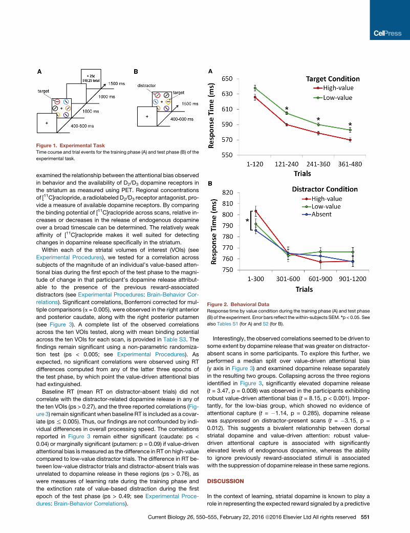
Figure 1. Experimental Task
Time course and trial events for the training phase (A) and test phase (B) of the
experimental task.
Figure 2. Behavioral Data
Response time by value condition during the training phase (A) and test phase
(B) of the experiment. Error bars reflect thewithin-subjects SEM. *p < 0.05. See
also Tables S1 (for A) and S2 (for B).
examined the relationship between the attentional bias observed
in behavior and the availability of D2/D3 dopamine receptors in
the striatum as measured using PET. Regional concentrations
of [11C]raclopride, a radiolabeled D2/D3 receptor antagonist, pro-
vide a measure of available dopamine receptors. By comparing
the binding potential of [11C]raclopride across scans, relative in-
creases or decreases in the release of endogenous dopamine
over a broad timescale can be determined. The relatively weak
affinity of [11C]raclopride makes it well suited for detecting
changes in dopamine release specifically in the striatum.
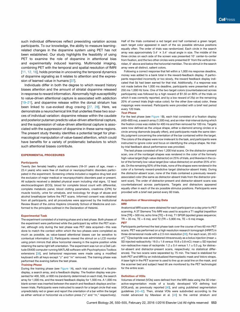
Within each of the striatal volumes of interest (VOIs) (see
Experimental Procedures), we tested for a correlation across
subjects of the magnitude of an individual’s value-based atten-
tional bias during the first epoch of the test phase to the magni-
tude of change in that participant’s dopamine release attribut-
able to the presence of the previous reward-associated
distractors (see Experimental Procedures: Brain-Behavior Cor-
relations). Significant correlations, Bonferroni corrected for mul-
tiple comparisons (a = 0.005), were observed in the right anterior
and posterior caudate, along with the right posterior putamen
(see Figure 3). A complete list of the observed correlations
across the ten VOIs tested, along with mean binding potential
across the ten VOIs for each scan, is provided in Table S3. The
findings remain significant using a non-parametric randomiza-
tion test (ps < 0.005; see Experimental Procedures). As
expected, no significant correlations were observed using RT
differences computed from any of the latter three epochs of
the test phase, by which point the value-driven attentional bias
had extinguished.
Baseline RT (mean RT on distractor-absent trials) did not
correlate with the distractor-related dopamine release in any of
the ten VOIs (ps > 0.27), and the three reported correlations (Fig-
ure 3) remain significant when baseline RT is included as a covar-
iate (ps% 0.005). Thus, our findings are not confounded by indi-
vidual differences in overall processing speed. The correlations
reported in Figure 3 remain either significant (caudate: ps <
0.04) or marginally significant (putamen: p = 0.09) if value-driven
attentional bias ismeasured as the difference in RT on high-value
compared to low-value distractor trials. The difference in RT be-
tween low-value distractor trials and distractor-absent trials was
unrelated to dopamine release in these regions (ps > 0.76), as
were measures of learning rate during the training phase and
the extinction rate of value-based distraction during the first
epoch of the test phase (ps > 0.49; see Experimental Proce-
dures: Brain-Behavior Correlations).
Current Biology 26, 550
Interestingly, the observed correlations seemed to be driven to
some extent by dopamine release that was greater on distractor-
absent scans in some participants. To explore this further, we
performed a median split over value-driven attentional bias
(y axis in Figure 3) and examined dopamine release separately
in the resulting two groups. Collapsing across the three regions
identified in Figure 3, significantly elevated dopamine release
(t = 3.47, p = 0.008) was observed in the participants exhibiting
robust value-driven attentional bias (t = 8.15, p < 0.001). Impor-
tantly, for the low-bias group, which showed no evidence of
attentional capture (t = �1.14, p = 0.285), dopamine release
was suppressed on distractor-present scans (t = �3.15, p =
0.012). This suggests a bivalent relationship between dorsal
striatal dopamine and value-driven attention: robust value-
driven attentional capture is associated with significantly
elevated levels of endogenous dopamine, whereas the ability
to ignore previously reward-associated stimuli is associated
with the suppression of dopamine release in these same regions.
DISCUSSION
In the context of learning, striatal dopamine is known to play a
role in representing the expected reward signaled by a predictive
–555, February 22, 2016 ª2016 Elsevier Ltd All rights reserved 551
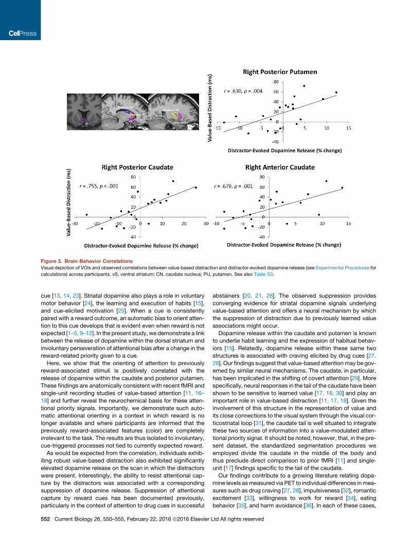
Figure 3. Brain-Behavior Correlations
Visual depiction of VOIs and observed correlations between value-based distraction and distractor-evoked dopamine release (see Experimental Procedures for
calculations) across participants. vS, ventral striatum; CN, caudate nucleus; PU, putamen. See also Table S3.
cue [13, 14, 23]. Striatal dopamine also plays a role in voluntary
motor behavior [24], the learning and execution of habits [15],
and cue-elicited motivation [25]. When a cue is consistently
paired with a reward outcome, an automatic bias to orient atten-
tion to this cue develops that is evident even when reward is not
expected [1–5, 9–12]. In the present study, we demonstrate a link
between the release of dopamine within the dorsal striatum and
involuntary perseveration of attentional bias after a change in the
reward-related priority given to a cue.
Here, we show that the orienting of attention to previously
reward-associated stimuli is positively correlated with the
release of dopamine within the caudate and posterior putamen.
These findings are anatomically consistent with recent fMRI and
single-unit recording studies of value-based attention [11, 16–
18] and further reveal the neurochemical basis for these atten-
tional priority signals. Importantly, we demonstrate such auto-
matic attentional orienting in a context in which reward is no
longer available and where participants are informed that the
previously reward-associated features (color) are completely
irrelevant to the task. The results are thus isolated to involuntary,
cue-triggered processes not tied to currently expected reward.
As would be expected from the correlation, individuals exhib-
iting robust value-based distraction also exhibited significantly
elevated dopamine release on the scan in which the distractors
were present. Interestingly, the ability to resist attentional cap-
ture by the distractors was associated with a corresponding
suppression of dopamine release. Suppression of attentional
capture by reward cues has been documented previously,
particularly in the context of attention to drug cues in successful
552 Current Biology 26, 550–555, February 22, 2016 ª2016 Elsevier
abstainers [20, 21, 26]. The observed suppression provides
converging evidence for striatal dopamine signals underlying
value-based attention and offers a neural mechanism by which
the suppression of distraction due to previously learned value
associations might occur.
Dopamine release within the caudate and putamen is known
to underlie habit learning and the expression of habitual behav-
iors [15]. Relatedly, dopamine release within these same two
structures is associated with craving elicited by drug cues [27,
28]. Our findings suggest that value-based attentionmay be gov-
erned by similar neural mechanisms. The caudate, in particular,
has been implicated in the shifting of covert attention [29]. More
specifically, neural responses in the tail of the caudate have been
shown to be sensitive to learned value [17, 18, 30] and play an
important role in value-based distraction [11, 17, 18]. Given the
involvement of this structure in the representation of value and
its close connections to the visual system through the visual cor-
ticostriatal loop [31], the caudate tail is well situated to integrate
these two sources of information into a value-modulated atten-
tional priority signal. It should be noted, however, that, in the pre-
sent dataset, the standardized segmentation procedures we
employed divide the caudate in the middle of the body and
thus preclude direct comparison to prior fMRI [11] and single-
unit [17] findings specific to the tail of the caudate.
Our findings contribute to a growing literature relating dopa-
mine levels asmeasured via PET to individual differences inmea-
sures such as drug craving [27, 28], impulsiveness [32], romantic
excitement [33], willingness to work for reward [34], eating
behavior [35], and harm avoidance [36]. In each of these cases,
Ltd All rights reserved
such individual differences reflect preexisting variation across
participants. To our knowledge, the ability to measure learning-
related changes in the dopamine system using PET has not
been established. Our study highlights the feasibility of using
PET to examine the role of dopamine in attentional bias
and experimentally induced learning. Multimodal imaging,
combining PET with the temporal and spatial resolution of fMRI
[11, 12, 16], holds promise in uncovering the temporal dynamics
of dopamine signaling as it relates to attention and the expres-
sion of learned value in humans [37].
Individuals differ in both the degree to which reward history
biases attention and the amount of striatal dopamine released
in response to reward information. Abnormally high susceptibility
to value-driven attentional capture is associated with addiction
[19–21], and dopamine release within the dorsal striatum has
been linked to cue-evoked drug craving [27, 28]. Here, we
demonstrate a neurochemical link that underlies these two sour-
ces of individual variation: dopamine release within the caudate
and posterior putamen predicts value-driven attentional capture,
and the suppression of value-driven attentional capture is asso-
ciated with the suppression of dopamine in these same regions.
The present study thereby identifies a potential target for phar-
macological manipulation of value-based attention, which could
have benefits for a variety of problematic behaviors to which
such attentional biases contribute.
EXPERIMENTAL PROCEDURES
Participants
Twenty (ten female) healthy adult volunteers (18–31 years of age, mean =
23.4 years) who were free of medical or neuropsychiatric disorders partici-
pated in the experiment. Screening criteria included a negative drug test and
the exclusion of major medical or neuropsychiatric disorders past or present.
All subjects received a detailed physical exam including vital signs, 12 lead
electrocardiogram (ECG), blood for complete blood count with differential,
complete metabolic panel, blood clotting parameters, creatinine (CPK) for
muscle toxicity, urine for urinalysis, and toxicology for drugs of abuse and
alcohol breathalyzer before the PET scans. Informed consent was obtained
from all participants, and all procedures were approved by the Institutional
Review Board of the Johns Hopkins University School of Medicine and con-
formed to the principles outlined in the Declaration of Helsinki.
Experimental Task
The experiment consisted of a training phase and a test phase. Both phases of
the experiment were performed while the participant lay within the PET scan-
ner, although only during the test phase was PET data acquired—this was
done to match the context within which the two phases were completed as
much as possible, as value-based attentional biases can be sensitive to
contextual information [5]. Participants viewed the stimuli on a LCD monitor
using prism mirrors that allow horizontal viewing in the supine position while
retaining the same right left orientation. The experiment was run on a Dell Lati-
tude E6400 computer running MATLAB software with Psychophysics Toolbox
extensions [38], and behavioral responses were made using a modified
keyboard with all keys except ‘‘z’’ and ‘‘m’’ removed. The training phase was
performed the evening before the test phase.
Training Phase
During the training phase (see Figure 1A), each trial consisted of a fixation
display, a search array, and a feedback display. The fixation display was pre-
sented for 400, 500, or 600 ms (randomly determined on each trial), the search
array for 1,000 ms, and the reward feedback display for 1,500 ms. A 1,000 ms
blank screen was inserted between the search and feedback displays and be-
tween trials. Participants were instructed to search for a target circle that was
unpredictably red or green and report the orientation of a bar within the target
as either vertical or horizontal via a button press (‘‘z’’ and ‘‘m,’’ respectively).
Current Biology 26, 550
Half of the trials contained a red target and half contained a green target;
each target color appeared in each of the six possible stimulus positions
equally often. The order of trials was randomized. Each circle in the search
array was approximately 3.4� 3 3.4� visual angle in size. The middle of the
three circles on each side of the screen was presented 10� center-to-center
from fixation, and the two other circles were presented 8� from the vertical me-
ridian, 6� above and below the horizontal meridian. The six stimuli in the search
array were all distinct, salient colors.
Following a correct response that fell within a 1,000 ms response deadline,
money was added to a bank total in the reward-feedback display. If partici-
pants responded incorrectly or too slowly, the reward feedback display indi-
cated that 0¢ had been earned for that trial. Additionally, if a response was
not made before the 1,000 ms deadline, participants were presented with a
250 ms 1,000 Hz tone. One of the two target colors (counterbalanced across
participants) was followed by a high reward of $1.50 on 80% of the trials on
which it was correctly reported, and by a low reward of 25¢ on the remaining
20% of correct trials (high-value color); for the other (low-value) color, these
mappings were reversed. Participants were provided with a brief rest period
every 120 trials.
Test Phase
For the test phase (see Figure 1B), each trial consisted of a fixation display
(400–600ms), a search array (1,500ms), and an inter-trial-interval duringwhich
the fixation cross was visible for 400 ms and then removed for 100 ms. Targets
were now defined as the unique shape, either a diamond among circles or a
circle among diamonds (equally often), and participants made the same iden-
tity judgment concerning the orientation of the bar contained within the target.
The colors of the shapes were now irrelevant to the task, and participants were
instructed to ignore color and focus on identifying the unique shape. No trial-
by-trial feedback about performance was provided.
The test phase consisted of two 1,200 trial scans. On the distractor-present
scan, one of the nontarget shapes was rendered in the color of the formerly
high-value target (high-value distractor) on 25%of trials, and likewise in the co-
lor of the formerly low-value target (low-value distractor) on another 25% of tri-
als. On the remaining 50%of the trials, none of the shapeswere rendered in the
color of a formerly reward-predictive target (distractor-absent trials). During
the distractor-absent scan, none of the trials contained a previously reward-
associated color (the same as distractor-absent trials from the distractor-pre-
sent scan). The order of distractor-present and distractor-absent scans was
counterbalanced across participants. Targets and distractors appeared
equally often in each of the six possible stimulus positions. Participants were
provided with a brief rest period every 60 trials.
Acquisition of Neuroimaging Data
MRI
Anatomical MRI scans were obtained for each participant on a day prior to PET
scanning. A 3T Siemens Trio MRI was used to acquire a T1 sagittal (repetition
time [TR] = 500 ms; echo time [TE] = 8 ms), T1 SPGR (spoiled grass sequence;
TR = 35 ms, TE = 6 ms), and T2 (TR = 5,900 ms, TE = 6 ms) image.
PET
Participants performed the test phase task over the course of two 60 min PET
scans. PET was performed on a high resolution research tomograph (HRRT) in
three-dimensional mode with a 2.5 mm resolution [39]. For each scan, 20 mCi
of [11C]raclopride was administered intravenously as a bolus injection (mean ±
SD injected radioactivity: 19.0 ± 1.6 versus 19.8 ± 0.8 mCi; mean ± SD injected
non-radioactive mass of raclopride: 1.2 ± 0.4 versus 1.1 ± 0.3 mg, for distrac-
tor-absent and distractor-present scans, respectively; no statistical differ-
ences). The two scans were separated by 75 min. The head is stabilized for
both PET and MRI by an individualized thermoplastic mask and Velcro straps.
A laser light in the PET scanner is used to line up an axial line on the mask, and
the scanner bed and subject head tilt are monitored by the PET technologist
for the entire scan.
Definition of VOIs
Volumes of interest (VOIs) were defined from the MRI data using the 3D inter-
active-segmentation mode of a locally developed VOI defining tool
(VOILand), as previously reported [40], and using published segmentation
guidelines [40–42]. Then, striatal VOIs were subdivided according to the
model advanced by Mawlawi et al. [43] to the ventral striatum and
–555, February 22, 2016 ª2016 Elsevier Ltd All rights reserved 553
anterior/posterior putamen and caudate nucleus (five subdivisions per side)
using a semi-automated method that incorporated anatomical guidance
based on post-mortem human materials [40, 44]. VOIs were transferred
from MRI to PET space according to MRI-to-PET coregistration parameters
obtained with the coregistration module [45] in SPM5 (The Statistical Para-
metric Mapping 5; The Wellcome Trust Centre for Neuroimaging; available
at http://www.fil.ion.ac.uk/spm) and applied to PET frames to obtain regional
time (radio-)activity curves (TACs).
Reconstruction of PET Data
Emission PET scans were reconstructed using the iterative ordered-subset
expectation-maximization algorithm correcting for attenuation, scatter, ran-
doms, and dead time [46] and including inter-frame head motion correction
including transmission-emission alignment for the individual frames [47]. The
radioactivity was corrected for physical decay to the injection time. Recon-
structions included dynamic PET frames of 256 (left-to-right) by 256 (nasion-
to-inion) by 207 (neck-to-cranium) voxels with 1.22 mm isotropic dimensions.
The frame schedules were four 15 s, four 30 s, three 1 min, two 2 min, five
4 min, and twelve 5 min frames.
Data Analysis
Behavior
Mean RT and accuracy were computed for each experimental condition. Only
correct RTs were included in the mean, and RTs faster than 200 ms or
exceeding 3 SD of the mean were trimmed as in prior studies [1–5, 19, 20].
Data from each phase of the experiment were further broken down into four
equally sized trial bins, as in [2].
PET
Nondisplaceable binding potential (BPND; [48]) of [11C]raclopride was obtained
by the reference tissue graphical analysis (RTGA; [49]) for striatum subdivi-
sions. Then, dopamine release (DARel in %; [50]) was obtained using the
following formula: (BPND[A] � BPND[P])/BPND[A] 3 100, where [A] and [P]
stands for BPND of the distractor-absent and distractor-present scans,
respectively. Data for one participant were unusable due to a technical error
that resulted in an asynchrony between PET data acquisition and the adminis-
tration of the experimental task.
Brain-Behavior Correlations
Within each of the striatal VOIs, we tested for a correlation (Pearson’s r) across
participants of the magnitude of an individual’s value-based attentional bias
(slowing of RT on high-value distractor trials compared to distractor-absent tri-
als during the first epoch, see Figure 2B) to themagnitude of dopamine release
attributable to distractor processing using the calculation described above.
Bonferroni correction was used to set the overall type I error rate at 0.05 (a =
0.005 for each of ten correlations). Significant correlations obtained using
Pearson’s r were further scrutinized via a randomization test in which the prob-
ability of each correlation was estimated non-parametrically by randomly shuf-
fling the xy pairings (n interations = 10,000). To examine the potential influence
of learning rate during the training phase on our measure of striatal dopamine
release, for each participant we defined learning rate both as the difference in
the RT facilitation by the high-value target (low-value minus high-value target
RT) between the first and last epoch, as well as a linear fit to the change in this
measure over all four epochs. To examine the potential influence of extinction
rate during the test phase, for each participant we computed the difference in
value-based attentional bias (slowing of RT on high-value distractor trials
compared to distractor-absent trials) during the first and second half of the first
epoch (i.e., trials 1–150 versus trials 151–300).
SUPPLEMENTAL INFORMATION
Supplemental Information includes three tables and can be found with this
article online at http://dx.doi.org/10.1016/j.cub.2015.12.062.
AUTHOR CONTRIBUTIONS
B.A.A. and S.Y. developed the study concept; B.A.A., H.K., D.F.W., and S.Y.
designed the experiment; D.F.W., E.G.G., J.R.B., N.G., and B.F. conducted
the experiment; B.A.A., H.K., D.F.W., and A.R. analyzed and chose the analytic
tools for the data; A.R. contributed custom software used in data analysis; all
554 Current Biology 26, 550–555, February 22, 2016 ª2016 Elsevier
authors contributed to the interpretation of the data and the writing and editing
of the manuscript.
ACKNOWLEDGMENTS
The reported research was supported by NIH grants R01-DA013165 to S.Y.
and S.M.C., S10-RR017219 to D.F.W., S10-RR023623 to D.F.W., and F31-
DA033754 to B.A.A. The funding sources played no role in the study beyond
financial support. The authors give special thanks to Andrew Crabb, MS,
Ayon Nandi, MS, and Joshua Roberts, PhD, for technical and/or editorial
assistance.
Received: October 5, 2015
Revised: November 30, 2015
Accepted: December 24, 2015
Published: February 11, 2016
REFERENCES
1. Anderson, B.A., Laurent, P.A., and Yantis, S. (2011). Value-driven atten-
tional capture. Proc. Natl. Acad. Sci. USA 108, 10367–10371.
2. Anderson, B.A., Laurent, P.A., and Yantis, S. (2011). Learned value mag-
nifies salience-based attentional capture. PLoS ONE 6, e27926.
3. Anderson, B.A., and Yantis, S. (2012). Value-driven attentional and oculo-
motor capture during goal-directed, unconstrained viewing. Atten.
Percept. Psychophys. 74, 1644–1653.
4. Anderson, B.A., and Yantis, S. (2013). Persistence of value-driven atten-
tional capture. J. Exp. Psychol. Hum. Percept. Perform. 39, 6–9.
5. Anderson, B.A. (2015). Value-driven attentional priority is context specific.
Psychon. Bull. Rev. 22, 750–756.
6. Della Libera, C., and Chelazzi, L. (2009). Learning to attend and to ignore is
a matter of gains and losses. Psychol. Sci. 20, 778–784.
7. Hickey, C., Chelazzi, L., and Theeuwes, J. (2010). Reward changes
salience in human vision via the anterior cingulate. J. Neurosci. 30,
11096–11103.
8. Raymond, J.E., and O’Brien, J.L. (2009). Selective visual attention and
motivation: the consequences of value learning in an attentional blink
task. Psychol. Sci. 20, 981–988.
9. Le Pelley, M.E., Pearson, D., Griffiths, O., and Beesley, T. (2015). When
goals conflict with values: counterproductive attentional and oculomotor
capture by reward-related stimuli. J. Exp. Psychol. Gen. 144, 158–171.
10. Anderson, B.A. (2013). A value-driven mechanism of attentional selection.
J. Vis. 13, 1–16.
11. Anderson, B.A., Laurent, P.A., and Yantis, S. (2014). Value-driven atten-
tional priority signals in human basal ganglia and visual cortex. Brain
Res. 1587, 88–96.
12. Hickey, C., and Peelen, M.V. (2015). Neural mechanisms of incentive
salience in naturalistic human vision. Neuron 85, 512–518.
13. Schultz, W., Dayan, P., and Montague, P.R. (1997). A neural substrate of
prediction and reward. Science 275, 1593–1599.
14. Waelti, P., Dickinson, A., and Schultz, W. (2001). Dopamine responses
comply with basic assumptions of formal learning theory. Nature 412,
43–48.
15. Graybiel, A.M. (2008). Habits, rituals, and the evaluative brain. Annu. Rev.
Neurosci. 31, 359–387.
16. Krebs, R.M., Boehler, C.N., Roberts, K.C., Song, A.W., andWoldorff, M.G.
(2012). The involvement of the dopaminergic midbrain and cortico-striatal-
thalamic circuits in the integration of reward prospect and attentional task
demands. Cereb. Cortex 22, 607–615.
17. Yamamoto, S., Kim, H.F., and Hikosaka, O. (2013). Reward value-contin-
gent changes of visual responses in the primate caudate tail associated
with a visuomotor skill. J. Neurosci. 33, 11227–11238.
18. Hikosaka, O., Yamamoto, S., Yasuda, M., and Kim, H.F. (2013). Why skill
matters. Trends Cogn. Sci. 17, 434–441.
Ltd All rights reserved
19. Anderson, B.A., Faulkner, M.L., Rilee, J.J., Yantis, S., and Marvel, C.L.
(2013). Attentional bias for nondrug reward is magnified in addiction.
Exp. Clin. Psychopharmacol. 21, 499–506.
20. Anderson, B.A., Kronemer, S.I., Rilee, J.J., Sacktor, N., and Marvel, C.L.
(2015). Reward, attention, and HIV-related risk in HIV+ individuals.
Neurobiol. Dis. Published online October 17, 2015. http://dx.doi.org/10.
1016/j.nbd.2015.10.018.
21. Field, M., and Cox, W.M. (2008). Attentional bias in addictive behaviors: a
review of its development, causes, and consequences. Drug Alcohol
Depend. 97, 1–20.
22. Anderson, B.A. (2015). Value-driven attentional capture in the auditory
domain. Atten. Percept. Psychophys. Published online October 22,
2015. http://dx.doi.org/10.3758/s13414-015-1001-7.
23. Zald, D.H., Boileau, I., El-Dearedy, W., Gunn, R., McGlone, F., Dichter,
G.S., and Dagher, A. (2004). Dopamine transmission in the human striatum
during monetary reward tasks. J. Neurosci. 24, 4105–4112.
24. Albin, R.L., Young, A.B., and Penney, J.B. (1989). The functional anatomy
of basal ganglia disorders. Trends Neurosci. 12, 366–375.
25. Berridge, K.C., and Robinson, T.E. (1998). What is the role of dopamine in
reward: hedonic impact, reward learning, or incentive salience? Brain Res.
Brain Res. Rev. 28, 309–369.
26. Stormark, K.M., Field, N.P., Hugdahl, K., and Horowitz, M. (1997).
Selective processing of visual alcohol cues in abstinent alcoholics: an
approach-avoidance conflict? Addict. Behav. 22, 509–519.
27. Volkow, N.D., Wang, G.J., Telang, F., Fowler, J.S., Logan, J., Childress,
A.R., Jayne, M., Ma, Y., andWong, C. (2006). Cocaine cues and dopamine
in dorsal striatum: mechanism of craving in cocaine addiction. J. Neurosci.
26, 6583–6588.
28. Wong, D.F., Kuwabara, H., Schretlen, D.J., Bonson, K.R., Zhou, Y., Nandi,
A., Brasi�c, J.R., Kimes, A.S., Maris, M.A., Kumar, A., et al. (2006).
Increased occupancy of dopamine receptors in human striatum during
cue-elicited cocaine craving. Neuropsychopharmacology 31, 2716–2727.
29. Fairhall, S.L., Indovina, I., Driver, J., and Macaluso, E. (2009). The brain
network underlying serial visual search: comparing overt and covert
spatial orienting, for activations and for effective connectivity. Cereb.
Cortex 19, 2946–2958.
30. Yamamoto, S., Monosov, I.E., Yasuda, M., and Hikosaka, O. (2012). What
and where information in the caudate tail guides saccades to visual ob-
jects. J. Neurosci. 32, 11005–11016.
31. Seger, C.A. (2013). The visual corticostriatal loop through the tail of the
caudate: circuitry and function. Front. Syst. Neurosci. 7, 104.
32. Buckholtz, J.W., Treadway, M.T., Cowan, R.L., Woodward, N.D., Li, R.,
Ansari, M.S., Baldwin, R.M., Schwartzman, A.N., Shelby, E.S., Smith,
C.E., et al. (2010). Dopaminergic network differences in human impulsivity.
Science 329, 532.
33. Takahashi, K., Mizuno, K., Sasaki, A.T., Wada, Y., Tanaka, M., Ishii, A.,
Tajima, K., Tsuyuguchi, N., Watanabe, K., Zeki, S., and Watanabe, Y.
(2015). Imaging the passionate stage of romantic love by dopamine dy-
namics. Front. Hum. Neurosci. 9, 191.
34. Treadway, M.T., Buckholtz, J.W., Cowan, R.L., Woodward, N.D., Li, R.,
Ansari, M.S., Baldwin, R.M., Schwartzman, A.N., Kessler, R.M., and
Zald, D.H. (2012). Dopaminergic mechanisms of individual differences in
human effort-based decision-making. J. Neurosci. 32, 6170–6176.
35. Volkow, N.D., Wang, G.J., Maynard, L., Jayne, M., Fowler, J.S., Zhu, W.,
Logan, J., Gatley, S.J., Ding, Y.S., Wong, C., and Pappas, N. (2003).
Brain dopamine is associated with eating behaviors in humans. Int. J.
Eat. Disord. 33, 136–142.
Current Biology 26, 550
36. Kim, J.H., Son, Y.D., Kim, H.K., Lee, S.Y., Cho, S.E., Kim, Y.B., and Cho,
Z.H. (2011). Association of harm avoidance with dopamine D2/3 receptor
availability in striatal subdivisions: a high resolution PET study. Biol.
Psychol. 87, 164–167.
37. Kohno, M., Ghahremani, D.G., Morales, A.M., Robertson, C.L., Ishibashi,
K., Morgan, A.T., Mandelkern, M.A., and London, E.D. (2013). Risk-taking
behavior: dopamine D2/D3 receptors, feedback, and frontolimbic activity.
Cereb. Cortex 25, 236–245.
38. Brainard, D.H. (1997). The psychophysics toolbox. Spat. Vis. 10, 433–436.
39. Sossi, V., de Jong, H.W.A.M., Barker, W.C., Bloomfield, P., Burbar, Z.,
Camborde, M.-L., Comtat, C., Eriksson, L.A., Houle, S., Keator, D., et al.
(2005). The second generation HRRT: amulti-centre scanner performance
investigation. IEEE Nucl. Sci. Symp. Conf. Rec. 4, 2195–2199.
40. Oswald, L.M., Wong, D.F., McCaul, M., Zhou, Y., Kuwabara, H., Choi, L.,
Bra�si�c, J., and Wand, G.S. (2005). Relationships among ventral striatal
dopamine release, cortisol secretion, and subjective responses to
amphetamine. Neuropsychopharmacology 30, 821–832.
41. Yushkevich, P.A., Piven, J., Hazlett, H.C., Smith, R.G., Ho, S., Gee, J.C.,
and Gerig, G. (2006). User-guided 3D active contour segmentation of
anatomical structures: significantly improved efficiency and reliability.
Neuroimage 31, 1116–1128.
42. Diedrichsen, J., Balsters, J.H., Flavell, J., Cussans, E., and Ramnani, N.
(2009). A probabilistic MR atlas of the human cerebellum. Neuroimage
46, 39–46.
43. Mawlawi, O., Martinez, D., Slifstein, M., Broft, A., Chatterjee, R., Hwang,
D.R., Huang, Y., Simpson, N., Ngo, K., Van Heertum, R., and Laruelle,
M. (2001). Imaging human mesolimbic dopamine transmission with posi-
tron emission tomography: I. Accuracy and precision of D(2) receptor
parameter measurements in ventral striatum. J. Cereb. Blood Flow
Metab. 21, 1034–1057.
44. Baumann, B., Danos, P., Krell, D., Diekmann, S., Leschinger, A., Stauch,
R., Wurthmann, C., Bernstein, H.G., and Bogerts, B. (1999). Reduced vol-
ume of limbic system-affiliated basal ganglia in mood disorders: prelimi-
nary data from a postmortem study. J. Neuropsychiatry Clin. Neurosci.
11, 71–78.
45. Ashburner, J., and Friston, K.J. (2003). Rigid body registration. In Human
Brain Function, Second Edition, R.S.J. Frackowiak, K.J. Friston, C. Frith,
R. Dolan, C.J. Price, S. Zeki, J. Ashburner, and W.D. Penny, eds.
(Academic Press).
46. Rahmim, A., Cheng, J.C., Blinder, S., Camborde, M.L., and Sossi, V.
(2005). Statistical dynamic image reconstruction in state-of-the-art high-
resolution PET. Phys. Med. Biol. 50, 4887–4912.
47. Keller, S.H., Sibomana, M., Olesen, O.V., Svarer, C., Holm, S., Andersen,
F.L., andHøjgaard, L. (2012). Methods formotion correction evaluation us-
ing 18F-FDG human brain scans on a high-resolution PET scanner.
J. Nucl. Med. 53, 495–504.
48. Innis, R.B., Cunningham, V.J., Delforge, J., Fujita, M., Gjedde, A., Gunn,
R.N., Holden, J., Houle, S., Huang, S.C., Ichise, M., et al. (2007).
Consensus nomenclature for in vivo imaging of reversibly binding radioli-
gands. J. Cereb. Blood Flow Metab. 27, 1533–1539.
49. Logan, J., Fowler, J.S., Volkow, N.D., Wang, G.J., Ding, Y.S., and Alexoff,
D.L. (1996). Distribution volume ratios without blood sampling from graph-
ical analysis of PET data. J. Cereb. Blood Flow Metab. 16, 834–840.
50. Innis, R.B., Malison, R.T., al-Tikriti, M., Hoffer, P.B., Sybirska, E.H., Seibyl,
J.P., Zoghbi, S.S., Baldwin, R.M., Laruelle, M., Smith, E.O., et al. (1992).
Amphetamine-stimulated dopamine release competes in vivo for [123I]
IBZM binding to the D2 receptor in nonhuman primates. Synapse 10,
177–184.
–555, February 22, 2016 ª2016 Elsevier Ltd All rights reserved 555
Related Documents