Nucleic Acids Research, 2009, 1–18 doi:10.1093/nar/gkp571 The role of deadenylation in the degradation of unstable mRNAs in trypanosomes Angela Schwede, Theresa Manful, Bhaskar Anand Jha, Claudia Helbig, Natalia Bercovich, Mhairi Stewart and Christine Clayton* Zentrum fu ¨ r Molekulare Biologie (ZMBH), DKFZ-ZMBH Alliance, Im Neuenheimer Feld 282, D-69120 Heidelberg, Germany Received May 15, 2009; Revised June 16, 2009; Accepted June 20, 2009 ABSTRACT Removal of the poly(A) tail is the first step in the degradation of many eukaryotic mRNAs. In meta- zoans and yeast, the Ccr4/Caf1/Not complex has the predominant deadenylase activity, while the Pan2/Pan3 complex may trim poly(A) tails to the correct size, or initiate deadenylation. In trypano- somes, turnover of several constitutively-expressed or long-lived mRNAs is not affected by depletion of the 5’–3’ exoribonuclease XRNA, but is almost completely inhibited by depletion of the deadeny- lase CAF1. In contrast, two highly unstable mRNAs, encoding EP procyclin and a phosphogly- cerate kinase, PGKB, accumulate when XRNA levels are reduced. We here show that degradation of EP mRNA was partially inhibited after CAF1 depletion. RNAi-targeting trypanosome PAN2 had a mild effect on global deadenylation, and on degradation of a few mRNAs including EP. By amplifying and sequencing degradation intermediates, we demon- strated that a reduction in XRNA had no effect on degradation of a stable mRNA encoding a ribosomal protein, but caused accumulation of EP mRNA frag- ments that had lost substantial portions of the 5’ and 3’ ends. The results support a model in which trypanosome mRNAs can be degraded by at least two different, partially independent, cytoplasmic degradation pathways attacking both ends of the mRNA. INTRODUCTION Messenger RNA degradation is an important component of the control of gene expression in eukaryotes. Messenger RNAs are generally protected from degradation by the interaction of poly(A)-binding protein (PABP) with both the poly(A) tail at the 3 0 -end, and the cap-binding complex at the 5 0 -end (1). In the simplest model, mRNA degrada- tion is initiated by digestion of the poly(A) tail; once the tail has been shortened below a certain threshold length, poly(A)-binding protein drops off, the interaction with the 5 0 -end is disrupted and the RNA ends become available for digestion (2,3). Three major deadenylases are known: PARN, Pan2/Pan3 and the Ccr4/Caf1/Not complex. The poly(A) ribonuclease PARN has specialized func- tions, such as deadenylation of stored mRNAs during the maturation of oocytes (4,5) and regulation of embryogen- esis in plants (6). It is stimulated by PABP (7) and by binding to the 5 0 cap (5). The Ccr4/Caf1/Not complex has the major deadenyla- tion activity in animal cells and yeast. (8,9). In some organisms, including yeast and mammals, it contains two catalytic subunits, Ccr4 and Caf1. Ccr4 is bound to Caf1, which in turn interacts with the scaffold protein Not1. Several other subunits, with stimulatory or other functions, are also associated with Not1 (10–12). Although Ccr4 is the most active deadenylase in the Saccharomyces cerevisiae Ccr4/Caf1/Not complex (13), Caf1 is more important in animal cells (8,14–16). Indeed, whereas Caf1 is conserved throughout eukaryotic evolution, Ccr4 is absent in many species. In some mammalian in vitro extracts, degradation of an mRNA substrate is stimulated by addition of poly(A) oligori- bonucleotides, suggesting that the major deadenylation activity is inhibited by PABP (3,17). This would corre- late with the known inhibition of Ccr4/Caf1/Not by PABP (18). Pan2 is the catalytic subunit of the Pan2/Pan3 deade- nylase and is a member of the DEDDh subfamily of exo- nuclease III enzymes. In contrast with Caf1, the activity of the complex is stimulated by PABP in vitro (19,20). *To whom correspondence should be addressed. Tel: 49 6221 546 876; Fax: 49 6221 545 894; Email: [email protected] Present addresses: Natalia Bercovich, Instituto de Investigaciones en Ingenierı´a Gene´tica y Biologı´a Molecular (INGEBI), FCEyN, Dto. FBMC, Universidad de Buenos Aires, Vuelta de Obligado 2490 2P, 1428, Buenos Aires, Argentina. Mhairi Stewart, Glasgow Biomedical Research Centre, University of Glasgow, G12 8QQ, UK. ß 2009 The Author(s) This is an Open Access article distributed under the terms of the Creative Commons Attribution Non-Commercial License (http://creativecommons.org/licenses/ by-nc/2.0/uk/) which permits unrestricted non-commercial use, distribution, and reproduction in any medium, provided the original work is properly cited. Nucleic Acids Research Advance Access published July 13, 2009 by guest on December 17, 2015 http://nar.oxfordjournals.org/ Downloaded from

Welcome message from author
This document is posted to help you gain knowledge. Please leave a comment to let me know what you think about it! Share it to your friends and learn new things together.
Transcript
Nucleic Acids Research, 2009, 1–18doi:10.1093/nar/gkp571
The role of deadenylation in the degradationof unstable mRNAs in trypanosomesAngela Schwede, Theresa Manful, Bhaskar Anand Jha, Claudia Helbig, Natalia Bercovich,
Mhairi Stewart and Christine Clayton*
Zentrum fur Molekulare Biologie (ZMBH), DKFZ-ZMBH Alliance, Im Neuenheimer Feld 282, D-69120 Heidelberg,Germany
Received May 15, 2009; Revised June 16, 2009; Accepted June 20, 2009
ABSTRACT
Removal of the poly(A) tail is the first step in thedegradation of many eukaryotic mRNAs. In meta-zoans and yeast, the Ccr4/Caf1/Not complex hasthe predominant deadenylase activity, while thePan2/Pan3 complex may trim poly(A) tails to thecorrect size, or initiate deadenylation. In trypano-somes, turnover of several constitutively-expressedor long-lived mRNAs is not affected by depletionof the 5’–3’ exoribonuclease XRNA, but is almostcompletely inhibited by depletion of the deadeny-lase CAF1. In contrast, two highly unstablemRNAs, encoding EP procyclin and a phosphogly-cerate kinase, PGKB, accumulate when XRNA levelsare reduced. We here show that degradation of EPmRNA was partially inhibited after CAF1 depletion.RNAi-targeting trypanosome PAN2 had a mild effecton global deadenylation, and on degradation ofa few mRNAs including EP. By amplifying andsequencing degradation intermediates, we demon-strated that a reduction in XRNA had no effect ondegradation of a stable mRNA encoding a ribosomalprotein, but caused accumulation of EP mRNA frag-ments that had lost substantial portions of the 5’and 3’ ends. The results support a model in whichtrypanosome mRNAs can be degraded by at leasttwo different, partially independent, cytoplasmicdegradation pathways attacking both ends of themRNA.
INTRODUCTION
Messenger RNA degradation is an important componentof the control of gene expression in eukaryotes. Messenger
RNAs are generally protected from degradation by theinteraction of poly(A)-binding protein (PABP) with boththe poly(A) tail at the 30-end, and the cap-binding complexat the 50-end (1). In the simplest model, mRNA degrada-tion is initiated by digestion of the poly(A) tail; once thetail has been shortened below a certain threshold length,poly(A)-binding protein drops off, the interaction with the50-end is disrupted and the RNA ends become availablefor digestion (2,3). Three major deadenylases are known:PARN, Pan2/Pan3 and the Ccr4/Caf1/Not complex.The poly(A) ribonuclease PARN has specialized func-
tions, such as deadenylation of stored mRNAs during thematuration of oocytes (4,5) and regulation of embryogen-esis in plants (6). It is stimulated by PABP (7) and bybinding to the 50 cap (5).The Ccr4/Caf1/Not complex has the major deadenyla-
tion activity in animal cells and yeast. (8,9). In someorganisms, including yeast and mammals, it containstwo catalytic subunits, Ccr4 and Caf1. Ccr4 is bound toCaf1, which in turn interacts with the scaffold proteinNot1. Several other subunits, with stimulatory or otherfunctions, are also associated with Not1 (10–12).Although Ccr4 is the most active deadenylase in theSaccharomyces cerevisiae Ccr4/Caf1/Not complex (13),Caf1 is more important in animal cells (8,14–16).Indeed, whereas Caf1 is conserved throughout eukaryoticevolution, Ccr4 is absent in many species. In somemammalian in vitro extracts, degradation of an mRNAsubstrate is stimulated by addition of poly(A) oligori-bonucleotides, suggesting that the major deadenylationactivity is inhibited by PABP (3,17). This would corre-late with the known inhibition of Ccr4/Caf1/Notby PABP (18).Pan2 is the catalytic subunit of the Pan2/Pan3 deade-
nylase and is a member of the DEDDh subfamily of exo-nuclease III enzymes. In contrast with Caf1, the activityof the complex is stimulated by PABP in vitro (19,20).
*To whom correspondence should be addressed. Tel: 49 6221 546 876; Fax: 49 6221 545 894; Email: [email protected] addresses:Natalia Bercovich, Instituto de Investigaciones en Ingenierıa Genetica y Biologıa Molecular (INGEBI), FCEyN, Dto. FBMC, Universidad deBuenos Aires, Vuelta de Obligado 2490 2P, 1428, Buenos Aires, Argentina.Mhairi Stewart, Glasgow Biomedical Research Centre, University of Glasgow, G12 8QQ, UK.
� 2009 The Author(s)This is an Open Access article distributed under the terms of the Creative Commons Attribution Non-Commercial License (http://creativecommons.org/licenses/by-nc/2.0/uk/) which permits unrestricted non-commercial use, distribution, and reproduction in any medium, provided the original work is properly cited.
Nucleic Acids Research Advance Access published July 13, 2009 by guest on D
ecember 17, 2015
http://nar.oxfordjournals.org/D
ownloaded from
Pan3 has stimulatory function: the C-terminal part inter-acts with Pan2, while the N-terminus interacts with PABP(20,21). Pan3 also has a central kinase domain, the role ofwhich is unknown. Pan2/3 is found in the yeast nucleusand cytoplasm. In the nucleus, it trims the poly(A) tails ofnewly synthesized mRNAs to specific lengths (22) whilethe cytoplasmic fraction plays a role in mature mRNAdeadenylation (12). Neither Pan2 nor Pan3 is essentialfor yeast growth (23,24) but their absence results inlonger bulk poly(A) tails in vivo. In Drosophila Schneidercells Pan2 contributes to the deadenylation of the hsp70mRNA during recovery from heat shock (25). Thereis evidence that in mammalian cells, the Pan2/Pan3deadenylase initiates cytoplasmic deadenylation (26).After deadenylation, mRNAs can be degraded either
unidirectionally from either end, or from both ends; thenature of the mRNA and the species determine whichpathway predominates. Results from work in S. cerevisiaeshowed that the major pathway for degradation ofsome unstable deadenylated mRNAs is decapping, by acomplex containing the MutT hydrolase Dcp2, thendegradation by the 50!30 exonuclease Xrn1 (27–29).Alternatively, or in addition, deadenylated transcriptscan be degraded by 30–50 exonucleases associated withthe exosome (30,31); in this case the residual cap structureis degraded by the scavenger decapping enzyme DcpS (32).Computer modelling of mRNA degradation in yeast indi-cated that modulation of deadenylation has a strongereffect on mRNA half-life and levels than changes in dec-apping or the 30–50 and 50–30 decay pathways (33).Mammalian mRNAs that are destabilized by AU-richelements (ARE) in the 30-untranslated region (UTR) aresubject to accelerated deadenylation (34–36) followed bydigestion by 50–30 and 30–50 pathways (37,38). 50 degrada-tion is also important in Drosophila cells (25). In contrast,the stability of the alpha globin mRNA depends on bind-ing of a complex that inhibits 30–50 decay (39). The 30
pathway has also been shown to be important duringdegradation of mammalian mRNAs with prematuretermination codons (nonsense-mediated mRNA decay,or NMD) (40–42). In mammalian cells, the decappingenzyme, Xrn1, Dhh1, Pan2 and Caf1 co-localize in cyto-plasmic processing bodies (P-bodies) (15) which arethought to be a site of mRNA decay.Despite the importance of deadenylation, other dead-
enylation-independent mRNA degradation pathwaysclearly exist. First, degradation may be initiated by anendonuclease: examples include mammalian mRNAs sub-ject to iron regulation (43), the alpha globin mRNA inmammalian erythroid cells (44) and the albumin RNAin Xenopus hepatocytes (45). In Drosophila and mamma-lian cells some mRNAs with premature terminationcodons (PTCs) are also attacked by an endonuclease(46–48). In yeast, RNAs with PTCs (49) and themRNAs encoding the ribosomal protein Rps28b (50)and the decapping enhancer Edc1 (51) are subject to dead-enylation-independent decapping. In contrast, degrada-tion of mRNAs lacking a stop codon depends primarilyon the exosome (52). Finally, degradation of mammalianhistone mRNAs, which are not polyadenylated, isinitiated by addition of poly(U) (53).
Trypanosomes are unicellular parasites which branchedfrom the Opisthokont (fungi/animal) line very early ineukaryotic evolution (54). In trypanosomes, transcriptionby RNA polymerase II is polycistronic (55), individualmRNAs being generated by processing (trans splicingand polyadenylation) (56,57). Control of polymerase IIinitiation—even for the polycistronic units—has notbeen documented. Nevertheless, gene expression is regu-lated in order to allow trypanosomes to survive and growin the mammalian host (the ‘bloodstream form’) andin the digestive tract of the Tsetse fly (the ‘procyclicform’) (58). To regulate gene expression, trypanosomesare therefore heavily dependent on mRNA degradation.The stability of trypanosome mRNAs is—as in othereukaryotes—determined mainly by sequences in the30-UTR (59). We have previously shown that degradationof relatively stable, constitutively expressed mRNAsdepends on CAF1 activity (14). (Trypanosomes lackCCR4.) Trypanosome extracts possess decapping activ-ities (60), but the proteins involved have not beenidentified. Of four XRN homologues, only XRNA hasso far been shown to play a role in mRNA decay(61). Trypanosome XRNA and DHH1 are found inP-bodies (62).
Our previous work on degradation of highly unstable,developmentally regulated mRNAs has focussed mainlyon two mRNAs that are highly expressed in the procyclicform trypanosomes, EP and PGKB. The EP mRNAsencode the major surface proteins of procyclic cells, theEP procyclins. Pairs of procyclin genes are arranged in atandem repeat which is transcribed by RNA polymerase I(63). EP transcription is approximately 10-fold less effi-cient in bloodstream forms than in procyclic forms, mostlikely a consequence of epigenetic control (64). The EPmRNAs have a half-life of about 5min in bloodstream-form trypanosomes, and over an hour in procyclic forms(65). Regulation of procyclin protein expression is com-pleted by blocks to translation and protein processing(66). PGKB encodes a cytosolic phosphoglycerate kinasethat is specific to procyclic forms; it is co-transcribed withthe PGKC mRNA, which encodes a phosphoglyceratekinase that is abundant in bloodstream forms (56). Theresults of mathematical modelling indicate that mRNAstability is the major determinant of PGKB and PGKCexpression (67).
Chloramphenicol acetyltransferase (CAT) reportermRNAs with 30-UTRs of either EP procyclin mRNA(CAT-EP) or PGKB mRNA (CAT-PGKB) are very unsta-ble in bloodstream forms, with half-lives of around 5min(68–70). The CAT-EP reporter thus has the same half-lifeas endogenous EP mRNAs. The half-life of PGKBmRNA has not been measured accurately because ofits low abundance (less than one molecule per cell), butavailable data suggests a value of around 5min (67).Mutational analyses on both reporters identified asingle-stranded poly(U) tract in the 30-UTR that wasrequired for mRNA instability (69–73). Analysis of thekinetics of CAT-EP mRNA degradation indicatedthat degradation occurs without accumulation of deade-nylated intermediates, and that the RNA is destroyed byboth 50–30 and 30–50 exoribonuclease activities (68).
2 Nucleic Acids Research, 2009
by guest on Decem
ber 17, 2015http://nar.oxfordjournals.org/
Dow
nloaded from

Correspondingly, depletion of either the exosome (74) orthe 50–30 exonuclease XRNA (61) delays degradationof the CAT-EP and CAT-PGKB mRNAs. The results ofXRNA depletion are more dramatic, with general disrup-tion of PGKB and PGKC regulation: steady-state PGKBmRNA increases in bloodstream forms, while PGKCmRNA appears in procyclics (61). Analysis of CAT-EPdegradation intermediates also suggested action ofboth deadenylation-dependent and -independent mRNAdegradation pathways. In this article we investigate theroles of trypanosome CAF1 and PAN2 in rapid mRNAdegradation.
MATERIALS AND METHODS
Sequence alignments
To find PAN2 and PAN3 genes, we searched variousgenomes using BLASTp, with the S. cerevisiae PAN2 orPAN3 protein sequences as the query, and then used thetop match (or matches) as queries for the S. cerevisiaegenome. Protein sequences that yielded S. cerevisiaePAN2 or PAN3 as the best match were categorized aspotential homologues. Sequence alignments were doneusing ClustalW, with DNAStar software. Functionalmotifs were found using Prosite and NCBI searches.
Trypanosome culture, RNAi and plasmids
Bloodstream form Trypanosoma brucei stably expressingthe tetracycline repressor (pHD 1313) and T7 RNA poly-merase (pHD 514) were grown, transfected and clonallyselected as described (75). The plasmids used in this studyare described in Table S1. For CAF1 and PAN3 RNAithe vector p2T7TA blue (75) was used to make dsRNA fromopposing tetracycline-inducible T7 promoters. In thePAN2 and MEX67 RNAi constructs, an inducible RNApolymerase I promoter drove expression of stem-loops(76). The plasmids were linearized with Not I and trans-fected into bloodstream-form trypanosomes expressingthe tet repressor and T7 RNA polymerase (77).Transfectants were selected with 10 mg/ml hygromycin.The RNAi was induced by adding 1 mg/ml tetracyclineto the medium. Samples were taken after 18.5 h of CAF1RNAi induction or after 2–3 days after PAN2 RNAiinduction.
In situ V5 tagging was done using the V5 plasmiddescribed in (78), while over-expression of open readingframes with two myc tags was done as in (79).
RNA preparation and blotting
To analyse mRNA decay the cells were treated withSinefungin (final concentration 2 mg/ml) for 5min thenActinomycin D was added (final concentration 10 mg/ml)(80). Total RNA was isolated using peqGold Trifast(peqLab, Germany). To remove poly(A) tails, 170 pmololigo d(T)20 was incubated with 10–12 mg total RNA at428C for 10min, 5U RNase H (New England Biolabs)and RNase H buffer were added and the mixture incu-bated for 40min at 378C.
To select polyadenylated RNA, oligo d(T) cellulose (GEHealthcare) was treated with 0.1M NaOH and washedin RNA-binding buffer (20mM HEPES pH 7.4, 5mMEDTA, 0.4% SDS, 500mM NaCl). A total of 20 mgRNA was dissolved in RNA denaturation buffer (20mMHEPES pH 7.4, 10mM EDTA, 1% SDS). The mixturewas heated at 658C for 10min then put on ice. A 1.5�volume of RNA dilution buffer (20mM HEPES pH 7.4,800mM NaCl) was added. The RNA was transferred to atube containing 10mg resuspended oligo d(T) celluloseand incubated for 10min at room temperature. Aftercentrifugation the supernatant contained the poly(A)–fraction. The oligo d(T) cellulose was washed three timeswith washing buffer (20mM HEPES, pH 7.4, 5mMEDTA, 0.5% SDS). Poly(A)+ RNA was eluted withwater.RNA was analysed by formaldehyde gel electrophor-
esis, blotting onto Nytran membranes (GE Healthcare)and hybridization with radioactive DNA (Prime-ITRmT Random Primer Labelling Kit, Stratagene) or anti-sense RNA (MAXIscriptT7/T3, Ambion) probes. Signalswere measured using a phosphorimager and normalizedrelative to the signal from a 7SL (signal recognition par-ticle) probe. Northerns were stripped by brief boiling in0.1% SDS; the efficacy of stripping assessed by phosphor-imager, and any residual signals left to decay as necessarybefore re-use.RNA half-lives were measured by plotting relative
signal intensities using Kaleidograph (Synergy software),including only the segments of the time course that gaveexponential decay curves (fitted with a linear correlationcoefficient generally exceeding 0.9). To mathematicallydescribe the half-life of the EP transcript, we usedMicrosoft Excel to build models, assuming the presenceof one or two exponential decay components. SinceXRNA depletion results in simple exponential kinetics(half-life about 12min) we varied one half-life aroundthis value. The other half-life was varied from 1.5minupwards; the proportions of mRNA affected by eachpathway were also varied. The root mean square deviationof the model from the data was calculated and the modelwith minimum deviation adopted.Poly(A) tail lengths were analysed as described pre-
viously (14). For pooled statistical analysis of timepoints 5min, 15min and 30min, a Wilcoxon signed ranktest was used (81).
Analysis of circular RNAs
RNA samples (60mg, 400 ml) were treated with 40 unitsof DNase I (Invitrogen) with 240 units of RNaseOUT(Invitrogen) for 30mins at room temperature. Thisand all following reactions were terminated by phenol-chloroform extraction and ethanol precipitation. Toremove the terminal cap, �10 mg of DNase-treated RNAwas incubated with 2.5 units of Tobacco AcidPyrophosphatase (TAP; Epicentre Biotechnologies) and40 units of RNaseOUT (Invitrogen) in a 20 ml reactionfor 1 h at 378C. To remove the cap with the modified50 nt, 10 mg of DNase-treated RNA was incubated with100 pmol of an oligonucleotide complementary to the
Nucleic Acids Research, 2009 3
by guest on Decem
ber 17, 2015http://nar.oxfordjournals.org/
Dow
nloaded from
first 15 nt of the spliced leader (50 TCTAATAATAGCGTT 30) at 378C in 33 ml of water. We than added 5 ml (1�RNase H buffer) of RNase H buffer, 1 ml (5U) of RNaseH and incubated for 1 h at 378C. The RNA was extractedwith phenol/chloroform and ethanol precipitated.Circularization of 10 mg RNA was done in a reaction
volume of 400 ml, using 40 units of T4 RNA ligase(New England Biolabs) and 80 units of RNaseOUT(Invitrogen), for 16 h at 168C. For reverse transcription�2 mg of RNA were incubated with 50 pmol of gene-specific reverse primer, 200 units SuperScript III RT(Invitrogen) and 40 units RNaseOUT (Invitrogen) in a20 ml reaction. PCR amplification was performed in a30 ml reaction [1 ml of cDNA (5%), 10 pmol each of for-ward and reverse primers, 2.5 units Taq DNA polymerase(New England Biolabs)] (30 s at 958C, 30 s at 528C and 45 sat 728C). For the RPL37A mRNA, we used 38 cycles; forEP, each PCR was for 30 cycles. PCR products were pur-ified and cloned into pGEMT-easy vector and randomlyselected clones sequenced. Details of the oligonucleotidesare in the Supplementary Data.
In situ hybridization
In situ hybridization followed approximately the protocolspublished previously (62,82). Gene-frames (Thermo scien-tific) were applied to glass slides, which were then coatedwith poly-L-Lysine. For each slide chamber, 106 to 107
cells were pelleted, washed with PBS, and fixed with500ml of freshly prepared 4% (w/v) paraformaldehydefor 18min. Fixed cells were pelleted, washed three timesin PBS, resuspended 200 ml PBS then allowed to settle onthe slides overnight at 48C. Cells were permeabilized with0.2% (v/v) TritonX-100 in PBS (30min), and washed threetimes with PBS. Total 50 ml hybridization buffer wasadded per chamber: 10� Denhardt’s solution, 4� SSC,1mM EDTA, 35% deionized formamide, 0.5 mg/mltRNA, 2mU/ml RNase OUT. After 30min 60 ng of50Cy3-oligo d(T)30 was mixed with 10 ml of hybridizationbuffer and added to the hybridization mix. The chamberwas covered with a coverslip, slides were heated to 658Cfor 3min., then incubated overnight at room temperaturein a humid box. Coverslips and gene frames were removedand the slides were washed twice with 2� SSC (15min),twice with 1� SSC (15min) and twice with PBS (5min).Cells were stained with DAPI solution (100 ng/ml,15min), washed twice with PBS (5min) and allowed todry for 1 h. Mounted preparations (Vectashield, Vectorlaboratories, H-1000) were viewed within 24 h.
Immunofluorescence
Totally 106 cells were fixed in 4% paraformaldehyde/PBS(w/v) for 20min. The cells were allowed to settle on apoly-lysine coated glass slide (25min), permeabilizedwith 0.2% (v/v) Triton X-100/PBS (room temperature,20min). Slides were blocked with 0.5% (w/v) gelatine(20min), incubated with a 1:100 dilution of mouse a-V5(Invitrogen) for 1 h, then a 1:500 dilution of the second-ary antibody Alexa Fluor 488 goat a-mouse IgG(Molecular probes) for 40min before repeated washing.
The kinetoplast and the nuclear DNA were stained with100 ng/ml DAPI/PBS for 10min.
Cell fractionation
The fractionation method was taken from (83). A totalof 2� 108 cells were washed in PBS and resuspended inbuffer A [150mM sucrose, 20mM KCl, 3mM MgCl2,20mM HEPES–KOH (pH 7.9), 1mM DTT, completeEDTA-free protease inhibitor (Roche)]. Igepal CA-630was added to a final concentration of 0.2%. The cellswere passed three times through a 27 g needle. After acentrifugation (13 000 rpm, minifuge, 10min) the super-natant (cytosolic fraction) was kept and purified by anadditional centrifugation step, while the nuclear pelletwas resuspended in buffer A and passed 15 times througha 27 g needle. The nuclear fraction was pelleted again(13 000 rpm, 10min) and washed once more before use.
RESULTS
The trypanosome genome encodes potential homologuesof PAN2 and PAN3
The trypanosome genome sequence encodes a singlepotential PAN2: the locus, Tb927.6.1670, is designatedas a pseudogene in the 927 strain and is absent from the(incomplete) genome sequence of the 427 strain, but thegene is intact in other Kinetoplastid genomes (Figure S1).Trypanosoma gambiense and T. brucei PAN2 genes areidentical in all but 19 (out of 3256) nt, but a T at position2697 in the T. gambiense sequence is missing in the corre-sponding T. brucei 927 sequence. To analyse the proteinsequence we therefore restored this residue; sequencing ofDNA from the 427 strain indeed revealed an intact openreading frame (M. Carrington and L. Ellis, University ofCambridge, personal communication).
Trypanosome PAN2, like the human and yeast pro-teins, has a C-terminal exonuclease III domain of theDEDDh RNase family (84), and this is the most conservedpart of the protein: it is 38% identical to the equivalentportion of human PAN2. We compared the sequence withthat of other, diverse eukaryotes (Figure 1; Figure S1shows the complete alignment). The PAN2 of Monosigabrevicollis, the nearest unicellular relative to animals,retains all three domains, as do the PAN2s ofChlamydomonas and Dictyostelium discoideum. TheChlamydomonas sequence has mutations in putativeactive site and catalytic residues (20,84); for example,Asp 1083 was shown to be essential for activity ofhuman PAN2 (20), but is missing in ChlamydomonasPAN2. Aureococcus anophagefferens (a chromalveolatealga), Naegleria (an amoeba), Micromonas pusilla (analga related to green plants) and Thalassiosira pseudonana(a marine diatom) have truncated versions containing onlythe exonuclease domain (Figures 1 and S1), but in all ofthese apart from Thalassiosira, N-terminal active site resi-dues are missing. We could not find PAN2 in Arabidopsis,Plasmodium, Entamoeba, Trichomonas, or Giardia: in theseorganisms the best match to PAN2 is another 30–50 exo-ribonuclease of S. cerevisiae, Rnh70, which is involved inmaturation of 5S rRNA and tRNAs.
4 Nucleic Acids Research, 2009
by guest on Decem
ber 17, 2015http://nar.oxfordjournals.org/
Dow
nloaded from
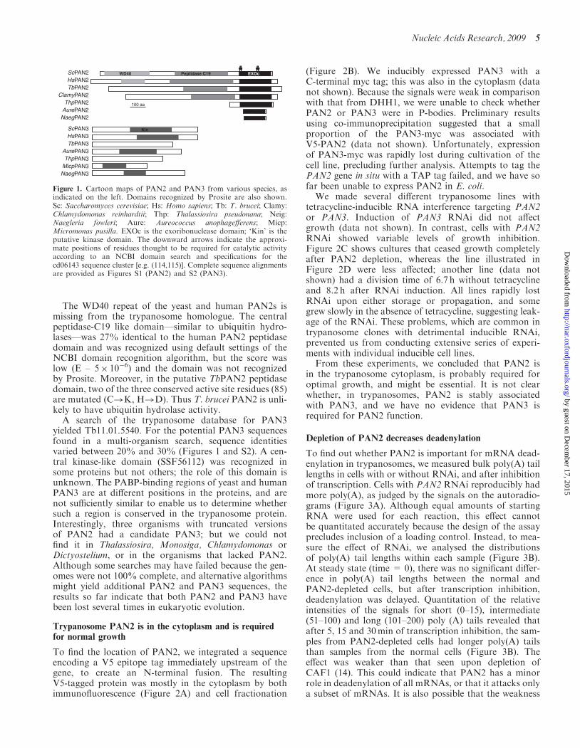
The WD40 repeat of the yeast and human PAN2s ismissing from the trypanosome homologue. The centralpeptidase-C19 like domain—similar to ubiquitin hydro-lases—was 27% identical to the human PAN2 peptidasedomain and was recognized using default settings of theNCBI domain recognition algorithm, but the score waslow (E – 5� 10�6) and the domain was not recognizedby Prosite. Moreover, in the putative TbPAN2 peptidasedomain, two of the three conserved active site residues (85)are mutated (C!K, H!D). Thus T. brucei PAN2 is unli-kely to have ubiquitin hydrolase activity.
A search of the trypanosome database for PAN3yielded Tb11.01.5540. For the potential PAN3 sequencesfound in a multi-organism search, sequence identitiesvaried between 20% and 30% (Figures 1 and S2). A cen-tral kinase-like domain (SSF56112) was recognized insome proteins but not others; the role of this domain isunknown. The PABP-binding regions of yeast and humanPAN3 are at different positions in the proteins, and arenot sufficiently similar to enable us to determine whethersuch a region is conserved in the trypanosome protein.Interestingly, three organisms with truncated versionsof PAN2 had a candidate PAN3; but we could notfind it in Thalassiosira, Monosiga, Chlamydomonas orDictyostelium, or in the organisms that lacked PAN2.Although some searches may have failed because the gen-omes were not 100% complete, and alternative algorithmsmight yield additional PAN2 and PAN3 sequences, theresults so far indicate that both PAN2 and PAN3 havebeen lost several times in eukaryotic evolution.
Trypanosome PAN2 is in the cytoplasm and is requiredfor normal growth
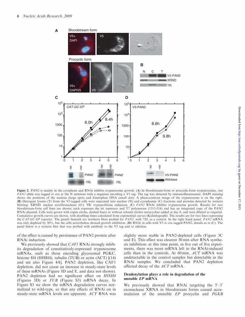
To find the location of PAN2, we integrated a sequenceencoding a V5 epitope tag immediately upstream of thegene, to create an N-terminal fusion. The resultingV5-tagged protein was mostly in the cytoplasm by bothimmunofluorescence (Figure 2A) and cell fractionation
(Figure 2B). We inducibly expressed PAN3 with aC-terminal myc tag; this was also in the cytoplasm (datanot shown). Because the signals were weak in comparisonwith that from DHH1, we were unable to check whetherPAN2 or PAN3 were in P-bodies. Preliminary resultsusing co-immunoprecipitation suggested that a smallproportion of the PAN3-myc was associated withV5-PAN2 (data not shown). Unfortunately, expressionof PAN3-myc was rapidly lost during cultivation of thecell line, precluding further analysis. Attempts to tag thePAN2 gene in situ with a TAP tag failed, and we have sofar been unable to express PAN2 in E. coli.We made several different trypanosome lines with
tetracycline-inducible RNA interference targeting PAN2or PAN3. Induction of PAN3 RNAi did not affectgrowth (data not shown). In contrast, cells with PAN2RNAi showed variable levels of growth inhibition.Figure 2C shows cultures that ceased growth completelyafter PAN2 depletion, whereas the line illustrated inFigure 2D were less affected; another line (data notshown) had a division time of 6.7 h without tetracyclineand 8.2 h after RNAi induction. All lines rapidly lostRNAi upon either storage or propagation, and somegrew slowly in the absence of tetracycline, suggesting leak-age of the RNAi. These problems, which are common intrypanosome clones with detrimental inducible RNAi,prevented us from conducting extensive series of experi-ments with individual inducible cell lines.From these experiments, we concluded that PAN2 is
in the trypanosome cytoplasm, is probably required foroptimal growth, and might be essential. It is not clearwhether, in trypanosomes, PAN2 is stably associatedwith PAN3, and we have no evidence that PAN3 isrequired for PAN2 function.
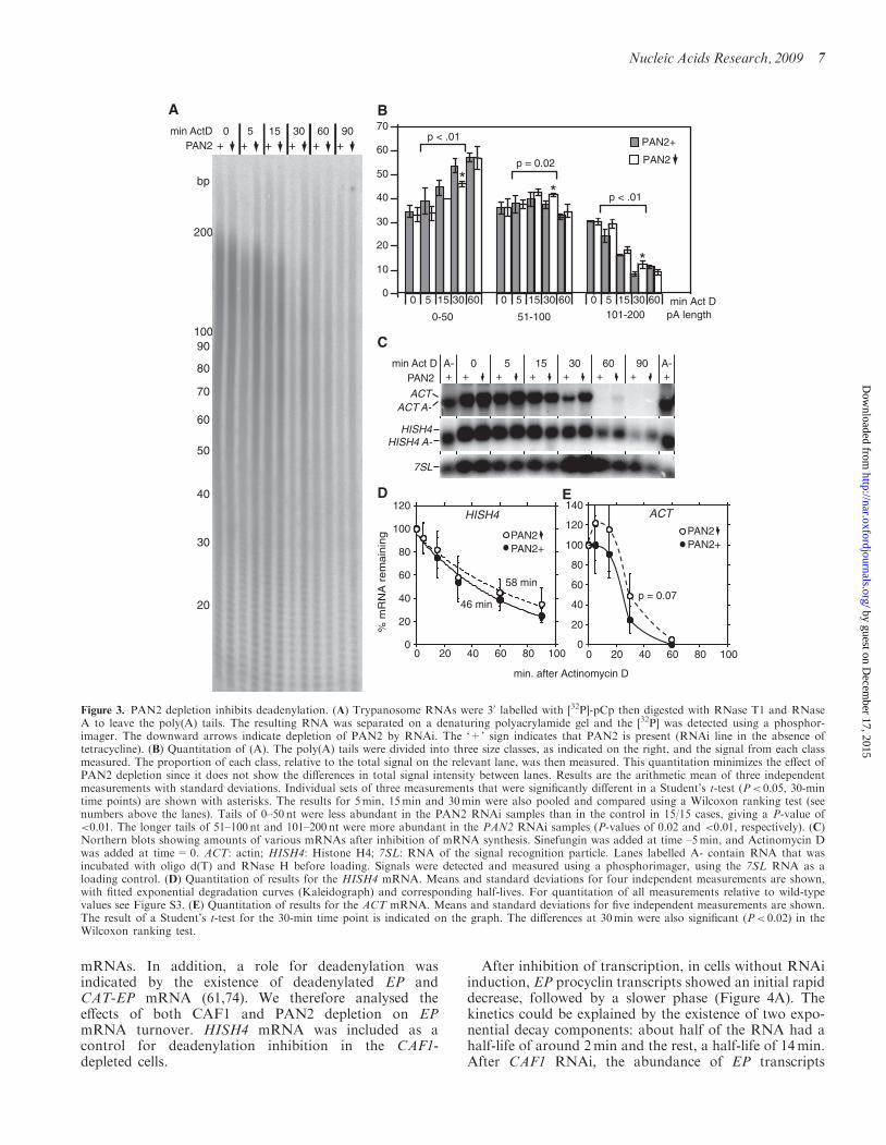
Depletion of PAN2 decreases deadenylation
To find out whether PAN2 is important for mRNA dead-enylation in trypanosomes, we measured bulk poly(A) taillengths in cells with or without RNAi, and after inhibitionof transcription. Cells with PAN2 RNAi reproducibly hadmore poly(A), as judged by the signals on the autoradio-grams (Figure 3A). Although equal amounts of startingRNA were used for each reaction, this effect cannotbe quantitated accurately because the design of the assayprecludes inclusion of a loading control. Instead, to mea-sure the effect of RNAi, we analysed the distributionsof poly(A) tail lengths within each sample (Figure 3B).At steady state (time = 0), there was no significant differ-ence in poly(A) tail lengths between the normal andPAN2-depleted cells, but after transcription inhibition,deadenylation was delayed. Quantitation of the relativeintensities of the signals for short (0–15), intermediate(51–100) and long (101–200) poly (A) tails revealed thatafter 5, 15 and 30min of transcription inhibition, the sam-ples from PAN2-depleted cells had longer poly(A) tailsthan samples from the normal cells (Figure 3B). Theeffect was weaker than that seen upon depletion ofCAF1 (14). This could indicate that PAN2 has a minorrole in deadenylation of all mRNAs, or that it attacks onlya subset of mRNAs. It is also possible that the weakness
ScPAN2 Peptidase C19WD40 EXOc
HsPAN2TbPAN2
ClamyPAN2ThpPAN2
ThpPAN3MicpPAN3
NaegPAN2
NaegPAN3
AurePAN3
AurePAN2
ScPAN3 Kin
HsPAN3TbPAN3
100 aa
Figure 1. Cartoon maps of PAN2 and PAN3 from various species, asindicated on the left. Domains recognized by Prosite are also shown.Sc: Saccharomyces cerevisiae; Hs: Homo sapiens; Tb: T. brucei; Clamy:Chlamydomonas reinhardtii; Thp: Thalassiosira pseudonana; Neig:Naegleria fowleri; Aure: Aureococcus anophagefferens; Micp:Micromonas pusilla. EXOc is the exoribonuclease domain; ‘Kin’ is theputative kinase domain. The downward arrows indicate the approxi-mate positions of residues thought to be required for catalytic activityaccording to an NCBI domain search and specifications for thecd06143 sequence cluster [e.g. (114,115)]. Complete sequence alignmentsare provided as Figures S1 (PAN2) and S2 (PAN3).
Nucleic Acids Research, 2009 5
by guest on Decem
ber 17, 2015http://nar.oxfordjournals.org/
Dow
nloaded from
of the effect is caused by persistence of PAN2 protein afterRNAi induction.We previously showed that CAF1 RNAi strongly inhib-
its degradation of constitutively-expressed trypanosomemRNAs, such as those encoding glycosomal PGKC,histone H4 (HISH4), tubulin (TUB) or actin (ACT) [(14)and see also Figure 4A]. PAN2 depletion, like CAF1depletion, did not cause an increase in steady-state levelsof these mRNAs (Figure 3D and E, and data not shown).PAN2 depletion had no significant effect on HISH4(Figures 3D) or TUB (Figure S3) mRNA decay. InFigure S3 we show the mRNA degradation curves nor-malized to wild-type, so that any effects of RNAi on insteady-state mRNA levels are apparent. ACT RNA was
slightly more stable in PAN2-depleted cells (Figure 3Cand E). This effect was clearest 30min after RNA synthe-sis inhibition: at this time point, in five out of five experi-ments, there was more mRNA left in the RNAi-inducedcells than in the controls. At 60min, ACT mRNA wasundetectable in the control samples but detectable in theRNAi samples. We concluded that PAN2 depletionaffected decay of the ACT mRNA.
Deadenylation plays a role in degradation of theunstable EP mRNA
We previously showed that RNAi targeting the 50–30
exonuclease XRNA in bloodstream forms caused accu-mulation of the unstable EP procyclin and PGKB
Bloodstream form
Procyclic form
B
A
V5-PAN2
XRND
TR
N
V5V5+DAPI
V5+V5DAPIV5
CAT-GC-EP
DC
9.8h
8.0h8.4h
8.6h
V5-PAN2
1
10
100
1000
104
105
1
10
100
1000
104
105
−tet
+te
t
PAN2
7SL
PAN2
7SL
−tet
+te
t
V5-PAN2Aldolase
−tet
+te
t0 1 2 3 4 5 6 0 1 2 3 4 5 6
C T
Figure 2. PAN2 is mainly in the cytoplasm and RNAi inhibits trypanosome growth. (A) In bloodstream-form or procyclic-form trypanosomes, onePAN2 allele was tagged in situ at the N terminus with a sequence encoding a V5 tag. The tag was detected by immunofluorescence. DAPI stainingshows the positions of the nucleus (large spot) and kinetoplast DNA (small dot). A phase-contrast image of the trypanosome is on the right.(B) Detergent lysates (T) from the V5-tagged cells were separated into nuclear (N) and cytoplasmic (C) fractions and proteins detected by westernblotting. XRND: nuclear exoribonuclease (61); TR: trypanothione reductase. (C) PAN2 RNAi inhibits trypanosome growth. Results for twobloodstream-form cell lines are shown; each expresses the tet repressor and T7 polymerase (1313-514) and has an integrated copy of the PAN2RNAi plasmid. Cells were grown with (open circles, dashed lines) or without (closed circles) tetracycline added at day 0, and were diluted as required.Cumulative growth curves are shown, with doubling times calculated from exponential curves (Kaleidograph). The results are for two lines expressingthe CAT-GC-EP reporter. The panels beneath are northern blots probed for PAN2, with 7SL as a control. In the right hand panel, PAN2 mRNAwas only depleted by 30%, but the cells nevertheless showed growth inhibition. (D) RNAi in cells with V5 in situ tagged PAN2, details as in (C). Thepanel below is a western blot that was probed with antibody to the V5 tag and to aldolase.
6 Nucleic Acids Research, 2009
by guest on Decem
ber 17, 2015http://nar.oxfordjournals.org/
Dow
nloaded from
mRNAs. In addition, a role for deadenylation wasindicated by the existence of deadenylated EP andCAT-EP mRNA (61,74). We therefore analysed theeffects of both CAF1 and PAN2 depletion on EPmRNA turnover. HISH4 mRNA was included as acontrol for deadenylation inhibition in the CAF1-depleted cells.
After inhibition of transcription, in cells without RNAiinduction, EP procyclin transcripts showed an initial rapiddecrease, followed by a slower phase (Figure 4A). Thekinetics could be explained by the existence of two expo-nential decay components: about half of the RNA had ahalf-life of around 2min and the rest, a half-life of 14min.After CAF1 RNAi, the abundance of EP transcripts
min ActDPAN2
0 5 15 30 60 90
200
10090
80
70
60
50
40
30
20
bp
A B
C
D E
min Act DpA length
PAN2
PAN2++ + + + + +
ACT A-ACT
7SL
HISH4 A-HISH4
PAN2min Act D
+ + +0A- 5 15 30 60 90 A-
+ + + ++
min. after Actinomycin D
HISH4
% m
RN
A r
em
ain
ing
600
p < .01
p = 0.02
p < .01
5 15 30600 5 15 30600 5
0-50 51-100
**
*
101-20015 30
0
10
20
30
40
50
60
70
PAN2
46 min
58 min
PAN2+
ACT
PAN2PAN2+
0
20
40
60
80
100
120
140
0 20 40 60 80 1000
20
40
60
80
100
120
0 20 40 60 80 100
p = 0.07
Figure 3. PAN2 depletion inhibits deadenylation. (A) Trypanosome RNAs were 30 labelled with [32P]-pCp then digested with RNase T1 and RNaseA to leave the poly(A) tails. The resulting RNA was separated on a denaturing polyacrylamide gel and the [32P] was detected using a phosphor-imager. The downward arrows indicate depletion of PAN2 by RNAi. The ‘+’ sign indicates that PAN2 is present (RNAi line in the absence oftetracycline). (B) Quantitation of (A). The poly(A) tails were divided into three size classes, as indicated on the right, and the signal from each classmeasured. The proportion of each class, relative to the total signal on the relevant lane, was then measured. This quantitation minimizes the effect ofPAN2 depletion since it does not show the differences in total signal intensity between lanes. Results are the arithmetic mean of three independentmeasurements with standard deviations. Individual sets of three measurements that were significantly different in a Student’s t-test (P< 0.05, 30-mintime points) are shown with asterisks. The results for 5min, 15min and 30min were also pooled and compared using a Wilcoxon ranking test (seenumbers above the lanes). Tails of 0–50 nt were less abundant in the PAN2 RNAi samples than in the control in 15/15 cases, giving a P-value of<0.01. The longer tails of 51–100 nt and 101–200 nt were more abundant in the PAN2 RNAi samples (P-values of 0.02 and <0.01, respectively). (C)Northern blots showing amounts of various mRNAs after inhibition of mRNA synthesis. Sinefungin was added at time –5min, and Actinomycin Dwas added at time=0. ACT: actin; HISH4: Histone H4; 7SL: RNA of the signal recognition particle. Lanes labelled A- contain RNA that wasincubated with oligo d(T) and RNase H before loading. Signals were detected and measured using a phosphorimager, using the 7SL RNA as aloading control. (D) Quantitation of results for the HISH4 mRNA. Means and standard deviations for four independent measurements are shown,with fitted exponential degradation curves (Kaleidograph) and corresponding half-lives. For quantitation of all measurements relative to wild-typevalues see Figure S3. (E) Quantitation of results for the ACT mRNA. Means and standard deviations for five independent measurements are shown.The result of a Student’s t-test for the 30-min time point is indicated on the graph. The differences at 30min were also significant (P< 0.02) in theWilcoxon ranking test.
Nucleic Acids Research, 2009 7
by guest on Decem
ber 17, 2015http://nar.oxfordjournals.org/
Dow
nloaded from
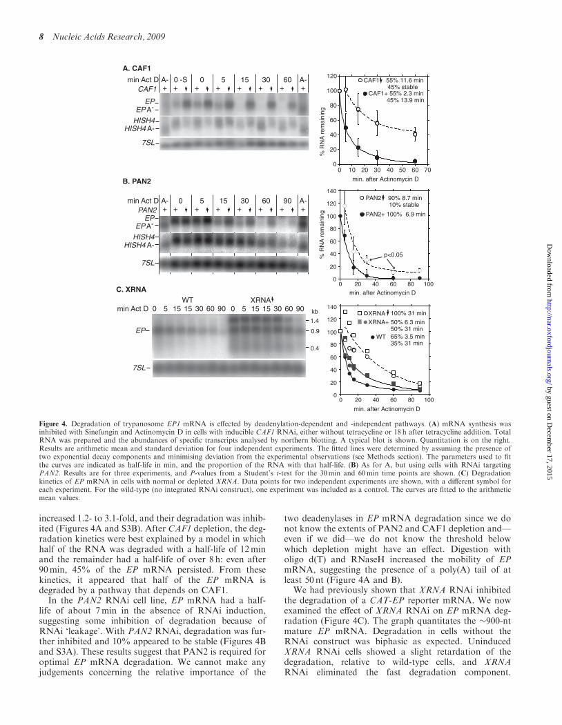
increased 1.2- to 3.1-fold, and their degradation was inhib-ited (Figures 4A and S3B). After CAF1 depletion, the deg-radation kinetics were best explained by a model in whichhalf of the RNA was degraded with a half-life of 12minand the remainder had a half-life of over 8 h: even after90min, 45% of the EP mRNA persisted. From thesekinetics, it appeared that half of the EP mRNA isdegraded by a pathway that depends on CAF1.In the PAN2 RNAi cell line, EP mRNA had a half-
life of about 7min in the absence of RNAi induction,suggesting some inhibition of degradation because ofRNAi ‘leakage’. With PAN2 RNAi, degradation was fur-ther inhibited and 10% appeared to be stable (Figures 4Band S3A). These results suggest that PAN2 is required foroptimal EP mRNA degradation. We cannot make anyjudgements concerning the relative importance of the
two deadenylases in EP mRNA degradation since we donot know the extents of PAN2 and CAF1 depletion and—even if we did—we do not know the threshold belowwhich depletion might have an effect. Digestion witholigo d(T) and RNaseH increased the mobility of EPmRNA, suggesting the presence of a poly(A) tail of atleast 50 nt (Figure 4A and B).
We had previously shown that XRNA RNAi inhibitedthe degradation of a CAT-EP reporter mRNA. We nowexamined the effect of XRNA RNAi on EP mRNA deg-radation (Figure 4C). The graph quantitates the �900-ntmature EP mRNA. Degradation in cells without theRNAi construct was biphasic as expected. UninducedXRNA RNAi cells showed a slight retardation of thedegradation, relative to wild-type cells, and XRNARNAi eliminated the fast degradation component.
min. after Actinomycin D
min. after Actinomycin D
min. after Actinomycin D
% R
NA
rem
aini
ng
CAF1 55% 11.6 min 45% stable
A. CAF1
XRNA
B. PAN2
C. XRNA
min Act D
0
20
40
60
80
100
120
0 10 20 30 40 50 60 70
WT
1.4
XRNA0 5 1515 30 60 90 0 5 1515 30 60 90
CAF1
EP A-EP
min Act D+ + +
0 -SA- 0 5 15 30 60 A-+ + + ++
7SL
HISH4 A-HISH4
PAN2+ 100% 6.9 min
p<0.05
PAN2 90% 8.7 min 10% stable
CAF1+ 55% 2.3 min 45% 13.9 min
EP A-EP
7SL
HISH4 A-HISH4
PAN2min Act D
+ + +0A- 5 15 30 60 90 A-
+ + + ++
0
20
40
60
80
100
120
140
0 20 40 60 80 100
XRNA+
WT
100% 31 min
50% 6.3 min50% 31 min65% 3.5 min35% 31 min
EP 0.9
0.4
kb
7SL
% R
NA
rem
aini
ng
0
20
40
60
80
100
120
140
0 20 40 60 80 100
Figure 4. Degradation of trypanosome EP1 mRNA is effected by deadenylation-dependent and -independent pathways. (A) mRNA synthesis wasinhibited with Sinefungin and Actinomycin D in cells with inducible CAF1 RNAi, either without tetracycline or 18 h after tetracycline addition. TotalRNA was prepared and the abundances of specific transcripts analysed by northern blotting. A typical blot is shown. Quantitation is on the right.Results are arithmetic mean and standard deviation for four independent experiments. The fitted lines were determined by assuming the presence oftwo exponential decay components and minimising deviation from the experimental observations (see Methods section). The parameters used to fitthe curves are indicated as half-life in min, and the proportion of the RNA with that half-life. (B) As for A, but using cells with RNAi targetingPAN2. Results are for three experiments, and P-values from a Student’s t-test for the 30min and 60min time points are shown. (C) Degradationkinetics of EP mRNA in cells with normal or depleted XRNA. Data points for two independent experiments are shown, with a different symbol foreach experiment. For the wild-type (no integrated RNAi construct), one experiment was included as a control. The curves are fitted to the arithmeticmean values.
8 Nucleic Acids Research, 2009
by guest on Decem
ber 17, 2015http://nar.oxfordjournals.org/
Dow
nloaded from
Modelling using our previous data for the CAT-EPmRNA (61) yielded almost identical results to those forEP (Figure S4A).
Surprisingly, northern blots of RNA from cells con-taining the XRNA RNAi construct had, in addition tothe expected band at 0.9 kb, two additional bands: oneat 1.3 kb and one at about 0.4 kb. These were faint, butdetectable, in RNA from cells grown without tetracycline(data not shown) and clearly visible after RNAi induction(Figure 4C). Both hybridized with an EP probe containingonly the coding region; the shorter product was notdetected in the poly(A)+ fraction (data not shown) andcould be a coding region fragment. The longer productwas polyadenylated; a band of similar size hybridized toan intergenic region probe (data not shown). This RNA istoo long to be a polyadenylated primary transcript, butcould be a previously detected minor alternatively splicedversion of EP2 or EP3 mRNA, extended at the 50-end(86,87). Thus XRNA seems to be implicated in destructionof abnormal, probably non-functional mRNAs from theprocyclin locus.
These results indicate that extremely rapid degradationof EP mRNA requires XRNA, but some EP mRNA isdegraded by a pathway that depends on the presence ofCAF1.
Degradation of the CAT-PGKB and PGKB mRNAs
The endogenous PGKB mRNA, produced by pol II, isvirtually undetectable by Northern blotting. Even using
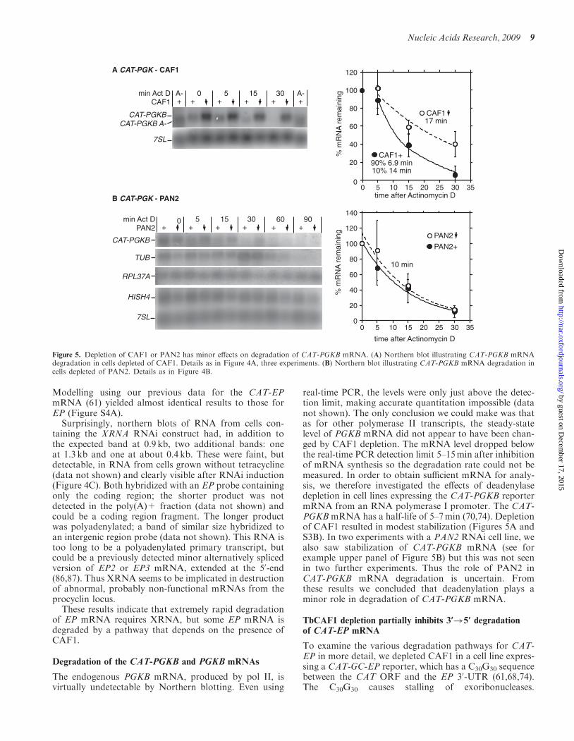
real-time PCR, the levels were only just above the detec-tion limit, making accurate quantitation impossible (datanot shown). The only conclusion we could make was thatas for other polymerase II transcripts, the steady-statelevel of PGKB mRNA did not appear to have been chan-ged by CAF1 depletion. The mRNA level dropped belowthe real-time PCR detection limit 5–15min after inhibitionof mRNA synthesis so the degradation rate could not bemeasured. In order to obtain sufficient mRNA for analy-sis, we therefore investigated the effects of deadenylasedepletion in cell lines expressing the CAT-PGKB reportermRNA from an RNA polymerase I promoter. The CAT-PGKB mRNA has a half-life of 5–7min (70,74). Depletionof CAF1 resulted in modest stabilization (Figures 5A andS3B). In two experiments with a PAN2 RNAi cell line, wealso saw stabilization of CAT-PGKB mRNA (see forexample upper panel of Figure 5B) but this was not seenin two further experiments. Thus the role of PAN2 inCAT-PGKB mRNA degradation is uncertain. Fromthese results we concluded that deadenylation plays aminor role in degradation of CAT-PGKB mRNA.
TbCAF1 depletion partially inhibits 3’!5’ degradationof CAT-EP mRNA
To examine the various degradation pathways for CAT-EP in more detail, we depleted CAF1 in a cell line expres-sing a CAT-GC-EP reporter, which has a C30G30 sequencebetween the CAT ORF and the EP 30-UTR (61,68,74).The C30G30 causes stalling of exoribonucleases.
time after Actinomycin D
time after Actinomycin D%
mR
NA
rem
aini
ng%
mR
NA
rem
aini
ng
CAF1+90% 6.9 min10% 14 min
PAN2+
CAF117 min
PAN2
10 min
B CAT-PGK - PAN2
A CAT-PGK - CAF1
CAF1min Act D
+ + +0A- 5 15 30 A-
+ ++
7SL
CAT-PGKB A-CAT-PGKB
PAN2min Act D
+ +0 5 15 30
+ +
7SL
HISH4
TUB
RPL37A
CAT-PGKB
0
20
40
60
80
100
120
140
0 5 10 15 20 25 30 35
60+
90+
0
20
40
60
80
100
120
0 5 10 15 20 25 30 35
Figure 5. Depletion of CAF1 or PAN2 has minor effects on degradation of CAT-PGKB mRNA. (A) Northern blot illustrating CAT-PGKB mRNAdegradation in cells depleted of CAF1. Details as in Figure 4A, three experiments. (B) Northern blot illustrating CAT-PGKB mRNA degradation incells depleted of PAN2. Details as in Figure 4B.
Nucleic Acids Research, 2009 9
by guest on Decem
ber 17, 2015http://nar.oxfordjournals.org/
Dow
nloaded from
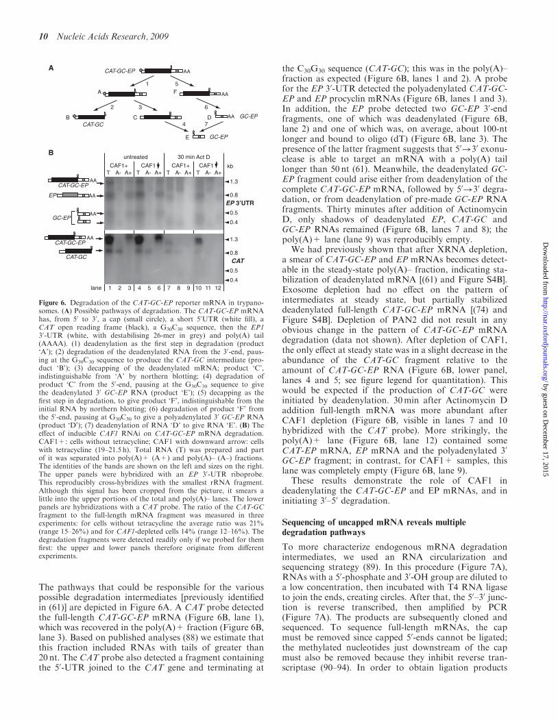
The pathways that could be responsible for the variouspossible degradation intermediates [previously identifiedin (61)] are depicted in Figure 6A. A CAT probe detectedthe full-length CAT-GC-EP mRNA (Figure 6B, lane 1),which was recovered in the poly(A)+ fraction (Figure 6B,lane 3). Based on published analyses (88) we estimate thatthis fraction included RNAs with tails of greater than20 nt. The CAT probe also detected a fragment containingthe 50-UTR joined to the CAT gene and terminating at
the C30G30 sequence (CAT-GC); this was in the poly(A)–fraction as expected (Figure 6B, lanes 1 and 2). A probefor the EP 30-UTR detected the polyadenylated CAT-GC-EP and EP procyclin mRNAs (Figure 6B, lanes 1 and 3).In addition, the EP probe detected two GC-EP 30-endfragments, one of which was deadenylated (Figure 6B,lane 2) and one of which was, on average, about 100-ntlonger and bound to oligo (dT) (Figure 6B, lane 3). Thepresence of the latter fragment suggests that 50!30 exonu-clease is able to target an mRNA with a poly(A) taillonger than 50 nt (61). Meanwhile, the deadenylated GC-EP fragment could arise either from deadenylation of thecomplete CAT-GC-EP mRNA, followed by 50!30 degra-dation, or from deadenylation of pre-made GC-EP RNAfragments. Thirty minutes after addition of ActinomycinD, only shadows of deadenylated EP, CAT-GC andGC-EP RNAs remained (Figure 6B, lanes 7 and 8); thepoly(A)+ lane (lane 9) was reproducibly empty.
We had previously shown that after XRNA depletion,a smear of CAT-GC-EP and EP mRNAs becomes detect-able in the steady-state poly(A)– fraction, indicating sta-bilization of deadenylated mRNA [(61) and Figure S4B].Exosome depletion had no effect on the pattern ofintermediates at steady state, but partially stabilizeddeadenylated full-length CAT-GC-EP mRNA [(74) andFigure S4B]. Depletion of PAN2 did not result in anyobvious change in the pattern of CAT-GC-EP mRNAdegradation (data not shown). After depletion of CAF1,the only effect at steady state was in a slight decrease in theabundance of the CAT-GC fragment relative to theamount of CAT-GC-EP RNA (Figure 6B, lower panel,lanes 4 and 5; see figure legend for quantitation). Thiswould be expected if the production of CAT-GC wereinitiated by deadenylation. 30min after Actinomycin Daddition full-length mRNA was more abundant afterCAF1 depletion (Figure 6B, visible in lanes 7 and 10hybridized with the CAT probe). More strikingly, thepoly(A)+ lane (Figure 6B, lane 12) contained someCAT-EP mRNA, EP mRNA and the polyadenylated 30
GC-EP fragment; in contrast, for CAF1+ samples, thislane was completely empty (Figure 6B, lane 9).
These results demonstrate the role of CAF1 indeadenylating the CAT-GC-EP and EP mRNAs, and ininitiating 30–50 degradation.
Sequencing of uncapped mRNA reveals multipledegradation pathways
To more characterize endogenous mRNA degradationintermediates, we used an RNA circularization andsequencing strategy (89). In this procedure (Figure 7A),RNAs with a 50-phosphate and 30-OH group are diluted toa low concentration, then incubated with T4 RNA ligaseto join the ends, creating circles. After that, the 50–30 junc-tion is reverse transcribed, then amplified by PCR(Figure 7A). The products are subsequently cloned andsequenced. To sequence full-length mRNAs, the capmust be removed since capped 50-ends cannot be ligated;the methylated nucleotides just downstream of the capmust also be removed because they inhibit reverse tran-scriptase (90–94). In order to obtain ligation products
lane
CAT
T A- A+ T A- A+ T A- A+ T A- A+
1 2 3 4 5 6 7 8 9 10 11 12
CAF1+
untreatedB
A
30 min Act D
CAF1+ CAF1CAF1
AA
AA
AA
kb
CAT-GC
EP
CAT-GC-EP
GC-EP
1.3
0.4
0.8
0.5
EP 3’UTR
1.3
0.4
0.8
0.5
AACAT-GC-EP
AA
AA
CAT-GC
CAT-GC-EP
GC-EP
GC-EP
AA
1A
C DB
F
E
3
5
6
74
2
Figure 6. Degradation of the CAT-GC-EP reporter mRNA in trypano-somes. (A) Possible pathways of degradation. The CAT-GC-EP mRNAhas, from 50 to 30, a cap (small circle), a short 50UTR (white fill), aCAT open reading frame (black), a G30C30 sequence, then the EP130-UTR (white, with destabilising 26-mer in grey) and poly(A) tail(AAAA). (1) deadenylation as the first step in degradation (product‘A’); (2) degradation of the deadenylated RNA from the 30-end, paus-ing at the G30C30 sequence to produce the CAT-GC intermediate (pro-duct ‘B’); (3) decapping of the deadenylated mRNA; product ‘C’,indistinguishable from ‘A’ by northern blotting; (4) degradation ofproduct ‘C’ from the 50-end, pausing at the G30C30 sequence to givethe deadenylated 30 GC-EP RNA (product ‘E’); (5) decapping as thefirst step in degradation, to give product ‘F’, indistinguishable from theinitial RNA by northern blotting; (6) degradation of product ‘F’ fromthe 50-end, pausing at G30C30 to give a polyadenylated 30 GC-EP RNA(product ‘D’); (7) deadenylation of RNA ‘D’ to give RNA ‘E’. (B) Theeffect of inducible CAF1 RNAi on CAT-GC-EP mRNA degradation.CAF1+: cells without tetracycline; CAF1 with downward arrow: cellswith tetracycline (19–21.5 h). Total RNA (T) was prepared and partof it was separated into poly(A)+ (A+) and poly(A)– (A–) fractions.The identities of the bands are shown on the left and sizes on the right.The upper panels were hybridized with an EP 30-UTR riboprobe.This reproducibly cross-hybridizes with the smallest rRNA fragment.Although this signal has been cropped from the picture, it smears alittle into the upper portions of the total and poly(A)– lanes. The lowerpanels are hybridizations with a CAT probe. The ratio of the CAT-GCfragment to the full-length mRNA fragment was measured in threeexperiments: for cells without tetracycline the average ratio was 21%(range 15–26%) and for CAF1-depleted cells 14% (range 12–16%). Thedegradation fragments were detected readily only if we probed for themfirst: the upper and lower panels therefore originate from differentexperiments.
10 Nucleic Acids Research, 2009
by guest on Decem
ber 17, 2015http://nar.oxfordjournals.org/
Dow
nloaded from
from mRNAs with intact 50-ends and cap structures, wetherefore pre-incubated samples with RNase H and anoligonucleotide complementary to the 50-end of the splicedleader. When considering the results, it is important tonote that if an mRNA had been decapped in vivo, butstill retained the 50 methylated nucleotides, it would bedetected only in RNase H treated samples. Such decappedRNAs are expected to accumulate after XRNA depletion.Tandemly repeated precursor RNAs should be sub-
strates for RNA-ligase-independent amplification, andare expected for both HISH4 and TUB. We thereforechose, as a control, the ribosomal protein L37a mRNA(RPL37A), which is encoded by a single locus, has a pre-dicted length of about 400 nt, and has a half-life exceeding90min in bloodstream-form trypanosomes (Figure 5B).RNA circle results for RPL37A are shown in Figure 7B,and sequencing results for a random selection of clonedproducts are in Tables 1 and S2, and Figure S5. ThemRNA is trans spliced at a single site, but there are severalalternative polyadenylation sites. Figure 7B shows theproducts of the RT–PCRs. The first PCR showed aladder in all lanes, except for the negative control withoutligase (lane 5). In lanes 1 and 3, showing products from 50
RNase H-treated RNA, there was a broad band around300 nt, which was absent in the products from RNA thathad not been RNase H treated. The predicted lengthfor amplification products without the poly(A) tail was159–210 bp, so the size of this band suggested averagepoly(A) tail lengths of about 100 nt. Cloning, however,instead gave poly(A/T) tracts of 36–37 bp. In the originalarticle describing the method (89), the poly(A) tails of fourrat mRNAs were measured by gel electrophoresis, withoutcloning. Amplification of three of them indicated poly(A)sof 20–50 nt; for one mRNAs there were two populations,corresponding to 20–50 nt and 120–200 nt. Since mam-malian poly(A) tails are expected to be 100–200 nt long,these results hint that the circle–PCR method may havean intrinsic bias against longer poly(A) tails. Our resultsshow that the bias is exacerbated if the products arecloned.RPL37A products from RNA that had not been treated
with RNase H were expected to correspond to mRNAsthat had already been decapped and had begun to bedegraded from the 50-end: they formed a smear in theagarose gel (Figure 7B). In order to reduce contaminationwith irrelevant cDNAs prior to cloning, we performed anested PCR. Surprisingly, the 50-ends of the degradationintermediates were not random. Instead, 25 out of 46clones had lost the first 27–31 nt of the spliced leader.About half of the degradation intermediates had lost thepoly(A) tail and parts of the 30-UTR as well. In contrast toobservations for mammalian histone RNAs (53), here wasno evidence for addition of U residues at the 30-ends ofthe degrading mRNAs. Depletion of XRNA did not affectthe patterns of degradation products. If poly(A) tailswere present they were almost always in the 30–40 ntsize range. Because of the bias noted earlier, this resultgives us the lower limit of poly(A) lengths for deadenyla-tion-dependent decapping, but does not tell us the upperthreshold. Just 2 of the 43 cloned intermediates had much
100
200
500
1000
1 2 3 4 5M
1 2 3 4 5M
a
b
XRNA
Primers 52 + 31
b sremirPa sremirP
1 2 3 4M
Primers 52 + 35
Primers 51 + 32
+ + +ligase + + + + -
+ - + - -
XRNA + +ligase + + + +
+ - + -
XRNA
RNase HRNase H
RNase H
+ + +ligase + + + + -
+ - + - -
XRNAligase
RNase H
+ + ++ + + + -+ - + - -
100
200
400300
100
200
500
1000
100200
500
1000
XRNAligase
RNase H
+ + ++ + + + -+ - +
5‘ ‘oligo, RNase H
RNA ligase
RT-PCR
RNA ligase
RT primer
PCR primer5‘ cap + methylations
Degradationintermediate
Intact
RT-PCR
- -
100
200
bp bp
bp
bp
bp
500
1000
1
a ab b
2 3 4 5M100
200
500
1000
1 2 3 4 5M
B RPL37A
Method
C EP
AAAAAAAA
AAAAAAAA
AAAAAAAA
AAAAAAAA
AAAAAAAA
AAAAA
RT52 35 31 32 33 3451
A
Figure 7. RT–PCR of circularized mRNAs. (A) Illustration of themethod for intact, capped RNA (right) and a degradation intermediate(left). The open reading frame is filled grey, other symbols are shownon the figure. For detailed description see text. (B) Ethidium-bromidestained gels of amplified products for RPL37A. A cartoon of themRNA, not to scale, showing the positions of the primers, is abovethe gels. Products from the first PCR (primers ‘a’) are shown on theleft, and from the second, nested PCR (primers ‘b’) on the right. (C)Ethidium-bromide stained gels of amplified products for EP. In thecartoon, the striped region represents the segment encoding EP repeats.After the first PCR with primers 52 and 31, DNA was isolated fromthe upper and lower portions of the gel, as indicated, and used for thenested PCR using primers 51 and 32. A separate picture shows theladders obtained using primers 35 and 52. In each case, the area ofthe gel containing most DNA was excised for purification and cloningof the PCR products, and clones were selected randomly for sequenc-ing. Gels for amplifications with 30 primers 33 or 34, with 50 primer 51,looked very similar to the lower panel for primers 51 and 32; the sameprocedure was followed but only clones with the longest inserts weresequenced.
Nucleic Acids Research, 2009 11
by guest on Decem
ber 17, 2015http://nar.oxfordjournals.org/
Dow
nloaded from
longer poly(A) tails (93 nt and 112 nt), illustrating theminor role of deadenylation-independent decay.We next repeated the experiment for EP mRNA.
In bloodstream forms, at steady state, EP mRNAs havea poly(A) tail of at least 50 nt, added about 300-nt down-stream of the stop codon (e.g. Figures 4 and 6B, lanes 2and 3). Primers 52 and 31 (Figures 7C and S6) weredesigned to allow detection of a broad range of degrada-tion products, including those that had lost long segmentsof the 50 and 30-ends. First, we examined RNase H-treatedmRNA, in order to see the full-length mRNA. The corre-sponding amplified product should have been 430 bp pluspoly(A). To our surprise, a band of this size was notseen and no clones corresponding to full-length mRNAwere obtained. Instead, the RNase H-treated RNAyielded EP products that were predominantly in the200 bp range (Figure 7C, lanes 1 and 3). Randomlyselected clones nearly all retained SL sequence, buteither lacked a poly(A) tail, or had (A)3–11 at positionsover 100-nt upstream of the usual site (Tables 1 and S3,and Figure S7).We next amplified EP cDNAs using RNaseH-treated
RNA from XRNA-depleted cells with primers 52 and31. The average size of the products was shorter thanbefore (Figure 7B, compare lane 1 with lane 3).Although all eight cloned products had retained theexpected portions of the spliced leader, only one of themhad a poly(A) tail (Table S3 and Figure S7). This illus-trates stabilization of deadenylated CAT-EP mRNA afterXRNA depletion (61)To find out whether the procedure could amplify full-
length EP mRNA from bloodstream forms at all, werepeated amplifications with primers 33 and 34 andselected only the longest inserts for sequencing (Table S4and Figure S8). The product distributions on agarose gelswere similar to those obtained with the first primer sets(data not shown). Even with primer 34, no cloneshad poly(A) in the expected position, although one was
polyadenylated a few nucleotides further downstream(Figure S8). This result is strange, since it is inconsistentwith the northern blot results. It will not be discussedfurther here, because a considerable number of furtherexperiments is required to determine the origin of theseproducts. In procyclic forms, the EP mRNA is very stableand polyadenylated. In preliminary experiments withRNaseH-treated mRNA from this form, using primers33 and 34, 6 out of 12 clones were polyadenylated in theexpected position at +297, with an average poly(A) lengthof 29 nt (maximum 58 nt). This shows that the procedurecan indeed amplify the full-length polyadenylated RNA,but probably biases towards shorter products and trun-cates the tails.
Returning to the bloodstream-form trypanosomes, wenext analysed circularized EP mRNAs without priorRNAase treatment. Using RNA from cells with normalXRNA levels, approximately half of the amplified pro-ducts using 30 primer 32 had an oligo(A) tail, wellupstream of the expected position; the intermediates with-out poly(A) had lost variable portions of the 30-UTR.It was obvious on the stained agarose gel that productsfrom cells with XRNA RNAi (Figure 7C, lane 2) weresmaller than those obtained from cells with a normalamount of XRNA (Figure 7C, lane 4). The clones fromthe XRNA-depleted cells mostly lacked poly(A) and hadbeen digested significantly further at the 50-end than thosefrom the normal cells: 56% had lost part of the codingregion, and only 1/18 had retained part of the splicedleader, while in XRNA positive cells, 14% had lostcoding sequence and 64% retained part of the splicedleader. Surprisingly although the coding region primerswere designed to hybridize equally to EP1, EP2 andEP3 sequences, all clones originated from EP1. Twoclones (no RNase H, normal XRNA) originated fromthe primary transcript, enabling precise mapping of thetranscription start site at position �115 relative to thestart codon.
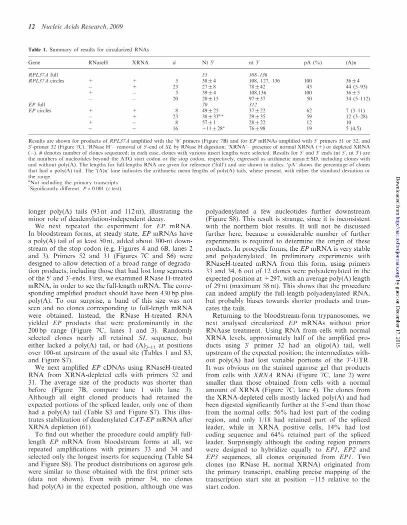
Table 1. Summary of results for circularized RNAs
Gene RNaseH XRNA # Nt 50 nt 30 pA (%) (A)n
RPL37A full 55 108–136RPL37A circles + + 5 38� 4 108, 127, 136 100 36� 4
� + 23 27� 8 78� 42 43 44 (5–93)+ � 5 39� 4 108,136 100 36� 5� � 20 20� 15 97� 37 50 34 (5–112)
EP full 70 312EP circles + + 8 49� 25 37� 22 62 7 (3–11)
� + 23 38� 33a,� 29� 35 59 12 (3–28)+ � 8 57� 1 28� 22 12 10� � 16 �11� 28� 76� 98 19 5 (4,5)
Results are shown for products of RPL37A amplified with the ‘b’ primers (Figure 7B) and for EP mRNAs amplified with 50 primers 51 or 52, and30-primer 32 (Figure 7C). ‘RNase H’—removal of 50-end of SL by RNase H digestion; ‘XRNA’—presence of normal XRNA (+) or depleted XRNA(�). # denotes number of clones sequenced; in each case, clones with various insert lengths were selected. Results for 50 and 30 ends (nt 50, nt 30) arethe numbers of nucleotides beyond the ATG start codon or the stop codon, respectively, expressed as arithmetic mean� SD, including clones withand without poly(A). The lengths for full-lengths RNA are given for reference (‘full’) and are shown in italics. ‘pA’ shows the percentage of clonesthat had a poly(A) tail. The ‘(A)n’ lane indicates the arithmetic mean lengths of poly(A) tails, where present, with either the standard deviation orthe range.aNot including the primary transcripts.�
Significantly different, P< 0.001 (t-test).
12 Nucleic Acids Research, 2009
by guest on Decem
ber 17, 2015http://nar.oxfordjournals.org/
Dow
nloaded from
Finally, results in Figure 4C had suggested that XRNAdepletion stabilized an intermediate of about 400 nt.Since this fragment had not been detected from CAT-EPmRNAs after XRNA depletion (61), we suspected that itmight depend on the EP coding region. We looked forsuch RNAs using primers 52 and 35; the latter hybridizeswithin the repetitive region encoding the EP repeats. Theresult was a clean ladder of products which was unaffectedby either RNase H decapping or XRNA depletion(Figure 5C). All clones obtained, (with or withoutXRNA depletion) started at position +51 downstreamof the ATG, and ended at position 10 in the 30-UTR.This was paradoxical since no clones obtained withother primers had these boundaries.
Two novel findings emerged from this approach.First, we saw hitherto unsuspected heterogeneity inpolyadenylation of EP mRNAs in bloodstream forms.Second, XRNA depletion caused accumulation of EPmRNA degradation intermediates that lacked not onlythe poly(A) tail, but also portions of the 50-end. Wewere disappointed that we could not establish an upperpoly(A) length threshold for deadenylation-dependentdecapping; nevertheless, it was interesting that cloned
EP mRNAs had much shorter poly(A)s than theRPL37A clones.
RNAi targeting a MEX67 homologue does notaccelerate EP mRNA decay
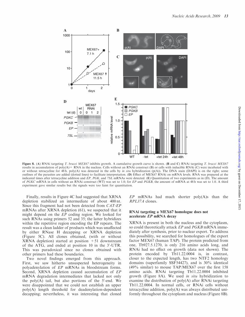
XRNA is present in both the nucleus and the cytoplasm,so could theoretically attack EP and PGKB mRNA imme-diately after synthesis, prior to nuclear export. To addressthis possibility, we searched for homologues of the exportfactor MEX67 (human TAP). The protein predicted fromone, Tb927.5.1270, is only 216 amino acids long, andRNAi had no effect on growth (data not shown). Theprotein encoded by Tb11.22.0004 is, in contrast,closer to the expected length, has two NTF2 homologydomains (superfamily SSF54427), and is 30% identical(49% similar) to mouse TAP/MEX67 over the first 158amino acids. RNAi targeting Tb11.22.0004 inhibitedgrowth (Figure 8A). We used in situ hybridization toexamine the distribution of poly(A) after RNAi targetingTb11.22.0004. In normal cells, or RNAi cells withouttetracycline addition, poly(A) was always distributed uni-formly throughout the cytoplasm and nucleus (Figure 8B).
DAPI
1
10
100
1000
0 1 2
7.1 h
11.5 hMEX67
MEX67+
days
DAPI
p(A)
p(A)
A
D
B
C
1.0
0.5
0WT −tet
rela
tive
mR
NA
leve
l
+tet 24h +tet 48h
1.5PGKCPGKBEP
0wt
0 1 2
PGKC
PGKB
EP
7SL
MEX67RNAi
tet (d)
E
Figure 8. (A) RNAi targeting T. brucei MEX67 inhibits growth. A cumulative growth curve is shown. (B and C) RNAi targeting T. brucei MEX67results in accumulation of poly(A)+ RNA in the nucleus. Cells without an RNAi construct (B) or cells with inducible RNAi (C) were incubated withor without tetracycline for 48 h. poly(A) was detected in the cells by in situ hybridization (p(A)). The DNA stain (DAPI) is on the right; someoutlines of the parasites are added (dotted lines) to facilitate interpretation. (D) Effect of MEX67 RNAi on mRNA levels. RNA was prepared at theindicated times after tetracycline addition and EP, PGK, and 7SL mRNAs were detected. (E) Quantitation of two experiments as in (D). The amountof PGKC mRNA in cells without an RNAi construct (WT) was set to 1.0; for EP and PGKB, the amount of mRNA at 48 h was set to 1.0. A thirdexperiment gave similar results but the signals were too faint for quantitation.
Nucleic Acids Research, 2009 13
by guest on Decem
ber 17, 2015http://nar.oxfordjournals.org/
Dow
nloaded from
After RNAi, the cells showed a range of phenotypes, vary-ing from normal poly(A) staining to strong retention ofpoly(A) in the nucleus (Figure 8C). These results, seenwith several independent RNAi lines, suggest that theTb11.22.0004 locus indeed encodes a MEX67-like proteinthat is required for mRNA export, so we have named thegene TbMEX67.If the XRNA pathway were in the nucleus, one would
expect a decrease in EP and PGKB mRNAs after MEX67RNAi. Instead, MEX67 RNAi reproducibly caused anincrease in both mRNAs (Figure 8D and E). At thesame time, PGKC mRNA decreased. In normal blood-stream forms, the PGKC:PGKB ratio was greater than10:1; after MEX67 RNAi, the ratio changed to about4:1. Results of a preliminary cell fractionation experimentsuggested that even after 48 h tetracycline treatment, mostof the EP mRNA was in the cytoplasm (data not shown).This is consistent with the in situ hybridization resultssuggesting that mRNA export from the nucleus hadslowed down, but not stopped. These results lent no sup-port to the notion that degradation of either EP or PGKBmRNA occurs in the nucleus.
DISCUSSION
Roles of PAN2 and PAN3 in trypanosomes
In yeast and animals, the roles of the PAN2/PAN3 com-plex may be mainly in quality control—trimming poly(A)tails in the nucleus and cytoplasm. The complex is notessential in yeast, and appears to have been lost manytimes in eukaryotic evolution. In addition, some organ-isms have truncated or mutant versions of PAN2 whichhave lost active site residues. PAN3 was present in mostorganisms that had PAN2, but absent in all that lacked it.Even in Plasmodium, which shows translational regulation(95) and has few potential regulatory transcription factors(96), we could find no PAN2/3.Homologues of PAN2 and PAN3 are present in the
trypanosome genome. Results from RNAi suggestedthat PAN2 is required for optimal trypanosome growth.We found no indication that PAN3 is required for PAN2activity, and evidence for a PAN2–PAN3 interaction wasweak. The effects of RNAi targeting PAN2 were difficultto analyse since the RNAi effect was rapidly lost duringculture. Nevertheless, it was clear that even when PAN2depletion was sufficient to stop trypanosome growth, thedegradation kinetics of several mRNAs were unchanged.In contrast, there was clear stabilization of the regulatedEP mRNA and mild inhibition of ACT and CAT-PGKBmRNA decay. These results suggest that PAN2 may havea specialized role in degrading some, but not all mRNAs.
Decapping in trypanosomes
Decapping has been seen in trypanosome extracts (60),and was also measured indirectly in Leishmania (97).RNAi targeting the only reasonable candidate for a try-panosome DCP2-like decapping enzyme, Tb10.70.2530,did not affect cell growth (for details see SupplementaryData). We therefore wondered whether trypanosomemRNAs were decapped by some other mechanism.
Since all mRNAs have a 50 SL, sequence-specific endonu-clease digestion within the SL could substitute for decap-ping. To analyse this we used RNA ligation and PCR toclone mRNA degradation intermediates. When we exam-ined decay intermediates of the mRNA encoding ribo-somal protein L37A, there was a favoured 50-end, atpositions 28–31 of the SL. However, the frequency ofthis was not increased after XRNA depletion, as wouldbe expected for an endonuclease product. Moreover, someL37A RNAs had more SL sequence, and a similar patternwas not seen for EP mRNA. The result for L37A could bean artefact of the method. If it is not, there might be anSL-specific endonuclease, affecting some, but not all,mRNAs. Alternatively, XRNA (or another unidentified50–30 exoribonuclease) may pause in vivo at position28–31 because of secondary structure or the presence ofbound protein.
CAF1 depletion increased the steady-state abundanceof EP mRNA
Depletion of CAF1 almost completely stopped degrada-tion of HISH4, TUB, ACT and PGKC mRNAs but thesteady-state abundances were unaffected (14). We pre-viously suggested that when mRNA degradation isstopped, a feedback mechanism might inhibit RNA poly-merase II transcription. In contrast, depletion of CAF1selectively increased the abundances of EP mRNA,which is made by RNA polymerase I. It is thereforepossible that if a feedback mechanism exists, RNA poly-merase I is less susceptible to it than RNA polymerase II.Also, it has recently been shown in S. cerevisiae that theRpb4 and Rpb7 subunits of RNA polymerase II playroles in mRNA degradation (98,99). It is conceivablethat these subunits also influence mRNA decay in trypa-nosomes (100).
Deadenylation and decapping of the L37A mRNA
In mammalian cells, decapping occurs on mRNAs thathave reduced poly(A) tail lengths; the threshold may cor-respond to a minimum number of 25-nt-binding sites forPABP (101,102). In studies of budding yeast, the dead-enylation limit is indeed 10–20 residues (103,104) thoughnot many transcripts have been analysed in detail. A studyof four mammalian liver mRNAs, using circularization,found that for one, decapped mRNAs had poly(A) tailsof 20–60 nt, but the three others had shorter tails; therewas no obvious relationship between the minimum taillength and the mRNA half-life (89). The stable beta-globin mRNA exhibits synchronous deadenylation untilthe poly(A) tail has a length of about 110 nt; after that,an asynchronous reduction to A(20) is observed before themRNA is degraded (26). In contrast, completely deadeny-lated cmyc mRNA has been detected (105).
Our previous experiments had suggested that thepoly(A) tails on histone H4 mRNA are reduced toabout 50 nt before the mRNA body is degraded (14,61).Our attempts to analyse overall poly(A) lengths of theL37A mRNA by sequencing RT–PCR amplified, circular-ized mRNAs were thwarted by methodological biastowards shorter products. Nevertheless, the majority of
14 Nucleic Acids Research, 2009
by guest on Decem
ber 17, 2015http://nar.oxfordjournals.org/
Dow
nloaded from
products were consistent with deadenylation-dependentdecay. XRNA depletion had no effect on the overall pat-tern, and we can suggest a lower limit of A(30–40) for thethreshold for decapping. The presence of short tailsin nearly half of the decapped intermediates implies atemporal and mechanistic discontinuity in the decay pro-cess: after deadenylation reaches the threshold, themRNA is decapped, but there is then a pause while theshort-tailed, decapped mRNA is ‘handed over’ to the 50–30
exoribonuclease and/or the exosome.
Polyadenylation of EP mRNA
All evidence from northern blotting indicates that mostEP mRNAs in bloodstream-form trypanosomes are poly-adenylated at approximately the same position as in pro-cyclic forms, about 300 nt downstream of the stop codon,and that the poly(A) tail is at least 50-nt long (see e.g.Figure 4). We were therefore surprised that after circle-PCR, almost all of the cloned polyadenylated EP RNAshad shorter 30-UTRs. If such mRNAs were as abundant asthe cloning results imply, the northern would have a smearand no full-length band at all. These mRNAs are never-theless probably present, albeit at much lower abundancethan the cloning results suggest. They could arise by use ofalternative processing signals: this has been seen for EPbefore, but only in reporters with deletions of the wild-type sequence (87,106), Alternatively, oligo(A) tails mighthave been added to degradation intermediates in the cyto-plasm. The exosome from some Archaea has the ability toadd nucleotides to RNA; adenosine is added preferentiallybecause ATP is most abundant in the cytoplasm (107).The trypanosome exosome lacks RNase PH activity(108), so is unlikely to be able to catalyse the polyadenyla-tion reaction, but some other cytoplasmic protein mightconceivably have this function.
Pathways of EP mRNA degradation
We previously suggested that in bloodstream-form trypa-nosomes, CAT-EP, EP and PGKB mRNAs are destroyedby two pathways, one dependent on XRNA and the otheron deadenylation. The results presented here suggest amore complicated picture. The presence of very active,and at least partially deadenylation- independent, 50–30
pathway for EP mRNA degradation is supported by thepresence of polyadenylated CAT-GC-EP degradationintermediates, and by the inhibition of degradation bydepletion of XRNA. However, we also found deadeny-lated CAT-GC-EP intermediates, and 30–50 exonucleaseproducts from both CAT-GC-EP and EP. The EPmRNA degradation kinetics included a fast, XRNA-dependent component and a slower one dependenton CAF1. Even the rapid degradation pathway, had a30-exonucleolytic component: it was made slower byCAF1 depletion, and was delayed by RNAi targetingthe exosome (74).
Yeast XRN1 is processive on a poly(A) substrate (109).If XRNA were processive on mRNA in vivo, a reductionin the XRNA level should cause accumulation of full-length, but decapped EP mRNA, but should not changethe overall distribution of mRNAs in which are ‘caught in
the act’ of 50 degradation at the time of RNA isolation.If XRNA were distributive in vivo, RNAi should causeaccumulation of many different 50 degradation products;the same picture would be obtained if, after XRNA deple-tion, decapped mRNAs were degraded by a different, dis-tributive, 50–30 exoribonuclease. Inhibition by secondarystructures, bound proteins and ribosomes could alsoaffect the pattern. We found that the cloned intermediatesfrom XRNA-depleted cells were significantly shorter atthe 50-end than those from normal cells. There are multiplepossible explanations, and we present just one, highlyspeculative, scenario here. Suppose that in normal cells,XRNA is responsible for most 50–30 degradation: its activ-ity is transiently delayed by the exon junction complex orother proteins bound to the spliced leader; but once overthis barrier, digestion is processive. This would explain thepredominance of intermediates that retain parts of thespliced leader in normal cells. In the absence of XRNA,decapped mRNA accumulates, and is either attacked fromthe 30-end, or degraded from the 50-end by other, distrib-utive exoribonucleases, or by endonucleases.When CAF1 was depleted, half of the EP mRNA was
degraded within 15min, but the rest appeared to be verystable, and so apparently not susceptible to degradationby other enzymes. This suggests mechanistic separationof the CAF1-dependent and -independent pathways.Temporal separation, with deadenylation-independentdegradation only within the first 15min after synthesis,would be easily understood if the XRNA pathway werelocated in the nucleus; but this is unlikely, since EPmRNA accumulated after depletion of MEX67. CAF1RNAi might also result in abnormal sequestering ofmRNAs in stress granules (62) or other structures (110).We looked for this, but 24 h after induction of RNAiagainst either CAF1 or NOT1 there was little, if anyeffect on the distribution of the helicase DHH1 (datanot shown), which is a stress granule marker (62).We suggest that the choice of mRNA degradation path-
way is dictated by binding of different combinations ofregulatory RNA-binding proteins. The RPL37A, HISH4and other relatively stable mRNAs are bound by combi-nations of RNA-binding proteins that actively preventdegradation (111); deadenylation is slow, but at a thresh-old (minimally, A30–40) there is a pause before subsequentattack at the 50 and 30 ends. Protein combinations specificfor EP, PGKB and related unstable mRNAs, in contrast,can actively recruit either XRNA and the elusive decap-ping enzyme, or deadenylases; this causes either very rapiddegradation initiated from the 50-end, or a rapid entryto the deadenylation-dependent pathway. If XRNA isnot available, the mRNA defaults to the deadenylation-dependent pathway. Possibly, the RNA-binding proteinsroute the highly unstable mRNAs directly to P-bodies.This scenario is entirely consistent with results from
other eukaryotes. For example, the alpha1 tubulinmRNA of Chlamydomonas reinhardtii is normallydegraded via deadenylation followed by decapping and50–30 digestion. After deflagellation, synthesis of themRNAs is induced, but their half-lives decrease to10–15min, and 50 degradation occurs without deadenyla-tion (112). Mammalian mRNAs with different AU-rich
Nucleic Acids Research, 2009 15
by guest on Decem
ber 17, 2015http://nar.oxfordjournals.org/
Dow
nloaded from
elements also show different deadenylation kinetics (113).The next challenge, for trypanosomes, consists in identify-ing the RNA-binding proteins that guide these choices.
SUPPLEMENTARY DATA
Supplementary Data are available at NAR Online.
ACKNOWLEDGEMENTS
The authors thank Prof. Luise Krauth-Siegel (BZH,Heidelberg) for providing antibody to trypanothionereductase, and Mark Carrington and Louise Ellis(University of Cambridge, England) for informationabout the PAN2 open reading frame. This work was sup-ported by the Deutsche Forschungsgemeinschaft. Theyalso thank Diana Inchaustegui for doing some of thecircle PCR experiments and Stuart Archer for checkingthe manuscript.
FUNDING
EMBO short-term fellowship to N.B. Funding for openaccess charge: State of Baden-Wurttemburg.
Conflict of interest statement. None declared.
REFERENCES
1. Caponigro,G. and Parker,R. (1995) Multiple functions for thepoly(A)-binding protein in mRNA decapping and deadenylationin yeast. Genes Dev., 9, 2421–2432.
2. Tharun,S. and Parker,R. (2001) Targeting an mRNA for decapping:displacement of translation factors and association of the Lsm1p-7pcomplex on deadenylated yeast mRNAs. Mol. Cell, 8, 1075–1083.
3. Wilusz,C.J., Gao,M., Jones,C.L., Wilusz,J. and Peltz,S.W. (2001)Poly(A)-binding proteins regulate both mRNA deadenylation anddecapping in yeast cytoplasmic extracts. RNA, 7, 1416–1424.
4. Korner,C.G., Wormington,M., Muckenthaler,M., Schneider,S.,Dehlin,E. and Wahle,E. (1998) The deadenylating nuclease (DAN)is involved in poly(A) tail removal during the meiotic maturation ofXenopus oocytes. EMBO J., 17, 5427–5437.
5. Dehlin,E., Wormington,M., Korner,C.G. and Wahle,E. (2000)Cap-dependent deadenylation of mRNA. EMBO J., 19, 1079–1086.
6. Reverdatto,S.V., Dutko,J.A., Chekanova,J.A., Hamilton,D.A. andBelostotsky,D.A. (2004) mRNA deadenylation by PARN isessential for embryogenesis in higher plants. RNA, 10, 1200–1214.
7. Korner,C.G. and Wahle,E. (1997) Poly(A) tail shortening by amammalian poly(A)-specific 30-exoribonuclease. J. Biol. Chem., 272,10448–10456.
8. Temme,C., Zaessinger,S., Meyer,S., Simonelig,M. and Wahle,E.(2004) A complex containing the CCR4 and CAF1 proteins isinvolved in mRNA deadenylation in Drosophila. EMBO J., 23,2862–2871.
9. Collart,M. and Timmers,H. (2004) The eukaryotic Ccr4-Notcomplex: A regulatory platform Integrating mRNA metabolismwith cellular signaling pathways? Prog. Nucleic Acid Res. Mol. Biol.,77, 289.
10. Albert,T.K., Lemaire,M., van Berkum,N.L., Gentz,R., Collart,M.A.and Timmers,H.T. (2000) Isolation and characterization of humanorthologs of yeast CCR4-NOT complex subunits. Nucleic AcidsRes., 28, 809–817.
11. Chen,J., Rappsilber,J., Chiang,Y.C., Russell,P., Mann,M. andDenis,C.L. (2001) Purification and characterization of the 1.0 MDaCCR4-NOT complex identifies two novel components of the com-plex. J. Mol. Biol., 314, 683–694.
12. Tucker,M., Valencia-Sanchez,M.A., Staples,R.R., Chen,J.,Denis,C.L. and Parker,R. (2001) The transcription factorassociated Ccr4 and Caf1 proteins are components of the majorcytoplasmic mRNA deadenylase in Saccharomyces cerevisiae. Cell,104, 377–386.
13. Tucker,M., Staples,R.R., Valencis-Sanchez,M.A., Muhlrad,D. andParker,R. (2002) Ccr4p is the catalytic subunit of a Ccr4p/Pop2p/Notp mRNA deadenylase complex in Saccharomyces cerevisiae.EMBO J., 21, 1427–1436.
14. Schwede,A., Ellis,L., Luther,J., Carrington,M., Stoecklin,G. andClayton,C. (2008) An essential role for Caf1 in mRNAdeadenylation in trypanosomes and human cells. Nucleic Acids Res.,36, 3374–3388.
15. Zheng,D., Ezzeddine,N., Chen,C.-Y., Zhu,W., He,X. andShyu,A.-B. (2008) Deadenylation is prerequisite for P-bodyformation and mRNA decay in mammalian cells. J. Cell Biol., 182,89–101.
16. Molin,L. and Puisieux,A. (2005) C. elegans homologue of the Caf1gene, which encodes a subunit of the CCR4-NOT complex, isessential for embryonic and larval development and for meioticprogression. Gene, 358, 73–81.
17. Gao,M., Fritz,D.T., Ford,L.P. and Wilusz,J. (2000) Interactionbetween a poly(A) specific ribonuclease and the 50 cap influencesmRNA deadenylation rates in vitro. Mol. Cell, 5, 479–488.
18. Daugeron,M.C., Mauxion,F. and Seraphin,B. (2001) The yeastPOP2 gene encodes a nuclease involved in mRNA deadenylation.Nucleic Acids Res., 29, 2448–2455.
19. Lowell,J.E., Rudner,D.Z. and Sachs,A.B. (1992) 30-UTR-dependentdeadenylation by the yeast poly(A) nuclease. Genes Dev., 6,2088–2099.
20. Uchida,N., Hoshino,S. and Katada,T. (2004) Identification of ahuman cytoplasmic poly(A) nuclease complex stimulated bypoly(A)-binding protein. J. Biol. Chem., 279, 1383–1391.
21. Siddiqui,N., Mangus,D.A., Chang,T.C., Palermino,J.M., Shyu,A.B.and Gehring,K. (2007) Poly(A) nuclease interacts with the C-term-inal domain of polyadenylate-binding protein domain from poly(A)-binding protein. J. Biol. Chem., 282, 25067–25075.
22. Brown,C.E. and Sachs,A.B. (1998) Poly(A) tail length control inSaccharomyces cerevisiae occurs by message-specific deadenylation.Mol. Cell Biol., 18, 6548–6559.
23. Boeck,R., Tarun,S. Jr, Rieger,M., Deardorff,J.A., Muller-Auer,S.and Sachs,A.B. (1996) The yeast Pan2 protein is required forpoly(A)-binding protein-stimulated poly(A)-nuclease activity.J. Biol. Chem., 271, 432–438.
24. Brown,C.E., Tarun,S.Z. Jr, Boeck,R. and Sachs,A.B. (1996) PAN3encodes a subunit of the Pab1p-dependent poly(A) nuclease inSaccharomyces cerevisiae. Mol. Cell Biol., 16, 5744–5753.
25. Bonisch,C., Temme,C., Moritz,B. and Wahle,E. (2007) Degradationof HSP70 and other mRNAs in Drosophila via the 50-30 pathwayand its regulation by heat shock. J. Biol. Chem., 282, 21818–21828.
26. Yamashita,A., Chang,T.C., Yamashita,Y., Zhu,W., Zhong,Z.,Chen,C.Y. and Shyu,A.B. (2005) Concerted action of poly(A)nucleases and decapping enzyme in mammalian mRNA turnover.Nat. Struct. Mol. Biol., 12, 1054–1063.
27. Decker,C.J. and Parker,R. (1993) A turnover pathway for bothstable and unstable mRNAs in yeast: evidence for a requirement fordeadenylation. Genes Dev., 7, 1632–1643.
28. Muhlrad,D., Decker,C.J. and Parker,R. (1994) Deadenylation ofthe unstable mRNA encoded by the yeast MFA2 gene leads todecapping followed by 50-30 digestion of the transcript. Genes Dev.,8, 855–866.
29. Muhlrad,D., Decker,C.J. and Parker,R. (1995) Turnovermechanisms of the stable yeast PGK1 mRNA. Mol. Cell Biol., 15,2145–2156.
30. Anderson,J. and Parker,R. (1998) The 30 to 50 degradation of yeastmRNAs is a general mechanism for mRNA turnover that requiresthe SKI2 DEVH box protein and 30 to 50 exonucleases of the exo-some complex. EMBO J., 17, 1497–1506.
31. Jacobs Anderson,J.S. and Parker,R.P. (1998) The 30 to 50
degradation of yeast mRNAs is a general mechanism for mRNAturnover that requires the SKI2 DEVH box protein and 30 to 50
exonucleases of the exosome complex. EMBO J., 17, 1497–1506.32. Wang,Z. and Kiledjian,M. (2001) Functional link between the
mammalian exosome and mRNA decapping. Cell, 107, 751–762.
16 Nucleic Acids Research, 2009
by guest on Decem
ber 17, 2015http://nar.oxfordjournals.org/
Dow
nloaded from
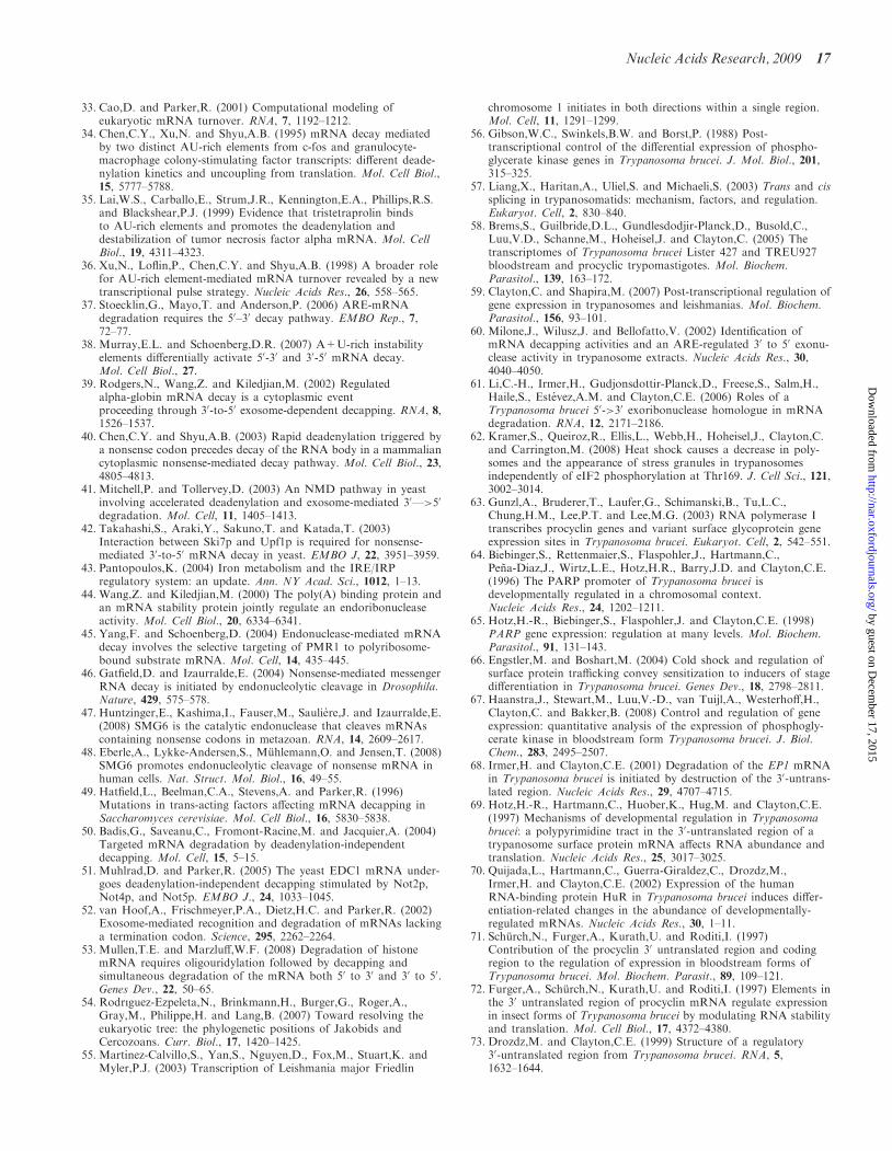
33. Cao,D. and Parker,R. (2001) Computational modeling ofeukaryotic mRNA turnover. RNA, 7, 1192–1212.
34. Chen,C.Y., Xu,N. and Shyu,A.B. (1995) mRNA decay mediatedby two distinct AU-rich elements from c-fos and granulocyte-macrophage colony-stimulating factor transcripts: different deade-nylation kinetics and uncoupling from translation. Mol. Cell Biol.,15, 5777–5788.
35. Lai,W.S., Carballo,E., Strum,J.R., Kennington,E.A., Phillips,R.S.and Blackshear,P.J. (1999) Evidence that tristetraprolin bindsto AU-rich elements and promotes the deadenylation anddestabilization of tumor necrosis factor alpha mRNA. Mol. CellBiol., 19, 4311–4323.
36. Xu,N., Loflin,P., Chen,C.Y. and Shyu,A.B. (1998) A broader rolefor AU-rich element-mediated mRNA turnover revealed by a newtranscriptional pulse strategy. Nucleic Acids Res., 26, 558–565.
37. Stoecklin,G., Mayo,T. and Anderson,P. (2006) ARE-mRNAdegradation requires the 50–30 decay pathway. EMBO Rep., 7,72–77.
38. Murray,E.L. and Schoenberg,D.R. (2007) A+U-rich instabilityelements differentially activate 50-30 and 30-50 mRNA decay.Mol. Cell Biol., 27.
39. Rodgers,N., Wang,Z. and Kiledjian,M. (2002) Regulatedalpha-globin mRNA decay is a cytoplasmic eventproceeding through 30-to-50 exosome-dependent decapping. RNA, 8,1526–1537.
40. Chen,C.Y. and Shyu,A.B. (2003) Rapid deadenylation triggered bya nonsense codon precedes decay of the RNA body in a mammaliancytoplasmic nonsense-mediated decay pathway. Mol. Cell Biol., 23,4805–4813.
41. Mitchell,P. and Tollervey,D. (2003) An NMD pathway in yeastinvolving accelerated deadenylation and exosome-mediated 30—>50
degradation. Mol. Cell, 11, 1405–1413.42. Takahashi,S., Araki,Y., Sakuno,T. and Katada,T. (2003)
Interaction between Ski7p and Upf1p is required for nonsense-mediated 30-to-50 mRNA decay in yeast. EMBO J, 22, 3951–3959.
43. Pantopoulos,K. (2004) Iron metabolism and the IRE/IRPregulatory system: an update. Ann. NY Acad. Sci., 1012, 1–13.
44. Wang,Z. and Kiledjian,M. (2000) The poly(A) binding protein andan mRNA stability protein jointly regulate an endoribonucleaseactivity. Mol. Cell Biol., 20, 6334–6341.
45. Yang,F. and Schoenberg,D. (2004) Endonuclease-mediated mRNAdecay involves the selective targeting of PMR1 to polyribosome-bound substrate mRNA. Mol. Cell, 14, 435–445.
46. Gatfield,D. and Izaurralde,E. (2004) Nonsense-mediated messengerRNA decay is initiated by endonucleolytic cleavage in Drosophila.Nature, 429, 575–578.
47. Huntzinger,E., Kashima,I., Fauser,M., Sauliere,J. and Izaurralde,E.(2008) SMG6 is the catalytic endonuclease that cleaves mRNAscontaining nonsense codons in metazoan. RNA, 14, 2609–2617.
48. Eberle,A., Lykke-Andersen,S., Muhlemann,O. and Jensen,T. (2008)SMG6 promotes endonucleolytic cleavage of nonsense mRNA inhuman cells. Nat. Struct. Mol. Biol., 16, 49–55.
49. Hatfield,L., Beelman,C.A., Stevens,A. and Parker,R. (1996)Mutations in trans-acting factors affecting mRNA decapping inSaccharomyces cerevisiae. Mol. Cell Biol., 16, 5830–5838.
50. Badis,G., Saveanu,C., Fromont-Racine,M. and Jacquier,A. (2004)Targeted mRNA degradation by deadenylation-independentdecapping. Mol. Cell, 15, 5–15.
51. Muhlrad,D. and Parker,R. (2005) The yeast EDC1 mRNA under-goes deadenylation-independent decapping stimulated by Not2p,Not4p, and Not5p. EMBO J., 24, 1033–1045.
52. van Hoof,A., Frischmeyer,P.A., Dietz,H.C. and Parker,R. (2002)Exosome-mediated recognition and degradation of mRNAs lackinga termination codon. Science, 295, 2262–2264.
53. Mullen,T.E. and Marzluff,W.F. (2008) Degradation of histonemRNA requires oligouridylation followed by decapping andsimultaneous degradation of the mRNA both 50 to 30 and 30 to 50.Genes Dev., 22, 50–65.
54. Rodr|guez-Ezpeleta,N., Brinkmann,H., Burger,G., Roger,A.,Gray,M., Philippe,H. and Lang,B. (2007) Toward resolving theeukaryotic tree: the phylogenetic positions of Jakobids andCercozoans. Curr. Biol., 17, 1420–1425.
55. Martinez-Calvillo,S., Yan,S., Nguyen,D., Fox,M., Stuart,K. andMyler,P.J. (2003) Transcription of Leishmania major Friedlin
chromosome 1 initiates in both directions within a single region.Mol. Cell, 11, 1291–1299.
56. Gibson,W.C., Swinkels,B.W. and Borst,P. (1988) Post-transcriptional control of the differential expression of phospho-glycerate kinase genes in Trypanosoma brucei. J. Mol. Biol., 201,315–325.
57. Liang,X., Haritan,A., Uliel,S. and Michaeli,S. (2003) Trans and cissplicing in trypanosomatids: mechanism, factors, and regulation.Eukaryot. Cell, 2, 830–840.
58. Brems,S., Guilbride,D.L., Gundlesdodjir-Planck,D., Busold,C.,Luu,V.D., Schanne,M., Hoheisel,J. and Clayton,C. (2005) Thetranscriptomes of Trypanosoma brucei Lister 427 and TREU927bloodstream and procyclic trypomastigotes. Mol. Biochem.Parasitol., 139, 163–172.
59. Clayton,C. and Shapira,M. (2007) Post-transcriptional regulation ofgene expression in trypanosomes and leishmanias. Mol. Biochem.Parasitol., 156, 93–101.
60. Milone,J., Wilusz,J. and Bellofatto,V. (2002) Identification ofmRNA decapping activities and an ARE-regulated 30 to 50 exonu-clease activity in trypanosome extracts. Nucleic Acids Res., 30,4040–4050.
61. Li,C.-H., Irmer,H., Gudjonsdottir-Planck,D., Freese,S., Salm,H.,Haile,S., Estevez,A.M. and Clayton,C.E. (2006) Roles of aTrypanosoma brucei 50->30 exoribonuclease homologue in mRNAdegradation. RNA, 12, 2171–2186.
62. Kramer,S., Queiroz,R., Ellis,L., Webb,H., Hoheisel,J., Clayton,C.and Carrington,M. (2008) Heat shock causes a decrease in poly-somes and the appearance of stress granules in trypanosomesindependently of eIF2 phosphorylation at Thr169. J. Cell Sci., 121,3002–3014.
63. Gunzl,A., Bruderer,T., Laufer,G., Schimanski,B., Tu,L.C.,Chung,H.M., Lee,P.T. and Lee,M.G. (2003) RNA polymerase Itranscribes procyclin genes and variant surface glycoprotein geneexpression sites in Trypanosoma brucei. Eukaryot. Cell, 2, 542–551.
64. Biebinger,S., Rettenmaier,S., Flaspohler,J., Hartmann,C.,Pena-Diaz,J., Wirtz,L.E., Hotz,H.R., Barry,J.D. and Clayton,C.E.(1996) The PARP promoter of Trypanosoma brucei isdevelopmentally regulated in a chromosomal context.Nucleic Acids Res., 24, 1202–1211.
65. Hotz,H.-R., Biebinger,S., Flaspohler,J. and Clayton,C.E. (1998)PARP gene expression: regulation at many levels. Mol. Biochem.Parasitol., 91, 131–143.
66. Engstler,M. and Boshart,M. (2004) Cold shock and regulation ofsurface protein trafficking convey sensitization to inducers of stagedifferentiation in Trypanosoma brucei. Genes Dev., 18, 2798–2811.
67. Haanstra,J., Stewart,M., Luu,V.-D., van Tuijl,A., Westerhoff,H.,Clayton,C. and Bakker,B. (2008) Control and regulation of geneexpression: quantitative analysis of the expression of phosphogly-cerate kinase in bloodstream form Trypanosoma brucei. J. Biol.Chem., 283, 2495–2507.
68. Irmer,H. and Clayton,C.E. (2001) Degradation of the EP1 mRNAin Trypanosoma brucei is initiated by destruction of the 30-untrans-lated region. Nucleic Acids Res., 29, 4707–4715.
69. Hotz,H.-R., Hartmann,C., Huober,K., Hug,M. and Clayton,C.E.(1997) Mechanisms of developmental regulation in Trypanosomabrucei: a polypyrimidine tract in the 30-untranslated region of atrypanosome surface protein mRNA affects RNA abundance andtranslation. Nucleic Acids Res., 25, 3017–3025.
70. Quijada,L., Hartmann,C., Guerra-Giraldez,C., Drozdz,M.,Irmer,H. and Clayton,C.E. (2002) Expression of the humanRNA-binding protein HuR in Trypanosoma brucei induces differ-entiation-related changes in the abundance of developmentally-regulated mRNAs. Nucleic Acids Res., 30, 1–11.
71. Schurch,N., Furger,A., Kurath,U. and Roditi,I. (1997)Contribution of the procyclin 30 untranslated region and codingregion to the regulation of expression in bloodstream forms ofTrypanosoma brucei. Mol. Biochem. Parasit., 89, 109–121.
72. Furger,A., Schurch,N., Kurath,U. and Roditi,I. (1997) Elements inthe 30 untranslated region of procyclin mRNA regulate expressionin insect forms of Trypanosoma brucei by modulating RNA stabilityand translation. Mol. Cell Biol., 17, 4372–4380.
73. Drozdz,M. and Clayton,C.E. (1999) Structure of a regulatory30-untranslated region from Trypanosoma brucei. RNA, 5,1632–1644.
Nucleic Acids Research, 2009 17
by guest on Decem
ber 17, 2015http://nar.oxfordjournals.org/
Dow
nloaded from
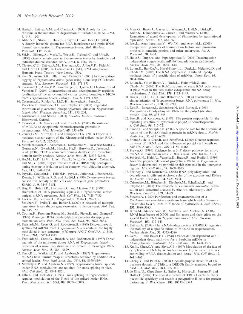
74. Haile,S., Estevez,A.M. and Clayton,C. (2003) A role for theexosome in the initiation of degradation of unstable mRNAs. RNA,9, 1491–1501.
75. Alibu,V.P., Storm,L., Haile,S., Clayton,C. and Horn,D. (2004)A doubly inducible system for RNA interference and rapid RNAiplasmid construction in Trypanosoma brucei. Mol. Biochem.Parasitol., 139, 75–82.
76. Shi,H., Djikeng,A., Mark,T., Wirtz,E., Tschudi,C. and Ullu,E.(2000) Genetic interference in Trypanosoma brucei by heritable andinducible double-stranded RNA. RNA, 6, 1069–1076.
77. Clayton,C.E., Estevez,A.M., Hartmann,C., Alibu,V.P., Field,M.and Horn,D. (2005) In Carmichael,G. (ed.), RNA interference.Humana Press, Totowa, New Jersey, USA.
78. Shen,S., Arhin,G.K., Ullu,E. and Tschudi,C. (2001) In vivo epitopetagging of Trypanosoma brucei genes using a one step PCR-basedstrategy. Mol. Biochem. Parasitol., 113, 171–173.
79. Colasante,C., Alibu,V.P., Kirchberger,S., Tjaden,J., Clayton,C. andVoncken,F. (2006) Characterisation and developmentally regulatedlocalisation of the mitochondrial carrier protein homologue MCP6from Trypanosoma brucei. Eukaryot Cell, 5, 1194–1205.
80. Colasante,C., Robles,A., Li,C.-H., Schwede,A., Benz,C.,Voncken,F., Guilbride,D.L. and Clayton,C. (2007) Regulatedexpression of glycosomal phosphoglycerate kinase in Trypanosomabrucei. Mol. Biochem. Parasitol., 151, 193–204.
81. Kirkwood,B. and Stern,J. (2003) Essential Medical Statistics.Blackwood, Oxford.
82. Cassola,A., De Gaudenzi,J. and Frasch,A. (2007) Recruitmentof mRNAs to cytoplasmic ribonucleoprotein granules intrypanosomes. Mol. Microbiol., 65, 655–670.
83. Zeiner,G.M., Sturm,N.R. and Campbell,D.A. (2003) Exportin 1mediates nuclear export of the Kinetoplastid spliced leader RNA.Eucaryot. Cell, 2, 222–230.
84. Marchler-Bauer,A., Anderson,J., Derbyshire,M., DeWeese-Scott,C.,Gonzales,N., Gwadz,M., Hao,L., He,S., Hurwitz,D., Jackson,J.et al. (2007) CDD: a conserved domain database for interactivedomain family analysis. Nucleic Acids Res., 35, D237–D240.
85. Hu,M., Li,P., Li,M., Li,W., Yao,T., Wu,J.-W., Gu,W., Cohen,R.and Shi,Y. (2002) Crystal Structure of a UBP-family deubiquiti-nating enzyme in isolation and in complex with ubiquitin aldehyde.Cell, 111, 1041–1054.
86. Pays,E., Coquelet,H., Tebabi,P., Pays,A., Jefferies,D., Steinert,M.,Koenig,E., Williams,R.O. and Roditi,I. (1990) Trypanosoma brucei:constitutive activity of the VSG and procyclin gene promoters.EMBO J., 9, 3145–3151.
87. Hug,M., Hotz,H.R., Hartmann,C. and Clayton,C.E. (1994)Hierarchies of RNA processing signals in a trypanosome surfaceantigen mRNA precursor. Mol. Cell Biol., 14, 7428–7435.
88. Lackner,D., Beilharz,T., Marguerat,S., Mata,J., Watt,S.,Schubert,F., Preiss,T. and Bahler,J. (2007) A network of multipleregulatory layers shapes gene expression in fission yeast. Mol. Cell,26, 145–155.
89. Couttet,P., Fromont-Racine,M., Steel,D., Pictet,R. and Grange,T.(1997) Messenger RNA deadenylylation precedes decapping inmammalian cells. Proc. Natl Acad. Sci. USA, 94, 5628–5633.
90. Freistadt,M., Cross,G. and Robertson,H. (1988) Discontinuouslysynthesized mRNA from Trypanosoma brucei contains the highlymethylated 50 cap structure, m7GpppA�A�C(2’-O)mU�A. J. Biol.Chem., 263, 15071–15075.
91. Freistadt,M., Cross,G., Branch,A. and Robertson,H. (1987) Directanalysis of the mini-exon donor RNA of Trypanosoma brucei:detection of a novel cap structure also present in messenger RNA.Nucleic Acids Res., 15, 9861–9879.
92. Perry,K.L., Watkins,K.P. and Agabian,N. (1987) TrypanosomemRNAs have unusual ‘‘cap 4’’ structures acquired by addition of aspliced leader. Proc. Natl Acad. Sci. USA, 84, 8190–8194.
93. McNally,K.P. and Agabian,N. (1992) Trypanosoma brucei spliced-leader RNA methylations are required for trans splicing in vivo.Mol. Cell Biol., 12, 4844–4851.
94. Ullu,E. and Tschudi,C. (1991) Trans splicing in trypanosomesrequires methylation of the 50 end of the spliced leader RNA.Proc. Natl Acad. Sci. USA, 88, 10074–10078.
95. Mair,G., Braks,J., Garver,L., Wiegant,J., Hall,N., Dirks,R.,Khan,S., Dimopoulos,G., Janse,C. and Waters,A. (2006)Regulation of sexual development of Plasmodium by translationalrepression. Science, 313, 667–669.
96. Iyer,L., Anantharaman,V., Wolf,M. and Aravind,L. (2008)Comparative genomics of transcription factors and chromatinproteins in parasitic protists and other eukaryotes. Int. J.Parasitol., 38, 1–31.
97. Haile,S., Dupe,A. and Papadopoulou,B. (2008) Deadenylation-independent stage-specific mRNA degradation in Leishmania.Nucleic Acids Res., 36, 1634–1644.
98. Lotan,R., Bar-On,V., Harel-Sharvit,L., Duek,L., Melamed,D. andChoder,M. (2005) The RNA polymerase II subunit Rpb4pmediates decay of a specific class of mRNAs. Genes Dev., 19,3004–3016.
99. Lotan,R., Goler-Baron,V., Duek,L., Haimovich,G. andChoder,M. (2007) The Rpb7p subunit of yeast RNA polymeraseII plays roles in the two major cytoplasmic mRNA decaymechanisms. J. Cell Biol., 178, 1133–1143.
100. Das,A., Li,H., Liu,T. and Bellofatto,V. (2006) Biochemicalcharacterization of Trypanosoma brucei RNA polymerase II. Mol.Biochem. Parasitol., 150, 201–210.
101. Deo,R., Bonanno,J., Sonenberg,N. and Burley,S. (1999)Recognition of polyadenylate RNA by the poly(A)-bindingprotein. Cell, 98, 835–845.
102. Baer,B. and Kornberg,R. (1983) The protein responsible for therepeating structure of cytoplasmic poly(A)-ribonucleoprotein.J. Cell Biol., 96, 717–721.
103. Simon,E. and Seraphin,B. (2007) A specific role for the C-terminalregion of the Poly(A)-binding protein in mRNA decay. NucleicAcids Res., 35, 6017–6028.
104. Prieto,S., de la Cruz,B. and Scheffler,I. (2000) Glucose-regulatedturnover of mRNA and the influence of poly(A) tail length onhalf-life. J. Biol. Chem., 275, 14155–14166.
105. Brewer,G. (1999) Evidence for a 30-50 decay pathway for c-mycmRNA in mammalian cells. J. Biol. Chem., 274, 16174–16179.
106. Schurch,N., Hehl,A., Vassella,E., Braun,R. and Roditi,I. (1994)Accurate polyadenylation of procyclin mRNAs in Trypanosomabrucei is determined by pyrimidine-rich elements in the intergenicregions. Mol. Cell Biol., 14, 3668–3675.
107. Portnoy,V. and Schuster,G. (2006) RNA polyadenylation anddegradation in different Archaea; roles of the exosome and RNaseR. Nucelic Acids Res., 34, 5923–5931.
108. Cristodero,M., Bottcher,B., Diepholz,M., Scheffzeck,K. andClayton,C. (2008) The exosome of Leishmania tarentolae: purifi-cation and structural analysis by electron microscopy. Mol.Biochem. Parasitol., 159, 24–29.
109. Stevens,A. (1980) Purification and characterization of aSaccharomyces cerevisiae exoribonuclease which yields 50-mono-nucleotides by a 50 leads to 30 mode of hydrolysis. J. Biol. Chem.,255, 3080–3085.
110. Biton,M., Mandelboim,M., Arvatz,G. and Michaeli,S. (2006)RNAi interference of XPO1 and Sm genes and their effect on thespliced leader RNA in Trypanosoma brucei. Mol. Biochem.Parasitol., 150, 132–143.
111. Estevez,A. (2008) The RNA-binding protein TbDRBD3 regulatesthe stability of a specific subset of mRNAs in trypanosomes.Nucleic Acids Res., 36, 4573–4586.
112. Gera,J.F. and Baker,E.J. (1998) Deadenylation-dependent and -independent decay pathways for a 1-tubulin mRNA inChlamydomonas reinhardtii. Mol. Cell Biol., 18, 1498–1505.
113. Xu,N., Chen,C.Y. and Shyu,A.B. (1997) Modulation of the fate ofcytoplasmic mRNA by AU-rich elements: key sequence featurescontrolling mRNA deadenylation and decay. Mol. Cell Biol., 17,4611–4621.
114. Cheng,Y. and Patel,D. (2004) Crystallographic structure of thenuclease domain of 30hExo, a DEDDh family member, bound torAMP. J. Mol. Biol., 343, 305–312.
115. de Silva,U., Choudhury,S., Bailey,S., Harvey,S., Perrino,F. andHollis,T. (2007) The crystal structure of TREX1 explains the 30
nucleotide specificity and reveals a polyproline II helix for proteinpartnering. J. Biol. Chem., 282, 10537–10543.
18 Nucleic Acids Research, 2009
by guest on Decem
ber 17, 2015http://nar.oxfordjournals.org/
Dow
nloaded from
Related Documents