The Role of Carboxydothermus hydrogenoformans in the Conversion of Calcium Phosphate from Amorphous to Crystalline State Mathieu Haddad 1,2 , Hojatollah Vali 3,4 , Jeanne Paquette 3 , Serge R. Guiot 1,2 * 1 Energy, Mining and Environment Portfolio, National Research Council Canada, Montreal, Quebec, Canada, 2 Department of Microbiology, Infectiology and Immunology, Universite ´ de Montre ´al, Montreal, Quebec, Canada, 3 Department of Earth and Planetary Sciences, McGill University, Montreal, Quebec, Canada, 4 Facility for Electron Microscopy Research, McGill University, Montreal, Quebec, Canada Abstract Two previously unknown modes of biomineralization observed in the presence of Carboxydothermus hydrogenoformans are presented. Following the addition of NaHCO 3 and the formation of an amorphous calcium phosphate precipitate in a DSMZ medium inoculated with C. hydrogenoformans, two distinct crystalline solids were recovered after 15 and 30 days of incubation. The first of these solids occurred as micrometric clusters of blocky, angular crystals, which were associated with bacterial biofilm. The second solid occurred as 30–50 nm nanorods that were found scattered among the organic products of bacterial lysis. The biphasic mixture of solids was clearly dominated by the first phase. The X-ray diffractometry (XRD) peaks and Fourier transform infrared spectroscopy (FTIR) spectrum of this biphasic material consistently showed features characteristic of Mg-whitlockite. No organic content or protein could be identified by dissolving the solids. In both cases, the mode of biomineralization appears to be biologically induced rather than biologically controlled. Since Mg is known to be a strong inhibitor of the nucleation and growth of CaP, C. hydrogenoformans may act by providing sites that chelate Mg or form complexes with it, thus decreasing its activity as nucleation and crystal growth inhibitor. The synthesis of whitlockite and nano-HAP-like material by C. hydrogenoformans demonstrates the versatility of this organism also known for its ability to perform the water-gas shift reaction, and may have applications in bacterially mediated synthesis of CaP materials, as an environmentally friendly alternative process. Citation: Haddad M, Vali H, Paquette J, Guiot SR (2014) The Role of Carboxydothermus hydrogenoformans in the Conversion of Calcium Phosphate from Amorphous to Crystalline State. PLoS ONE 9(2): e89480. doi:10.1371/journal.pone.0089480 Editor: Vladimir N. Uversky, University of South Florida College of Medicine, United States of America Received November 19, 2013; Accepted January 21, 2014; Published February 26, 2014 Copyright: ß 2014 Haddad et al. This is an open-access article distributed under the terms of the Creative Commons Attribution License, which permits unrestricted use, distribution, and reproduction in any medium, provided the original author and source are credited. Funding: One of the authors (M.H.) was supported by the Natural Sciences and Engineering Research Council of Canada (grant 185778-2009). The funders had no role in study design, data collection and analysis, decision to publish, or preparation of the manuscript. Competing Interests: The authors have declared that no competing interests exist. * E-mail: [email protected] Introduction Biomineralization as described by Lowenstam [1] is the ability of living organisms to form minerals as well as materials composed of an organic and inorganic phase [2,3]. Among more than 60 biominerals formed by bacteria discovered so far, 25% are amorphous and 75% crystalline. Several authors [3–5] have investigated the mechanism of biomineralization and found that organisms across different phyla control biomineralization in a distinct manner and that biominerals have different functions. According to Mann [6] biomineralization occurs at the organic- inorganic interface where a molecular recognition system is involved in the control of crystal nucleation and growth. Biomineralization processes fall in two categories: biologically induced mineralization (BIM) and biologically controlled miner- alization (BCM) [1]. In BIM, biomineralization occurs outside the cell and none of the cell components are serving as a template for nucleation and growth of the precipitate. In this case, cellular activity results in changes in the microenvironment and anionic and cationic precipitation [3]. Biominerals produced by BIM are characterized by poor crystallinity and high variations in morphology, water content, structure, particle size as well as the presence of trace elements [7]. In BCM, also known as inorganic matrix-mediated mineralization [1], the cell controls all of the above described stages of mineralization from nucleation to crystal-formation, leading to a highly specie-specific product [8]. BCM is based on a site-specific matrix (cytoplasm or on the cell wall) that enables the formation of a compartmentalized environ- ment with its own chemical composition. Nucleation is then made possible by sequestering specific ions leading to supersaturation and precipitation in the matrix [9]. Bacteria living under high temperature conditions are known as thermophiles (40–69uC) and hyperthermophiles (70–110uC). Biomineralization processes in this latter group of bacteria have not been extensively explored yet. Indeed, known processes describe magnetite and realgar formation [10] as well as reductive precipitation of uranium, manganese and other toxic metals [11]. In this study, we report that C. hydrogenoformans a carboxydo- trophic hydrogenogenic hyperthermophilic bacterium [12] con- verts an amorphous calcium phosphate phase into a fully crystalline whitlockite mineral and spherulitic clusters that we interpret to be hydroxyapatite-like nanocrystals. In addition to conventional microbiological analysis, Fourier transform infrared spectroscopy (FTIR), X-ray diffractometry (XRD) and electron microscopy techniques were applied. We demonstrate that an PLOS ONE | www.plosone.org 1 February 2014 | Volume 9 | Issue 2 | e89480

Welcome message from author
This document is posted to help you gain knowledge. Please leave a comment to let me know what you think about it! Share it to your friends and learn new things together.
Transcript
The Role of Carboxydothermus hydrogenoformans in theConversion of Calcium Phosphate from Amorphous toCrystalline StateMathieu Haddad1,2, Hojatollah Vali3,4, Jeanne Paquette3, Serge R. Guiot1,2*
1 Energy, Mining and Environment Portfolio, National Research Council Canada, Montreal, Quebec, Canada, 2 Department of Microbiology, Infectiology and Immunology,
Universite de Montreal, Montreal, Quebec, Canada, 3 Department of Earth and Planetary Sciences, McGill University, Montreal, Quebec, Canada, 4 Facility for Electron
Microscopy Research, McGill University, Montreal, Quebec, Canada
Abstract
Two previously unknown modes of biomineralization observed in the presence of Carboxydothermus hydrogenoformans arepresented. Following the addition of NaHCO3 and the formation of an amorphous calcium phosphate precipitate in a DSMZmedium inoculated with C. hydrogenoformans, two distinct crystalline solids were recovered after 15 and 30 days ofincubation. The first of these solids occurred as micrometric clusters of blocky, angular crystals, which were associated withbacterial biofilm. The second solid occurred as 30–50 nm nanorods that were found scattered among the organic productsof bacterial lysis. The biphasic mixture of solids was clearly dominated by the first phase. The X-ray diffractometry (XRD)peaks and Fourier transform infrared spectroscopy (FTIR) spectrum of this biphasic material consistently showed featurescharacteristic of Mg-whitlockite. No organic content or protein could be identified by dissolving the solids. In both cases, themode of biomineralization appears to be biologically induced rather than biologically controlled. Since Mg is known to be astrong inhibitor of the nucleation and growth of CaP, C. hydrogenoformans may act by providing sites that chelate Mg orform complexes with it, thus decreasing its activity as nucleation and crystal growth inhibitor. The synthesis of whitlockiteand nano-HAP-like material by C. hydrogenoformans demonstrates the versatility of this organism also known for its abilityto perform the water-gas shift reaction, and may have applications in bacterially mediated synthesis of CaP materials, as anenvironmentally friendly alternative process.
Citation: Haddad M, Vali H, Paquette J, Guiot SR (2014) The Role of Carboxydothermus hydrogenoformans in the Conversion of Calcium Phosphate fromAmorphous to Crystalline State. PLoS ONE 9(2): e89480. doi:10.1371/journal.pone.0089480
Editor: Vladimir N. Uversky, University of South Florida College of Medicine, United States of America
Received November 19, 2013; Accepted January 21, 2014; Published February 26, 2014
Copyright: � 2014 Haddad et al. This is an open-access article distributed under the terms of the Creative Commons Attribution License, which permitsunrestricted use, distribution, and reproduction in any medium, provided the original author and source are credited.
Funding: One of the authors (M.H.) was supported by the Natural Sciences and Engineering Research Council of Canada (grant 185778-2009). The funders hadno role in study design, data collection and analysis, decision to publish, or preparation of the manuscript.
Competing Interests: The authors have declared that no competing interests exist.
* E-mail: [email protected]
Introduction
Biomineralization as described by Lowenstam [1] is the ability
of living organisms to form minerals as well as materials composed
of an organic and inorganic phase [2,3]. Among more than 60
biominerals formed by bacteria discovered so far, 25% are
amorphous and 75% crystalline. Several authors [3–5] have
investigated the mechanism of biomineralization and found that
organisms across different phyla control biomineralization in a
distinct manner and that biominerals have different functions.
According to Mann [6] biomineralization occurs at the organic-
inorganic interface where a molecular recognition system is
involved in the control of crystal nucleation and growth.
Biomineralization processes fall in two categories: biologically
induced mineralization (BIM) and biologically controlled miner-
alization (BCM) [1]. In BIM, biomineralization occurs outside the
cell and none of the cell components are serving as a template for
nucleation and growth of the precipitate. In this case, cellular
activity results in changes in the microenvironment and anionic
and cationic precipitation [3]. Biominerals produced by BIM are
characterized by poor crystallinity and high variations in
morphology, water content, structure, particle size as well as the
presence of trace elements [7]. In BCM, also known as inorganic
matrix-mediated mineralization [1], the cell controls all of the
above described stages of mineralization from nucleation to
crystal-formation, leading to a highly specie-specific product [8].
BCM is based on a site-specific matrix (cytoplasm or on the cell
wall) that enables the formation of a compartmentalized environ-
ment with its own chemical composition. Nucleation is then made
possible by sequestering specific ions leading to supersaturation
and precipitation in the matrix [9].
Bacteria living under high temperature conditions are known as
thermophiles (40–69uC) and hyperthermophiles (70–110uC).
Biomineralization processes in this latter group of bacteria have
not been extensively explored yet. Indeed, known processes
describe magnetite and realgar formation [10] as well as reductive
precipitation of uranium, manganese and other toxic metals [11].
In this study, we report that C. hydrogenoformans a carboxydo-
trophic hydrogenogenic hyperthermophilic bacterium [12] con-
verts an amorphous calcium phosphate phase into a fully
crystalline whitlockite mineral and spherulitic clusters that we
interpret to be hydroxyapatite-like nanocrystals. In addition to
conventional microbiological analysis, Fourier transform infrared
spectroscopy (FTIR), X-ray diffractometry (XRD) and electron
microscopy techniques were applied. We demonstrate that an
PLOS ONE | www.plosone.org 1 February 2014 | Volume 9 | Issue 2 | e89480
abiotic soluble CaP precursor is converted, in the presence of an
active culture of C. hydrogenoformans, to a biphasic mixture of
granular aggregates of whitlockite and spherulitic hydroxyapatite.
This phase is then converted in the crystalline whitlockite by C.
hydrogenoformans activity.
Microbial calcification is a widespread phenomenon, which
includes the formation of phosphate salts of calcium (CaP) [13].
CaP displays high biocompatibility and biodegradability due to
their chemical similarity to calcified tissue [14–16]. A large range
of CaP, which differ in origin, composition and form, are currently
used in medicine for regeneration of hard tissues [17]. Depending
on the required characteristic (bioactive or resorbable material) for
CaP applications (bone replacement, filling or coating, functiona-
lized nanoparticle), different phases of CaP ceramics are used (b-
tri-calcium phosphate (b-TCP), hydroxyapatite or biphasic CaP)
[15,18]. The chemical synthesis of CaP and CaP-based materials,
while being very effective on one hand, are relatively expensive
and eco-hazardous, requiring extremes of temperature and pH
[14]. Thus, the present work offers an alternative biological
approach with a more environmentally friendly process making C.
hydrogenoformans a possible ecofriendly nanofactory for CaP
synthesis.
Materials and Methods
Bacterial strain and growth conditionsC. hydrogenoformans (DSM 6008) was obtained from the German
Collection of Microorganisms and Cell Cultures (DSMZ,
Braunschweig, Germany). Microorganisms were cultivated under
strictly anaerobic conditions in basal mineral bicarbonate-phos-
phate buffered medium that contained (in g/L of demineralized
water): KCl (0.33), MgCl2?6H2O (0.52), CaCl2?2H2O (0.29),
NH4Cl (0.33), KH2PO4 (0.33). The medium was supplemented
with 10 mL?L21 of trace metals solution. The medium was boiled
and then introduced anaerobically in sterilized serum bottles
under N2 air flush. After autoclaving, it was then complemented
with (in mL?L21 of medium): 5% NaHCO3 stock solution (20),
2.5% Na2S?9H2O stock solution (10), 0.5% yeast extract solution
(10) and vitamin solution (1). The trace metals and vitamin stock
solutions were prepared as described elsewhere [19]. All stock
solutions were autoclaved, except the vitamin solution, which was
sterilized by filtration through 0.22 mm filter membranes. After
complementation, the pH was between 6.8 and 7.0. All
experiments were carried out at 70uC, 150 rpm in 500 mL
bottles. Bottles contained 200 mL of medium inoculated with the
same amount of biomass under a 300 mL headspace. Initial
headspace composition was set at 100% CO and 1 atm.
Control experimentsIn control experiments, the bacterial biomass was resuspended
in a modified medium described by Zhao and coll. [20] in which
no precipitation of amorphous calcium phosphate was observed.
The modified medium differed from the DSMZ one only in
MgCl2?6H2O, CaCl2?2H2O, KH2PO4 and NaHCO3 concentra-
tions, which were (in g?L21 of demineralized water): 0.102, 0.015,
0.136, 0.401, respectively. In that medium, no amorphous CaP
was observed to form abiotically over a period of 30 days, and the
addition of a live bacterial culture did not induce detectable
precipitation of CaP. The modified medium was also used to
determine the proteomic profile of C. hydrogenoformans when no
biomineralization took place (see biomolecular techniques).
Another control experiment was conducted to verify if proteins
or amino acids released in the medium by the bacteria had a direct
or indirect role in the crystallization of the precipitate. Dry
amorphous precipitate obtained in the sterile DSMZ medium was
incubated for 15 days at 70uC in the filtered (0.33 mm) inoculated
DSMZ medium from which crystalline phases had been recov-
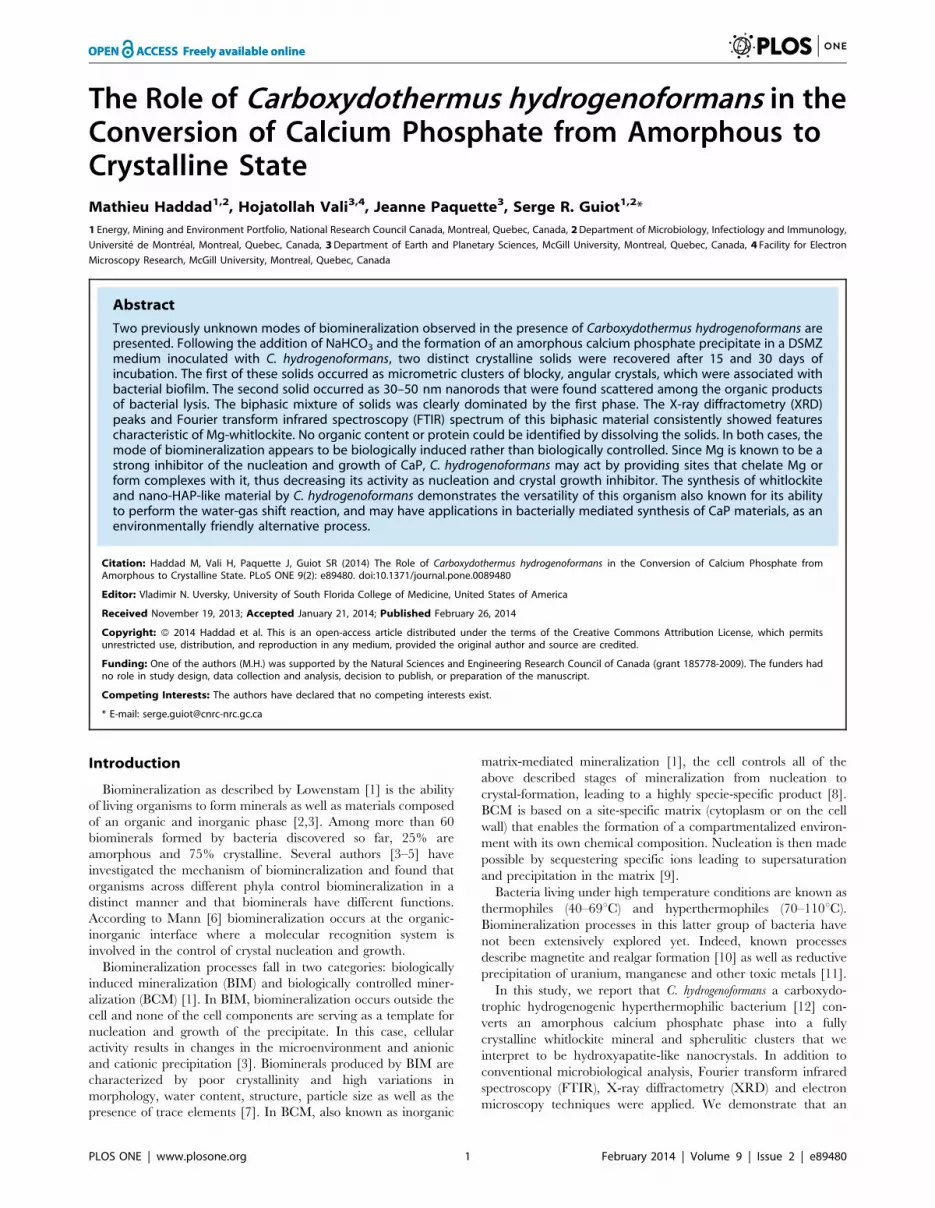
Figure 1. Change with time of dissolved total phosphate concentration in the sterile (dash) and inoculated (triangle) DSMZ mediumafter complementation with NaHCO3 (at time 0).doi:10.1371/journal.pone.0089480.g001
Table 1. Elemental analysis of a washed sample of C.hydrogenoformans culture grown on DSMZ mediumcompared to biomass elemental composition from literature[51].
Chemicalelement Proportion (% wt) Atomic fraction
This study Literature This study Literature
C 2.8160.09 48 1 1
H 0.8560.03 7.3 3.62 1.8
N 0.4960.02 11.3 0.15 0.2
O 2.1860.14 32.5 0.58 0.5
S 0 0.01 0 0
Total 6.3360.27 99.1
Molecular weight (g?mol21) 27 24.6
doi:10.1371/journal.pone.0089480.t001
Biomineralization by C. hydrogenoformans
PLOS ONE | www.plosone.org 2 February 2014 | Volume 9 | Issue 2 | e89480

ered. The result of this control experiment was also negative as
XRD analysis showed that the CaP precipitate remained
amorphous.
In order to exclude the precipitation of an amorphous calcium
carbonate in the DSMZ medium, the third abiotic control
experiment was carried out using NH3OH as buffer instead of
NaHCO3. A similar precipitate appeared and its energy
dispersive X-ray spectrometry (EDX) patterns showed Ca and P
peaks identical to those of the solid produced by NaHCO3
addition. This confirmed that the amorphous precipitate formed
in the DSMZ medium was dominantly a calcium phosphate
phase.
Sampling proceduresAll measurements that were carried out on the DSMZ medium
were processed immediately after sampling in order to avoid any
time related alteration. For precipitate characterization, samples
were first concentrated by centrifugation during 10 min at
Table 2. Comparison of the elemental chemical composition of whitlockite [27], hydroxyapatite [52], octacalcium [53] to theelemental composition of suspended solids obtained after 39 days of C. hydrogenoformans growth on DSMZ medium. N.D.: notdetermined.
Chemical element Proportion (% wt)
Suspended Solids whitlockite hydroxyapatite octacalcium phosphate
Calcium (Ca) 27.3061.70 33.91 39.9 32.63
Phosphorus (P) 17.9561.16 20.38 18.5 18.91
Hydrogen (H) N.D. 0.076 0.2 1.23
Oxygen (O) N.D. 42.11 41.4 40.71
Metals 4.1060.45 N.D. N.D. N.D.
Ca/P ratio (weight) 1.5260.01 1.66 2.15 1.72
Ca/P ratio (molar) 1.1760.01 1.28 1.66 1.33
Formula Ca9(Mg, Fe2+)(PO4)6(PO3OH) Ca5(PO4)3(OH) Ca8H2(PO4)6.5H2O
doi:10.1371/journal.pone.0089480.t002
Figure 2. XRD spectra. Black: dried precipitate formed and obtained after 30 days of C. hydrogenoformans growth in the DSMZ medium. Red:whitlockite from the JCPDS (Joint Committee on Powder Diffraction Standards) database (number 01-070-1786).doi:10.1371/journal.pone.0089480.g002
Biomineralization by C. hydrogenoformans
PLOS ONE | www.plosone.org 3 February 2014 | Volume 9 | Issue 2 | e89480

Figure 3. XRD spectra of the dried precipitate recovered after 30 days of C. hydrogenoformans growth in the DSMZ medium(identified as ‘Dried sample’), the dried sample after having been calcinated (identified as ‘Calcinated sample’), the commercialsintered b-TCP, and the whitlockite, calculated according to the JCPDS (Joint Committee on Powder Diffraction Standards)database (number 01-070-1786).doi:10.1371/journal.pone.0089480.g003
Figure 4. XRD pattern of the dried precipitate formed and sampled after 30 days of aging in the sterile DSMZ medium.doi:10.1371/journal.pone.0089480.g004
Biomineralization by C. hydrogenoformans
PLOS ONE | www.plosone.org 4 February 2014 | Volume 9 | Issue 2 | e89480

15000 rpm. Supernatant was removed and the pellet washed 3
times in MilliQ water to remove any remaining of the medium.
Since the abiotic precipitate obtained in the absence of C.
hydrogenoformans was highly soluble in water, the pellet obtained by
centrifuging the control samples was washed only once in MilliQ
water prior to any characterization.
Figure 5. FTIR analysis of precipitate after 30 days of aging. Dried precipitate from sterile DSMZ medium (continuous line) and calcinatedprecipitate from the C. hydrogenoformans culture in the DSMZ medium (triangles).doi:10.1371/journal.pone.0089480.g005
Figure 6. SEM-EDX analysis of two areas from a calcinated precipitate isolated after 30 days of C. hydrogenoformans growth in theDSMZ medium. Images on the left show two levels of magnification of same area. Images on the right show EDX spectrum of two distinct areas ofthe sample.doi:10.1371/journal.pone.0089480.g006
Biomineralization by C. hydrogenoformans
PLOS ONE | www.plosone.org 5 February 2014 | Volume 9 | Issue 2 | e89480

Experimental parametersDissolved total phosphate. Dissolved phosphate ions con-
centration was measured on aliquots sampled from bottles
inoculated with active cultures of C. hydrogenoformans. Sterile control
series were also conducted on DSMZ medium. 2 mL of medium
was sampled every 24 hours and centrifuged. Supernatant was
analyzed on a Hamilton PRP-X100 (Hamilton Company, Reno,
NV, USA) polymer-based chromatography column (250641 mm
O.D.) in a high-performance liquid chromatograph TSP model
P4000 & AS 3000 (TSP, San Jose, CA, USA). Conductivity data
were obtained by using a Waters Millipore detector model 432.
The mobile phase was p-hydroxybenzoic acid at pH 8.5 with
2.5% methanol at a flow rate of 1.8 mL.min21 at 40uC.
Organics and Inorganics. The suspended solids (SS) and
volatile suspended solids (VSS) were determined according to
Standard Methods [21]. The sample was dried at 105uC over
night, weighed then placed in a muffle furnace at 600uC for two
hours. VSS is determined from the weight loss from ignition.
Volatile fatty acids (VFA). VFAs (i.e. acetic, propionic and
butyric acids) were measured on an Agilent 6890 (Wilmington,
DE) gas chromatograph (GC) equipped with a flame ionization
detector (FID) on 0.2 ml samples diluted 1:1 (vol./vol.) with an
internal standard of iso-butyric acid in 6% formic acid, directly
injected on a glass column of 1 m62 mm Carbopack C (60–
80 mesh) coated with 0.3% Carbowax 20 M and 0.1% H3PO4.
The column was held at 130uC for 4 min. Helium was the carrier
gas fed at a rate of 20 mL?min21. Both injector and detector were
maintained at 200uC.
Solvents. For measurement of solvents (methanol, ethanol,
acetone, 2-propanol, tert-butanol, n-propanol, sec-butanol, n-
butanol) 100 mL of liquid was transferred into a vial that had
20 mL of headspace and was crimp sealed with a Teflon-coated
septum. The vial was heated at 80uC for 2 min, then 1000 ml of
headspace gas was injected onto a DB-ACL2 capillary column of
30 m6530 mm62 mm using a Combipal autosampler (CTC
Analytics AG, Zwingen, Swizerland). The column was held at
40uC for 10 min. Helium was the carrier gas at a head pressure of
5 psi. The injector and the detector were maintained at 200uC and
250uC, respectively.
Mono and disaccharides. Mono and disaccharides were
measured using an HPLC from Waters Corporation (Milford,
MA) consisting of a pump (model 600, Waters Corporation) and
an auto sampler model 717 Plus equipped with a refractive index
detector (model 2414, Waters Corporation). Organics acids are
monitored using a PDA detector (model 2996, Waters Corpora-
tion). The column used for the separation is Transgenomic ICSep
IC-ION-300 (300 mm67.8 mm OD) (Transgenomics, San Jose,
CA, USA). The mobile phase is 0.01N H2SO4 at 0.4 mL min21.
Analysis is carried out at 35uC.
Sample characterizationThe goal of sample characterization was to compare the solid
precipitate obtained from experiments carried out in inoculated
and sterile DSMZ media. The characterized solid was obtained by
centrifugation of the sampled medium and could not be physically
separated from the biomass. In some cases, the bacterial biomass
was eliminated by calcination (heating to 600uC for 2 hours) but
most observations were carried out on a dried (105uC overnight)
composite material made of bacterial biomass intimately mixed
with the CaP precipitate. Precipitates from inoculated medium
(dried and calcinated to remove all organic matter) and from
sterile medium (dried only) were analyzed by XRD. Number and
positions of XRD peaks were unchanged from dried-only to dried
and calcinated precipitates. Also, XRD patterns of dried
precipitate from the sterile medium consistently showed broad
humps of an amorphous material, showing that drying did not
Figure 7. SEM-EDX analysis of two areas from a precipitate recovered and dried after 30 days of aging in the sterile DSMZ medium.Images on the left show two levels of magnification of same area. Images on the right show EDX spectrum of two distinct areas of the sample.doi:10.1371/journal.pone.0089480.g007
Biomineralization by C. hydrogenoformans
PLOS ONE | www.plosone.org 6 February 2014 | Volume 9 | Issue 2 | e89480

induce crystallinity. To eliminate the signature of the biomass,
FTIR and scanning electron microscopy (SEM) analysis were
conducted on the calcinated and dried sample from inoculated
media, and compared to those of dried-only samples from sterile
media.
Elemental analysis of the biotic precipitate. Elemental
analysis was performed on a dry sample of a 30-day culture of C.
hydrogenoformans in the DSMZ medium. Standard Methods were
used for determination of elemental carbon, hydrogen, nitrogen,
oxygen and sulfur [22] [23]. The sample was combusted at
1030uC. The combustion gases produced are then passed on a GC
(ECS 4010, Costech Analytical Technologies, Valencia, CA) using
ultra high purity helium as the carrier gas and equipped with a
TCD, which analyzes the concentrations of CO2, N2, H2O and
SO2 from which percentages of carbon, hydrogen, nitrogen and
sulfur are derived. The same procedure was utilized for oxygen
analysis using a combustion elemental analyzer EA 1108 (Fisons/
Carlo Erba, Milan, Italy). Similar samples were analyzed at two
different analytical facilities (Dept. of Chemistry, Universite de
Montreal, Montreal, QC and Chemisar Inc., Guelph, ON) and
resulted in the same elemental content.
Metals and phosphorus content of the biotic
precipitate. A centrifuged sample from a 39 days C. hydrogenofor-
mans culture was washed twice and resuspended in milliQ water.
Phosphorus was determined using colorimetric methods (method
365.1, [24]). Calcium and metals were determined by Agatlabs
Inc. (Montreal, QC) using inductively coupled plasma mass
spectrometry (Elan 9000, Perkin-Elmer, Uberlingen, Germany)
[25].
Figure 8. Images (A, C) and corresponding TEM-EDS analysis (B, D) of two areas in a whole-mount sample recovered after 30 daysfrom the culture in the DSMZ medium. (C) Magnification of the area in (A) showing biofilm covering and binding the granules, (B) Spectrumfrom the granule labelled B. (D) Spectrum from the organic material labelled D. (E, F) HR-TEM images of the granules’ edges showing lattice fringes.doi:10.1371/journal.pone.0089480.g008
Biomineralization by C. hydrogenoformans
PLOS ONE | www.plosone.org 7 February 2014 | Volume 9 | Issue 2 | e89480

X-ray diffractometry of the abiotic and biotic
precipitate. Phase analysis was performed on a Bruker D8
Advance X-ray diffractometer (Bruker, Germany) using Cu Karadiation (1.5417A) at 40 kV and 40 mA. The scanning range (2h)
was from 5u to 80u at a scan speed of 0.15u min21 (for the dried
sample) and 0.075u min21 (for the calcinated sample) with a step
size of 0.025u.Phases were identified by matching the peaks to the JCPDS
(Joint Committee on Powder Diffraction Standards) database. As
b-TCP and whitlockite have similar XRD profiles [26–28]
diffractograms were compared to one obtained from a commercial
100% crystalline b-TCP (based on the manufacturer’s description,
$98% b-phase basis, Sigma-Aldrich Co., St Louis, MO, USA).
The relative crystallinity (Cr) of the magnesium whitlockite powder
was determined as described elsewhere [29]. In short, the most
intense peak (31.4u at 2h) of the powders was compared to the
same peak of the reference b-TCP according to:
Cr %ð Þ~ A(31:4 2h) 100
As(31:4 2h)
where Cr is the relative crystallinity of the measured magnesium
whitlockite powder, and As(31.4h) and A(31.4h) are the integrated
area intensity of the 31.4 2h peak of the b-TCP standard and the
biomass powder respectively. TOPASH software (Bruker AXS)
was used for profile fitting and crystallite size calculations.
Fourier transform infrared spectroscopy
(FTIR). Attenuated Total Reflectance (ATR) Fourier transform
infrared (FT-IR) spectra of pure powdered solids were obtained
using a Bruker Tensor Series FT-IR (Bruker, Germany)
spectrometer equipped with a zinc selenide crystal. Each
Spectrum (sum of 64 scans) was collected from 4000 to
500 cm21 at a spectral resolution of 4 cm21. An air spectrum
was also obtained at the beginning of the analysis to measure the
water and carbon dioxide content in the air and these were
subtracted from the sample spectra. The spectra obtained from
both biotic and abiotic precipitates were compared with that of the
commercial reference material b-tricalcium phosphate (b-TCP, $
98% b-phase basis, Sigma-Aldrich Co., St Louis, MO, USA).
Scanning Electron Microscopy (SEM). SEM imaging was
carried out on two 30 days aged samples in order to compare: (1)
the precipitate obtained from the sterile medium and (2) the
Figure 9. TEM imaging of solids identified in sterile and inoculated DSMZ media. (A) Sample recovered from the sterile DSMZ mediumafter 15 days of aging. ACP granules embedded in resin are visible. (B, C, D) Images from samples recovered after 3, 8 and 15 days respectively from atime course experiment in inoculated DSMZ medium. Three solid phases are distinct and interpreted to be either amorphous CaP (ACP) or whitlockite(W) and nanocrystalline hydroxyl-apatite (HAP). Bacteria (B) are also visible.doi:10.1371/journal.pone.0089480.g009
Biomineralization by C. hydrogenoformans
PLOS ONE | www.plosone.org 8 February 2014 | Volume 9 | Issue 2 | e89480
·

Figure 10. TEM imaging of the inoculated DSMZ medium sampled after 6 days of culture. A to D show magnification of cell lysis andspatial association of the lysed vesicle of C. hydrogenoformans and the interpreted HAP.doi:10.1371/journal.pone.0089480.g010
Biomineralization by C. hydrogenoformans
PLOS ONE | www.plosone.org 9 February 2014 | Volume 9 | Issue 2 | e89480

Figure 11. Backscatter electron image and EDS analysis by SEM of samples recovered after 15 days, also shown in Figure 9. (A)Sample recovered from sterile DSMZ medium, and (B) EDS analysis of its precipitate. (C) EDS analysis of the embedding epoxy matrix. (D) Samplerecovered after 15 days of C. hydrogenoformans growth in the DSMZ medium, and (E) EDS analysis of its solid precipitate, (F) EDS analysis of itsembedding epoxy matrix showing that Ca and P are present.doi:10.1371/journal.pone.0089480.g011
Biomineralization by C. hydrogenoformans
PLOS ONE | www.plosone.org 10 February 2014 | Volume 9 | Issue 2 | e89480
precipitate that aged in the presence of C. hydrogenoformans. In both
cases, a 40 mL sample was centrifuged, washed and dried, but the
precipitate from the inoculated medium was also calcinated.
Specimens were then mounted on SEM stubs with double side
carbon tape. In order to avoid any interference during elemental
analysis, no coating was applied. Examination and elemental
analysis was done using a S-4700 Hitachi FE-SEM (Tokyo,
Japan) working under vacuum at an acceleration voltage of 2.0 kV
coupled to an Oxford INCA energy dispersive spectrometer
(EDX) detector.
Backscattering electron (BSE) imaging was performed on an
environmental SEM (ESEM, Quanta 200 FEG, FEI Company
Hillsboro, OR) equipped with an energy dispersive X-ray (EDX)
spectrometer (Genesis 2000, XMS System 60 with a Sapphire Si/
Li Detector from EDAX Inc., Mahwah, NJ). Imaging was also
done under the high vacuum mode of the ESEM microscope at an
accelerating voltage of 20 kV and a working distance of 5–10 mm.
Transmission Electron Microscopy (TEM). Whole
mounts were prepared from 1 mL sample of an active 30 days
bacterial culture of C. hydrogenoformans suspended in distilled water.
They were imaged using a CM200 TEM (Philips, Netherlands),
operating at an accelerating voltage of 200 kV. It was equipped
with an AMT 2 k62 k CCD Camera and an EDAX Genesis
(EDAX Inc, Mahwah, NJ) energy dispersive spectrometer (EDS).
To document the evolution of the solids in the presence of the
bacterial culture, a time course experiment was carried on a 27
days culture. Every 3 days, a 50 mL aliquot of medium was
sampled and centrifuged. The resulting pellet was washed in a
0.1 M sodium cacodylate buffer and then fixed in l mL of fixative
solution (2.5% glutaraldehyde in 0.1 M sodium cacodylate buffer).
Samples were then centrifuged for 5 min at 5000 rpm and post-
fixed with 1% aqueous OsO4+1.5% aqueous potassium ferrocy-
anide for 2 h, and washed 3 times with washing buffer. Samples
were then dehydrated in a graded acetone series, infiltrated with
graded Epon:acetone and embedded in Epon. Sections were
polymerized for at least 120 h at 58uC. Sections that were 90–
100 nm thick were cut using a diamond knife on a Reichert
Ultracut II microtome, collected on 200-mesh copper grids, and
stained with uranyl acetate and Reynold’s lead for 6 and 5 min,
respectively. Samples were imaged with a FEI Tecnai 12
transmission electron microscope (FEI Company, Hillsboro, OR)
operating at an accelerating voltage of 120 kV equipped with an
AMT XR-80C 8 megapixel CCD camera (Advanced Microscopy
Techniques, Corp. Woburn, MA).
Biomolecular techniquesTo assess the potential role of proteins in the biomineralization
process, protein extraction within and adsorbed to the precipitate
was carried out on four independent cultures (200 mL each) after
21 days of C. hydrogenoformans growth. Each culture was centrifuged
at 10000 rpm during 10 min at 4uC. The pellet was washed in
20 mL of sterile PBS buffer to remove any residual medium and
then centrifuged. After its resuspension in a 10 mL crystal
dissolving solution (151 U/mg trypsine in in 0.2 M EDTA), it
was sonicated 5 times during 20 seconds at 40 Watts on ice using a
Vibra-Cell Ultrasonic Processor (Sonics & Materials Inc., Dan-
bury, CT, USA). This solution was then decanted for 1 hour and
centrifuged. Potentially adsorbed proteins released in the super-
natant were then analyzed by SDS-PAGE using a Criterion XT
Precast Gel, 4–12% Bis-Tris (Bio-Rad, Hercules, CA, USA). SDS-
PAGE was run at 200 V for 60 min in a Bio-Rad Criterion Cell.
The running buffer was XT MOPS (Bio-Rad) and the gel was
stained with the Bio-Rad Silver Stain Plus Kit according to the
manufacturer’s procedure. The same steps were also applied to
samples drawn from the control experiments in the sterile DSMZ
and inoculated modified DSMZ media.
Results and Discussion
A white precipitate appeared immediately after addition of
NaHCO3 to the DSMZ medium inoculated with C. hydrogenofor-
mans. Its appearance coincided with a sharp decrease in the total
phosphate concentration of the solution (Figure 1). The same
phenomenon was noted following an addition of NaHCO3 to the
same DSMZ medium without bacterial inoculation (hereafter
referred to as the sterile DSMZ medium).
The centrifuged reaction product from 30-day culture in the
DSMZ medium showed a 5.0 wt.% VSS/SS ratio, suggesting 95%
inorganic precipitate and 5% biomass. This is inconsistent with the
usual composition of microbial biomass where the inorganic
portion represents typically less than 10% of dry weight [30].
Chemical analysis of the organic and inorganic components
obtained from bacterial culture confirmed that 6.33% was organic
(Table 1). This SS, expressed as absolute concentration, was
0.31+/20.03 gSS.L21 after 30 days, and was identical to what
was measured within 2 hours following the initial addition of
NaHCO3 in either sterile or inoculated media. The most
abundant elements (in weight) were calcium and phosphorus (27
and 18% dry wt) while the total of other metals did not exceed 4%
(Table 2). The metals detected were Mg, Mn, Cu, Ba and Al, with
respective abundances of 3.81, 0.15, 0.13, 0.01, 0.01% dry wt.
Converted to molar ratios, these relative abundances fall in the
compositional range of the Ca-phosphate (CaP) phases listed in
Table 2.
The identical initial decrease, following medium complementa-
tion with NaHCO3, in phosphate concentration and the similarly
steady pH maintained in sterile and inoculated media suggest that
bacterial growth did not influence these parameters to induce the
initial precipitation of the solid detected in our experiments.
Within 25 hours of the initial formation of this precipitate, the
dissolved phosphate concentration dropped slightly below
100 mg/L and varied very little for the remaining 29 days, in
the sterile DSMZ medium. In contrast, measurements from
several experiments on inoculated bottles showed a large scatter in
values at 25 hours (resulting in the large error bar shown on
Figure 1, at 25 hours) before a subsequent decline to a level
comparable to the concentrations observed in the sterile medium
after 48 hours.
Following repeated washing and recentrifugation, the precipi-
tate in the sterile medium was invariably dissolved. This was not
the case for the precipitate recovered from the inoculated DSMZ
medium. The precipitate formed in the sterile DSMZ medium was
therefore consistently more soluble in water.
The XRD pattern (Figure 2) of solids recovered after 30 days in
the inoculated DSMZ medium showed sharp peaks at the same
angles as the one of whitlockite XRD pattern from the JCPDS
database (file number 01-070-1786). Lattice parameters of these
solids were determined as a = 10,330 Au and c = 37,103 Au, in
agreement with those reported from natural whitlockite [26]. The
calculated value of crystallite size was 30 nm compared to the
crystallite size of 102 nm for the commercial b-TCP. The
calculated crystallinity of the dried-only solid was 91.7%. After
calcination (600uC) in air for 1 h, the solid showed 100%
crystallinity (Figure 3). By comparison, the XRD spectrum
(Figure 4) of the solid recovered after 30 days of aging in the
sterile DSMZ medium showed a broad hump around 30u and no
sharp peaks, suggesting either a total lack of crystallinity or barely
incipient nanocrystallinity.
Biomineralization by C. hydrogenoformans
PLOS ONE | www.plosone.org 11 February 2014 | Volume 9 | Issue 2 | e89480

The FTIR spectrum of the calcinated solids recovered from a 30
days experiment in inoculated DSMZ medium showed multiple
split bands (Figure 5) associated with distinct absorption domains
assigned to phosphate groups. Two groups of bands were
observed: P–O stretching in HPO4 and PO4 groups at 1110,
1075, 1058, 1023, 962 603 cm21 and the whitlockite specific
bands at 990 and 555 cm21. According to literature, these latter
bands correspond to the phosphate groups with different structural
environments present in whitlockite [31–33].
The FTIR spectra of the sample recovered from a sterile DSMZ
medium (Figure 5) showed two broad and unsplit phosphate
absorption bands between 1250 and 900 cm21 and 650 and
500 cm21. No bands related to carbonate groups were detected.
Similar broad bands have been reported from FTIR spectra of
amorphous calcium phosphate in previous studies where FTIR
spectrum without any well-defined absorption bands, which
indicated a disordered environment [34].
SEM imaging of solids from the 30-day inoculated DSMZ
medium showed granules of 1–2 mm diameter consisting of
angular particles approximately 50 nm across (Figure 6), which
is a size consistent with the one determined from XRD spectra.
Five EDS analyses from different granules revealed constant
proportions of Ca (41), P (22), O (32), Mg (2) (% dry wt) without
detectable spatial variation.
In contrast SEM imaging of the solid recovered from the sterile
DSMZ medium revealed the presence of smooth spherical
aggregates of 1–2 mm diameter (Figure 7). Their EDS analysis
showed considerable variation in elemental composition within the
following ranges: Ca (30–42), O (28–44) and Mg (4–5) while
phosphorus remained constant around 24 dry wt.%.
To document the evolution of CaP phases with time, from an
amorphous precursor phase to crystalline phases, samples incu-
bated in the presence of C. hydrogenoformans were analysed using
analytical TEM and biochemical techniques. An unstained whole-
mount (see TEM analysis in methods) of a sample from the 30-
days inoculated DSMZ medium showed granules made of angular
particles similar in size and shape to those imaged by SEM. EDX
confirmed their uniform concentrations of P, Ca and Mg (Figure 8,
A, B). The granules were covered with a biofilm (Figure 8, C).
Lattice fringes (Figure 8, E, F) were observed at the edges of the
granules, confirming the crystalline character of their constituent
particles.
Ultrathin sections of samples obtained from the time course
experiment shed additional light on the evolution of the
amorphous precipitate. Because of its high solubility, the only
evidence of the solid granules produced in the sterile DSMZ
medium were holes left in the epoxy matrix (Figure 9, A). The
granules were dissolved during the sectioning process, which
exposed them to low-pH water. After 3 days of C. hydrogenoformans
growth, a nanocrystalline phase composed of 30–50 nm rod-like
crystals, distinct from the previously characterized whitlockite, was
observed (Figure 10, D). These nanorods resembled hydroxyap-
atite produced by bacteria and mammalian cells such as bone and
calcified tissue [35]. In this sample, well-preserved bacteria were
observed (Figure 9, B). Backscatter analysis was carried out on the
cutting face of the epoxy blocks used for the sectioning.
Distribution and chemical composition of the amorphous CaP
precursor confirmed the results previously obtained with TEM
and SEM analysis (Figure 11, A, B). No traces of CaP material
were detected in the epoxy matrix for the sample recovered after
15 days from the sterile DSMZ medium (Figure 11, C). The
backscatter analysis of the blocks containing the sample recovered
after 15 days of incubation in an inoculated DSMZ medium
(Figure 11, D) revealed a presence both larger crystals and the
chemical signature of a CaP material dispersed throughout the
matrix, which could be mixture of disaggregated granular
whitlockite and nanorods (Figure 11, E, F). With time, there was
increasing visible evidence for bacterial lysis (Figure 10) and the
nanorods were always associated with those degraded bacterial
remnants (Figure 10, C, D). Disruption of cytoplasmic membrane
led to the formation of vesicles that could have served as
nucleation site for the precipitation of hydroxyapatite (Figure 10,
A, B).
In summary, we report the formation of two distinct CaP
crystalline solids in the presence of C. hydrogenoformans grown in a
phosphate and calcium rich medium under a near-steady pH and
at a controlled temperature. For both CaP phases, the path of
biomineralization appears to follow biologically induced mineral-
ization (BIM) [7] in contrast to biologically controlled minerali-
zation (BCM) [8]. Both phases only appeared in the inoculated
DSMZ medium.
One mode of mineralization involves conversion of a granular
amorphous CaP precipitate to polycrystalline granules of whitlock-
ite. The presence in the whole mount of a microbial biofilm that
covers and binds the granules suggests that the biofilm creates the
conditions for a dissolution-reprecipitation mechanism. This is
strongly supported by the phosphate resolubilization (Figure 1)
observed during the first 25 hours following DSMZ medium
inoculation with C. hydrogenoformans. It has been reported elsewhere
that whitlockite formation was caused by the binding of the
amorphous CaP precursor with phospholipids, with the magne-
sium content in the precursor inhibiting apatite to the benefit of
whitlockite formation [36]. In our system, the bacterial biofilm
could have played a similar role in the conversion of amorphous
CaP to whitlockite.
A number of biochemical analysis were conducted on the
precipitate (see Biomolecular techniques in methods section) to
look for evidence of biomolecules occluded or strongly adsorbed to
its surface. The analyses of metabolic by-products of bacteria
(mono or disaccharides, VFAs and alcohols) and protein content in
the crystalline material dissolved to detect them were negative.
Since this precipitate, according to XRD, is dominantly whitlock-
ite (or a Mg-stabilized b-TCP), we conclude to a conversion by a
biologically induced mechanism rather than a biologically
controlled one. The inhibiting effect of Mg on a conversion from
an amorphous CaP precursor to hydroxyapatite is widely
documented [37,38]. It is therefore plausible that the biofilm
counteracts this by chelating Mg and decreasing its capacity to
inhibit either dissolution of the amorphous CaP or nucleation of
other crystalline CaP phases.
The second mode of mineralization involves formation of
nanorods interpreted to be hydroxyapatite-like (HA). The bacterial
cells were lysed and fragmented leading to formation of vesicles,
and there was a direct association between the HA nanorods and
the fragmented membrane material (Figure 9, B, C, D; Figure 10;
Figure 11, F). According to Mann [6], a cellular membrane can
serves as a template for the nucleation of HA. Our nanorods
resemble closely those obtained in other cases of induced
biomineralization described in Serratia species, where presence of
high concentration of calcium and phosphate in the growth
medium coupled to the presence of an organic matrix (EPS),
triggered hydroxyapatite nucleation [39].
Bioresorption or biodegradation of the CaP ceramics is a
biological mechanism during which part of (or all) grafted CaP
disappear partially (or completely) over a period of time in vivo
[40]. The major factor accelerating CaP resorption is local pH
diminution, which can be caused either chemically or biologically
[41]. This feature allows avoiding lifetime implants of foreign
Biomineralization by C. hydrogenoformans
PLOS ONE | www.plosone.org 12 February 2014 | Volume 9 | Issue 2 | e89480

bodies and stronger newly formed bone [40]. All synthetic calcium
phosphate ceramics are bioresorbable to a certain extent, from
most to least: amorphous calcium phosphate, tetracalcium
phosphate, a-TCP, b-TCP, hydroxyapatite (HA) [17]. Resorption
rate increases with surface area increase and decreases with an
increase of crystallinity, grain size and ionic substitutions of
CO322, Mg2+ and Sr2+ in HA [41]. Little is known about
bioresorption of whitlockite. A previous study has concluded that,
in vivo, whitlockite was biodegraded at a faster rate than HA -
because of differences in density and pore diameter - but at a
slower rate than b-TCP, due probably to the presence of metal
oxides that may make the material less resorbable [42].
Magnesium incorporation was also shown to stimulate human
osteoblast proliferation [43] making whitlockite less quickly
resorbable than b-TCP but more osteoinductive, thus with an
interesting range of application in bone engineering.
Conclusion
C. hydrogenoformans is a carboxydotrophic bacterium that was first
isolated from a hot spring in Kunashir Island, Russia [44]. This
bacterium has been subject to an extensive investigation with
respect to its genomic and metabolic activities [44–47]. The main
interest in this organism has been its ability to produce hydrogen
from carbon monoxide, which is a potential biological based
alternative to the currently conventional chemical catalysed water-
gas shift reaction [48–50]. So far, there has not been any report on
biomineralization activities associated with C. hydrogenoformans. The
results presented here show two previously unknown modes of
biomineralization carried out in the presence of C. hydrogenoformans.
The suggested BIM pathway for HAP nanorods follows a
typical pattern, widely documented in the literature [7], but the
association of nanorods with cellular components resulting from C.
hydrogenoformans lysis is unusual. The whitlockite, however, is a
novel aspect where a biofilm is involved but acts on an amorphous
precipitate, by a different mechanism possibly neutralizing the
inhibiting effect of Mg on the dissolution of the amorphous CaP
and the nucleation of whitlockite. The result is a biphasic
crystalline product induced by the bacterial activity and decay.
Further investigation of the mechanisms by which this biominer-
alization proceeds could lead to interesting applications in the field
of CaP bioceramics.
Acknowledgments
The authors wish to thank R. Cimpoia, J. Mui, L. Mongeon, X.D. Liu, A.
Corriveau and S. Deschamps for their assistance and discussions. Paper
No. NRC-EME-55633.
Author Contributions
Conceived and designed the experiments: MH SRG. Performed the
experiments: MH. Analyzed the data: MH HV JP. Contributed reagents/
materials/analysis tools: MH HV JP. Wrote the paper: MH HV JP SRG.
References
1. Lowenstam HA (1981) Minerals formed by organisms. Science 211: 1126–1131.
doi:10.1126/science.7008198
2. Bauerlein E (2007) Growth and Form: What is the Aim of Biomineralization? In:
Buerlein E, editor. Handbook of Biomineralization. Weinheim, Germany:
Wiley-VCH Verlag GmbH. pp. 1–20. doi:10.1002/9783527619443.ch1
3. Weiner S (2003) An Overview of Biomineralization Processes and the Problem
of the Vital Effect. Rev Mineral Geochemistry 54: 1–29. doi:10.2113/0540001
4. Colfen H, Qi L (2001) A Systematic Examination of the Morphogenesis of
Calcium Carbonate in the Presence of a Double-Hydrophilic Block Copolymer.
Chem – A Eur J 7: 106–116. doi:10.1002/1521-3765(20010105)7:1,106::AID-
CHEM106.3.0.CO;2-D
5. Berman A, Hanson J, Leiserowitz L, Koetzle TF, Weiner S, et al. (1993)
Biological control of crystal texture: a widespread strategy for adapting crystal
properties to function. Science 259: 776–779. doi:10.1126/science.259.5096.776
6. Mann S (1993) Molecular tectonics in biomineralization and biomimetic
materials chemistry. Nature 365: 499–505. doi:10.1038/365499a0
7. Frankel RB, Bazylinski DA (2003) Biologically Induced Mineralization by
Bacteria. Rev Mineral Geochemistry 54: 95–114. doi:10.2113/0540095
8. Bazylinski DA, Richard FB (2003) Biologically Controlled Mineralization in
Prokaryotes. Rev Mineral Geochemistry 54: 217–247. doi:10.2113/0540217
9. Bazylinski DA, Frankel RB, Konhauser KO (2007) Modes of Biomineralization
of Magnetite by Microbes. Geomicrobiol J 24: 465–475. doi:10.1080/
01490450701572259
10. Huber R, Huber H, Stetter KO (2000) Towards the ecology of hyperthermo-
philes: biotopes, new isolation strategies and novel metabolic properties. FEMS
Microbiol Rev 24: 615–623. doi:10.1111/j.1574-6976.2000.tb00562.x
11. Kashefi K, Lovley DR (2000) Reduction of Fe(III), Mn(IV), and Toxic Metals at
100 C by Pyrobaculum islandicum. Appl Environ Microbiol 66: 1050–1056.
doi:10.1128/AEM.66.3.1050-1056.2000
12. Gerhardt M, Svetlichny V, Sokolova TG, Zavarzin GA, Ringpfeil M (1991)
Bacterial CO utilization with H 2 production by the strictly anaerobic
lithoautotrophic thermophilic bacterium Carboxydothermus hydrogenus DSM
6008 isolated from a hot swamp. FEMS Microbiol Lett 83: 267–271.
doi:10.1111/j.1574-6968.1991.tb04475.x
13. Sidaway DA (1978) A microbiological study of dental calculus. J Periodontal Res
13: 349–359. doi:10.1111/j.1600-0765.1978.tb00189.x
14. Dorozhkin SV, Epple M (2002) Biological and medical significance of calcium
phosphates. Angew Chem Int Ed Engl 41: 3130–3146. doi:10.1002/1521-
3773(20020902)41:17,3130::AID-ANIE3130.3.0.CO;2-1
15. Chevalier J, Gremillard L (2009) Ceramics for medical applications: A picture
for the next 20 years. J Eur Ceram Soc 29: 1245–1255. doi:10.1016/
j.jeurceramsoc.2008.08.025
16. Yuan H, Yang Z, Li Y, Zhang X, De Bruijn JD, et al. (1998) Osteoinduction by
calcium phosphate biomaterials. J Mater Sci Mater Med 9: 723–726.
doi:10.1023/A:1008950902047
17. LeGeros RZ (2002) Properties of Osteoconductive Biomaterials: Calcium
Phosphates. Clin Orthop Relat Res 395: 81–98. doi:10.1097/00003086-
200202000-00009
18. Epple M, Kovtun A (2010) Functionalized Calcium Phosphate Nanoparticles for
Biomedical Application. Key Eng Mater 441: 299–305. doi:10.4028/www.
scientific.net/KEM.441.299
19. Stams AJ, Van Dijk JB, Dijkema C, Plugge CM (1993) Growth of syntrophic
propionate-oxidizing bacteria with fumarate in the absence of methanogenic
bacteria. Appl Environ Microbiol 59: 1114–1119.
20. Zhao Y, Cimpoia R, Liu Z, Guiot SR (2011) Orthogonal optimization of
Carboxydothermus hydrogenoformans culture medium for hydrogen produc-
tion from carbon monoxide by biological water-gas shift reaction. Int J Hydrogen
Energy 36: 10655–10665. doi:10.1016/j.ijhydene.2011.05.134
21. Eaton A, Clesceri L, Rice E, Greenberg A (2005) Standard methods for the
examination of water and wastewater. 21st ed. Washington, D.C.: American
Public Health Association, American Water Works Association, Water
Environment Federation.
22. ASTM Standard D5373 (2008) Instrumental Determination of Carbon,
Hydrogen, and Nitrogen in Laboratory Samples of Coal. West Conshohocken,
PA.
23. ASTM Standard D5291 (2010) Instrumental determination of carbon,
hydrogen, and nitrogen in petroleum products and lubricants. West Con-
shohocken, PA.
24. Pfaff JD (1993) US EPA Method 300.0, Methods for the determination of
inorganic substances in environmental samples. EPA-600/R-93-100, NTIS
PB94-121811. Cincinnati, OH, USA.
25. CEAEQ (2004) Determination de la speciation de l’arsenic: methode par
chromatographie a haute pression couple a un spectrometre de masse a source
ionisante au plasma d’argon. MA.200-As 1.1. Quebec, QC, Canada.
26. Gopal R, Calvo C (1972) Structural Relationship of Whitlockite and
bCa3(PO4)2. Nat Phys Sci 237: 30–32. doi:10.1038/physci237030a0
27. Gopal R, Calvo C, Ito J, Sabine WK (1974) Crystal Structure of Synthetic Mg-
Whitlockite, Ca18Mg2H2(PO4)14. Can J Chem 52: 1155–1164. doi:10.1139/
v74-181
28. Frondel C (1943) Mineralogy of the calcium phosphates in insular phosphate
rock. Am Mineral 28: 215–232.
29. Kweh SW, Khor K, Cheang P (2000) Plasma-sprayed hydroxyapatite (HA)
coatings with flame-spheroidized feedstock: microstructure and mechanical
properties. Biomaterials 21: 1223–1234. doi:10.1016/S0142-9612(99)00275-6
30. Rouf MA (1964) Spectrochemical Analysis of Inorganic Elements in Bacteria.
J Bacteriol 88: 1545–1549.
31. Mandel S, Tas CA (2010) Brushite (CaHPO4?2H2O) to octacalcium phosphate
(Ca8(HPO4)2(PO4)4?5H2O) transformation in DMEM solutions at 36.5uC.
Mater Sci Eng C 30: 245–254. doi:10.1016/j.msec.2009.10.009
Biomineralization by C. hydrogenoformans
PLOS ONE | www.plosone.org 13 February 2014 | Volume 9 | Issue 2 | e89480

32. Rey C, Shimizu M, Collins B, Glimcher MJ (1991) Resolution-enhanced fourier
transform infrared spectroscopy study of the environment of phosphate ion inthe early deposits of a solid phase of calcium phosphate in bone and enamel and
their evolution with age: 2. Investigations in thev 3 PO4 domain. Calcif Tissue
Int 49: 383–388. doi:10.1007/BF0255584733. Rey C, Shimizu M, Collins B, Glimcher MJ (1990) Resolution-enhanced fourier
transform infrared spectroscopy study of the environment of phosphate ions inthe early deposits of a solid phase of calcium-phosphate in bone and enamel, and
their evolution with age. I: Investigations in thev 4 PO4 domain. Calcif Tissue
Int 46: 384–394. doi:10.1007/BF0255496934. Layrolle P, Ito A, Tateishi T (1998) Sol-Gel Synthesis of Amorphous Calcium
Phosphate and Sintering into Microporous Hydroxyapatite Bioceramics. J AmCeram Soc 81: 1421–1428. doi:10.1111/j.1151-2916.1998.tb02499.x
35. Azari F, Vali H, Guerquin-Kern J-L, Wu T-D, Croisy A, et al. (2008)Intracellular precipitation of hydroxyapatite mineral and implications for
pathologic calcification. J Struct Biol 162: 468–479. doi:http://dx.doi.org/10.
1016/j.jsb.2008.03.00336. Lagier R, Baud C-A (2003) Magnesium Whitlockite, a Calcium Phosphate
Crystal of Special Interest in Pathology. Pathol - Res Pract 199: 329–335.doi:http://dx.doi.org/10.1078/0344-0338-00425
37. Blumenthal NC (1989) Mechanisms of inhibition of calcification. Clin Orthop
Relat Res 247: 279–289.38. Boskey AL, Posner AS (1974) Magnesium stabilization of amorphous calcium
phosphate: A kinetic study. Mater Res Bull 9: 907–916. doi:http://dx.doi.org/10.1016/0025-5408(74)90169-X
39. Medina Ledo H, Thackray AC, Jones IP, Marquis PM, Macaskie LE, et al.(2008) Microstructure and composition of biosynthetically synthesised hydroxy-
apatite. J Mater Sci Mater Med 19: 3419–3427. doi:10.1007/s10856-008-3485-
340. Blokhuis TJ, Termaat MF, den Boer FC, Patka P, Bakker FC, et al. (2000)
Properties of Calcium Phosphate Ceramics in Relation to Their In VivoBehavior. J Trauma Inj Infect Crit Care 48: 179. doi:10.1097/00005373-
200001000-00037
41. Hench LL (1991) Bioceramics: From Concept to Clinic. J Am Ceram Soc 74:1487–1510. doi:10.1111/j.1151-2916.1991.tb07132.x
42. Ramselaar MMA, Driessens FCM, Kalk W, Wijn JR, Mullem PJ (1991)Biodegradation of four calcium phosphate ceramics;in vivo rates and tissue
interactions. J Mater Sci Mater Med 2: 63–70. doi:10.1007/BF00703460
43. Sader MS, Legeros RZ, Soares GA (2009) Human osteoblasts adhesion and
proliferation on magnesium-substituted tricalcium phosphate dense tablets.
J Mater Sci Mater Med 20: 521–527. doi:10.1007/s10856-008-3610-3
44. Svetlichny VA, Sokolova TG, Gerhardt M, Ringpfeil M, Kostrikina NA, et al.
(1991) Carboxydothermus hydrogenoformans gen. nov., sp. nov., a CO-utilizing
Thermophilic Anaerobic Bacterium from Hydrothermal Environments of
Kunashir Island. Syst Appl Microbiol 14: 254–260. doi:10.1016/S0723-
2020(11)80377-2
45. Wu M, Ren Q, Durkin AS, Daugherty SC, Brinkac LM, et al. (2005) Life in hot
carbon monoxide: the complete genome sequence of Carboxydothermus
hydrogenoformans Z-2901. PLoS Genet 1: e65. doi:10.1371/journal.pgen.
0010065
46. Henstra AM, Stams AJM (2004) Novel physiological features of Carboxy-
dothermus hydrogenoformans and Thermoterrabacterium ferrireducens. Appl
Environ Microbiol 70: 7236–7240. doi:10.1128/AEM.70.12.7236-7240.2004
47. Henstra AM, Stams AJM (2011) Deep Conversion of Carbon Monoxide to
Hydrogen and Formation of Acetate by the Anaerobic Thermophile
Carboxydothermus hydrogenoformans. Int J Microbiol 2011: 4 pages.
doi:10.1155/2011/641582
48. Newsome DS (1980) The Water-Gas Shift Reaction. Catal Rev 21: 275–318.
doi:10.1080/03602458008067535
49. Henstra AM, Sipma J, Rinzema A, Stams AJM (2007) Microbiology of synthesis
gas fermentation for biofuel production. Curr Opin Biotechnol 18: 200–206.
doi:10.1016/j.copbio.2007.03.008
50. Zhao Y, Haddad M, Cimpoia R, Liu Z, Guiot SR (2013) Performance of a
Carboxydothermus hydrogenoformans-immobilizing membrane reactor for
syngas upgrading into hydrogen. Int J Hydrogen Energy 38: 2167–2175.
doi:http://dx.doi.org/10.1016/j.ijhydene.2012.11.038
51. Flickinger M, Drew S (2002) Fermentation, Biocatalysis and Bioseparation. In:
Flickinger MC, Drew SW, editors. Encyclopedia of Bioprocess Technology, 1st
ed. Hoboken, NJ, USA: John Wiley & Sons, Inc., Vol. 1. pp. 267–291.
doi:10.1002/0471250589
52. Emsley J (1991) The elements. 2d ed. Oxford, UK.: Clarendon Press.
53. Tung MS (1998) Calcium Phosphates in Biological and Industrial Systems. In:
Amjad Z, editor. Biological and industrial systems. Boston, MA: Springer US.
pp. 1–19. doi:10.1007/978-1-4615-5517-9
Biomineralization by C. hydrogenoformans
PLOS ONE | www.plosone.org 14 February 2014 | Volume 9 | Issue 2 | e89480
Related Documents


