© 2015 Desai et al. This work is published by Dove Medical Press Limited, and licensed under Creative Commons Attribution – Non Commercial (unported, v3.0) License. The full terms of the License are available at http://creativecommons.org/licenses/by-nc/3.0/. Non-commercial uses of the work are permitted without any further permission from Dove Medical Press Limited, provided the work is properly attributed. Permissions beyond the scope of the License are administered by Dove Medical Press Limited. Information on how to request permission may be found at: http://www.dovepress.com/permissions.php ImmunoTargets and Therapy 2015:4 13–26 ImmunoTargets and erapy Dovepress submit your manuscript | www.dovepress.com Dovepress 13 REVIEW open access to scientific and medical research Open Access Full Text Article http://dx.doi.org/10.2147/ITT.S76720 The role of autophagy in microbial infection and immunity Mayura Desai 1 Rong Fang 2 Jiaren Sun 1 1 Department of Microbiology and Immunology, 2 Department of Pathology, University of Texas Medical Branch at Galveston, Galveston, TX, USA Correspondence: Mayura Desai Department of Microbiology and Immunology, University of Texas Medical Branch, 301 University Boulevard, Galveston, TX 77555-1070, USA Tel +1 409 772 4911 Fax +1 409 747 6869 Email [email protected] Abstract: The autophagy pathway represents an evolutionarily conserved cell recycling process that is activated in response to nutrient deprivation and other stress signals. Over the years, it has been linked to an array of cellular functions. Equally, a wide range of cell-intrinsic, as well as extracellular, factors have been implicated in the induction of the autophagy pathway. Microbial infections represent one such factor that can not only activate autophagy through specific mecha- nisms but also manipulate the response to the invading microbe’s advantage. Moreover, in many cases, particularly among viruses, the pathway has been shown to be intricately involved in the replication cycle of the pathogen. Conversely, autophagy also plays a role in combating the infec- tion process, both through direct destruction of the pathogen and as one of the key mediating factors in the host defense mechanisms of innate and adaptive immunity. Further, the pathway also plays a role in controlling the pathogenesis of infectious diseases by regulating inflammation. In this review, we discuss various interactions between pathogens and the cellular autophagic response and summarize the immunological functions of the autophagy pathway. Keywords: autophagy, xenophagy, antiviral, antibacterial Introduction Autophagy is a cellular degradation pathway by which cytoplasmic cellular constituents are directed to the lysosome. As a regulated catabolic process activated in response to starvation and other cellular stresses, autophagy plays a critical role in the main- tenance of cell homeostasis by eliminating and/or recycling unwanted cell products and defunct organelles. It involves a series of dynamic membrane-rearrangement reactions that lead to either a nonselective “bulk” or selective engulfment of cargo by double-membraned autophagosomes, which then fuse with the lysosome, where their content is degraded. 1,2 Genetic analysis in yeast has identified over 35 components that are required for different steps of the autophagy process, called Atg1–Atg36. Most of these genes have known mammalian counterparts, and many of the core aspects of the process are conserved. 3 The Atg proteins are grouped under four broad categories according to their function. The pathway is initiated by a protein serine/threonine kinase complex (Atg1/ULK1, Atg13, and Atg17) that responds to upstream signals. This activates the formation of the phagophore by a lipid kinase signaling complex consisting of the class III phosphoinositide 3-kinase (PI3K) VPS34, VPS15, AMBRA1, ATG6/ Beclin1 and ATG14, which together mediate vesicle nucleation. Atg9, an integral membrane protein, provides lipids to the isolation membrane by cycling between dis- tinct subcellular compartments. Vesicle expansion and formation of autophagosomes

Welcome message from author
This document is posted to help you gain knowledge. Please leave a comment to let me know what you think about it! Share it to your friends and learn new things together.
Transcript

© 2015 Desai et al. This work is published by Dove Medical Press Limited, and licensed under Creative Commons Attribution – Non Commercial (unported, v3.0) License. The full terms of the License are available at http://creativecommons.org/licenses/by-nc/3.0/. Non-commercial uses of the work are permitted without any further
permission from Dove Medical Press Limited, provided the work is properly attributed. Permissions beyond the scope of the License are administered by Dove Medical Press Limited. Information on how to request permission may be found at: http://www.dovepress.com/permissions.php
ImmunoTargets and Therapy 2015:4 13–26
ImmunoTargets and Therapy Dovepress
submit your manuscript | www.dovepress.com
Dovepress 13
R e v I e w
open access to scientific and medical research
Open Access Full Text Article
http://dx.doi.org/10.2147/ITT.S76720
The role of autophagy in microbial infection and immunity
Mayura Desai1
Rong Fang2
Jiaren Sun1
1Department of Microbiology and Immunology, 2Department of Pathology, University of Texas Medical Branch at Galveston, Galveston, TX, USA
Correspondence: Mayura Desai Department of Microbiology and Immunology, University of Texas Medical Branch, 301 University Boulevard, Galveston, TX 77555-1070, USA Tel +1 409 772 4911 Fax +1 409 747 6869 email [email protected]
Abstract: The autophagy pathway represents an evolutionarily conserved cell recycling process
that is activated in response to nutrient deprivation and other stress signals. Over the years, it
has been linked to an array of cellular functions. Equally, a wide range of cell-intrinsic, as well
as extracellular, factors have been implicated in the induction of the autophagy pathway. Microbial
infections represent one such factor that can not only activate autophagy through specific mecha-
nisms but also manipulate the response to the invading microbe’s advantage. Moreover, in many
cases, particularly among viruses, the pathway has been shown to be intricately involved in the
replication cycle of the pathogen. Conversely, autophagy also plays a role in combating the infec-
tion process, both through direct destruction of the pathogen and as one of the key mediating
factors in the host defense mechanisms of innate and adaptive immunity. Further, the pathway
also plays a role in controlling the pathogenesis of infectious diseases by regulating inflammation.
In this review, we discuss various interactions between pathogens and the cellular autophagic
response and summarize the immunological functions of the autophagy pathway.
Keywords: autophagy, xenophagy, antiviral, antibacterial
IntroductionAutophagy is a cellular degradation pathway by which cytoplasmic cellular constituents
are directed to the lysosome. As a regulated catabolic process activated in response
to starvation and other cellular stresses, autophagy plays a critical role in the main-
tenance of cell homeostasis by eliminating and/or recycling unwanted cell products
and defunct organelles. It involves a series of dynamic membrane-rearrangement
reactions that lead to either a nonselective “bulk” or selective engulfment of cargo by
double-membraned autophagosomes, which then fuse with the lysosome, where their
content is degraded.1,2
Genetic analysis in yeast has identified over 35 components that are required for
different steps of the autophagy process, called Atg1–Atg36. Most of these genes
have known mammalian counterparts, and many of the core aspects of the process are
conserved.3 The Atg proteins are grouped under four broad categories according to
their function. The pathway is initiated by a protein serine/threonine kinase complex
(Atg1/ULK1, Atg13, and Atg17) that responds to upstream signals. This activates
the formation of the phagophore by a lipid kinase signaling complex consisting of
the class III phosphoinositide 3-kinase (PI3K) VPS34, VPS15, AMBRA1, ATG6/
Beclin1 and ATG14, which together mediate vesicle nucleation. Atg9, an integral
membrane protein, provides lipids to the isolation membrane by cycling between dis-
tinct subcellular compartments. Vesicle expansion and formation of autophagosomes
ImmunoTargets and Therapy 2015:4submit your manuscript | www.dovepress.com
Dovepress
Dovepress
14
Desai et al
is controlled by two ubiquitin-like conjugation processes: the
Atg12-Atg5-Atg16L1 complex formation (mediated by Atg7
and Atg10), and the conjugation of Atg8/LC3 to phosphati-
dylethanolamine (PE) (mediated by Atg7 and Atg3). Finally,
the newly formed autophagosomes fuse with lysosomes in
a process that requires recruitment of the small GTPase
Rab7 and involves interaction between distinct soluble
N-ethylmaleimide-sensitive fusion attachment protein recep-
tors (SNAREs) present on the surfaces of autophagosomes
and lysosomes.4–6
Though i t can media te ce l l dea th th rough
self-cannibalization, autophagy is primarily a cell survival
mechanism. Similar to apoptosis, it is induced by cell
intrinsic signals such as DNA damage, nutrient deprivation,
and oxidative stress, and is also regulated through common
molecular factors like the Bcl-2 family members and various
transcription factors. The mammalian target of rapamycin
(mTOR) kinase that controls protein synthesis and nutri-
ent import plays a vital role in the control of autophagy.
The mTORC1 complex integrates signals from multiple
upstream signaling pathways such as the class I PI3K,
the Akt/PKB (serine/threonine protein kinase B), and the
Ras-related small GTPases (RAG). The downstream tar-
gets of mTORC1 through which it regulates autophagy are
ULK1 and ULK2.7–9 Alternatively, the energy-sensing kinase
adensine monophosphate-activated protein kinase (AMPK)
and the tumor suppressor p53 act as positive regulators of
autophagy by restraining mTOR under conditions of meta-
bolic and genotoxic stress, respectively.10,11 Additionally,
the mitogen-activated kinases (MAPKs), extracellular signal
regulated kinase (ERK), p38, and c-Jun N-terminal kinase
(JNK), which mediate responses to extracellular stimuli,
also induce autophagy in response to various stresses. The
anti-apoptotic factors of the Bcl-2 protein family (Bcl-2,
Bcl-xl) act as repressors of the Beclin1–VPS34 complex. In
response to specific activator signals, the phosphorylation
of Bcl-2 by JNK1 decreases the interaction between Bcl-2
and Beclin1 and triggers autophagy. Equally, death signals,
pathogens, and hypoxia that activate factors such as DAP
kinase, TRIF/MyD88, and BNIP3, respectively, also disrupt
the Bcl-2–Beclin1 interaction to induce autophagy. Further,
autophagy is also induced by the stress-related eukaryotic
initiation factor α (eIFα) kinase pathway.7,12 To survive
endoplasmic reticulum (ER) stress, cells mount an unfolded
protein response (UPR) to decrease the ER protein load and
enhance protein-folding capacity. The UPR is triggered
through the activation of ER stress transducers inositol-
requiring kinase 1 (IRE1), activating transcription factor 6
(ATF6), and PKR-like ER kinase (PERK) that dissociate
from the folding chaperone BiP to induce downstream signal-
ing pathways. While various studies link autophagy to UPR,
the induction signal appears to be cell-type-dependent.13
Autophagy has been implicated in various physiological
processes, and perturbations in the autophagy response are
associated with different disease pathologies, including can-
cer, neurodegeneration, cardiovascular complications, and
microbial infection. During the past decade, evidence for the
role of autophagy in functions of innate and specific immu-
nity, such as pathogen clearance, lymphocyte development,
antigen presentation, and immunoglobulin production, have
been described, thereby providing a link between autophagy
and the control of microbial infections.14–17 Furthermore, evi-
dence has been presented describing strategies that are used
by pathogenic microorganisms to overcome or manipulate
the autophagy response to prevent their clearance, allowing
them to establish infection. Alternatively, virus pathogens
have been shown to utilize autophagic structures for virus
particle assembly and possibly egress.15,18 In this review,
we summarize some of the interactions between pathogens
and the autophagy pathway and discuss the pro- and anti-
pathogenic ramifications of the process. A brief overview of
the pathogen–autophagy interplay is provided in Table 1.
Virus interactions with autophagyBeing obligate intracellular pathogens, viruses depend on
the host cellular apparatus for their survival and replication.
Conceivably, given the importance of autophagy in diverse
cellular functions, many viruses have evolved to exploit
the cellular autophagy machinery for their survival and
replication. On the other hand, the autophagy pathway has
emerged as a key effector as well as regulator of antiviral
immunity. The following section offers an overview of the
interplay between autophagy and viruses.
Proviral autophagyWhile viruses have evolved to subvert the autophagy path-
ways, some have also devised mechanisms to manipulate
the autophagy machinery for their replication cycles. This is
achieved either through deregulation of host stress responses
and/or by blocking the autophagic flux2,19,20 (Figure 1). One
of the best studied viruses in this regard, the hepatitis C
virus, triggers autophagy for its replication cycle. At the
very outset, the virus induces autophagy to enhance transla-
tion of the incoming RNA.2 Further, as the ER responds to
an overload of viral proteins generated through translation
of viral RNA, it leads to a UPR. Several UPR modulators,

ImmunoTargets and Therapy 2015:4 submit your manuscript | www.dovepress.com
Dovepress
Dovepress
15
Autophagy in infection and immunity
eg, PERK, IRE1α, CCAAT/enhancer-binding protein-
homologous protein (CHOP), and ATF6, seem to play a
role in HCV-induced autophagy.21 It has been proposed that
the HCV-induced autophagic membrane may be used as a
membrane-associated compartment for the replication of
viral RNA, as the viral NS5A, NS5B, proteins, and nascent
viral RNA are colocalized with the autophagosome.21,22
Likewise, viral NS4B and the envelope glycoproteins E1
and E2 are also said to contribute to ER stress activation.
In addition to its pivotal role in viral RNA replication,
Table 1 Summary of pathogen interactions with the cellular autophagy machinery
Pathogen Interaction with autophagy
Hepatitis C virus (HCv) Triggers autophagy through endoplasmic reticulum (ER) stress and blocks autophagic flux to enhance viral RNA translation and replication in autophagic-membrane-associated compartments. virus-induced mitophagy protects infected cells from apoptosis.20–23
Polio virus (Pv) viral 2BC enhances lipidation of LC3 and 3A inhibits autophagosome movement along microtubules to establish a replication compartment. virus exits the host cell by an autophagy-related secretary pathway.2,24–28
Coxsackie B (CBv) Virus induction of autophagy and regulation of autophagic flux enhances virus replication and maximizes dissemination.2,29,30
Influenza A virus (IAV) Proteolytic cleavage of viral HA increases autophagy, while M2 inhibits autophagosome maturation, compromising survival of host cells. M2 also promotes relocalization of LC3 to the plasma membrane to support filamentous budding of virions. Autophagy in dying IAv-infected cells potentiated IAv Ag presentation by DCs to MHC class I–restricted cytotoxic T lymphocytes.5,32–35
Japanese encephalitis virus (Jev)
while autophagy proteins play a proviral role in virus replication, conventional autophagy may be antiviral for the virus.36,37
Human immunodeficiency virus (HIv)
Virus upregulates autophagy during primary infection and viral Nef blocks autophagosome acidification through Beclin1 interaction. Autophagy is essential for Gag processing. In CD4+ T cells gp41 fusion activity induces autophagy. Alternatively, during the productive phase Nef interacts with IRGM to inhibit autophagy. TLR7/8 activation in virus-infected cells induces autophagy.2,20,38,39,69
Hepatitis B virus (HBv) Viral HBx induces autophagy to promote viral DNA replication and envelopment and blocks autophagic degradation through repression of lysosomes.38,40–43
epstein-Barr virus (eBv) In the latent phase of infection the virus induces autophagy to counter the eR stress-related apoptotic factors, while during the lytic phase autophagosomes are hijacked to promote virus production. The EBNA1 protein is presentated on MHC class II through autophagy.7,44–47
Herpes simplex virus (HSv) viral ICP34.5 attenuates autophagy by binding Beclin1 and through inhibition of the PKR-eIF2α pathway. In the late-stage of infection viral Us11 inhibits eIF2α phosphorylation. Autophagy is required for MHC class II cross-presentation of viral Ags by dendritic cells (DCs). viral capsid Ag processing is impaired by the ICP34.5 inhibition of autophagy.7,44,48,49,77
Human cytomegalovirus (HCMv)
Inhibits autophagy through activation of the mammalian target of rapamycin (mTOR) pathway. MHC class I presentation of viral pUL138 is mediated by an autophagy-dependent mechanism.5,7,44,50
Kaposi’s sarcoma-associated herpesvirus (KSHv)
virus encoded homologues of Bcl-2 and FLIP interact with Beclin1 and Atg3 respectively, leading to inhibition of autophagy to enhance viral proliferation.7,19
Listeria monocytogenes Activation of NLRs induces autophagic sequestration of invading bacteria. Additionally, bacterial pore-forming toxin LLO also induces autophagy via mTOR inhibition. On the other hand, LLO and actin polymerization protein ActA prevent entrapment of bacteria in autophagosomes. Also, bacterial phospholipase C enzymes mediate autophagy evasion through disruption of autophagosome inner membrane. The bacterial PRR, PGRP-Le, mediates autophagic targeting of bacteria in Drosophila melanogaster.52–54,61–63,71,72
Group A Streptococcus (GAS) Bacteria enter host cells through endocytosis and are susceptible to xenophagic killing.58
Mycobacterium tuberculosis Bacteria block phagosome maturation and induction of autophagy facilitates phagosome-lysosome fusion. IFN-γ-induced autophagy mediates bacterial clearance.59,60,74
Rickettsia conorii Bacteria are susceptible to IFN-γ-induced autophagy.74
Salmonella enterica serovar typhimurium
NOD2-mediated autophagy in DCs is required for the generation of CD4+ T-cell responses during bacterial infection.81
Coxiella burnetti Bacteria survive in Coxiella-replicative vacuoles that are decorated with LC3 and Beclin1 and inhibition of autophagy impairs bacterial replication.65,66
Anaplasma phagocytophilum Ats-1 hijacks the Beclin1–Atg14L autophagy initiation pathway. Stimulation of autophagy facilitates infection by providing bacteria access to host cytosolic nutrients.67,68
Toxoplasma gondii CD40 ligand induces autophagy-mediated fusion of bacteria-containing phagosomes with lysosomes through CD40 signaling.74
Chlamydia species Autophagy plays a role in preprocessing of intracellular bacterial Ags before loading onto recycling MHC I complexes.5
Escherichia coli CD4+ T-cell responses are generated through NOD2-mediated autophagy in DCs. Suppression of prolonged NFκ-B activation in infected macrophages leads to upregulation of autophagy and promotes cell survival.4,81
Abbreviations: NLR, NOD-like receptors; DCs, dendritic cells; IFN-γ, interferon-γ; PRR, pattern recognition receptor; PGRP, peptidoglycan-recognition protein; LLO, listeriolysin O; HA, hemagglutinin.

ImmunoTargets and Therapy 2015:4submit your manuscript | www.dovepress.com
Dovepress
Dovepress
16
Desai et al
autophagy was shown to regulate the assembly of infectious
virions and protection of infected cells from death.22 Several
autophagy proteins, such as Beclin1, LC3, Atg4B, Atg5,
Atg7, and Atg12, are deemed necessary for productive HCV
infection.20 Moreover, autophagy proteins also contribute to
HCV particle assembly and/or egress.22 HCV also induced
mitochondrial fission and mitophagy to attenuate apoptosis
and possibly facilitate viral persistence.23
The poliovirus (PV) induces autophagy while inhibiting
autophagosome degradation.24 Autophagy can be induced by
the combination of two viral proteins, termed 2BC and 3A.
The virus 2BC increases the lipidation of LC3, and 3A
inhibits autophagosome movement along microtubules to
block autophagosome–lysosome fusion.2,25 The incomplete
autophagic process allows the virus to establish a replica-
tive niche within the cytoplasm.26 According to one model,
the PV replication complex is initially present inside single
membrane vesicles, which eventually morph to autophago-
somes and amphisomes. The vesicle acidification is critical
for the virus life cycle, as the acidic amphisomes promote the
late, post-RNA replication step of PV particle maturation.27
Further, the virus exits the cell by an autophagy-related sec-
retary pathway.28 On the other hand, the related picornavirus,
Coxsackievirus B, induces autophagy through its CVB3 and
CVB4 proteins.2 Further, CVB3 regulates the autophagic
flux by inhibiting the maturation of autophagosomes.29
Recently, it has been demonstrated that the virus exits the
host cell in shed microvesicles displaying autophagosomal
markers.30 It is known that viruses exploit cellular microve-
sicle pathways to maximize dissemination.31
Influenza A virus (IAV) subverts autophagy by mimicking
a host short-linear protein-protein interaction motif. The abil-
ity of IAV to evade autophagy depends on the Matrix2 (M2)
ion-channel protein. The cytoplasmic tail of IAV M2 interacts
directly with the essential autophagy protein LC3 and pro-
motes LC3 re-localization to the unexpected destination of the
plasma membrane. LC3 binding is key for virion stability and
filamentous budding.32 Proteolytic cleavage of the influenza
hemagglutinin (HA) protein also increases autophagy. On
the other hand, the viral NS1 stimulates autophagy indirectly
by upregulating the synthesis of HA and M2.33 Moreover, by
interacting with Beclin1, M2 blocks autophagic flux through
inhibition of autophagosome maturation.34 However, inhibi-
tion of autophagosome maturation compromises survival of
IAV-infected cells, thereby enhancing the proapoptotic effect
of the viral protein PB-F1.35
The cellular autophagy process is also involved in the
early stages of the Japanese encephalitis virus (JEV) infec-
tion, and the inoculated viral particles traffic to autophago-
somes for subsequent steps of viral infection. Viral replication
was seen to be reduced in cells with downregulated Atg5 or
Beclin1 expression, which is suggestive of a pro-viral role of
Isolationmembrane
HSV-(ICP34.5, gB,US11)
KSHV-(vBcl2, vFLIP)
HCMV-(TRS1, UL38,US2, US11)
HCV-(E1, E2, NS4B, NS5A, NS5B)
IAV-(M2, HA, NS1)
PV-(2BC)
JEV-(NS1)
HBV-(HBx)
EBV-(LMP1) HIV-(Nef)PV-(3A)EBV
HBV
Lysosome
Autolysosome fusionAutophagosome
HCV
CVB-(CVB3, CVB4)HIV-(Gag, gp41)
IAV-(M2)
Figure 1 virus manipulation of the cellular autophagy pathway.Notes: viruses have evolved to either activate (green arrows) or inhibit (red lines) different stages of the autophagic response. Many viral proteins interact with components of the autophagy machinery or modulate the autophagy-related signaling pathways for their survival and/or replication.Abbreviations: HCV, hepatitis C virus; PV, poliovirus; CVB, Coxsackievirus B; IAV, Influenza A virus; JEV, Japanese encephalitis virus; HIV, human immunodeficiency virus; HBV, hepatitis B virus; EBV, Epstein-Barr virus; HSV, herpes simplex virus; HCMV, human cytomegalovirus; KSHV, Kaposi’s sarcoma-associated herpesvirus.

ImmunoTargets and Therapy 2015:4 submit your manuscript | www.dovepress.com
Dovepress
Dovepress
17
Autophagy in infection and immunity
autophagy in JEV replication.36 Conversely, a recent study
indicated that, while autophagy is primarily antiviral for JEV,
nonlipidated LC3 plays an important autophagy-independent
function in the virus life cycle. This was implied as colocal-
ization of the viral nonstructural protein NS1, and LC3 was
observed even in Atg5-deficient cells that contain only the
nonlipidated form of LC3.37
The human immunodeficiency virus (HIV) subverts
autophagy to promote its own replication. About ten
autophagy genes have been linked to HIV replication. These
include four genes involved in the nucleation and elongation
of autophagosomes (Atg7, LC3, Atg12, Atg16L2) and two
involved in lysosomal function (CLN3 and LAPTM5) that
are essential for HIV replication.38 Silencing of Beclin1 and
Atg5 in macrophages and Beclin1 and Atg7 in monocytes
has been shown to inhibit HIV replication. Autophagy is
said to be involved in HIV Gag processing, and Gag-derived
proteins colocalize with LC3B-II enriched membranes,
suggesting a role of autophagy in the production of nascent
virions.20,39 While the virus upregulates autophagy during the
initial stages of primary infection, HIV needs to control the
antiviral proteolytic and degradative late stages of autophagy
to avoid its self-degradation. The viral Nef protein blocks
autophagosome acidification by interacting with Beclin1 and
sequestering Beclin1 to the Golgi complex.2,20,39 Further, the
immunity-associated GTPase (IRG) family M, which inter-
acts with Atg5 and Atg10, has been reported to be another
target of Nef for the accumulation of autophagosomes and
HIV-1 production.38 In the case of HIV infection of CD4+
T cells, there is an initial induction of autophagy associated
with the fusion activity of the HIV gp41 protein. However,
during productive infection autophagy is inhibited.39
The hepatitis B virus (HBV) induces autophagy to favor
its own replication. The exact steps impacted are not clear,
but it appears that autophagy either enhances viral DNA
replication or facilitates envelopment. The viral HBX pro-
tein, a multifunctional protein, has been shown to induce
autophagy either through upregulation of PI3KC3 or through
upregulation of Beclin1 expression.38 HBX also induced
autophagy through the dephosphorylation/activation of the
death-associated protein kinase (DAPK).40 Additionally,
HBX has a repressive effect on lysosomal function, which
results in the inhibition of autophagic degradation, and
this may be critical to the development of HBV-associated
hepatocellular carcinoma (HCC).41 Besides this, HBV also
activates the ER-associated degradation (ERAD) pathway,
which, in turn, reduces the amount of envelope proteins, pos-
sibly as a mechanism to control the level of virus particles
in infected cells and facilitate the establishment of chronic
infections. The ER degradation-enhancing mannosidase-like
proteins (EDEMs) are thought to play an important role in
relieving ER stress during UPR. Synthesis of EDEMs is
significantly upregulated in cells with persistent or transient
HBV replication.42 Further, mutation analysis showed that
the HBV small surface protein (SHBs) could also induce
UPR, and the blockage of UPR signaling pathways abrogated
the SHB-induced lipidation of LC3-I. This response was
required for HBV envelopment but not for the efficiency
of HBV release.43
The Epstein–Barr virus (EBV) establishes its latent phase
of infection by regulating both the UPR and autophagy.44
During its lytic phase, the virally encoded transcription fac-
tor Rta induces autophagy via the ERK pathway to promote
replication,45 and, in the latent phase, the latent membrane
protein, LMP1, oncogene of EBV, induces UPR by activat-
ing PERK, ATF6, and IRE-1.46 To counter the proapoptotic
effects of prolonged PERK activation, LMP-1 activates
autophagy. This activation, in turn, promotes degradation of
the high levels of LMP1 to reset the cell’s physiology to drive
proliferation and further increase expression of LMP1. Thus,
through a cyclic induction of UPR and autophagy, the cell
maintains a supraphysiological level of the LMP1 oncogene.
This control of UPR promotes XBP-1-spliced RNA, a plasma
cell differentiation factor, and the subsequent secretion of
immunoglobulins.45,46 Conversely, during the lytic phase of
infection, EBV blocks autolysosome fusion to hijack the
autophagic vesicles for its intracellular transportation in order
to enhance viral production.47
Viral attenuation of xenophagyXenophagy is a form of autophagy that specifically targets
intracellular pathogens for lysosomal degradation. Viruses
regulate xenophagy to evade immune detection and often to
promote survival during latency48 (Figure 1). A virus family
that has developed diverse mechanisms to antagonize the
cellular autophagic processes is Herpesviridae.7 In the case
of the herpes simplex virus (HSV) 1, the virus subverts host
autophagy through multiple mechanisms. The neurovirulence
factor ICP34.5 blocks the translation repression of PKR by
activating a cellular phosphatase PP1α that mediates the
dephosphorylation of eIF2α. The PKR–eIF2α pathway posi-
tively regulates autophagy, and its inhibition ensures efficient
translation and accumulation of viral proteins.44 In addition,
ICP34.5, a multifunctional protein, attenuates autophagy by
binding with Beclin1 and also blocks the UPR. The inhibi-
tion of autophagy through binding of Beclin1 is said to be

ImmunoTargets and Therapy 2015:4submit your manuscript | www.dovepress.com
Dovepress
Dovepress
18
Desai et al
linked to PKR.7 Further, the viral glycoprotein B (gB) has
also been shown to block the activation of PERK in infected
cells with experimentally induced UPR. PERK inhibition not
only controls UPR but also blocks the synthesis of CHOP,
thus preventing apoptosis of infected cells.44 Another protein
Us11, which is synthesized at the late stage of HSV1 infec-
tion, like ICP34.5, also inhibits phosphorylation of eIF2α.
Thus the two proteins act in concert to regulate the turnover
of viral proteins during infection.49
The human cytomegalovirus (HCMV) counteracts
autophagic degradation in the late stage of infection through
its TRS1 protein. Previously, it was shown that TRS1 could
neutralize the PKR antiviral effector molecule.7 However,
later the PKR-binding domain of TRS1 was seen to be
dispensable to its inhibitory effect. Subsequently, it was
demonstrated that TRS1 interacts with -Beclin1 to inhibit
autophagy.50 Additionally, HCMV could activate the
mTOR signaling pathway in primary human fibroblasts,
and rendered infected cells resistant to rapamycin-induced
autophagy. Moreover, the infected cells also became resis-
tant to the stimulation of autophagy by lithium chloride, an
mTOR-independent inducer of autophagy. It was suggested
that the viral early gene UL38 might play a role in the mTOR
signaling.7 Additionally, two viral gene products, US2 and
US11, were reported to bind the chaperone BiP, which is
diagnostic of UPR. The virus uses BiP for its assembly.44
The γ-herpesvirus, Kaposi’s sarcoma-associated her-
pesvirus (KSHV), encodes viral homologues of the cellular
proteins Bcl-2 and FLIP (FLICE-like inhibitor protein). The
KSHV Bcl-2 inhibits autophagy by interacting with Beclin1.
Moreover, contrary to the cellular Bcl-2–Beclin1 interaction,
which can be modulated by nutrient conditions, the viral Bcl2
associates with cellular Beclin1 irreversibly, resulting in a
steady repression of autophagy in virus-infected cells, which
could promote oncogenic events.7 The viral FLIP (vFLIP)
represses autophagy by interacting with Atg3 to inhibit
the conjugation of LC3, which is essential for autophagic
membrane biogenesis. This inhibition of autophagy is said
to enhance KSHV proliferation.19
Autophagy and bacterial infectionThe first observation of infection-triggered autophagy
was described in an infection with intracellular bacteria.51
Autophagy not only plays a role in bacteria recognition
and restriction but also has many other functions in the
immune system. Here, we focus on molecular mechanisms of
autophagic recognition, targeting and elimination of intracel-
lular bacteria, and manipulation of autophagy by bacteria.
Autophagy induction by bacteriaListeria monocytogenes is one of the best studied examples of
bacterial induction of autophagy.52 Upon entry into host cells,
L. monocytogenes rapidly escapes, by using the pore-forming
toxin listeriolysin O (LLO) from its phagosome into the
cytosol, where it is able to replicate proficiently.53 Once in the
cytosol, wild-type L. monocytogenes recruit LC3 to bacteria,
and at 1 hour post-infection, a population of 37% intracellular
bacteria colocalizes with this autophagy marker.52 This level of
LC3 recruitment does not occur during infection by an hly (the
gene encoding LLO) deletion strain, which points to the pos-
sible requirement of LLO for induction of autophagy. Further
studies have demonstrated that LLO can activate AMPK and
thereby downregulate mTORC1, a control node in the regula-
tion of starvation-induced autophagy54 (Figure 2).
In addition, bacterial pathogen-associated molecular
patterns (PAMPs) are also considered important virulence
factors that induce autophagy. The induction of autophagy
by bacterial PAMPs has been discussed in the section on
autophagy in innate immunity.
Autophagic elimination of bacteriaAutophagy has been demonstrated to be a key defense
mechanism for the control of bacterial infection both in vitro
and in vivo. Extensive work has been done to determine
the mechanisms of xenophagic elimination of bacteria.55
Autophagy limits the growth of diverse species of bacteria,
including Group A Streptococcus (GAS), Mycobacterium
tuberculosis, Rickettsia conorii, Salmonella typhimurium,
and S. flexneri.56,57 The pathway can target intracellular bac-
teria, within phagosomes (eg, M. tuberculosis), in damaged
vacuoles (eg, S. enterica serovar Typhimurium), or in the
cytosol (eg, GAS), and kill them via the autolysosome.
Though typically extracellular bacteria, GAS can enter
the cytosol of host cells when internalized into endosomes,
which are then captured by autophagosomes. GAS-containing
autophagosomes have been found to eventually fuse with
lysosomes, resulting in killing of most intracellular GAS and
preventing GAS replication.58
Studies have demonstrated that stimulation of autophagy
suppressed the intracellular survival of M. tuberculosis
in vitro.59 Upon infection of macrophages, M. tuberculosis
blocks phagosomal maturation in order to survive. Induction
of autophagy facilitates mycobacterial phagosome fusion with
lysosomes and degradation of the pathogen.59 Furthermore,
M. tuberculosis infection of autophagy-gene-deficient
conditional knockout mice resulted in increased bacterial
burden as well as excessive tissue inflammation compared to

ImmunoTargets and Therapy 2015:4 submit your manuscript | www.dovepress.com
Dovepress
Dovepress
19
Autophagy in infection and immunity
autophagy-proficient littermates.60 Thus, autophagy in vivo is
important not only in bacterial clearance but also in preven-
tion of host tissue destruction.
On the other, phagolysosomal killing can also occur
through the alternate mechanism of LC3-associated phago-
cytosis (LAP). As opposed to canonical autophagy, in this
case, following the uptake of an invading bacterium by con-
ventional phagocytosis, the autophagy machinery enhances
the maturation of the phagosomes through Beclin1–VP34
complexes and LC3 conjugation systems, independently
of ULK1.4
Bacterial manipulation of autophagyIn order to survive in host cells, intracellular bacteria have
evolved mechanisms to evade (eg, Shigella flexneri), inhibit
(eg, Legionella pneumophila), and subvert (Coxiella burnetii)
autophagy. Cytosolic L. monocytogenes utilizes several viru-
lence factors, including LLO and the actin polymerization
protein ActA, to avoid entrapment in autophagosomes.52 LLO
damages the membrane of autophagosomes. Expression of
ActA on the bacterial surface recruits the host cell proteins
Arp2/3 complex and actin, which help prevent marking of the
bacteria by ubiquitination and recognition by components of
the autophagic pathway.61,62 L. monocytogenes can replicate
in LAMP1-positive spacious Listeria-containing phagosomes
(SLAPs), the formation of which is dependent on both LLO
and the host autophagy pathway63 (Figure 2). Most recently,
it was reported that L. monocytogenes hijacks the host major
vault protein through interaction with InIK, a listerial viru-
lence factor, thus preventing their ubiquitination and escape
from autophagic recognition.64 Two bacterial phospholipase
C (PLC) enzymes, with substrate preferences for phosphati-
dylinositol (PI-PLC) or phosphatidylcholine and other phos-
phoinositides (PC-PLC), may mediate autophagy evasion
by disrupting the inner membrane of the autophagosomes.52
Mutant bacteria lacking PI-PLC or PC-PLC expression were
targeted by autophagy at later times during infection.
In contrast to the bacteria that try to evade autophagic
elimination, certain bacteria actively exploit autophagy to
support creation of the specialized vacuole in which they
replicate. C. burnetii survives in large Coxiella-replicative
vacuoles (CRVs) that are decorated with the autophagy com-
ponents LC3 and Beclin1. Overexpressing LC3 or Beclin1
promotes bacterial infection and increases the number and
size of the CRVs during early infection, while the inhibi-
tion of autophagy impairs CRV formation and bacterial
replication.65,66 Anaplasma phagocytophilum actively induces
autophagy by secreting Anaplasma translocated substrate
1 (Ats-1), a type IV secretion effector, which hijacks the
Beclin1–Atg14L autophagy initiation pathway.67 Stimulation
of autophagy with rapamycin facilitates A. phagocytophilum
Degraded inautolysosome
Autophagosome
Phagophore
Cytosol
Cytoplasmmembrane
AMP-activatedproteinkinase
Lysosome
mTORC1LC3
LC3
NLR
L. monocytogenes
Induction of autophagy Manipulation of autophagy
L. monocytogenes
LLO
ActA
Actin
Arp2/3proteincomplex
SLAPs
Damagedautophagosome
Atg16L1LLO
PGRP-LE
Figure 2 Interaction of autophagy with Listeria monocytogenes.Notes: At the early stage of infection, Listeria induces autophagy via LLO, activation of a peptidoglycan-recognition protein member, PGRP-LE, NOD1, and NOD2. At a later stage of infection, Listeria utilizes several virulence factors, including LLO, InIK, and the actin polymerization protein ActA to avoid entrapment in autophagosomes.Abbreviations: LLO, listeriolysin O; SLAPs, spacious Listeria-containing phagosomes; NOD, nucleotide-binding oligomerization domain.

ImmunoTargets and Therapy 2015:4submit your manuscript | www.dovepress.com
Dovepress
Dovepress
20
Desai et al
infection, which may mean that the autophagosome provides
Anaplasma with direct access to host cytosolic nutrients with-
out the need for transport across the inclusion membrane.68
Autophagy in innate immunityAutophagy has been widely recognized as an important innate
immune mechanism due to its role in pattern recognition
receptor (PRR) recognition of pathogen components and in
regulation of type I IFN induction pathways. These functions
are mediated through feedback loops by which autophagy
either upregulates the activation of type I interferon (IFN)
responses or downregulates type I IFN signaling following a
period of productive induction.69 Pathogen recognition is the
first step of innate immunity. The response to pathogens by
the innate immune system is initiated through the detection of
PAMPs by a variety of host PRRs. Among the cellular PRRs,
the toll-like receptors (TLRs) are the first class of PRRs that
were associated to autophagy. TLR engagement with their
cognate ligand triggers the production of cytokines. TLRs are
membrane-bound proteins that are expressed predominantly
in intracellular endosomal compartments. Autophagy assists
TLRs in meeting their cognate ligands by sequestering the
cytosolic PAMPs and delivering them to the endosomally
located and luminally oriented TLRs69,70 (Figure 3).
Autophagy has also emerged as an important player in
regulating innate immune responses induced through the
alternate PRRs, the RIG-I-like receptors (RLRs), which
recognize dsRNA and the sensors of intracellular DNA.
Mitochondria serve as coordinating sites of RLR signaling,
and activation of autophagic processes regulates RLR sig-
naling, by promoting clearance of reactive oxygen species
(ROS)-containing dysfunctional mitochondria. Further, the
Atg5–Atg12 conjugate, a key factor of autophagy, negatively
regulates the type I IFN signaling by direct association with
RLR and IFN signaling by direct association with RLR
and mitochondrial antiviral signaling protein (MAVS).70
Additionally, autophagy has also been implicated in the
turnover of the ER-associated adaptor, stimulator of inter-
feron genes (STING), an important transducer of the innate
signaling response. Atg9, a key protein in the autophagosome
membrane, regulates the assembly of TBK1 with STING after
dsDNA sensing69 (Figure 4).
Activation of both nucleotide-binding oligomerization
domain 1 (NOD1) and NOD2 by NOD-like receptors (NLRs)
activates autophagy by recruiting Atg16L1 to the plasma
membrane at the entry site of the invading L. monocytogenes,
leading to their efficient sequestration in autophagosomes and
subsequent killing.71 Further, it has been demonstrated that a
cytosolic PRR, a peptidoglycan-recognition protein (PGRP)
member, PGRP-LE, which recognizes diaminopimelic
acid-type peptidoglycan, induces autophagy. PGRP-LE is
crucial for autophagy targeting of Listeria in Drosophila
TLR
Endosome
Autophagosome
Phagosome
Amphisome
Figure 3 Autophagy promotes pathogen sensing by promoting delivery of pathogen-associated molecular patterns (PAMPs) to the endosomal toll-like receptors (TLR)s.

ImmunoTargets and Therapy 2015:4 submit your manuscript | www.dovepress.com
Dovepress
Dovepress
21
Autophagy in infection and immunity
melanogaster and therefore protects the fruit fly from Listeria
monocytogenes infection.72
On the other hand, autophagy has also been suggested to
be one of the downstream effectors by which TLR3 mediates
elimination of invading viruses, and Beclin1 is a key initiator
of this response. Infection with HIV, a virus that activates
TLR7/8, induced autophagy in HeLa cells. Conversely,
vesicular stomatitis virus (VSV) infection failed to upregulate
autophagy in plasmacytoid dendritic cells (DCs), and the
failure was attributed to high baseline autophagy in DCs.69
Previously, we showed that the type I IFN subtypes IFN-α
and IFN-β induce a differential autophagic sequestration of
ER and/or mitochondria-associated proteins through the RLR
signaling adaptor MAVS, and only a high relative induc-
tion of IFN-β exclusively favors autolysomal degradation
of the sequestered proteins.73 Alternatively, IFN-γ induces
macroautophagy and mycobacterial clearance through
IRGs. It has also been reported to enhance M. tuberculosis
and Ricksettia conorii degradation by autophagy in infected
cells.74 The Atg5–Atg12/Atg16L1 complex performs a piv-
otal, nondegradative role in IFN-γ-mediated antiviral defense,
establishing that multicellular organisms have evolved to
selectively use portions of the autophagy machinery for host
defense. IFN-γ, via Atg5–Atg12/Atg16L1, inhibited the
formation of the membranous cytoplasmic murine norovirus
(MNV) replication complex, where Atg16L1 localized.75
Recently, a paradigm has emerged in which Th1 cytokines
induce autophagy, while Th2 cytokines inhibit autophagy.76
Tumour necrosis factor (TNF)-α upregulates autophagy
in cells lacking NF-κB activation. The TNF-related
apoptosis-inducing ligand (TRAIL) has been described to
induce autophagy in human epithelial cells, and the TRAIL
induction of autophagy is regulated through the inactiva-
tion of Fas-associated death domain (FADD), the signaling
adapter protein of the TRAIL receptor. Likewise, the CD40
ligand, also a TNF family member, has been shown to induce
autophagy-mediated fusion of Toxoplasma gondii-containing
phagosomes with lysosomes through CD40 signaling in
macrophages.74
Autophagy and adaptive immunityAutophagy enables the immune surveillance for intracellular
antigens by aiding the induction and execution of adap-
tive immune responses. MHC class II protein expression
is induced during a type I IFN signaling response. MHC
class II subunits assemble in the ER and transit to endosomal
compartments. Studies have shown that autophagy is involved
in the MHC class II processing and presentation of various
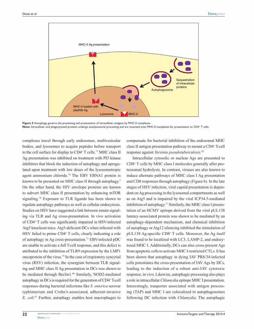
intracellular Ags to CD4+ T cells (Figure 5). Physical intersec-
tion of autophagy pathways with endosomes and lysosomes is
critical in promoting cytosolic and nuclear Ag processing and
presentation by MHC class II molecules. The MHC class II
Autolysosome
Autophagosome
ROS-containing mitochondria
MAVS
Atg5 Atg12RIG-I
STING
ER
STINGGolgi
STING
IFN-βIFN-βIFN-βA B C
TBK1
Atg9
Figure 4 Autophagy negatively regulates type I interferon induction through multiple mechanisms.Notes: (A) The Atg5–Atg12 complex blocks RLR–MAvS interaction. (B) Mitophagy eliminates reactive oxygen species (ROS)-containing mitochondria. (C) Atg9 controls the assembly of stimulator of interferon genes (STING) with TBK1 following its translocation from the endoplasmic reticulum (ER) to Golgi.
ImmunoTargets and Therapy 2015:4submit your manuscript | www.dovepress.com
Dovepress
Dovepress
22
Desai et al
complexes travel through early endosomes, multivesicular
bodies, and lysosomes to acquire peptides before transport
to the cell surface for display to CD4+ T cells.77 MHC class II
Ag presentation was inhibited on treatment with PI3 kinase
inhibitors that block the induction of autophagy and upregu-
lated upon treatment with low doses of the lysosomotropic
agent ammonium chloride.78 The EBV EBNA1 protein is
known to be presented on MHC class II through autophagy.7
On the other hand, the HIV envelope proteins are known
to subvert MHC class II presentation by enhancing mTOR
signaling.78 Exposure to TLR ligands has been shown to
regulate autophagy pathways as well as cellular endocytosis.
Studies on HSV have suggested a link between innate signal-
ing via TLR and Ag cross-presentation. In vivo activation
of CD4+ T cells was significantly impaired in HSV-infected
Atg5 knockout mice. Atg5-deficient DCs when infected with
HSV failed to prime CD4+ T cells, clearly indicating a role
of autophagy in Ag cross-presentation.77 EBV-infected pDC
are unable to activate a full T-cell response, and this defect is
attributed to the inhibition of TLR9 expression by the LMP1
oncoprotein of the virus.79 In the case of respiratory syncytial
virus (RSV) infection, the synergism between TLR signal-
ing and MHC class II Ag presentation in DCs was shown to
be mediated through Beclin1.80 Similarly, NOD2-mediated
autophagy in DCs is required for the generation of CD4+ T-cell
responses during bacterial infections like S. enterica serovar
typhimurium and Crohn’s-associated, adherent-invasive
E. coli.81 Further, autophagy enables host macrophages to
compensate for bacterial inhibition of the endosomal MHC
class II antigen presentation pathway to mount a CD4+ T-cell
response against Yersinia pseudotuberculosis.82
Intracellular cytosolic or nuclear Ags are presented to
CD8+ T cells by MHC class I molecules generally after pro-
teosomal hydrolysis. In contrast, viruses are also known to
induce alternate pathways of MHC class I Ag presentation
and CD8 responses through autophagy (Figure 6). In the late
stages of HSV infection, viral capsid presentation is depen-
dent on Ag processing in the lysosomal compartments as well
as on Atg5 and is impaired by the viral ICP34.5-mediated
inhibition of autophagy.77 Similarly, the MHC class I presen-
tation of an HCMV epitope derived from the viral pUL138
latency-associated protein was shown to be mediated by an
autophagy-dependent mechanism, and chemical inhibition
of autophagy or Atg12 silencing inhibited the stimulation of
pUL138 Ag-specific CD8+ T cells. Moreover, the Ag itself
was found to be localized with LC3, LAMP-2, and endocy-
tosed MHC I. Additionally, DCs can also cross-present Ags
from apoptotic cells to activate MHC I-restricted CTLs. It has
been shown that autophagy in dying IAV PR8/34-infected
cells potentiates the cross-presentation of IAV Ags by DCs,
leading to the induction of a robust anti-IAV cytotoxic
response, in vivo. Likewise, autophagic processing also plays
a role in intracellular Chlamydia epitope MHC I presentation.
Interestingly, tranporter associated with antigen process-
ing (TAP) and MHC I are colocalized to autophagosomes
following DC infection with Chlamydia. The autophagic
MHC-II loaded withpeptide Ag
MHC-II
Sequestrationof intracellularproteins
Autophagosome
MHC-II Ag presentation
Lysosome
Figure 5 Autophagy governs the processing and presentation of intracellular antigens by MHC-II complexes.Note: Intracellular and phagocytosed proteins undergo autolysosomal processing and are mounted onto MHC-II complexes for presentation to CD4+ T cells.

ImmunoTargets and Therapy 2015:4 submit your manuscript | www.dovepress.com
Dovepress
Dovepress
23
Autophagy in infection and immunity
targeting allows preprocessing of the bacterial antigens in the
lysosomes, which is then followed by their cytosolic release
and further processing by the proteasome before loading onto
recycling MHC I complexes.5
Additionally, autophagy is involved in multiple aspects
of lymphocyte development and function and is essential for
both T and B lymphocyte survival and proliferation.83 The
pathway is highly induced in effector T cells and has been
shown to promote the cytokine-dependent survival of primary
T cells. In contrast, studies also suggest that autophagy is an
important death pathway in T cells lacking FADD activity,
caspase-8, or immunity-related GTPase family M protein
(IRGM)-1. Thus, autophagy has been shown to have both
pro-survival and pro-death roles in T cells.84 On the other
hand, Atg5 and an intact autophagy pathway are required
at specific stages in B-cell development and differentially
required for distinct, but closely related, cell lineages.85
Autophagic regulation of inflammationAutophagy not only plays a role in pathogen sensing and
restriction but also has many other functions in the immune
system, including processing of PAMPs for PRR recognition,
inflammasome regulation, and unconventional secretion of
alarmins.70,86 Inflammasomes are protein complexes that
respond to PAMPs and damage-associated molecular patterns
(DAMPs) by inducing proteolytic processing and secretion of
IL-1β and IL-18. Increasing evidence from various studies86–90
supports that autophagy negatively regulates inflammasome
activation. It has been suggested that basal levels of autophagy
control the set point of inflammasome activation by clearing
cytosolic debris, protein aggregates, and defective organelles.4
More specifically, autophagy has been reported to control the
production of IL-1β through at least two separate mechanisms:
by targeting pro-IL-1β for lysosomal degradation, and by
regulating activation of the NLRP3 inflammasome. Following
treatment of macrophages with TLR ligands, pro-IL-1β was
seen to be sequestered in autophagosomes, whereas specific
activation of autophagy with rapamycin induced the deg-
radation of pro-IL-1β and blocked secretion of the mature
cytokine. Conversely, the inhibition of autophagy promoted
the secretion of IL-1β by macrophages in a NLRP3- and
TRIF-dependent fashion.87 Similarly, autophagy inhibition in
dendritic cells also promotes the secretion of both IL-1β and
IL-23, and supernatants from these cells stimulated the innate
secretion of IL-17, IFN-γ, and IL-22 by γδ T cells.88 In vivo,
the deficiency of Atg16L1 represents a source of sterile inflam-
mation that leads to inflammasome activation and increased
IL-1β processing. Further, autophagy inhibits the cytosolic
release of NALP3 inflammasome-mediated mitochondrial
DNA, which is an endogenous source of inflammasome
agonists.89 In contrast, a proinflammatory export pathway
that mediates an unconventional secretion of IL-1β, IL-18
and the DAMP HMGB1 depends on specific Atg factors and
the mammalian Golgi reassembly stacking protein (GRASP)
paralogue, GRASP55.4
Lysosomal processing of antigens
Recycling endosome
MHC-I
Alternate MHC-I Ag presentation
ViralAg
Endogenouspeptides
Autophagosome
Sequestration of viral proteins
Figure 6: Autophagy facilitates processing and loading of sequestered cytosolic proteins to MHC-I complexes.Notes: Recycling endosomes capture MHC-I and fuse with autolysosomes to serve as sites for peptide exchange, so as to allow the loading of lysosomally processed viral proteins to MHC-I complexes and subsequent presentation to CD8+ T cells.

ImmunoTargets and Therapy 2015:4submit your manuscript | www.dovepress.com
Dovepress
Dovepress
24
Desai et al
Additionally, autophagy also mediates degradation of
other proinflammatory factors such as the NF-κB signal-
ing components, NIKs (NF-κB-inducing kinases), and the
IKK (the inhibitor of NF-κB) protein family, and complex
interactions have been reported between both pathways.90
With respect to viral infection, the murine cytomegalovirus
(MCMV) M45 protein binds to the NF-κB subunit NEMO
targeting it for autophagic degradation. In contrast, TNF-
dependent activation of NF-κB represses autophagy through
the activation of mTOR. In macrophages exposed to E. coli,
the suppression of prolonged NF-κB activity promotes
autophagy to advance cell survival, while NF-κB-proficient
macrophages undergo cell death under the same conditions.
Defects in the autophagic response can lead to inflammatory
and autoimmune disorders.4,5,90
Concluding remarksAs the role of autophagy in eukaryotic cells has evolved much
beyond its basic metabolic function, the pathway appears to be
integrated with all stages of antimicrobial host defense. Not
surprisingly, thus, pathogens have devised strategies to evade as
well as exploit the process for their survival and proliferation. It
is becoming increasingly evident that the pathway plays a vital
role in determining the disease course of infection. In light of
the intricate interplay between autophagy and pathogens, the
pathway has often been suggested as a target for new inter-
ventional approaches against infectious diseases. However,
its dichotomous role in limiting as well as in favoring the
propagation of pathogens and its involvement in a range of
biologic processes could complicate its therapeutic application.
The difficulties are further compounded by the fact that stimuli
that activate autophagy also trigger other stress responses. Thus,
extreme dissection of the molecular mechanisms underlying the
pathogen–autophagy interactions is warranted for the selective
harnessing of the host-beneficial potential of the response.
AcknowledgmentsWe apologize to all researchers in the field whose work has
not been cited and in some cases having cited reviews instead
of primary articles due to space limitations. The work was
supported by NIHAI109100.
DisclosureThe authors report no conflicts of interest in this work.
References1. Rubinsztein DC, Codogno P, Levine B. Autophagy modulation as a
potential therapeutic target for diverse diseases. Nat Rev Drug Discov. 2012;11:709–730.
2. Dreux M, Chisari FV. Viruses and the autophagy machinery. Cell Cycle. 2010;9(7):1295–1307.
3. Kraft C, Kijanska M, Kalie E, et al. Binding of the Atg1/ULK1 kinase to the ubiquitin-like protein Atg8 regulates autophagy. EMBO J. 2012;31:3691–3703.
4. Deretic V, Saitoh T, Akira S. Autophagy in infection, inflammation and immunity. Nat Rev Immunol. 2013;13:722–737.
5. Perot BP, Ingersoll MA, Albert ML. The impact of macroautophagy on CD8(+) T-cell-mediated antiviral immunity. Immunol Rev. 2013;255: 40–56.
6. Tang D, Kang R, Coyne CB, Zeh HJ, Lotze MT. PAMPs and DAMPs: signal 0s that spur autophagy and immunity. Immunol Rev. 2012;249: 158–175.
7. Cavignac Y, Esclatine A. Herpesviruses and autophagy: catch me if you can! Viruses. 2010;2:314–333.
8. Kampa-Schittenhelm KM, Heinrich MC, Akmut F, et al. Cell cycle-dependent activity of the novel dual PI3K-MTORC1/2 inhibitor NVP-BGT226 in acute leukemia. Mol Cancer. 2013;12:46.
9. Efeyan A, Zoncu R, Chang S, et al. Regulation of mTORC1 by the Rag GTPases is necessary for neonatal autophagy and survival. Nature. 2013;493:679–683.
10. Alers S, Loffler AS, Wesselborg S, Stork B. Role of AMPK-mTOR-Ulk1/2 in the regulation of autophagy: cross talk, shortcuts, and feedbacks. Mol Cell Biol. 2012;32:2–11.
11. Hasty P, Sharp ZD, Curiel TJ, Campisi J. mTORC1 and p53: clash of the gods? Cell Cycle. 2012;12:20–25.
12. Cabrera R, Tu Z, Xu Y, et al. An immunomodulatory role for CD4(+)CD25(+) regulatory T lymphocytes in hepatitis C virus infection. Hepatology. 2004;40:1062–1071.
13. Jheng JR, Ho JY, Horng JT. ER stress, autophagy, and RNA viruses. Front Microbiol. 2014;5:388.
14. Pengo N, Scolari M, Oliva L, et al. Plasma cells require autophagy for sus-tainable immunoglobulin production. Nat Immunol. 2013;14:298–305.
15. Deretic V. Autophagy in immunity and cell-autonomous defense against intracellular microbes. Immunol Rev. 2011;240:92–104.
16. Levine B, Mizushima N, Virgin HW. Autophagy in immunity and inflammation. Nature. 2011;469:323–335.
17. Deretic V. Autophagy in innate and adaptive immunity. Trends Immunol. 2005;26:523–528.
18. Kirkegaard K, Jackson WT. Topology of double-membraned vesicles and the opportunity for non-lytic release of cytoplasm. Autophagy. 2005;1:182–184.
19. Dong X, Levine B. Autophagy and viruses: adversaries or allies? J Innate Immun. 2013;5:480–493.
20. Jordan TX, Randall G. Manipulation or capitulation: virus interactions with autophagy. Microbes Infect. 2012;14:126–139.
21. Dreux M, Chisari FV. Impact of the autophagy machinery on hepatitis C virus infection. Viruses. 2011;3:1342–1357.
22. Ke PY, Chen SS. Autophagy in hepatitis C virus-host interactions: potential roles and therapeutic targets for liver-associated diseases. World J Gastroenterol. 2014;20:5773–5793.
23. Kim SJ, Syed GH, Khan M, et al. Hepatitis C virus triggers mitochondrial fission and attenuates apoptosis to promote viral persistence. Proc Natl Acad Sci U S A. 2014;111:6413–6418.
24. Kirkegaard K. Subversion of the cellular autophagy pathway by viruses. Curr Top Microbiol Immunol. 2009;335:323–333.
25. Taylor MP, Kirkegaard K. Potential subversion of autophagosomal pathway by picornaviruses. Autophagy. 2008;4:286–289.
26. Shi J, Luo H. Interplay between the cellular autophagy machinery and positive-stranded RNA viruses. Acta Biochim Biophys Sin (Shanghai). 2012;44:375–384.
27. Richards AL, Jackson WT. Intracellular vesicle acidif ication promotes maturation of infectious poliovirus particles. PLoS Pathog. 2012;8:e1003046.
28. Richards AL, Jackson WT. That which does not degrade you makes you stronger: infectivity of poliovirus depends on vesicle acidification. Autophagy. 2013;9:806–807.

ImmunoTargets and Therapy 2015:4 submit your manuscript | www.dovepress.com
Dovepress
Dovepress
25
Autophagy in infection and immunity
29. Kemball CC, Alirezaei M, Flynn CT, et al. Coxsackievirus infection induces autophagy-like vesicles and megaphagosomes in pancreatic acinar cells in vivo. J Virol. 2010;84:12110–12124.
30. Robinson SM, Tsueng G, Sin J, et al. Coxsackievirus B exits the host cell in shed microvesicles displaying autophagosomal markers. PLoS Pathog. 2014;10:e1004045.
31. Wurdinger T, Gatson NN, Balaj L, Kaur B, Breakefield XO, Pegtel DM. Extracellular vesicles and their convergence with viral pathways. Adv Virol. 2012;2012:767694.
32. Beale R, Wise H, Stuart A, Ravenhill BJ, Digard P, Randow FA. LC3-interacting motif in the influenza A virus M2 protein is required to subvert autophagy and maintain virion stability. Cell Host Microbe. 2014;15:239–247.
33. Zhirnov OP, Klenk HD. Influenza A virus proteins NS1 and hemagglutinin along with M2 are involved in stimulation of autophagy in infected cells. J Virol. 2013;87:13107–13114.
34. Gannagé M, Dormann D, Albrecht R, et al. Matrix protein 2 of influenza A virus blocks autophagosome fusion with lysosomes. Cell Host Microbe. 2009;6:367–380.
35. Munz C. Beclin-1 targeting for viral immune escape. Viruses. 2011;3: 1166–1178.
36. Li JK, Liang JJ, Liao CL, Lin YL. Autophagy is involved in the early step of Japanese encephalitis virus infection. Microbes Infect. 2012;14: 159–168.
37. Sharma M, Bhattacharyya S, Nain M, et al. Japanese encephalitis virus replication is negatively regulated by autophagy and occurs on LC3-I- and EDEM1-containing membranes. Autophagy. 2014;10(9): 1637–1651.
38. Tang SW, Ducroux A, Jeang KT, Neuveut C. Impact of cellular autophagy on viruses: Insights from hepatitis B virus and human retroviruses. J Biomed Sci. 2012;19:92.
39. Campbell GR, Spector SA. Inhibition of human immunodeficiency virus type-1 through autophagy. Curr Opin Microbiol. 2013;16:349–354.
40. Zhang HT, Chen GG, Hu BG, et al. Hepatitis B virus x protein induces autophagy via activating death-associated protein kinase. J Viral Hepat. 2014;21:642–649.
41. Liu B, Fang M, Hu Y, et al. Hepatitis B virus X protein inhibits autophagic degradation by impairing lysosomal maturation. Autophagy. 2014;10:416–430.
42. Lazar C, Macovei A, Petrescu S, Branza-Nichita N. Activation of ERAD pathway by human hepatitis B virus modulates viral and subviral particle production. PLoS One. 2012;7:e34169.
43. Li J, Liu Y, Wang Z, et al. Subversion of cellular autophagy machinery by hepatitis B virus for viral envelopment. J Virol. 2011;85:6319–6333.
44. Lee DY, Lee J, Sugden B. The unfolded protein response and autophagy: herpesviruses rule! J Virol. 2009;83:1168–1172.
45. Hung CH, Chen LW, Wang WH, et al. Regulation of autophagic activation by Rta of Epstein-Barr virus via ERK-kinase pathway. J Virol. 2014;88(20):12133–12145.
46. Pratt ZL, Zhang J, Sugden B. The latent membrane protein 1 (LMP1) oncogene of Epstein-Barr virus can simultaneously induce and inhibit apoptosis in B cells. J Virol. 2012;86:4380–4393.
47. Granato M, Santarelli R, Farina A, et al. Epstein-barr virus blocks the autophagic flux and appropriates the autophagic machinery to enhance viral replication. J Virol. 2014;88:12715–12726.
48. Silva LM, Jung JU. Modulation of the autophagy pathway by human tumor viruses. Semin Cancer Biol. 2013;23:323–328.
49. Cheng CY, Chi PI, Liu HJ. Commentary on the regulation of viral proteins in autophagy process. Biomed Res Int. 2014;2014:962915.
50. Chaumorcel M, Lussignol M, Mouna L, et al. The human cytomegalovirus protein TRS1 inhibits autophagy via its interaction with Beclin 1. J Virol. 2012;86:2571–2584.
51. Rikihisa Y. Glycogen autophagosomes in polymorphonuclear leuko-cytes induced by rickettsiae. Anat Rec. 1984;208:319–327.
52. Birmingham CL, Canadien V, Gouin E, et al. Listeria monocytogenes evades killing by autophagy during colonization of host cells. Autophagy. 2007;3:442–451.
53. Portnoy DA, Jacks PS, Hinrichs DJ. Role of hemolysin for the intracellular growth of Listeria monocytogenes. J Exp Med. 1988;167:1459–1471.
54. Kloft N, Neukirch C, Bobkiewicz W, et al. Pro-autophagic signal induction by bacterial pore-forming toxins. Med Microbiol Immunol. 2010;199:299–309.
55. Levine B. Eating oneself and uninvited guests: autophagy-related pathways in cellular defense. Cell. 2005;120:159–162.
56. Deretic V, Levine B. Autophagy, immunity, and microbial adaptations. Cell Host Microbe. 2009;5:527–549.
57. Walker DH, Popov VL, Crocquet-Valdes PA, Welsh CJ, Feng HM. Cytokine-induced, nitric oxide-dependent, intracellular antirickettsial activity of mouse endothelial cells. Lab Invest. 1997;76:129–138.
58. Nakagawa I, Amano A, Mizushima N, et al. Autophagy defends cells against invading group A Streptococcus. Science. 2004;306: 1037–1040.
59. Gutierrez MG, Master SS, Singh SB, Taylor GA, Colombo MI, Deretic V. Autophagy is a defense mechanism inhibiting BCG and Mycobacterium tuberculosis survival in infected macrophages. Cell. 2004;119:753–766.
60. Castillo EF, Dekonenko A, Arko-Mensah J, et al. Autophagy protects against active tuberculosis by suppressing bacterial burden and inflammation. Proc Natl Acad Sci U S A. 2012;109:E3168–E3176.
61. Ogawa M, Yoshikawa Y, Mimuro H, Hain T, Chakraborty T, Sasakawa C. Autophagy targeting of Listeria monocytogenes and the bacterial countermeasure. Autophagy. 2011;7:310–314.
62. Yoshikawa Y, Ogawa M, Hain T, et al. Listeria monocytogenes ActA-mediated escape from autophagic recognition. Nat Cell Biol. 2009;11: 1233–1240.
63. Birmingham CL, Canadien V, Kaniuk NA, Steinberg BE, Higgins DE, Brumell JH. Listeriolysin O allows Listeria monocytogenes replication in macrophage vacuoles. Nature. 2008;451:350–354.
64. Dortet L, Mostowy S, Samba-Louaka A, et al. Recruitment of the major vault protein by InlK: a Listeria monocytogenes strategy to avoid autophagy. PLoS Pathog. 2011;7:e1002168.
65. Gutierrez MG, Vázquez CL, Munafó DB, et al. Autophagy induction favours the generation and maturation of the Coxiella-replicative vacuoles. Cell Microbiol. 2005;7:981–993.
66. Vazquez CL, Colombo MI. Coxiella burnetii modulates Beclin 1 and Bcl-2, preventing host cell apoptosis to generate a persistent bacterial infection. Cell Death Differ. 2009;17:421–438.
67. Niu H, Xiong Q, Yamamoto A, Hayashi-Nishino M, Rikihisa Y. Autophagosomes induced by a bacterial Beclin 1 binding protein facilitate obligatory intracellular infection. Proc Natl Acad Sci U S A. 2012;109:20800–20807.
68. Niu H, Yamaguchi M, Rikihisa Y. Subversion of cellular autophagy by Anaplasma phagocytophilum. Cell Microbiol. 2008;10:593–605.
69. Delgado M, Singh S, De Haro S, et al. Autophagy and pattern recogni-tion receptors in innate immunity. Immunol Rev. 2009;227:189–202.
70. Deretic V. Autophagy as an innate immunity paradigm: expanding the scope and repertoire of pattern recognition receptors. Curr Opin Immunol. 2012;24:21–31.
71. Travassos LH, Carneiro LA, Ramjeet M, et al. Nod1 and Nod2 direct autophagy by recruiting ATG16L1 to the plasma membrane at the site of bacterial entry. Nat Immunol. 2010;11:55–62.
72. Yano T, Mita S, Ohmori H, et al. Autophagic control of listeria through intracellular innate immune recognition in drosophila. Nat Immunol. 2008;9:908–916.
73. Desai MM, Gong B, Chan T, et al. Differential, type I interferon-mediated autophagic trafficking of Hepatitis C virus proteins in mouse liver. Gastroenterology. 2011;141:674–685, 685. e671–e676.
74. Lunemann JD, Munz C. Autophagy in CD4+ T-cell immunity and tolerance. Cell Death Differ. 2009;16:79–86.
75. Hwang S, Maloney NS, Bruinsma MW, et al. Nondegradative role of Atg5-Atg12/Atg16L1 autophagy protein complex in antiviral activity of interferon gamma. Cell Host Microbe. 2012;11:397–409.
76. Yordy BIA. Autophagy in Antiviral Immunity. 1st ed. Austin, TX: Landes Bioscience; 2013.

ImmunoTargets and Therapy
Publish your work in this journal
Submit your manuscript here: http://www.dovepress.com/immunotargets-and-therapy-journal
ImmunoTargets and Therapy is an international, peer-reviewed open access journal focusing on the immunological basis of diseases, potential targets for immune based therapy and treatment protocols employed to improve patient management. Basic immunology and physiology of the immune system in health, and disease will be also covered. In addition, the journal will focus on the impact of manage-
ment programs and new therapeutic agents and protocols on patient perspectives such as quality of life, adherence and satisfaction. The manuscript management system is completely online and includes a very quick and fair peer-review system, which is all easy to use. Visit http://www.dovepress.com/testimonials.php to read real quotes from published authors.
ImmunoTargets and Therapy 2015:4submit your manuscript | www.dovepress.com
Dovepress
Dovepress
Dovepress
26
Desai et al
77. Crotzer VL, Blum JS. Autophagy and adaptive immunity. Immunology. 2010;131:9–17.
78. Oh JE, Lee HK. Autophagy in innate recognition of pathogens and adaptive immunity. Yonsei Med J. 2012;53:241–247.
79. Zhang L, Sung JJ, Yu J, et al. Xenophagy in Helicobacter pylori- and Epstein-Barr virus-induced gastric cancer. J Pathol. 2014;233: 103–112.
80. Reed M, Morris SH, Jang S, Mukherjee S, Yue Z, Lukacs NW. Autophagy-inducing protein beclin-1 in dendritic cells regulates CD4 T cell responses and disease severity during respiratory syncytial virus infection. J Immunol. 2013;191:2526–2537.
81. Cooney R, Baker J, Brain O, et al. NOD2 stimulation induces autophagy in dendritic cells influencing bacterial handling and antigen presentation. Nat Med. 2010;16:90–97.
82. Rüssmann H, Panthel K, Köhn B, et al. Alternative endogenous protein processing via an autophagy-dependent pathway compensates for Yersinia-mediated inhibition of endosomal major histocompatibility complex class II antigen presentation. Infect Immun. 2010;78: 5138–5150.
83. Pua HH, He YW. Autophagy and lymphocyte homeostasis. Curr Top Microbiol Immunol. 2009;335:85–105.
84. Kovacs JR, Li C, Yang Q, et al. Autophagy promotes T-cell survival through degradation of proteins of the cell death machinery. Cell Death Differ. 2011;19:144–152.
85. Miller BC, Zhao Z, Stephenson LM, et al. The autophagy gene ATG5 plays an essential role in B lymphocyte development. Autophagy. 2008;4:309–314.
86. Kuballa P, Nolte WM, Castoreno AB, Xavier RJ. Autophagy and the immune system. Annu Rev Immunol. 2012;30:611–646.
87. Harris J, Hartman M, Roche C, et al. Autophagy controls IL-1beta secretion by targeting pro-IL-1beta for degradation. J Biol Chem. 2011;286:9587–9597.
88. Peral de Castro C, Jones SA, Ní Cheallaigh C, et al. Autophagy regulates IL-23 secretion and innate T cell responses through effects on IL-1 secretion. J Immunol. 2012;189:4144–4153.
89. Nakahira K, Haspel JA, Rathinam VA, et al. Autophagy proteins regulate innate immune responses by inhibiting the release of mitochondrial DNA mediated by the NALP3 inflammasome. Nat Immunol. 2011;12:222–230.
90. Trocoli A, Djavaheri-Mergny M. The complex interplay between autophagy and NF-kappaB signaling pathways in cancer cells. Am J Cancer Res. 2011;1:629–649.
Related Documents