THE ROLE OF ASTROCYTES AND COMPLEMENT SYSTEM IN NEURAL PLASTICITY Milos Pekny,* Ulrika Wilhelmsson,* Yalda Rahpeymai Bogesta ˚l, y and Marcela Pekna y *Center for Brain Repair and Rehabilitation, Department of Clinical Neuroscience and Rehabilitation, Institute for Neuroscience and Physiology at Sahlgrenska Academy Go ¨teborg University, 405 30 Go ¨teborg, Sweden y Department of Medical Chemistry and Cell Biology, Institute of Biomedicine at Sahlgrenska Academy, Go ¨teborg University, 405 30 Go ¨teborg, Sweden I. Introduction II. Astrocytes, GFAP, and Astrocyte Intermediate Filaments III. Reactive Gliosis, Neurotrauma, and CNS Transplants IV. The Complement System References In neurotrauma, brain ischemia or neurodegenerative diseases, astrocytes become reactive (which is known as reactive gliosis) and this is accompanied by an altered expression of many genes. Two cellular hallmarks of reactive gliosis are hypertrophy of astrocyte processes and the upregulation of the part of the cytoskeleton known as intermediate filaments, which are composed of nestin, vimentin, and GFAP. Our aim has been to better understand the function of reactive astrocytes in CNS diseases. Using mice deficient for astrocyte intermediate fila- ments (GFAP –/– Vim –/– ), we were able to attenuate reactive gliosis and slow down the healing process after neurotrauma. We demonstrated the key role of reactive astrocytes in neurotrauma—at an early stage after neurotrauma, reactive astrocytes have a neuroprotective eVect; at a later stage, they facilitate the formation of posttraumatic glial scars and inhibit CNS regeneration, specifically, they seem to compromise neural graft survival and integration, reduce the extent of synaptic regeneration, inhibit neurogenesis in the old age, and inhibit regeneration of severed CNS axons. We propose that reactive astrocytes are the future target for the therapeutic strategies promoting regeneration and plasticity in the brain and spinal cord in various disease conditions. Through its involvement in inflammation, opsonization, and cytolysis, com- plement protects against infectious agents. Although most of the complement proteins are synthesized in CNS, the role of the complement system in the normal INTERNATIONAL REVIEW OF 95 NEUROBIOLOGY, VOL. 82 Copyright 2007, Elsevier Inc. All rights reserved. DOI: 10.1016/S0074-7742(07)82005-8 0074-7742/07 $35.00

Welcome message from author
This document is posted to help you gain knowledge. Please leave a comment to let me know what you think about it! Share it to your friends and learn new things together.
Transcript

THE ROLE OF ASTROCYTES AND COMPLEMENT SYSTEMIN NEURAL PLASTICITY
Milos Pekny,* Ulrika Wilhelmsson,* Yalda Rahpeymai Bogestal,y andMarcela Peknay
*Center for Brain Repair and Rehabilitation, Department of Clinical Neuroscienceand Rehabilitation, Institute for Neuroscience and Physiology at Sahlgrenska Academy
Goteborg University, 405 30 Goteborg, SwedenyDepartment of Medical Chemistry and Cell Biology, Institute of Biomedicine at Sahlgrenska
Academy, Goteborg University, 405 30 Goteborg, Sweden
I. I
INTE
NEU
DOI:
ntroduction
RNATIONAL REVIEW OF 95ROBIOLOGY, VOL. 82
Copyright 2007, Elsevier In
All rights reserve
10.1016/S0074-7742(07)82005-8 0074-7742/07 $35.0
II. A
strocytes, GFAP, and Astrocyte Intermediate FilamentsIII. R
eactive Gliosis, Neurotrauma, and CNS TransplantsIV. T
he Complement SystemR
eferencesIn neurotrauma, brain ischemia or neurodegenerative diseases, astrocytes
become reactive (which is known as reactive gliosis) and this is accompanied
by an altered expression of many genes. Two cellular hallmarks of reactive gliosis
are hypertrophy of astrocyte processes and the upregulation of the part of the
cytoskeleton known as intermediate filaments, which are composed of nestin,
vimentin, andGFAP.Our aim has been to better understand the function of reactive
astrocytes in CNS diseases. Using mice deficient for astrocyte intermediate fila-
ments (GFAP –/–Vim–/– ), we were able to attenuate reactive gliosis and slow down
the healing process after neurotrauma. We demonstrated the key role of reactive
astrocytes in neurotrauma—at an early stage after neurotrauma, reactive astrocytes
have a neuroprotective eVect; at a later stage, they facilitate the formation of
posttraumatic glial scars and inhibit CNS regeneration, specifically, they seem to
compromise neural graft survival and integration, reduce the extent of synaptic
regeneration, inhibit neurogenesis in the old age, and inhibit regeneration of severed
CNS axons. We propose that reactive astrocytes are the future target for the
therapeutic strategies promoting regeneration and plasticity in the brain and spinal
cord in various disease conditions.
Through its involvement in inflammation, opsonization, and cytolysis, com-
plement protects against infectious agents. Although most of the complement
proteins are synthesized in CNS, the role of the complement system in the normal
c.
d.
0
96 PEKNY et al.
or ischemic CNS remains unclear. Complement activiation in the CNS has been
generally considered as contributing to tissue damage. However, growing body of
evidence suggests that complement may be a physiological neuroprotective mech-
anism as well as it may participate in maintenance and repair of the adult brain.
I. Introduction
In a striking contrast to the peripheral nervous system, the regenerative capacity
of the adult brain and spinal cord (e.g., synaptic and axonal regeneration, neuro-
genesis) is extremely limited, despite the fact that neural stem cells are present in
CNS throughout the whole life. Both the endogenous neural stem cells and neural
implants grafted to replace lost neurons fail to form functional connections to the
extent that would influence the clinical outcome in conditions such as neurotrauma,
stroke, or neurodegenerative diseases. Moreover shortly after birth, axons in the
adult mammalian CNS lose their ability to grow and regenerate following injury.
We suggest that the environment, in particular astrocytes, and the immune system
are important modulators of CNS regeneration.
II. Astrocytes, GFAP, and Astrocyte Intermediate Filaments
Astrocytes are the most numerous cells in the CNS, and they were implicated
to be involved in many CNS pathologies such as trauma, ischemia, or neuro-
degenerative diseases. In response to any kind of injury in the CNS, astrocytes
change their appearance and undergo a characteristic hypertrophy of their
cellular processes. This phenomenon is known as reactive gliosis or astrogliosis
its hallmark being upregulation of intermediate filament (IF) proteins GFAP and
vimentin, reexpression of nestin as well as altered expression profiles of many
proteins (Eddleston and Mucke, 1993; Hernandez et al., 2002). The IFs can be
considered the least understood part of the cytoskeleton. The family of IF proteins
expressed in vertebrates is large (in humans 65 diVerent IF proteins have been
identified) (Herrmann and Aebi, 2004; Herrmann et al., 2003), and there is a
complex expression pattern of IF proteins unique for each cell type as well as
during diVerent developmental stages.
The dynamic feature of the IF network depends both on the equilibrium
between filaments and unassembled subunits and the regulation of filament
assembly/disassembly by phosphorylation of the head domain of the IF proteins.
IFs were at first considered to be static structures primarily responsible for

THE ROLE OF ASTROCYTES AND COMPLEMENT SYSTEM 97
maintaining the cell shape (Renner et al., 1981; Rueger et al., 1979). However,
later studies both in vitro (Angelides et al., 1989; Nakamura et al., 1991) and in vivo
(Miller et al., 1991; Vikstrom et al., 1992; Wiegers et al., 1991; Yoon et al., 1998)
revealed the rather dynamic nature of IFs and the existence of a dynamic
equilibrium between the assembled filaments and the pool of soluble subunits
(reviewed in Goldman et al., 1999).
In vivo, IFs are often, if not always, heteropolymeric (Herrmann and Aebi,
2000). For determining the partnership in the formation of IF heteropolymers in
astrocytes, transgenic mice deficient in individual IF proteins were instrumental.
In nonreactive astrocytes, IFs are formed of GFAP and vimentin, while in reactive
astrocytes, nestin can be found as the additional partner in the IF network (Pekny
et al., 1998; Table I). In addition, some reactive astrocytes, for example in
neurotrauma, express another IF protein, synemin ( Jing et al., 2007). The studies
of astrocytes lacking GFAP and/or vimentin revealed that GFAP can form IFs on
its own in vimentin deficient (Vim–/– ) astrocytes, but such filaments form more
compact bundles than in wild-type astrocytes, suggesting that at least a low level of
vimentin is needed for normal IF formation in the astrocytes (Eliasson et al., 1999;
Lepekhin et al., 2001; Menet et al., 2001). Studies in mice deficient in GFAP
(GFAP–/– ) showed that vimentin does not form IF on its own, or it does so only
with a very low eYciency (McCall et al., 1996; Pekny et al., 1995). In contrast, the
reactiveGFAP –/– astrocytes contain IFs since vimentin can polymerize with nestin,
which is expressed in reactive astrocytes (Eliasson et al., 1999). GFAP does not
polymerize with nestin in reactive Vim–/– astrocytes and consequently, the IFs
contain only GFAP and exhibit the characteristic tight bundling similar to Vim–/–
nonreactive astrocytes. In reactive astrocytes lacking both GFAP and vimentin
(GFAP–/– Vim–/– ) no IFs are formed, and both the nestin and synemin proteins
which are produced, stay in a nonfilamentous form (Eliasson et al., 1999; Jing et al.,
2007). Nesti n was proposed to facili tate phosp horylation -depend ent disas semb ly
TABLE I
COMPOSITION OF IFS IN NONREACTIVE AND REACTIVE ASTROCYTES OF WILD-TYPE MICE AND MICE
DEFICIENT IN GFAP AND/OR VIMENTINa
Genotype
Composition of IFsReactive astrocytes:
IF amount/bundlingNonreactive astrocytes Reactive astrocytes
Wild type GFAP, vimentin GFAP, vimentin, nestin Normal/normal
GFAP–/– No IFs (nonfilamentous
vimentin)
Vimentin, nestin Decreased/normal
Vim–/– GFAP GFAP (nonfilamentous nestin) Decreased/tight
GFAP –/–Vim–/– No IFs No IFs (nonfilamentous nestin) –
aSome reactive astrocytes contain another IF protein, synemin ( Jing et al., 2007).
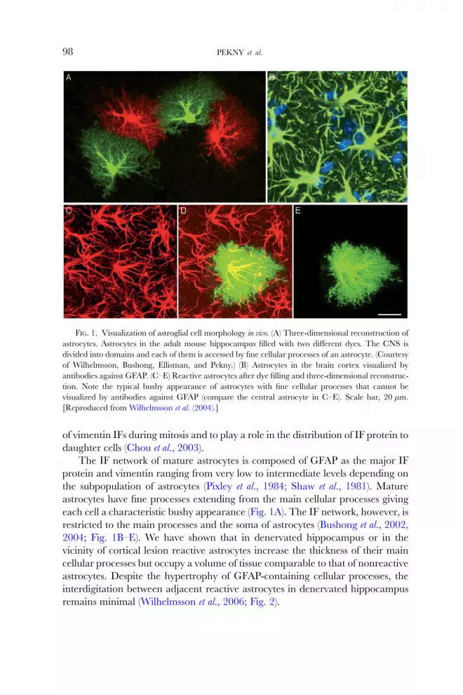
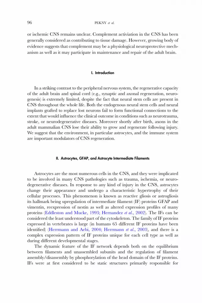
FIG. 1. Visualization of astroglial cell morphology in vivo. (A) Three-dimensional reconstruction of
astrocytes. Astrocytes in the adult mouse hippocampus filled with two diVerent dyes. The CNS is
divided into domains and each of them is accessed by fine cellular processes of an astrocyte. (Courtesy
of Wilhelmsson, Bushong, Ellisman, and Pekny.) (B) Astrocytes in the brain cortex visualized by
antibodies against GFAP. (C–E) Reactive astrocytes after dye filling and three-dimensional reconstruc-
tion. Note the typical bushy appearance of astrocytes with fine cellular processes that cannot be
visualized by antibodies against GFAP (compare the central astrocyte in C–E). Scale bar, 20 �m.
[Reproduced from Wilhelmsson et al. (2004).]
98 PEKNY et al.
of vimen tin IFs during mitosis and to play a ro le in the distrib ution of IF protein to
daug hter cells (Chou et al ., 2003 ).
The IF network of mature astrocytes is composed of GFAP as the major IF
protein and vimentin ranging from very low to intermediate levels depending on
the subpopulation of astrocytes (Pixley et al., 1984; Shaw et al., 1981). Mature
astrocytes have fine processes extending from the main cellular processes giving
each cell a characteristic bushy appearance (Fig. 1A). The IF network, however, is
restricted to the main processes and the soma of astrocytes (Bushong et al., 2002,
2004; Fig. 1B–E). We have shown that in denervated hippocampus or in the
vicinity of cortical lesion reactive astrocytes increase the thickness of their main
cellular processes but occupy a volume of tissue comparable to that of nonreactive
astrocytes. Despite the hypertrophy of GFAP-containing cellular processes, the
interdigitation between adjacent reactive astrocytes in denervated hippocampus
remains minimal (Wilhelmsson et al., 2006; Fig. 2).
FIG. 2. The domains of nonreactive and reactive astrocytes—a concept. (A) Interdigitation of fine
cellular processes in a 3D reconstruction of dye filled astrocytes in the dentate gyrus of the hippocam-
pus. The yellow zone shows the border area where cellular processes of two adjacent astrocytes
interdigitate. (B) Reactive astrocytes stay within their domains, but their main cellular processes get
thicker, making them visible over a greater distance (illustrated here by the gray circles). [Reproduced
from Wilhelmsson et al. (2006).]
THE ROLE OF ASTROCYTES AND COMPLEMENT SYSTEM 99
It was proposed that GFAP-positive astroglial cells are involved in the baseline
neurogenesis in the adult mammalian CNS. Recent data suggest that astrocytes
positively control neurogenesis in the two regions of the adult CNS, specifically in the
dentate gyrus of the hippocampus and in the subventricular zone, that is, the only
two CNS regions in which new neurons are generated in relatively high numbers
even in the adult (Song et al., 2002). It was suggested that the majority of neural stem
cells in the adult CNS are at some point GFAP positive, that is, could be defined as
astroglial cells (Doetsch et al., 1999; Imura et al., 2003; Laywell et al., 2000;Morshead
et al., 2003). Thus, astroglial cells might be both the cell type that controls adult
neurogenesis as well as the precursors to neurons that are added in the adult life.

100 PEKNY et al.
III. Reactive Gliosis, Neurotrauma, and CNS Transplants
To assess the role of IF upregulation in reactive astrocytes in CNS injury, several
trauma models were applied to mice deficient in GFAP and/or vimentin. Fine
needle injury of the brain cortex and transection of the dorsal funiculus in
the upper thoracic spinal cord were two of the models used. The responses of
wild-type, GFAP–/– and Vim–/– mice were indistinguishable. In GFAP–/–Vim–/–
mice, however, the posttraumatic glial scarring was looser and less organized,
suggesting that upregulation of IFs is an important step in astrocyte activation.
These data also imply that reactive astrocytes play a role in posttraumatic healing
(Pekny et al., 1999; Fig. 3).
Extended healing period following CNS injury was also reported in mice in
which dividing astrocytes had been ablated by GFAP-driven expression of Herpes
simplex virus thymidine kinase and administration of ganciclovir (Bush et al.,
1999; Faulkner et al., 2004). Another group used hemisections of the lower
thoracic spinal cord and reported increased axonal sprouting and better function-
al recovery in GFAP–/–Vim–/– mice than wild-type controls (Menet et al., 2003).
Two groups addressed the role of astrocyte IFs in neurite outgrowth in vitro (Menet
et al., 2000, 2001; Xu et al., 1999). One group reported that GFAP–/–Vim–/– and
GFAP–/– astrocytes were a better substrate for the outgrowth of neurites in vitro
than wild-type astrocytes (Menet et al., 2000, 2001). The other group found
comparable neurite outgrowth when neurons were cultured on wild-type and
GFAP–/– astrocytes (Xu et al., 1999). The latter finding is in agreement with the
normal axonal sprouting and regeneration assessed after dorsal hemisection of the
spinal cord in GFAP–/– mice (Wang et al., 1997). Extensive axonal regeneration
was reported in the severed optic nerve of young GFAP –/–Vim–/– mice that also
carried a transgene overexpressing Bcl-2 in neurons (Cho et al., 2005).
Another model that was used to study the involvement of astrocytes in
neurotrauma was entorhinal cortex lesion. This lesion interrupts axonal connec-
tions (known as the perforant path) between the entorhinal cortex and the
projection area in the outer molecular layer of the dentate gyrus of the hippo-
campus (Turner et al., 1998) where degenerating neurons trigger extensive reac-
tive gliosis. The distance between these two regions allows assessment of astrocyte
response, degeneration, and subsequent regeneration in the hippocampus, that is,
the region that is not directly aVected by the surgery. By utilizing this model,
we showed that reactive astrocytes devoid of IFs (GFAP–/–Vim–/–) exhibited only
limited hypertrophy of cell processes. Many processes of GFAP–/–Vim–/– astro-
cytes were shorter and less straight than those of wild-type astrocytes, albeit the
volume of the CNS tissue reached by a single astrocyte was comparable to wild-
type mice (Wilhelmsson et al., 2004). These results, along with in vitro data on the
morphology of IF-depleted astrocytes in primary cultures (Lepekhin et al., 2001),

Wild type
Uninjured
2
weeks
2
weeks
GFAP−/− Vimentin−/−
A B
C D
E F
FIG. 3. Wound healing in the absence of astrocyte IF proteins after transection of the dorsal
funiculus in the upper thoracic spinal cord takes longer in GFAP–/–Vim–/– than wild-type mice. H & E
staining. Scale bar, 300 �m in A–D and 100 �m in E and F. [Reproduced from Pekny et al. (1999).]
THE ROLE OF ASTROCYTES AND COMPLEMENT SYSTEM 101
show a novel role for IFs in determining astrocyte morphology. In GFAP–/–Vim–/–
mice, loss of neuronal synapses in the outer molecular layer of the hippocampal
dentate gyrus was prominent 4 days after lesioning (Fig. 4A,B, and E). Most
interestingly, there was remarkable synaptic regeneration 10 days later (at 14 days
after lesions) (Fig. 4C–E). In contrast to wild type, GFAP –/–Vim–/– reactive

wt
A
C
1000
750
500
250
0
Injured side
wtDay 4
*
*
Day 14 Day 4 Day 14GV wt GV wt GV wt GV
Uninjured side
D
E
B
4 D
ays
afte
r le
sion
14 D
ays
afte
r le
sion
Syn
aptic
com
plex
es/1
000 mm
2
GV
FIG. 4. Synaptic regeneration after entorhinal cortex lesion in the projection area of entorhinal cortex
in the dentate gyrus of the hippocampus in GFAP –/–Vim–/– (GV) and wild-type (wt) mice. At day 4
after lesioning, the synaptic loss and the signs of neurodegeneration were more prominent in GFAP –/–
Vim–/– than wild-type mice (A, B, and E). At day 14 after lesioning, the number of synapses in
GFAP –/–Vim–/–, but not wild-type mice, recovered reaching the levels comparable with the uninjured
hemisphere (C–E). Asterisks, degenerated axons; arrows, synaptic complexes; D, dendritic profile;
B, synaptic bouton; *, p < 0.05. [Reproduced from Wilhelmsson et al. (2004).]
102 PEKNY et al.

THE ROLE OF ASTROCYTES AND COMPLEMENT SYSTEM 103
astrocytes did not upregulate the expression of endothelin B receptors, suggesting
that the upregulation of this novel marker of reactive astrocytes (Baba, 1998;
Ishikawa et al., 1997; Koyama et al., 1999; Peters et al., 2003) is IF dependent
(Wilhelmsson et al., 2004). Thus, the eVect of reactive astrocytes after CNS trauma
seems to be twofold: reactive astrocytes play a beneficial role in the acute stage
after CNS injury, however later on act as inhibitors of CNS regeneration. Support
for the concept of reactive gliosis as an inhibiting factor with respect to posttrau-
matic repair and functional recovery was provided also by studies using transgenic
mice expressing an NF-�B inhibitor in astrocytes (Brambilla et al., 2005) and in
mice deficient for EphA4 (Goldshmit et al., 2004).
Because of their morphology and abundance in the adult CNS, astrocytes have
direct physical contact with any cell that moves from one place to another. To assess
the impact of astrocyte IFs on the fate of cells migrating from neural transplants, the
Chen and Pekny groups transplanted dissociated retinal cells from 0- to 3-week-old
donor mice that ubiquitously express enhanced green fluorescent protein (Okabe
et al., 1997) into the retinas of adult wild-type and GFAP –/–Vim–/– recipients and
compared the eYciency of long-term integration of such grafts in the retina
(Kinouchi et al., 2003). In wild-type hosts, few transplanted cells migrated from the
transplantation site and few integrated into the retina. In GFAP–/–Vim–/– hosts,
however, the transplanted cells eVectively moved through the retina, diVerentiatedinto neurons, integrated into the ganglion cell layer, and some of them even
extended neurites about 1 mm into the optic nerve (Fig. 5A–D). The single mutants
exhibited a dose eVect (Fig. 5E–I). Six months after transplantation, the cells
remained alive and well-integrated in the GFAP –/–Vim–/– hosts (Kinouchi et al.,
2003). These results show that the absence of IFs in astroglial cells (astrocytes and
Muller cells) of the retina increases the permissiveness of the retinal environment for
integration of neural transplants through yet unknown mechanism. It is possible to
speculate that IF depletion in astroglial cells alters their diVerentiation state, turningthem into cells functionally similar to more immature astrocytes, and thereby also
more supportive of CNS regeneration (Emsley et al., 2004).
By aVecting the abundance or the composition of IFs, it might be possible to
alter the state of cellular diVerentiation and thus many cellular functions, which
ultimately allow control of complex processes such as the permissiveness of the
CNS for regeneration (Pekny et al., 2004; Quinlan and Nilsson, 2004).
IV. The Complement System
Complement, a component of the humoral immune system, is involved in
inflammation, opsonization, and cytolysis. More than 20 plasma proteins partici-
pate in the activation and regulation of complement, most of them functioning as

50
40
30
20
Num
ber
of n
euro
ns r
epop
ulat
ed
10
0wt
Eye
Brain
***
***
*
GV
wt0
50
100
150
200
250
300
Num
ber
of r
epop
ulat
ed n
euro
ns
G V GV
wt
wt G V GV
GV GV
FIG. 5. Integration of neural transplants in GFAP –/–Vim–/– mice. Retinal transplants from mice
expressing enhanced green fluorescent protein integrated much better in GFAP –/–Vim–/– (GV) than
wild-type (wt) recipients (A–D). In GFAP –/–Vim–/– recipients, transplanted cells migrated more
eYciently from the transplantation site and integrated into the ganglion cell layer (GCL, D), exhibiting
typical morphology of ganglion cells with axon-like process parallel to the retinal surface (arrowhead)
and branched dendritic treelike structures (arrow, B). Some of these neurons even extended axons into
the optic nerve (C). In single mutant recipients (G or V), the transplanted cells spread out more
extensively than in wild-type but less eYciently than in GFAP –/–Vim–/– recipients (E–I). *, p< 0.05; ***,
p < 0.001. Scale bar, 5 �m in A and B, 50 �m in C, and 100 �m in E–H. Data represent mean� SD.
[Reproduced from Kinouchi et al. (2003).]
104 PEKNY et al.

THE ROLE OF ASTROCYTES AND COMPLEMENT SYSTEM 105
enzymes, enzyme inhibitors, or enzyme cofactors. In addition, there are more
than 10 membrane proteins that regulate complement activation or serve as
receptors for proteolytic fragments generated during activation of the cascade.
Complement activation results in the formation of C3-convertase, an enzymatic
complex that activates the central molecule of the cascade, the third complement
component (C3). The proteolytic activation of C3 generates C3a, a small frag-
ment with anaphylatoxic properties, and C3b, that binds to an activating surface
and triggers the terminal part of the cascade, generating C5a through the
proteolytic activation of C5 and culminating in the assembly of the cytolytic
membrane attack complex on the target surface.
The primary site of complement protein synthesis is the liver; however, local
complement production in the CNS is now well established in microglia, astro-
cytes, and neurons (Gasque et al., 1992, 1993, 1995; Thomas et al., 2000). Local
expression of complement proteins by resident cells is increased following brain
infection (Dandoy-Dron et al., 1998; Dietzschold et al., 1995; Stahel et al., 1997a,b)
and ischemia (Schafer et al., 2000; van Beek et al., 2000b). Cerebral ischemia leads
also to an increased expression of receptors for the complement-derived anaphy-
latoxic peptides C3a and C5a (C3aR and C5aR) in the ischemic cortex in mice
(van Beek et al., 2000a).
Although the role of complement in normal CNS is unknown, in injury such
as ischemia, complement activation has been suggested to exacerbate the
inflammatory response, therefore contributing to secondary tissue damage.
Systemic complement depletion reduced complement-mediated tissue damage
after transient cerebral ischemia in rats (Vasthare et al., 1998). Treatment with a
bifunctional molecule, designed to inhibit both complement activation and
selectin-mediated inflammatory cell migration, reduced the infarct volume
following transient cerebral ischemia in mice (Huang et al., 1999). Treatments
with antibodies against complement receptor 3 or with C1 inhibitor reduced
infarct size after transient cerebral ischemia (De Simoni et al., 2003; Zhang et al.,
1995).
Notably, complement activation has been implicated in tissue regeneration.
C3 mRNA and protein were specifically expressed in the blastema cell layer of
the regenerating amphibian limbs but not in developing limbs (Del Rio-Tsonis
et al., 1998). In a study, C3 and C5 were detected in regenerating but not intact
newt limb and lens (Kimura et al., 2003) suggestive of specific role for comple-
ment components in the regenerative process. In addition, the newt ortholog of
CD59, termed Prod 1, has been implicated in blastema positional identity
during adult limb regeneration (Morais da Silva et al., 2002). More importantly,
complement has also been implicated in the regenerative process in higher
vertebrates. In mice, C3a and C5a are critical for liver regeneration by pro-
moting hepatocyte proliferation (Daveau et al., 2004; Markiewski et al., 2004;

106 PEKNY et al.
Mastellos et al., 2001; Strey et al., 2003), whereas C3 activation–derived C3b/
iC3b seem to contribute to the clearance of injured tissue (Markiewski et al.,
2004).
We have recently identified an additional and novel role for complement in
the CNS. We have shown that neural stem cells in vitro as well as on neuroblasts
in vivo express both C3aR and C5aR. Basal neurogenesis was impaired in
C3aR deficient, C3aR antagonist treated as well as C3-deficient mice implicating
signaling through C3aR as a positive regulator of adult neurogenesis (Rahpeymai
et al., 2006). In contrast, signaling through C5aR does not appear to be involved
in this process as basal neurogensis was not aVected in C5aR deficient mice
(Rahpeymai Bogestal et al., in press). Remarkably, the C3-deficient mice showed
impaired ischemia-induced neurogenesis despite the larger infarct volume
(Rahpeymai et al., 2006). Thus, complement appears to function as a positive
regulator of both basal and ischemia-induced adult mammalian neurogenesis.
Inhibition of complement activation aggravated amyloid plaque formation and
neurodegeneration in an experimental model of Alzheimer’s disease (Wyss-Coray
et al., 2002) and exposure to C3a induced de novo expression of nerve growth factor,
a molecule involved in neuronal growth and survival, in microglial cells in vitro
(Heese et al., 1998), both observations consistent with the involvement of intrace-
rebral complement in brain tissue repair. Interestingly, C3a and C5a were also
shown to be neuroprotective (Mukherjee and Passinetti, 2001; O’Barr et al., 2001;
van Beek et al., 2001).
Complement activation in the CNS may have a dual role. Although it has
generally been considered detrimental, it may be a physiological protective
mechanism as well as participate in maintenance and repair of the adult brain.
A detailed understanding of the nonimmune functions of the complement system
and other components of the immune system in the normal as well as injured and
diseased CNS will conceivably aid in the development of novel therapeutic
strategies to promote tissue repair and to prevent or reverse neurological deficits
following CNS injury or disease.
Acknowledgments
This work was supported by grants from the Swedish Research Council (projects 11548, 20116,
and 5174), the Region of Vastra Gotaland (RUN), Swedish Stroke Foundation, Torsten and Ragnar
Soderberg foundation, Heart-Lung Foundation, the Swedish Society for Medicine, W. and M.
Lundgren Foundation, John and Brit Wennerstrom’s Foundation for Neurological Research and
Foundation Edit Jacobson’s Donation Fund, Trygg-Hansa, Hjarnfonden, ALF Goteborg, and
Ahlen-stiftelsen.

THE ROLE OF ASTROCYTES AND COMPLEMENT SYSTEM 107
References
Angelides, K. J., Smith, K. E., and Takeda, M. (1989). Assembly and exchange of intermediate
filament proteins of neurons: Neurofilaments are dynamic structures. J. Cell Biol. 108, 1495–1506.
Baba, A. (1998). Role of endothelin B receptor signals in reactive astrocytes. Life Sci. 62, 1711–1715.
Brambilla, R., Bracchi-Ricard, V., Hu,W. H., Frydel, B., Bramwell, A., Karmally, S., Green, E. J., and
Bethea, J. R. (2005). Inhibition of astroglial nuclear factor kappaB reduces inflammation and
improves functional recovery after spinal cord injury. J. Exp. Med. 202, 145–156.
Bush, T. G., Puvanachandra, N., Horner, C. H., Polito, A., Ostenfeld, T., Svendsen, C. N., Mucke, L.,
Johnson, M. H., and Sofroniew, M. V. (1999). Leukocyte infiltration, neuronal degeneration, and
neurite outgrowth after ablation of scar-forming, reactive astrocytes in adult transgenic mice.
Neuron 2, 297–308.
Bushong, E. A., Martone, M. E., Jones, Y. Z., and Ellisman, M. H. (2002). Protoplasmic astrocytes in
CA1 stratum radiatum occupy separate anatomical domains. J. Neurosci. 22, 183–192.
Bushong, E. A., Martone, M. E., and Ellisman, M. H. (2004). Maturation of astrocyte morphology and
the establishment of astrocyte domains during postnatal hippocampal development. Int. J. Dev.
Neurosci. 22, 73–86.
Cho, K. S., Yang, L., Lu, B., Feng Ma, H., Huang, X., Pekny, M., and Chen, D. F. (2005).
Re-establishing the regenerative potential of central nervous system axons in postnatal mice.
J. Cell Sci. 118, 863–872.
Chou, Y. H., Khuon, S., Herrmann, H., and Goldman, R. D. (2003). Nestin promotes the
phosphorylation-dependent disassembly of vimentin intermediate filaments during mitosis. Mol.
Biol. Cell 14, 1468–1478.
Dandoy-Dron, F., Guillo, F., Benboudjema, L., Deslys, J. P., Lasmezas, C., Dormont, D., Tovey,M. G.,
andDron,M. (1998). Gene expression in scrapie. Cloning of a new scrapie-responsive gene and the
identification of increased levels of seven other mRNA transcripts. J. Biol. Chem. 273, 7691–7697.
Daveau, M., Benard, M., Scotte, M., Schouft, M.-T., Hiron, M., Francois, A., Salier, J.-P., and
Fontaine, M. (2004). Expression of a functional C5a receptor in regenerating hepatocytes and
its involvement in a proliferative signalling pathway in rat. J. Immunol. 173, 3418–3424.
De Simoni, M. G., Storini, C., Barba, M., Catapano, L., Arabia, A. M., Rossi, E., and
Bergamaschini, L. (2003). Neuroprotection by complement (C1) inhibitor in mouse transient
brain ischemia. J. Cereb. Blood Flow Metab. 23, 232–239.
Del Rio-Tsonis, K., Tsonis, P. A., Zarkadis, I. K., Tsagas, A. G., and Lambris, J. D. (1998). Expression
of the third component of complement, C3, in regenerating limb blastema cells of urodeles.
J. Immunol. 161, 6819–6824.
Dietzschold, B., Schwaeble, W., Schafer, M. K., Hooper, D. C., Zehng, Y. M., Petry, F., Sheng, H.,
Fink, T., Loos, M., Koprowski, H., and Weihe, E. (1995). Expression of C1q, a subcomponent of
the rat complement system, is dramatically enhanced in brains of rats with either Borna disease or
experimental allergic encephalomyelitis. J. Neurol. Sci. 130, 11–16.
Doetsch, F., Caille, I., Lim, D. A., Garcia-Verdugo, J. M., and Alvarez-Buylla, A. (1999). Subven-
tricular zone astrocytes are neural stem cells in the adult mammalian brain. Cell 97, 703–716.
Eddleston, M., and Mucke, L. (1993). Molecular profile of reactive astrocytes—implications for their
role in neurologic disease. Neuroscience 54, 15–36.
Eliasson, C., Sahlgren, C., Berthold, C. H., Stakeberg, J., Celis, J. E., Betsholtz, C., Eriksson, J. E.,
and Pekny, M. (1999). Intermediate filament protein partnership in astrocytes. J. Biol. Chem.
274, 23996–24006.
Emsley, J. G., Arlotta, P., and Macklis, J. D. (2004). Star-cross’d neurons: Astroglial eVects on neural
repair in the adult mammalian CNS. Trends Neurosci. 27, 238–240.

108 PEKNY et al.
Faulkner, J. R., Herrmann, J. E., Woo, M. J., Tansey, K. E., Doan, N. B., and Sofroniew, M. V. (2004).
Reactive astrocytes protect tissue and preserve function after spinal cord injury. J. Neurosci. 24,
2143–2155.
Gasque, P., Julen, N., Ischenko, A. M., Picot, C., Mauger, C., Chauzy, C., Ripoche, J., and
Fontaine, M. (1992). Expression of complement components of the alternative pathway by glioma
cell lines. J. Immunol. 149, 1381–1387.
Gasque, P., Ischenko, A., Legoedec, J., Mauger, C., Schouft, M. T., and Fontaine, M. (1993).
Expression of the complement classical pathway by human glioma in culture. A model for
complement expression by nerve cells. J. Biol. Chem. 268, 25068–25074.
Gasque, P., Fontaine, M., and Morgan, B. P. (1995). Complement expression in human brain.
Biosynthesis of terminal pathway components and regulators in human glial cells and cell lines.
J. Immunol. 154, 4726–4733.
Goldman, R. D., Chou, Y. H., Prahlad, V., and Yoon, M. (1999). Intermediate filaments: Dynamic
processes regulating their assembly, motility, and interactions with other cytoskeletal systems.
FASEB J. 13(Suppl. 2), S261–S265.
Goldshmit, Y., Galea, M. P., Wise, G., Bartlett, P. F., and Turnley, A. M. (2004). Axonal regeneration
and lack of astrocytic gliosis in EphA4-deficient mice. J. Neurosci. 24, 10064–10073.
Heese, K., Hock, C., and Otten, U. (1998). Inflammatory signals induce neurotropin expression in
human microglial cells. J. Neurochem. 70, 699–707.
Hernandez, M. R., Agapova, O. A., Yang, P., Salvador-Silva, M., Ricard, C. S., and Aoi, S. (2002).
DiVerential gene expression in astrocytes from human normal and glaucomatous optic nerve head
analyzed by cDNA microarray. Glia 38, 45–64.
Herrmann, H., and Aebi, U. (2000). Intermediate filaments and their associates: Multi-talented
structural elements specifying cytoarchitecture and cytodynamics. Curr. Opin. Cell Biol. 12, 79–90.
Herrmann, H., and Aebi, U. (2004). Intermediate filaments: Molecular structure, assembly mecha-
nism, and integration into functionally distinct intracellular scaVolds. Annu. Rev. Biochem. 73,
749–789.
Herrmann, H., Hesse, M., Reichenzeller, M., Aebi, U., and Magin, T. M. (2003). Functional
complexity of intermediate filament cytoskeletons: From structure to assembly to gene ablation.
Int. Rev. Cytol. 223, 83–175.
Huang, J., Kim, L. J., Mealey, R., Marsh, H. C., Jr., Zhang, Y., Tenner, A. J., Connolly, E. S., Jr., and
Pinsky, D. J. (1999). Neuronal protection in stroke by an sLex-glycosylated complement inhibitory
protein. Science 285, 595–599.
Imura, T., Kornblum, H. I., and Sofroniew, M. V. (2003). The predominant neural stem cell isolated
from postnatal and adult forebrain but not early embryonic forebrain expresses GFAP. J. Neurosci.
23, 2824–2832.
Ishikawa, N., Takemura, M., Koyama, Y., Shigenaga, Y., Okada, T., and Baba, A. (1997). Endothelins
promote the activation of astrocytes in rat neostriatum through ET(B) receptors. Eur. J. Neurosci. 9,
895–901.
Jing, R., Wilhelmsson, U., Goodwill, W., Li, L., Pan, Y., Pekny, M., and Skalli, O. (2007). Synemin is
expressed in reactive astrocytes in neurotrauma and interacts differentially with vimentin and
GFAP intermediate filament networks. J. Cell Sci. 12, 1267–1277.
Kimura, Y., Madhavan, M., Call, M. K., Santiago, W., Tsonis, P. A., Lambris, J. D., and Del Rio-
Tsonis, K. (2003). Expression of complement 3 and complement 5 in newt limb and lens
regeneration. J. Immunol. 170, 2331–2339.
Kinouchi, R., Takeda, M., Yang, L., Wilhelmsson, U., Lundkvist, A., Pekny, M., and Chen, D. F.
(2003). Robust neural integration from retinal transplants in mice deficient in GFAP and
vimentin. Nat. Neurosci. 6, 863–868.

THE ROLE OF ASTROCYTES AND COMPLEMENT SYSTEM 109
Koyama, Y., Takemura, M., Fujiki, K., Ishikawa, N., Shigenaga, Y., and Baba, A. (1999). BQ788,
an endothelin ET(B) receptor antagonist, attenuates stab wound injury-induced reactive astro-
cytes in rat brain. Glia 26, 268–271.
Laywell, E. D., Rakic, P., Kukekov, V. G., Holland, E. C., and Steindler, D. A. (2000). Identification of
a multipotent astrocytic stem cell in the immature and adult mouse brain. Proc. Natl. Acad. Sci. USA
97, 13883–13888.
Lepekhin, E. A., Eliasson, C., Berthold, C. H., Berezin, V., Bock, E., and Pekny, M. (2001). Interme-
diate filaments regulate astrocyte motility. J. Neurochem. 79, 617–625.
Markiewski, M. M., Mastellos, D., Tudoran, R., DeAngelis, R. A., Strey, C. W., Franchini, S.,
Wetsel, R. A., Erdei, A., and Lambris, J. D. (2004). C3a and C3b activation products of the
third component of complement (C3) are critical for normal liver recovery after toxic injury.
J. Immunol. 173, 747–754.
Mastellos, D., Papadimitriou, J. C., Franchini, S., Tsonis, P. A., and Lambris, J. D. (2001). A novel role
of complement: Mice deficient in the fifth component of complement (C5) exhibit impaired liver
regeneration. J. Immunol. 166, 2479–2486.
McCall, M. A., Gregg, R. G., Behringer, R. R., Brenner, M., Delaney, C. L., Galbreath, E. J.,
Zhang, C. L., Pearce, R. A., Chiu, S. Y., and Messing, A. (1996). Targeted deletion in astrocyte
intermediate filament (GFAP) alters neuronal physiology. Proc. Natl. Acad. Sci. USA 93, 6361–6366.
Menet, V., Gimenez, Y. R. M., Sandillon, F., and Privat, A. (2000). GFAP null astrocytes are a
favorable substrate for neuronal survival and neurite growth. Glia 31, 267–272.
Menet, V., Gimenez y Ribotta, M., Chauvet, N., Drian, M. J., Lannoy, J., Colucci-Guyon, E., and
Privat, A. (2001). Inactivation of the glial fibrillary acidic protein gene, but not that of vimentin,
improves neuronal survival and neurite growth by modifying adhesion molecule expression.
J. Neurosci. 21, 6147–6158.
Menet, V., Prieto, M., Privat, A., and Gimenez y Ribotta, M. (2003). Axonal plasticity and functional
recovery after spinal cord injury in mice deficient in both glial fibrillary acidic protein and
vimentin genes. Proc. Natl. Acad. Sci. USA 100, 8999–9004.
Miller, R. K., Vikstrom, K., and Goldman, R. D. (1991). Keratin incorporation into intermediate
filament networks is a rapid process. J. Cell Biol. 113, 843–855.
Morais da Silva, S., Gates, P. B., and Brockers, J. P. (2002). The newt ortholog of CD59 is implicated in
proximodistal identity during amphibian limb regeneration. Dev. Cell 3, 547–555.
Morshead, C. M., Garcia, A. D., Sofroniew, M. V., and van Der Kooy, D. (2003). The ablation of glial
fibrillary acidic protein-positive cells from the adult central nervous system results in the loss of
forebrain neural stem cells but not retinal stem cells. Eur. J. Neurosci. 18, 76–84.
Mukherjee, P., and Passinetti, G. M. (2001). Complement anaphylatoxin C5a neuroprotects through
mitogen-activated protein kinase-dependent inhibition of caspase 3. J. Neurochem. 77, 43–49.
Nakamura, Y., Takeda, M., Angelides, K. J., Tada, K., Hariguchi, S., and Nishimura, T. (1991).
Assembly, disassembly, and exchange of glial fibrillary acidic protein. Glia 4, 101–110.
O’Barr, S. A., Caguioa, J., Gruol, D., Perkins, G., Ember, J. A., Hugli, T., and Cooper, N. R. (2001).
Neuronal expression of a functional receptor for the C5a complement activation fragment.
J. Immunol. 166, 4154–4162.
Okabe, M., Ikawa, M., Kominami, K., Nakanishi, T., and Nishimune, Y. (1997). ‘Green mice’ as a
source of ubiquitous green cells. FEBS Lett. 407, 313–319.
Pekny, M., Leveen, P., Pekna, M., Eliasson, C., Berthold, C. H., Westermark, B., and Betsholtz, C.
(1995). Mice lacking glial fibrillary acidic protein display astrocytes devoid of intermediate
filaments but develop and reproduce normally. EMBO J. 14, 1590–1598.
Pekny, M., Eliasson, C., Chien, C. L., Kindblom, L. G., Liem, R., Hamberger, A., and Betsholtz, C.
(1998). GFAP-deficient astrocytes are capable of stellation in vitro when cocultured with neurons
and exhibit a reduced amount of intermediate filaments and an increased cell saturation density.
Exp. Cell Res. 239, 332–343.

110 PEKNY et al.
Pekny, M., Johansson, C. B., Eliasson, C., Stakeberg, J., Wallen, A., Perlmann, T., Lendahl, U.,
Betsholtz, C., Berthold, C. H., and Frisen, J. (1999). Abnormal reaction to central nervous system
injury in mice lacking glial fibrillary acidic protein and vimentin. J. Cell Biol. 145, 503–514.
Pekny, M., Pekna, M., Wilhelmsson, U., and Chen, D. F. (2004). Response to Quinlan and Nilsson:
Astroglia sitting at the controls? Trends Neurosci. 27, 243–244.
Peters, C. M., Rogers, S. D., Pomonis, J. D., Egnazyck, G. F., Keyser, C. P., Schmidt, J. A.,
Ghilardi, J. R., Maggio, J. E., and Mantyh, P. W. (2003). Endothelin receptor expression in the
normal and injured spinal cord: Potential involvement in injury-induced ischemia and gliosis. Exp.
Neurol. 180, 1–13.
Pixley, S. K., Kobayashi, Y., and de Vellis, J. (1984). A monoclonal antibody against vimentin:
Characterization. Brain Res. 317, 185–199.
Quinlan, R., and Nilsson,M. (2004). Reloading the retina by modifying the glial matrix.Trends Neurosci.
27, 241–242.
Rahpeymai, Y., Hietala, M. A., Wilhelmsson, U., Fotheringham, A. P., Davies, I., Nilsson, A.-K.,
Zwirner, J., Wetsel, R. A., Gerard, C., Pekny, M., and Pekna, M. (2006). Complement: A novel
factor in basal and ischemia-induced neurogenesis. EMBO J. 25, 1364–1374.
Rahpeymai Bogestal, Y., Barnum, S. R., Smith, P. L. P., Mattisson, V., Pekny, M., and Pekna, M.
(2007). Signaling through C5aR is not involved in basal neurogenesis.. J. Neurosci. Res. (in press).
Renner, W., Franke, W. W., Schmid, E., Geisler, N., Weber, K., and Mandelkow, E. (1981). Reconsti-
tution of intermediate-sized filaments from denatured monomeric vimentin. J. Mol. Biol. 149,
285–306.
Rueger, D. C., Huston, J. S., Dahl, D., and Bignami, A. (1979). Formation of 100 A filaments from
purified glial fibrillary acidic protein in vitro. J. Mol. Biol. 135, 53–68.
Schafer, M. K., Schwaeble, W. J., Post, C., Salvati, P., Calabresi, M., Sim, R. B., Petry, F., Loos, M.,
and Weihe, E. (2000). Complement C1q is dramatically up-regulated in brain microglia in
response to transient global cerebral ischemia. J. Immunol. 164, 5446–5452.
Shaw, G., Osborn, M., and Weber, K. (1981). An immunofluorescence microscopical study of the
neurofilament triplet proteins, vimentin and glial fibrillary acidic protein within the adult rat
brain. Eur. J. Cell Biol. 26, 68–82.
Song, H., Stevens, C. F., and Gage, F. H. (2002). Astroglia induce neurogenesis from adult neural stem
cells. Nature 417, 39–44.
Stahel, P. F., Frei, K., Fontana, A., Eugster, H. P., Ault, B. H., and Barnum, S. R. (1997a). Evidence for
intrathecal synthesis of alternative pathway complement activation proteins in experimental
meningitis. Am. J. Pathol. 151, 897–904.
Stahel, P. F., Nadal, D., Pfister, H. W., Paradisis, P. M., and Barnum, S. R. (1997b). Complement C3
and factor B cerebrospinal fluid concentrations in bacterial and aseptic meningitis. Lancet 349,
1886–1887.
Strey, C. W., Markiewski, M., Mastellos, D., Tudoran, R., Spruce, L. A., Greenbaum, L. E., and
Lambris, J. D. (2003). The proinflammatory mediators C3a and C5a are essential for liver
regeneration. J. Exp. Med. 198, 913–923.
Thomas, A., Gasque, P., Vaudry, D., Gonzalez, B., and Fontaine, M. (2000). Expression of a complete
and functional complement system by human neuronal cells in vitro. Int. Immunol. 12, 1015–1023.
Turner, D. A., Buhl, E. H., Hailer, N. P., and Nitsch, R. (1998). Morphological features of the
entorhinal-hippocampal connection. Prog. Neurobiol. 55, 537–562.
van Beek, J., Bernaudin, M., Petit, E., Gasque, P., Nouvelot, A., MacKenzie, E. T., and Fontaine, M.
(2000a). Expression of receptors for complement anaphylatoxins C3a and C5a following perma-
nent focal cerebral ischemia in the mouse. Exp. Neurol. 161, 373–382.
van Beek, J., Chan, P., Bernaudin, M., Petit, E., MacKenzie, E. T., and Fontaine, M. (2000b). Glial
responses, clusterin, and complement in permanent focal cerebral ischemia in the mouse. Glia 31,
39–50.
THE ROLE OF ASTROCYTES AND COMPLEMENT SYSTEM 111
van Beek, J., Nicole, O., Ali, C., Ischenko, A., MacKenzie, E. T., Buisson, A., and Fontaine, M. (2001).
Complement anaphylatoxin C3a is selectively protective against NMDA-induced neuronal cell
death. Neuroreport 12, 289–293.
Vasthare, U. S., Barone, F. C., Sarau, H. M., Rosenwasser, R. H., DiMartino, M., Young, W. F., and
Tuma, R. F. (1998). Complement depletion improves neurological function in cerebral ischemia.
Brain Res. Bull. 45, 413–419.
Vikstrom, K. L., Lim, S. S., Goldman, R. D., and Borisy, G. G. (1992). Steady state dynamics of
intermediate filament networks. J. Cell Biol. 118, 121–129.
Wang, X., Messing, A., and David, S. (1997). Axonal and nonneuronal cell responses to spinal cord
injury in mice lacking glial fibrillary acidic protein. Exp. Neurol. 148, 568–576.
Wiegers, W., Honer, B., and Traub, P. (1991). Microinjection of intermediate filament proteins into
living cells with and without preexisting intermediate filament network. Cell. Biol. Int. Rep. 15,
287–296.
Wilhelmsson, U., Li, L., Pekna, M., Berthold, C. H., Blom, S., Eliasson, C., Renner, O., Bushong, E.,
Ellisman, M., Morgan, T. E., and Pekny, M. (2004). Absence of glial fibrillary acidic protein and
vimentin prevents hypertrophy of astrocytic processes and improves post-traumatic regeneration.
J. Neurosci. 24, 5016–5021.
Wilhelmsson, U., Bushong, E. A., Price, D. L., Smarr, B. L., Phung, V., Terada, M., Ellisman, M. H.,
and Pekny, M. (2006). Redefining the concept of reactive astrocytes as cells that remain within
their unique domains upon reaction to injury. Proc. Natl. Acad. Sci. USA 103, 17513–17518.
Wyss-Coray, T., Yan, F., Lin, A. H., Lambris, J. D., Alexander, J. J., Quigg, R. J., and Masliah, E.
(2002). Prominent neurodegeneration and increased plaque formation in complement-inhibited
Alzheimer’s mice. Proc. Natl. Acad. Sci. USA 99, 10837–10842.
Xu, K., Malouf, A. T., Messing, A., and Silver, J. (1999). Glial fibrillary acidic protein is necessary for
mature astrocytes to react to beta-amyloid. Glia 25, 390–403.
Yoon, M., Moir, R. D., Prahlad, V., and Goldman, R. D. (1998). Motile properties of vimentin
intermediate filament networks in living cells. J. Cell Biol. 143, 147–157.
Zhang, Z. G., Chopp, M., Tang, W. X., Jiang, N., and Zhang, R. L. (1995). Post-ischemic treatment
(2–4 h) with anti-CD11b and anti-CD18 monoclonal antibodies are neuroprotective after
transient (2 h) cerebral ischemia in the rat. Brain Res. 698, 79–85.
Related Documents


