The RNA world: hypotheses, facts and experimental results. Marie-Christine Maurel, Anne-Lise Haenni To cite this version: Marie-Christine Maurel, Anne-Lise Haenni. The RNA world: hypotheses, facts and experimen- tal results.. M. Gargaud, B. Barbier, H. Martin, J. Reisse. Lectures in Astrobiology. Vol 1, Springer-Verlag, pp.571-594, 2005, copyright Springer-Verlag. <hal-00008134> HAL Id: hal-00008134 https://hal.archives-ouvertes.fr/hal-00008134 Submitted on 23 Aug 2005 HAL is a multi-disciplinary open access archive for the deposit and dissemination of sci- entific research documents, whether they are pub- lished or not. The documents may come from teaching and research institutions in France or abroad, or from public or private research centers. L’archive ouverte pluridisciplinaire HAL, est destin´ ee au d´ epˆ ot et ` a la diffusion de documents scientifiques de niveau recherche, publi´ es ou non, ´ emanant des ´ etablissements d’enseignement et de recherche fran¸cais ou ´ etrangers, des laboratoires publics ou priv´ es.

Welcome message from author
This document is posted to help you gain knowledge. Please leave a comment to let me know what you think about it! Share it to your friends and learn new things together.
Transcript

The RNA world: hypotheses, facts and experimental
results.
Marie-Christine Maurel, Anne-Lise Haenni
To cite this version:
Marie-Christine Maurel, Anne-Lise Haenni. The RNA world: hypotheses, facts and experimen-tal results.. M. Gargaud, B. Barbier, H. Martin, J. Reisse. Lectures in Astrobiology. Vol 1,Springer-Verlag, pp.571-594, 2005, copyright Springer-Verlag. <hal-00008134>
HAL Id: hal-00008134
https://hal.archives-ouvertes.fr/hal-00008134
Submitted on 23 Aug 2005
HAL is a multi-disciplinary open accessarchive for the deposit and dissemination of sci-entific research documents, whether they are pub-lished or not. The documents may come fromteaching and research institutions in France orabroad, or from public or private research centers.
L’archive ouverte pluridisciplinaire HAL, estdestinee au depot et a la diffusion de documentsscientifiques de niveau recherche, publies ou non,emanant des etablissements d’enseignement et derecherche francais ou etrangers, des laboratoirespublics ou prives.

The RNA world: Hypotheses, facts and
experimental results
Marie-Christine Maurel and Anne-Lise Haenni
Institut Jacques-Monod-CNRS-Universités Paris 6 et Paris 7
Tour 43, 2 place Jussieu 75251 Paris. France
A biochemical world that would have existed before the contem-
porary DNA-RNA-Protein world, and baptized in 1986 «The RNA
World» by Walter Gilbert (Gilbert, 1986), such a world had already
been proposed during the preceding decades by Carl Woese, Francis
Crick and Leslie Orgel (Woese, 1965; Crick, 1968; Orgel, 1968).
By demonstrating the remarkable diversity of the RNA molecule,
Molecular Biology proved these predictions. RNA present in all liv-
ing cells, performs structural and metabolic functions many of which
were unsuspected only a few years ago. A truly modern «RNA
world» exists in each cell; it contains RNAs in various forms, short
and long fragments, single and double-stranded, endowed with mul-
tiple roles (informational, catalytic, that can serve as templates,
guides, defense…), certain molecules being even capable of carrying
out several of these functions.
Are the sources of this RNA world to be found in the bygone liv-
ing world?

2
1. The modern RNA world
1.1 Where in the living cell is RNA found?
Synthesized (transcribed) in the nucleus, mature messenger RNAs
(mRNAs), transfer RNAs (tRNAs) and ribosomal RNAs (rRNAs)
are exported as single strands to the cytoplasm of the cell after vari-
ous maturation steps. A ribonucleic acid (RNA) is formed by linking
nucleotides1, themselves composed of heterocyclic bases associated
with a sugar, β-D-ribofuranose, and a phosphate molecule (phospho-
ric acid). The four main nucleotides contain the heterocyclic purine
(adenine and guanine) or pyrimidine (cytosine and uracil) bases2.
However RNAs, in particular rRNAs and tRNAs contain a very
large diversity of modified nucleotides, since more that a hundred
modified nucleotides3 have now been identified in these two classes
of molecules (Grosjean and Benne, 1998).
RNAs are usually single-stranded4. Nevertheless, these strands can
base pair locally or over long stretches (intramolecular pairing). Fi-
nally, from a structural point of view, they contain a reactive hy-
droxyl group in the 2' position of the ribose (a group that is absent
from DNA). The stacking forces and pairing of bases produce
«stems and helices»; defined structures bring together the helices
and the regions separating them, into «motifs».
1 To yield a polyribonucleotide 2 Adenine, A; guanine, G; cytosine, C; uracil, U 3 Post-transcriptional modifications 4 Paired two-stranded RNAs are exceptions found in a few rare viruses

The RNA world: Hypotheses, facts and experimental results 3
3
RNA helices: Through the action of the stacking forces, the skele-
ton of the single strand by itself tends to take the shape of a simple,
right-handed and irregular helix. However, the important conforma-
tion is the double helix composed of two strands of RNA or of
RNA/DNA (hybrids formed transiently during transcription) or that
occurs when two distantly located complementary segments of the
same RNA base pair.
The motifs identified are bulges, elbows, or loops.
Hairpins are other important structural motifs related to certain
functions of RNAs. They can lead to interactions with special se-
quences, such as the GNRA loops5, seven base-long loops, etc.
Large RNAs possess independent domains formed by the arrange-
ment of a certain number of motifs. An RNA molecule can adopt
several reversible conformations, depending on the presence of ions,
specific surfaces or bound ligands. RNAs possess a repertory of
structures reminiscent of proteins (motifs or domains) allowing them
to express certain functions such as catalysis. Finally, non Watson-
Crick base pairs6 are frequently encountered in RNAs (G-U pairs are
common) and modified bases are involved, and by their strong steric
hindrance with the bases, the 2' OH groups of the ribose moieties
tend to prevent folding in the B helical conformation7.
5 N is any nucleotide, R is a purine nucleotide 6 See Index. Watson-Crick pairings are the standard pairs (A-U and G-C) 7 The bends they impose to the plane of the bases – of about 20° – on the axis results in a
structure resembling the A conformation (also designated RNA 11 to stress the 11 base pairs per turn). The A form of RNA double helices is caracterized by 11 base pairs per helical turn (instead of 10 for the B form), and by bending of the base pairs by 16°/helical axis (instead of 20° for DNA A)

4
1.1.1 The three large classes of RNA
Messenger RNAs (mRNAs of 400 to 6 000 nucleotides) are the
copy of DNA genes8. The RNA transcripts are considerably modi-
fied in the nucleus during maturation, and during transcription of
DNA into RNA, short hybrids of the A conformation appear. Their
life is short in prokaryotes (a few minutes to a few dozen minutes)
and can be of several hours in higher eukaryotes; mRNAs corre-
spond to only a few percent of the total cellular RNAs. The step by
step decoding of the mRNA by the ribosome known as translation is
regulated by specific proteins, and in some cases also by hairpin mo-
tifs and/or by pseudoknots (see chapter of Barbier, the origin of the
genetic code). Pseudoknots result from base-pairing between nucleo-
tides within a loop and complementary nucleotides outside of the
loop.
Transfer RNAs (tRNAs) are small molecules whose maximum
length is about 100 nucleotides. They are strongly conserved and are
involved in the central metabolism of all types of cells. Their main
function is to ensure the interaction between the codon presented by
the mRNA and the specific amino acid corresponding to this codon
and contained in the anticodon of the aminoacyl tRNA. tRNAs pos-
sess two extremely specific sites: the first is the sequence CCA lo-
cated at the 3' OH of the molecule; the second site is located in a
loop that contains the anticodon. The cloverleaf-shaped secondary
structure (Fig. 1) possesses several motifs. tRNAs also serve as
8 A gene is a fragment of DNA whose information is expressed via the genetic code

The RNA world: Hypotheses, facts and experimental results 5
5
primers during replication of certain viruses and are involved in the
activity of telomerases. Synthesized as pre-tRNAs they undergo a
maturation step during which RNAse P cleaves off a short fragment
from the 5' end of the RNA (Guerrier-Takada et al., 1983). As al-
ready mentioned, tRNAs contain a large number of modified bases
that are probably the most visible «relics» of an ancient RNA world
(Cermakian and Cedergren, 1998).
Fig. 1: Secondary cloverleaf structure of a tRNA. Arrows indicate number of nucleotides in the loop, stem and bulge.
N N N anticodon
A – amino acid
C
C
A
3’
acceptor arm
N N N anticodonN N NN N N anticodon
A – amino acid
C
C
A
3’
acceptor arm

6
The size of the ribosomal RNAs (rRNAs) is variable, from 120 to
4 718 nucleotides. rRNAs are located in the ribosome, the site of
protein synthesis. In addition to about fifty proteins, the prokaryotic
ribosome contains three rRNAs and the eukaryotic ribosome four
rRNAs. The rRNAs are methylated (sometimes in the 2'OH position
of the ribose, protecting the polymer from hydrolysis). Their typical
secondary structure is remarkably conserved (Fig. 2).
They possess complex global tertiary conformations that compact
the molecule into different domains, and it has now been clearly
demonstrated that the rRNA catalyzes the formation of the peptide
bond during protein biosynthesis (Ban et al., 2000; Nissen et al.,
2000).
Fig. 2: Typical secondary structure 1) 16S rRNA of the bacterium Escherichia coli, 2) 18S rRNA of the yeast Saccharomyces cerevisiae.

The RNA world: Hypotheses, facts and experimental results 7
7
1.1.2 Non-coding RNAs (NCRNAs)
In addition to rRNAs, tRNAs and mRNAs a variety of RNA mole-
cules have been discovered that possess very diverse functions in the
living cell (Maurel, 1992; Meli et al., 2001; Zamore, 2002; Gross-
hans and Slack, 2002; Westhof, 2002). Before involvement of the ri-
bosome, the RNA transcripts must undergo maturation steps. In eu-
karyotes, these post-transcriptional modification steps require the
participation of small RNAs, the snoRNAs (small nucleolar RNAs)
that together with proteins, form the snoRNP (small nucleolar Ribo-
nucleoprotein Particles). Over 150 snoRNPs have been described in
eukaryotes (in different lineages). They form a snoRNP complex,
the snorposome, that participates in RNA maturation. The origin of
the modification systems is still unknown. One of the various hy-
potheses put forward suggests that the snoRNAs of the RNA world
would have been involved in the assembly of the protoribosomes,
and more generally in the scaffolding of ribozymes (Terns and
Terns, 2002).
Moreover, large snRNPs (small nuclear Ribonucleoprotein Parti-
cles) responsible for intron excision from pre-mRNAs have been
identified. Each snRNP is composed of snRNA and about a dozen
snRNP proteins. Two classes of such spliceosomes cleave different
introns, whereas excision and ligation of the exons is achieved by
the same biochemical mechanism (Tarn and Steitz, 1997). Spli-
ceosomes are restricted to eukaryotes, even though bacteria have
been reported that contain introns.

8
The telomerase is an enzyme that uses a small RNA as primer
during replication to elongate the linear DNA located at the end of
eukaryotic chromosomes (Maizels et al., 1999).
Vault RNAs are ribonucleoprotein particles located in the cyto-
plasm of eukaryotes (Kong et al., 2000). They are associated with
the nuclear «pore complex»; their function has not been clearly de-
fined, but their structure suggests that they may be involved in cell
transport or in the assembly of macromolecules. The history of the
evolution of Vault RNAs remains unknown, but these RNAs could
have participated in primitive compartmentation.
Finally, an RNA-protein complex, the SrpRNA (Signal recogni-
tion particle RNA) is highly conserved in the three kingdoms (Wild
et al., 2002). It is involved in translation, and during secretion of
proteins from the plasma membrane or from the endoplasmic reticu-
lum.
About 15 years ago, the existence of a correcting mechanism, ed-
iting, was demonstrated (Lamond, 1988). This co- or post-
transcriptional mechanism modifies the sequence of the mRNA by
the insertion or deletion of nucleotides, or by the modification of
bases. Up to 55 % modifications can take place with respect to the
gene (in this case it is designated «cryptogene»). The sites where ed-
iting takes place are determined by the structure of the RNA, or by
guide-RNAs (Stuart and Panigrahi, 2002). In kinetoplastid protozoa,
guide RNAs are required to edit mitochondrial pre-mRNAs by in-

The RNA world: Hypotheses, facts and experimental results 9
9
serting or deleting uridylate residues in precise sites (Kable et al.,
1997).
Finally, the tmRNA (transfer-messenger RNA) is a stable cyto-
plasmic RNA found in eubacteria. TmRNAs contain a tRNAAla-like
structure (with pairing between the 5' and 3' ends) and an internal
reading frame that codes for a short peptide (peptide tag) (Fig. 3). It
is thus at variance with the strict definition of snRNAs, since it en-
compasses a short reading frame. It performs a new type of recently
discovered translation, known as trans-translation, during which a
peptide is synthesized starting from two distinct mRNAs. TmRNA
acts as tRNA and as mRNA to «help» ribosomes that are blocked on
a trunctated mRNA lacking a termination codon. TmRNA partici-
pates by adding alanine to the growing peptide chain. Thus, tmRNA
plays a dual role: as tRNAAla it can be aminoacylated by the corre-
sponding alanyl-tRNA synthetase, and as mRNA its open reading
frame can be translated by the ribosome (Withey and Friedman,
2002; Valle et al., 2003). Could tmRNA be a bacterial adaptation, or
could it have been lost by the archae and the eukaryae?

10
Fig. 3: How tmRNA functions.
A eukaryotic system distantly related to tmRNA has recently been
described (Barends et al., 2003) in the single-stranded Turnip yellow
mosaic virus (TYMV) RNA. The 3' end of the viral genome harbors
a tRNA-like structure that is indispensable for viability of the virus
and can be valylated. During protein biosynthesis programmed with
valylated TYMV RNA, the valine residue is N-terminally incorpo-
rated into the viral polyprotein, thereby introducing a novel mecha-
nism of initiating protein synthesis (Fig. 4). Here again, the viral
RNA would be bifunctional, serving both as tRNA and as mRNA.

The RNA world: Hypotheses, facts and experimental results 11
11
It will be interesting to determine whether other viral RNAs
whose 3’ end bears an aminoacylatable tRNA-like structure (Fechter
et al., 2001) can also donate their amino acid for mRNA translation.
Fig. 4: Model of the tRNA-like structure-mediated internal initiation mechanism of TYMV RNA for polyprotein translation. I : Coat protein gene II: Polyprotein gene III: Movement protein gene Adapted from Barends et al., 2003.
Viroids are subviral plant pathogens responsible for economically
important diseases. They are small (246-401 nucleotides), single-
stranded closed circular RNA molecules characterized by a highly
compact secondary structure. They are devoid of coding capacity
and replicate autonomously in the plant host. Two families of viroids
have been characterized, the Pospiviroidae (type-member: Potato
spindle tuber viroid, PSTVd) that replicates in the nucleus, and the
Avsunviroidae (type-member: Avocado sun blotch viroid, ASBVd)
that replicates in chloroplasts and possesses conserved hammerhead

12
structures in the viroid and in the complementary RNA orientation.
It has been suggested that the presence of hammerhead structures
could reflect the early appearance of viroids in the course of evolu-
tion; they could correspond to “living fossils” of the primitive RNA
world (Diener, 2001).
The few ncRNAs described here are probably but the tip of a huge
iceberg (Bachellerie et al., 2002) since most of the transcriptional
output of superior eukaryotes is non-protein coding (97% for hu-
man). These ncRNAs could constitute a real RNA world in complex
organisms (Eddy, 2001; Mattick, 2003). Their study may open new
perspectives about the importance of RNA in primitive life. Certain
RNAs that are presently being investigated, are those involved in
RNA interference (RNAi) : the RNAs responsible for RNAi are the
small interfering RNAs that target and cleave mRNAs (Nykanen et
al., 2001). Micro RNAs, another class of small RNAs, are involved
in translation regulation (Grosshans and Slack, 2002). In eukaryotes,
guide snoRNAs participate in selecting the sites on rRNAs that un-
dergo modifications such as Ψ formation or 2’-O-methylation (La-
fontaine and Tollervey, 1998).
2. An RNA world at the origin of life?
The scenario of evolution postulates that an ancestral molecular
world existed originally that was common to all the present forms of
life; the functional properties of nucleic acids and proteins as we see

The RNA world: Hypotheses, facts and experimental results 13
13
them today would have been produced by molecules of ribonucleic
acids (Joyce, 1989; Orgel, 1989; Benner et al., 1989, 1993; Joyce
and Orgel, 1999; Gesteland et al., 1999; Bartel and Unrau, 1999;
McGinness et al., 2002; Joyce, 2002).
2.1 Facts
As we have seen, RNAs occupy a pivotal role in the cell metabo-
lism of all living organisms and several biochemical observations re-
sulting from the study of contemporary metabolism should be
stressed. For instance, throughout its life cycle, the cell produces de-
oxyribonucleotides required for the synthesis of DNA that derive
from ribonucleotides of the RNA. Thymine a base specific of DNA
is obtained by transformation (methylation) of uracil a base specific
of RNA, and RNAs serve as obligatory primers during DNA synthe-
sis (Fig. 5). Finally, the demonstration that RNAs act as catalysts is
an additional argument in favor of the presence during evolution of
RNAs before DNA.

14
Fig. 5: Facts in favor of an RNA world. a: Synthesis of deoxyribonucleotide b: Structure of uracil and thymine c: DNA synthesis primed by RNA.
2.2 Hypotheses
DNA replication triggered by ribonucleotide primers can be con-
sidered as a modified transcription process during which polymeri-
sation of RNA is «replaced» by that of DNA. In addition, DNA a
double-stranded molecule lacking a hydroxyl group in 2' of the
desoxyfuranose, appears more stable than RNA. Therefore it seems

The RNA world: Hypotheses, facts and experimental results 15
15
highly likely that RNA arose before DNA during biochemical evolu-
tion, and for this reason DNA is sometimes considered as modified
RNA better suited for the conservation of genetic information. This
genetic privilege would constitute a logical step in an evolutionary
process during which other molecules could have preceded RNA
and transmitted genetic information.
The idea of an «RNA» world rests primarily on three fundamental
hypotheses, developed by Joyce and Orgel (1999):
- during a certain period in evolution, genetic continuity was as-
sured by RNA replication;
- replication was based on Watson-Crick type base pairing;
- genetically coded proteins were not involved in catalysis.
2.3 But what do we know of primitive replication?
Synthesis of a strand complementary of the template was studied
extremely thoroughly in vitro in the group of Orgel (Inoue and Or-
gel, 1983; Joyce and Orgel, 1986; Orgel, 1992). During this directed
synthesis, the mononucleotides activated in the form of 5'-
phosphorimidazolides are positioned according to the Watson and
Crick paring rules opposite a preformed polypyrimidine template.
Since these monomers are activated, they can bind to one another to
form the complementary strand (Fig. 6). Orgel and his coworkers
showed that starting from activated monomers, it is possible in cer-
tain conditions to copy a large number of oligonucleotide sequences

16
containing one or two different nucleotides in the absence of enzyme
(Hill et al., 1993).
Fig. 6: Template-directed RNA synthesis.
Ferris and his coworkers spent some 15 years studying the assem-
bly of RNA oligomers on the surface of montmorillonite (clay of
Montmorillon in the Vienne region in France) (Ferris, 1987; Ferris
and Ertem, 1992). The monomers used, nucleoside 5'-

The RNA world: Hypotheses, facts and experimental results 17
17
phosphorimidazolides, were probably not prebiotic molecules. Nev-
ertheless, experimental results demonstrated that minerals which
serve as adsorbing surfaces and as catalysts (Paecht-Horowitz et al.,
1970; Ferris et al., 1996), can lead to accumulation of long oligonu-
cleotides, given that activated monomers are available. One can thus
envisage that activated mononucleotides assembled into oligomers
on the montmorillonite surface or on an equivalent mineral surface.
The longest strands serving as templates, direct synthesis of a com-
plementary strand starting from monomers or short oligomers, and
double-stranded RNA molecules accumulate. Finally, a double RNA
helix of which one strand is endowed with RNA polymerase activ-
ity, would dissociate to copy the complementary strand to produce a
second polymerase that would copy the first to produce a second
complementary strand, and so forth. The RNA world would thus
have emerged from a mixture of activated nucleotides. However, a
mixture of activated nucleotides would need to have been available!
In addition, this nucleotide chemistry is restricted in another way,
since a copy of the template can be started only if the nucleotides are
homochirals (Joyce et al., 1987).
Finally, when either the first replicative molecule, the template or
one of its elements (nucleotides) is to be synthesized from the origi-
nal building blocks in particular the sugars that are constituants of
nucleotides, a certain number of difficulties are encountered (Suther-
land and Whitfield, 1997). Synthesis of the sugars from formalde-
hyde produces a complexe mixture in which ribose is in low

18
amounts. On the other hand, production of a nucleoside from a base
and a sugar leads to numerous isomers, and no synthesis of
pyrimidine nucleosides has so far been achieved in prebiotic condi-
tions. Finally, phosphorylation of nucleosides also tends to produce
complex mixtures (Ferris, 1987). Onset of nucleic acid replication is
nearly inconceivable if one does not envisage a simpler mechanism
for the prebiotic synthesis of nucleotides. Eschenmoser succeeded in
producing 2,4-diphosphate ribose during a potentially prebiotic reac-
tion between glycol aldehyde9 monophosphate and formaldehyde
(Eschenmoser, 1999). It is thus possible that direct prebiotic nucleo-
tide synthesis can occur by an alternative chemical pathway. Never-
theless, it is more likely that a certain organized form of chemistry
preceded the RNA world, hence the notion of «genetic take-over».
Since the ribose-phosphate skeleton is theoretically not indispensa-
ble for the transfer of genetic information, it is logical to propose
that a simpler replication system would have appeared before the
RNA molecule.
3. A pre-RNA world
3.1 Evolutive usurpation
During the evolutionary process, a first genetic material, mineral
in nature would have been replaced by another totally distinct mate-
9 Recently shown to exist in space (Cooper et al., 2001)
The RNA world: Hypotheses, facts and experimental results 19
19
rial of organic nature. The hypothesis of a precursor of nucleic acid10
(Cairns-Smith, 1966, 1982) is a relatively ancient idea, but it is only
within the last few years that research has been oriented towards the
study of simpler molecules than present day RNAs, yet capable of
auto-replication. Models with predictably retroactive activities can
thus be tested experimentally.
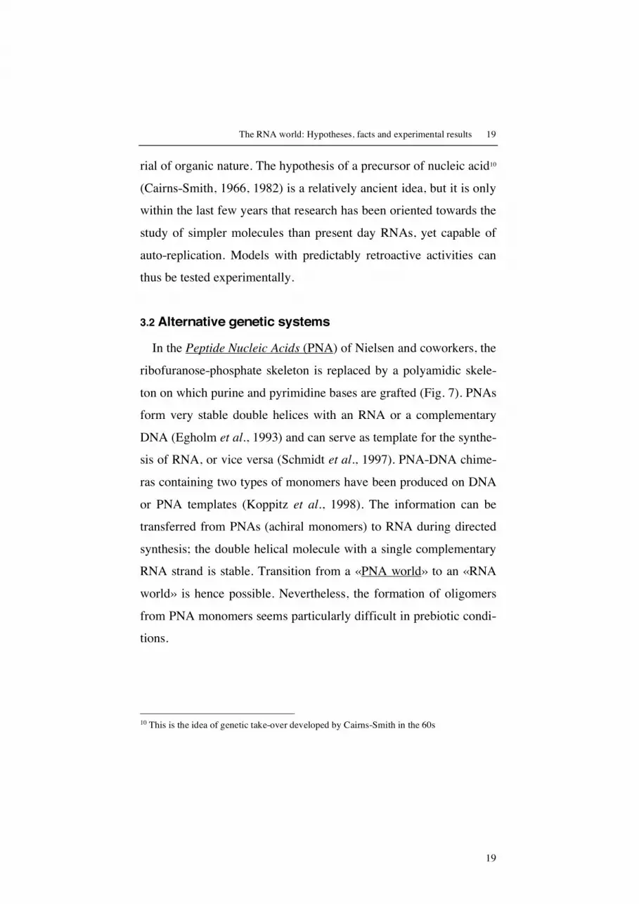
3.2 Alternative genetic systems
In the Peptide Nucleic Acids (PNA) of Nielsen and coworkers, the
ribofuranose-phosphate skeleton is replaced by a polyamidic skele-
ton on which purine and pyrimidine bases are grafted (Fig. 7). PNAs
form very stable double helices with an RNA or a complementary
DNA (Egholm et al., 1993) and can serve as template for the synthe-
sis of RNA, or vice versa (Schmidt et al., 1997). PNA-DNA chime-
ras containing two types of monomers have been produced on DNA
or PNA templates (Koppitz et al., 1998). The information can be
transferred from PNAs (achiral monomers) to RNA during directed
synthesis; the double helical molecule with a single complementary
RNA strand is stable. Transition from a «PNA world» to an «RNA
world» is hence possible. Nevertheless, the formation of oligomers
from PNA monomers seems particularly difficult in prebiotic condi-
tions.
10 This is the idea of genetic take-over developed by Cairns-Smith in the 60s

20
Fig. 7: Alternative genetic systems (B = base).
Eschenmoser (1994) explored the properties of nucleic acid ana-
logues in which ribofuranose is replaced by one of its isomers, ri-
bopyranose (Furanose, 5-membered ring; pyranose, 6-membered
ring). p-RNAs (pyranosyl RNAs) (Fig. 7) form more stable double
helices (with Watson-Crick pairings) than RNA with ribofuranose.
In addition, the double helices of p-RNA wind and unwind more
easily than those formed with standard nucleic acids, and this should
facilitate their separation during replication. p-RNAs could therefore
constitute good candidates as precursor genetic systems, but a p-
RNA strand cannot pair with an RNA of complementary sequence,
and this makes it difficult to imagine a transition from p-RNA to
RNA.

The RNA world: Hypotheses, facts and experimental results 21
21
The group of Eschenmoser recently replaced the ribose moiety by
a four-carbon sugar, threose, whose prebiotic synthesis seems easier.
The resulting oligonucleotides designated TNAs, (3’->2’)-α-L-
threose nucleic acid (Fig. 8), can form a double helix with RNA
(Schöning et al., 2000). TNA is capable of antiparallel, Watson-
Crick pairing with complementary DNA, RNA and TNA oligonu-
cleotides. Furthermore Szostak and his collaborators have recently
found that certain DNA polymerases can copy limited stretches of a
TNA template, despite significant differences in the sugar-phosphate
backbone, (Chaput et al., 2003).
Fig. 8: Structure of TNA and RNA.
Finally, Hexitol Nucleic Acids (HNA) (Fig. 7), whose skeleton is
composed of 1, 5-anhydrohexitol (six-membered cyclic hexitol) and
their isomers Altritol Nucleic Acids (ANA), form stable duplexes
B
O
O
O
O OP
O
B
OO
B
OO
OHO
O
O OP
B
OO
OH
TNA RNA

22
with complementary oligonucleotides, and are very efficient tem-
plates since they favor assembly of a complementary strand during
directed synthesis (Kozlov et al., 1999a, 1999b, 2000). The shape of
the duplexes formed is reminiscent of that of DNA in the A form.
Double-helical DNA is mainly in the B form11, whereas the double
helices of RNA in the DNA-RNA hybrids adopt the A form12.
Kozlov et al. (1999c) have demonstrated that the more the template
is in the A form, the better the efficiency of directed synthesis.
Based on these studies one can imagine an entire series of templates
that would supply the «good» structural pre-organization. Further-
more, these same authors have shown that RNA partially pre-
organized in the A form, is a more efficient matrix than single-
stranded DNA. Finally, whatever the precursor skeleton adapted to
the formation of stable duplexes may have been, the bond at the
mineral surface could have imposed the necessary geometrical con-
straints: yet this still remains to be experimentally demonstrated.
This leads us to two major conclusions, namely that on one hand a
transition may have occurred between two different systems without
loss of information, and that on the other hand the HNA and ANA
nucleic acids are very efficient templates. Even if it is difficult to
imagine prebiotic synthesis of these molecules, they are good model
11 d-ribose in the 2'-endo form. The characteristics of these forms are indicated above in the
text 12 In this case, the sugar is in the 3'-endo conformation. In the A form of RNA double heli-
ces, there are 11 base pairs per helical turn (instead of 10 for the B form); the inclination of the base pairs is 16°/helical axis (20° for DNA A)

The RNA world: Hypotheses, facts and experimental results 23
23
systems that show the importance of a necessary structural pre-
organization for directed synthesis by a template.
From the point of view of evolution, the studies described previ-
ously demonstrate that other molecules capable of transmitting he-
reditary information may have preceded our present day nucleic ac-
ids. This is what Cairns-Smith coined the «take-over» (Cairns-
Smith, 1982), the evolutionary encroachment or genetic take-over,
or to some extent what François Jacob (1970) calls genetic tinkering,
in other words, making new material from the old. This also sheds
light on the precision with which the various elements or processes
progressively adjusted themselves, thanks to successive trials and er-
rors.
4. Optimizing the functional capacities of ribonucleic acids
4.1 Coenzymes and modified nucleosides
The nucleotides that by post-transcriptional modification can to-
day acquire the majority of functional groups present in amino acids,
possess a great potential diversity that is expressed at the level of ri-
bonucleotide coenzymes (several coenzymes derive from AMP), and
of the modified bases of tRNAs (Fig. 9). The role of cofactors at all
steps of the metabolism and their distribution within the three king-
doms suggest that a great variety of nucleotides was present in the
ancestor common to all forms of life.

24
Several authors have underscored the possible presence of coen-
zymes before the appearance of the translation machinery (White,
1976). Proteins would have appeared only at a later stage, coen-
zymes and ribozymes being fossil traces of past catalysts. Indeed, in
the living cell, only a minority of enzymes function without coen-
zyme; they are mostly hydrolases, and apart from this group, 70% of
the enzymes require a coenzyme. If metal coenzymes involved in ca-
talysis are considered, the number of enzymes that depend on coen-
zymes increases further. Present-day coenzymes, indispensable co-
factors for many proteins, would be living fossils of catalysts of
primitive metabolism.
Coenzyme R R’ R’’ n
Activated
methionine
methionine H H 0
Amino acid
adenylate
amino acid H H 1
Activated
sulfate
SO32-
H PO32-
1
Cyclic 3’-5’
AMP
H H PO32-
1
NAD H H 2
NADP PO32-
H 2
FAD H H 2
O
N
N
N
N
C
H2
OPO
O
R
O
n
OR'
NH2
OR''
CoA-SH H H 2
Fig. 9: List of coenzymes derived from AMP.
Most coenzymes are nucleotides (NAD, NADP, FAD, coenzyme
A, ATP…) or contain heterocyclic nitrogen bases that can originate

The RNA world: Hypotheses, facts and experimental results 25
25
from nucleotides (thiamine pyrophosphate, tetrahydrofolate, pyri-
doxal phosphate, etc.).
Coenzymes would be vestiges of catalytic nucleic enzymes that
preceded ribosomal protein synthesis, and tRNAs can be viewed as
large coenzymes participating in the transfer of amino acids. It is
even possible to consider that catalytic groups that were part of nu-
cleic enzymes were incorporated in specific amino acids rather than
being «retained» as coenzymes. This could be the case of imidazol,
the functional group of histidine, whose present synthesis in the cell
is triggered by a nucleotide.
The modified nucleosides present today in RNAs result from post-
transcriptional modifications. Nevertheless, modified nucleosides
could have been present in the primitive world and their distribution
would have become established in the RNAs of the three living
kingdoms (Cermakian and Cedergren, 1998).
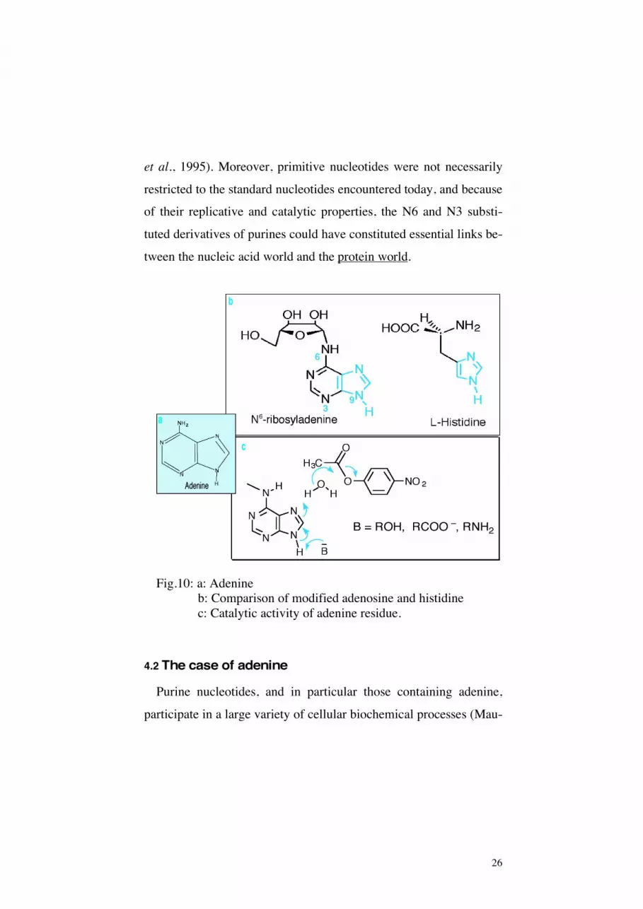
Our working hypothesis is based on the demonstration of esterase
activity in a nucleoside analogue N6-ribosyladenine (Fuller et al.,
1972; Maurel and Ninio, 1987). This activity which is due to the
presence of an imidazol group that is free and available for catalysis,
is comparable to that of histidine placed in the same conditions (Fig.
10). We have studied the kinetic behavior of this type of catalyst
(Ricard et al., 1996) and have shown that the catalytic effect in-
creases greatly when the catalytic element, pseudohistidine, is
placed in a favorable environment within a macromolecule (Décout
26
et al., 1995). Moreover, primitive nucleotides were not necessarily
restricted to the standard nucleotides encountered today, and because
of their replicative and catalytic properties, the N6 and N3 substi-
tuted derivatives of purines could have constituted essential links be-
tween the nucleic acid world and the protein world.
Fig.10: a: Adenine b: Comparison of modified adenosine and histidine c: Catalytic activity of adenine residue.
4.2 The case of adenine
Purine nucleotides, and in particular those containing adenine,
participate in a large variety of cellular biochemical processes (Mau-

The RNA world: Hypotheses, facts and experimental results 27
27
rel and Décout, 1999). Their best-known function is that of mono-
meric precursors of RNAs and DNAs. Nevertheless, derivatives of
adenine are universal cofators. They serve in biological systems as
source of energy (ATP), allosteric regulators of enzymatic activity
and regulation signals (cyclic AMP). They are also found as accep-
tors during oxidative phosphorylation (ADP), as components of co-
enzymes (such as in FAD, NAD, NADP, coenzyme A), as transfer
agents of methyl groups and of S-adenosylmethionine, as possible
precursors of polyprenoids in C5 (adenosylhopane) (Neunlist et al.,
1987), and – last but not least – adenine 2451 conserved within the
large rRNA in the three kingdoms, would be involved in acid-base
catalysis during the formation of the peptide bond (Muth et al.,
2000). However this role of adenine has been refuted based on
mutagenesis studies and phylogenetic comparisons (Muth et al,
2001; Green and Lorsch, 2002).
On the other hand, biosynthesis of an amino acid, histidine, that
would have appeared late in evolution, begins with 5-
phosphoribosyl-1-phosphate (PRPP) that forms N'-(5-
phosphoribosyl)-ATP by condensation with ATP. This reaction is
akin to the initial reaction of purine biosynthesis. Finally, the ease
with which purine bases are formed in prebiotic conditions13 (Orò,
1960) suggests that these bases were probably essential components
of an early genetic system. The first genetic system was probably
capable of forming base pairs of the Watson-Crick type, Hoogsteen
13 Purines have also been found in the meteorite Murchison

28
and other atypical associations, by hydrogen bonds as they still ap-
pear today in RNA. It probably contained a different skeleton from
that of RNA, and no doubt also modified bases, thereby adding
chemical functions, but also hydrophobic groups, and functions such
as amine, thiol, imidazole, etc. Wächterhaüser (1988) also suggested
novel pairings of the purine-purine type.
Originally, the principle probably rested on forced cooperation of
genetic and functional components, rather than on selection by indi-
vidual competition. It may have first entailed testing and improve-
ments (learning by trial and error) of the informational content of the
genes, i. e. linking the genotype (sequence) to the phenotype
(shape). One can consider that in such a system the unforseen was
faced, so that the living organism would need to adapt favorably and
rapidly.
4.3 Mimicking Darwinian evolution
Most of the «rational» biochemical approaches consist in deduc-
ing the active sequence of a nucleic acid or protein from a primary
sequence, or in synthesizing a defined compound by modelling and
structural analysis. However, «real life», that of our ancestors as also
that of our cells, does not proceed in this manner. The hunters-
gatherers of prehistory survived only thanks to their extraordinary
capacity to recognize objects. In addition, survival of a population in
a new environment is often linked to the appearance of a few vari-
ants to which random mutations conferred the power to adapt and

The RNA world: Hypotheses, facts and experimental results 29
29
exploit the new situation to their advantage. Combinatorial methods,
by modelling these observations14, have now become the alternative
to the rational concept. Selection in vitro requires no information
concerning the sequence of the molecules, and replaces the pre-
established adjustments between the molecule and its target. What is
needed, is to mimick the processes of evolution at the molecular
level.
Indeed, it is known since the experiments of Spiegelman (1971)
and his colleagues (Kramer et al., 1974) that populations of different
molecules capable of reproducing themselves in a hereditary man-
ner, can evolve and adapt to an appropriate environment. Spiegel-
man, the inventor of non-natural selection indeed demonstrated in
the 60s, that RNA populations can evolve when they replicate with
the help of an enzyme, the replicase of the bacteriophage Qβ. A
population of macromolecules can thus comply with the prerequi-
sites of Darwinian theory, and must find a form adapted to recogni-
tion of the target in a sufficiently rich population. Coexistence in the
same entity of shape and sequence, can favor the emergence of fa-
vorable candidates by means of a selection step (linked to the shape)
and an amplification step (linked to the sequence) at the end of this
molecular evolutionary process. A selection of this type could have
occurred during early molecular evolution, some 3.5 billion years
ago…
14 And by giving access to many related molecules that can be sorted

30
The original polymers more or less related to RNA and formed in
the primitive world must have randomly contained the A, U, G and
C bases. There are over one million possible sequences for a de-
canucleotide composed of 10 monomers A, U, G, C, and over 1012
sequences for a polynucleotide of 20 monomers15. Nature does not
appear to have exploited all the possible combinations before having
reached the remarkable functional unity of the living world, and
given the immense number of possibilities it is also useless to try to
explore experimentally, one by one, all the potentially functional se-
quences.
The SELEX method (Systematic Evolution of Ligands by Expo-
nential enrichment) (Tuerk and Gold, 1990) is an efficient, quasi
automatic method based on repeated cycles of reproductive selection
of those individuals that are best adapted to a given function. Estab-
lished in the 90s, this method makes it possible to obtain new struc-
tures, aptamers, selected through their aptitude to recognize other
molecules (Ellington and Szostak, 1990). Aptamers are capable of
recognizing targets as small as metal ions, or as large as cells. They
can interact with a great variety of molecules that are important for
primitive metabolism, be they amino acids, porphyrines, nucleotide
factors, coenzymes, small peptides and short oligonucleotides (Illan-
gasekare and Yarus, 1997; Jadhav and Yarus, 2002; Joyce, 2002;
McGinness et al., 2002; Reader and Joyce, 2002).
15 For a nucleic acid of 200 nucleotides, 10120 different sequences are theoretically possible,
and for a small protein containing 200 amino acids, 10280 arrangements are possible!

The RNA world: Hypotheses, facts and experimental results 31
31
At the molecular level, the Darwinian behavior requires that a
method of selection (RNA-aptamers), of amplification of selected
species, and of mutations (introduction of variants in the population
by means of mutations) be established. Through several cycles of se-
lection, amplification and mutations, populations of molecules are
«pushed» to evolve towards novel properties. The molecules pre-
senting the best «aptitudes» are selected and a new generation will
thereby see the day. Evolutionary processes performed experimen-
tally thus make it possible for molecules to emerge that have not yet
been produced by Nature, or allow the re-emergence of precursor
molecules that have strongly diverged or naturally disappeared.
Fig. 11: The SELEX method (adapted from Wilson and Szostak,
1999).
16 Which also applies to the protein world (phage display and combinatorial synthesis of
peptides)

32
In practice, how does one proceed? A «bank» of oligonucleotides
is a population of shapes among which a shape adapted to recogni-
tion of the target of interest is present (Fig. 11). The protocol is
composed of five steps: the creation of double-stranded DNA carry-
ing the random «box»16 flanked by regions required for amplifica-
tion; transcription of this DNA into single-stranded RNA; selection;
production of a DNA population by reverse transcription and PCR
of the sequences retained during the selection step, then cloning and
sequencing of the strands obtained after a certain number of selec-
tion and amplification cycles.
From a vast combination of nucleic acids, one can isolate aptam-
ers that possess catalytic properties (RNA ligation, cleavage or syn-
thesis of a peptide bond, transfer of an aminoacyl group, etc.). The
first nucleic acids could possess independent domains, separated by
flexible segments, creating reversible conformational motifs, de-
pendent on ions and bound ligands. Thus, a 10 amino acid-long pep-
tide can recognize fine structural differences within a micro RNA
helix (discrimination can be made between two closely-placed mi-
crohelices). Just as protein and antibodies, RNA molecules can pre-
sent hollows, cavities, or slits that make these specific molecular
recognitions possible. RNAs must “behave as proteins”. Whatever
the chronology and the order of appearance of the various classes of
molecules, the importance lies in the shape, the scaffolding and the
architecture that have allowed functional associations.
16 That is, a region of defined length, for instance of some 50 randomly aligned nucleotides

The RNA world: Hypotheses, facts and experimental results 33
33
Starting from a heterogenous population of RNAs with 1015 vari-
ants (a population of 1015 different molecules) we have selected 5
populations of RNAs capable of specifically recognizing adenine af-
ter about ten generations (Meli et al., 2002). When cloned, se-
quenced and modelled, the best one among the individuals of these
populations, has a shape reminiscent of a claw capable of grasping
adenine. Is it the exact copy of a primitive ribo-organism that feeds
on prebiotic adenine in prebiotic conditions? Functional and struc-
tural studies presently under way will highlight other activities, other
conformations…
Following this line of investigation we have selected two adenine-
dependent ribozymes capable of triggering reversible cleavage reac-
tions (Fig. 12). One of them is also active with imidazol alone. This
result leads to very important perspectives (Meli et al., 2003).
Fig 12: Adenine-dependent hairpin ribozymes (ADHR).
Arrowheads: cleavage sites
Grey dots: degenerated (mutated) sites
Vertical bars: separation between the primer binding region and
the random sequence.

34
A considerable amount of research has been focused on the selec-
tion of ribozymes in vitro. Recently, it was demonstrated that a ri-
bozyme is capable of continuous evolution, adding successively up
to 3 nucleotides to the initial molecule (McGuinnes, 2002). It is also
possible to construct a ribozyme with only two different nucleotides,
2,6-diaminopurine and uracil (Reader and Joyce, 2002). Finally,
Bartel and coworkers have selected a ribozyme-polymerase, capable
of self-amplification (Johnston et al., 2001).
4.4 Other perspectives
Very little is known to date about the behavior of macromolecules
in «extreme» environments. How do structures behave? What are
the major modifications observed? What are the conditions of struc-
tural and functional stability? How are the dynamics of the macro-
molecules and their interactions affected? What are the possibilities
of conserving biological macromolecules in very ancient soils or in
meteorites? Can we find traces of these macromolecules as molecu-
lar biosignatures, and if so in what form (Maurel and Zaccaï, 2001;
Tehei et al., 2002)?
The selection of thermohalophilic aptamers, RNAs resistant to
high temperatures (80°C) in the presence of salt (halites 30 million
years old), undertaken in our laboratory, will maybe allow us to an-

The RNA world: Hypotheses, facts and experimental results 35
35
swer some of these questions, that are fundamental for the search of
past traces of life, and of life on other planets…
5. Conclusion
The RNA world thus contains innumerable perspectives. The
combination of methods available today are the best adapted to ex-
plore the vast combinations of nucleic acids but also of peptides.
Will they make it possible to reconstitute the first steps of the living
world? Attractive simulations may emerge, opening new evolution-
ary paths that have not been envisaged or that Nature has not yet ex-
plored.
The RNA world, at whatever step we situate it in the history of the
living world, must be considered as a step in the history of life, an
important step in the evolution of the contemporary cellular world.
Because of its strong explanatory power, it also constitutes an im-
portant opening in the scientific study of the origin of life. Even if
this concept does not explain how life appeared, it nevertheless
promises a great number of experimental breakthroughs.
Acknowledgements : Figure 4 is reprinted from Cell, 2003,112, Bar-ends S., Bink H.H.J., van den Worm S.H.E., Pleij C.W.A., Kraal B. Entrapping ribosomes for viral translation: tRNA mimicry as a mo-lecular trojan horse. Copyright 2003, with permission from Elsevier. We thank Dr. G.F Joyce for his constructive comments on the manu-script.

36
References: Bachellerie J.P., Cavaillé J., Hüttenhofer A. (2002). The expanding
snoRNA world. Biochimie, 84, 775-790.
Ban N., Nissen P., Hansen J., Moore P.B., Steitz, T.A. (2000). The
complete atomic structure of the large ribosomal subunit at 2.4 Å
resolution. Science, 289, 905-920.
Barends S., Bink H.H.J., van den Worm S.H.E., Pleij C.W.A., Kraal
B. (2003). Entrapping ribosomes for viral translation: tRNA mim-
icry as a molecular trojan horse. Cell, 112, 123-129.
Bartel D.P., Unrau P.J. (1999). Constructing an RNA world. Trends
Biochem. Sci., 24, 9-13.
Benner S.A., Ellington A.D., Tauer A. (1989). Modern metabolism
as a palimpsest of the RNA world. Proc. Natl. Acad. Sci. USA, 86,
7054-7058.
Benner S.A., Cohen M.A., Gonnet G.H., Berkowitz D.B., Johnson
K.P. (1993). Reading the palimpsest: contemporary biochemical
data and the RNA World, in The RNA World, ed. Gesteland R.F. At-
kins J.F. p. 27-70, Cold Spring Harbor Laboratory Press: Cold
Spring Harbor NY.
Cairns-Smith A.G. (1966). The origin of life and the nature of the
primitive gene. J. Theor. Biol., 10, 53-88.
Cairns-Smith A.G. (1982). Genetic Takeover and the Mineral Ori-
gins of Life, Cambridge University Press, Cambridge.
Cermakian N., Cedergren, R. (1998). Modified nucleosides always
were: an evolutionary model, in Modification and Editing of RNA,

The RNA world: Hypotheses, facts and experimental results 37
37
ed. H. Grosjean and R. Benne, p. 535-541, ASM Press, Washington,
D.C.
Chaput J.C., Szostak J.W. (2003). TNA synthesis by DNA polym-
erase. J. Am. Chem. Soc.,125, 9274-9275.
Cooper G., Kimmich N., Belisle W., Sarinana J., Brabham K., Gar-
rel L. (2001). Sugar-related organic compounds in carbonaceous
meteorites. Nature, 414, 879-883.
Crick F.H. (1968). The origin of the genetic code. J. Mol. Biol. 38,
367-379.
Décout J-L., Vergne J., Maurel M-C. (1995). Synthesis and catalytic
activity of adenine containing polyamines. Macromol. Chem. Phys.,
196, 2615-2624.
Diener TO. (2001). The viroid: biological oddity or evolutionary
fossil? Adv. Virus. Res. 57, 137-184.
Eddy S.R. (2001). Non-coding RNA genes and the modern RNA
world. Nature Reviews Genetic, 2, 919-929.
Egholm M., Buchardt O., Christensen L., Behrens C., Freier S.M.,
Driver D.A., Berg R.H., Kim S.K., Norden B., Nielsen P.E. (1993).
PNA hybridizes to complementary oligonucleotides obeying the
Watson-Crick hydrogen-bonding rules. Nature, 365, 566-568.
Ellington, A.D., Szostak, J.W. (1990). In vitro selection of RNA
molecules that bind specific ligands. Nature. 346, 818-822.
Eschenmoser A. (1994). Chemistry of potentially prebiological natu-
ral products. Origins Life Evol. Biosphere, 24, 389-423.

38
Eschenmoser A. (1999). Chemical etiology of nucleic acid structure.
Science, 284, 2118-2124.
Fechter P., Rudonger-Thirion J., Florentz C., Giegé R. (2001). Novel
features in the tRNA-like world of plant viral RNAs. Cell. Mol. Life.
Sci. 58, 1547-1561.
Ferris J.P. (1987). Prebiotic synthesis: problems and challenges Cold
Spring Harbor Symp. Quant. Biol. LII, 29-39.
Ferris J.P., Ertem G. (1992). Oligomerization of ribonucleotides on
montmorillonite: reaction of the 5’ phosphorimidazolide of adeno-
sine. Science, 257, 1387-1389.
Ferris J.P., Hill A.R., Liu R., Orgel L.E. (1996). Synthesis of long
prebiotic oligomers on mineral surfaces. Nature, 381, 59-61.
Fuller W.D, Sanchez R.A, Orgel L.E. (1972). Studies in prebiotic
synthesis. J. Mol. Biol., 67, 25-33.
Gesteland R.F., Cech T.R., Atkins J.F. (ed) (1999). The RNA World,
second edition, Cold Spring Harbor Laboratory Press, Cold Spring
Harbor, NY.
Gilbert W. (1986). The RNA world. Nature, 319, 618.
Guerrier-Takada C., Gardiner K., Marsh T., Pace N., Altman S.
(1983). The RNA moiety of ribonuclease P is the catalytic subunit of
the enzyme. Cell, 35, 849-857.
Green R., Lorsch J.R. (2002). The path to perdition is paved with
protons. Cell, 110, 665-668.
Grosjean H., Benne R. (ed.) (1998). Modification and Editing of
RNA, ASM Press, Washington, D.C.

The RNA world: Hypotheses, facts and experimental results 39
39
Grosshans H., Slack F.J. (2002). Micro-RNAs: small is plentiful. J.
Cell Biol., 156, 17-21.
Hill A.R., Orgel L.E., Wu T. (1993). The limits of template-directed
synthesis with nucleoside-5’-phosphoro (2-methyl) imidazolides.
Orig. Life Evol. Biosphere, 23, 285-290.
Illangasekare M., Yarus M. (1997). Small-molecule substrate inter-
actions with a self aminoacylating ribozyme. J. Mol. Evol., 54, 298-
311.
Inoue T., Orgel L.E. (1983). A non-enzymatic RNA polymerase
model. Science, 219, 859-862.
Jacob F. (1970). La logique du vivant, Gallimard, Paris.
Jadahv V.R., Yarus M. (2002). Coenzymes as coribozymes. Biochi-
mie, 84, 877-888.
Johnston W.K., Unrau P.J., Lawrence M.S., Glasner M.E., Bartel
D.P. (2001). RNA-catalyzed RNA polymerization: Accurate and
general RNA-templated primer extension. Science, 292, 1319-1325.
Joyce G.F., Orgel L.E. (1986). Non-enzymic template-directed syn-
thesis on RNA random copolymers: poly (C,G) templates. J. Mol.
Biol., 188, 433-441.
Joyce G.F., Schwartz A.W., Miller S.L., Orgel L.E. (1987). The case
for an ancestral genetic system involving simple analogues of the
nucleotides. Proc. Natl. Acad. Sci. USA, 84, 4398-4402.
Joyce G.F. (1989). RNA evolution and the origins of life. Nature,
338, 217-224.

40
Joyce G.F., Orgel L.E. (1999). Prospects for understanding the ori-
gin of the RNA world, in The RNA World, ed. Gesteland R.F., Cech
T.R., Atkins J.F. p. 49-77, Cold Spring Harbor Laboratory Press:
Cold Spring Harbor NY.
Joyce G.F. (2002). The antiquity of RNA-based evolution. Nature,
418, 214-221.
Kable M.L., Heidmann S., Stuart K.D. (1997). RNA editing: getting
U into RNA. Trends Biochem. Sci., 22, 162-166.
Kong L.B., Siva A.C., Kickhoefer V.A., Rome L.H., Stewart P.L.
(2000). RNA location and modeling of a WD40 repeat domain
within the vault. RNA, 6, 890-900.
Koppitz M., Nielsen P.E., Orgel, L.E. (1998). Formation of oligonu-
cleotide-PNA-chimeras by template-directed ligation. J. Am. Chem.
Soc., 120, 4563-4569.
Kozlov I., Politis P.K., Pitsch S., Herdewijn P., Orgel L.E. (1999a).
A highly enantio-selective hexitol nucleic acid template for nonen-
zymatic oligoguanylate synthesis. J. Am. Chem. Soc., 121, 1108-
1109.
Kozlov I., De Bouvere B., Van Aerschot A. Herdewijn P., Orgel
L.E. (1999b). Efficient transfer of information from hexitol nucleic
acids to RNA during nonenzymatic oligomerization. J. Am. Chem.
Soc., 121, 5856-5859.
Kozlov I., Politis P.K., Van Aerschot A. Busson R., Herdewijn P.,
Orgel L.E. (1999c). Nonenzymatic synthesis of RNA and DNA oli-

The RNA world: Hypotheses, facts and experimental results 41
41
gomers on hexitol nucleic acid templates: the importance of A struc-
ture. J. Am. Chem. Soc., 121, 2653-2656.
Kozlov I., Zielinski M., Allart B., Kerremans L., Van Aerschot A,
Busson R., Herdewijn P., Orgel, L.E. (2000). Nonenzymatic tem-
plate-directed reactions on altritol oligomers, preorganized ana-
logues of oligonucleotides. Chem. Eur. J., 6, 151-155.
Kramer F.R., Mills D.R., Cole P.E., Nishihara T., Spiegelman S.
(1974). J. Mol. Biol., 89, 719-736.
Lafontaine D.L., Tollervey D. (1998). Birth of the snoRNPs: the
evolution of the modification-guide snoRNAs. Trends Biochem.
Sci., 83, 383-388.
Lamond A.I. (1988). RNA editing and the mysterious undecovered
genes of trypanosomatid mitochondria. Trends Biochem. Sci., 13,
283-284.
Maizels N., Weiner A.M., Yue D., Shi P.Y. (1999). New evidence
for the genomic tag hypothesis: archaeal CCA-adding enzymes and
DNA substrates. Biol. Bull., 196, 331-333.
Mattick J.S. (2003). Challenging the dogma: the hidden layer of
non-protein-coding RNAs in complex organisms. BioEssays, 25,
930-939.
Maurel M.-C., Ninio J. (1987). Catalysis by a prebiotic nucleotide
analog of histidine. Biochimie, 69, 551-553.
Maurel M.-C. (1992). RNA in evolution. J. Evol. Biol., 2, 173-188.
Maurel M.-C., Décout J.-L. (1999). Origins of life: molecular foun-
dations and new approaches. Tetrahedron, 55, 3141-3182.

42
Maurel M.-C., Zaccaï G. (2001). Why Biologists should support the
exploration of Mars. BioEssays, 23, 977-978.
McGinness K.E., Wright M.C., Joyce G.F. (2002). Continuous in vi-
tro evolution of a ribozyme that catalyzes three successive nucleoti-
dyl addition reactions. Chem. Biol., 9, 585-596.
Meli M., Albert-Fournier B., Maurel M.-C. (2001). Recent findings
in the modern RNA world. Int. Microbio. 4, 5-11.
Meli M., Vergne J., Décout J.-L., Maurel M.-C. (2002). Adenine-
aptamer complexes. A bipartite RNA site which binds the adenine
nucleic base. J. Biol. Chem., 277, 2104-2111.
Meli M., Vergne J., Maurel M.-C. (2003). In vitro selection of ade-
nine-dependent hairpin ribozymes. J. Biol. Chem., 278, 9835-9842.
Muth G.W., Ortoleva-Donnelly L., Strobel S.A. (2000). A single
adenosine with a neutral pKa in the ribosomal peptidyl transferase
center. Science, 289, 947-950.
Muth G.W., Chen L., Kosek A.B. Strobel S.A. (2001). PH-
dependent conformational flexibility within the ribosomal peptidyl
transferase center. RNA, 7, 1403-1415.
Neunlist S., Bisseret P., Rohmer M. (1987). The hopanoids of the
purple non-sulfur bacteria Rhodopseudomonas palustris and
Rhodopseudomonas acidophila and the absolute configuration of
bacteriohopanetetrol. Eur. J. Biochem., 87, 245-252.
Nissen P., Hansen J., Ban N., Moore P.B., Steitz T.A. (2000). The
structural basis of ribosome activity in peptide bond synthesis. Sci-
ence, 289, 920-930.

The RNA world: Hypotheses, facts and experimental results 43
43
Nykanen A., Haley B., Zamore P.D. (2001). ATP requirements and
small interfering RNA structure in the RNA interference pathway.
Cell, 107, 309-321.
Orgel L.E. (1968). Evolution of the genetic apparatus. J. Mol. Biol.,
38, 381-393.
Orgel L.E. (1989). Was RNA the first genetic polymer? in Evolu-
tionary tinkering in gene expression ed. M. Grunberg-Manago et al.,
p. 215-224, Plenum Press London.
Orgel L.E. (1992). Molecular replication. Nature, 358, 203-209.
Orò J. (1960). Biochem. Biophys. Res. Comm., 2, 407-412.
Paecht-Horowitz M., Berger J., Katchalsky A. (1970). Prebiotic
synthesis of polypeptides by heterogeneous polycondensation of
amino acid adenylates. Nature, 7, 847-850.
Reader J.S., Joyce G.F. (2002). A ribozyme composed of only two
different nucleotides. Nature, 420, 841-844.
Ricard J., Vergne J., Décout J.-L., Maurel M.-C. (1996). The origin
of kinetic cooperativity in prebiotic catalysts. J. Mol. Evol. 43, 315-
325.
Schmidt J.G., Nielsen P.E., Orgel, L.E. (1997). Information transfer
from peptide nucleic acid RNA by template-directed syntheses.
Nucl. Acids Res., 25, 4797-4802.
Schöning K.-U., Scholz P., Guntha S., Wu X., Krishnamurthy R.,
Eschenmoser A. (2000). Chemical etiology of nucleic acid struc-
ture: the α-threo-furanosyl-(3’->2’) oligonucleotide system. Science,
290, 1347-1351.

44
Spiegelman S. (1971). An in vitro analysis of a replicating molecule.
Quaterly Review Biophys., 4, 213-253.
Stuart K., Panigrahi A.K. (2002). RNA editing: complexity and
complications. Mol. Microbiol., 45, 591-596.
Sutherland J.D., Whitfield J.N. (1997). Prebiotic chemistry: a bioor-
ganic perspective. Tetrahedron, 53, 11493-11527.
Tarn W.Y., Steitz J.A. (1997). Pre-mRNA splicing: the discovery of
a new spliceosome doubles the challenge. Trends Biochem. Sci., 22,
132-137.
Tehei M., Franzetti B., Maurel M.-C., Vergne J., Hountondji C.,
Zaccaï G. (2002). Salt and the search for traces of life. Extremo-
philes, 6 , 427-430.
Terns M.P., Terns R.M. (2002). Small nucleolar RNAs: versatile
trans-acting molecules of ancient evolutionary origin. Gene Expr.,
10, 17-39.
Tuerk,C., Gold, L., (1990 ). Systematic evolution of ligands by ex-
ponential enrichment: RNA ligands to bacteriophage T4 DNA po-
lymerase.Science. 249, 505-510.
Valle M., Gillet R., Kaur S., Henne A., Ramakrishnan V., Frank J.
(2003). Visualizing tmRNA entry into a stalled ribosome. Science,
300, 127-130.
Wächtershäuser G. (1988). An all-purine precursor of nucleic acids.
Proc. Natl. Acad. Sci. USA, 85, 1134-1135.
Westhof E. (2002). Foreword. Biochimie, 84, 687-689.

The RNA world: Hypotheses, facts and experimental results 45
45
White H.B. (1976). Coenzymes as fossils of an earlier metabolic
state. J. Mol. Evol., 7, 101-104.
Wild K., Weichenrieder O., Strub K., Sinning I., Cusack S. (2002).
Towards the structure of the mammalian signal recognition particle.
Curr. Opinion Struct. Biol., 12, 72-81.
Wilson D.S., Szostak J.W. (1999). In vitro selection of functional
nucleic acids. Annu. Rev. Biochem., 68, 611-647.
Withey J.H., Friedman D.I. (2002). The biological roles of trans-
translation. Curr. Opinion Microbiol., 5, 154-159.
Woese C.R. (1965). On the evolution of the genetic code. Proc.
Natl. Acad. Sci. USA., 54, 1546-1552.
Related Documents