University of New Mexico UNM Digital Repository Biomedical Sciences ETDs Electronic eses and Dissertations 12-1-2012 e RNA binding protein KSRP destabilizes GAP-43 mRNA to limit axonal elongation in cultured hippocampal neurons Clark Bird Follow this and additional works at: hps://digitalrepository.unm.edu/biom_etds is Dissertation is brought to you for free and open access by the Electronic eses and Dissertations at UNM Digital Repository. It has been accepted for inclusion in Biomedical Sciences ETDs by an authorized administrator of UNM Digital Repository. For more information, please contact [email protected]. Recommended Citation Bird, Clark. "e RNA binding protein KSRP destabilizes GAP-43 mRNA to limit axonal elongation in cultured hippocampal neurons." (2012). hps://digitalrepository.unm.edu/biom_etds/65

Welcome message from author
This document is posted to help you gain knowledge. Please leave a comment to let me know what you think about it! Share it to your friends and learn new things together.
Transcript
University of New MexicoUNM Digital Repository
Biomedical Sciences ETDs Electronic Theses and Dissertations
12-1-2012
The RNA binding protein KSRP destabilizesGAP-43 mRNA to limit axonal elongation incultured hippocampal neuronsClark Bird
Follow this and additional works at: https://digitalrepository.unm.edu/biom_etds
This Dissertation is brought to you for free and open access by the Electronic Theses and Dissertations at UNM Digital Repository. It has beenaccepted for inclusion in Biomedical Sciences ETDs by an authorized administrator of UNM Digital Repository. For more information, please [email protected].
Recommended CitationBird, Clark. "The RNA binding protein KSRP destabilizes GAP-43 mRNA to limit axonal elongation in cultured hippocampalneurons." (2012). https://digitalrepository.unm.edu/biom_etds/65
i
ii
The RNA binding protein KSRP destabilizes GAP-43 mRNA to limit axonal
elongation in cultured hippocampal neurons
By
Clark W. Bird
B.A., Molecular, Cellular, and Developmental Biology
and
Psychology
University of Colorado at Boulder, 2005
DISSERTATION
Submitted in partial fulfillment of the requirements for the degree of
Doctor of Philosophy
Biomedical Sciences
The University of New Mexico
Albuquerque, New Mexico
December 2012
iii
Acknowledgements
None of the work contained within this dissertation would have been possible
without the knowledge and patience of my mentor, Nora Perrone-Bizzozero. Her
enthusiasm for science and encyclopedic knowledge of all things related to molecular
biology and neuroscience were invaluable to my laboratory experience. I couldn’t have
asked for a better mentor.
I would like to thank all members of my dissertation committee for their valuable
input and help during the last five years. Fernando Valenzuela and Kevin Caldwell were
awesome rotation mentors and great teachers. Rebecca Hartley and her RNA journal
club were very helpful in getting me interested in the biology of RNA, and was a
fantastic teacher to boot. Their friendly faces, attitudes, and knowledge were extremely
helpful to getting through my dissertation research. I also would like to thank Xinyu
Zhao, a former member of my committee, for her assistance during my first few years at
UNM.
The members of the Perrone-Bizzozero lab, both past and present, also deserve a
measure of gratitude. Sayuri Nixon in particular was a fantastic lab manager, and made
performing experiments an easy process, as the lab was wonderfully organized and
protocols were easy to understand. The knowledge and experience of post-doc Amy
Gardiner provided a great laboratory resource. Joanna Mounce was a great lab-mate, and
didn’t complain when I played terrible rock and roll around the clock. All these people
made going to work every day a more pleasurable experience.
The most important person I need to thank is my mother, Mary Bird. Without her
none of this would have been possible, as she raised me to be an ambitious, well-
mannered, sarcastic member of society. She “gently” pushed me into being more than
just a sandwich shop manager and restaurant waiter. I can’t thank her enough for being
there for me whenever I needed her.
My girlfriend, Lindsay Rogash, has also been wonderful during my graduate
experience. Dealing with a stressed-out graduate student can’t be the easiest thing in the
world, and she has always been encouraging, and is great motivation to do well and be a
better person. Lastly, I have to thank my cats: Dale, Eddie, and Floofball, constant
companions who lifted my spirits when I needed it.
iv
The RNA binding protein KSRP destabilizes GAP-43 mRNA to limit
axonal elongation in cultured hippocampal neurons
By
Clark W. Bird
B.A., Molecular, Cellular and Developmental Biology
and
Psychology
University of Colorado at Boulder
Abstract
The KH-type splicing regulatory protein (KSRP) promotes the decay of AU-rich
element (ARE) containing mRNAs. Although KSRP is expressed in the developing and
mature nervous system, very little is known about its role in regulating gene expression in
the brain. In this study, we utilized in vitro binding and decay studies to examine
whether KSRP regulates the stability of the GAP-43 transcript, an ARE-containing
neuronal mRNA whose protein product plays a role in axonal growth and synaptic
plasticity. We found KSRP destabilizes GAP-43 mRNA by binding to the GAP-43 ARE,
v
a process that depends on the presence of the fourth KH domain in the protein.
Furthermore, KSRP competed with another GAP-43 mRNA binding protein, the
stabilizing factor HuD, for binding to these ARE sequences. Given that GAP-43
expression is crucial for accurate axonal outgrowth during neuronal development, we also
examined the functional consequences of KSRP overexpression and depletion on the
axonal outgrowth from primary hippocampal neurons. Overexpression of either full
length KSRP or KSRP without the nuclear localization signal hindered axonal outgrowth
in these cultures, while overexpression of a mutant protein without the KH4 domain did
not have any effect. In contrast, depletion of KSRP led to a dramatic increase in axonal
length. Concurrent overexpression of GAP-43 and KSRP rescued the axonal outgrowth
deficits seen with KSRP overexpression, but only when the GAP-43 mRNA was targeted
to axons using GAP-43 or amphoterin 3’ UTR sequences. Together, our results suggest
that KSRP is an important regulator of GAP-43 mRNA stability and neuronal
differentiation that works in direct opposition to HuD.
vi
Table of Contents
Acknowledgements .......................................................................................................... iii
Abstract ............................................................................................................................. iv
List of Figures ................................................................................................................. viii
1. Introduction ................................................................................................................... 1
1.1 The AU rich element and associated proteins ........................................................................................... 1
1.2 GAP-43 and HuD ...................................................................................................................................... 6
1.3 KSRP ......................................................................................................................................................... 9
1.4 Summary ................................................................................................................................................. 13
2. Rationale, hypothesis, and specific aims ................................................................... 14
2.1 Rationale.................................................................................................................................................. 14
2.2 Hypothesis ............................................................................................................................................... 14
2.3 Specific aims ........................................................................................................................................... 14
2.3.1 Specific aim 1: ...................................................................................................................................... 14
2.3.2 Specific aim 2: ...................................................................................................................................... 15
3. Regulation of GAP-43 stability by KSRP in vitro .................................................... 16
3.1 Introduction ............................................................................................................................................. 16
3.2 Materials and methods ............................................................................................................................. 17
3.3 Results ..................................................................................................................................................... 19
3.3.1 KSRP binds to GAP-43 mRNA in vitro: RNA electrophoretic mobility shift ..................................... 19
3.3.2 KSRP binds to GAP-43 mRNA in vitro: biotinylated GAP-43 mRNA pulldown (Experiment
performed by Dan Tanner) .............................................................................................................. 21
3.3.3 HuD and KSRP compete for binding to the GAP-43 ARE .................................................................. 21
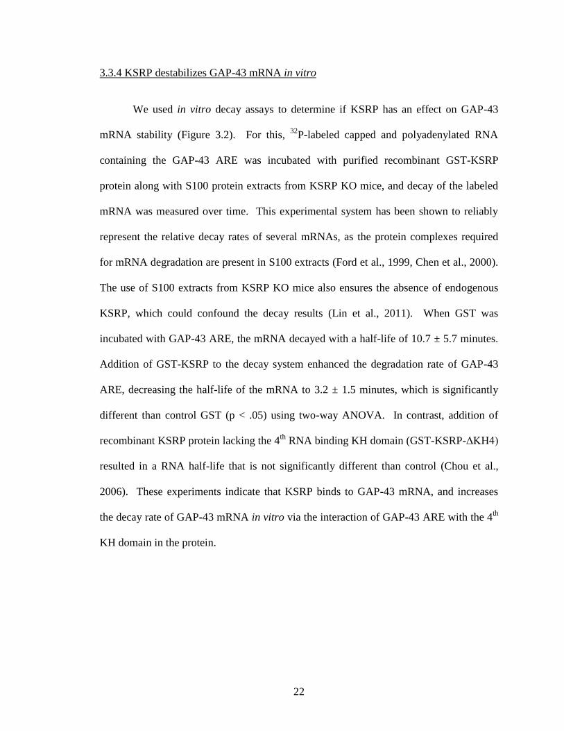
3.3.4 KSRP destabilizes GAP-43 mRNA in vitro ......................................................................................... 22
3.4 Discussion ............................................................................................................................................... 24
4. KSRP inhibits axonal outgrowth in cultured hippocampal neurons ..................... 26
4.1 Introduction ............................................................................................................................................. 26
4.2 Materials and methods ............................................................................................................................. 27
4.3 Results ..................................................................................................................................................... 31
4.3.1 KSRP and HuD localize to separate cytoplasmic granules in developing hippocampal neurons ......... 31
4.3.2 KSRP limits axonal outgrowth in cultured rat E17 hippocampal neurons ........................................... 32
4.3.3 Knockdown of KSRP enhances axonal outgrowth in E17 rat hippocampal neurons ........................... 35
4.3.4 Rescue of limited axonal outgrowth in KSRP transfected neurons by co-expression of GAP-43 ....... 37
vii
4.4 Discussion ............................................................................................................................................... 39
5. Discussion..................................................................................................................... 42
Abbreviations Used ......................................................................................................... 50
References ........................................................................................................................ 52
Appendix A: KSRP and GAP-43 mRNA expression in shRNA transfected PC12
cells ....................................................................................................................... 59
Appendix B: Submitted short-report article to J. Neuroscience co-authored by
Clark Bird ............................................................................................................ 60
Appendix C: Review article in press, Frontiers in Bioscience. Co-authored by Clark
Bird ....................................................................................................................... 82
viii
List of Figures
Figure 1.1 Overview of KSRP binding to the ARE of GAP-43 .................................................................... 11
Figure 3.1 Binding of KSRP to GAP-43 ARE .............................................................................................. 20
Figure 3.2 KSRP increases GAP-43 ARE decay in vitro .............................................................................. 23
Figure 4.1 GFP-KSRP constructs cloned for KSRP overexpression studies. ................................................ 28
Figure 4.2 KSRP and HuD localized to separate locations in the cytoplasm of cultured hippocampal
neurons ............................................................................................................................................ 32
Figure 4.3 Overexpression of KSRP limits axonal outgrowth in E17 hippocampal neurons ........................ 34
Figure 4.4 shRNA knockdown of KSRP in transfected hippocampal neurons increases axonal length. ...... 36
Figure 4.5 Overexpression of GAP-43 rescues limited axonal outgrowth in KSRP transfected E17
hippocampal neurons. ..................................................................................................................... 38
Figure 5.1 Theoritical model for GAP-43 regulation during axonal elongation ........................................... 46
Appendix A: shRNA knockdown of KSRP in PC12 cells increases GAP-43 mRNA expression ................ 59
1
1. Introduction
1.1 The AU rich element and associated proteins
Gene expression from any given gene is dependent on a number of cellular events
working in parallel. The initial transcription of a gene determines the amount of pre-
mRNA ready for processing. Mature mRNA levels are dependent on the speed at which
post-transcriptional processes occur, which include splicing, capping, and
polyadenylation. Once processed, the transcript still needs to be exported to the
cytoplasm, so nuclear export rates need to be considered. Subsequent stability of the
mRNA also determines the available pool of transcript available for translation. The next
step determining final protein output is the rate of translation of the mRNA by ribosomes.
Once the final protein product is produced, the amount of functional protein is still
determined by post-translational processing and folding events, as well as degradation
processes mediated by cellular proteases. Any one of the preceding events can have a
significant impact on the overall expression of a given gene.
For a number of genes, the primary factor governing final protein output is the
stability of mRNA following nuclear export. Many proteins in a cell do not need to be
constitutively produced, but rather are needed for specific cellular processes. Sometimes
these proteins need to be produced relatively quickly to impact a cellular event, such as
the production of inducible nitric oxide synthase (iNOS) following an immunologic insult
to generate nitric oxide, which has anti-microbial and anti-apoptotic functions (Linker et
al., 2005). In human hepatocytes, the iNOS promoter exhibits high basal activity, but the
mRNA product is virtually undetectable. Following cytokine induction, the promoter
2
activity of iNOS is only increased by two- to five-fold, but there is a much more
significant accumulation of the mRNA (Kleinert et al., 2004). This indicates that the
stability of the mRNA is the major determining factor determining iNOS levels following
cytokine induction.
In 1986, a conserved sequence in the 3’ untranslated regions (3’UTRs) of
numerous mRNAs encoding for cytokines was identified, having the adenine (A) and
uracil (U) containing consensus sequence of UUAUUUAU (Caput et al., 1986). Other
investigators of the time separately noticed the phenomenon, noting that many
lymphokine, cytokine, and proto-oncogene mRNAs contained stretches of A and U
nucleotides (Shaw and Kamen, 1986). The conserved nature of this nucleotide sequence
prompted researchers to postulate that the sequence had a specific functionality. When
the AU rich sequence-containing 3’UTR of granulocyte-monocyte colony stimulating
factor (GM-CSF) was attached to the normally stable mRNA encoding for β-globin, the
mRNA became very unstable (Shaw and Kamen, 1986). Other investigators also noted
these AU rich sequences in a number of labile mRNAs, including c-fos, c-myc, β-
interferon, IL-3, IL-8, and many others (Treisman, 1985, Brewer and Ross, 1988, Peppel
et al., 1991, Stoecklin et al., 1994, Winzen et al., 1999). The A and U rich region in the
3’UTRs of short-lived mRNAs has since been coined the AU-rich element (ARE)
(Brewer, 1991).
Since their original identification, ARE’s have been categorized into three
separate subgroups based on their sequence composition (Chen and Shyu, 1995). Class I
ARE’s contain 1-3 copies of an AUUUA pentamer surrounded by a U rich region. This
type of ARE is contained in c-fos and c-myc (Treisman, 1985, Chen and Shyu, 1994).
3
The class II ARE, which is found in many unstable cytokine mRNAs, comprises at least
two overlapping copies of a UUAUUUA(U/A)(U/A) nonamer (Shaw and Kamen, 1986).
The least well defined ARE sequence is that for the class III ARE, which does not
contain a canonical sequence, but is rather a span of mostly A and U nucleotides.
Examples of class III ARE containing mRNAs are those encoding for c-jun and the
neuronally expressed growth associated protein 43 (GAP-43) mRNAs (Kohn et al., 1996,
Peng et al., 1996). While there are many mRNAs that have AU rich stretches within their
3’UTRs, they are not defined as AREs unless they confer instability to the transcript
(Chen and Shyu, 1995). An estimated 5-8% of mRNAs contain typical AREs according
to the ARE database (ARED), which may be an underestimate of the actual number of
ARE containing genes due to the ambiguous nature of the class III ARE (Bakheet et al.,
2006).
Degradation of an ARE-containing mRNA involves a number of different steps.
The first process typically involved in mammalian mRNA decay is deadenylation, which
is most likely mediated by the poly(A) ribonuclease (PARN) (Wilusz et al., 2001). Initial
reports indicated that the next step in ARE-mediated mRNA decay (AMD) was 3’ to 5’
exonucleotic digestion by the exosome complex (Chen et al., 2001). Later evidence also
implicates cytoplasmic processing bodies (p-bodies) in the degradation of ARE
containing mRNA, which is initiated by Dcp decapping proteins followed by 5’ to 3’
exonucleolytic cleavage by the enzyme Xrn1 (Fenger-Gron et al., 2005). Interestingly, 3’
to 5’ degradation by the exosome complex is aided by decapping, indicating that 5’ to 3’
and 3’ to 5’ digestion may be functionally linked (Murray and Schoenberg, 2007). The
pathway that AMD follows may depend on the type of ARE that the mRNA contains, or
4
may be a function of a trans acting factor: an ARE-binding protein (ARE-BP) (Barreau et
al., 2005).
A number of ARE-BPs have been identified to date, which can serve to stabilize
or destabilize bound ARE-containing transcripts. The most well-studied stabilizing
ARE-BPs are the Hu family proteins HuR (also known as HuA), HuB (also known as
He1-N1), HuC, and HuD (Bolognani and Perrone-Bizzozero, 2008). These proteins are
the human homologues of a Drosophila protein that causes an embryonic lethal abnormal
vision phenotype (ELAV) (Szabo et al., 1991). All the Hu family proteins contain a
similar structure and sequence, and contain three RNA recognition motifs (RRMs)
(Brennan and Steitz, 2001). The most well studied of the ELAV proteins, HuR, has been
implicated in the stabilization of a number of mRNAs involved in multiple cellular
processes ranging from immediate early gene expression (c-fos and c-myc), to cell cycle
progression (Cyclin A, Cyclin B1, Cyclin D1) and differentiation (MyoD and VEGF)
(Reviewed in (Brennan and Steitz, 2001, Barreau et al., 2005)). Binding of Hu family
proteins to the ARE protects the mRNA from degradation, and may help to enhance
translational rates by associating with polysomes (Keene, 1999). HuR is mainly nuclear
but can shuttle to the cytoplasm, leading some to postulate that HuR initially binds to
ARE-containing mRNA in the nucleus, protecting the mRNA from both nuclear and
cytoplasmic decay processes (Fan and Steitz, 1998).
Functioning in opposition to the ELAV family proteins are a number of ARE-BPs
that serve to destabilize target mRNA. The first ARE-BP identified serving to destabilize
bound mRNA is the protein AUF1 (also known as hnRNP D), which was shown to
promote the degradation of bound c-myc mRNA (Brewer, 1991). AUF1 has also been
5
implicated in stabilizing IL-3 mRNA in NIH 3T3 cells, while destabilizing IL-3 mRNA
in K562 cells, providing a somewhat ambiguous role for this ARE-BP (Loflin et al.,
1999, Ming et al., 2001). Other destabilizing RNA binding proteins were later identified,
such as KH-splice regulatory factor (KSRP), tristetraprolin (TTP), and butyrate-regulated
factor-1 (BRF1) (Reviewed in (Wu and Brewer, 2012)). KSRP mainly associates with
components of the exosomal degradation machinery, while TTP and BRF1 promote
mRNA degradation via both exosomes and p-bodies (Gherzi et al., 2004, Lykke-
Andersen and Wagner, 2005).
How and when ARE-BPs bind to, and compete for binding to ARE-containing
mRNAs will determine the functional protein output of a specific gene. For example,
iNOS can be destabilized by KSRP, but HuR also competes for the same binding site in
the iNOS 3’UTR (Linker et al., 2005). It seems that before cytokine induction of iNOS,
KSRP is bound to the mRNA to negatively regulate its stability. Following an antigenic
insult and cytokine signaling, KSRP is dislodged from the iNOS mRNA, allowing HuR
to bind to and stabilize the transcript for translation (Linker et al., 2005, Wu and Brewer,
2012). This post-transcriptional “operon” of stabilizing and destabilizing proteins and
their interaction with ARE-containing mRNA serves as a dynamic regulator of gene
expression and provides regulation of gene expression beyond simple transcriptional and
translational activation (Keene and Lager, 2005).
6
1.2 GAP-43 and HuD
The ARE-containing mRNA encoding for GAP-43 is the most well studied labile
mRNA specific to neurons. GAP-43 was initially identified as a protein present in
retinal ganglion cells during axonal regeneration following optic nerve crush (Skene and
Willard, 1981). GAP-43 protein was later shown to be highly expressed in dissociated rat
cerebrocortical cultures soon after plating and also in PC12 cells following neurite
outgrowth induced by NGF stimulation (Perrone-Bizzozero et al., 1986, Van Hooff et al.,
1989). During axonal outgrowth GAP-43 accumulates in axonal growth cones and helps
to direct axonal outgrowth, and knockdown of GAP-43 leads to regression of the axon
(Skene et al., 1986, Caroni and Becker, 1992). Once axons reach their post-synaptic
target, GAP-43 levels diminish as a functional synapse is formed (Schreyer and Skene,
1991, Karimi-Abdolrezaee et al., 2002). Transgenic mice overexpressing GAP-43
exhibit aberrant axonal sprouting of mossy fibers in the hippocampus, while GAP-43
knockout mice have deficits in axonal pathfinding and die during development (Aigner et
al., 1995, Strittmatter et al., 1995, Klein et al., 1999). Clearly, the coordinated expression
of GAP-43 during development is critical to proper nervous system development.
The actual mechanism of action of GAP-43 in promoting axonal outgrowth is not
completely understood, but there are a number of indications as to its cellular function.
The N-terminal end of the GAP-43 protein can activate the GTP-binding protein G0, and
subsequently causes filopodial extension (Strittmatter et al., 1994). Another potential
mechanism of GAP-43 mediated axonal outgrowth involves protein kinase C (PKC)
activity, which is induced by NGF stimulation (Perrone-Bizzozero et al., 1993). Active
PKC phosphorylates GAP-43, which leads to changes in GAP-43 binding to different
7
cellular molecules (Perrone-Bizzozero et al., 1993). Unphosphorylated GAP-43 binds to
a large number of actin filaments, and is proposed to act as a cap to actin filaments,
preventing further actin polymerization and limiting filopodial extension at the growth
cone (He et al., 1997). When GAP-43 is phosphorylated by PKC, it has less affinity for
actin, and is then proposed to act as a lateral stabilizer of actin filaments and promote
filopodial extension (He et al., 1997, Denny, 2006). In a separate model,
unphosphorylated GAP-43 binds readily to phosphatidylinositol 4,5-bisphosphate [PI
(4,5) P2], and clusters this phospholipid into rafts. Upon phosphorylation by PKC, GAP-
43 releases PI (4,5) P2, allowing PI (4,5) P2 to bind profilin, cofilin, and gelsolin, which
are molecules that act to prevent actin polymerization. With these actin-binding
molecules sequestered to the membrane, actin is free to polymerize and promote
filopodia extension (Laux et al., 2000, Denny, 2006). These two models could also be
working cooperatively, enhancing actin polymerization and promoting axonal outgrowth.
Control of GAP-43 expression is regulated at both the transcriptional and post-
transcriptional level. The promoter for GAP-43 restricts its transcription to the nervous
system (Reinhard et al., 1994, Weber and Skene, 1997). Enhancer elements upstream of
the GAP-43 gene in the 5’ regulatory region can be bound by basic helix-loop-helix
transcription factors, promoting GAP-43 transcription (Chiaramello et al., 1996).
Following initial transcriptional events, GAP-43 expression is also regulated by post-
transcriptional mechanisms. GAP-43 transcription occurs at similar rates in quiescent
and developing rat cortical neurons, but steady state levels of GAP-43 mRNA are greatly
increased in the developing neurons, indicating that this transcript is regulated by a post-
transcriptional mechanism (Perrone-Bizzozero et al., 1991). Later investigations
8
identified a pyrimidine rich stretch in the 3’UTR of the GAP-43 mRNA that regulates its
stability (Kohn et al., 1996). When this instability conferring sequence was attached to
the normally stable β-globin mRNA it promoted its degradation (Tsai et al., 1997). This
pyrimidine stretch, rich in U and cytosine (C) nucleotides, is classified as a class III ARE,
as it is U rich but does not contain a canonical AUUUA sequence (Chen and Shyu, 1995,
Kohn et al., 1996). The GAP-43 ARE is also the binding site for the stabilizing ARE-BP
HuD (Chung et al., 1997).
The neuronally expressed ELAV protein HuD has been implicated repeatedly as a
critical determinant regulating GAP-43 mRNA stability. Overexpression of HuD in NGF
induced PC12 cells led to increased neurite outgrowth and elevated GAP-43 protein
levels (Anderson et al., 2000). Transfection of a HuD overexpression construct into
PC12 cells or E19 rat cortical neurons accelerated neurite outgrowth, as well as increased
both mRNA and protein levels of GAP-43 (Mobarak et al., 2000, Anderson et al., 2001).
Conversely, knockdown of HuD in PC12 cells led to a significant decrease in GAP-43
gene products (Mobarak et al., 2000). Stabilization of GAP-43 mRNA requires all three
RRMs of HuD (Anderson et al., 2000). The first two RRMs of ELAV proteins bind to
ARE sequences, while the third RRM binds to poly(A) stretches, i.e. the poly-A tail (Liu
et al., 1995, Abe et al., 1996, Ma et al., 1997). HuD binds preferentially to GAP-43
mRNAs with a long poly-A tail, and inhibits deadenylation of the transcript, protecting it
from the first step in AMD (Beckel-Mitchener et al., 2002).
HuD has not only been demonstrated to enhance the stability of GAP-43 in vitro
and in cell culture, but also in vivo by utilizing transgenic animal models. Transgenic
mice overexpressing HuD display elevated levels of GAP-43 mRNA in granule cells of
9
the dentate gyrus, in the lateral amygdala, and in layer V of the neocortex (Bolognani et
al., 2006). Increased GAP-43 mRNA stability in transgenic HuD overexpressing mice
was further confirmed by utilizing in vitro mRNA decay assays using S100 extracts from
the mutant mice (Bolognani et al., 2006). In S100 extracts from HuD KO mice GAP-43
mRNA is much less stable than in S100 extracts from WT mice (Bolognani et al., 2007a).
HuD and GAP-43 expressions are induced by a few different cellular processes
besides initial neuron outgrowth. In the peripheral nervous system, injury to the
peripheral branch of a dorsal root ganglion (DRG) leads to an increase in GAP-43 mRNA
levels during regeneration (Schreyer and Skene, 1993). It was later demonstrated that
this was accompanied by an increase in HuD protein levels in the same DRG neurons
(Anderson et al., 2003). Learning and memory processes also affect HuD and GAP-43
expression. Following Morris water maze and radial arm maze training, there is a
significant increase in the levels of HuD and GAP-43 mRNA, as well as increased
association of HuD protein with GAP-43 mRNA, indicating that these brain specific
molecules are important not only in the development of the nervous system but also in its
everyday function (Quattrone et al., 2001, Pascale et al., 2004)
1.3 KSRP
The ARE-BP KSRP was originally identified as a far upstream element (FUSE)
binding protein (FBP), named FBP2, and implicated in activating transcription of the c-
myc gene (Davis-Smyth et al., 1996). KSRP was next associated with post-
transcriptional processing as a splicing factor, serving to include the neural specific N1
exon in neuronally expressed c-src (Min et al., 1997). It was in 2001 that the ARE
10
binding properties of KSRP were identified. CY Chen et al., demonstrated the exosome
does not bind ARE-containing mRNAs directly, but through intermediary proteins, one
of which was shown to be KSRP (Chen et al., 2001). Further experiments showed that
depleting KSRP in S100 extracts from several different cell types inhibited the decay of
many ARE containing transcripts (Gherzi et al., 2004). Tethering KSRP to a transcript
that does not contain an ARE instability conferring element led to the rapid decay of the
bound mRNA, further demonstrating that KSRP serves to promote the degradation of
target mRNAs (Chou et al., 2006).
Structurally, the main important features of KSRP are the 4 KH binding domains
it contains (Davis-Smyth et al., 1996, Min et al., 1997). KH domains are conserved RNA
binding domains that were originally identified in the protein/RNA hnRNP K complex,
where the KH domain was shown to be essential for the RNA binding activity of the
protein (Siomi et al., 1993). The N-terminal end of KSRP contains a proline/glycine rich
domain, and also contains a bipartite nuclear localization sequence consisting of non-
basic amino acids flanked by basic amino acids (Hall et al., 2004). The 4 KH domains
are located in the central portion of the protein and are separated by intervening
sequences, followed by a glutamate rich domain at the C-terminal end. Also located at
the C-terminal end of KSRP are four degenerate copies of the sequence
DYTKAWEEYYKK, which are implicated in protein-protein interactions, although their
specific function relating to KSRP protein interactions has not been identified (Hall et al.,
2004).
Gherzi et al., (2004) investigated the role of each of the KH domains in binding to
ARE-containing mRNA and how they impact subsequent AMD. Utilizing different
11
KSRP deletion mutants, KH domains 3 and 4 were shown to bind to AREs with high
affinity. Mutants lacking KH domain 4 did bind to ARE-containing mRNA efficiently,
leading the researchers to conclude that the fourth KH domain was the one primarily
involved in ARE binding. The third KH domain of KSRP proved to be essential for
binding to the exosome and PARN, and is key for the decay promoting activity of KSRP.
KSRP functions most efficiently, however, when it contains all four of its KH domains,
implicating the first two KH domains in aiding KSRP binding to ARE containing mRNA
(Gherzi et al., 2004).
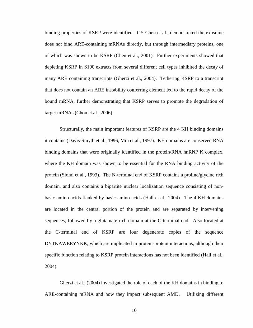
Figure 1.1 Overview of KSRP binding to the ARE of GAP-43
Shown here is a schematic of how KSRP binds to ARE-containing mRNA. The fourth KH domain of
KSRP has been demonstrated to be crucial for binding to KSRP target mRNAs. The third KH domain is
critical for recruiting the exosome and PARN to the bound transcript, where deadenylation and decapping
events take place, followed by 3’ to 5’ degradation by the exosome.
Identifying a specific consensus sequence for KSRP binding has proven elusive.
KSRP can promote the decay of mRNA reporter constructs containing any of the 3 ARE
classes (Gherzi et al., 2004). A binding study examining the affinity of the different KH
domains for RNA tetramers of varying composition failed to identify a motif that KSRP
12
preferred to bind, as the KH domains bound most of the tetramers with similar affinity,
with the exclusion of the KH3 domain which has a slight affinity for guanine (G) rich
sequences (Garcia-Mayoral et al., 2008). A subsequent experiment by the same group
showed that the third and fourth KH domains bind AU rich sequences similar to the type
II ARE with a relatively high affinity, but still failed to identify a consensus sequence
(Diaz-Moreno et al., 2010). The most fruitful experiments to examining KSRP targets
have involved immunoprecipitation of KSRP and subsequent identification of bound
mRNAs by microarray analysis. One such study performed this type of
immunoprecipitation experiment, and also knocked down KSRP to see what mRNAs
were upregulated. Investigators analyzed the data obtained from both types of
experiments to see what types of mRNAs were regulated by KSRP, and found a large
number of targets, including cytokines and cellular growth factors (Winzen et al., 2007).
Post-transcriptional regulation of these mRNA targets by KSRP still needs to be verified,
however, which is a daunting task given the number of mRNA binding targets found.
Recently, KSRP has also been identified to function as an enhancer of microRNA
(miRNA) biogenesis. The initial processing of primary miRNA transcripts occurs in the
nucleus when the hairpin transcript is cleaved by the ribonuclease Drosha, producing a
pre-miRNA. Following nuclear export of the pre-miRNA it is once again cleaved, this
time by the ribonuclease Dicer, after which one or both strands of the RNA duplex is
incorporated into the RNA-induced silencing complex (RISC). Depending on the
sequence homology of a miRNA for its mRNA target, the mRNA may be translationally
repressed or actively degraded [for a review of miRNA biogenesis see (Kim, 2005)].
KSRP is able to bind to Drosha, Dicer and stem loop sequences in some pre-miRNAs,
13
promoting the biogenesis of a subset of miRNAs, including let-7 and miR-155 (Ruggiero
et al., 2009, Trabucchi et al., 2009). KSRP serves many cellular functions, and it is
interesting to note that it functions in two completely different post-transcriptional
regulatory processes with similar outcomes on gene expression.
1.4 Summary
Precise spatio-temporal regulation of GAP-43 expression during neuronal growth
and differentiation is essential for correct nervous system development. The instability-
conferring ARE in the GAP-43 3’UTR is critical in this process, as are the trans-acting
ARE-BPs that can bind to this mRNA to modulate the stability of the transcript. HuD
has repeatedly been shown to be a stabilizer of GAP-43 mRNA and functions to promote
axonal outgrowth. KSRP, a negative regulator of ARE-containing mRNA expression,
has the potential to bind to and promote the decay of GAP-43 mRNA. In this dissertation
we examine this possibility, and show that KSRP does indeed function in neurons to
modulate the expression of GAP-43.
14
2. Rationale, hypothesis, and specific aims
2.1 Rationale
Stabilization of the GAP-43 transcript by HuD has been extensively studied, but the
opposite side of the spectrum has yet to be investigated: are there other ARE-BPs
associated with GAP-43 mRNA that destabilize this transcript when neurons mature?
Preliminary investigations in our laboratory demonstrated that the destabilizing factor
KSRP binds to GAP-43 transcripts.
2.2 Hypothesis
KSRP functions in direct opposition to HuD, destabilizing GAP-43 mRNA during neurite
development to counteract HuD’s growth stimulating effect. Overexpression of KSRP is
hypothesized to limit GAP-43 transcript levels and limit primary axonal outgrowth, while
knockdown of KSRP will increase GAP-43 mRNA levels and axonal growth.
2.3 Specific aims
The above hypothesis was investigated with two specific aims:
2.3.1 Specific aim 1:
How does KSRP affect GAP-43 mRNA stability in vitro? In order to demonstrate that
KSRP functions in opposition to HuD’s stabilizing effect on this target mRNA, we will
need to determine whether KSRP serves to promote GAP-43 mRNA degradation. We
will accomplish this aim utilizing in vitro binding and mRNA decay assays.
15
2.3.2 Specific aim 2:
How does KSRP expression affect axonal outgrowth in primary neuronal cultures?
2.3.2.1 Specific aim 2A:
Will overexpression or knockdown of KSRP in developing neurons lead to limited or
enhanced axonal outgrowth, respectively? To examine the involvement of KSRP in
developing axons, we will transfect primary hippocampal neuronal cultures with a GFP
conjugated plasmid overexpressing KSRP, or a GFP-shRNA construct to knockdown
KSRP. We can then examine axon outgrowth through fluorescence microscopy.
2.3.2.2 Specific aim 2B:
In neurons overexpressing KSRP, can the resulting neuronal phenotype be rescued with
overexpression of GAP-43? Performing this experiment will be critical in demonstrating
that KSRP’s effect on axon growth is due to its modulation of GAP-43 mRNA stability.
16
3. Regulation of GAP-43 stability by KSRP in vitro
3.1 Introduction
The regulation of gene expression by stabilization or degradation of mRNA
transcripts is a well-documented process important during a number of cellular events,
including cytokine production and cellular differentiation (Wilusz and Wilusz, 2004).
GAP-43 is important in developing neurons, where GAP-43 protein promotes axonal
outgrowth in response to growth factor signaling (Goslin et al., 1988, Perrone-Bizzozero
et al., 1993). GAP-43 is also important in mature synapses, and is expressed during
learning and memory processes. The mRNA for GAP-43 is unstable, a trait conferred to
it by the class III ARE present in its 3’UTR. The ARE-BP HuD has been repeatedly
proven to be a post-transcriptional regulator of GAP-43 mRNA, stabilizing the transcript
in developing and regenerating neurons (Anderson et al., 2000, Anderson et al., 2003).
KSRP, a mRNA destabilizing protein, has been implicated in contributing to the
degradation of a variety of transcripts. KSRP binds to the ARE sequence present in many
labile transcripts, and targets them for degradation by binding to the exosomal
degradation machinery (Chen et al., 2001). KSRP contains 4 RNA binding KH domains
that are important for binding to AU rich elements, but the third and fourth domains are
the most essential for its degradatory function (Gherzi et al., 2004). The fourth domain is
important for binding to target mRNAs, while the third domain is essential for binding to
the exosome. KSRP is highly expressed in brain tissue, but little is known about its
function in the nervous system (Gu et al., 2002).
17
In the following in vitro studies, we examine the role of KSRP in regulating GAP-
43 mRNA stability. We approach these studies using a combination of in vitro binding
and decay studies to see if KSRP degrades GAP-43 transcripts. We also investigate
whether or not KSRP and HuD compete for binding to the same sequence in the GAP-43
3’UTR.
3.2 Materials and methods
Biotinylated GAP-43 mRNA protein pull-down. (Performed by Dan Tanner)
Biotinylated mRNA was prepared and pull-down assays performed as described
by Ambrosino et al. (2003) with minor modifications (Ambrosino et al., 2003). Briefly, a
biotinylated RNA containing GAP-43 ARE was prepared from EcoRI linearized pGAP/B
plasmid (Kohn et al., 1996) by in vitro transcription using SP6 RNA polymerase and
biotin-11-UTP (Enzo Life Sciences, Farmingdale, NY). RNA (2 µg) was resuspended in
1X TENT buffer (20 mM Tris-HCl pH 8.0, 2mM EDTA pH 8.0, 500 mM NaCl, 1%
[vol/vol] Triton X-100) and then incubated with 300 µg of S100 extract prepared from rat
cortices and 40 units RNasin (Promega, Madison, WI) for 30 minutes at room
temperature. Control reactions did not include biotinylated RNA. Pre-washed
streptavidin-coated Dynabeads (Life Technologies) were then added to each reaction and
allowed to incubate for 30 more minutes at room temperature. Beads were collected by
centrifugation, washed twice with TENT buffer, then resuspended in SDS-PAGE loading
buffer. Samples were then run on a 10% SDS-PAGE gel. Western blotting was
performed as described previously (Anderson et al., 2001) using α-KSRP AB5 antibody
18
provided by Dr. Doug Black and affinity purified anti-HuD antibody (Mobarak et al.,
2000).
RNA electrophoretic mobility-shift assay (REMSA)
32P-labeled GAP-43 ARE mRNA was prepared by in vitro transcription from
EcoRI linearized pGAP/B plasmid using SP6 RNA polymerase and [α-32
P] UTP (3000
Ci/mmol, Perkin-Elmer, Waltham, MA). REMSA assay was performed as described by
Li et al. with minor modifications (Li et al., 2004). Briefly, 100,000 CPM (45 nCi) of
32P-UTP labeled GAP-43 3’ARE mRNA was incubated with increasing amounts of
purified GST or recombinant GST-KSRP in a buffer containing 50 mM Tris-HCl pH 7.0,
150 mM NaCl, 0.25 mg/ml tRNA, 0.25 mg/ml bovine serum albumin, and 5% glycerol
for 10 min at 37˚C. RNA-protein complexes were then run on a non-denaturing 10%
polyacrylamide gel in TBE buffer for 45 min. at 200V. The gel was then dried and
exposed to a phosphor screen overnight before radioactivity was measured using a Bio-
Rad Personal Molecular Imager FX (Bio-Rad, Hercules, CA).
Competitive RNA binding assay
Protein G dynabeads (Life Technologies) were washed and pre-bound with HuD
E-1 antibody (Santa Cruz). 32
P-labeled GAP-43 ARE was prepared as described above.
Labeled GAP-43 ARE was incubated with 1.5 nmol GST-HuD protein and increasing
amounts of GST or GST-KSRP in a binding buffer containing 10 mM HEPES pH 7.4,
100 mM KCl, 5 mM MgCl2, and 0.5% NP40. Assays were then incubated for 10 minutes
at 4˚C before being exposed to UV light for 30 minutes at 4˚C. Pre-bound beads were
then added to the binding assays along with 40 U of RNasin RNase inhibitor (Promega).
19
Samples were then mixed at 4˚C for 1 hour on a rotating mixer, then washed three times
with binding buffer. Samples were then resuspended in Tris-EDTA buffer and the
radiation measured by scintillation count.
In vitro mRNA decay assay
KSRP KO mouse S100 extracts were prepared from cortical brain tissue as
described (Ford et al., 1999). GST protein and recombinant GST-KSRP protein was
expressed in BL21 E. coli and purified using the MagneGST™ Protein Purification
System (Promega) according to the manufacturer’s protocol. 32
P-labeled GAP-43 3’UTR
mRNA was prepared and decay reactions were performed as described previously
(Bolognani et al., 2006), with minor modifications: 50 fmol 32
P-labeled GAP-43 3’UTR
mRNA was incubated with 20 µg KSRP KO S100 protein and 50 ng purified
recombinant protein.
3.3 Results
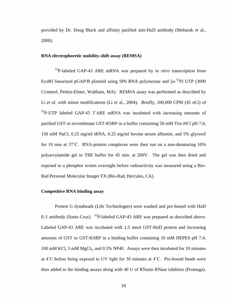
3.3.1 KSRP binds to GAP-43 mRNA in vitro: RNA electrophoretic mobility shift
Given that KSRP is known to bind ARE sequences, initial studies used two
separate in vitro binding assays to determine if this RBP directly binds to GAP-43
mRNA. First, we utilized REMSA using radiolabeled GAP-43 ARE. As shown in Figure
3.1A, incubation with 25 ng of GST-KSRP protein was sufficient to cause a shift in the
migration of a GAP-43 ARE containing RNA, and bands were completely shifted in the
presence of 100 ng KSRP protein. In contrast, there was no shift in the mobility of the
mRNA when incubated with up to 10 µg of the GST protein control. Previous research
20
Figure 3.1 Binding of KSRP to GAP-43 ARE
A. REMSA shift assays used increasing amounts of purified GST, GST-KSRP, or GST-KSRP-ΔKH4
protein and 32
P-labeled GAP-43 ARE. B. RNA pull-down assay was performed with biotinylated GAP-43
ARE and S100 cytosolic protein extracts from mouse brain as indicated in the Methods (Performed by Dan
Tanner). Western blots (WB) show the presence of KSRP and HuD proteins in the RNA pull down. C.
Competitive binding assay of HuD and KSRP. 32
P-labeled GAP-43 ARE was incubated with HuD and
increasing amounts of either GST or KSRP competitor protein, before HuD was pulled down and bound
RNA measured by scintillation counting. **, p < .05 using Student’s t-test (n=2).
21
showed that KSRP binds ARE sequences primarily via its 4th
KH domain (Gherzi et al.,
2004). Thus, we used a KSRP protein lacking this domain (GST-KSRP-ΔKH4) for
binding GAP-43 ARE. GST-KSRP-ΔKH4 bound to the GAP-43 ARE, but with
significantly less affinity than wild-type KSRP (Figure 3.1A)(Gherzi et al., 2004).
3.3.2 KSRP binds to GAP-43 mRNA in vitro: biotinylated GAP-43 mRNA pulldown
(Experiment performed by Dan Tanner)
In a separate binding assay, biotinylated GAP-43 ARE-containing RNA was
incubated with rat S100 protein extracts, and mRNA/protein complexes pulled down
using streptavidin coated beads. Analysis of the bound proteins by western blot revealed
that KSRP, as well as HuD (Figure 3.1B) bound the GAP-43 ARE.
3.3.3 HuD and KSRP compete for binding to the GAP-43 ARE
Given that both HuD and KSRP bind to the same ARE sequence, we then
examined the ability of KSRP to compete with HuD for binding to this RNA. For these
studies, 32
P-labeled GAP-43 ARE was incubated with 1.5 nmol of GST-HuD protein and
increasing amounts of either GST or GST-KSRP. HuD was then immunoprecipitated
and the amount of mRNA bound was measured by scintillation counting. Increasing
amounts of GST protein did not interfere with HuD binding to GAP-43 ARE, while
increasing amounts of KSRP displaced HuD from the transcript (Figure 3.1C). When
equal molar amounts of KSRP and HuD were used in the assay HuD binding to GAP-43
ARE decreased by roughly half, indicating that KSRP and HuD have a similar affinity for
binding GAP-43 ARE (Figure 3.1C).
22
3.3.4 KSRP destabilizes GAP-43 mRNA in vitro
We used in vitro decay assays to determine if KSRP has an effect on GAP-43
mRNA stability (Figure 3.2). For this, 32
P-labeled capped and polyadenylated RNA
containing the GAP-43 ARE was incubated with purified recombinant GST-KSRP
protein along with S100 protein extracts from KSRP KO mice, and decay of the labeled
mRNA was measured over time. This experimental system has been shown to reliably
represent the relative decay rates of several mRNAs, as the protein complexes required
for mRNA degradation are present in S100 extracts (Ford et al., 1999, Chen et al., 2000).
The use of S100 extracts from KSRP KO mice also ensures the absence of endogenous
KSRP, which could confound the decay results (Lin et al., 2011). When GST was
incubated with GAP-43 ARE, the mRNA decayed with a half-life of 10.7 ± 5.7 minutes.
Addition of GST-KSRP to the decay system enhanced the degradation rate of GAP-43
ARE, decreasing the half-life of the mRNA to 3.2 ± 1.5 minutes, which is significantly
different than control GST (p < .05) using two-way ANOVA. In contrast, addition of
recombinant KSRP protein lacking the 4th
RNA binding KH domain (GST-KSRP-ΔKH4)
resulted in a RNA half-life that is not significantly different than control (Chou et al.,
2006). These experiments indicate that KSRP binds to GAP-43 mRNA, and increases
the decay rate of GAP-43 mRNA in vitro via the interaction of GAP-43 ARE with the 4th
KH domain in the protein.
23
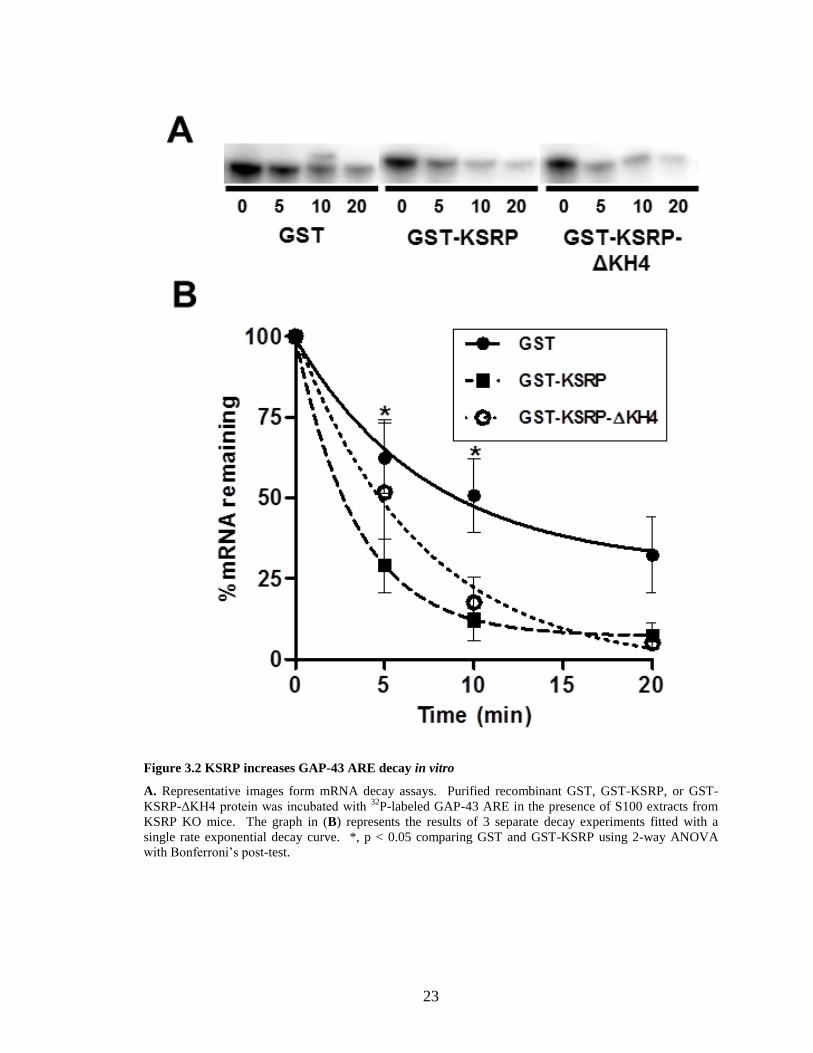
Figure 3.2 KSRP increases GAP-43 ARE decay in vitro
A. Representative images form mRNA decay assays. Purified recombinant GST, GST-KSRP, or GST-
KSRP-ΔKH4 protein was incubated with 32
P-labeled GAP-43 ARE in the presence of S100 extracts from
KSRP KO mice. The graph in (B) represents the results of 3 separate decay experiments fitted with a
single rate exponential decay curve. *, p < 0.05 comparing GST and GST-KSRP using 2-way ANOVA
with Bonferroni’s post-test.
24
3.4 Discussion
In this study, we show for the first time that KSRP binds to and destabilizes GAP-43
mRNA in vitro. In the REMSA experiments, GAP-43 mRNA migration through the gel
was shifted completely by 100 ng of KSRP protein, demonstrating that KSRP closely
associates with GAP-43 ARE. KSRP-ΔKH4 protein also bound to the GAP-43 ARE, but
with less affinity than full length KSRP protein. This was expected, based on the fact
that the fourth KSRP KH domain was found to be primarily responsible for ARE binding
(Gherzi et al., 2004). The KSRP-ΔKH4 protein still binds to the GAP-43 ARE sequence
with some affinity, because the first two KH domains aid in binding ARE sequences
(Gherzi et al., 2004). These binding studies help to confirm the results obtained from the
in vitro decays, which showed that full length KSRP protein led to the fastest degradation
rate of GAP-43 ARE. KSRP-ΔKH4 also enhanced the decay rate of the GAP-43 when
compared to the GST control, but less so than the full length KSRP protein, supposedly
due to the reduced affinity of the KSRP-ΔKH4 protein for the ARE sequence.
Performing these decays in S100 extracts from the brains of KSRP KO mice provided by
CY Chen was valuable in running a clean assay, as there was no endogenous KSRP to
confound results (Lin et al., 2011).
Future experiments that could be performed to enhance the results of these decay
studies would further utilize the KSRP KO mouse. In vivo decay assays analyzing GAP-
43 mRNA stability could be performed in cortical neuronal cultures from these mice,
using Actinomycin D to inhibit transcription. Cultures from wild-type and KO mice
would be cultured at the same time, and at a specific time point after plating Actinomycin
D would be added to the media, and neurons collected at measured time points. The
25
collected neurons would be flash-frozen to inhibit further RNA degradation, and
measured using RT-PCR. Such a decay experiment would be relatively easy to perform.
The reason such a study has not been performed to date is purely logistical: we have not
had access to a breeding colony of KSRP KO mice until recently. Most likely we will
perform these experiments in the near future to further strengthen our results, however,
the evidence that KSRP enhances the decay of GAP-43 ARE obtained from our assays
performed is already quite strong.
It is interesting to note that KSRP and HuD appear to compete for the same
binding site in the GAP-43 ARE. Analyzing the actual binding affinities for each of
these ARE-BPs will be integral to understanding how they affect GAP-43 mRNA
stability during different cellular conditions, to see if KSRP would displace HuD from the
transcript. During axonal outgrowth GAP-43 mRNA is bound by HuD, stabilizing the
transcript for translation. After the axon has reached its target and the neuron matures,
GAP-43 and HuD levels both drop off (Skene et al., 1986, Bolognani et al., 2007b). It
would be interesting to know exactly when KSRP binds to GAP-43 mRNA after the axon
has reached its target. Performing high-resolution in situ hybridization and
immunocytochemistry at different points during neuron development, both in cultured
neurons and in vivo could lend some insight as to the precise time-course of KSRP, HuD,
and GAP-43 expression in the developing brain.
26
4. KSRP inhibits axonal outgrowth in cultured hippocampal neurons
4.1 Introduction
In developing neurons, GAP-43 mRNA expression is essential for axonal growth
and pathfinding processes (Skene et al., 1986). GAP-43 is critical during development,
as GAP-43 KO mice die before birth, and exhibit impairments in axonal targeting
(Strittmatter et al., 1995). If GAP-43 is overexpressed, aberrant axonal sprouting is
observed (Aigner et al., 1995, Klein et al., 1999). The ARE-BP HuD stabilizes the GAP-
43 transcript, and overexpressing HuD increases the rate of process outgrowth in both
NGF-stimulated PC12 cells and cultured cortical neurons (Anderson et al., 2000,
Anderson et al., 2001). HuD overexpressor mice have elevated GAP-43 mRNA levels in
the dentate gyrus of the hippocampus, demonstrating that HuD not only regulates GAP-
43 expression in vitro and in cell culture, but in vivo as well (Bolognani et al., 2006).
Regulation of GAP-43 expression is imperative to proper neural development,
and knowledge about regulatory processes impacting GAP-43 mRNA stability are
evolving as we gain knowledge about ARE binding proteins and their targets. Work
presented in the previous chapter of this dissertation demonstrated that the destabilizing
protein KSRP is able to bind to the GAP-43 ARE in vitro, and serves to destabilize GAP-
43 mRNA. The following experiments examine the functional consequences of KSRP
expression in hippocampal cultures using KSRP overexpression vectors, as well as
shKSRP vectors to achieve knockdown of this protein. We also were able to measure
GAP-43 protein levels in neurons in which KSRP has been knocked down, and mRNA
levels in PC12 cells transfected with shKSRP and sorted to attain an enriched population
of KSRP deficient cells.
27
4.2 Materials and methods
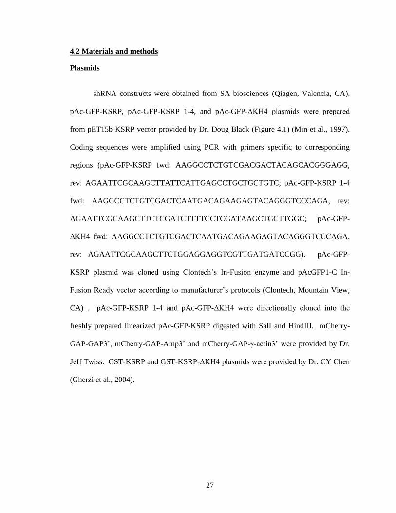
Plasmids
shRNA constructs were obtained from SA biosciences (Qiagen, Valencia, CA).
pAc-GFP-KSRP, pAc-GFP-KSRP 1-4, and pAc-GFP-ΔKH4 plasmids were prepared
from pET15b-KSRP vector provided by Dr. Doug Black (Figure 4.1) (Min et al., 1997).
Coding sequences were amplified using PCR with primers specific to corresponding
regions (pAc-GFP-KSRP fwd: AAGGCCTCTGTCGACGACTACAGCACGGGAGG,
rev: AGAATTCGCAAGCTTATTCATTGAGCCTGCTGCTGTC; pAc-GFP-KSRP 1-4
fwd: AAGGCCTCTGTCGACTCAATGACAGAAGAGTACAGGGTCCCAGA, rev:
AGAATTCGCAAGCTTCTCGATCTTTTCCTCGATAAGCTGCTTGGC; pAc-GFP-
ΔKH4 fwd: AAGGCCTCTGTCGACTCAATGACAGAAGAGTACAGGGTCCCAGA,
rev: AGAATTCGCAAGCTTCTGGAGGAGGTCGTTGATGATCCGG). pAc-GFP-
KSRP plasmid was cloned using Clontech’s In-Fusion enzyme and pAcGFP1-C In-
Fusion Ready vector according to manufacturer’s protocols (Clontech, Mountain View,
CA) . pAc-GFP-KSRP 1-4 and pAc-GFP-ΔKH4 were directionally cloned into the
freshly prepared linearized pAc-GFP-KSRP digested with SalI and HindIII. mCherry-
GAP-GAP3’, mCherry-GAP-Amp3’ and mCherry-GAP-γ-actin3’ were provided by Dr.
Jeff Twiss. GST-KSRP and GST-KSRP-ΔKH4 plasmids were provided by Dr. CY Chen
(Gherzi et al., 2004).
28
Figure 4.1 GFP-KSRP constructs cloned for KSRP overexpression studies.
GFP-KSRP-KH 1-4 lacks a NLS, while the GFP-KSRP-ΔKH4 lacks both a NLS and the 4th
KH domain.
Fluorescence activated cell sorting
Fluorescence activated cell sorting (FACS) was performed in the UNM Flow
Cytometry Facility by dedicated personnel. 10 cm dishes of PC12 cells near 100%
confluence were incubated overnight in Opti-MEM media supplemented with 4% serum
before being transfected with either control non-targeting or shKSRP GFP plasmids.
Transfections were performed with 24 µg of plasmid per dish using Lipofectamine 2000
(Life Technologies) according to manufacturer’s protocol. Transfection media was
replaced the next day with Opti-MEM + 4% serum. Neurons were grown for 48 hours
post transfection before cells were trypsinized and pelleted, then resuspended in sorting
medium (cation free PBS supplemented with 0.2% fetal bovine serum, 10mM HEPES pH
7.3, 1 mM EDTA) at a concentration of 5 x 10⁶
cells/ml. Cells were then sorted, using
gating to collect the brightest 3% of GFP positive cells. Cells were collected in sorting
medium, then pelleted by centrifugation at 10,000 x g for 1 min and flash frozen on dry
ice before RNA was extracted.
29
Real-time PCR
Quantitative real-time PCR (qRT-PCR) was performed as described previously
(Bullock et al., 2008). Briefly, RNA was extracted from sorted PC12 cells using an
RNeasy mini kit (Qiagen). cDNA was synthesized using 1µg total RNA using
Superscript II reverse transcriptase (Invitrogen) according to manufacturer’s protocol.
Exon spanning primer pairs were designed using Primer Express 3.0 (Life Technologies)
and validated using NCBI primer BLAST software. Primers used for analysis were:
KSRP (forward 5’-GGACTCAGGCTGCAAAGTTC, reverse 5’-
CCAGGATCATCTTTGCCTTT), GAP-43 (forward 5’-
AGCCAAGGAGGAGCCTAAAC, reverse 5’-CTGTCGGGCACTTTCCTTAG), and
GAPDH (forward 5’-TGTGATGGGTGTGAACCACGAGAA, reverse 5’-
GAGCCCTTCCACAATGCCAAAGTT). qRT-PCR reactions were performed on an
Applied Biosystems 7300 Real Time PCR System. Gene expression levels were
analyzed using SYBR Green (Life Technologies). No evidence of primer dimerization
was evident by dissociation curve analysis. Primers for KSRP and GAP-43 were
validated against GAPDH and were within optimal amplification values (slope <|0.1|).
Samples were run in triplicate, and relative levels of expression compared to GAPDH
were calculated using the 2-ΔΔCt
method (Livak and Schmittgen, 2001).
Primary hippocampal cultures
Primary hippocampal neuronal cultures were isolated and grown as described
previously (Smrt et al., 2010). Briefly, hippocampi from E17 Sprague-Dawley rats were
pooled, triturated with a fire-polished Pasteur pipette and dissociated neurons plated on
30
poly-D-lysine/laminin coated coverslips (BD Biosciences, San Jose, CA). Cultures were
grown in Neurobasal medium (Life Technologies, Grand Island, NY) supplemented with
B27 supplement (Life Technologies), Penicillin/Streptomycin (Life Technologies), 0.5
mM L-glutamine and 25 µM glutamate for 4 days in vitro (DIV) before being transfected
with GFP plasmid constructs using Lipofectamine 2000 and Opti-MEM (Life
Technologies) according to manufacturer’s protocol. Cells were incubated with
transfection medium for 48 hours before being fixed with 4% paraformaldehyde.
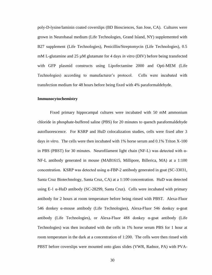
Immunocytochemistry
Fixed primary hippocampal cultures were incubated with 50 mM ammonium
chloride in phosphate-buffered saline (PBS) for 20 minutes to quench paraformaldehyde
autofluorescence. For KSRP and HuD colocalization studies, cells were fixed after 3
days in vitro. The cells were then incubated with 1% horse serum and 0.1% Triton X-100
in PBS (PBST) for 30 minutes. Neurofilament light chain (NF-L) was detected with α-
NF-L antibody generated in mouse (MAB1615, Millipore, Billerica, MA) at a 1:100
concentration. KSRP was detected using α-FBP-2 antibody generated in goat (SC-33031,
Santa Cruz Biotechnology, Santa Cruz, CA) at a 1:100 concentration. HuD was detected
using E-1 α-HuD antibody (SC-28299, Santa Cruz). Cells were incubated with primary
antibody for 2 hours at room temperature before being rinsed with PBST. Alexa-Fluor
546 donkey α-mouse antibody (Life Technologies), Alexa-Fluor 546 donkey α-goat
antibody (Life Technologies), or Alexa-Fluor 488 donkey α-goat antibody (Life
Technologies) was then incubated with the cells in 1% horse serum PBS for 1 hour at
room temperature in the dark at a concentration of 1:200. The cells were then rinsed with
PBST before coverslips were mounted onto glass slides (VWR, Radnor, PA) with PVA-
31
DABCO mounting media. Mounted coverslips were dried overnight in the dark at room
temperature before imaging.
Microscopy and image analysis
For quantitation of axonal outgrowth, transfected neurons were imaged with an
Olympus BX60 fluorescence microscope with a 20X objective and images collected
using an Olympus DP71 camera. For neurons that were too large to be imaged in a single
20X field, multiple overlapping images of the same neuron were taken and the images
merged together using Adobe Photoshop Elements (Adobe, San Jose, CA). For
quantitation of KSRP shRNA knockdown and analysis of KSRP and HuD localization
transfected primary hippocampal neurons were imaged with a Zeiss LSM 510 confocal
microscope. Axonal outgrowth of transfected primary hippocampal neurons was
measured using Neurolucida (MBF biosciences, Williston, VT). Cell bodies and primary
axons were traced using the program, and overall axonal length per neuron was
calculated. Quantitation of axonal outgrowth was calculated from multiple transfection
experiments run in duplicate. In each coverslip, roughly 6 GFP positive neuron axons
were measured for overall length, and these lengths were averaged together to generate a
single n for statistical purposes. About 50 total cells were measured per condition.
4.3 Results
4.3.1 KSRP and HuD localize to separate cytoplasmic granules in developing
hippocampal neurons
We examined whether HuD or KSRP colocalize to the same cytoplasmic granules
in the neuronal cytoplasm. Because HuD stabilizes and KSRP destabilizes bound ARE
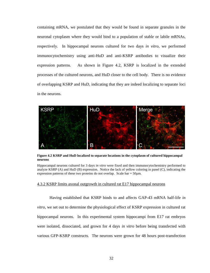
32
containing mRNA, we postulated that they would be found in separate granules in the
neuronal cytoplasm where they would bind to a population of stable or labile mRNAs,
respectively. In hippocampal neurons cultured for two days in vitro, we performed
immunocytochemistry using anti-HuD and anti-KSRP antibodies to visualize their
expression patterns. As shown in Figure 4.2, KSRP is localized in the extended
processes of the cultured neurons, and HuD closer to the cell body. There is no evidence
of overlapping KSRP and HuD, indicating that they are indeed localizing to separate loci
in the neurons.
Figure 4.2 KSRP and HuD localized to separate locations in the cytoplasm of cultured hippocampal
neurons
Hippocampal neurons cultured for 3 days in vitro were fixed and then immunocytochemistry performed to
analyze KSRP (A) and HuD (B) expression. Notice the lack of yellow coloring in panel (C), indicating the
expression patterns of these two proteins do not overlap. Scale bar = 50µm.
4.3.2 KSRP limits axonal outgrowth in cultured rat E17 hippocampal neurons
Having established that KSRP binds to and affects GAP-43 mRNA half-life in
vitro, we set out to determine the physiological effect of KSRP expression in cultured rat
hippocampal neurons. In this experimental system hippocampi from E17 rat embryos
were isolated, dissociated, and grown for 4 days in vitro before being transfected with
various GFP-KSRP constructs. The neurons were grown for 48 hours post-transfection
33
and then fixed and immunostained with anti-NF-L antibody to distinguish neurons from
astrocytes (data not shown).
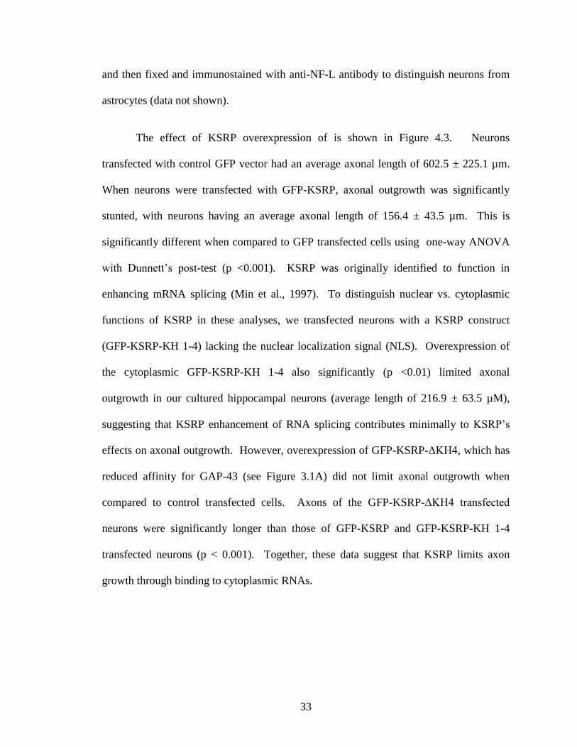
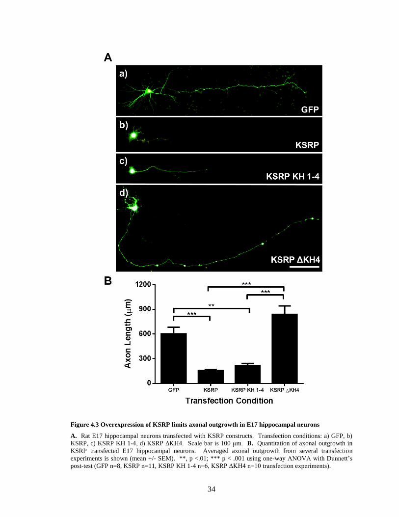
The effect of KSRP overexpression of is shown in Figure 4.3. Neurons
transfected with control GFP vector had an average axonal length of 602.5 ± 225.1 µm.
When neurons were transfected with GFP-KSRP, axonal outgrowth was significantly
stunted, with neurons having an average axonal length of 156.4 ± 43.5 µm. This is
significantly different when compared to GFP transfected cells using one-way ANOVA
with Dunnett’s post-test (p <0.001). KSRP was originally identified to function in
enhancing mRNA splicing (Min et al., 1997). To distinguish nuclear vs. cytoplasmic
functions of KSRP in these analyses, we transfected neurons with a KSRP construct
(GFP-KSRP-KH 1-4) lacking the nuclear localization signal (NLS). Overexpression of
the cytoplasmic GFP-KSRP-KH 1-4 also significantly (p <0.01) limited axonal
outgrowth in our cultured hippocampal neurons (average length of 216.9 ± 63.5 µM),
suggesting that KSRP enhancement of RNA splicing contributes minimally to KSRP’s
effects on axonal outgrowth. However, overexpression of GFP-KSRP-ΔKH4, which has
reduced affinity for GAP-43 (see Figure 3.1A) did not limit axonal outgrowth when
compared to control transfected cells. Axons of the GFP-KSRP-ΔKH4 transfected
neurons were significantly longer than those of GFP-KSRP and GFP-KSRP-KH 1-4
transfected neurons (p < 0.001). Together, these data suggest that KSRP limits axon
growth through binding to cytoplasmic RNAs.
34
Figure 4.3 Overexpression of KSRP limits axonal outgrowth in E17 hippocampal neurons
A. Rat E17 hippocampal neurons transfected with KSRP constructs. Transfection conditions: a) GFP, b)
KSRP, c) KSRP KH 1-4, d) KSRP ΔKH4. Scale bar is 100 µm. B. Quantitation of axonal outgrowth in
KSRP transfected E17 hippocampal neurons. Averaged axonal outgrowth from several transfection
experiments is shown (mean +/- SEM). **, p <.01; *** p < .001 using one-way ANOVA with Dunnett’s
post-test (GFP n=8, KSRP n=11, KSRP KH 1-4 n=6, KSRP ΔKH4 n=10 transfection experiments).
35
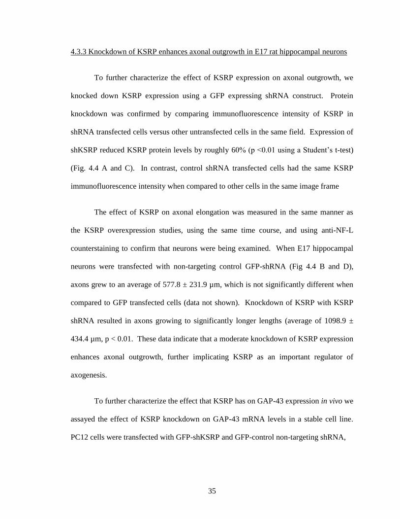
4.3.3 Knockdown of KSRP enhances axonal outgrowth in E17 rat hippocampal neurons
To further characterize the effect of KSRP expression on axonal outgrowth, we
knocked down KSRP expression using a GFP expressing shRNA construct. Protein
knockdown was confirmed by comparing immunofluorescence intensity of KSRP in
shRNA transfected cells versus other untransfected cells in the same field. Expression of
shKSRP reduced KSRP protein levels by roughly 60% (p <0.01 using a Student’s t-test)
(Fig. 4.4 A and C). In contrast, control shRNA transfected cells had the same KSRP
immunofluorescence intensity when compared to other cells in the same image frame
The effect of KSRP on axonal elongation was measured in the same manner as
the KSRP overexpression studies, using the same time course, and using anti-NF-L
counterstaining to confirm that neurons were being examined. When E17 hippocampal
neurons were transfected with non-targeting control GFP-shRNA (Fig 4.4 B and D),
axons grew to an average of 577.8 ± 231.9 µm, which is not significantly different when
compared to GFP transfected cells (data not shown). Knockdown of KSRP with KSRP
shRNA resulted in axons growing to significantly longer lengths (average of 1098.9 ±
434.4 µm, p < 0.01. These data indicate that a moderate knockdown of KSRP expression
enhances axonal outgrowth, further implicating KSRP as an important regulator of
axogenesis.
To further characterize the effect that KSRP has on GAP-43 expression in vivo we
assayed the effect of KSRP knockdown on GAP-43 mRNA levels in a stable cell line.
PC12 cells were transfected with GFP-shKSRP and GFP-control non-targeting shRNA,
36
Figure 4.4 shRNA knockdown of KSRP in transfected hippocampal neurons increases axonal length.
A. Representative images of shRNA transfections. Control (a-c) or anti-KSRP (d-f) GFP-shRNA plasmids
were transfected into primary hippocampal neuronal cultures, and KSRP expression was quantified by
comparing KSRP immunofluorescence levels with untransfected cells in the same field. Scale bar is 25
µm. B. Rat E17 hippocampal neurons transfected with shRNA constructs. Transfection conditions: a)
control shRNA, b) α-KSRP shRNA. Scale bar is 100 µm. C. Quantification of KSRP knockdown by anti-
KSRP shRNA. Knockdown was measured by comparing KSRP immunofluorescence intensity in shRNA
transfected cells vs. untransfected cells in the same image frame. Plotted graphs are relative mean (+/-
SEM) levels of KSRP fluorescence intensity. **, p < .01, Student’s t-test (n=7 transfections for control
shRNA; n=14 transfections for shKSRP). D. Quantitation of axonal outgrowth in shRNA transfected E17
hippocampal neurons. Averaged axonal outgrowth from 10 separate transfection experiments is shown
(mean +/- SEM). **, p < .01 using Student’s t-test.
37
and incubated for 48 hours. Transfected cells were then purified using fluorescent
activated cell sorting to enrich for the brightest 2.5% of transfected GFP positive cells
(Appendix A, images A and B). KSRP and GAP-43 mRNA expression was then
analyzed by qRT-PCR. Knockdown of KSRP by the shRNA construct was robust,
lowering KSRP mRNA levels by roughly 95% (Appendix A, graph C). GAP-43 mRNA
levels were increased 1.7 fold in cells with reduced KSRP (Appendix A, graph D). These
results indicate that KSRP is a negative regulator of GAP-43 expression, as knockdown
of KSRP can increase GAP-43 expression.
4.3.4 Rescue of limited axonal outgrowth in KSRP transfected neurons by co-expression
of GAP-43
Based on the results of the overexpression and knockdown of KSRP in affecting
axonal outgrowth, and the knowledge that KSRP binds to and affects the stability of
GAP-43 mRNA, we sought to determine if the phenotype resulting from KSRP
overexpression could be rescued by overexpressing GAP-43 in the same neurons. These
rescue experiments were performed by transfecting E17 hippocampal neurons with GFP-
KSRP along with different mCherry-GAP-43 constructs. We used three different GAP-
43 constructs, each with a different 3’UTR. The mCherry-GAP-GAP3’ plasmid contains
GAP-43 coding region along with GAP-43 3’UTR, which contains the ARE and the
element necessary for targeting GAP-43 to axonal growth cones, where its localized
translation is required for promoting axonal extension (Donnelly et al., 2011). mCherry-
GAP-AMP3’ contains the GAP-43 coding region with the 3’UTR from amphoterin,
which does not contain an ARE but does contain an axonal targeting sequence. As
38
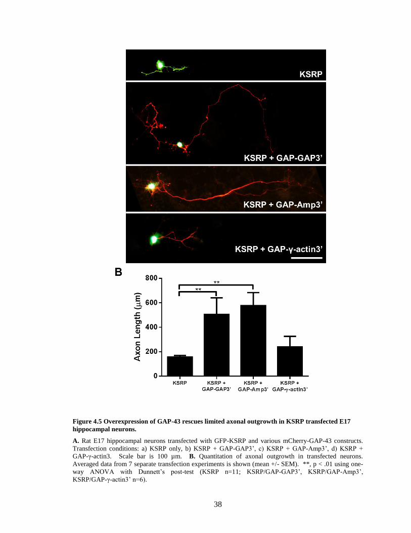
Figure 4.5 Overexpression of GAP-43 rescues limited axonal outgrowth in KSRP transfected E17
hippocampal neurons.
A. Rat E17 hippocampal neurons transfected with GFP-KSRP and various mCherry-GAP-43 constructs.
Transfection conditions: a) KSRP only, b) KSRP + GAP-GAP3’, c) KSRP + GAP-Amp3’, d) KSRP +
GAP-γ-actin3. Scale bar is 100 µm. B. Quantitation of axonal outgrowth in transfected neurons.
Averaged data from 7 separate transfection experiments is shown (mean +/- SEM). **, p < .01 using one-
way ANOVA with Dunnett’s post-test (KSRP n=11; KSRP/GAP-GAP3’, KSRP/GAP-Amp3’,
KSRP/GAP-γ-actin3’ n=6).
39
control we used mCherry-GAP-γ-actin3’, which has the GAP-43 coding region with γ-
actin 3’UTR, and does not contain an ARE or an axonal targeting sequence.
Hippocampal neurons transfected with both GFP-KSRP and mCherry-GAP-
GAP3’ had axons grow to lengths significantly longer (505.6 ± 331.1 µm vs. 156.4 ±
43.5 µm, p < 0.01 using ANOVA with Dunnett’s post-test) than neurons transfected with
GFP-KSRP alone (Fig. 4.5). Neurons transfected with GFP-KSRP/mCherry-GAP-
AMP3’ also grew to lengths (average 577 ± 260.5 µm) significantly longer than neurons
transfected with GFP-KSRP alone and comparable to the levels of control GFP-
transfected neurons (Figure 4.3). In contrast, mCherry-GAP-γ-actin3’ did not rescue
axonal outgrowth in KSRP transfected cells, indicating that axonal targeting of GAP-43
mRNA is required for its correct function.
4.4 Discussion
Overexpression of KSRP in cultured hippocampal neurons led to a significant
reduction in axon outgrowth compared to control neurons. Knockdown of KSRP had the
opposite effect, resulting in axons growing out to significantly longer lengths. The
overexpression of KSRP could be rescued with GAP-43 overexpression, indicating that
KSRP regulates GAP-43 expression in neurons and has a significant impact on axonal
outgrowth during development.
KSRP appears to function in direct opposition to HuD, which is supported by the
finding that KSRP and HuD do not colocalize in the cytoplasm of cultured hippocampal
neurons. This supports the notion that HuD and KSRP are binding to separate pools of
mRNAs, with one group being stabilized for translation by HuD and another group
40
targeted for degradation by KSRP. This study only examined the locations of HuD and
KSRP after neurons were cultured for 3DIV. A more detailed time course study of ARE-
BP localization during neuronal development, both in cell culture and in vivo, will
provide valuable insight into how these proteins interact and localize within the cell to
post-transcriptionally regulate developmentally related ARE containing mRNA.
The shKSRP construct that we utilized led to a robust knockdown of KSRP
mRNA in PC12 cells, reducing KSRP transcript levels to 5% of control levels. In
cultured hippocampal neurons, shKSRP decreased KSRP protein levels by 60% based on
immunocytochemical analysis of KSRP expression. The difference in knockdown levels
between KSRP mRNA and protein could be attributed to the fact that turnover of RNA in
a cell is much faster than protein turnover, so a quick knockdown of mRNA levels will be
followed by a slower knockdown of the corresponding protein (Wu et al., 2004). In
addition, some differences in knockdown efficiencies can also be attributed to the two
different cells types utilized for these studies, i.e. PC12 cells vs. cultured hippocampal
neurons.
One of the most exciting results from these culture studies was the rescue of the
limited outgrowth in KSRP transfected neurons by concomitant overexpression of GAP-
43. Especially intriguing is the fact that axonal targeting of GAP-43 mRNA is necessary
for this rescue. The class III ARE of GAP-43 also contains a zip code targeting sequence
required for targeting this mRNA to axons. Recent studies have shown that
overexpression of GFP containing the β-actin 3’UTR, which also contains a zip code
sequence, leads to decreased GAP-43 mRNA in axons, and hinders axonal outgrowth
during development (Donnelly et al., 2011). Overexpression of a GAP-43 construct with
41
the γ-actin 3’UTR did not rescue limited axonal outgrowth caused by KSRP
overexpression, demonstrating that localization of GAP-43 mRNA to axons, and most
likely subsequent localized translation of GAP-43 is essential to promote axonal
elongation during neuronal maturation.
42
5. Discussion
RNA-binding protein-mediated mRNA stabilization is an important mechanism of
gene expression control during cell growth and differentiation. This is especially true in
the nervous system, where the neuronal RBP HuD was shown to stabilize the mRNAs of
several growth-related genes, such as GAP-43, AChE, tau, MARCKS and neuroserpin
controlling neuron development and function [for review see (Bolognani and Perrone-
Bizzozero, 2008)]. Despite the evident role of these post-transcriptional mechanisms in
neurons, very little is known of the factors responsible for destabilizing mRNAs upon
maturation. In this study, we have identified KSRP as a negative regulator of GAP-43
mRNA stability in developing neurons, which functions in an opposite manner to HuD,
promoting mRNA decay and limiting axonal outgrowth.
In vitro studies examining how KSRP interacts with GAP-43 mRNA clearly
indicate that KSRP binds to the highly conserved type III ARE present in the 3’UTR
(Chung et al., 1996). Furthermore, KSRP and HuD compete for binding to the same site
in GAP-43 mRNA with similar affinity. However, binding of KSRP to the GAP-43 ARE
has the opposite effect of HuD, decreasing mRNA stability by almost 3-fold. While in
vitro work is important in demonstrating KSRP modulation of GAP-43 transcript
stability, there are a number of other factors that interact in cells to affect gene
expression. To validate this in vitro result we performed shRNA knock-down studies and
found increased levels of GAP-43 mRNA in PC12 cells. Using primary hippocampal
neurons, we found that knockdown of KSRP drastically increases axonal outgrowth,
while overexpression stunts growth. The proper control of axonal outgrowth during
development is critical for forming functional synapses, so it seems that careful control of
43
both KSRP and HuD expression and their interaction with specific transcripts during
differentiation are important for regulating axonal elongation and hence normal neuronal
development.
Given that KSRP is highly expressed in brain tissue, it is interesting that there are
relatively few studies examining how it affects neuronal development. One of these
studies reported that the chick KSRP homolog zip code-binding protein 2 (ZBP2) aids the
other zip-code binding protein ZBP1 binding to β-actin mRNA in the nucleus, and
shuttles the mRNA to neurites for localized translation during development (Pan et al.,
2007). ZBP1 expression has also been shown to be crucial for proper axonal localization
of GAP-43 mRNA, because limited availability of ZBP1 depletes axonally localized
GAP-43 and hampers axonal outgrowth (Donnelly et al., 2011). Knock-down of ZBP2 in
a mouse neuroblastoma cell line hindered neurite outgrowth once the cells were
differentiated, supposedly due to decreased loading of ZBP1 onto β-actin mRNA (Pan et
al., 2007). In the present study, we observed the opposite effect in primary cultures with
KSRP knocked down. This apparent discrepancy is likely due to the differences in the
cell types used and the timing of the knockdown of KSRP expression, as we let the
neurons establish themselves for 4 days in vitro before performing shRNA knockdown.
Perhaps early in development KSRP functions to help load ZBP1 onto zip code
containing mRNAs in the nucleus, while it can subsequently function in the cell body and
processes to destabilize growth related transcripts to fine-tune neuronal development. It
is important to note that the ARE in GAP-43 3’ UTR also acts as a zip-code element that
is critical for its correct axonal localization, as shown by our rescue study showing that
44
overexpression of GAP-43 lacking these sequences (mCherry-GAP-γ-Actin3’) did not
rescue the phenotype induced by KSRP overexpression (Donnelly et al., 2011).
One factor crucial to understanding where KSRP is localized and how it binds
mRNAs during development is its phosphorylation state. KSRP can be phosphorylated
by p38 MAP kinase (p38 MAPK), which inhibits KSRP binding to ARE containing
mRNAs and prevents their subsequent decay (Briata et al., 2005). Phosphorylation of
KSRP’s KH1 domain also allows it to be bound by the regulatory 14-3-3ζ protein, after
which KSRP is localized to the nucleus where it cannot bind to cytoplasmically localized
labile transcripts (Diaz-Moreno et al., 2009). It is interesting to consider the possibility
that during HuD stabilizing events KSRP is mainly localized to the nucleus, aiding in the
export of zip-code containing mRNAs that are stabilized by HuD in the cytoplasm.
These HuD stabilizing events need not be limited to initial neuron outgrowth during
development, as there are other cellular processes in which HuD protein levels are up-
regulated to promote stability of GAP-43 mRNA. As mentioned in the introduction,
HuD levels are increased in the neuronal cell body of DRG neurons following a nerve
crush injury (Anderson et al., 2003). HuD protein and GAP-43 mRNA levels are also
increased during learning and memory processes (Quattrone et al., 2001, Pascale et al.,
2004). In the course of our studies, we have seen that KSRP and HuD do not colocalize
within the cytoplasm, lending support to the notion that these two RNA binding proteins
could be interacting with separate populations of mRNAs at a given time point. Studies
examining KSRP and HuD localization during these cellular events in which GAP-43 is
stabilized could lend insight into how these two RBPs with opposing effects on mRNA
stability operate in processes critical for neuronal growth and everyday neuronal function.
45
Based on the above information, I propose the following model for how KSRP
and HuD function during initial axonal pathfinding and later target acquisition (Figure
5.1). As axons of developing neurons are finding their targets, KSRP is localized to the
nucleus, which is regulated by p38 MAPK signaling and 14-3-3 ζ binding. This notion is
supported by the fact that NGF stimulation of PC12 cells also activates p38 MAPK
signaling, which leads to KSRP phosphorylation (Takeda and Ichijo, 2002, Briata et al.,
2005). KSRP, localized to the nucleus in a developing neuron, would aid in loading
ZBP1 to GAP-43 mRNA, which would then be exported from the cytoplasm and
localized to the axon (Donnelly et al., 2011). Once exported from the nucleus,
cytoplasmic HuD would bind to the GAP-43 mRNA transcript, stabilizing it and
promoting its translation via association with polysomes (Bolognani et al., 2004,
Tiruchinapalli et al., 2008). Once translated into a functional protein, GAP-43 would be
phosphorylated by PKC activated by growth factor signaling, which would then serve to
promote actin polymerization and filopodia extension of the axon terminal to reach its
dendritic target (Sofroniew et al., 2001, Denny, 2006). Once the axon terminal of the
developing neuron acquires its post-synaptic target and forms a functional synapse,
growth factor signaling would decrease, leading to subsequent decreases in p38 MAPK
and PKC activity. Cellular phosphatases would dephosphorylate GAP-43 protein at the
axon terminal, which would in turn lead to decreased levels of actin polymerization. In
the nucleus, active phosphatases would dephosphorylate KSRP allowing it to dissociate
from 14-3-3ζ, at which point KSRP would shuttle to the cytoplasm, where it is free to
bind to GAP-43 mRNA and promote exosome binding and decay of the GAP-43
transcript (Diaz-Moreno et al., 2009). This hypothetical model is based on the both
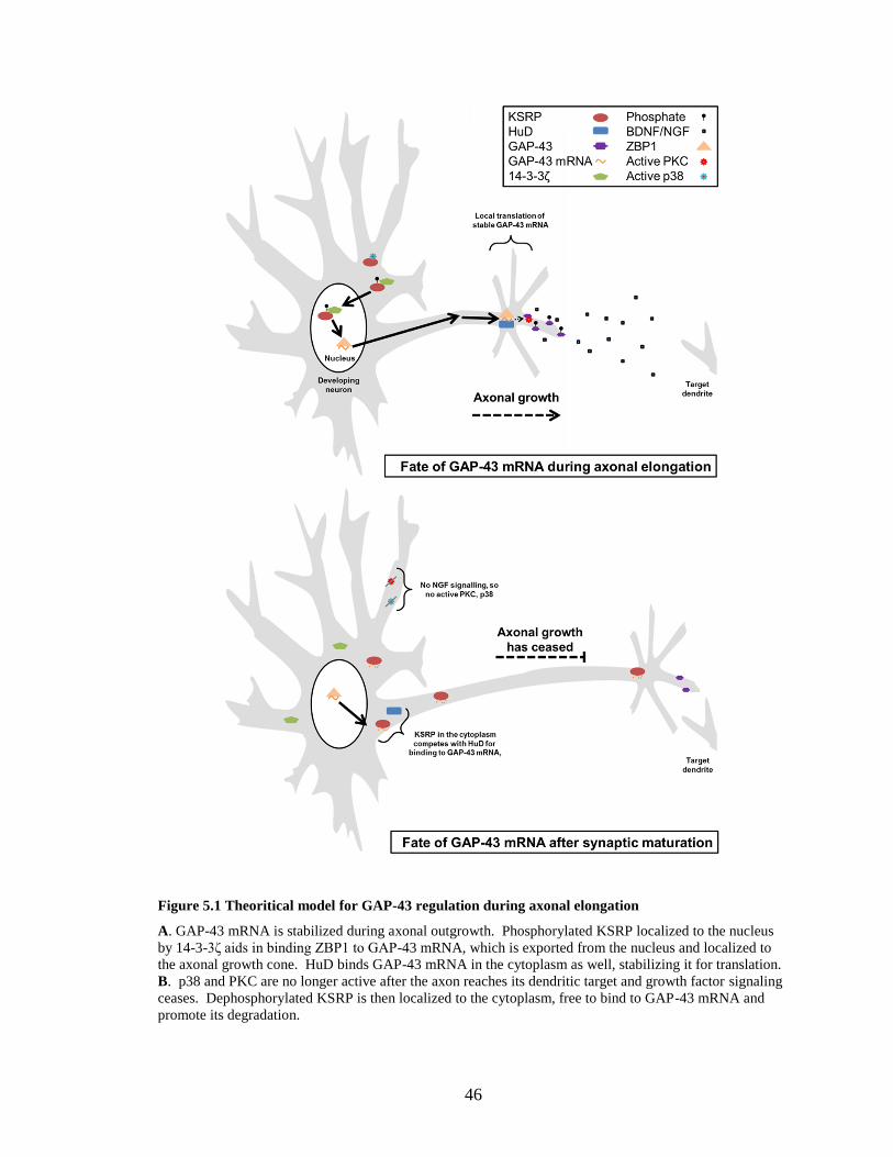
46
Figure 5.1 Theoritical model for GAP-43 regulation during axonal elongation
A. GAP-43 mRNA is stabilized during axonal outgrowth. Phosphorylated KSRP localized to the nucleus
by 14-3-3ζ aids in binding ZBP1 to GAP-43 mRNA, which is exported from the nucleus and localized to
the axonal growth cone. HuD binds GAP-43 mRNA in the cytoplasm as well, stabilizing it for translation.
B. p38 and PKC are no longer active after the axon reaches its dendritic target and growth factor signaling
ceases. Dephosphorylated KSRP is then localized to the cytoplasm, free to bind to GAP-43 mRNA and
promote its degradation.
47
the results obtained and data from the literature. Subsequent investigations will need to
tease apart the specific processes mediating axonal outgrowth associated with post-
transcriptional regulation of the GAP-43 gene.
Another mechanism promoting mRNA degradation also needs to be considered
when discussing KSRP’s function as recent evidence demonstrates KSRP’s involvement
in promoting the biogenesis of a subset of miRNAs. Thus far KSRP has been shown to
be a required factor in the maturation of two different miRNAs, let-7a and miR-155
(Ruggiero et al., 2009, Trabucchi et al., 2009). Knockdown of KSRP in P19 cells led to a
decrease in the maturation of a number of miRNAs (Trabucchi et al., 2009), one of which
(miR-21) has the potential to bind to GAP-43 mRNA with 100% sequence
complementarity (MicroCosm Targets Version 5, http://www.ebi.ac.uk/enright-
srv/microcosm/htdocs/targets/v5/). Initial investigations in our laboratory have shown
KSRP potentially binds to a number of miRNA targets, and may be involved in
promoting their maturation as well (Gardiner et al., unpublished observations). Perhaps
one of these miRNAs can also bind to GAP-43 mRNA and promote GAP-43 transcript
degradation through a decay pathway independent of AMD.
KSRP regulation of GAP-43 mRNA stability may not be the only mechanism
affecting neuron differentiation and development. As shown by Winzen et al. (2007)
several transcripts bind KSRP in HeLa cell extracts including a number of mRNAs that
encode for growth related proteins or signaling molecules relating to cellular
development. Furthermore, using UV-crosslinking and immunoprecipitation assays, we
have recently demonstrated binding of KSRP to not only GAP-43 mRNA but also to
other axonally localized mRNAs (Gardiner et al., unpublished observations). Therefore,
48
it will be important to examine the interplay of KSRP with HuD in the stabilization and
transport of target mRNAs during neuronal development. HuD and KSRP show a similar
expression pattern during brain development, with KSRP and HuD highly expressed
early in development and their levels dropping off during adolescence (Gu et al., 2002,
Bolognani et al., 2007a). Although both proteins are present in neurons, preliminary
studies indicate that they are not co-localized to the same cytoplasmic granules. Along
these lines, it is interesting to note that both HuD and KSRP have been shown to localize
to peripheral nerve axons via binding to SMN, and improper localization of these RBPs
can affect motor neuron function (Tadesse et al., 2008, Akten et al., 2011, Fallini et al.,
2011, Hubers et al., 2011).
As with any scientific study, there are some limitations to the work presented in
this dissertation. The in vitro decay study is a powerful tool for examining the stability of
an mRNA, but because it takes place outside the cell it does not include other cellular
proteins and factors that could be having an effect on mRNA expression. In order to
strengthen our decay studies, we could perform decay studies in cells transfected with
isoforms of KSRP, and inhibit transcription using actinomycin D. Following
transcriptional inhibition, we could collect RNA at different time points and examine the
stability of GAP-43 over time. Another drawback to the studies performed involves the
cell sorting experiments. At the time this dissertation was written, the experiment had
been performed once in duplicates, so the data is limited in that respect. More
transfection and sorting experiments could be done to further confirm that the KSRP
knockdown in PC12 cells increases the expression of GAP-43 mRNA. Lastly,
performing cell culture and transfection experiments to examine the effect of KSRP on
49
axonal elongation is a good model system, but does not capture the complete
environment of the intact brain. To truly show that KSRP restricts axonal elongation in
vivo, in utero transfection experiments could be performed to truly show that excess
KSRP expression limits axonal growth in the developing brain. Unfortunately, these
types of studies require extensive training and specialized equipment, which are currently
unavailable in the laboratory.
In conclusion, the research contained within this dissertation shows that KSRP
binds to and destabilizes GAP-43 mRNA in vitro, and serves to limit axonal outgrowth in
primary hippocampal cultures. These data add another layer of complexity to the post-
transcriptional operon that regulates GAP-43 mRNA stability, showing that tight control
of this gene’s expression is an important factor regulating axonal growth processes.
50
Abbreviations Used
3’UTR Three prime untranslated region
A Adenine
AChE Acetylcholine esterase
AMD AU-rich element mediated mRNA decay
ARE AU-rich element
ARE-BP ARE binding protein
ARED ARE database
BRF1 Butyrate regulated factor one
C Cytosine
cDNA Complementary deoxyribonucleic acid
Dcp Decapping protein
DIV Days in vitro
ELAV Embryonic lethal abnormal vision
FBP Far upstream element binding protein
FUSE Far upstream element
G Guanine
GAP-43 Growth associated protein 43
GFP Green fluorescent protein
GM-CSF Granulocyte-monocyte colony stimulating factor
IL Interleukin
iNOS Inducible nitric oxide synthase
KH K homology
KSRP K homology splicing regulatory protein
MAPK Mitogen activated protein kinase
51
mRNA Messenger ribonucleic acid
miRNA Micro ribonucleic acid
NGF Nerve growth factor
NLS Nuclear localization signal
PARN Poly(A) ribonuclease
PKC Protein kinase C
RBP RNA-binding protein
REMSA RNA electrophoretic mobility shift assay
RRM RNA recognition motif
RT-PCR Real time polymerase chain reaction
shRNA Short hairpin RNA
SMN Survival of motor neuron protein
TTP Tristetraprolin
U Uracil
WB Western blot
Xrn1 Exoribonuclease 1
ZBP Zip code binding protein
52
References
Abe R, Sakashita E, Yamamoto K, Sakamoto H (1996) Two different RNA binding
activities for the AU-rich element and the poly(A) sequence of the mouse
neuronal protein mHuC. Nucleic Acids Res 24:4895-4901.
Aigner L, Arber S, Kapfhammer JP, Laux T, Schneider C, Botteri F, Brenner HR, Caroni
P (1995) Overexpression of the neural growth-associated protein GAP-43 induces
nerve sprouting in the adult nervous system of transgenic mice. Cell 83:269-278.
Akten B, Kye MJ, Hao le T, Wertz MH, Singh S, Nie D, Huang J, Merianda TT, Twiss
JL, Beattie CE, Steen JA, Sahin M (2011) Interaction of survival of motor neuron
(SMN) and HuD proteins with mRNA cpg15 rescues motor neuron axonal
deficits. Proc Natl Acad Sci U S A 108:10337-10342.
Ambrosino C, Mace G, Galban S, Fritsch C, Vintersten K, Black E, Gorospe M, Nebreda
AR (2003) Negative feedback regulation of MKK6 mRNA stability by p38alpha
mitogen-activated protein kinase. Mol Cell Biol 23:370-381.
Anderson KD, Merhege MA, Morin M, Bolognani F, Perrone-Bizzozero NI (2003)
Increased expression and localization of the RNA-binding protein HuD and GAP-
43 mRNA to cytoplasmic granules in DRG neurons during nerve regeneration.
Exp Neurol 183:100-108.
Anderson KD, Morin MA, Beckel-Mitchener A, Mobarak CD, Neve RL, Furneaux HM,
Burry R, Perrone-Bizzozero NI (2000) Overexpression of HuD, but not of its
truncated form HuD I+II, promotes GAP-43 gene expression and neurite
outgrowth in PC12 cells in the absence of nerve growth factor. J Neurochem
75:1103-1114.
Anderson KD, Sengupta J, Morin M, Neve RL, Valenzuela CF, Perrone-Bizzozero NI
(2001) Overexpression of HuD accelerates neurite outgrowth and increases GAP-
43 mRNA expression in cortical neurons and retinoic acid-induced embryonic
stem cells in vitro. Exp Neurol 168:250-258.
Bakheet T, Williams BR, Khabar KS (2006) ARED 3.0: the large and diverse AU-rich
transcriptome. Nucleic Acids Res 34:D111-114.
Barreau C, Paillard L, Osborne HB (2005) AU-rich elements and associated factors: are
there unifying principles? Nucleic Acids Res 33:7138-7150.
Beckel-Mitchener AC, Miera A, Keller R, Perrone-Bizzozero NI (2002) Poly(A) tail
length-dependent stabilization of GAP-43 mRNA by the RNA-binding protein
HuD. J Biol Chem 277:27996-28002.
Bolognani F, Merhege MA, Twiss J, Perrone-Bizzozero NI (2004) Dendritic localization
of the RNA-binding protein HuD in hippocampal neurons: association with
polysomes and upregulation during contextual learning. Neurosci Lett 371:152-
157.
Bolognani F, Perrone-Bizzozero NI (2008) RNA-protein interactions and control of
mRNA stability in neurons. J Neurosci Res 86:481-489.
Bolognani F, Qiu S, Tanner DC, Paik J, Perrone-Bizzozero NI, Weeber EJ (2007a)
Associative and spatial learning and memory deficits in transgenic mice
overexpressing the RNA-binding protein HuD. Neurobiol Learn Mem 87:635-
643.
53
Bolognani F, Tanner DC, Merhege M, Deschenes-Furry J, Jasmin B, Perrone-Bizzozero
NI (2006) In vivo post-transcriptional regulation of GAP-43 mRNA by
overexpression of the RNA-binding protein HuD. J Neurochem 96:790-801.
Bolognani F, Tanner DC, Nixon S, Okano HJ, Okano H, Perrone-Bizzozero NI (2007b)
Coordinated expression of HuD and GAP-43 in hippocampal dentate granule cells
during developmental and adult plasticity. Neurochem Res 32:2142-2151.
Brennan CM, Steitz JA (2001) HuR and mRNA stability. Cell Mol Life Sci 58:266-277.
Brewer G (1991) An A + U-rich element RNA-binding factor regulates c-myc mRNA
stability in vitro. Mol Cell Biol 11:2460-2466.
Brewer G, Ross J (1988) Poly(A) shortening and degradation of the 3' A+U-rich
sequences of human c-myc mRNA in a cell-free system. Mol Cell Biol 8:1697-
1708.
Briata P, Forcales SV, Ponassi M, Corte G, Chen CY, Karin M, Puri PL, Gherzi R (2005)
p38-dependent phosphorylation of the mRNA decay-promoting factor KSRP
controls the stability of select myogenic transcripts. Mol Cell 20:891-903.
Caput D, Beutler B, Hartog K, Thayer R, Brown-Shimer S, Cerami A (1986)
Identification of a common nucleotide sequence in the 3'-untranslated region of
mRNA molecules specifying inflammatory mediators. Proc Natl Acad Sci U S A
83:1670-1674.
Caroni P, Becker M (1992) The downregulation of growth-associated proteins in
motoneurons at the onset of synapse elimination is controlled by muscle activity
and IGF1. J Neurosci 12:3849-3861.
Chen CY, Gherzi R, Andersen JS, Gaietta G, Jurchott K, Royer HD, Mann M, Karin M
(2000) Nucleolin and YB-1 are required for JNK-mediated interleukin-2 mRNA
stabilization during T-cell activation. Genes Dev 14:1236-1248.
Chen CY, Gherzi R, Ong SE, Chan EL, Raijmakers R, Pruijn GJ, Stoecklin G, Moroni C,
Mann M, Karin M (2001) AU binding proteins recruit the exosome to degrade
ARE-containing mRNAs. Cell 107:451-464.
Chen CY, Shyu AB (1994) Selective degradation of early-response-gene mRNAs:
functional analyses of sequence features of the AU-rich elements. Mol Cell Biol
14:8471-8482.
Chen CY, Shyu AB (1995) AU-rich elements: characterization and importance in mRNA
degradation. Trends Biochem Sci 20:465-470.
Chiaramello A, Neuman T, Peavy DR, Zuber MX (1996) The GAP-43 gene is a direct
downstream target of the basic helix-loop-helix transcription factors. J Biol Chem
271:22035-22043.
Chou CF, Mulky A, Maitra S, Lin WJ, Gherzi R, Kappes J, Chen CY (2006) Tethering
KSRP, a decay-promoting AU-rich element-binding protein, to mRNAs elicits
mRNA decay. Mol Cell Biol 26:3695-3706.
Chung S, Eckrich M, Perrone-Bizzozero N, Kohn DT, Furneaux H (1997) The Elav-like
proteins bind to a conserved regulatory element in the 3'-untranslated region of
GAP-43 mRNA. J Biol Chem 272:6593-6598.
Chung S, Jiang L, Cheng S, Furneaux H (1996) Purification and properties of HuD, a
neuronal RNA-binding protein. J Biol Chem 271:11518-11524.
54
Davis-Smyth T, Duncan RC, Zheng T, Michelotti G, Levens D (1996) The far upstream
element-binding proteins comprise an ancient family of single-strand DNA-
binding transactivators. J Biol Chem 271:31679-31687.
Denny JB (2006) Molecular mechanisms, biological actions, and neuropharmacology of
the growth-associated protein GAP-43. Curr Neuropharmacol 4:293-304.
Diaz-Moreno I, Hollingworth D, Frenkiel TA, Kelly G, Martin S, Howell S, Garcia-
Mayoral M, Gherzi R, Briata P, Ramos A (2009) Phosphorylation-mediated
unfolding of a KH domain regulates KSRP localization via 14-3-3 binding. Nat
Struct Mol Biol 16:238-246.
Diaz-Moreno I, Hollingworth D, Kelly G, Martin S, Garcia-Mayoral M, Briata P, Gherzi
R, Ramos A (2010) Orientation of the central domains of KSRP and its
implications for the interaction with the RNA targets. Nucleic Acids Res 38:5193-
5205.
Donnelly CJ, Willis DE, Xu M, Tep C, Jiang C, Yoo S, Schanen NC, Kirn-Safran CB,
van Minnen J, English A, Yoon SO, Bassell GJ, Twiss JL (2011) Limited
availability of ZBP1 restricts axonal mRNA localization and nerve regeneration
capacity. Embo J 30:4665-4677.
Fallini C, Zhang H, Su Y, Silani V, Singer RH, Rossoll W, Bassell GJ (2011) The
survival of motor neuron (SMN) protein interacts with the mRNA-binding protein
HuD and regulates localization of poly(A) mRNA in primary motor neuron axons.
J Neurosci 31:3914-3925.
Fan XC, Steitz JA (1998) HNS, a nuclear-cytoplasmic shuttling sequence in HuR. Proc
Natl Acad Sci U S A 95:15293-15298.
Fenger-Gron M, Fillman C, Norrild B, Lykke-Andersen J (2005) Multiple processing
body factors and the ARE binding protein TTP activate mRNA decapping. Mol
Cell 20:905-915.
Ford LP, Watson J, Keene JD, Wilusz J (1999) ELAV proteins stabilize deadenylated
intermediates in a novel in vitro mRNA deadenylation/degradation system. Genes
Dev 13:188-201.
Garcia-Mayoral MF, Diaz-Moreno I, Hollingworth D, Ramos A (2008) The sequence
selectivity of KSRP explains its flexibility in the recognition of the RNA targets.
Nucleic Acids Res 36:5290-5296.
Gherzi R, Lee KY, Briata P, Wegmuller D, Moroni C, Karin M, Chen CY (2004) A KH
domain RNA binding protein, KSRP, promotes ARE-directed mRNA turnover by
recruiting the degradation machinery. Mol Cell 14:571-583.
Goslin K, Schreyer DJ, Skene JH, Banker G (1988) Development of neuronal polarity:
GAP-43 distinguishes axonal from dendritic growth cones. Nature 336:672-674.
Gu W, Pan F, Zhang H, Bassell GJ, Singer RH (2002) A predominantly nuclear protein
affecting cytoplasmic localization of beta-actin mRNA in fibroblasts and neurons.
J Cell Biol 156:41-51.
Hall MP, Huang S, Black DL (2004) Differentiation-induced colocalization of the KH-
type splicing regulatory protein with polypyrimidine tract binding protein and the
c-src pre-mRNA. Mol Biol Cell 15:774-786.
He Q, Dent EW, Meiri KF (1997) Modulation of actin filament behavior by GAP-43
(neuromodulin) is dependent on the phosphorylation status of serine 41, the
protein kinase C site. J Neurosci 17:3515-3524.
55
Hubers L, Valderrama-Carvajal H, Laframboise J, Timbers J, Sanchez G, Cote J (2011)
HuD interacts with survival motor neuron protein and can rescue spinal muscular
atrophy-like neuronal defects. Hum Mol Genet 20:553-579.
Karimi-Abdolrezaee S, Verge VM, Schreyer DJ (2002) Developmental down-regulation
of GAP-43 expression and timing of target contact in rat corticospinal neurons.
Exp Neurol 176:390-401.
Keene JD (1999) Why is Hu where? Shuttling of early-response-gene messenger RNA
subsets. Proc Natl Acad Sci U S A 96:5-7.
Keene JD, Lager PJ (2005) Post-transcriptional operons and regulons co-ordinating gene
expression. Chromosome Res 13:327-337.
Kim VN (2005) MicroRNA biogenesis: coordinated cropping and dicing. Nat Rev Mol
Cell Biol 6:376-385.
Klein RL, McNamara RK, King MA, Lenox RH, Muzyczka N, Meyer EM (1999)
Generation of aberrant sprouting in the adult rat brain by GAP-43 somatic gene
transfer. Brain Res 832:136-144.
Kleinert H, Pautz A, Linker K, Schwarz PM (2004) Regulation of the expression of
inducible nitric oxide synthase. Eur J Pharmacol 500:255-266.
Kohn DT, Tsai KC, Cansino VV, Neve RL, Perrone-Bizzozero NI (1996) Role of highly
conserved pyrimidine-rich sequences in the 3' untranslated region of the GAP-43
mRNA in mRNA stability and RNA-protein interactions. Brain Res Mol Brain
Res 36:240-250.
Laux T, Fukami K, Thelen M, Golub T, Frey D, Caroni P (2000) GAP43, MARCKS, and
CAP23 modulate PI(4,5)P(2) at plasmalemmal rafts, and regulate cell cortex actin
dynamics through a common mechanism. J Cell Biol 149:1455-1472.
Li Y, Jiang Z, Chen H, Ma WJ (2004) A modified quantitative EMSA and its application
in the study of RNA--protein interactions. J Biochem Biophys Methods 60:85-96.
Lin WJ, Zheng X, Lin CC, Tsao J, Zhu X, Cody JJ, Coleman JM, Gherzi R, Luo M,
Townes TM, Parker JN, Chen CY (2011) Posttranscriptional control of type I
interferon genes by KSRP in the innate immune response against viral infection.
Mol Cell Biol 31:3196-3207.
Linker K, Pautz A, Fechir M, Hubrich T, Greeve J, Kleinert H (2005) Involvement of
KSRP in the post-transcriptional regulation of human iNOS expression-complex
interplay of KSRP with TTP and HuR. Nucleic Acids Res 33:4813-4827.
Liu J, Dalmau J, Szabo A, Rosenfeld M, Huber J, Furneaux H (1995) Paraneoplastic
encephalomyelitis antigens bind to the AU-rich elements of mRNA. Neurology
45:544-550.
Livak KJ, Schmittgen TD (2001) Analysis of relative gene expression data using real-
time quantitative PCR and the 2(-Delta Delta C(T)) Method. Methods 25:402-
408.
Loflin P, Chen CY, Shyu AB (1999) Unraveling a cytoplasmic role for hnRNP D in the
in vivo mRNA destabilization directed by the AU-rich element. Genes Dev
13:1884-1897.
Lykke-Andersen J, Wagner E (2005) Recruitment and activation of mRNA decay
enzymes by two ARE-mediated decay activation domains in the proteins TTP and
BRF-1. Genes Dev 19:351-361.
56
Ma WJ, Chung S, Furneaux H (1997) The Elav-like proteins bind to AU-rich elements
and to the poly(A) tail of mRNA. Nucleic Acids Res 25:3564-3569.
Min H, Turck CW, Nikolic JM, Black DL (1997) A new regulatory protein, KSRP,
mediates exon inclusion through an intronic splicing enhancer. Genes Dev
11:1023-1036.
Ming XF, Stoecklin G, Lu M, Looser R, Moroni C (2001) Parallel and independent
regulation of interleukin-3 mRNA turnover by phosphatidylinositol 3-kinase and
p38 mitogen-activated protein kinase. Mol Cell Biol 21:5778-5789.
Mobarak CD, Anderson KD, Morin M, Beckel-Mitchener A, Rogers SL, Furneaux H,
King P, Perrone-Bizzozero NI (2000) The RNA-binding protein HuD is required
for GAP-43 mRNA stability, GAP-43 gene expression, and PKC-dependent
neurite outgrowth in PC12 cells. Mol Biol Cell 11:3191-3203.
Murray EL, Schoenberg DR (2007) A+U-rich instability elements differentially activate
5'-3' and 3'-5' mRNA decay. Mol Cell Biol 27:2791-2799.
Pan F, Huttelmaier S, Singer RH, Gu W (2007) ZBP2 facilitates binding of ZBP1 to beta-
actin mRNA during transcription. Mol Cell Biol 27:8340-8351.
Pascale A, Gusev PA, Amadio M, Dottorini T, Govoni S, Alkon DL, Quattrone A (2004)
Increase of the RNA-binding protein HuD and posttranscriptional up-regulation
of the GAP-43 gene during spatial memory. Proc Natl Acad Sci U S A 101:1217-
1222.
Peng SS, Chen CY, Shyu AB (1996) Functional characterization of a non-AUUUA AU-
rich element from the c-jun proto-oncogene mRNA: evidence for a novel class of
AU-rich elements. Mol Cell Biol 16:1490-1499.
Peppel K, Vinci JM, Baglioni C (1991) The AU-rich sequences in the 3' untranslated
region mediate the increased turnover of interferon mRNA induced by
glucocorticoids. J Exp Med 173:349-355.
Perrone-Bizzozero NI, Cansino VV, Kohn DT (1993) Posttranscriptional regulation of
GAP-43 gene expression in PC12 cells through protein kinase C-dependent
stabilization of the mRNA. J Cell Biol 120:1263-1270.
Perrone-Bizzozero NI, Finklestein SP, Benowitz LI (1986) Synthesis of a growth-
associated protein by embryonic rat cerebrocortical neurons in vitro. J Neurosci
6:3721-3730.
Perrone-Bizzozero NI, Neve RL, Irwin N, Lewis S, Fischer I, Benowitz LI (1991) Post-
transcriptional regulation of GAP-43 rnRNA levels during neuronal
differentiation and nerve regeneration. Mol Cell Neurosci 2:402-409.
Quattrone A, Pascale A, Nogues X, Zhao W, Gusev P, Pacini A, Alkon DL (2001)
Posttranscriptional regulation of gene expression in learning by the neuronal
ELAV-like mRNA-stabilizing proteins. Proc Natl Acad Sci U S A 98:11668-
11673.
Reinhard E, Nedivi E, Wegner J, Skene JH, Westerfield M (1994) Neural selective
activation and temporal regulation of a mammalian GAP-43 promoter in
zebrafish. Development 120:1767-1775.
Ruggiero T, Trabucchi M, De Santa F, Zupo S, Harfe BD, McManus MT, Rosenfeld
MG, Briata P, Gherzi R (2009) LPS induces KH-type splicing regulatory protein-
dependent processing of microRNA-155 precursors in macrophages. FASEB
57
journal : official publication of the Federation of American Societies for
Experimental Biology 23:2898-2908.
Schreyer DJ, Skene JH (1991) Fate of GAP-43 in ascending spinal axons of DRG
neurons after peripheral nerve injury: delayed accumulation and correlation with
regenerative potential. J Neurosci 11:3738-3751.
Schreyer DJ, Skene JH (1993) Injury-associated induction of GAP-43 expression
displays axon branch specificity in rat dorsal root ganglion neurons. J Neurobiol
24:959-970.
Shaw G, Kamen R (1986) A conserved AU sequence from the 3' untranslated region of
GM-CSF mRNA mediates selective mRNA degradation. Cell 46:659-667.
Siomi H, Matunis MJ, Michael WM, Dreyfuss G (1993) The pre-mRNA binding K
protein contains a novel evolutionarily conserved motif. Nucleic Acids Res
21:1193-1198.
Skene JH, Jacobson RD, Snipes GJ, McGuire CB, Norden JJ, Freeman JA (1986) A
protein induced during nerve growth (GAP-43) is a major component of growth-
cone membranes. Science 233:783-786.
Skene JH, Willard M (1981) Changes in axonally transported proteins during axon
regeneration in toad retinal ganglion cells. J Cell Biol 89:86-95.
Smrt RD, Szulwach KE, Pfeiffer RL, Li X, Guo W, Pathania M, Teng ZQ, Luo Y, Peng
J, Bordey A, Jin P, Zhao X (2010) MicroRNA miR-137 regulates neuronal
maturation by targeting ubiquitin ligase mind bomb-1. Stem Cells 28:1060-1070.
Sofroniew MV, Howe CL, Mobley WC (2001) Nerve growth factor signaling,
neuroprotection, and neural repair. Annu Rev Neurosci 24:1217-1281.
Stoecklin G, Hahn S, Moroni C (1994) Functional hierarchy of AUUUA motifs in
mediating rapid interleukin-3 mRNA decay. J Biol Chem 269:28591-28597.
Strittmatter SM, Fankhauser C, Huang PL, Mashimo H, Fishman MC (1995) Neuronal
pathfinding is abnormal in mice lacking the neuronal growth cone protein GAP-
43. Cell 80:445-452.
Strittmatter SM, Fishman MC, Zhu XP (1994) Activated mutants of the alpha subunit of
G(o) promote an increased number of neurites per cell. J Neurosci 14:2327-2338.
Szabo A, Dalmau J, Manley G, Rosenfeld M, Wong E, Henson J, Posner JB, Furneaux
HM (1991) HuD, a paraneoplastic encephalomyelitis antigen, contains RNA-
binding domains and is homologous to Elav and Sex-lethal. Cell 67:325-333.
Tadesse H, Deschenes-Furry J, Boisvenue S, Cote J (2008) KH-type splicing regulatory
protein interacts with survival motor neuron protein and is misregulated in spinal
muscular atrophy. Hum Mol Genet 17:506-524.
Takeda K, Ichijo H (2002) Neuronal p38 MAPK signalling: an emerging regulator of cell
fate and function in the nervous system. Genes Cells 7:1099-1111.
Tiruchinapalli DM, Ehlers MD, Keene JD (2008) Activity-dependent expression of RNA
binding protein HuD and its association with mRNAs in neurons. RNA Biol
5:157-168.
Trabucchi M, Briata P, Garcia-Mayoral M, Haase AD, Filipowicz W, Ramos A, Gherzi
R, Rosenfeld MG (2009) The RNA-binding protein KSRP promotes the
biogenesis of a subset of microRNAs. Nature 459:1010-1014.
Treisman R (1985) Transient accumulation of c-fos RNA following serum stimulation
requires a conserved 5' element and c-fos 3' sequences. Cell 42:889-902.
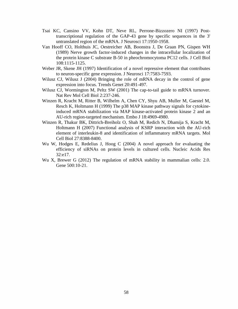
58
Tsai KC, Cansino VV, Kohn DT, Neve RL, Perrone-Bizzozero NI (1997) Post-
transcriptional regulation of the GAP-43 gene by specific sequences in the 3'
untranslated region of the mRNA. J Neurosci 17:1950-1958.
Van Hooff CO, Holthuis JC, Oestreicher AB, Boonstra J, De Graan PN, Gispen WH
(1989) Nerve growth factor-induced changes in the intracellular localization of
the protein kinase C substrate B-50 in pheochromocytoma PC12 cells. J Cell Biol
108:1115-1125.
Weber JR, Skene JH (1997) Identification of a novel repressive element that contributes
to neuron-specific gene expression. J Neurosci 17:7583-7593.
Wilusz CJ, Wilusz J (2004) Bringing the role of mRNA decay in the control of gene
expression into focus. Trends Genet 20:491-497.
Wilusz CJ, Wormington M, Peltz SW (2001) The cap-to-tail guide to mRNA turnover.
Nat Rev Mol Cell Biol 2:237-246.
Winzen R, Kracht M, Ritter B, Wilhelm A, Chen CY, Shyu AB, Muller M, Gaestel M,
Resch K, Holtmann H (1999) The p38 MAP kinase pathway signals for cytokine-
induced mRNA stabilization via MAP kinase-activated protein kinase 2 and an
AU-rich region-targeted mechanism. Embo J 18:4969-4980.
Winzen R, Thakur BK, Dittrich-Breiholz O, Shah M, Redich N, Dhamija S, Kracht M,
Holtmann H (2007) Functional analysis of KSRP interaction with the AU-rich
element of interleukin-8 and identification of inflammatory mRNA targets. Mol
Cell Biol 27:8388-8400.
Wu W, Hodges E, Redelius J, Hoog C (2004) A novel approach for evaluating the
efficiency of siRNAs on protein levels in cultured cells. Nucleic Acids Res
32:e17.
Wu X, Brewer G (2012) The regulation of mRNA stability in mammalian cells: 2.0.
Gene 500:10-21.
59
Appendix A: KSRP and GAP-43 mRNA expression in shRNA
transfected PC12 cells
Appendix A: shRNA knockdown of KSRP in PC12 cells increases GAP-43 mRNA expression
A and B. Graphs of the gating used to collect GFP positive transfected PC12 cells. Control non-targeting
GFP-shRNA (A) and GFP-shKSRP (B) transfected PC12 cells were enriched to collect the brightest 2.5%
fraction of cells. C. Graph of KSRP mRNA knockdown in transfected PC12 cells. KSRP levels were
knocked down ~95%. D. Graph of GAP-43 mRNA levels in transfected cells. PC12 cells transfected with
shKSRP expressed 1.7 fold more GAP-43 mRNA than control non-targeting GFP-shRNA transfected cells.
60
Appendix B: Submitted short-report article to J. Neuroscience co-
authored by Clark Bird
References for this article are self-contained within this appendix.
HuD Promotes BDNF Expression via Selective Stabilization of the BDNF Long
3’UTR mRNA
Wei Feng1,3
, Clark Bird2,3
, Megan Allen1,3
, Guanglu Liu1,Wenqi Li
1, Nora I Perrone-
Bizzozero2, and Yue Feng
1
1Department of Pharmacology, Emory University School of Medicine
2Department of Neurosciences, University of New Mexico School of Medicine
3These people contributed equally to the work.
Abstract
Complex regulation of the brain-derived neurotrophic factor (BDNF) governs its
intricate functions in brain development and neuronal plasticity. Besides tight
transcriptional control, alternative 3’end processing of the BDNF transcripts generates
either a long or a short 3’untranslated region (3’UTR). The distinct RNA sequence in the
BDNF 3’UTRs differentially regulates BDNF production in brain neurons, conceivably
due to differential interactions with undefined trans-acting factors. In this study, we
report that the neuronal selective RNA-binding protein HuD interacts with a highly
conserved AU-rich element (ARE) specifically located in the BDNF long 3’UTR. Such
interaction is sufficient for selective stabilization of the BDNF long 3’UTR mRNA in
vitro and in vivo, leading to increased levels of BDNF long 3’UTR mRNA and BDNF
protein in the hippocampal mossy fiber (MF) pathway in HuD transgenic mice. Our
results identify the first trans-acting factor that enhances BDNF expression specifically
through the long 3’UTR and a novel mechanism that differentially regulates BDNF
production in selected neuronal populations.
61
Introduction
Brain-derived neurotrophic factor (BDNF) plays pivotal roles in governing a
broad spectrum of brain functions including neuronal survival, neural network
development, and synaptic plasticity. To accommodate such intricate functions, BDNF
expression is under precise regulation. Furthermore, dysregulation of BDNF is indicated
in the pathogenesis of neurological and psychiatric diseases (K. Martinowich et al., 2007;
A. H. Nagahara and M. H. Tuszynski, 2011). Transcription of BDNF can be initiated
from at least eight different promoters in mammals in response to various stimulation
cues (Q. R. Liu et al., 2006; T. Aid et al., 2007). In addition, alternative poly-adenylation
results in two pools of BDNF mRNAs; each carrying either a short or a long
3’untranslated region (UTR) regardless of which promoter drives BDNF transcription.
While the entire sequence of the short 3’UTR is contained within the long 3’UTR, it is
the unique sequence in the BDNF long 3’ UTR that mediates differential regulation of
BDNF production through multiple posttranscriptional mechanisms. The long 3’UTR,
but not the short 3’UTR, mediates neuronal activity-dependent translation of BDNF (A.
G. Lau et al., 2010) and dendritic localization of BDNF mRNA (J. J. An et al., 2008).
Additionally, the short and long 3’UTR differentially affect BDNF mRNA stability, with
the long transcript having a much shorter half-life than its short counterpart (E. Castren et
al., 1998). The unique sequence in the BDNF long 3’UTR is predicted to provide binding
sites for trans-acting RNA-binding proteins (RBPs) and/or microRNAs to achieve
differential posttranscriptional regulation of BDNF mRNA isoforms. However, no
previous studies have identified RBPs that specifically bind and regulate the BDNF long
3’UTR mRNA.
62
We report here that HuD, a neuronal RBP that plays critical roles in governing
neuronal circuitry development and plasticity via controlling mRNA stability and/or
translation (J. Deschenes Furry et al., 2006), selectively binds the BDNF long 3’UTR
through a highly conserved AU-rich element (ARE) and stabilizes the BDNF long
mRNA. Moreover, we show that a HuD transgene increases the levels of the BDNF long
3’UTR mRNA and BDNF protein primarily in the hippocampal mossy fiber (MF)
pathway, offering a novel mechanism for posttranscriptional regulation of distinct BDNF
mRNA isoforms.
Methods
Cell culture, plasmids and transfection
The ARE at the distal end of BDNF long 3’UTR was removed by PCR
amplification of the pcLuc-BDNFlongUTR plasmid (A. G. Lau et al., 2010) with primers
of ccgctcgagTGGATTTATGTTGTATAGATTAT (forward) and
gctctagaACATGGTGAATAATATCTTTACC (reverse), and subcloned between the
XhoI/XabI sites to replace the full length BDNF 3’UTR in pcLuc-BDNF longUTR
(pcLuc-BDNF longUTRΔARE). Luciferase reporter constructs were co-transfected with
myc-HuD (K. D. Anderson et al., 2003) or pcDNA into CAD cells using Lipofectamine
2000 (Invitrogen). The pRL-TK renilla luciferase construct was also co-transfected.
Reporter expression was quantified using the Dual-Luciferase assay (Promega).
UV-crosslinking-immunoprecipitation (CLIP) and RT-PCR
63
Crosslinking was performed as previously described (J. Ule et al., 2003) with
modifications. Briefly, cells were UV crosslinked using a Stratalinker (Stratagene, 400
mJ). The postnuclear supernatant was precleared with IgG-conjugated protein A-
Sepharose beads in the presence of 0.001% SDS. Anti-c-myc antibody (SantaCruz) 1:500
conjugated to protein A beads was used for immunoprecipitation (L. Zhao et al., 2006).
The immunoprecipitated complexes were proteinase K-treated before RNA extraction (L.
Zhao et al., 2010). RT-PCR was performed with Primer A:
ATCAGGCAAGGATATGGGCTCACT (forward) and
TCCAGATCCACAACCTTCGCTTCA (reverse); Primer B:
TGGCCTAACAGTGTTTGCAG (forward) and GGATTTGAGTGTGGTTCTCC
(reverse); and Primer C: CAGTGGCTGGCTCTCTTACC (forward) and
GGCCACAGACATTTACTTACAGTTT (reverse).
In vitro mRNA binding and decay
A 164 bp fragment containing the BDNF ARE (nt 2640- 2746) was PCR-
amplified and cloned into the XbaI/XhoI sites of pBSKII (Invitrogen). Radiolabeled
BDNF-ARE RNA was prepared by in vitro transcription using 32P-UTP as described (A.
C. Beckel-Mitchener et al., 2002). RNA electrophoretic mobility shift assay [REMSA,
(Y. Li et al., 2004)] used 100,000 cpm of 32P-UTP labeled BDNF-ARE RNA, increasing
amounts of purified GST or GST-HuD (S. Chung et al., 1997), and 0.25 mg/ml yeast
tRNA and 0.25 mg/ml of BSA to minimize non-specific interactions. Specific
competition was carried out with a 10-fold molar excess of cold BDNF-ARE RNA. In
vitro mRNA decay reactions were performed using ~200 fmol (100,000 cpm) of capped
and polyadenylated radiolabeled BDNF-ARE RNA and 20 μg S100 protein from HuD-

64
KO mice. Reactions were supplemented with either 50 ng of GST-HuD or GST and the
half-life of the mRNA was calculated as described (F. Bolognani et al., 2007).
Treatment of Primary cultures of embryonic cortical neurons
Neuronal cell cultures were prepared from E17 C57BL/6 mice (P. Washbourne et
al., 2002), and were grown for 24 hours before infection with HSV-HuD or control HSV-
lacZ as described (K. D. Anderson et al., 2001). After 72 h, total RNA was isolated and
BDNF long 3’UTR mRNA levels were measured by RTqPCR using GAPDH as
reference.
In situ hybridization and immunofluorescence
Fluorescent in situ hybridization (FISH) was performed using a digoxigenin-
labeled antisense oligonucleotide complementary to nucleotides 2508-2556 in the BDNF
long 3’ UTR (5’ GGGTGTATACAATAACTTTTATCTGCAAACACGTTAGG-
CCATATTAC) as described (N.I. Perrone-Bizzozero et al., 2011). Duplicate adjacent
sections from HuD-Tg mice and nontransgenic wild type littermates (WT) were analyzed
in parallel and images acquired using the same condition. To assess BDNF protein levels
in hippocampus, brain slices derived from 2- month old HuD-Tg mice and WT
littermates were subjected to immunofluorescent staining followed by image acquisition
as described previously (A. G. Lau et al., 2010). Fluorescent signals were quantified
using ImageJ (NIH) and the density of IF was calculated.
Results
65
HuD selectively enhances expression of the BDNF long 3’UTR reporter through a
highly conserved ARE
AREs are prominent motifs found in the 3’UTRs that recruit various ARE-
binding proteins (ARE-BPs) to stabilize or destabilize target mRNAs (N. Xu et al., 1997).
AREs can be divided into three different classes: Class I ARE contains a core penta-
nucleotide AUUUA flanked by A/U, Class II ARE contains overlapping AUUUA motifs,
and Class III does not contain typical AUUUA motifs but long stretches of U-rich
sequences. Using the ARED-Organisms database (http://brp.kfshrc.edu.sa/ARED/), we
identified a highly conserved Class I ARE specifically located in the BDNF long 3’UTR
immediately up-stream of the distal poly-adenylation site (Figure 1A), suggesting that
ARE-BPs may preferentially regulate stability of the BDNF long 3’UTR mRNA. HuD is
a neuronal specific ARE-BP (F. Bolognani et al., 2009) that displays a functional
spectrum substantially overlapping with that of BDNF (N. Perrone-Bizzozero and F.
Bolognani, 2002). To directly test whether HuD can regulate mRNA expression
selectively via the BDNF long 3’UTR, we co-expressed c-myc-tagged HuD (K. D.
Anderson et al., 2000) with luciferase reporters carrying the short BDNF 3’UTR (S-
3’UTR), the full-length BDNF long 3'UTR (L-3’UTR) or the BDNF long 3'UTR lacking
the ARE (ΔARE) in the brain neuron cell line CAD. As shown in Figure 1B, c-myc-HuD
significantly increased luciferase reporter expression when full length BDNF L-3'UTR is
present. In contrast, c-myc-HuD did not have any effect to the BDNF S-3’UTR reporter.
Furthermore, removing the ARE in BDNF long 3’UTR completely abolished the effect of
HuD (Figure 1C). Importantly, myc-HuD significantly increased BDNF L-3'UTR
reporter mRNA levels in an ARE-dependent manner (Figure 1D), recapitulating the HuD
66
response measured by luciferase activity (Figure 1C). Thus, although HuD can also
promote translation initiation (A. Fukao et al., 2009), the BDNF ARE appears to mediate
regulation by HuD primarily at the level of mRNA stability. The expression of each
luciferase reporter with expected sequence of BDNF 3’UTRs was confirmed by RT-PCR
using multiple primer pairs (Figure 1E).
To test whether HuD associates with the BDNF long 3’UTR through the ARE in
transfected cells, we performed UV-cross linking immunoprecipitation (CLIP) followed
by RTPCR. As shown in Figure 1F, only the long 3’UTR reporter mRNA, but not the
short 3’UTR reporter mRNA was co-immunoprecipitated with HuD (left panel).
Furthermore, removing the ARE in the long 3’UTR significantly attenuated the
association with HuD (Figure 1F, right panel, and Figure 1G). These results suggest that
selective association of HuD with the long 3’UTR mediates the biological effect of HuD,
and the ARE serves as a key site for interaction with HuD.
HuD directly binds and stabilizes the BDNF long 3’UTR mRNA
To test whether HuD can directly bind to the BDNF long 3’UTR segment that
contains the ARE, we performed RNA-mobility shift assays using recombinant GST-
HuD or GST. As shown in Figure 2A, GST-HuD bound the BDNF-ARE segment with
high affinity. A mobility shift of the RNA can be visualized even with 25 ng of GST-
HuD. In contrast, GST alone did not bind the RNA at any concentrations examined. The
specificity of HuD-ARE interaction was further demonstrated by the displacement of the
radiolabeled RNA with a cold ARE competitor (Figure 2A, right panel).
To further examine whether interactions between HuD and the BDNF-ARE could
result in mRNA stabilization, we used in vitro transcribed capped and polyadenylated
67
BDNF-ARE RNA and GST-HuD for in vitro decay assays (Figure 2B). Addition of GST-
HuD to brain extracts resulted in a significant stabilization of the RNA, with an almost 2-
fold reduction in the initial rate of decay. We next examined the effect of HuD on
endogenous long-BDNF mRNA in primary cultured cortical neurons treated with HSV
vectors expressing either HuD or the control LacZ gene (K. D. Anderson et al., 2001). As
shown in Figure 3A, overexpression of HuD significantly increased the levels of the
BDNF long 3’ UTR mRNA, whereas the pan BDNF mRNA levels were unaltered, in
which the short BDNF mRNA is the major isoform (A. G. Lau et al., 2010).
Reciprocally, shRNA-mediated HuD knockdown reduced the levels of the BDNF long
3’UTR mRNA in both the soma and processes (data not shown), suggesting that HuD
governs BDNF long mRNA expression throughout the entire neuron.
A HuD transgene selectively up-regulates BDNF long 3’UTR mRNA and BDNF
protein in the hippocampal MF pathway
Finally, we tested whether elevated HuD expression can regulate neuronal BDNF
production in vivo through the long 3’UTR. Quantitative fluorescent in situ hybridization
(FISH) was performed using brain slices of HuD transgenic mice (HuD-Tg) that express
myc-tagged HuD under the CamKIIα promoter (F. Bolognani et al., 2006). Non-
transgenic WT littermates were processed in parallel as baseline controls. Consistent with
the preferential increase of HuD in hippocampal dentate gyrus granule cells (DGCs) of
the HuD-Tg (F. Bolognani et al., 2006), BDNF long 3’UTR mRNA is significantly up-
regulated in the DGCs of HuD-Tg (Figure. 3B). In contrast, BDNF long 3’ UTR mRNA
was not increased in CA3 or CA1 neurons that do not harbor significant over-expression
of transgenic HuD (data not shown). Furthermore, pan BDNF mRNA that contains 80%
68
of BDNF short 3’UTR mRNA is not affected in HuD-Tg, indicating preferential
regulation of the BDNF long 3’UTR mRNA by HuD in vivo.
BDNF protein synthesized in DGCs is primarily transported to and stored in
hippocampal MF axons that project through the hilus to form large synapses with CA3
dendrites (S. C. Danzer and J. O. McNamara, 2004). Consistent with the enhanced
expression of BDNF long 3’UTR mRNA in DGCs (Figure 3B), significantly increased
BDNF protein immunofluorescence was detected in the hilus (Figure. 4B), the CA3 strata
lucidum and radiatum (Figure. 4C). Transgenic HuD expression also increased BDNF
protein in cells of the DG subgranular zone (SGZ) that is enriched of adult neural stem
cells (W. Guo et al. 2011), particularly in the characteristic long processes extended into
the DGC layer (arrows in Figure. 4B). In contrast, BDNF protein levels in CA1 neurons
are not significantly altered (Figure. 4F). Given the vigorous regulation of endogenous
HuD in DGCs (F. Bolognani et al., 2007), HuD-dependent stabilization of BDNF long
3’UTR mRNA is a novel mechanism that controls BDNF production in the hippocampal
MF pathway, a critical circuitry that governs hippocampal excitatory activities in
physiological and pathological plasticity during epileptogenesis (S. C. Danzer and J. O.
McNamara, 2004).
Discussion
Our studies identify HuD as the first RBP that selectively binds to and stabilizes
the BDNF long 3’UTR mRNA but not the short BDNF mRNA isoform. Such function is
mediated by a highly conserved ARE in the BDNF long 3’UTR. Furthermore, we showed
that elevated HuD expression can enhance BDNF protein production in brain neurons in
69
culture and in vivo, suggesting that the quantity of HuD controls BDNF levels in neurons
through preferential regulation of the long 3’UTR BDNF mRNA.
Consistent with the fact that mRNAs carrying AREs in the 3’UTR often display
short half-lives (N. Xu et al., 1997; C. Y. Chen et al., 2001), the BDNF long 3’UTR
mRNA is less stable than the BDNF short 3’UTR mRNA (E. Castren et al., 1998), likely
due to recruitment of undefined destabilizing RBPs to the unique sequence in the long
3’UTR. Such instability provides a practical opportunity for stabilizing RBPs,
represented by HuD, to differentially regulate BDNF mRNAs that carry the two distinct
3’UTRs. However, although the long 3’UTR can mediate dendritic localization of BDNF
mRNA (J. J. An et al., 2008), manipulating HuD expression reduced the long BDNF
mRNA isoform in both neuronal soma and processes. Thus, HuD-dependent stabilization
governs the overall levels of the BDNF long 3’UTR mRNA, which is known to mediate
neuronal activity-dependent translation (A. G. Lau et al., 2010). Importantly, the
abundance of HuD is vigorously regulated during neuronal development and functional
changes. HuD is highly abundant in the brain during embryonic and neonatal
development, down-regulated in adult, but markedly up-regulated upon neuronal
activation in multiple plasticity paradigms (F. Bolognani et al., 2004; A. Pascale et al.,
2004; F. Bolognani et al., 2007; D. M. Tiruchinapalli et al., 2008). Moreover, marked
regulation of HuD is found in various neuronal subpopulations (H. J. Okano and R. B.
Darnell, 1997; N. Perrone-Bizzozero and F. Bolognani, 2002). Together with the
differential levels of BDNF mRNAs that carry the long or the short 3’UTR in distinct
brain regions (J. J. An et al., 2008), HuD-dependent stabilization of the BDNF mRNA
70
through the long 3’UTR provides a posttranscriptional mechanism that increases the
complexity of BDNF regulation to accommodate brain development and function.
A particular interesting case is the regulation of HuD in hippocampal MFs. HuD
protein is not normally detected in adult hippocampal DGCs (F. Bolognani et al., 2007).
However, upon seizure induction, HuD is drastically up-regulated in DGCs. In contrast,
although HuD is present in CA1 and CA3 neurons at rest, it is only moderately increased
after seizures. Recapitulating the robust increase of HuD in DGCs in response to seizure,
resting HuD-Tg mice display the highest fold increase of HuD protein in DGCs (F.
Bolognani et al., 2006; N. I. Perrone-Bizzozero et al. 2011), although HuD transgene
expression can be detected in all forebrain neurons. As a result, BDNF protein expression
is preferentially increased in DGCs and stored in the MF pathway. Considering the well-
known effects of BDNF in promoting neuronal processes growth (R. Koyama et al.,
2004), the increased BDNF expression in hippocampal MFs is likely an contributing
factor for the MF over-projection in HuD-Tg (N. I. Perrone-Bizzozero et al. 2011).
Conceivably, the seizure-induced HuD up-regulation in DGCs could contribute to the
wellcharacterized preferential increase of BDNF in the MF terminals and MF sprouting
during epileptogenesis (S. C. Danzer et al., 2004; S. C. Danzer and J. O. McNamara,
2004).
Taken together, our study reveals a novel mechanism that controls BDNF
expression through HuD-dependent stabilization of the BDNF long 3’UTR mRNA.
Because BDNF is also under rigorous transcriptional regulation, and conventional HuD
knockout results in abnormal neuronal development (W. Akamatsu et al., 2005), a
definitive answer regarding the biological function of HuD-dependent BDNF mRNA
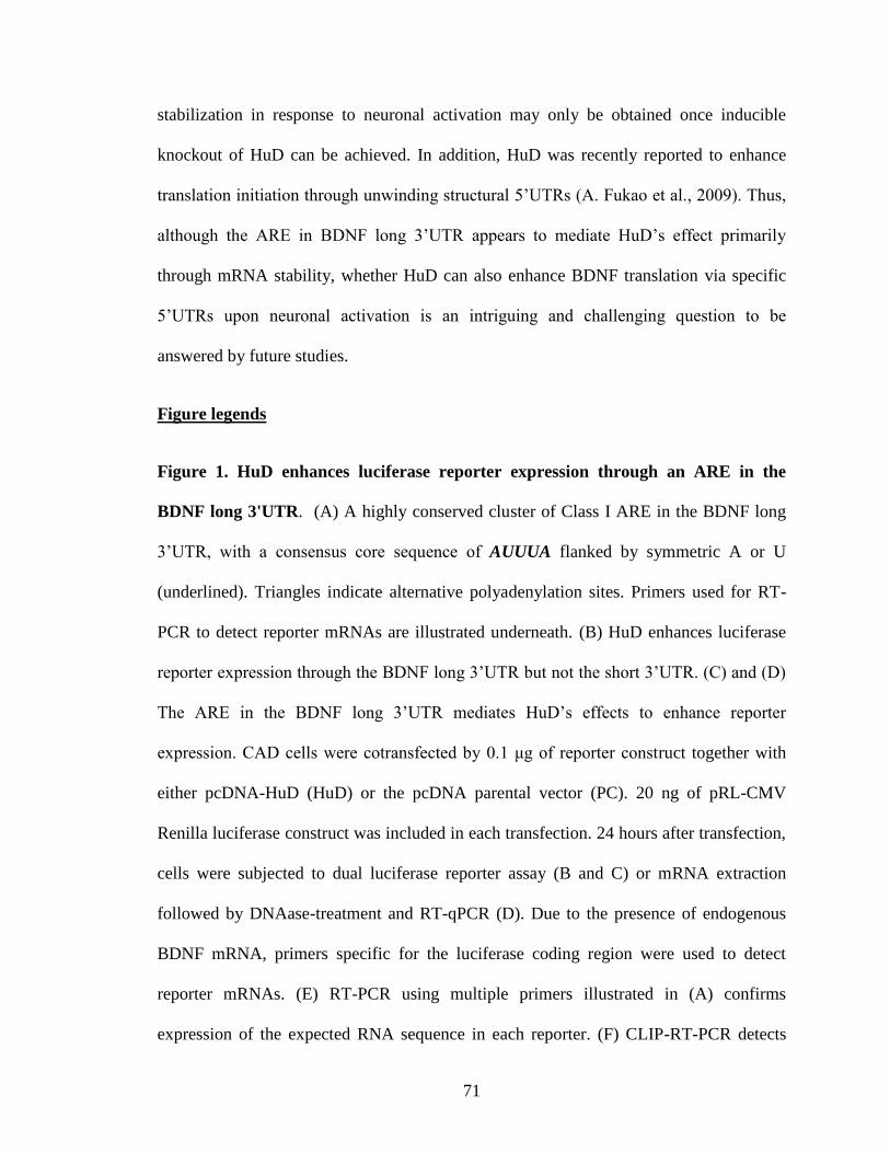
71
stabilization in response to neuronal activation may only be obtained once inducible
knockout of HuD can be achieved. In addition, HuD was recently reported to enhance
translation initiation through unwinding structural 5’UTRs (A. Fukao et al., 2009). Thus,
although the ARE in BDNF long 3’UTR appears to mediate HuD’s effect primarily
through mRNA stability, whether HuD can also enhance BDNF translation via specific
5’UTRs upon neuronal activation is an intriguing and challenging question to be
answered by future studies.
Figure legends
Figure 1. HuD enhances luciferase reporter expression through an ARE in the
BDNF long 3'UTR. (A) A highly conserved cluster of Class I ARE in the BDNF long
3’UTR, with a consensus core sequence of AUUUA flanked by symmetric A or U
(underlined). Triangles indicate alternative polyadenylation sites. Primers used for RT-
PCR to detect reporter mRNAs are illustrated underneath. (B) HuD enhances luciferase
reporter expression through the BDNF long 3’UTR but not the short 3’UTR. (C) and (D)
The ARE in the BDNF long 3’UTR mediates HuD’s effects to enhance reporter
expression. CAD cells were cotransfected by 0.1 μg of reporter construct together with
either pcDNA-HuD (HuD) or the pcDNA parental vector (PC). 20 ng of pRL-CMV
Renilla luciferase construct was included in each transfection. 24 hours after transfection,
cells were subjected to dual luciferase reporter assay (B and C) or mRNA extraction
followed by DNAase-treatment and RT-qPCR (D). Due to the presence of endogenous
BDNF mRNA, primers specific for the luciferase coding region were used to detect
reporter mRNAs. (E) RT-PCR using multiple primers illustrated in (A) confirms
expression of the expected RNA sequence in each reporter. (F) CLIP-RT-PCR detects
72
BDNF long 3’UTR reporter mRNA, but not short 3’UTR reporter mRNA, in
immunoprecipitated HuD complex. Deletion of the ARE in the long 3’UTR reduced the
association with HuD. Expression of the corresponding reporters in the input (inp) was
shown on top panels. (G) Quantification of reporter mRNAs coimmunoprecipitated with
HuD relevant to the corresponding mRNA levels in the input by RTqPCR. * indicates
P<0.05 by Student’s t-test (n=3).
Figure 2. Binding of HuD to the ARE in the BDNF-long 3’UTR increases the
stability of the mRNA. (A) Specific binding of HuD to the ARE in BDNF-long 3’UTR
was demonstrated by REMSA using recombinant proteins and radiolabeled RNA along
with a 10-fold molar excess of cold ARE competitor. (B) In vitro decay assay of capped
and poly-adenylated RNA containing the BDNF-ARE with left lane showing RNA
molecular weight markers. Analysis of the rate of decay in three independent experiment
revealed that the mRNA is stabilized in the presence of HuD. Half-life for GST-treated
mRNA is 7.0 ± 0.90 min, and for GST-HuD-treated mRNA is 12.0±1.0 min
Figure 3. Overexpression of HuD increases the BDNF long 3’UTR mRNA in
primary cultured neurons and in vivo (A) E17 cortical neurons were infected with
HSV-HuD or HSVlacZ virus and the levels of long 3’UTR BDNF mRNA and pan BDNF
mRNA were determined by qRT-PCR. (B) Fluorescent in situ hybridization of BDNF
long 3’UTR mRNA in hippocampal sections derived from HuD-Tg and WT littermates.
Left panels show representative images (bar= 75 μm) and the right panel displays the
quantification of four pairs of HuD-Tg and WT controls. *p<0.05 and ** p<0.01
(Student’s t-test, n=4).
73
Figure 4. The HuD transgene enhances BDNF protein levels in the hippocampal MF
pathway. Representative images of BDNF immunofluorescence (green) in the
hippocampal hilus (A and B), CA3 (C and D) and CA1 regions in HuD-Tg (B, D, and F)
and WT littermates (A, C, and E). DAPI stained nuclei (blue) mark the aforementioned
principle neuron layers. Asterisks indicate enhanced BDNF signals in the long processes
of adult neural stem cells in the SGZ. Quantification of BDNF immunofluorescence in
the hilus (G), CA3 strata lucidum and radiatum (H) and CA1 stratum radiatum (I) are
graphically displayed. *p<0.05 (Student’s t-test, n=3).
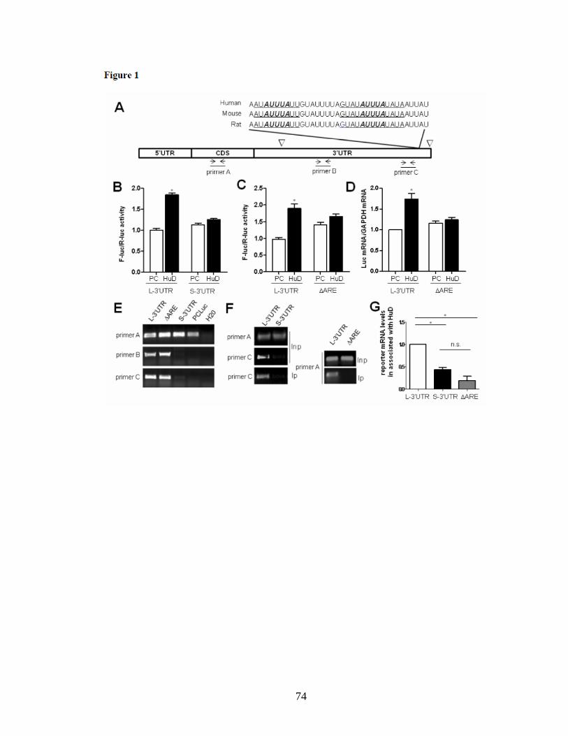
74
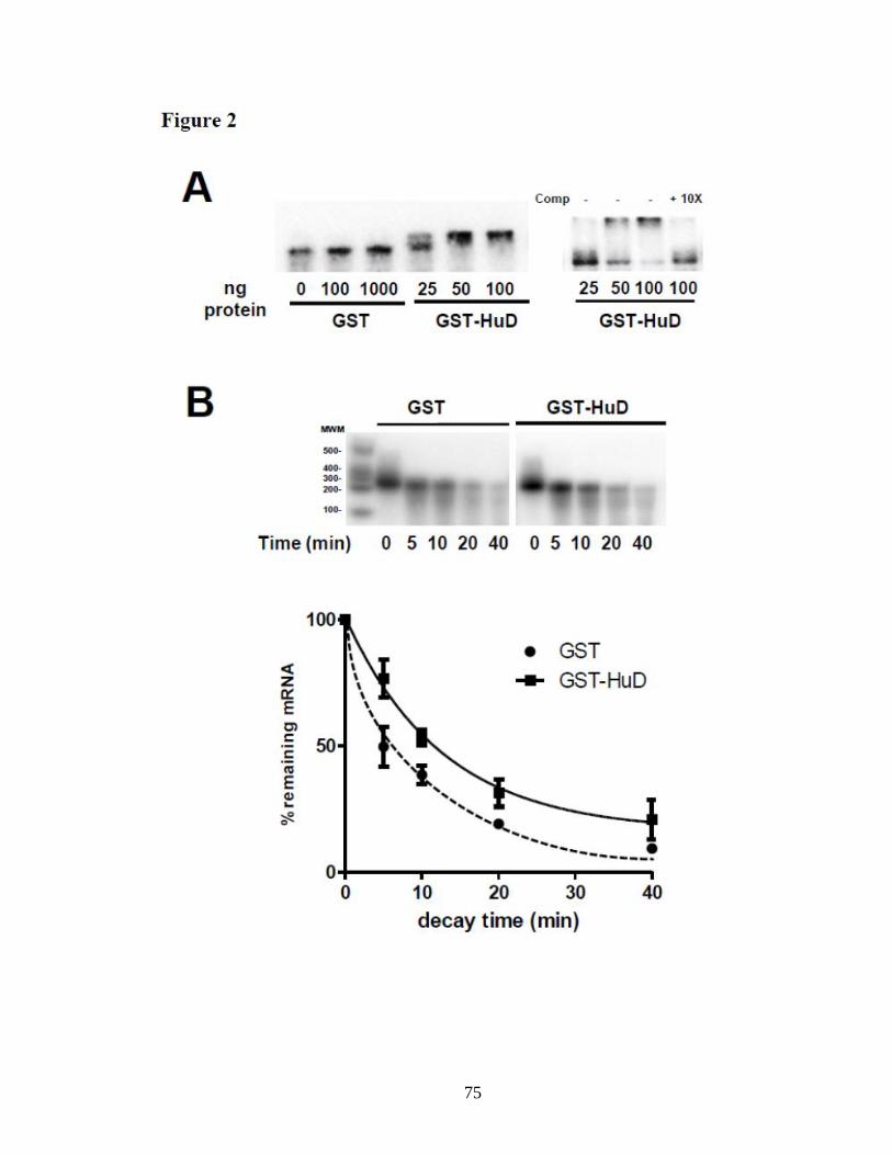
75
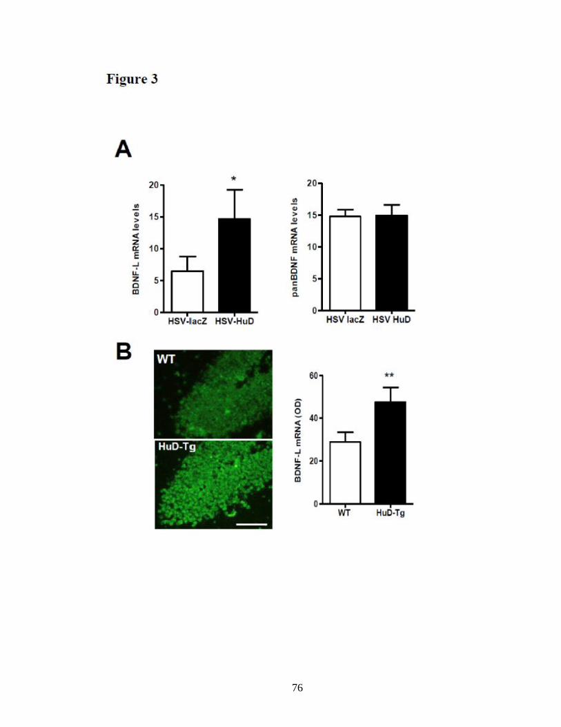
76
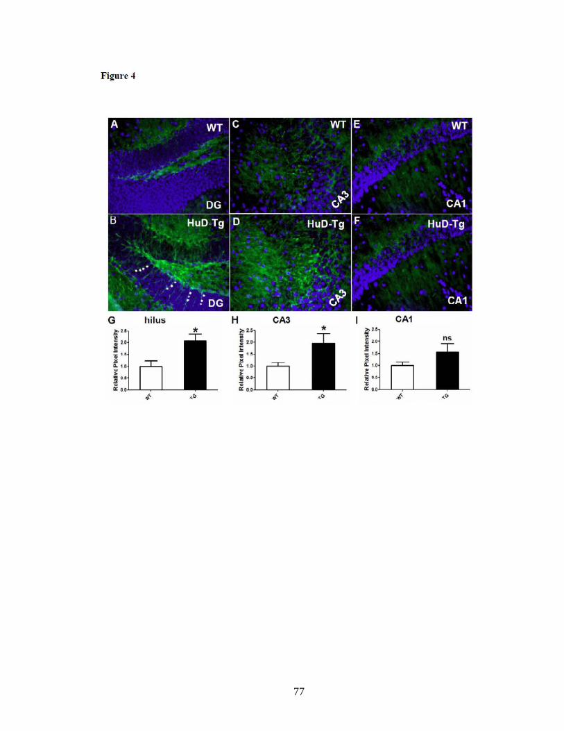
77
78
Reference Cited
Aid T, Kazantseva A, Piirsoo M, Palm K, Timmusk T (2007) Mouse and rat BDNF gene
structure and expression revisited. J Neurosci Res 85:525-535.
Akamatsu W, Fujihara H, Mitsuhashi T, Yano M, Shibata S, Hayakawa Y, Okano HJ,
Sakakibara S, Takano H, Takano T, Takahashi T, Noda T, Okano H (2005) The
RNAbinding protein HuD regulates neuronal cell identity and maturation. Proc
Natl Acad Sci U S A 102:4625-4630.
An JJ, Gharami K, Liao GY, Woo NH, Lau AG, Vanevski F, Torre ER, Jones KR, Feng
Y, Lu B, Xu B (2008) Distinct Role of Long 3' UTR BDNF mRNA in Spine
Morphology and Synaptic Plasticity in Hippocampal Neurons. Cell 134:175-187.
Anderson KD, Merhege MA, Morin M, Bolognani F, Perrone-Bizzozero NI (2003)
Increased expression and localization of the RNA-binding protein HuD and GAP-
43 mRNA to cytoplasmic granules in DRG neurons during nerve regeneration.
Exp Neurol 183:100- 108.
Anderson KD, Sengupta J, Morin M, Neve RL, Valenzuela CF, Perrone-Bizzozero NI
(2001) Overexpression of HuD accelerates neurite outgrowth and increases GAP-
43 mRNA expression in cortical neurons and retinoic acid-induced embryonic
stem cells in vitro. Exp Neurol 168:250-258.
Anderson KD, Morin MA, Beckel-Mitchener A, Mobarak CD, Neve RL, Furneaux HM,
Burry R, Perrone-Bizzozero NI (2000) Overexpression of HuD, but not of its
truncated form HuD I+II, promotes GAP-43 gene expression and neurite
outgrowth in PC12 cells in the absence of nerve growth factor. J Neurochem
75:1103-1114.
Beckel-Mitchener AC, Miera A, Keller R, Perrone-Bizzozero NI (2002) Poly(A) tail
length-dependent stabilization of GAP-43 mRNA by the RNA-binding protein
HuD. J Biol Chem 277:27996-28002.
Bolognani F, Contente-Cuomo T, Perrone-Bizzozero NI (2009) Novel recognition motifs
and biological functions of the RNA-binding protein HuD revealed by genome-
wide identification of its targets. Nucleic Acids Res 38:117-130.
Bolognani F, Merhege MA, Twiss J, Perrone-Bizzozero NI (2004) Dendritic localization
of the RNA-binding protein HuD in hippocampal neurons: association with
polysomes and upregulation during contextual learning. Neurosci Lett 371:152-
157.
Bolognani F, Tanner DC, Merhege M, Deschenes-Furry J, Jasmin B, Perrone-Bizzozero
NI (2006) In vivo post-transcriptional regulation of GAP-43 mRNA by
overexpression of the RNA-binding protein HuD. J Neurochem 96:790-801.
79
Bolognani F, Tanner DC, Nixon S, Okano HJ, Okano H, Perrone-Bizzozero NI (2007)
Coordinated expression of HuD and GAP-43 in hippocampal dentate granule cells
during developmental and adult plasticity. Neurochem Res 32:2142-2151.
Castren E, Berninger B, Leingartner A, Lindholm D (1998) Regulation of brain-derived
neurotrophic factor mRNA levels in hippocampus by neuronal activity. Prog
Brain Res 117:57-64.
Chen CY, Gherzi R, Ong SE, Chan EL, Raijmakers R, Pruijn GJ, Stoecklin G, Moroni C,
Mann M, Karin M (2001) AU binding proteins recruit the exosome to degrade
ARE-containing mRNAs. Cell 107:451-464.
Chung S, Eckrich M, Perrone-Bizzozero N, Kohn DT, Furneaux H (1997) The Elav-like
proteins bind to a conserved regulatory element in the 3'-untranslated region of
GAP-43 mRNA. J Biol Chem 272:6593-6598.
Danzer SC, McNamara JO (2004) Localization of brain-derived neurotrophic factor to
distinct terminals of mossy fiber axons implies regulation of both excitation and
feedforward inhibition of CA3 pyramidal cells. J Neurosci 24:11346-11355.
Danzer SC, He X, McNamara JO (2004) Ontogeny of seizure-induced increases in BDNF
immunoreactivity and TrkB receptor activation in rat hippocampus. Hippocampus
14:345-355.
Deschenes-Furry J, Perrone-Bizzozero N, Jasmin BJ (2006) The RNA-binding protein
HuD: a regulator of neuronal differentiation, maintenance and plasticity.
Bioessays 28:822-833.
Fukao A, Sasano Y, Imataka H, Inoue K, Sakamoto H, Sonenberg N, Thoma C, Fujiwara
T (2009) The ELAV protein HuD stimulates cap-dependent translation in a
Poly(A)- and eIF4A-dependent manner. Mol Cell 36:1007-1017.
Guo W, Allan AM, Zong R, Zhang L, Johnson EB, Schaller EG, Murthy AC, Goggin SL,
Eisch AJ, Oostra BA, Nelson DL, Jin P, Zhao X (2011) Ablation of Fmrp in adult
neural stem cells disrupts hippocampus-dependent learning. Nat Med 17:559-565.
Lau AG, Irier HA, Gu J, Tian D, Ku L, Liu G, Xia M, Fritsch B, Zheng JQ, Dingledine
R, Xu B, Lu B, Feng Y (2010) Distinct 3'UTRs differentially regulate activity-
dependent translation of brain-derived neurotrophic factor (BDNF). Proc Natl
Acad Sci U S A 107:15945-15950.
Li Y, Jiang Z, Chen H, Ma WJ (2004) A modified quantitative EMSA and its application
in the study of RNA--protein interactions. J Biochem Biophys Methods 60:85-96.
80
Liu QR, Lu L, Zhu XG, Gong JP, Shaham Y, Uhl GR (2006) Rodent BDNF genes, novel
promoters, novel splice variants, and regulation by cocaine. Brain Res 1067:1-12.
Martinowich K, Manji H, Lu B (2007) New insights into BDNF function in depression
and anxiety. Nat Neurosci 10:1089-1093.
Nagahara AH, Tuszynski MH (2011) Potential therapeutic uses of BDNF in neurological
and psychiatric disorders. Nat Rev Drug Discov 10:209-219.
Okano HJ, Darnell RB (1997) A hierarchy of Hu RNA binding proteins in developing
and adult neurons. J Neurosci 17:3024-3037.
Pascale A, Gusev PA, Amadio M, Dottorini T, Govoni S, Alkon DL, Quattrone A (2004)
Increase of the RNA-binding protein HuD and posttranscriptional up-regulation
of the GAP-43 gene during spatial memory. Proc Natl Acad Sci U S A 101:1217-
1222.
Perrone-Bizzozero N, Bolognani F (2002) Role of HuD and other RNA-binding proteins
in neural development and plasticity. J Neurosci Res 68:121-126.
Perrone-Bizzozero NI, Tanner DC, Mounce J, Bolognani F (2011) Increased expression
of axogenesis-related genes and mossy fibre length in dentate granule cells from
adult HuD overexpressor mice. ASN Neuro 3:259-270.
Tiruchinapalli DM, Caron MG, Keene JD (2008) Activity-dependent expression of
ELAV/Hu RBPs and neuronal mRNAs in seizure and cocaine brain. J Neurochem
107:1529-1543.
Ule J, Jensen KB, Ruggiu M, Mele A, Ule A, Darnell RB (2003) CLIP identifies Nova-
regulated RNA networks in the brain. Science 302:1212-1215.
Washbourne P, Thompson PM, Carta M, Costa ET, Mathews JR, Lopez-Bendito G,
Molnar Z, Becher MW, Valenzuela CF, Partridge LD, Wilson MC (2002) Genetic
ablation of the t- SNARE SNAP-25 distinguishes mechanisms of
neuroexocytosis. Nat Neurosci 5:19-26.
Xu N, Chen CY, Shyu AB (1997) Modulation of the fate of cytoplasmic mRNA by AU-
rich elements: key sequence features controlling mRNA deadenylation and decay.
Mol Cell Biol 17:4611-4621.
Zhao L, Mandler MD, Yi H, Feng Y (2010) Quaking I controls a unique cytoplasmic
pathway that regulates alternative splicing of myelin-associated glycoprotein.
Proc Natl Acad Sci U S A 107:19061-19066.

81
Zhao L, Tian D, Xia M, Macklin WB, Feng Y (2006) Rescuing qkV dysmyelination by a
single isoform of the selective RNA-binding protein QKI. J Neurosci 26:11278-
11286.
82
Appendix C: Review article in press, Frontiers in Bioscience. Co-
authored by Clark Bird
References for this article are self-contained within this appendix.
Role of HuD in neurological diseases
Nora Perrone-Bizzozero1, Clark W. Bird
1
1Department of Neurosciences, University of New Mexico School of Medicine,
Albuquerque, NM 87131
TABLE OF CONTENTS
1. Abstract
2. Introduction
3. Hu protein function in neurons
3.1. Role of Hu proteins in neuronal development
3.2. Role of HuD in synaptic plasticity
3.3. Role of HuD in nerve regeneration
4. Control of HuD function and its target mRNAs
4.1. Transcriptional, post-transcriptional and post-translational control
4.2. Mechanism of HuD-mediated stabilization of neuronal mRNAs
4.3. HuD targets in neurological disorders
5. Association of HuD with neuropsychiatric disorders
5.1. HuD and the genetics of Parkinson’s disease
5.2. HuD and Alzheimer’s disease
5.3. HuD and schizophrenia
5.4. HuD levels in epilepsy and drug abuse
5.5. HuD and spinal muscular ataxia
6. Involvement of HuD in neuroblastomas
6.1. HuD levels in different tumor subtypes
6.2. ELAVL4 haploinsufficiency and tumor malignancy
7. Perspectives
8. Acknowledgement
9. References
1. ABSTRACT
83
Hu proteins are a family of RNA-binding proteins (RBPs) that are homologs of
Drosophila ELAV, a protein required for nervous system development. Three of these
proteins (HuB, HuC, and HuD) are developmentally regulated and expressed in neurons.
The fourth member, HuR is also expressed in other tissues. At the molecular level, Hu
proteins are known to interact with AU-rich instability conferring sequences in the 3’
UTR of specific target mRNAs, stabilizing the mRNAs. These proteins are not only the
best known mRNA stabilizers but also the earliest markers of the neuronal cell lineage.
Among the neuronal Hu proteins, HuD has been shown to accelerate neuronal
differentiation and axonal outgrowth in neurons both in culture and in vivo. In addition,
HuD and other Hu proteins participate in synaptic plasticity mechanisms in the mature
central nervous system and promote regeneration of peripheral nerves. Furthermore, HuD
has been implicated in pathological conditions from neurodegenerative disorders such as
Parkinson’s and Alzheimer’s disease to childhood brain tumors. This review will focus
on the involvement of HuD in nervous system function and pathology.
2. INTRODUCTION
Hu proteins are a family of RNA-binding proteins that were first detected as the
targets of autoantibodies found in patients with paraneoplastic encephalomyelitis (1).
These proteins are homologs of ELAV (embryonic lethal abnormal vision), a Drosophila
RNA-binding protein identified because of the lethality shown by its deletion (2).
Although there is only one ELAV protein in Drosophila, four mammalian ELAV-like Hu
proteins have been identified [HuR, HuB (a.k.a.Hel-N1), HuC and HuD]. Three of these
proteins (HuB, HuC, and HuD) are expressed in neurons while the fourth member, HuR,
is ubiquitously-expressed in all tissues. At the molecular level, all four ELAV-like Hu
84
proteins contain three RNA recognition motifs (RRMs), a highly conserved 80 amino
acid region that was first recognized in splicing factors and poly(A)-binding protein (3,
4). The RRMs are highly conserved among members of the family while the amino
terminus and a basic domain between RRMs 2 and 3 are very diverse. RRMs 1 and 2 in
Hu proteins bind AU-rich elements (ARE) found in the 3’UTRs of several unstable
mRNAs involved in cell growth and differentiation (5, 6). In contrast, the third RRM is
important for the interaction of the protein with long poly (A) tails (7, 8) (Figure 1).
Recent studies indicate that Hu proteins are involved in various aspects of mRNA
regulation, from mRNA processing and stability to translation (9-13).
3. Hu PROTEIN FUNCTION IN NEURONS
3.1. Role of Hu proteins in neuronal development
In Drosophila, deletion of the elav gene is embryonic lethal due to the failure of
neurons to differentiate (2, 14). The continued expression of ELAV in adult neurons is
essential for brain function, as temperature-sensitive mutants become incapacitated at
non-permissive temperatures (15). In higher vertebrates and mammals, Hu proteins are
one of the earliest markers expressed in neurons (16). HuR is the first protein to be
expressed in chicken embryos where it is thought to be involved in cell proliferation (17).
HuR is mainly nuclear and shuttles to the cytosol (18, 19) while HuD and other Hu
proteins are localized to the cytoplasm (20-22). The expression of HuD coincides with
the earliest stages of neuronal differentiation and is maintained through the maturation of
neurons (17). Similar types of expression patterns have been observed in the developing
mouse (23) and rat (24, 25) brains. The involvement of Hu proteins in different stages of
85
neuronal differentiation was confirmed by overexpression and knockout studies.
Overexpression of HuB, HuC or HuD in PC12 cells and in vivo was shown to increase
the rate of neuronal differentiation (22, 26-29). Down-regulation of these proteins in
neural cell lines results in the opposite phenotype, with cells failing to grow neurites (29,
30). Neuronal Hu proteins also have a role in neural stem cell differentiation as seen by
the phenotype of HuD KO mice, which show increased proliferation of stem cells in the
subventricular zone but decreased production of mature neurons (31). Altogether, these
findings support the notion that Hu proteins play a critical role in nervous system
development.
3.2. Role of Hu proteins in synaptic plasticity
Although Hu proteins were initially described as early markers of development,
in certain mature neurons significant levels of Hu proteins persist throughout life,
particularly in the cortex and hippocampus. As shown in Bolognani et al., 2004, (ref. 20)
HuD is present in the soma and dendrites of pyramidal cells in the hippocampus and
neocortex in close association with polysomes. In contrast this protein is not detected in
mature dentate granule cells. The spatial pattern of expression of HuB is similar to that
of HuD but distinct from HuC, which is normally expressed at high levels in dentate
granule cells (23). The function of these proteins in mature neurons is not completely
understood but several lines of evidence indicate that they are involved in synaptic
plasticity mechanisms. First, HuD protein levels are increased in the hippocampus after
different learning and memory tasks (20, 32, 33). Second, antisense-mediated knock
down of HuC in the hippocampus impairs learning in the radial maze task in mice (33).
Third, overexpression of HuD in transgenic mice leads to profound deficits in the
86
performance of two associative learning and memory tests, fear conditioning and the
Morris water maze (34). Finally, recent analysis of the HuD targets in the mature brain
(35) revealed that several of these mRNAs are associated with long-term potentiation, a
phenomenon thought to underlie learning and memory (Figure 2).
3.3. Role of HuD in nerve regeneration
In addition to participating in synaptic changes in adult neurons in the central
nervous system, HuD has been implicated in the response of peripheral neurons to injury.
Following sciatic nerve crush, HuD protein and transcript levels increase in dorsal root
ganglia sensory neurons within 7 days and remain elevated for up to three weeks (36).
This increase in HuD expression is accompanied by a dramatic increase in GAP-43
mRNA levels, a known HuD target that encodes for a growth-associated protein involved
in axonal outgrowth. In regenerating dorsal root ganglion neurons, HuD protein co-
localizes with GAP-43 transcripts and ribosomal proteins in cytoplasmic granules (36),
suggesting that HuD contributes to stabilize GAP-43 mRNA before translation. Another
study also demonstrated the co-localization of HuD and GAP-43 mRNA in ribosome
containing granules in axons and growth cones (37), the sites of localized GAP-43
protein synthesis (Moon, Twiss and Perrone-Bizzozero, unpublished results). The role of
HuD in nerve regeneration was further investigated using a viral vector to express
exogenous human HuD in rat superior cervical ganglion neurons following axotomy (38).
HuD overexpression prevented the acute downregulation of acetylcholinesterase (AChE)
and GAP-43 mRNAs, leading to faster response to the injury. Together, these findings
demonstrate that in addition to its role in neuronal development and plasticity HuD is
87
important for the increased expression of growth-associated genes during nerve
regeneration.
4. CONTROL OF HuD FUNCTION AND ITS TARGET mRNAs
4.1. Transcriptional, post-transcriptional and post-translational control
Analysis of the structure of the four genes encoding each of the Hu proteins
demonstrated that these proteins not only have a high degree of sequence conservation
but also that their genes have a similar genomic organization (39-41, for review see ref.
10). Although the transcriptional control of Hu protein expression has not been fully
characterized, elements in the promoter regions of HuD, HuB and HuC were shown to
control neuron-specific expression of these proteins (41-45). In addition, HuD
transcription is known to be repressed by thyroid hormone (46), suggesting a potential
dysregulation in both hypo- and hyperthyroidism. Hu proteins are also subjected to
alternative splicing between RRMs 2 and 3. In the case of HuD, three alternatively-
spliced isoforms have been identified: HuDpro (with exons 6 and 7 inclusion), HuD (with
exon 6 inclusion) and HuDmex (excluding both exons 6 and 7). Among these isoforms,
HuD is the most abundant protein in neurons followed by HuDpro. Interestingly, it was
found that although Hu proteins block inclusion of specific exons in the mRNA encoding
neurofibromatosis 1 (NF1) and calcitonin gene-related peptide (CGRP) (12), they
promote inclusion of exon 6 in HuD (47), explaining the increased abundance of this
isoform in neurons.
Another post-transcriptional mechanism involves the regulation of Hu protein
levels by microRNAs. The effects of mir-519 and mir-125a on HuR levels have been
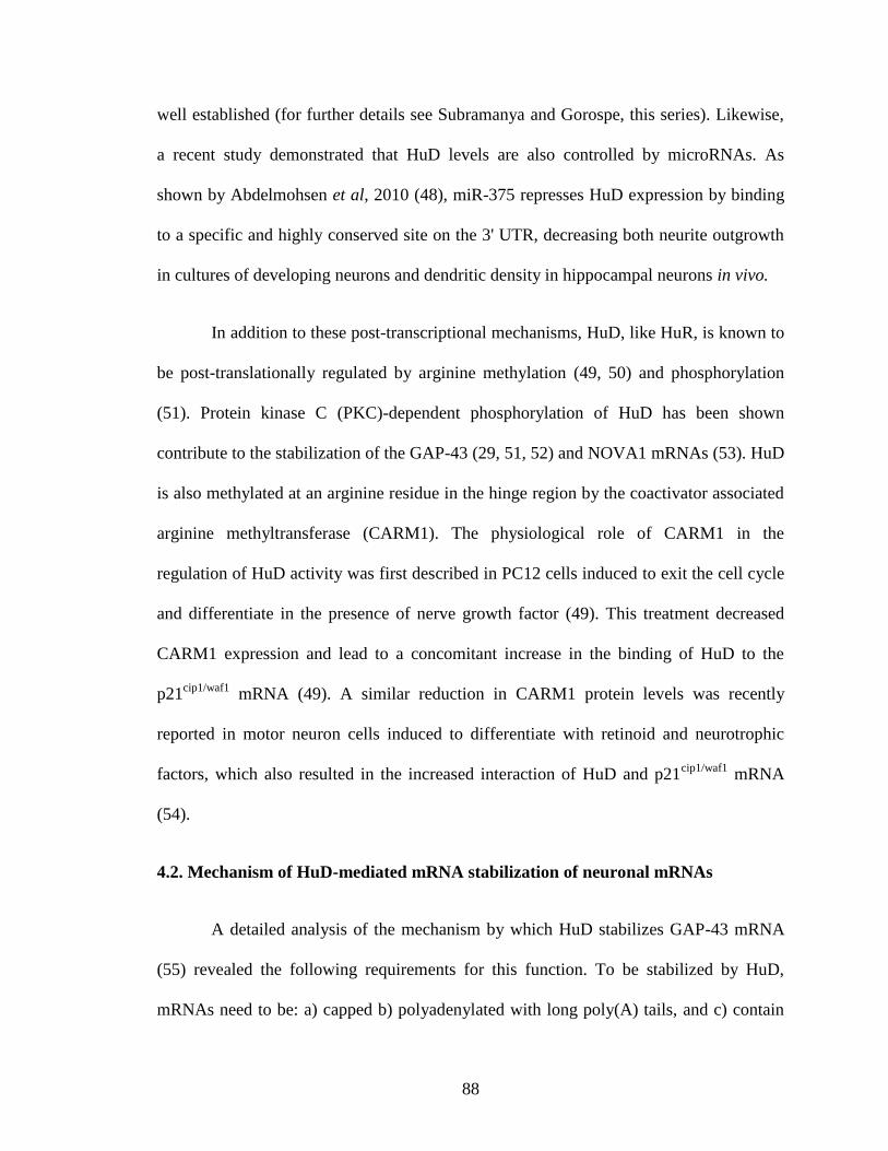
88
well established (for further details see Subramanya and Gorospe, this series). Likewise,
a recent study demonstrated that HuD levels are also controlled by microRNAs. As
shown by Abdelmohsen et al, 2010 (48), miR-375 represses HuD expression by binding
to a specific and highly conserved site on the 3' UTR, decreasing both neurite outgrowth
in cultures of developing neurons and dendritic density in hippocampal neurons in vivo.
In addition to these post-transcriptional mechanisms, HuD, like HuR, is known to
be post-translationally regulated by arginine methylation (49, 50) and phosphorylation
(51). Protein kinase C (PKC)-dependent phosphorylation of HuD has been shown
contribute to the stabilization of the GAP-43 (29, 51, 52) and NOVA1 mRNAs (53). HuD
is also methylated at an arginine residue in the hinge region by the coactivator associated
arginine methyltransferase (CARM1). The physiological role of CARM1 in the
regulation of HuD activity was first described in PC12 cells induced to exit the cell cycle
and differentiate in the presence of nerve growth factor (49). This treatment decreased
CARM1 expression and lead to a concomitant increase in the binding of HuD to the
p21cip1/waf1
mRNA (49). A similar reduction in CARM1 protein levels was recently
reported in motor neuron cells induced to differentiate with retinoid and neurotrophic
factors, which also resulted in the increased interaction of HuD and p21cip1/waf1
mRNA
(54).
4.2. Mechanism of HuD-mediated mRNA stabilization of neuronal mRNAs
A detailed analysis of the mechanism by which HuD stabilizes GAP-43 mRNA
(55) revealed the following requirements for this function. To be stabilized by HuD,
mRNAs need to be: a) capped b) polyadenylated with long poly(A) tails, and c) contain
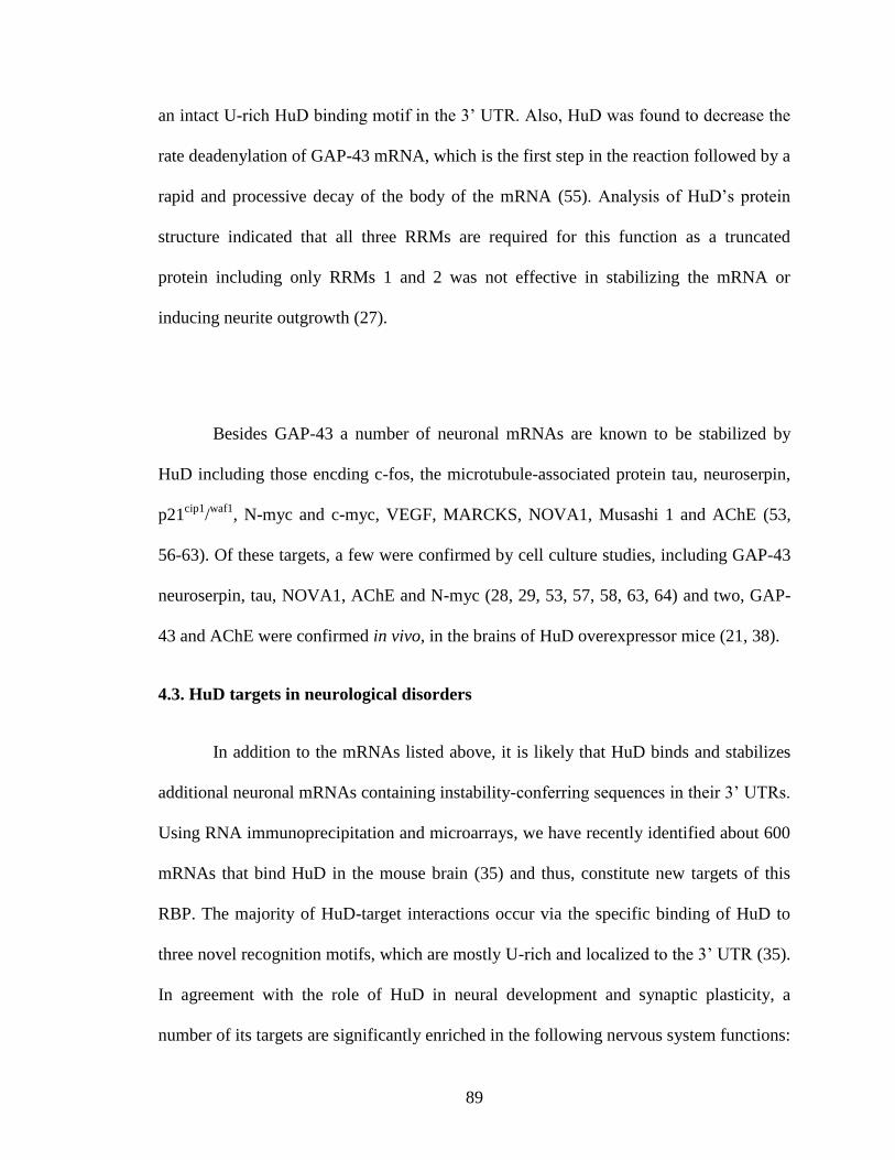
89
an intact U-rich HuD binding motif in the 3’ UTR. Also, HuD was found to decrease the
rate deadenylation of GAP-43 mRNA, which is the first step in the reaction followed by a
rapid and processive decay of the body of the mRNA (55). Analysis of HuD’s protein
structure indicated that all three RRMs are required for this function as a truncated
protein including only RRMs 1 and 2 was not effective in stabilizing the mRNA or
inducing neurite outgrowth (27).
Besides GAP-43 a number of neuronal mRNAs are known to be stabilized by
HuD including those encding c-fos, the microtubule-associated protein tau, neuroserpin,
p21cip1
/waf1
, N-myc and c-myc, VEGF, MARCKS, NOVA1, Musashi 1 and AChE (53,
56-63). Of these targets, a few were confirmed by cell culture studies, including GAP-43
neuroserpin, tau, NOVA1, AChE and N-myc (28, 29, 53, 57, 58, 63, 64) and two, GAP-
43 and AChE were confirmed in vivo, in the brains of HuD overexpressor mice (21, 38).
4.3. HuD targets in neurological disorders
In addition to the mRNAs listed above, it is likely that HuD binds and stabilizes
additional neuronal mRNAs containing instability-conferring sequences in their 3’ UTRs.
Using RNA immunoprecipitation and microarrays, we have recently identified about 600
mRNAs that bind HuD in the mouse brain (35) and thus, constitute new targets of this
RBP. The majority of HuD-target interactions occur via the specific binding of HuD to
three novel recognition motifs, which are mostly U-rich and localized to the 3’ UTR (35).
In agreement with the role of HuD in neural development and synaptic plasticity, a
number of its targets are significantly enriched in the following nervous system functions:
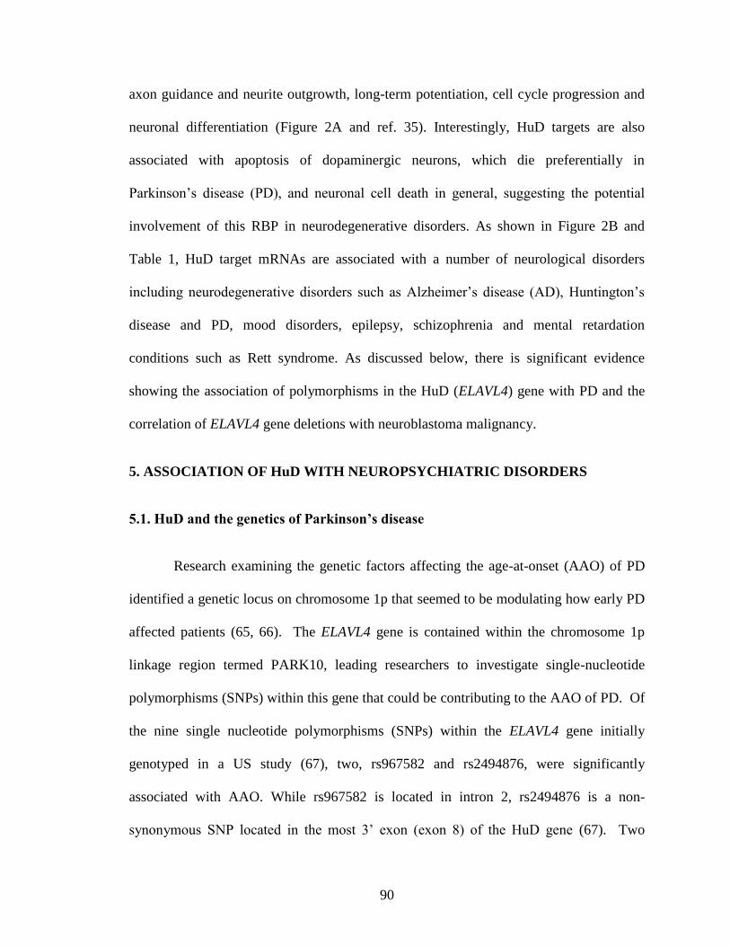
90
axon guidance and neurite outgrowth, long-term potentiation, cell cycle progression and
neuronal differentiation (Figure 2A and ref. 35). Interestingly, HuD targets are also
associated with apoptosis of dopaminergic neurons, which die preferentially in
Parkinson’s disease (PD), and neuronal cell death in general, suggesting the potential
involvement of this RBP in neurodegenerative disorders. As shown in Figure 2B and
Table 1, HuD target mRNAs are associated with a number of neurological disorders
including neurodegenerative disorders such as Alzheimer’s disease (AD), Huntington’s
disease and PD, mood disorders, epilepsy, schizophrenia and mental retardation
conditions such as Rett syndrome. As discussed below, there is significant evidence
showing the association of polymorphisms in the HuD (ELAVL4) gene with PD and the
correlation of ELAVL4 gene deletions with neuroblastoma malignancy.
5. ASSOCIATION OF HuD WITH NEUROPSYCHIATRIC DISORDERS
5.1. HuD and the genetics of Parkinson’s disease
Research examining the genetic factors affecting the age-at-onset (AAO) of PD
identified a genetic locus on chromosome 1p that seemed to be modulating how early PD
affected patients (65, 66). The ELAVL4 gene is contained within the chromosome 1p
linkage region termed PARK10, leading researchers to investigate single-nucleotide
polymorphisms (SNPs) within this gene that could be contributing to the AAO of PD. Of
the nine single nucleotide polymorphisms (SNPs) within the ELAVL4 gene initially
genotyped in a US study (67), two, rs967582 and rs2494876, were significantly
associated with AAO. While rs967582 is located in intron 2, rs2494876 is a non-
synonymous SNP located in the most 3’ exon (exon 8) of the HuD gene (67). Two
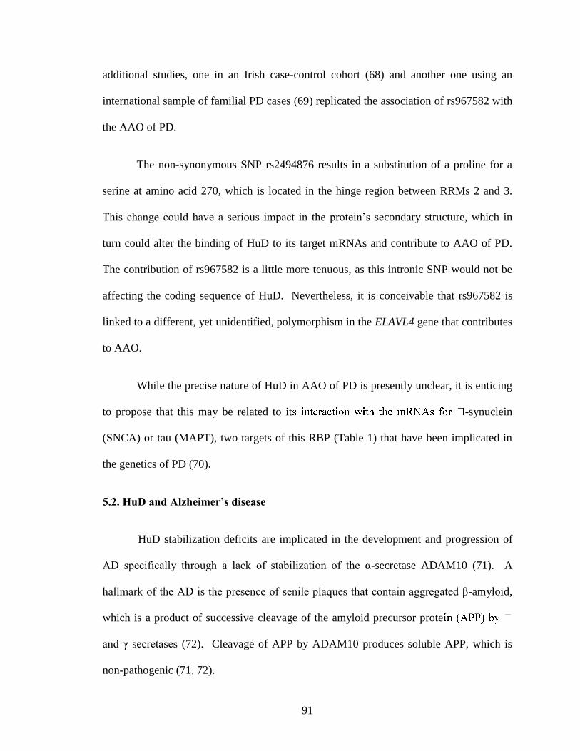
91
additional studies, one in an Irish case-control cohort (68) and another one using an
international sample of familial PD cases (69) replicated the association of rs967582 with
the AAO of PD.
The non-synonymous SNP rs2494876 results in a substitution of a proline for a
serine at amino acid 270, which is located in the hinge region between RRMs 2 and 3.
This change could have a serious impact in the protein’s secondary structure, which in
turn could alter the binding of HuD to its target mRNAs and contribute to AAO of PD.
The contribution of rs967582 is a little more tenuous, as this intronic SNP would not be
affecting the coding sequence of HuD. Nevertheless, it is conceivable that rs967582 is
linked to a different, yet unidentified, polymorphism in the ELAVL4 gene that contributes
to AAO.
While the precise nature of HuD in AAO of PD is presently unclear, it is enticing
to propose that this may be related to it -synuclein
(SNCA) or tau (MAPT), two targets of this RBP (Table 1) that have been implicated in
the genetics of PD (70).
5.2. HuD and Alzheimer’s disease
HuD stabilization deficits are implicated in the development and progression of
AD specifically through a lack of stabilization of the α-secretase ADAM10 (71). A
hallmark of the AD is the presence of senile plaques that contain aggregated β-amyloid,
which is a product of successive cleavage of the amyloid precursor prote
and γ secretases (72). Cleavage of APP by ADAM10 produces soluble APP, which is
non-pathogenic (71, 72).
92
ADAM10 and HuD protein levels are reduced in AD, suggesting that HuD could
be regulating ADAM10 protein levels through post-transcriptional regulation (73, 74).
Computational analysis of ADAM10 mRNA sequence demonstrated the presence of an
ARE, which also contains the putative HuD binding site. Immunoprecipitation of mRNPs
containing HuD demonstrated an enrichment of ADAM10 mRNA in these complexes,
further implicating HuD in the regulation of ADAM10 gene expression (73).
Binding of HuD to target mRNAs is regulated in part by PKCα isoenzyme
signaling (51), which may play a role in HuD regulation of ADAM10 levels. PKC
isoenzyme levels are decreased in the post-mortem brain of AD patients, leading to the
interesting possibility that a deficit of PKC signaling could be impairing the binding of
HuD to its mRNA targets, including ADAM10 (71). As shown in Table 1, in addition to
ADAM10, a number of AD associated protein mRNAs are HuD targets. Among these is
the mRNA for the β-secretase BACE1, which is involved in the production of β-amyloid
and a new target for the treatment of AD (72, 75). However HuD’s effect on the levels of
this mRNA is yet to be established.
5.3. HuD and schizophrenia
A DNA microarray study of mRNAs expressed in the prefrontal cortex of patients
with schizophrenia (76) revealed that HuD mRNA levels are increased in this disorder.
Clustering analyses of the data showed that not only was HuD increased in patients but
also and, most-importantly, that GAP-43 was tightly co-regulated with this protein and so
were other HuD targets such as neuroserpin and MARCKS. Considering that these
mRNAs are also developmentally-regulated and that the expression of at least one of
93
them, GAP-43, is increased in these patients (77), it is enticing to propose that the
observed changes in these mRNAs could be due to their HuD-induced stabilization.
5.4. HuD levels in epilepsy and drug abuse
As shown in Table 1 and Figure 2B, a number of HuD targets are associated with
epilepsy. Supporting a role of HuD in this disorder HuD mRNA levels were shown to
increase in rat hippocampal dentate granule cells 24 hours following kainic acid induced
seizures (78) and similar findings were reported in CA1 and CA3 region of the
hippocampus after pilocarpine induced seizures (79). Furthermore, HuD expression is
also affected by exposure to drugs of abuse such as cocaine, as shown by the increases in
the levels of this mRNA either in the whole brain (79) or in the nucleus accumbens
(Perrone-Bizzozero and Neisewander, unpublished observations) 24 hours after rats
received a single sensitizing injection of cocaine.
5.5. HuD and spinal muscular ataxia
Spinal muscular ataxia (SMA) is an autosomal recessive neuromuscular disease
characterized by the selective degeneration of lower motor neurons of the spinal cord.
SMA is caused by deletions or loss-of-function mutations in the Survival of Motor
Neuron (SMN) gene. SMN is a protein connecting neuronal splicing and axonal transport
and a recent study demonstrated that HuD co-localizes with this protein in axons (54).
Furthermore, SMN was shown to recruit HuD and its target mRNAs into RNA granules,
a process that depends on the presence of the Tudor domain in SMN. Finally, it was
shown that HuD overexpression could compensate for the differentiation defects
94
observed in SMN haploinsufficient motor neurons, suggesting that increasing HuD levels
in these cells could lead to better treatments for SMA (54).
6. ASSOCIATION OF HuD WITH NEUROBLASTOMAS
6.1. HuD levels in different neuroblastoma subtypes
Neuroblastomas (NB) arise from embryonic neural crest and primarily affect
young children. The proto-oncogene encoding N-myc (MYCN) is found to be amplified
as well as overexpressed in a number of these cancers (64). A number of NB-derived cell
lines have been used to understand the mechanisms of carcinogenesis. Phenotypically
these lines are diverse but can be classified into two main subtypes: neuroblastic N-type
cells, which are non-adherent and very tumorigenic and substrate adherent S-type cells,
which are not tumorigenic. N-myc is believed to maintain the proliferative,
undifferentiated state of cells during development (80). HuD expression levels are high in
N-type but absence in S type cells, creating the possibility that HuD may stabilize N-myc
transcripts and push the cells towards a cancerous fate (81).
In vitro experiments showed that HuD binds to elements in the 3’ UTR of N-myc
mRNA (81). Ectopic HuD expression in stable cell lines leads to the stabilization of a
reporter gene expressing the N-myc 3’UTR (64). Conversely, treatment of cells with anti-
sense oligomers against HuD lead to a decrease in N-myc reporter levels, implicating
HuD as the main factor regulating the stability of this mRNA (64). In addition to
stabilization of the mature N-myc transcripts, HuD-induced pre-mRNA processing and
stability has been reported in these cells (82).
95
6.2. ELAVL4 haploinsufficiency and NB malignancy
The posttranscriptional regulation of N-myc transcripts is not the only mechanism
by which HuD is involved in neuroblastoma progression. About 30% of neuroblastomas
with a clinically poor prognosis contain an amplification of the MYCN gene and a
deletion in the small arm of chromosome 1 where the ELAVL4 gene is located (83, 84).
Moreover, higher HuD expression levels in neuroblastoma cells were correlated with a
better clinical outcome in patients (85). Supporting a role of HuD in decreasing
malignancy, overexpression of HuD in two neuroblastoma cell lines with high MYCN
amplification was shown to decrease both cell proliferation and MYCN gene copy
numbers (86). In contrast, knockdown of HuD levels in non-amplified SY5Y cells had
the opposite effect, causing decreased HuD levels and selecting for cells with multiple
copies of the MYCN gene (86). Although the precise mechanism of MYCN amplification
remains to be established, one possible explanation for these findings is that cells with
HuD haploinsufficiency, which normally would have lower N-myc levels, will not
survive in culture unless they contain multiple MYCN copies, which will give them a
growth advantage over non-amplified cells (86).
7. PERSPECTIVE
Altogether the data presented above supports the idea that HuD and other
neuronal Hu proteins play a critical role in the post-transcriptional control of gene
expression during nervous system development and plasticity. As shown in Figure 2A,
HuD targets are involved in different aspects in the life of a neuron from cell cycle
progression and neuronal differentiation to proper maturation and cell death. Therefore, it
96
is not surprising that many of these same target mRNAs are associated with various
neuropsychiatric disorders, from mental retardation and schizophrenia to
neurodegenerative disorders such as Parkinson’s and Alzheimer’s disease (Table 1 and
Figure 2B). The role of HuD in Parkinson’s disease is also supported by genetic linkage
studies demonstrating a significant association of two polymorphisms in the ELAVL4
gene with AAO of PD. The recent findings that deletions in the ELAVL4 gene are
associated with increased malignancy in neuroblastoma cell lines not only highlight the
function of this RBP in cell cycle arrest but also suggest that gene therapy directed at
increasing HuD levels in neuroblastoma cells could lead to more effective treatments of
those NB patients with the worst prognosis. Likewise, it is possible that that HuD
overexpression could help rescue part of the motor neuron death phenotype in patients
with SMA. Finally, the changes in HuD expression in response to epileptic seizures and
cocaine exposure suggest a role of HuD in these disorders. From a biomedical
perspective, the elucidation of the mechanisms controlling how HuD regulates the
stability and translation of its target mRNAs and how these RNA-protein interactions
respond to environmental cues has potential implications for the understanding a broad
range of conditions from normal development and synaptic plasticity to
neurodevelopmental disorders and neurodegenerative diseases.
8. ACKNOWLEDGEMENTS
We wish to thank Joanna Beeson Mounce for performing biological pathway and
disease association analyses of HuD targets shown in Table 1 and Figure 2. Part of this
work was supported by NIH grants (NS30255 and DA25992 to NPB).
97
9. REFERENCES
1. Dalmau, J., H. M. Furneaux, R. J. Gralla, M. G. Kris & J. B. Posner: Detection of the
anti-Hu antibody in the serum of patients with small cell lung cancer--a quantitative
western blot analysis. Ann Neurol, 27, 544-52. (1990)
2. Robinow, S., A. R. Campos, K. M. Yao & K. White: The elav gene product of
Drosophila, required in neurons, has three RNP consensus motifs. Science, 242, 1570-2
(1988)
3. Kenan, D. J., C. C. Query & J. D. Keene: RNA recognition: towards identifying
determinants of specificity. Trends Biochem Sci, 16, 214-20 (1991)
4. Query, C. C., R. C. Bentley & J. D. Keene: A common RNA recognition motif
identified within a defined U1 RNA binding domain of the 70K U1 snRNP protein. Cell,
57, 89-101 (1989)
5. Keene, J. D.: Why is Hu where? Shuttling of early-response-gene messenger RNA
subsets. Proc Natl Acad Sci U S A, 96, 5-7. (1999)
6. Wilusz, C. J. & J. Wilusz: Bringing the role of mRNA decay in the control of gene
expression into focus. Trends Genet, 20, 491-7 (2004)
7. Abe, R., K. Yamamoto & H. Sakamoto: Target specificity of neuronal RNA-binding
protein, Mel-N1: direct binding to the 3' untranslated region of its own mRNA. Nucleic
Acids Res, 24, 2011-6 (1996)
8. Ma, W. J., S. Chung & H. Furneaux: The Elav-like proteins bind to AU-rich elements
and to the poly(A) tail of mRNA. Nucleic Acids Res, 25, 3564-9 (1997)
9. Bolognani, F. & N. I. Perrone-Bizzozero: RNA-protein interactions and control of
mRNA stability in neurons. J Neurosci Res, 86, 481-9 (2008)
10. Deschenes-Furry, J., N. Perrone-Bizzozero & B. J. Jasmin: The RNA-binding protein
HuD: a regulator of neuronal differentiation, maintenance and plasticity. Bioessays, 28,
822-33 (2006)
11. Fukao, A., Y. Sasano, H. Imataka, K. Inoue, H. Sakamoto, N. Sonenberg, C. Thoma
& T. Fujiwara: The ELAV protein HuD stimulates cap-dependent translation in a
Poly(A)- and eIF4A-dependent manner. Mol Cell, 36, 1007-17 (2009)
12. Hinman, M. N. & H. Lou: Diverse molecular functions of Hu proteins. Cell Mol Life
Sci, 65, 3168-81 (2008)
13. Perrone-Bizzozero, N. & F. Bolognani: Role of HuD and other RNA-binding proteins
in neural development and plasticity. J Neurosci Res, 68, 121-6. (2002)
98
14. Campos, A. R., D. Grossman & K. White: Mutant alleles at the locus elav in
Drosophila melanogaster lead to nervous system defects. A developmental-genetic
analysis. J Neurogenet, 2, 197-218 (1985)
15. Homyk, T., Jr., K. Isono & W. L. Pak: Developmental and physiological analysis of a
conditional mutation affecting photoreceptor and optic lobe development in Drosophila
melanogaster. J Neurogenet, 2, 309-24 (1985)
16. Marusich, M. F., H. M. Furneaux, P. D. Henion & J. A. Weston: Hu neuronal proteins
are expressed in proliferating neurogenic cells. J Neurobiol, 25, 143-55 (1994)
17. Wakamatsu, Y. & J. A. Weston: Sequential expression and role of Hu RNA-binding
proteins during neurogenesis. Development, 124, 3449-60. (1997)
18. Fan, X. C. & J. A. Steitz: Overexpression of HuR, a nuclear-cytoplasmic shuttling
protein, increases the in vivo stability of ARE-containing mRNAs. EMBO J, 17, 3448-60
(1998)
19. Peng, S. S.-Y., Chen, C.-Y.A., Xu, N., and A.-B. Shyu: RNA stabilization of the AU-
rich element binding protein, HuR, an ELAV protein. EMBO J, 17, 3461-3470 (1998)
20. Bolognani, F., Merhege, M. A., Twiss, J. and Nora I. Perrone-Bizzozero: Dendritic
localization of the RNA-binding protein HuD in hippocampal neurons: association with
polysomes and upregulation during contextual learning. Neurosci Letters, 371, 152-157
(2004)
21. Bolognani, F., D. C. Tanner, M. Merhege, J. Deschenes-Furry, B. Jasmin & N. I.
Perrone-Bizzozero: In vivo post-transcriptional regulation of GAP-43 mRNA by
overexpression of the RNA-binding protein HuD. J Neurochem, 96, 790-801 (2006)
22. Kasashima, K., K. Terashima, K. Yamamoto, E. Sakashita & H. Sakamoto:
Cytoplasmic localization is required for the mammalian ELAV-like protein HuD to
induce neuronal differentiation. Genes Cells, 4, 667-83 (1999)
23. Okano, H. J. & R. B. Darnell: A hierarchy of Hu RNA binding proteins in developing
and adult neurons. J Neurosci, 17, 3024-37. (1997)
24. Clayton, G. H., Perez, G.M., Smith, R.L., and G.C. Owens: Expression of mRNA for
the elav-like neural-specific RNA binding protein, HuD, during nervous system
development Brain Res. Dev Brain Res, 109, 271-280 (1998)
25. Hambardzumyan, D., S. Sergent-Tanguy, R. Thinard, V. Bonnamain, M. Masip, A.
Fabre, H. Boudin, I. Neveu & P. Naveilhan: AUF1 and Hu proteins in the developing rat
brain: implication in the proliferation and differentiation of neural progenitors. J Neurosci
Res, 87, 1296-309 (2009)
26. Akamatsu, W., H. J. Okano, N. Osumi, T. Inoue, S. Nakamura, S. Sakakibara, M.
Miura, N. Matsuo, R. B. Darnell & H. Okano: Mammalian ELAV-like neuronal RNA-
99
binding proteins HuB and HuC promote neuronal development in both the central and the
peripheral nervous systems. Proc Natl Acad Sci U S A, 96, 9885-90 (1999)
27. Anderson, K. D., M. A. Morin, A. Beckel-Mitchener, C. D. Mobarak, R. L. Neve, H.
M. Furneaux, R. Burry & N. I. Perrone-Bizzozero: Overexpression of HuD, but not of its
truncated form HuD I+II, promotes GAP-43 gene expression and neurite outgrowth in
PC12 cells in the absence of nerve growth factor. J Neurochem, 75, 1103-14. (2000)
28. Anderson, K. D., J. Sengupta, M. Morin, R. L. Neve, C. F. Valenzuela & N. I.
Perrone-Bizzozero: Overexpression of HuD accelerates neurite outgrowth and increases
GAP-43 mRNA expression in cortical neurons and retinoic acid-induced embryonic stem
cells in vitro. Exp Neurol, 168, 250-8. (2001)
29. Mobarak, C. D., K. D. Anderson, M. Morin, A. Beckel-Mitchener, S. L. Rogers, H.
Furneaux, P. King & N. I. Perrone-Bizzozero: The RNA-binding protein HuD is required
for GAP-43 mRNA stability, GAP-43 gene expression, and PKC-dependent neurite
outgrowth in PC12 cells. Mol Biol Cell, 11, 3191-203. (2000)
30. Dobashi, Y., Shoji, M., Wakata, Y., and T. Kameya: Expression of HuD protein is
essential for initial phase of neuronal differentiation in rat pheochromocytoma cells.
Biochem Biophys Res Comm, 244, 226-229 (1998)
31. Akamatsu, W., H. Fujihara, T. Mitsuhashi, M. Yano, S. Shibata, Y. Hayakawa, H. J.
Okano, S. Sakakibara, H. Takano, T. Takano, T. Takahashi, T. Noda & H. Okano: The
RNA-binding protein HuD regulates neuronal cell identity and maturation. Proc Natl
Acad Sci U S A, 102, 4625-30 (2005)
32. Pascale, A., P. A. Gusev, M. Amadio, T. Dottorini, S. Govoni, D. L. Alkon & A.
Quattrone: Increase of the RNA-binding protein HuD and posttranscriptional up-
regulation of the GAP-43 gene during spatial memory. Proc Natl Acad Sci U S A, 101,
1217-22 (2004)
33. Quattrone, A., A. Pascale, X. Nogues, W. Zhao, P. Gusev, A. Pacini & D. L. Alkon:
Posttranscriptional regulation of gene expression in learning by the neuronal ELAV-like
mRNA-stabilizing proteins. Proc Natl Acad Sci U S A, 98, 11668-73. (2001)
34. Bolognani, F., S. Qiu, D. C. Tanner, J. Paik, N. I. Perrone-Bizzozero & E. J. Weeber:
Associative and spatial learning and memory deficits in transgenic mice overexpressing
the RNA-binding protein HuD. Neurobiol Learn Mem, 87, 635-43 (2007)
35. Bolognani, F., T. Contente-Cuomo & N. I. Perrone-Bizzozero: Novel recognition
motifs and biological functions of the RNA-binding protein HuD revealed by genome-
wide identification of its targets. Nucleic Acids Res, 38, 117-30 (2010)
36. Anderson, K. D., M. A. Merhege, M. Morin, F. Bolognani & N. I. Perrone-Bizzozero:
Increased expression and localization of the RNA-binding protein HuD and GAP-43
mRNA to cytoplasmic granules in DRG neurons during nerve regeneration. Exp Neurol,
183, 100-8 (2003)
100
37. Smith, C. L., R. Afroz, G. J. Bassell, H. M. Furneaux, N. I. Perrone-Bizzozero & R.
W. Burry: GAP-43 mRNA in growth cones is associated with HuD and ribosomes. J
Neurobiol, 61, 222-35 (2004)
38. Deschenes-Furry, J., K. Mousavi, F. Bolognani, R. L. Neve, R. J. Parks, N. I.
Perrone-Bizzozero & B. J. Jasmin: The RNA-binding protein HuD binds
acetylcholinesterase mRNA in neurons and regulates its expression after axotomy. J
Neurosci, 27, 665-75 (2007)
39. Cairns, P., K. Okami, P. King, J. Bonacum, S. Ahrendt, L. Wu, L. Mao, J. Jen & D.
Sidransky: Genomic organization and mutation analysis of Hel-N1 in lung cancers with
chromosome 9p21 deletions. Cancer Res, 57, 5356-9 (1997)
40. Inman, M. V., S. Levy, B. A. Mock & G. C. Owens: Gene organization and
chromosome location of the neural-specific RNA binding protein Elavl4. Gene, 208, 139-
45 (1998)
41. King, P. H.: Cloning the 5' flanking region of neuron-specific Hel-N1: evidence for
positive regulatory elements governing cell-specific transcription. Brain Res, 723, 141-7
(1996)
42. Nassar, F. & M. Wegnez: Characterization of two promoters of the Xenopus laevis
elrD gene. Biochem Biophys Res Commun, 283, 392-8 (2001)
43. Park, H. C., C. H. Kim, Y. K. Bae, S. Y. Yeo, S. H. Kim, S. K. Hong, J. Shin, K. W.
Yoo, M. Hibi, T. Hirano, N. Miki, A. B. Chitnis & T. L. Huh: Analysis of upstream
elements in the HuC promoter leads to the establishment of transgenic zebrafish with
fluorescent neurons. Dev Biol, 227, 279-93 (2000)
44. Zhao, C., X. He, C. Tian & A. Meng: Two GC-rich boxes in huC promoter play
distinct roles in controlling its neuronal specific expression in zebrafish embryos.
Biochem Biophys Res Commun, 342, 214-20 (2006)
45. Yao, K. M. & K. White: Neural specificity of elav expression: defining a Drosophila
promoter for directing expression to the nervous system. J Neurochem, 63, 41-51 (1994)
46. Cuadrado, A., C. Navarro-Yubero, H. Furneaux & A. Munoz: Neuronal HuD gene
encoding a mRNA stability regulator is transcriptionally repressed by thyroid hormone. J
Neurochem, 86, 763-73 (2003)
47. Wang, H., J. Molfenter, H. Zhu & H. Lou: Promotion of exon 6 inclusion in HuD pre-
mRNA by Hu protein family members. Nucleic Acids Res, 38, 3760-70 (2010)
48. Abdelmohsen, K., E. R. Hutchison, E. K. Lee, Y. Kuwano, M. M. Kim, K. Masuda,
S. Srikantan, S. S. Subaran, B. S. Marasa, M. P. Mattson & M. Gorospe: miR-375
101
inhibits differentiation of neurites by lowering HuD levels. Mol Cell Biol, 30, 4197-210
(2010)
49. Fujiwara, T., Y. Mori, D. L. Chu, Y. Koyama, S. Miyata, H. Tanaka, K. Yachi, T.
Kubo, H. Yoshikawa & M. Tohyama: CARM1 regulates proliferation of PC12 cells by
methylating HuD. Mol Cell Biol, 26, 2273-85 (2006)
50. Li, H., S. Park, B. Kilburn, M. A. Jelinek, A. Henschen-Edman, D. W. Aswad, M. R.
Stallcup & I. A. Laird-Offringa: Lipopolysaccharide-induced methylation of HuR, an
mRNA-stabilizing protein, by CARM1. Coactivator-associated arginine
methyltransferase. J Biol Chem, 277, 44623-30 (2002)
51. Pascale, A., M. Amadio, G. Scapagnini, C. Lanni, M. Racchi, A. Provenzani, S.
Govoni, D. L. Alkon & A. Quattrone: Neuronal ELAV proteins enhance mRNA stability
by a PKCalpha-dependent pathway. Proc Natl Acad Sci U S A, 102, 12065-70 (2005)
52. Perrone-Bizzozero, N. I., V. V. Cansino & D. T. Kohn: Posttranscriptional regulation
of GAP-43 gene expression in PC12 cells through protein kinase C-dependent
stabilization of the mRNA. J Cell Biol, 120, 1263-70. (1993)
53. Ratti, A., C. Fallini, C. Colombrita, A. Pascale, U. Laforenza, A. Quattrone & V.
Silani: Post-transcriptional regulation of neuro-oncological ventral antigen 1 by the
neuronal RNA-binding proteins ELAV. J Biol Chem, 283, 7531-41 (2008)
54. Hubers, L., H. Valderrama-Carvajal, J. Laframboise, J. Timbers, G. Sanchez & J.
Cote: HuD interacts with survival motor neuron protein and can rescue spinal muscular
atrophy-like neuronal defects. Hum Mol Genet (2010)
55. Beckel-Mitchener, A. C., A. Miera, R. Keller & N. I. Perrone-Bizzozero: Poly(A) tail
length dependent stabilization of GAP-43 mRNA by the RNA binding protein HuD. J
Biol Chem, 28, 28 (2002)
56. Chung, S., L. Jiang, S. Cheng & H. Furneaux: Purification and properties of HuD, a
neuronal RNA-binding protein. J Biol Chem, 271, 11518-24 (1996)
57. Aranda-Abreu, G. E., L. Behar, S. Chung, H. Furneaux & I. Ginzburg: Embryonic
lethal abnormal vision-like RNA-binding proteins regulate neurite outgrowth and tau
expression in PC12 cells. J Neurosci, 19, 6907-17 (1999)
58. Cuadrado, A., C. Navarro-Yubero, H. Furneaux, J. Kinter, P. Sonderegger & A.
Munoz: HuD binds to three AU-rich sequences in the 3'-UTR of neuroserpin mRNA and
promotes the accumulation of neuroserpin mRNA and protein. Nucleic Acids Res, 30,
2202-11 (2002)
59. Joseph, B., M. Orlian & H. Furneaux: p21(waf1) mRNA contains a conserved
element in its 3'-untranslated region that is bound by the Elav-like mRNA-stabilizing
proteins. J Biol Chem, 273, 20511-6. (1998)
102
60. Ross, R. A., D. L. Lazarova, G. T. Manley, P. S. Smitt, B. A. Spengler, J. B. Posner
& J. L. Biedler: HuD, a neuronal-specific RNA-binding protein, is a potential regulator of
MYCN expression in human neuroblastoma cells. Eur J Cancer, 33, 2071-4 (1997)
61. King, P. H.: RNA-binding analyses of HuC and HuD with the VEGF and c-myc 3'-
untranslated regions using a novel ELISA-based assay. Nucleic Acids Res, 28, E20
(2000)
62. Wein, G., M. Rossler, R. Klug & T. Herget: The 3'-UTR of the mRNA coding for the
major protein kinase C substrate MARCKS contains a novel CU-rich element interacting
with the mRNA stabilizing factors HuD and HuR. Eur J Biochem, 270, 350-365 (2003)
63. Deschenes-Furry, J., G. Belanger, N. Perrone-Bizzozero & B. J. Jasmin: Post-
transcriptional regulation of acetylcholinesterase mRNAs in nerve growth factor-treated
PC12 cells by the RNA-binding protein HuD. J Biol Chem, 278, 5710-7 (2003)
64. Manohar, C. F., M. L. Short, A. Nguyen, N. N. Nguyen, D. Chagnovich, Q. Yang &
S. L. Cohn: HuD, a neuronal-specific RNA-binding protein, increases the in vivo stability
of MYCN RNA. J Biol Chem, 277, 1967-73 (2002)
65. Li, Y. J., W. K. Scott, D. J. Hedges, F. Zhang, P. C. Gaskell, M. A. Nance, R. L.
Watts, J. P. Hubble, W. C. Koller, R. Pahwa, M. B. Stern, B. C. Hiner, J. Jankovic, F. A.
Allen, Jr., C. G. Goetz, F. Mastaglia, J. M. Stajich, R. A. Gibson, L. T. Middleton, A. M.
Saunders, B. L. Scott, G. W. Small, K. K. Nicodemus, A. D. Reed, D. E. Schmechel, K.
A. Welsh-Bohmer, P. M. Conneally, A. D. Roses, J. R. Gilbert, J. M. Vance, J. L. Haines
& M. A. Pericak-Vance: Age at onset in two common neurodegenerative diseases is
genetically controlled. Am J Hum Genet, 70, 985-93 (2002)
66. Hicks, A. A., H. Petursson, T. Jonsson, H. Stefansson, H. S. Johannsdottir, J. Sainz,
M. L. Frigge, A. Kong, J. R. Gulcher, K. Stefansson & S. Sveinbjornsdottir: A
susceptibility gene for late-onset idiopathic Parkinson's disease. Ann Neurol, 52, 549-55
(2002)
67. Noureddine, M. A., X. J. Qin, S. A. Oliveira, T. J. Skelly, J. van der Walt, M. A.
Hauser, M. A. Pericak-Vance, J. M. Vance & Y. J. Li: Association between the neuron-
specific RNA-binding protein ELAVL4 and Parkinson disease. Hum Genet, 117, 27-33
(2005)
68. Haugarvoll, K., M. Toft, O. A. Ross, J. T. Stone, M. G. Heckman, L. R. White, T.
Lynch, J. M. Gibson, Z. K. Wszolek, R. J. Uitti, J. O. Aasly & M. J. Farrer: ELAVL4,
PARK10, and the Celts. Mov Disord, 22, 585-7 (2007)
69. DeStefano, A. L., J. Latourelle, M. F. Lew, O. Suchowersky, C. Klein, L. I. Golbe,
M. H. Mark, J. H. Growdon, G. F. Wooten, R. Watts, M. Guttman, B. A. Racette, J. S.
Perlmutter, L. Marlor, H. A. Shill, C. Singer, S. Goldwurm, G. Pezzoli, M. H. Saint-
Hilaire, A. E. Hendricks, A. Gower, S. Williamson, M. W. Nagle, J. B. Wilk, T.
103
Massood, K. W. Huskey, K. B. Baker, I. Itin, I. Litvan, G. Nicholson, A. Corbett, M.
Nance, E. Drasby, S. Isaacson, D. J. Burn, P. F. Chinnery, P. P. Pramstaller, J. Al-Hinti,
A. T. Moller, K. Ostergaard, S. J. Sherman, R. Roxburgh, B. Snow, J. T. Slevin, F.
Cambi, J. F. Gusella & R. H. Myers: Replication of association between ELAVL4 and
Parkinson disease: the GenePD study. Hum Genet, 124, 95-9 (2008)
70. Hardy, J.: Genetic Analysis of Pathways to Parkinson Disease. Neuron, 68, 201-206
(2010)
71. Alkon, D. L., M. K. Sun & T. J. Nelson: PKC signaling deficits: a mechanistic
hypothesis for the origins of Alzheimer's disease. Trends Pharmacol Sci, 28, 51-60
(2007)
72. Buoso, E., C. Lanni, G. Schettini, S. Govoni & M. Racchi: [beta]-Amyloid precursor
protein metabolism: focus on the functions and degradation of its intracellular domain.
Pharmacological Research, 62, 308-317 (2010)
73. Amadio, M., A. Pascale, J. Wang, L. Ho, A. Quattrone, S. Gandy, V. Haroutunian, M.
Racchi & G. M. Pasinetti: nELAV proteins alteration in Alzheimer's disease brain: a
novel putative target for amyloid-beta reverberating on AbetaPP processing. J Alzheimers
Dis, 16, 409-19 (2009)
74. Colciaghi, F., B. Borroni, L. Pastorino, E. Marcello, M. Zimmermann, F. Cattabeni,
A. Padovani & M. Di Luca: [alpha]-Secretase ADAM10 as well as [alpha]APPs is
reduced in platelets and CSF of Alzheimer disease patients. Mol Med, 8, 67-74 (2002)
75. Luo, X. & R. Yan: Inhibition of BACE1 for therapeutic use in Alzheimer's disease.
Int J Clin Exp Pathol, 3, 618-28 (2010)
76. Hakak, Y., J. R. Walker, C. Li, W. H. Wong, K. L. Davis, J. D. Buxbaum, V.
Haroutunian & A. A. Fienberg: Genome-wide expression analysis reveals dysregulation
of myelination-related genes in chronic schizophrenia. Proc Natl Acad Sci U S A, 98,
4746-51 (2001)
77. Perrone-Bizzozero, N. I., A. C. Sower, E. D. Bird, L. I. Benowitz, K. J. Ivins & R. L.
Neve: Levels of the growth-associated protein GAP-43 are selectively increased in
association cortices in schizophrenia. Proc Natl Acad Sci U S A, 93, 14182-7 (1996)
78. Bolognani, F., D. C. Tanner, S. Nixon, H. J. Okano, H. Okano & N. I. Perrone-
Bizzozero: Coordinated Expression of HuD and GAP-43 in Hippocampal Dentate
Granule Cells During Developmental and Adult Plasticity. Neurochem Res (2007)
79. Tiruchinapalli, D. M., M. G. Caron & J. D. Keene: Activity-dependent expression of
ELAV/Hu RBPs and neuronal mRNAs in seizure and cocaine brain. J Neurochem, 107,
1529-43 (2008)
80. Cole, M. D. & S. B. McMahon: The Myc oncoprotein: a critical evaluation of
transactivation and target gene regulation. Oncogene, 18, 2916-24 (1999)

104
81. Chagnovich, D., B. E. Fayos & S. L. Cohn: Differential activity of ELAV-like RNA-
binding proteins in human neuroblastoma. J Biol Chem, 271, 33587-91 (1996)
82. Lazarova, D. L., B. A. Spengler, J. L. Biedler & R. A. Ross: HuD, a neuronal-specific
RNA-binding protein, is a putative regulator of N-myc pre-mRNA processing/stability in
malignant human neuroblasts. Oncogene, 18, 2703-10 (1999)
83. Maris, J. M., P. S. White, C. P. Beltinger, E. P. Sulman, R. P. Castleberry, J. J.
Shuster, A. T. Look & G. M. Brodeur: Significance of chromosome 1p loss of
heterozygosity in neuroblastoma. Cancer Res, 55, 4664-9 (1995)
84. Muresu, R., A. Baldini, T. Gress, J. B. Posner, H. M. Furneaux & M. Siniscalco:
Mapping of the gene coding for a paraneoplastic encephalomyelitis antigen (HuD) to
human chromosome site 1p34. Cytogenet Cell Genet, 65, 177-8 (1994)
85. Ball, N. S. & P. H. King: Neuron-specific hel-N1 and HuD as novel molecular
markers of neuroblastoma: a correlation of HuD messenger RNA levels with favorable
prognostic features. Clin Cancer Res, 3, 1859-65 (1997)
86. Grandinetti, K. B., B. A. Spengler, J. L. Biedler & R. A. Ross: Loss of one HuD allele
on chromosome #1p selects for amplification of the N-myc proto-oncogene in human
neuroblastoma cells. Oncogene, 25, 706-12 (2006)
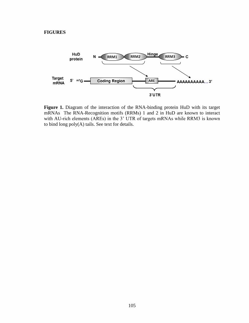
105
FIGURES
Figure 1. Diagram of the interaction of the RNA-binding protein HuD with its target
mRNAs The RNA-Recognition motifs (RRMs) 1 and 2 in HuD are known to interact
with AU-rich elements (AREs) in the 3’ UTR of targets mRNAs while RRM3 is known
to bind long poly(A) tails. See text for details.
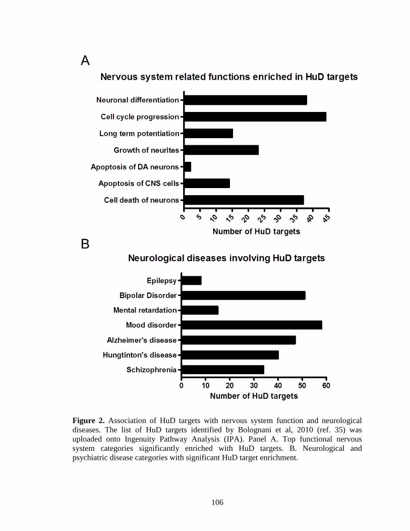
106
Figure 2. Association of HuD targets with nervous system function and neurological
diseases. The list of HuD targets identified by Bolognani et al, 2010 (ref. 35) was
uploaded onto Ingenuity Pathway Analysis (IPA). Panel A. Top functional nervous
system categories significantly enriched with HuD targets. B. Neurological and
psychiatric disease categories with significant HuD target enrichment.
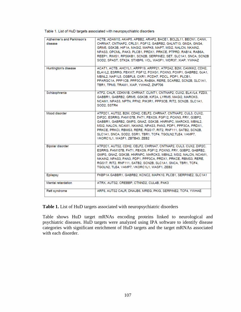
107
Table 1. List of HuD targets associated with neuropsychiatric disorders
Table shows HuD target mRNAs encoding proteins linked to neurological and
psychiatric diseases. HuD targets were analyzed using IPA software to identify disease
categories with significant enrichment of HuD targets and the target mRNAs associated
with each disorder.
Related Documents