Eur Respir J 1990, 3, 1166-1172 The reversible effect of lignocaine on the stimulated metabolic activity of bronchoalveolar lavage cells M. Duddridge, C.A. Kelly, C. Ward, D.J. Hendrick, E.H. Waiters The reversible effect of lignocaine on the stimulated metabolic activity of bronchoalveolar lavage cells. M. Duddridge, CA. Kelly, C. Ward, D.J. Hendrick, EH. Waiters. ABSTRACT: Lignocaine concentrations were measured In the aspirate from a low volume (100 ml) broncboalveolar lavage (BAL) In twenty patients who bad received topical 4% lignocaine as required, and compared to those found In the aspirate from a 180 ml BAL In ten patients who bad received 1.5% Isotonic lignocaine. The median BAL supernatant lignocaine concentration was significantly lower at 0.14 mM (range 0.07-0.44 mM) In the group who received 1.5% lignocaine, compared to 1.08 mM (range 0.03-7.05 mM) In those given 4% lignocaine (p<0.01). The effect of Increasing concentrations of lignocaine on BAL neutrophil and pulmonary macrophage metabolic activity, as assessed by latex-sttmulated lumlnol- and luclgenin-ampllfled chemiluminescence (CL), respecttvely, In mixed BAL cell populations, were measured following preincubation of ''washed" harvested BAL cells with 0.4-8.0 mM lignocaine. There was no demonstrable decline In either cell activity with llgnocaine concentrations of up to 2 mM, with dose-dependent Inhibition of both above this thresh· old. Cell vlablllty was unaffected. In a further experiment, the Inhibition Induced by 8 mM lignocaine on both pulmonary macrophage (lucigenin- amplified CL of harvested BAL cells) and Isolated peripheral blood neutrophil metabolic activity was completely reversed by a single ''wash", following both 30 and 60 min lncubatlons at 4°C, which was equivalent to resuspendlng harvested BAL cells In fresh medium after separation from BAL supernatant. Thus, although lignocaine, In concentrations obtained during BAL, can affect measurement of BAL cell metabolic activity, the use of 1.5% or 4% ll.gnocalne at BAL carries negligible risk of doing so, provided the harvested cells are promptly separated from the supernatant and resuspended In fresh cell medium before further analysis Is undertaken. Eur Respir J., 1990, 3, ll66-ll72. The Chest Unit, Newcastle General Hospital, University of Newcastle upon Tyne, Newcastle upon Tyne, UK. Correspondence: Dr E.H. Waiters, Chest Unit, Newcastle General Hospital, Westgate Road, Newcastle upon Tyne, NE4 6BE, UK. Keywords: Bronchoalveolar lavage; chemilu- minescence; lignocaine; lucigenin; lwninol; neutrophil; pulmonary macrophage. Received: August 9, 1989; accepted after revision April 26, 1990. This work was supported in part by grants from the Asthma Research Council and the Newcastle upon Tyne District Research Committee. Although it had been established that lignocaine can inhibit the oxidative function of cells, including those obtained at bronchoalveolar lavage (BAL), in a dose- related fashion [1-4], and may also alter their morphol- ogy [1, 5, 6) and cell membrane receptor expression [7], these effects have only been demonstrated at concentra- tions of lignocaine infrequently achieved in BAL fluid [8-10]. The effects of high concentrations of lignocaine have been shown to be reversible following short peri- ods of incubation and one or more cycles of cell washing [1, 2, S-7]. However, lignocaine-induced inhibition of phagocytOsis and oxidative function have previously been shown to be irreversible following incubations of longer than 25 min [2]. The time between the collection of BAL aspirate and the separation of BAL cells from the supematant in the laboratory may therefore be of great importance. With increasing interest in the functional activity of BAL cells in health and disease, it is important to know whether the lignocaine used during the bronchoscopic procedure will influence ex-vivo results. In this study, we have measured lignocaine levels in BAL supematants obtained from two similar groups of patients undergoing bronchoscopy and BAL: the first group received topical 4% lignocaine prior to a low volume (lOO ml) lavage, while the second group received 1.5% isotonic ligno- caine before undergoing a 180 ml lavage, thus covering the likely "extremes" of current practice. We examined the effect of lignocaine, in the range of concentrations found, on harvested BAL cell latex stimulated amplified chemiluminescence (CL) in order to establish the threshold of effect. In these mixed BAL cell populations, luminol-amplified CL was used as a marker of neutrophil metabolic activity [11], and in the

Welcome message from author
This document is posted to help you gain knowledge. Please leave a comment to let me know what you think about it! Share it to your friends and learn new things together.
Transcript

Eur Respir J 1990, 3, 1166-1172
The reversible effect of lignocaine on the stimulated metabolic activity of bronchoalveolar lavage cells
M. Duddridge, C.A. Kelly, C. Ward, D.J. Hendrick, E.H. Waiters
The reversible effect of lignocaine on the stimulated metabolic activity of bronchoalveolar lavage cells. M. Duddridge, CA. Kelly, C. Ward, D.J. Hendrick, EH. Waiters. ABSTRACT: Lignocaine concentrations were measured In the aspirate from a low volume (100 ml) broncboalveolar lavage (BAL) In twenty patients who bad received topical 4% lignocaine as required, and compared to those found In the aspirate from a 180 ml BAL In ten patients who bad received 1.5% Isotonic lignocaine. The median BAL supernatant lignocaine concentration was significantly lower at 0.14 mM (range 0.07-0.44 mM) In the group who received 1.5% lignocaine, compared to 1.08 mM (range 0.03-7.05 mM) In those given 4% lignocaine (p<0.01). The effect of Increasing concentrations of lignocaine on BAL neutrophil and pulmonary macrophage metabolic activity, as assessed by latex-sttmulated lumlnoland luclgenin-ampllfled chemiluminescence (CL), respecttvely, In mixed BAL cell populations, were measured following preincubation of ''washed" harvested BAL cells with 0.4-8.0 mM lignocaine. There was no demonstrable decline In either cell activity with llgnocaine concentrations of up to 2 mM, with dose-dependent Inhibition of both above this thresh· old. Cell vlablllty was unaffected. In a further experiment, the Inhibition Induced by 8 mM lignocaine on both pulmonary macrophage (lucigeninamplified CL of harvested BAL cells) and Isolated peripheral blood neutrophil metabolic activity was completely reversed by a single ''wash", following both 30 and 60 min lncubatlons at 4°C, which was equivalent to resuspendlng harvested BAL cells In fresh medium after separation from BAL supernatant. Thus, although lignocaine, In concentrations obtained during BAL, can affect measurement of BAL cell metabolic activity, the use of 1.5% or 4% ll.gnocalne at BAL carries negligible risk of doing so, provided the harvested cells are promptly separated from the supernatant and resuspended In fresh cell medium before further analysis Is undertaken. Eur Respir J., 1990, 3, ll66-ll72.
The Chest Unit, Newcastle General Hospital, University of Newcastle upon Tyne, Newcastle upon Tyne, UK.
Correspondence: Dr E.H. Waiters, Chest Unit, Newcastle General Hospital, Westgate Road, Newcastle upon Tyne, NE4 6BE, UK.
Keywords: Bronchoalveolar lavage; chemiluminescence; lignocaine; lucigenin; lwninol; neutrophil; pulmonary macrophage.
Received: August 9, 1989; accepted after revision April 26, 1990.
This work was supported in part by grants from the Asthma Research Council and the Newcastle upon Tyne District Research Committee.
Although it had been established that lignocaine can inhibit the oxidative function of cells, including those obtained at bronchoalveolar lavage (BAL), in a doserelated fashion [1-4], and may also alter their morphology [1, 5, 6) and cell membrane receptor expression [7], these effects have only been demonstrated at concentrations of lignocaine infrequently achieved in BAL fluid [8-10]. The effects of high concentrations of lignocaine have been shown to be reversible following short periods of incubation and one or more cycles of cell washing [1, 2, S-7]. However, lignocaine-induced inhibition of phagocytOsis and oxidative function have previously been shown to be irreversible following incubations of longer than 25 min [2]. The time between the collection of BAL aspirate and the separation of BAL cells from the supematant in the laboratory may therefore be of great importance.
With increasing interest in the functional activity of BAL cells in health and disease, it is important to know whether the lignocaine used during the bronchoscopic procedure will influence ex-vivo results. In this study, we have measured lignocaine levels in BAL supematants obtained from two similar groups of patients undergoing bronchoscopy and BAL: the first group received topical 4% lignocaine prior to a low volume (lOO ml) lavage, while the second group received 1.5% isotonic lignocaine before undergoing a 180 ml lavage, thus covering the likely "extremes" of current practice. We examined the effect of lignocaine, in the range of concentrations found, on harvested BAL cell latex stimulated amplified chemiluminescence (CL) in order to establish the threshold of effect. In these mixed BAL cell populations, luminol-amplified CL was used as a marker of neutrophil metabolic activity [11], and in the
THE REVERSIBLE EFFECT OF LIGNOCAINE ON BAL CELLS 1167
presence of only low percentages of polymorphonuclear leucocytes, using latex particles as the stimulus [12], lucigenin-amplified CL was used LO reflect pulmonary macrophage metabolic activity [13, 14]. Finally, we have investigated whether the effect of lignocaine on stimulated harvested BAL cell lucigenin-amplified CL (pulmonary macrophage metabo)jc activity) and isolated peripheral blood neutrophil metabolic activity could be reversed, following a 30 or 60 min incubation, by washing the cells. The "wash" was chosen to equate to our routine initial resuspension of harvested BAL cells after their separation from supernatant BAL fluid.
Methods
Patients were studied with informed consent and the permission of the Newcastle upon Tyne Ethics Committee. They were randomly selected from those undergoing diagnostic fibreoptic bronchoscopy, premedicated with 10 mg papaveretum and 0.6 mg atropine given intramuscularly 30 min prior to the procedure, and local anaesthesia of the nose and throat was achieved with 4% lignocaine spray in all patients. Per nasal bronchoscopy (Olympus BF PIO, Keymed Ltd) was then perfonned without further sedation, anaesthesia of the vocal cords and airways achieved as subsequently described for the different groups, and BAL performed with the bronchoscope wedged in a subsegmental bronchus (of normal appearance) of the middle lobe, or lingula if local pathology was thought LO be present in the right lung.
Patient details
The indications for bronchoscopy were either unexplained haemoptysis or the evaluation of a unilateral chest radiograph opacity, with carcinoma of the bronchus as the final diagnosis in 73% of patients and no abnormality found in the remainder. In these patients the final diagnosis was of haemoptysis secondary to a previous infective exacerbation of chronic bronchitis (no infection within 8 weeks of bronchoscopy) or persistent radiological shadowing following a previous pneumonia. Of the 41 patients, 33 were men and 32 were current smokers, with a median age of 58 yrs (range 36-77 yrs) and a median forced expiratory volume in one second (FEV,) of78% of predicted (range 29-125%). There were no marked differences between the groups undergoing the different BAL protocols, as subsequently described, and all patients tolerated the procedure equally well, regardless of the anaesthetic regimen used [15).
BAL details
In the fust group of twenty patients, 2 ml aliquots of 4% lignocaine were used to anaesthetize the vocal cords and major airways by direct per-bronchoscopic injection as required for relief of cough and patient discomfort throughout the procedure. The LOLal dose of lignocaine
used was recorded. Although anaesthetic was generally instilled into the main bronchi, none was specifically instilled into the middle lobe/lingula (as was also the case in groups 2-4). BAL was performed using five 20 ml aliquots of sterile buffered saline at 37°C. Each aliquot was aspirated immediately and collected into a siliconized glass bottle at 4°C. The supernatanl was separated within one hour of BAL by centrifugation at 200 g for 10 min at 4°C, following filtration through stainless steel mesh (pore size 200 llffi), and was stored at -20°C for later assay of lignocaine levels.
Following premedication, the second group of ten patients inhaled 4 ml of 1.5% isotonic lignocaine generated by an uluasonic nebulizer (Porta-Ncb, Medic-Aid Ltd) over a 10 min period, as recently described [15]. They then underwent standard per nasal bronchoscopy. and received 2 ml aliquots of 1.5% lignocaine by direct per-bronchoscopic injection, again as required, throughout the procedure. The total dose administered was recorded. BAL was performed using three 60 ml aliquots of sterile buffered saline, the whole aspirate collected as for group 1, and the supematant separated and stored as above.
The third group of five patients underwent the same BAL procedure as described for the second group above. However, each of the three 60 ml aliquots were aspirated and collected individually, and the supematants separated and stored, as above, for individual assays of lignocaine concenuations. The BAL cells obtained from groups 2 and 3 were subsequently used to study the effects of lignocaine on cell function (sec below).
Six other patients. the fourth group, also underwent the BAL procedure described for the second and third groups; the BAL cells harvested from the supematant as above, and these were used in the final experiment examining the effect of a cell "wash" on lignocaineinduced inhibition of stimulated metabolic activity. In this final experiment peripheral blood neutrophils, which had not previously been exposed to lignocaine, were also used. Twenty millilitres of peripheral blood was obtained by venesection ~rom five healthy volunteers and the neutrophils separated by dextran sedimentation and Ficoll gradient centrifugation [16) . They were resuspended at 106 per ml in phosphate-buffered saline (PBS).prior to use.
Lignocaine assay
The supernatant lignocaine concentrations in each specimen were measured by high pressure liquid chromatography, according to the method described by KEENAGHAN (17]. Three assays were performed on each sample; the intra-assay coefficient of variation was less than 2%.
The effect of lignocaine on cellular activity
The cell pellet obtained after centrifugation of the aspirated BAL fluid in patient groups 2, 3 and 4 was

1168 M. DUDDRIDGE liT AL.
immediately resuspended in a cell medium containing calcium and magnesium, but without phenol red (cell medium 199, Gibco). A cell count was made by two independent observers using a Neubauer chamber, and the harvested BAL cell suspension adjusted to 1()6 per ml. Differential cell counts (Wright-Giemsa) were made on cytocentrifuged specimens (Shandon Cytospin ll, Shandon Southern Instruments), the two observers counting a minimum of 600 cells each. All work subsequently described was completed within three hours of BAL.
One millilitre aliquots of resuspended BAL cells from the ten patients in group 2 were incubated with equal volumes of sterile lignocaine solutions (in PBS) at 4°C for 60 min at final lignocaine concentrations of 0.4, 0.6, 0.88 and 1.0 mM. PBS without lignocaine was added to a further aliquot of cells as a control incubation. Cell viability was estimated by trypan blue exclusion following each incubation. Separate 500 J.l.l aliquots of these incubations, each containing 2.5x10S BAL cells, were then used to measure peak latex-stimulated luminol- and lucigenin-amplified CL [18]. 1n brief, the cell suspensions were incubated with 900 J.1.l of 1()4 M lurninol or lucigenin for 10 min at 37°C and 100 J.l.l 5% latex particles added. Peak CL was measured in duplicate samples from each incubation, using an LKB 1250 lurninometer, and the mean values at each lignocaine concentration expressed as a percentage of the peak CL of the control incubation in the absence of lignocaine.
In order to examine the effect of higher concentrations of lignocaine on cell function, a similar experiment was performed using final lignocaine concentrations of 0, 1, 2, 4, 6 and 8 mM with BAL cell suspensions (pooled from the 3 aspirates) obtained from the five patients studied in group 3.
The effect of cell ''washing"
The effect of a cell "wash" on lignocaine-induced inhibition of CL was studied in the final experiment using BAL cells harvested from the six patients of group 4 and the separated blood neutrophils from the five healthy volunteers. Lignocaine was prepared in cell medium 199 for the studies with the BAL cells and in PBS for the studies with the blood neutrophils. Two millilitres of resuspended cells (2x1()6 cells) were incubated with an equal volume of lignocaine solution, at a final lignocaine concentration of 8 mM, for 30 or 60 m in, at 4°C. AJiquots of cells were also incubated with lignocaine-free cell medium or PBS as controls. The cells were then pelleted by centrifugation at 200 g for 10 min , and resuspended in fresh cell medium, or PBS, with or without 8 mM lignocaine. A further two cell counts were performed and the cell suspensions made to 5x l OS per ml. This "wash", for those cells resuspended in lignocaine-free cell medium or PBS, was chosen to equate to our routine initial resuspension of BAL cells after tJteir separation from supematant BAL flu id. Cell viability was assessed by acridine orange uptake [19) for each incubation. Latexstimulated lucigenin-amplified CL was measured in triplicate, as described above but using a Lumac M2010
luminometer, for each incubation of harvested BAL cells to reflect pulmonary macrophage metabolic activity, and similarly luminol-amplified CL for each incubation of isolated peripheral blood neutrophils. Peak CL for the ''washed cells" (those resuspended for 8 mM lignocaine into lignocaine-free medium) and the "positive control cells" (those resuspended into fresh 8 mM lignocaine from 8 mM lignocaine) was expressed as a percentage of that for the "negative control" incubation (those suspended in lignocaine-free medium throughout).
Statistical analysis
Wilcoxon's two sample rank sum test was used to compare the BAL supematant lignocaine concentrations found, and the total amount of lignocaine used as topical anaesthesia, in the patients of groups 1 and 2. The threshold of the inhibitory effect of increasing concentrations of lignocaine on BAL neutrophil (luminol-amplified CL) and pulmonary macrophage (lucigenin-amplified CL) metabolic activity was determined by comparing the 95% confidence intervals (95% Cl) for the mean activity, calculated using the t distribution, at each lignocaine concentration with the standardized control in the absence of lignocaine (100% activity). Analysis of variance, using a randomized block design [20], with the Genstat 4.04B statistical package [21] was used to examine the effect of the cell"wash" on both pulmonary macrophage (lucigenin-amplified CL) and
0
4
00
00 - o -
00
I
- ·-o -'------'"'-"'---------'~---
Group 1 100 ml BAL 4% lignocaine
Group 2 180ml BAL 1.5% lignocaine
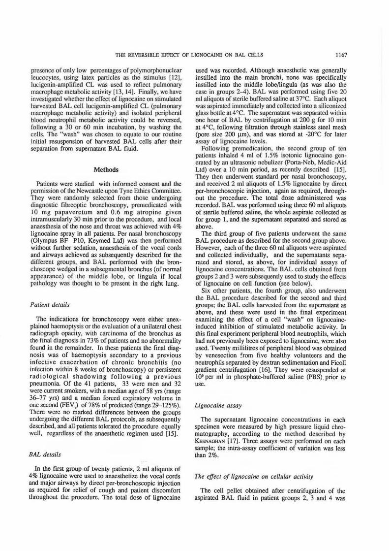
Fig. l. - The BAL supematant lignocaine concentrations found in 20 patients undergoing a 100 ml BAL following topical anaesthesia with 4% lignocaine (group I) (open circles) were significantly higher than those found in 10 paticnu in whom a 180 ml BAL was perfom1ed following the use of topical 1.5% i1otonic Lignocaine (group 2) (closed circles), p<O.Ol. Median values are marlced by the horizontal lines. BAL: bronchoalveolar lavage.

THE REVERSIBLE EFFECf OF LIGNOCAINE ON BAL CELLS 1169
separated blood neutrophil (luminol-amplified CL) metabolic activity following incubation with 8 mM lignocaine for both 30 and 60 min. Probability values of <0.05 (p<0.05) were taken to be significant
Results
The median volume of aspirated BAL fluid in the twenty patients undergoing the 100 mllavage (group 1) was 37 ml (range 12-52 ml), and in the ten patients undergoing the 180 mllavage (group 2) was 88 ml (range 48-122 ml). Similar aspirated volumes were recorded for both groups 3 and 4, as for group 2.
The BAL differential cell counts in groups 2-4 were similar, with a median of 91% pulmonary macrophages (range 80-99%), 8% lymphocytes (range 1-19%) and 1% neutrophils (range 0-3%). The mean purity of the peripheral blood neutrophil preparations from the five healthy volunteers was 96% (sEM 2%).
BAL supernatant lignocaine concentrations
Measurable concentrations of lignocaine were present in the BAL supematants of all patients in groups 1 and 2 (fig. 1). The median concentration of lignocaine in the twenty patients who received 4% topical lignocaine as local anaesthesia (group 1) was 1.08 mM (range 0.03-7.05 mM), with a significant negative correlation between the aspirated BAL volume and BAL supematant lignocaine concentration (r=-0.68, p<0.01). Significantly lower lignocaine levels were found in the BAL sopematants of the ten patients given topical 1.5% lignocaine (group 2), with a median of 0.14 mM (range 0.07-0.44 mM) (p<O.Ol). In the three individually aspirated 60 ml aliquots of BAL, from the five patients given 1.5% lignocaine (group 3), the lignocaine concentration was maximal in the first aspirate, median 0.50 mM (range 0.10-0.90 mM), falling to 0.10 mM (0.05-0.20 mM) in the second aspirate, and to 0.05 mM (0.03-0.06 mM) in the third aspirate.
The total amount of lignocaine used as topical anaesthesia per patient was less in the second group, with a median of 160 mg (range 120-200 mg), compared to that in the first group, median 280 mg (range 200-480 mg), (p<O.OOI). All patients tolerated the procedure equally well, regardless of the topical anaesthetic regimen used.
The effect of lignocaine on cellular activity
Figure 2 shows the dose-response relationship for the effect of lignocaine on both luminol-amplified (BAL neutrophil activity) and lucigenin-amplified (pulmonary macrophage activity) latex-stimulated CL of harvested BAL cells. Data for groups 2 and 3 have been pooled. Neither cell activity was inhibited by the 60 min preincubation of "washed" BAL cells with I mM lignocaine (mean luminol CL 98%, 95% Cl 83-113%; mean
..J 0
2 t
100
80
8 60
40
20
0.6 0.8 1.0 2 4 8
lignocaine concentration mM
Fig. 2. - The dose-dependent inhibition of BAL macrophage metabolic activity (latex-stimulated lucigenin-amplified CL: open circles/ dashed line) and of BAL neutrophil metabolic a~ivity (latex-stimulated luminol-amplified CL: closed circles/solid line) following a 60 min preincubation of harvested mixed populations of BAL ceUs with lignocaine. The peak CL at each lignocaine concentration is expressed as a percentage of its control incubation in the absence of lignocaine, and the mean (si!M) percentage activity plotted as the ordinate and log
10 lignocaine concentration as the abscissa. The data from the two separate experiments have been pooled. The mean absolute values for luminol- and lucigenin-amplified CL in the control inwbations in the absence of lignocaine were 2.0 mV and 4.4 m V, respectively. BAL: bronchoalveolar lavage; CL: chemiluminescence.
lucigenin CL 95%, 95% Cl 89- 101 %) and below. The threshold of inhibition was at 2 mM for both types of cell (mean Iuminol CL 86%, 95% Cl 60-111 %; mean lucigenin CL 90%, 9)% Cl 62- 118%), with both significantly inhibited following preincubation with 4 mM lignocaine (mean luminol CL 50%, 95% Cl 10-89%; mean lucigenin CL 52%, 95% Cl 3~74%) and above. Cell viability was unaffected, being greater than 90% after all the incubations.
The effect of cell "washing" on lignocaine-induced inhibition
Table 1 shows the effect of a simple cell wash on 8 mM lignocaine-induced inhibition of both BAL macrophage and peripheral blood neutrophil activity. The inhibitory effect was totally reversed by the cell wash after both the 30 and 60 min incubations. In the presence of 8 mM lignocaine ("positive control cells") pulmonary macrophage activity (mean lucigenin CL 30%, 95% Cl 12-48%) was significantly inhibited compared with that of the "washed cells" (mean 98%, 95% Cl 80-116%) (p<O.OOI), whose activity was indistinguishable from that of the "negative control cells" (100%).

1170 M. DUDDRIDGE ET AL.
Table 1.- The effect of a cell wash on the inhibition of BAL macrophage (lucigeninamplified CL) and blood neutrophil (luminol-amplified CL) metabolic activity• following preincubation of harvested BAL cells and isolated peripheral blood neutrophits, respectively, with 8 mM lignocaine.
BAL macrophage activity % Blood neutrophil activity %
"Positive control'' "Washed cells" "Positive control" "Washed cells" 30 min 60 min 30 min 60 min 30 min 60 min 30 min 60 min
1 10 17 97 92 22 32 12 26 2 111 35 74 121 10 10 93 98 3 3 1 110 100 2 4 95 121 4 73 77 80 95 7 12 117 84 5 10 16 106 154 2 4 115 90 6 7 6 73 78
Mean 36 25 90 107 9 12 108 84
·: metabolic activity is expressed as a percentage of the peak CL of control incubations in the absence of lignocaine for cells resuspended in 8 mM lignocaine ("positive control") or in lignocaine-free mediwn ("washed cells"); BAL: bronchoa!veolar lavage; CL: chemiluminescence.
Similarly, in the presence of 8 mM lignocaine, peripheral blood neutrophil activity (mean luminal CL 10%, 95% er 0-26%) was significantly inhibited compared with that of the "washed cells" (mean luminol CL 96%, 95% Cl 80-112%) (p<O.OOJ), whose activity was again indistinguishable from that of the "negative control cells". There was no significant difference in the effect of the cell wash following either the 30 or 60 min incubation of cells with 8 mM lignocaine for the BAL macrophage (p=0.80) or the peripheral blood neutrophil (p=0.34) activity. Cell viability was again unaffected, being greater than 92% after all incubations.
Discussion
This study was designed to assess whether the lignocaine concentrations found in BAL supematants could potentially affect ex-vivo assays of BAL cell metabolic activity as measured by amplified stimulated chemiluminescence, a technique being increasingly used to evaluate cellular function in respiratory disease [18, 22-27]. The literature is currently confused, as some previous studies [1, 2] have concluded that BAL cell function may be inhibited by the lignocaine concentrations found in BAL supematants [8], while others [9, 101 have more recently described lower lignocaine levels in lavage fluid whose effect on BAL cell function have not been investigated. The lower lignocaine concentrations found by STRANGE et al. [IOJ, 0.01-0.32 mM, and by ourselves in the group undergoing a 180 ml BAL following topical 1.5% lignocaine, were well below !he threshold level of 2 mM now described by ourselves, below which no significant inhibition of cellular metabolic activity occurred. Lignocaine-induced inhibition of cell function is therefore unlikely to be a problem if BAL supematant concentrations of lignocaine are kept low, i.e. if topical 1.5% lignocaine is used at bronchoscopy, even in the first BAL aspirate.
In contrast, 55% of the group receiving 4% topical lignocaine at a 100 ml BAL had BAL supernatant lignocaine concentrations in excess of 1 mM, approaching the threshold level described, and 15% had concentrations above this. This compares with only two of our subj~ts achieving a BAL supematant lignocaine concentration near the thresholds of 4 and 8 mM described by HoiDAL et a/ [1]. Our data raise the possibility of a serious potential source of artefact in studies on BAL cells when using high concentrations of topical lignocaine or small BAL volumes, especially when the volume of BAL aspirate is low. The low volume of the fust BAL aspirate probably also explains HoiDAL et al. [8) fmding higher supematant lignocaine concentrations in the first aspirate of a 3x60 ml BAL (mean 5.5 mM), compared with the later aspirates (mean 0.8 and 0.4 mM), following the use of 4% lignocaine above the vocal cords. Fortunately, the lignocaine-induced inhibition at a concentration in excess of the maximal found in BAL supematant was completely reversed by a simple, single cells wash.
Local anaesthetic-induced alterations of cell morphology and membrane receptor function have been said to be reversible following short incubations and one or more conventional cycles of cell washing [5-7]. However, reversibility of local anaesthetic-induced inhibition of cellular metabolic activity had never previously been subjected to statistical scrutiny, as in our data, with authors using rather subjective descriptive statements such as "responded comparably" and "largely reversed" to describe the effects of cell washing, without providing supportive data [1, 2, 4]. Our analysis is therefore of importance to those studying BAL cell function in general, and metabolic activity/reactive oxygen species generation in particular.
In contrast to earlier work [2], where 12 mM lignocaine-induced inhibition of blood neutrophil and monocyte metabolic activity was shown to be irreversible following incubations of longer than 25 min, we have

THE REVERSIBLE EFFECf OF LIGNOCAJNE ON BAL CELLS 1171
shown the inhibition of both blood neutrophil and pulmonary macrophage metabolic activity to be reversible following either 30 or 60 m in incubations with a higher concentration of lignocaine (8 mM) than found in our BAL supematants, by simply resuspending the cells in fresh lignocaine-free medium. There was no difference in cell viability between the two studies. The lower concentration of lignocaine used by ourselves may be an important difference, but higher BAL supematant concentrations can be avoided as shown by our results.
The cell "wash" was chosen to equate with our routine handling of BAL fluid and the harvested cells, in particular the separation of the cells from the lignocainecontaining supematant and their resuspension in fresh cell medium. As pulmonary macrophages responded similarly to blood nel.itrophils, naive to lignocaine prior to their in vitro exposure, it would be unlikely that BAL cells exposed, for up to sixty minutes, to the lower levels of lignocaine found in most BAL supematants would be "inhibited" when resuspended in fresh cell medium prior to ex-vivo assays.
Table 1 shows that in one case the cell wash did not reverse the inhibitory effect of 8 mM lignocaine on peripheral blood neutrophil activity following the 60 min preincubation. With the small sample population this is difficult to intercept, but it could imply that some neutrophils are susceptible to irreversible inhibition of metabolic function when exposed to high concentrations of lignocaine for a protracted period. However, with the low BAL supematant concentrations achieved following the used of 1.5% topical lignocaine, this should not be a practical problem.
During cell enumeration, we and others [28] have observed that the cells in BAL supematant appear smaller and more rounded than cells from the same subject following a simple wash, consistent with the previously described morphological changes produced by lignocaine [5, 6]. This would suggest an even lower threshold dose for the effect of lignocaine on cell morphology, as we are aware of these changes in all our other 180 ml BAL studies following the use of 1.5% lignocaine as topical anaesthesia, whether the cells were harvested from normal subjects, or patients with asthma, sarcoidosis or interstitial pulmonary fibrosis. In all cases the morphological changes are reversed by a single-step wash, suggesting that there is no difference in the reversibility of the changes between different pathological entities.
Previous studies have shown that the inhibition produced by lignocaine is not due to activity as a superoxide anion scavenger [3, 4]. Current evidence suggests that local anaesthetics bind to one of the three glycoproteins of the cell membrane sodium channels, so blocking depolarization changes [29]. The sodium/calcium exchange channel may be implicated, as calcium is an antagonist of the effect of lignocaine in vitro [29]. With calcium fluxes important in stimulus transduction in the cell membrane, the above mode of action would explain the wide range of different cell functions inhibited by lignocaine.
In summary, although lignocaine concentrations which can affect measurement of BAL cell metabolic activity
can be achieved during BAL, the use of 1.5% or 4% topical lignocaine at BAL carries negligible risk of doing so provided the harvested cells are separated from the supematant within 60 min and resuspended in fresh cell medium before further analysis is undertaken. This is true for both pulmonary macrophage and neutrophils metabolic function, and we suggest that it is also likely to be true for other cellular activities that are inhibited by lignocaine, owing to its mechanism of action.
Acknowledgements: The authors thank J. Fletcher of the Phannacy Dept, Newcastle General Hospital, for preparing lhe lignocaine solutions, D. Henderson of the Wolfson Unit of Clinical Phannacology, University of Newcastle upon Tyne, for assaying the lignocaine concentrations in r.he BAL supematant, and A. Avery of the Dept of Medical Statistics, University of Newcastle upon Tyne for advice and assisiAtlce in lhe statistical analysis.
References
1. Hoidal JR, White JG, Repine JE. - Influence of cationic local anaesthetics on the metabolism and ultrastructure of human alveolar macrophages. J Lab Clin Med, 1979, 93, 857-866. 2. Baser Y, deShazo RD, Barkrn.an HW, Nordberg J. -Lidocaine effects on immunocompetent cells. Implications for studies of cells obtained by bronchoavleolar lavage. Chest, 1982, 82, 32:>-328. 3. Cullen BF, Haschke RH. - Local anaesthetic inhibition of phagocytosis and metabolism of human leukocytes. Anesthesiology, 1974, 40, 142-146. 4. Goldstein IM. Lind S. Hoffstein S, Weissmann G. -Influence of local anesthetics upon human polymorphonuclear leukocyte ftmction in vitro. Reduction of lysosomal enzyme release and superoxide anion production. J Exp Med, 1977, 146, 483-494. 5. Nicolson GL, Smith JR, Poste G. - Effects of local anaesthetics on cell morphology and membl'ane-associated cytoskeletal organisation in BALB/31'3 cells. J Cell Bioi, 1976, 68, 395-402. 6. Rabinovitch M, deStefano MI. - Cell shape changes induced by cationic anaesthetics. J Exp Med, 1976, 143, 290-304. 7. Ryhanen P. - Lidocaine inhibition of rosette formation. Med Bioi, 1979, 57, 196-198. 8. Hoidal JR, White JG, Repine JE. - Impairment of human alveolar macrophage oxygen consumption, and superoxide anion production by local anaesthetics used in bronchoscopy. Chest, 1979, 75, 243-246. 9. Davidson AC, Barbosa I, Kooner JS, Hamblin AS, Stokes TC, Bateman NT, Gray B. - The influence of topical anaesthetic agent at bronchoalveolar lavage upon cellular yield and identification and upon macrophage flDlction. Br J Dis Chest, 1986. 80, 385- 390. 10. Strange C, Barbarash RA, Heffner JE. - Lidocaine concentrations in bronchoscopic specimens. Chest, 1988, 93, 547-549. 11. Williarns AJ, Cole PJ. - Human bronchoalveolar cells and luminol-dependent chemilwninescence. J Clin Path, 1981, 34, 167-171. 12. Cumutte IT, Tauber AI. - Failure to detect superoxide in human neutrophils stimulated with latex particles. PediAJr Res, 1983. 17, 281-284.

1172 M. DUDDRJDGB Er AL.
13. Williams AI, Cole PI. - Investigation of alveolar macrophage function using lucigenin-dependent chemilwninescence. Thorax, 1981, 36, 866-869. 14. Ward C, Kelly CA, Stenton SC, Duddridge M, Hendrick DJ, Waiters EH. - Macrophage and neutrophil chernilwninescence in bronchoalveolar lavage fluid. Clin Sci, 1987, 73, 33P. 15. Gove RI, Wiggins I, Stableforth DE. - A study of the use of ultrasonically nebulised lignocaine for local anaesthesia during fibreoptic bronchoscopy. Br J Dis Chest, 1985, 79, 49-59. 16. Boyum A. - Isolation of mononuclear cells and granulocytes from human blood. Isolation of mononucelar cells by one centrifugation, and of granulocytes by combining centrifugation and sedimentation at 1 g. Scand J Clin Lab Invest, 1968, 21 (Suppl. 97), 77--89. 17. Keenaghan ffi. - The determination of lidocaine or prilocaine in whole blood by gas chromatography. Anesthesiology, 1968, 29, 110-112. 18. Kelly CA, Ward C, Stenton SC, Bird G, Hendrick DJ, Waiters EH. - Number and activity of inflammatory cells in bronchoalveolar lavage fluid in asthma and their relation to airway responsiveness. Thorax, 1988, 43, 684-692. 19. Parks DR, Byran VM, Oi Vf, Herzenberg LA. - Antigen specific identification and cloning of hybridomas with a fluorescence activated cell sorter. Proc Nat/ Acad Sci USA, 1979, 76, 1962-1966. 20. Armitage P, Berry G. - In: Statistical methods in medical research. 2nd edn. Blackwell Scientific Publications, Oxford, 1987, pp. 186--263. 21. Alvery NG, Banfield CF, Baxter RI. - Genstat (4.04) -a general statistical programme. The Numerical Algorithm Group, Oxford, 1983. 22. Martin RR, Lawrence EC, Teague RB, Gottlieb MS, Putman M. - Cherniluminscence of hmg macrophages and blood leukocytes in sarcoidosis. Am Rev Respir Dis, 1986, 133, 298-301. 23. Ward C, Kelly CA, Stenton SC, Duddridge M, Hendrick DJ, Waiters EH. - Luminol-amplified chemiluminescence as a marker of neutrophil metabolic activity in asthma. Thorax, 1988, 43, 248P. 24. Kelly CA, Ward C, Stenton SC, Hendrick DJ, Waiters EH. - Assessment of pulmonary macrophage and neutrophil function in sequential bronchoalvcolar lavage aspirates in sarcoidosis. Thorax, 1988, 43, 787- 791. 25. Wallaert B, Bonnicre P, Prin L, Conot A, Tonne! AB, Voisin C. - Primary biliary cirrhosis. SubcliJUcal inflammatory alveolitis in patients with normal chest roentgenograms Chest, 1986, 90, 842-848. 26. Wallaen B, Bart F, Aens C, Ouaissi A, Hatron PY, Tonne! AB, Voisin C. - Activated alveolar macrophages in subclinical pulmonary inflammation in collagen vascular diseases. Thorax, 1988, 43, 24-30. 27. Perez Th, Farre JM, Gosset Ph. Wallaert B, Duquesnoy B, Voisin C, Delcambre B, Tonne] AB. - Subc1inical alveolar
inflammation in rheumatoid arthritis: superoxide anion, neutrophil chemotactic activity in fibronection generation by alveolar macrophages. Eur Respir J, 1989, 2, 7-13. 28. Saltini C, Hance AI, Ferrans VI, Basset F, Bitterman PB, Crystal R. - Accurate quantification of cells recovered by bronchoalveolar lavage. Am Rev Respir Dis, 1984, 130, 650-658. 29. Ritchie JM, Greene NM. - Local anaesthetics. In: The pharmacological basis of therapeutics. 7th edn. A.G. Gilman, L.S. Goodrnan, T.W. Rail, F. Murad eds, Macrnillan, New York, 1985, pp. 302-321.
Effet reversible de la lignocaine sur l'activite mitabolique provoquee des cellules du lavage alveolaire. M. Duddridge, CA. Kelly, C. Ward, DJ. Hendrick, EH. Waiters. RESUME: Les concentrations de lignocaine ont ete mesurees dans les produits d'un petit (100 ml) lavage broncho-alveolaire (BAL) chez 20 patients qui avaient subi une anesthesie locale classique avec de la lignocaine a 4%, et elles ont ete comparees a celles obtenues dans un BAL de 180 ml chez 10 patients traites par la lignocaine isotonique a 1.5%. La concentration mediane du surnage a du BAL en lignocaine est significativement plus basse apres lignocaine a 1.5% (0.14 mM; extremes 0.07-0.44) qu'apres lignocaiil.e a 4% (1.08 mM; extremes 0.03-7.05 mM) (p<0.01). Les effets de concentrations croissantes de lignocaine sur l'activite metabolique des neutrophiles et des macrophages du BAL (apprecies par chemo-lurninescence amplifiee respectivement par luminol et lucigenine apres stimulation au latex sur des populations cellulaires rnixtes de BAL) ont ete mesures apres pre-incubation des cellules "lavees" recueillies du BAL, dans 0.4-8.0 mM de lignocaine. On n'a pu demontrer de decroissance d'activite dans aucun des types cellulaires avec des concentrations de lignocaine allant jusqu'a 2 mM, !'inhibition etant dose-dependante au-delAB de ce seuil. La viabilite cellulaire est restee intacte. Dans une experience ulteneure, !'inhibition induite par 8mM de lignocaine sur les macrophages alveolaires (CL amplifie par lucigerune dans les cellules recueillies par BAL) et sur l'activite metabolique des neutrophiles isoles de sang peripherique, a ete completement eliminee par un simple lavage, faisant suite a des incubations de 30 et 60' a 4°C, ce qui equivalait a une resuspension des cellules prelevees par BAL dans un milieu frais apres s~aration du sumageant du BAL. Done, quoique la lignocaine, aux concentrations obtenues pendant le BAL, peut affecter les determinations de l'activite metabolique des cellules du BAL, l'emploi de lignocaine a 1.5 ou 4% lors du BAL ne comporte qu'un risque negligeable d'en faire autant, pourvu que Jes cellules prelevees soient rapidement separees du sumageant et rernises en suspension dans un milieu frais pour cellules, avant que !'analyse ulterieure ne soit entreprise. Eur Respir J., 1990, 3, 1166-1172.
Related Documents