The response of rat cerebellar granule neurons (rCGNs) to various polyhydroxyalkanoate (PHA) films Bo-Yi Yu a,b , Chi-Ruei Chen b , Yi-Ming Sun a,c,d, *, Tai-Horng Young b a Department of Chemical Engineering and Materials Science, Yuan Ze University, Chung-Li, Taoyuan, Taiwan 320, Republic of China Tel. +886-3-4638800 ext. 2558; Fax +886-3-4559373; email: [email protected] b Institute of Medical Engineering, National Taiwan University, Taipei, Taiwan 100, Republic of China c Graduate School of Biotechnology and Bioengineering, Yuan Ze University, Chung-Li, Taoyuan, Taiwan 320, Republic of China d R&D Center for Membrane Technology, Chung Yuan University, Chung-Li, Taoyuan, Taiwan 320, Republic of China Received 30 June 2008; revised 04 January 2009; accepted 09 February 2009 Abstract The aim of this study is to control the behavior of rat cerebellar granule neurons (rCGNs) by adjusting the surface characteristics of polyhydroxyalkanoate (PHA) films which were created by using compression-mold- ing, solvent-casting, and electrospinning methods. The compression-molded PHA membranes were dense and flat substrates, the cast ones showed higher roughness than the compression-molded ones, and the electrospun membranes were fibrous substrates. RCGNs could aggregate into three-dimensional (3-D) spheroid and develop many synapses on the compression-molded and solvent-cast membranes, and they aggregated into two-dimen- sional (2-D) flat sheet on the electrospun film in contrast. The viability of rCGNs on the electrospun membranes was higher than that on the other PHA films because the nutrients and metabolizes could easily transport through the highly fibrous structure of the electrospun films. RCGNs did not respond to the environmental stimuli created by the surface characteristics of the compression-molded and solvent-cast films, while they showed obvious difference to the specific fibrous characteristics of electrospun film in terms of morphology and via- bility. Keywords: Cell-substrate interaction; Electrospun fibrous film Desalination 246 (2009) 266–273 *Corresponding author. Presented at the conference Engineering with Membranes 2008; Membrane Processes: Development, Monitoring and Modelling – From the Nano to the Macro Scale – (EWM 2008), May 25–28, 2008, Vale do Lobo, Algarve, Portugal. 0011-9164/09/$– See front matter © 2009 Elsevier B.V. All rights reserved. doi: 10.1016/j.desal.0000.00.000

Welcome message from author
This document is posted to help you gain knowledge. Please leave a comment to let me know what you think about it! Share it to your friends and learn new things together.
Transcript
The response of rat cerebellar granule neurons (rCGNs) to
various polyhydroxyalkanoate (PHA) films
Bo-Yi Yua,b, Chi-Ruei Chenb, Yi-Ming Suna,c,d,*, Tai-Horng Youngb
aDepartment of Chemical Engineering and Materials Science, Yuan Ze University, Chung-Li, Taoyuan,Taiwan 320, Republic of China
Tel. +886-3-4638800 ext. 2558; Fax +886-3-4559373; email: [email protected] of Medical Engineering, National Taiwan University, Taipei, Taiwan 100, Republic of China
cGraduate School of Biotechnology and Bioengineering, Yuan Ze University, Chung-Li, Taoyuan, Taiwan 320,Republic of China
dR&D Center for Membrane Technology, Chung Yuan University, Chung-Li, Taoyuan, Taiwan 320,Republic of China
Received 30 June 2008; revised 04 January 2009; accepted 09 February 2009
Abstract
The aim of this study is to control the behavior of rat cerebellar granule neurons (rCGNs) by adjusting the
surface characteristics of polyhydroxyalkanoate (PHA) films which were created by using compression-mold-
ing, solvent-casting, and electrospinning methods. The compression-molded PHA membranes were dense and
flat substrates, the cast ones showed higher roughness than the compression-molded ones, and the electrospun
membranes were fibrous substrates. RCGNs could aggregate into three-dimensional (3-D) spheroid and develop
many synapses on the compression-molded and solvent-cast membranes, and they aggregated into two-dimen-
sional (2-D) flat sheet on the electrospun film in contrast. The viability of rCGNs on the electrospun membranes
was higher than that on the other PHA films because the nutrients and metabolizes could easily transport through
the highly fibrous structure of the electrospun films. RCGNs did not respond to the environmental stimuli
created by the surface characteristics of the compression-molded and solvent-cast films, while they showed
obvious difference to the specific fibrous characteristics of electrospun film in terms of morphology and via-
bility.
Keywords: Cell-substrate interaction; Electrospun fibrous film
Desalination 246 (2009) 266–273
*Corresponding author.
Presented at the conference Engineering with Membranes 2008; Membrane Processes: Development, Monitoring andModelling – From the Nano to the Macro Scale – (EWM 2008), May 25–28, 2008, Vale do Lobo, Algarve, Portugal.
0011-9164/09/$– See front matter © 2009 Elsevier B.V. All rights reserved.
doi: 10.1016/j.desal.0000.00.000

B.-Y. Yu et al. / Desalination 246 (2009) 266–273 267
1. Introduction
Polyhydroxyalkanoates (PHAs) are a class of
polyesters produced by microorganisms as intra-
cellular carbon and energy storage polymers under
unbalanced growth conditions. More than 150
kinds of PHAs consisting of various co-monomers
have been reported, but only a few of them have
been considered for commercial production, such
as poly(3-hydroxybutyrate) (PHB), poly(3-
hydroxybutyrate-co-3-hydroxyvalerate) (PHBV),
and poly(3-hydroxybutyrate-co-3- hydroxyhexa-
noate) (PHBHHx). In general, PHAs are native
polymers whose thermal and mechanical proper-
ties can be adjusted from thermoplastic to elas-
tomeric by using various compositions of their
copolymeric components. The advantageous char-
acteristics of PHAs are biocompatible, biodegrad-
able, and nontoxic [1–5]. Recently, PHAs have
been applied widely in regenerative medicine and
tissue engineering [6–7].
Cerebellar granule neurons (CGNs) constitute
the largest homogeneous neuronal population in
mammalian brain. The postnatal neurogenesis of
cerebellar granule neurons suggested that cere-
bella explanted from neonatal rats could be
advantageously used as an easy source of primary
neurons to be grown in vitro. Since the culture of
CGNs was established, the cells have become one
of the most important in vitro model to study all
the aspects of developmental, functional, and
pathological neurobiology in a rather homoge-
neous population of neurons [8]. Furthermore,
CGNs were highly sensitive to the stimulus from
the environment, so there could be a model cell to
detect the different surface characteristics of bio-
materials [9].
Electrospinning is a technology with some his-
tory as the first patent on this area was issued in
the 1930s [10]. This technique has recently been
applied to produce polymeric fibrous scaffolds for
cell culture and tissue engineering. Meshes of ran-
dom and orientated fibers with an average diam-
eter ranging from 100 nm to over 1 μm have been
prepared through the controlling of various
processing parameters. Previous studies showed
that cells on these fibrous meshes could have bet-
ter performances of proliferation, differentiation,
and metabolism than that on other films [10–15].
The micro-environment provided by the
fibrous PHA mesh was superior to that by com-
pression-molded and solvent-cast PHA films for
the proliferation of human mesenchymal stem
cells (hMSCs) in our previous study [16]. The
compression-molded PHA membranes were
dense and flat substrates, the cast ones showed
higher roughness than the compression-molded
ones and contained some dents, and the electro-
spun membranes were fibrous nonwoven sub-
strates. More detailed information about the
surface characteristics of PHA films has been dis-
cussed in our previous study [16–17], and the
behaviors of rCGNs cultured on those films are
presented here. In this study, PHAs are used as
the substrate materials. The behaviors of rCGNs
are modulated by adjusting the surface character-
istics of PHA films. It is intended to demonstrate
that the micro-environment provided by various
PHA films is also suitable for the proliferation of
rCGNs.
2. Materials and methods
2.1. Materials
Poly(3-hydroxybutyrate-co-5 mol%-3-
hydroxyvalerate) (PHBV5) and poly(3-hydroxy-
butyrate-co-12 mol%-3-hydroxyvalerate)
(PHBV12) were purchased from Aldrich, Inc.
(USA). Poly(3-hydroxybutyrate-co-8.3 mol%-3-
hydroxyhexanoate) was kindly provided by the
Procter & Gamble Co. (West Chester, Ohio,
USA). The mol% indicates the molar content of
hydroxyvalerate (HV) or hydroxyhexanoate
(HHx), respectively, in those copolymers.
2.2. Membrane preparation
The compression-molded PHBV5, PHBV12,
and PHBHHx membranes were prepared by a
268 B.-Y. Yu et al. / Desalination 246 (2009) 266–273
compression molding machine (Gotech GT-
7014). The polymer samples were placed
between two aluminum plates, heated without
any pressure at 170°C for 5 min, and then pressed
under a pressure of 9.8 MPa at 170°C for 15 min.
The melted samples were cooled to 120°C in a
period of 30 min and then cooled to ambient tem-
perature in another 10 min (step cooling). The
cast PHA films were prepared as follows. The
casting solution was prepared by dissolving 10
wt% PHBV12 (or 5 wt% for PHBHHx) in chlo-
roform (CHCl3, Mallinckrodt), and then was
poured onto a glass plate. A film-casting knife
(Braive) was pulled over the solution with con-
trolled clearance to adjust the thickness. After the
solvent evaporated in the air for 24 h, a cast PHB-
HHx film was obtained. The solvent evaporation
procedures were carried out at two temperatures,
18 and 32°C, in order to obtain different topo-
graphic morphology or surface roughness on the
cast PHA films. The apparent thickness of com-
pression-molded and solvent-cast films ranged
from 50 to 60 μm. The films were washed with
deionized water at least three times before further
applications or treatments.
Fibrous PHA membranes were prepared via
electrospinning. The polymer was dissolved in an
organic solvent mixture of chloroform (CHCl3,
Mallinckrodt) and N,N-dimethylformamide
(DMF, Tedia). The polymer solution was loaded
in a syringe capped with 21 gauge metal needle.
An electric field was created by a power supply at
12 kV between the needle and the rectangular
stainless steel receiver at the distance of 25 cm.
The polymer solution was drawn from the needle
under an accurate controlled syringe pump and
then sprayed onto the receiver by combined
forces of gravity and electrostatic charge.
2.3. Primary rCGNs culture
Cerebellar granule neurons were obtained
from 7-day-old Wistar rats according to Levi et
al . [18] with slight modification. Briefly, neurons
were dissociated from freshly dissected cerebella
by mechanical disruption in the presence of
trypsin and DNase. A variety of the PHA films
were placed in 24-well tissue culture plates
(Orange). The films were cut into 13 mm-diame-
ter discs and were tightly wedged into the culture
wells. In order to avoid film floating in the pres-
ence of medium, Teflon O-rings were used to fix
the films on the bottom of the wells. Subse-
quently, neurons were added to the culture wells
at a density of 1 × 10⁶ cells/well in basal Eagle
medium (BME, Gibco) supplemented with 10%
fetal calf serum, 25 mM KCl, penicillin G (100
IU/ml) and streptomycin (100 mg/mL). Cultures
were maintained at 37°C in a 95% humidified
atmosphere with 5% CO2
in air. Cytosine arabi-
noside (10 mM) was added to the culture medium
to prevent replication of non-neuronal cells 1 day
after plating [18]. The samples were cultured up
to 21 days, and there was no medium change dur-
ing the process of this study. The cell behaviors
were observed by using phase contrast
microscopy every day.
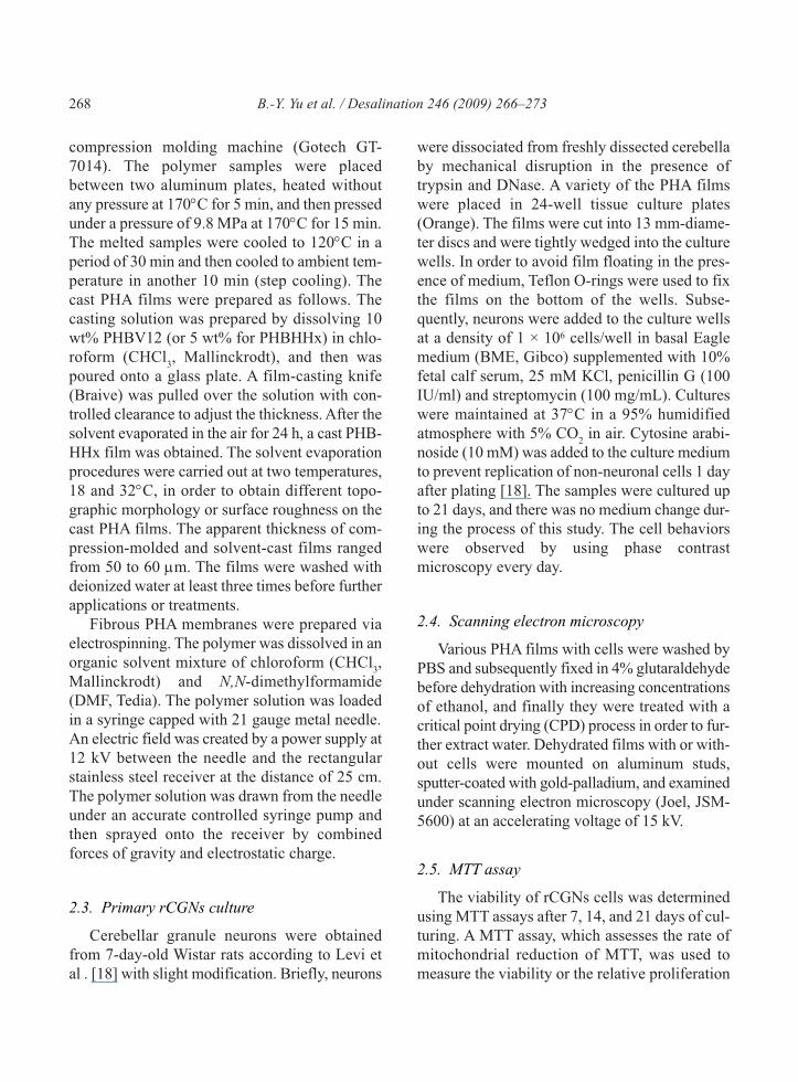
2.4. Scanning electron microscopy
Various PHA films with cells were washed by
PBS and subsequently fixed in 4% glutaraldehyde
before dehydration with increasing concentrations
of ethanol, and finally they were treated with a
critical point drying (CPD) process in order to fur-
ther extract water. Dehydrated films with or with-
out cells were mounted on aluminum studs,
sputter-coated with gold-palladium, and examined
under scanning electron microscopy (Joel, JSM-
5600) at an accelerating voltage of 15 kV.
2.5. MTT assay
The viability of rCGNs cells was determined
using MTT assays after 7, 14, and 21 days of cul-
turing. A MTT assay, which assesses the rate of
mitochondrial reduction of MTT, was used to
measure the viability or the relative proliferation

B.-Y. Yu et al. / Desalination 246 (2009) 266–273 269
activity of cells. The MTT assay kit (KPL) was
used as the procedures suggested by the supplier.
Each test was quadruplicated by using an ELISA
reader (Thermo Lab systems).
3. Results and discussions
The surface characteristics of various PHA
films via compression-molding (thermal-press-
ing), solvent-casting, and electrospinning meth-
ods could obviously affect the behaviors of
rCGNs in cell morphology and proliferation. The
morphology of rCGNs on compression-molded
PHBV12 film after being seeded for 6 days is
shown in Fig. 1. Not only could rCGNs develop
many synapses, but they also established synapsis
network structure. During the same time, rCGNs
could aggregate and form 3-D cell clusters (Fig.
1b). In addition, the glia cells also adhered to the
surface and built some kinds of synapses network
structure (Fig. 1c). It was noted that rCGNs could
(a)
(b)
(c)
Fig. 1. The morphology of rCGNs on the compression-
molded PHBV12 membranes: (a) ×150, (b) and (c)
×1000 after being cultured for 6 days.
(a)
(b)
Fig. 2. The morphology of rCGNs on the compression-
molded PHBHHX membranes: (a) ×150 and (b) ×1000
after being cultured for 6 days.

270 B.-Y. Yu et al. / Desalination 246 (2009) 266–273
not be completely isolated by the current method
of rCGNs purification, but the existence of glia
cells could benefit the construction of neural net-
work. It indicated that the micro-environment of
compression-molded PHBV12 film was suitable
for rCGNs to proliferate and differentiate mor-
phologically. The behaviors of rCGNs on the
compression-molded PHBHHx film are shown in
Fig 2. RCGNs showed the same behaviors as that
on the PHBV12 film (Fig. 1). There were com-
plicated synapsis networks among the 3-D cell
clusters and glia cells (Fig. 2b). The same cell
behaviors were also found on the compression-
molded PHBV5 film and the solvent-cast
PHBV12 films.
The morphology of rCGNs on various PHA
membranes after being seeded for 11 days is
presented in Fig. 3. It was obvious that the length of
the neurites reduced and the neuronal network
structure demolished with time for rCGNs on the
cast PHBV12 films prepared at 18°C under
unchanged medium condition (Fig. 3a). RCGNs on
the compression-molded PHBV5, PHBHHx film,
and the solvent-cast PHBHHx film (prepared at
18°C) also showed the same behaviors as that on
the solvent-cast PHBV12 film. Those results
demonstrated that rCGNs could not recognize the
difference of chemical composition among various
compression-molded and solvent-cast PHA films.
The evaporation rate of the solvent, which
depends on the casting temperature, has an obvi-
ous effect on the surface morphology of films.
The surface roughness of the cast films increased
with an increase of temperature. Namely, there
(a) (c)
(b) (d)
Fig. 3. The morphology of rCGNs (a) on cast PHBV12 prepared at 18°C, (b) cast PHBV12 membrane prepared at 32°C,
(c) and (d) electrospun PHBHHx membrane after being seeded for 11 days.

B.-Y. Yu et al. / Desalination 246 (2009) 266–273 271
was a porous structure on the surface of the sol-
vent-cast film at 32°C (Fig. 3b), and the pores
were significantly larger than that (if there was
any) on the solvent-cast film at 18°C (Fig. 3a).
The films with the different degree of surface
roughness or morphologic topography were pre-
pared in order to discuss the efforts of that on the
behaviors of rCGNs. RCGNs first piled up on
each other and built a 3-D cell cluster on the com-
pression-molded PHBV12 film (Fig. 1b), and the
behaviors of rCGNs on the solvent-cast PHBV12
film also showed the same as that on the com-
pression-molded PHBV12 film. Even the surface
roughness of the cast PHBV12 film was obvi-
ously higher than that of compression-molded
PHBV12 film (Fig. 3a and b). The results illus-
trated that the surface roughness or morphologic
topography didn’t have significant effect on the
aggregation and the formation of 3-D cell clusters
for rCGNs. However, rCGNs displayed a good
spread and developed many synapses on the elec-
trospun PHBHHx film after being cultured for 11
days (Fig. 3c). This cellular behavior was signif-
icantly different from that on the compression-
molded or solvent-cast PHBHHx film. This result
indicated that the interconnected space between
fibers in the interior of the electrospun film still
had influence on the morphology of rCGNs,
although rCGNs could only adhere to the surface
and could not migrate into the interior of this film
with time. Even the cell size of rCGNs, after
being cultured for 11 days, was smaller than 10
μm, rCGNs on the electrospun film could not fall
in the space between the electrospun fibers,
which was larger than 10 μm (Fig. 3(c)).
The results of MTT assay of rCGNs on various
PHA films under unchanged medium are shown
in Fig. 4. RCGNs on the electrospun PHBHHx
film presented much higher viability than those on
the other PHA films and TCPS (as reference) on
the 7th day. Because the medium was not replaced,
the nutrients in the medium would decrease and
the metabolites from rCGNs would accumulate
with time, and then the environment became
MTT assay
BV5 BV12 BV12C HX HX-ES TCPS
O.D
. val
ue 5
70 n
m
0.10
0.15
0.20
0.25
0.30
0.35
0.40
0.457 days14 days21 days
Fig. 4. MTT assay results of rCGNs cultured on the com-
pression-molded PHBV5 (BV5), PHBV12 (BV12), and
PHBHHx (HX), solvent-cast PHBV12 (BV12C), elec-
trospun PHBHHx (HX-ES), and TCPS films after being
cultured for 7, 14, and 21days, respectively.
unsuitable for rCGNs to survive. On the 14th day,
the viabilities of rCGNs on most PHA films and
TCPS were lower that on the 7th day, however, the
viability of rCGNs on the electrospun film was
kept about the same. Even on the 21st days, the
viability of rCGNs on the electrospun film was
significantly higher than that on other PHA films.
Moreover, rCGNs on other PHA films would
aggregate and detach from the surface of film and
then suspend in the medium (Fig. 5), but this phe-
nomenon was never found on the electrospun
film. Those results suggested that the electrospun
PHBHHx film provided a suitable micro-environ-
ment for rCGNs to have a better viability in long-
term culture. Although the films with various
surface properties (chemical composition, surface
roughness, texture, and hydrophilic nature) were
prepared, rCGNs were not sensitive to those dif-
ferences and presented similar behaviors except
that on the electrospun film.
It was of interest to discuss the effect of surface
morphology of films on the behaviors of rCGNs.
Now, we focused on the results from PHBHHx
films with various surface roughness or topo-
graphic morphology, and discussed the effect of
inner fibrous structure of electrospun films on the
behaviors of rCGNs. RCGNs on the solvent-cast

272 B.-Y. Yu et al. / Desalination 246 (2009) 266–273
film and compression-molded film had similar
morphology and viability, although the surface
characteristics of those films were significantly dif-
ferent. Even though the surfaces of both solvent-
cast films and electrospun films were porous,
rCGNs on those films showed obviously different
behaviors in morphology and viability. Those
results demonstrated that the fibrous characteristics
(including surface and bulk) of electrospun film
played an important role on the cellular behaviors
in terms of adhesion, morphology, and viability.
Although rCGNs only adhered to the surface and
could not migrate into the interior of the electrospun
film, the interconnected space between fibers of
electrospun film still had influence on the cell
behaviors. We speculated that the interconnected
space facilitated the transport of nutrients and meta-
bolic products. In other words, the specific fibrous
structure of electrospun film could induce rCGNs
to perform better spread and higher viability.
4. Conclusion
The PHA films with various surface character-
istics (surface roughness and topographic mor-
phology) were prepared by compression-molding,
solvent-casting, and electrospinning methods.
RCGNs could form 3-D cell clusters and build
synapsis network on various PHA films. It
demonstrated that PHAs were suitable materials
for rCGNs, although rCGNs were not sensitive to
the change of the surface properties of films
except that on the electrospun film. Furthermore,
the viability of rCGNs on the electrospun PHB-
HHx film was higher than that on the compres-
sion- molded and solvent-cast PHBHHx films and
TCPS, and it demonstrated that the specific
fibrous characteristics of electrospun film was
suitable for rCGNs to perform better viability in
long-term culture. Because the interconnected
space between fibers of electrospun film can ben-
efit the transport of nutrients and metabolic prod-
ucts, the inner fibrous structure still hade influence
on the behaviors of rCGNs in terms of cell mor-
phology and viability.
Acknowledgments
This work was supported by the National Sci-
ence Council of the Republic of China through
the grant of NSC95-2218-E-155-001. The
authors would like to thank Dr. Isao Noda of the
Procter & Gamble Co. for his kindness in provid-
ing PHBHHx.
References
[1] Z. Gugala and S. G.ogolewski, Differentiation,
growth and activity of rat bone marrow stromal cells
on resorbable poly(L/DL-lactide) membranes, Bio-
materials, 25 (2004) 2299–2307.
[2] Z. Zheng, F.F. Bei, H.L. Tian and G.Q. Chen,
Effects of crystallization of polyhydroxyalkanoate
blend on surface physicochemical properties and
interactions, Biomaterials, 26 (2005) 3537–3548.
[3] M. Yang, S. Zhu , Y. Chen , Z. Chang, G. Chen, Y.
Gong, N. Zhao and X. Zhang, Studies on bone mar-
row stromal cells affinity of poly(3-hydroxybu-
tyrate-co-3-hydroxyhexanoate), Biomaterials, 25
(2004) 1365–1373.
[4] X.H. Qu, Q. Wu, K.Y. Zhang and G.Q. Chen,
In vivo studies of (3-hydroxybutyrate-co-3- hydrox-
yhexanoate) based polymers: Biodegradation
and tissue reactions, Biomaterials, 27 (2006)
3540–3548.
Fig. 5. The morphology of rCGNs on the compression-
molded PHBHHx film after being seeded for 21 days
(image by optical microscopy).
B.-Y. Yu et al. / Desalination 246 (2009) 266–273 273
[5] I. Noda, P.R. Green, M.M. Satkowski and L.A.
Schechtman, Preparation and properties of a novel
class of polyhydroxyalkanoate copolymers, Bio-
macromolecules, 6 (2005) 580–586.
[6] Y.W. Wang, Q. Wu, J. Chen and G.Q. Chen, Evalu-
ation of three-dimensional scaffolds made of blends
of hydroxyapatite and poly(3-hydroxybutyrate-co-
3-hydroxyhexanoate) for bone reconstruction, Bio-
materials, 26 (2005) 899–904.
[7] Y. Gao, L. Kong, L. Zhang, Y. Gong, G. Chen, N.
Zhao and X. Zhang, Improvement of mechanical
properties of poly(DL-lactide) films by blending of
poly(3-hydroxybutyrate-co-3- hydroxyhexanoate),
Eur. Polym. J., 42 (2006) 764–775.
[8] A. Contestabile, Cerebellar granule cells as a model
to study mechanisms of neuronal apoptosis or sur-
vival in vivo and in vitro, The Cerebellum, 1 (2002)
41–55.
[9] C.R. Chen and T.H. Young, The effect of gallium
nitride on long-term culture induced aging of neu-
ritic function in cerebellar granule cells, Biomateri-
als, 29 (2008) 1573–1582.
[10] Y.R.V. Shih, C.N. Chen, S.W. Tsai, Y.J. Wang and
O.K. Lee, Growth of mesenchymal stem cells on
electrospun type I collagen nanofibers, Stem Cells,
24 (2006) 2391–2397.
[11] R.V.J. Langer, Tissue engineering, Science, 260
(1993) 920–926.
[12] R. Murugan and S. Ramakrishna, Nano-featured
scaffolds for tissue engineering: a review of spin-
ning methodologies, Tissue Engineering, 12 (2006)
435–467.
[13] X.J. Xin, M. Hussain and J.J. Mao, Continuing dif-
ferentiation of human mesenchymal stem cells and
induced chondrogenic and osteogenic lineages in
electrospun PLGA nanofiber scaffold, Biomaterials,
28 (2007) 316–325.
[14] W.J. Li, R. Tuli, X. Huang, P. Laquerriere and R.S.
Tuan, Multilineage differentiation of human mes-
enchymal stem cells in a three-dimensional nanofi-
brous scaffold, Biomaterials, 26 (2005) 5158–5166.
[15] S.Y. Chew, R. Mi, A. Hoke and K.W. Leong, The
effect of the alignment of electrospun fibrous scaf-
folds on Schwann cell maturation, Biomaterials, 29
(2008) 653–661.
[16] B.Y. Yu, P.-Y. Chen, Y.-M. Sun, Y.-T. Lee and T.-H.
Young, The behaviors of human mesenchymal stem
cells on the poly(3-hydroxybutyrate-co-3-
hydroxylhexanoate) (PHBHHx) films, Desalina-
tion, 34 (2008) 204–211.
[17] B.Y. Yu, P.-Y. Chen, Y.-M. Sun, Y.-T. Lee and
T.-H. Young, Effects of the surface characteristics
of polyhydroxyalkanoates (PHAs) on the meta-
bolic activities and morphology of human mes-
enchymal stem cells, J Biomater Sci. Polym. Ed.,
in press (2008).
[18] G. Levi, F. Aloisi, M.T. Ciotti and V. Gallo,
Autoradiographic localization and depolarization-
induced release of acidic amino acids in differen-
tiating cerebellar granule cell cultures, Brain Res.,
290 (1984) 77–86.
Related Documents


