The Respiratory System Chapter 23

The Respiratory System Chapter 23. Introduction n The trillions of cells making up the body require a continuous supply of oxygen to carry out vita functions.
Dec 19, 2015
Welcome message from author
This document is posted to help you gain knowledge. Please leave a comment to let me know what you think about it! Share it to your friends and learn new things together.
Transcript

The Respiratory System
Chapter 23

Introduction The trillions of cells making up the body
require a continuous supply of oxygen to carry out vita functions
We can survive only a few minutes without oxygen
As cells use oxygen, they give off carbon dioxide a waste product of cellular respiration

Introduction The major function of the respiratory system
is to supply the body with oxygen and dispose of carbon dioxide
To achieve this function four distinct processes, collectively called respiration occur– Pulmonary ventilation– External respiration– Transport of respiratory gases– Internal respiration

Introduction Pulmonary ventilation
– Air must be moved in and out of the lungs so that the gases in the air sacs (alveoli) of the lungs are continually changed and refreshed
– This air movement is commonly called ventilation or breathing

Introduction External respiration
– Gas exchange (oxygen loading and carbon dioxide unloading) between the blood and the air-filled chambers of the lungs must occur

Introduction Transport of respiratory gases
– Oxygen and carbon dioxide must be transported between the lungs and tissue cells of the body
– This is accomplished by the cardiovascular system, which uses blood as the transporting fluid

Introduction Internal respiration
– At systemic capillaries, gas exchanges (oxygen unloading and carbon dioxide loading) must be made between the blood and tissue cells

Respiratory System The organs of the
respiratory system include the nasal cavity, pharynx, larynx, trachea, bronchi, lungs
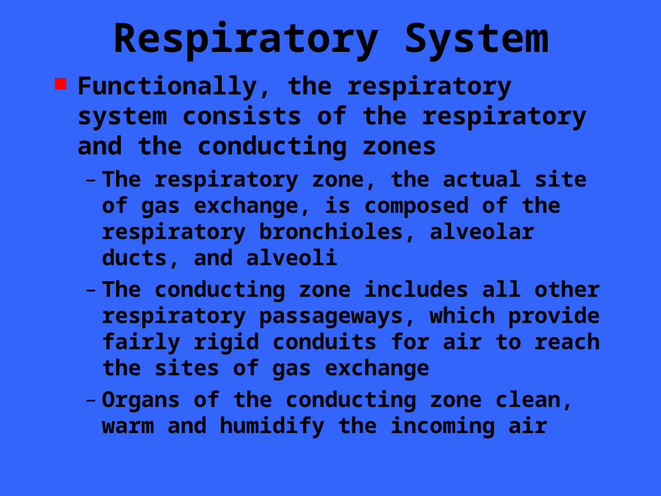
Respiratory System Functionally, the respiratory system
consists of the respiratory and the conducting zones– The respiratory zone, the actual site of gas
exchange, is composed of the respiratory bronchioles, alveolar ducts, and alveoli
– The conducting zone includes all other respiratory passageways, which provide fairly rigid conduits for air to reach the sites of gas exchange
– Organs of the conducting zone clean, warm and humidify the incoming air

The Nose The nose is the
only visible part of the respiratory system
The external framework of the nose

The Nose The functions of the nose include
– Providing an airway for respiration– Moistening and warming entering air– Filtering inspired air and cleansing it of
foreign matter– Serving as a resonating chamber for speech– Housing the olfactory receptors

The Nose The structures of
the nose are divided into the– External nose
– Nasal cavity Surface features
– Root (between eyes)
– Bridge
– Dorsum nasi
– Apex
– Philtrum
– External nares
– Alae

The Nose The nasal cavity lies in and posterior to the
external nose During breathing air enters the external
cavity by passing through the external nares or nostrils
The nasal cavity is divided by a midline nasal septum
The nasal cavity is continuous posteriorly with the nasal portion of the pharynx through the internal nares

The Nose The roof of the nasal cavity is formed by
the ethmoid and sphenoid bones of the skull
The floor is formed by the palate which separates it from the oral cavity below
Anteriorly, where the palate is supported by the maxillary processes and the palatine bones is considered the hard palate
The unsupported posterior portion is the muscular soft palate

The Nose
The vestibule is lined with skin containing sebaceous and sweat glands and numerous hair follicles
The hair or vibrissae filter coarse particles from inspired air

The Nose The nasal cavity is lined with two types of
mucous membrane The olfactory mucosa, lining the slitlike
superior region of the nasal cavity, contain the receptors for the sense of smell
The balance of the nasal cavity is lined with respiratory mucosa which is made up of pseudostratified columnar epithelium, containing scattered goblet cells, that rests on a lamina propria richly supplied with mucous and serous glands

The Nose Each day the mucous glands secrete about
a quart of sticky mucous containing lysozyme, an antibacterial enzyme
The mucous traps inspired dust, bacteria and other debris, while lysozyme attacks and destroys bacteria chemically
The epithelial cells of the respiratory mucosa also secrete defensins, natural antibotics that help to get rid of invading microbes

The Nose The ciliated cells of the respiratory
mucosa create a gentle current that moves the sheet of contaminated mucus posteriorly toward the throat where it is swallowed and digested by stomach juices
These ciliated cells become sluggish in cold weather allowing mucus to accumulate in the nasal cavity where it “runs” on a cold day

The Nose A rich plexus of thin walled veins
underlies the nasal epithelium and warms the incoming air as it flows across the mucosal surface
Blood flow increases when the weather turns cold
Because of its superficial location and the extent of vessels, nosebleeds are common and often profuse

The Nose Protruding medially
from each lateral wall of the nasal cavity are three mucosa-covered projections, the superior, middle and inferior conchae
The conchae serve to increase nasal turbulence in the nasal cavity
Mucus/sneeze

The Paranasal Sinuses The nasal cavity is
surround by sinuses located in the frontal, sphenoid, ethmoid and maxillary bones
They function to– Produce mucus
– Lighten the skull
– Warm the air
– Voice resonance
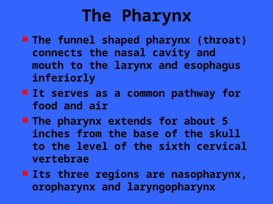
The Pharynx The funnel shaped pharynx (throat)
connects the nasal cavity and mouth to the larynx and esophagus inferiorly
It serves as a common pathway for food and air
The pharynx extends for about 5 inches from the base of the skull to the level of the sixth cervical vertebrae
Its three regions are nasopharynx, oropharynx and laryngopharynx

The Nasopharynx The nasopharynx lies
above the point of food entry, it serves only as an air passageway
During swallowing the uvula reflects posteriorly to close off the nasopharynx and prevent food from entering the nasal cavity

The Nasopharynx The nasopharynx is
continuous with the nasal cavity through the internal nares
It ciliated pseudo- stratified epithelium produces mucus
Mucosa high on the posterior wall contains masses of lymphatic tissue, the pharyngeal tonsils or adenoids

The Oropharynx The oropharynx lies
posterior to the oral cavity and is continuous with it through an archway called the fauces
Both swallowed food and air pass through
Lined with stratified squamous epithelium for protection from food abrasion and chemical trauma

The Oropharynx Three tonsils lie
embedded in the oropharyngeal mucosa– Paired palatine
tonsils
– Lingual tonsil

The Laryngopharynx The laryngopharynx
serves as a common pathway for food and air and is lined with stratified squamous epithelium
It lies directly posterior to the upright epiglottis and extends to the larynx where the digestive and respiratory pathways diverge

The Laryngopharynx The esophagus
conducts food to the stomach while air enters the larynx anteriorly
During swallowing food has the “right of way” and air passage temporarily stops

The Larynx The larynx attaches
to the hyoid bone superiorly and opens into the laryngopharynx
Inferiorly is is continuous with the trachea

The Larynx The larynx has three important functions
– It provides an airway for respiration– Act as a switching mechanism to route air
and food into the proper channels– Vocal cords housed in larynx are used in
voice production

The Larynx The framework of
the larynx is an arrangement of nine cartilages connected by membranes and ligaments
Except for the epiglottis, all laryngeal cartilages are made of hyaline

The Larynx The large, shield
shaped thyroid cartilage is formed by the fusion of two cartilage plates
The laryngeal prominence marks the midline fusion point
The cricoid cartilage is anchored to the trachea inferiorly

The Larynx Three pairs of
small cartilages, the arytenoid, cuneiform and corniculate form part of the lateral and posterior walls of the larynx
The arytenoid anchors the vocal cords

The Larynx The ninth cartilage
the flexible, spoon shaped epiglottis is composed of elastic cartilage
It is almost entirely covered by mucosa
The epiglottis extends from the posterior aspect of the tongue to its anchoring point on the thyroid cartilage

The Larynx When only air is
flowing into the larynx, the inlet to the larynx is open wide and the free edge of the epiglottis projects upward
During swallowing the larynx is pulled superiorly and the epiglottis tips to cover the laryngeal inlet
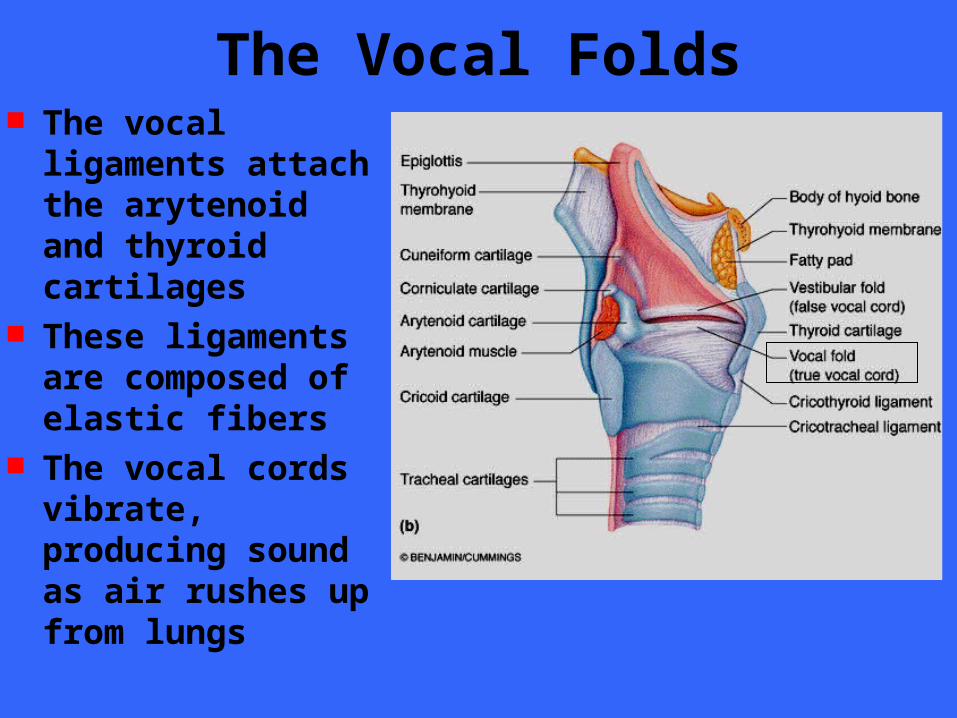
The Vocal Folds The vocal ligaments
attach the arytenoid and thyroid cartilages
These ligaments are composed of elastic fibers
The vocal cords vibrate, producing sound as air rushes up from lungs

The Vocal Folds The opening
through which air passes is the glottis
Superior to the vocal cords are the vestibular cords which play no part in voice production
Vocal Folds Stratified squamous epithelium lines the
superior portion of the larynx, an area subject to food contact
Below the vocal folds the epithelium is pseudostratified ciliated columnar epithelium
Cilia move the mucus away from our lungs

Voice Production Speech involves the intermittent release
of expired air and opening and closing of the glottis
The length of the true vocal cords and the size of the glottis are altered by the action of the intrinsic laryngeal muscles most of which move the arytenoid cartilages
As the length and tension of the vocal folds change, the pitch of the sound is altered

Voice Production The glottis is wide when we produce deep
tones and narrows to a slit for high pitched sounds
Length and thickness of the vocal folds changes for males during puberty
Loudness of the voice depends on the force with which the airstream rushes across the vocal cords
The greater the force, the stronger the vibration and the louder the sound

Sphincter Functions of Larynx The vestibular folds can perform a
sphincter function under certain conditions In abdominal straining associated with
defecation and urination, inhaled air is held temporarily in the lower respiratory tract by closing the epiglottis
The abdominal muscle then contract and the interabdominal pressure rises
The action know as the Valsalva manuever can also stabilize the trunk when one lifts a heavy load

The Trachea The trachea
descends from the larynx through the neck and into the mediastinum
It ends by dividing into the two primary bronchi at midthorax
10 cm long and 2.5 cm in diameter
The trachea is very flexible and mobile

The Tracheal Wall
The tracheal wall consists of several layers that are common in many tubular organs of the body

The Tracheal Wall
From internal to external these layers are the mucosa, submucosa, and adventitia

The Tracheal Wall
The mucosa contains the same goblet cells containing pseudostratifed epithelium that occurs throughout most the of respiratory tract

The Tracheal Wall
Its cilia continually propel mucus, loaded with dust particles and other debris, toward the larynx

The Tracheal Wall Smoking inhibits and ultimately destroys
the cilia in the mucosa layer When their function is lost, coughing is
the only means of preventing mucus from accumulating in the lungs
Smokers with respiratory congestion should avoid medications that inhibit the cough reflex

The Tracheal Wall
The submucosa, a connective tissue layer, contains seromucous glands that help produce the mucus “sheets” within the trachea

The Tracheal Wall
The adventitia is a connective tissue layer that is reinforced by 16 to 20 C-shaped rings of hyaline cartilage
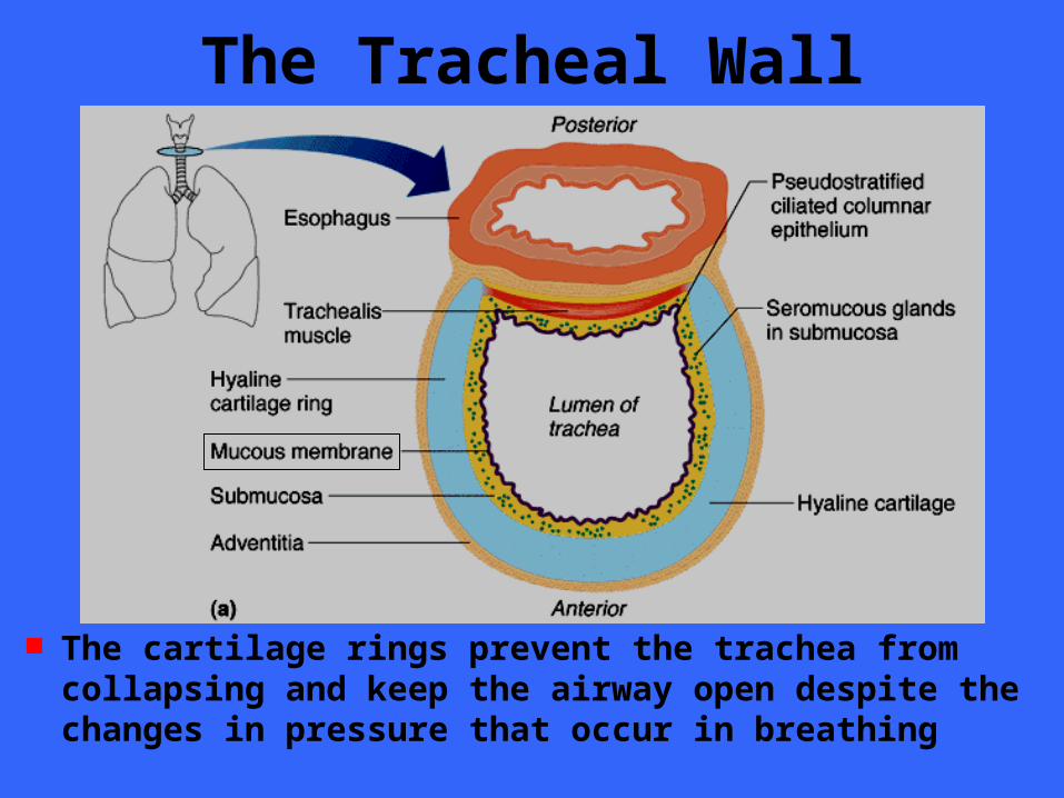
The Tracheal Wall
The cartilage rings prevent the trachea from collapsing and keep the airway open despite the changes in pressure that occur in breathing
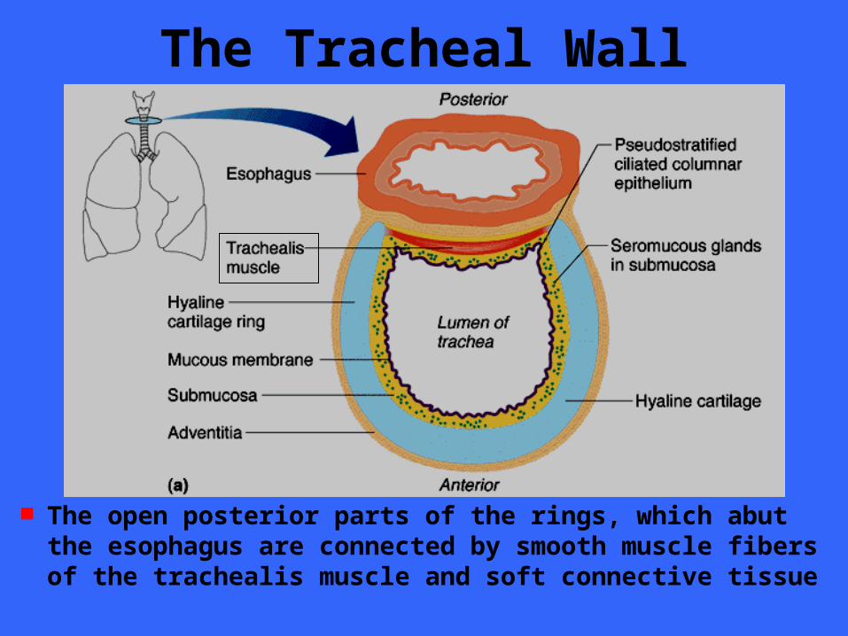
The Tracheal Wall
The open posterior parts of the rings, which abut the esophagus are connected by smooth muscle fibers of the trachealis muscle and soft connective tissue

The Tracheal Wall
Since this portion of the tracheal wall is not rigid, the esophagus can expand anteriorly as swallowed food passes through it

The Trachea The last tracheal
cartilage is expanded and a spar of cartilage called the carina projects posteriorly from its inner surface, marking the point where the trachea splits
Contacting this point results in violent coughing
The Trachea Tracheal obstruction is life threatening The Heimlich maneuver was developed
to expel an obstruction using the residual air in the victim’s lungs
The maneuver creates interthoracic pressure that drives the obstruction from its lodging point

The Conducting Zone
The right and left primary bronchi are formed by the division of the trachea at the level of T5
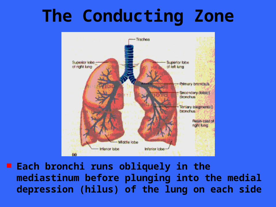
The Conducting Zone
Each bronchi runs obliquely in the mediastinum before plunging into the medial depression (hilus) of the lung on each side
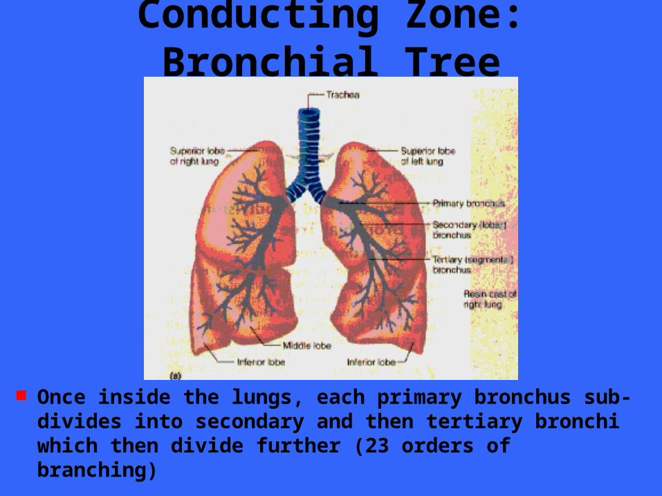
Conducting Zone: Bronchial Tree
Once inside the lungs, each primary bronchus sub- divides into secondary and then tertiary bronchi which then divide further (23 orders of branching)
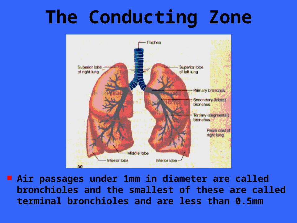
The Conducting Zone
Air passages under 1mm in diameter are called bronchioles and the smallest of these are called terminal bronchioles and are less than 0.5mm

The Conducting Zone The tissue composition of the walls of the
primary bronchi mimics that of the trachea but as the conducting tubes become smaller, a number of structural changes occurs– The cartilage supports change
• Rings are replaced by plates and then none at all
– The epithelium type changes• Pseudostratified columnar, to columnar, to cuboidal
• Debris removed by macrophages at bronchiole level
– The amount of smooth muscle increases• A complete layer of circular smooth muscle allows for
vasoconstriction and vasodilation

The Respiratory Zone
The respiratory zone begins as the terminal bronchioles feed into respiratory bronchioles within the lungs
Protruding from these smallest bronchioles are scattered alveoli

The Respiratory Zone
The respiratory bronchioles lead into alveolar ducts The ducts lead into terminal clusters of alveoli called
alveolar sacs Respiration takes place within the alveoli

The Respiratory Membrane The walls of the
alveoli are composed primarily of a single cell layer of squamous epithelial cells, called Type I cells underlain by a flimsy basal lamina
The cell walls are extremely thin to allow for ease of gas exchange

The Respiratory Membrane The external surfaces
of the alveoli are densely covered with a web of pulmonary capillaries
Together the alveolar and capillary walls and their fused basal lamina form the respiratory membrane with gas on one side and blood on the other

The Respiratory Membrane Gas exchange occurs
by simple diffusion across the respiratory membrane
Oxygen from the alveoli passes into the blood and carbon dioxide leaves the blood to enter the alveoli

The Respiratory Membrane Scattered amid the
type I squamous cells that form the alveoli walls are cuboidal type II cells
Type II cells secrete a fluid containing a surfactant that coats the alveolar surfaces which reduces the surface tension of the alveolar fluid
Type II Cell

The Respiratory Membrane Lung alveoli have
three other features– Surrounded by fine
elastic fibers
– Open pores connect adjacent alveoli
• Allow for pressure equalization
• Alternative air routes for blocked bronchi
– Alveolar macrophages crawl freely along the internal alveolar surfaces
Pores

The Lung
The lungs occupy all of the thoracic cavity except the mediastinum
Each cone shaped lung is suspended in its own pleural cavity and connected to the mediastinum

The Lung
The anterior, lateral and posterior lung surfaces lie in close contact with the ribs and forms a curving surface called the costal surface
The apex is the superior tip of the lung
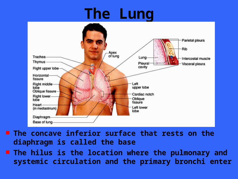
The Lung
The concave inferior surface that rests on the diaphragm is called the base
The hilus is the location where the pulmonary and systemic circulation and the primary bronchi enter

The Lung The left lung is divided
into two lobes (upper and lower) while the right has three lobes (upper, middle, lower)
Each of the lobes contains a number of bronchopulmonary segments separated by connective tissue
Each lung has 10 segment

The Pluera The pleura is a thin,
double layered serosa The parietal pleura
lines the thoracic wall and superior surface of the diaphragm
The visceral pleura covers the external surface of the lung
The pleura produce the fluid that lubricate the membrane

End of Material
Chapter 23
Related Documents