The Renal System Peter McCluggage Paul Óg Cooper

Welcome message from author
This document is posted to help you gain knowledge. Please leave a comment to let me know what you think about it! Share it to your friends and learn new things together.
Transcript

The Renal System
Peter McCluggage
Paul Óg Cooper

Topics for Discussion
• Function
• Anatomy
• Physiology
• Pharmacology
• Pathophysiology
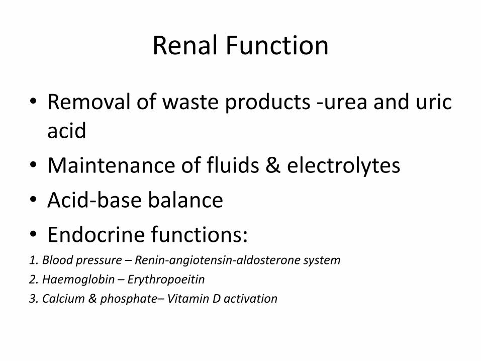
Renal Function
• Removal of waste products -urea and uric acid
• Maintenance of fluids & electrolytes
• Acid-base balance
• Endocrine functions: 1. Blood pressure – Renin-angiotensin-aldosterone system
2. Haemoglobin – Erythropoeitin
3. Calcium & phosphate– Vitamin D activation
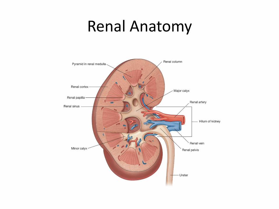
Renal Anatomy

Renal Anatomy• Each kidney has outer cortex
and inner medulla.
• Urine is formed within functional subunits known as nephrons.
• Each nephron contains a glomerulus, consisting of a tuft of capillaries with an afferent and efferent arteriole.
• The glomerulus is surrounded by epithelium of the Bowman’s capsule.
• Glomerulus and Bowman’s capsule form renal corpuscle.

Renal Anatomy
• This initial filtrate is then modified by a variety of secretory and reabsorptive processes as it passes through:
1. Proximal convoluted tubule
2. Loop of Henle
3. Distal convoluted tubule
4. Collecting duct
• The glomeruli and convoluted tubules lie within outer cortex and loop of Henle and collecting duct extend into medullary region.
• End product, urine, is delivered via renal pelvis to ureter.

Regulation of Fluids & Electrolytes
• Fluid balance is the concept of homeostasis, that the amount of fluid lost is equal to the amount taken in.
• For the normal function of the body it is vital that fluid balance is maintained.
• Euvolaemia is the state of normal body fluid volume.
• The major source of fluid loss is urine.
• Fluid loss is regulated in the kidney
hormonally. Largely through
RAAS and ADH.

Hormonal RegulationRenin-angiotensin-aldosterone system
Ren
in
Secreted from juxta-glomerular apparatus –macula densa
Secreted in response to renal hypoperfusion
Conversion of angiotensinogen to angiotensin I
Direct Effects
Vasodilatation of afferent arteriole
Direct Na + loss
An
gio
ten
sin
II Very potent vasoconstrictor of peripheral & efferent arterioles
Stimulates aldosterone secretion
Ald
ost
ero
ne End product of RAA axis
Produced by adrenal cortex
Acts on DCT causing reabsorption of Na+ & water
Increases ECF increases blood pressure

Hormonal RegulationAntidiuretic hormone
An
tid
iure
tic
ho
rmo
ne Secreted from posterior pituitary
Secretion is ↑ if:
1. ↑ osmolality (ECF)
2. ↓ volume
3. ↓ atrial pressure
Promotes water reabsorption in the distal convoluted tubule and collecting duct
Also known as vasopressin – a direct vasoconstrictor
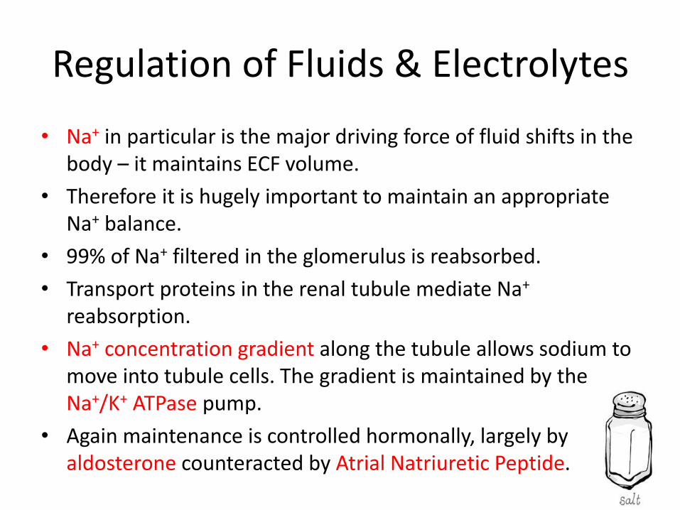
Regulation of Fluids & Electrolytes
• Na+ in particular is the major driving force of fluid shifts in the body – it maintains ECF volume.
• Therefore it is hugely important to maintain an appropriate Na+ balance.
• 99% of Na+ filtered in the glomerulus is reabsorbed.
• Transport proteins in the renal tubule mediate Na+
reabsorption.
• Na+ concentration gradient along the tubule allows sodium to move into tubule cells. The gradient is maintained by the Na+/K+ ATPase pump.
• Again maintenance is controlled hormonally, largely by aldosterone counteracted by Atrial Natriuretic Peptide.

Acid Base Regulation
• H+ levels are regulated through two buffering systems:– Chemical buffers – binds to H+ e.g. Bicarbonate buffering system
– Physiological buffer – controls excretion of acids or bases (kidneys) or volatile acids (lungs)
• Bicarbonate buffering system:– CO2 + H2O H2CO3 H+ + HCO3
-
• The kidney acts as a physiological buffer:– When pH is low excess H+ ions are secreted in the tubules via the
Na+/H+ exchanger and more bicarbonate is reabsorbed.
– When pH is high less bicarbonate is reabsorbed.

Assessment of Renal Function
Measurement of plasma [urea]
and [creatinine]
Measurement of GFR
Creat. Clearnce = UV/P
U=urinary [creatinine]
V= urinary output in 24 hours
P= plasma [creatinine]
Was
te p
rod
uct
s o
f p
rote
in m
etab
olis
mC
reatinin
e clearance = U
V/P

GFR
• Normal values are:
• ♂ 90-110 ml/min
• ♀ 80-125ml/min• In clinical practice an
estimated GFR (eGFR) is provided based on:
1. Plasma [creatinine]
2. Age
3. Gender
4. Ethnicity

Renal PharmacologyProximal Convoluted Tubule
Glucose, bicarbonate, amino acids reabsorbed
2/3 of sodium reabsorbed
Descending Loop of Henle
Permeable only to water
Ascending Loop of Henle
Impermeable to water
Active reabsorption of Na+, K+ and Cl- mediated by Na-K-2Cl co-transporter
Distal Convoluted Tubule
10% of NaCL reaborbed
Ca2+ excretion is regulated by PTH
Collecting Duct
Stimulation of aldosterone
receptors Na+ reabsorption & K+
secretion

Sites of Diuretic Effect
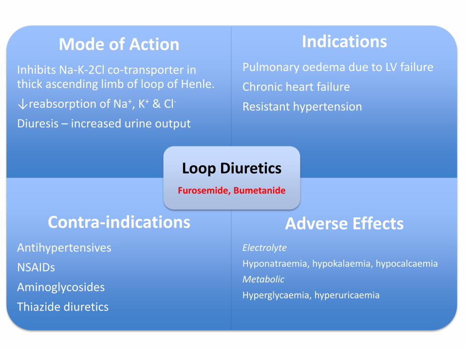
Mode of Action
Inhibits Na-K-2Cl co-transporter in thick ascending limb of loop of Henle.
↓reabsorption of Na+, K+ & Cl-
Diuresis – increased urine output
Indications
Pulmonary oedema due to LV failure
Chronic heart failure
Resistant hypertension
Contra-indications
Antihypertensives
NSAIDs
Aminoglycosides
Thiazide diuretics
Adverse EffectsElectrolyte
Hyponatraemia, hypokalaemia, hypocalcaemia
Metabolic
Hyperglycaemia, hyperuricaemia
Loop DiureticsFurosemide, Bumetanide

Mode of ActionInhibits the reabsorption of Na+ & Cl- in the cortical diluting segment of DCT.
Enhances Ca2+ reabsorption in the distal tubule.
Natriuresis and ↓blood volume and pressure.
Indications
Hypertension
Mild to moderate heart failure
InteractionsOther antihypertensives
Antidiabetic drugs
NSAIDs
Other drugs causing hyponatraemia
Adverse EffectsHyperuricaemia, hyperglycaemia
Hypokalaemia, hyponatraemia, hypomagnesaemia
Postural hypotension, impotence
Thiazide Diuretics
Bendroflumethiazide

Mode of ActionAldosterone-dependent potassium sparing diuretics.
Inhibits Na+/K+ exchange in distal tubule and collecting duct.
Promotes K+ retention and Na+ and water loss.
Hypotensive effect.
Indications
Oedema in CCF
Ascites
Primary hyperaldosteronism
Nephrotic syndrome
Contra-indications
Severe renal impairment
Addison’s disease
Adverse Effects
Hyperkalaemia
Acute kidney injury
Gynaecomastia, impotence and testicular atrophy with spironolactone
K+ SparingSpironolactone

Urinary Tract Infections Lower UTI – Cystitis
Frequency, dysuria, urgency, suprapubicpain, haematuria
Upper UTI – Pyelonephritis
High fever, rigors, vomiting,
loin pain
OrganismsE. coli
Staphylococcus saprophyticus
Proteus mirabilis
TrimethoprimBacterial dihydrofolate reductaseinhibitor
Limits bacterial reproduction
NitrofurantoinInhibits bacterial enzymes involved in carbohydrate metabolism and cell wall synthesis
UTI

Acute Kidney Injury
• Acute reduction in renal perfusion
• Hypovolaemic shock, acute cardiac failure, obstruction of renal vasculature
• Compensatory mechanisms result in increased urine osmolality, increased urine specific gravity, reduced urine [Na+]
• Rx - Fluid resuscitation to restore renal perfusion
Pre-renal
• Renal parenchymatous disease / acute tubular necrosis often from ischaemic damage
• Reduced Na+ and water reabsorption, reduced tubular K+ secretion, reduced tubular H+ secretion, reduced GFR
• Rx - Limit Na+, K+, fluid and protein intake. D
• Dialysis as needed until renal function recovers
Renal
• Urinary tract obstruction by abnormalities in lumen, wall or outside wall of urinary tract
• Rx - Relieve obstruction e.g. urinary catheter, treat underlying cause, prevent/ treat infections, common in urinary tract obstruction with stasis
Post-renal
Reduction in GFR resulting in rise in urea and creatinineReduced urinary output
Clinical Features
Reflect build up of nitrogenous waste
products
Anorexia, nausea & vomiting
Pruritis
Confusion, reduced consciousness

Chronic Kidney Disease
Causes
1. Congenital / inherited e.g. ADPKD
2. Glomerular disease – primary glomerulonephritis, secondary to diabetes
3. Vascular disease
4. Urinary tract obstruction
• 70% cases caused by diabetes, hypertension and atherosclerosis.
Kidney damage or reduced GFR for more than 3
months

Chronic Kidney DiseaseComplications
• Fluid retention – tissue oedema and heart failure
• Reduced metabolite excretion – uraemia, increased lipids, increased plasma urate and [creatinine]
Reduced GFR
• Reduced fluid reabsorption causing polyuria
• Reduced K+ secretion resulting in hyperkalaemia
• Reduced acid secretion resulting in metabolic acidosisReduced tubular function
• Reduced erythropoietin production – normocytic normochromic anaemiaAnaemia
• Reduced vitamin D activation leads to decreased Ca2+ absorption from GIT
• Plasma phosphate elevated due to reduced renal excretion and reduced calcium
• Reduced levels of vitamin D and reduced calcium stimulate parathormone secretion secondary hyperparathyroidism
Renal bone disease
• Activation of RAAS – increases PR hypertension
• Fluid retention leading to heart failure
• Increased cholesterol atherosclerosisCardiovascular complications
• Pruritis caused by nitrogenous waste compoundsSkin disease
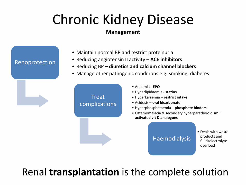
Chronic Kidney DiseaseManagement
Renoprotection
• Maintain normal BP and restrict proteinuria
• Reducing angiotensin II activity – ACE inhibitors
• Reducing BP – diuretics and calcium channel blockers
• Manage other pathogenic conditions e.g. smoking, diabetes
Treat complications
• Anaemia - EPO
• Hyperlipidaemia - statins
• Hyperkalaemia – restrict intake
• Acidosis – oral bicarbonate
• Hyperphosphataemia – phosphate binders
• Ostemomalacia & secondary hyperparathyroidism –activated vit D analogues
Haemodialysis• Deals with waste
products and fluid/electrolyte overload
Renal transplantation is the complete solution

All the best in the upcoming exams!

Related Documents