ANALYTICAL BIOC‘HEMISTRY 137, 236-247 ( 1984) The Release and Purification of Sialic Acids from Glycoconjugates: Methods to Minimize the Loss and Migration of 0-Acetyl Groups AJIT VARKI’ AND SANDRA DIAZ The UCSD Cancer Center, Department of’Medicine. T-01 1. Unirrrsity oJ’Callftirnia a[ San Diego, San Diego, Califirnia 92093 Received September 9. 1983 The sialic acids can have O-acetyl esters at the 4, 7, 8. and 9 positions. Most methods for the detailed study of such molecules require their release from glycosidic linkage, followed by extensive purification. The currently used methods for release and purification of sialic acids allow a reasonable qualirative analysis of the diversity of sialic acids from a given biological source. However, for several reasons. quantitariw assessment of the degree and type of O-acetylation is not possible with these methods. Previously known problems include the incomplete and non- random release of the different sialic acids by both enzymatic and chemical means, and extensive destruction of the 0acetyl esters (de-O-acetylation) during the release and purification. An additional problem, that migration of O-acetyl groups from the 7 or 8 positions to the Y position can occur under the conditions of release and purification. particularly when the pH is above 6 or below 3.0, is demonstrated here. It is also shown that the U-acetyl esters on free sialic acids are relatively more stable under acid conditions but more labile under basic conditions than similar esters on bound sialic acids. An analysis of the various steps of the conventional purification procedure showed that exposure to the basic anion-exchange resin is the critical step that results in de-O-acetylation and O-acetyl migration. Based upon these and other findings some new methods have been devised. and several modifications of the existing methods have been suggested. that allow the quantitative release and purification of sialic acids with minimal loss of 0acetyl groups. The migration of 0acetyl groups is also decreased by these modifications. but cannot be completely controlled. KEY WORDS: sialic acids: O-acctylation; O-acetyl migration: neuraminidases; ion-exchange chromatography; mucins. The sialic acids are a family of N- and O- substituted derivatives of neuraminic acid, a nine-carbon polyhydroxyamino-ketoacid sugar (Samino 3,5 di-deoxy-D-glycero-D-gal- acto-nonulosonic acid). The two principal N- substituted sialic acids, N-acetylneuraminic acid (NeuSAc)’ and N-glycolylneuraminic ’ To whom all correspondence should be addressed. ’ Abbreviations used: The various sialic acids are des- (NeuSGc) can also be O-substituted at the 4, 7, 8, and 9 positions, giving rise to a great variety of possible compounds and isomers (see Fig. 1) (I 2). For several reasons, early studies of the sialic acidsdid not fully recognize the extent of this complexity. First, many con- ventional methods for the analysis of glyco- conjugates, such as methanolysis, hydrazin- olysis, methylation analysis,and &elimination could result in destruction of the 0-substituent ignated by combinations of Neu = Neuraminic acid: AC = Acetyl: Cc = Glycolyl: and Lac = Lactyl. The amino group at the 5 position is always substituted with an acetyl (AC) or glycolyl (Cc) group. Other substitution positions are indicated by numerals. For example. Wacetyl-9-mono- O-acetyl-neuraminic acid may be written as Neu5.9Ac, and A’-glycolyl-7,8.9.-tri-O-acetyl-neuraminic acid as Ncu7.X.VAc~SGc. (After Schauer and others) (2.24). Other abbreviations used include BSM. bovine submaxillary mucin; ESM. equine submaxillary mucin: CM. collocalia mucoid: BHT. butylated hydroxytoluene: GLC/MS, gas- liquid chromatography/mass spectrometry: TMSI. tri- methylsilylimidarole: ‘TBA. 2-thiobarbituric acid. 0003-2697/84 $3.00 Copyright ‘~8 ,484 by Academy Pro, Ini All nghtr of rcpreductmn I” an, form rcscrved. 236

Welcome message from author
This document is posted to help you gain knowledge. Please leave a comment to let me know what you think about it! Share it to your friends and learn new things together.
Transcript

ANALYTICAL BIOC‘HEMISTRY 137, 236-247 ( 1984)
The Release and Purification of Sialic Acids from Glycoconjugates: Methods to Minimize the Loss and Migration of 0-Acetyl Groups
AJIT VARKI’ AND SANDRA DIAZ
The UCSD Cancer Center, Department of’ Medicine. T-01 1. Unirrrsity oJ’Callftirnia a[ San Diego, San Diego, Califirnia 92093
Received September 9. 1983
The sialic acids can have O-acetyl esters at the 4, 7, 8. and 9 positions. Most methods for the detailed study of such molecules require their release from glycosidic linkage, followed by extensive purification. The currently used methods for release and purification of sialic acids allow a reasonable qualirative analysis of the diversity of sialic acids from a given biological source. However, for several reasons. quantitariw assessment of the degree and type of O-acetylation is not possible with these methods. Previously known problems include the incomplete and non- random release of the different sialic acids by both enzymatic and chemical means, and extensive destruction of the 0acetyl esters (de-O-acetylation) during the release and purification. An additional problem, that migration of O-acetyl groups from the 7 or 8 positions to the Y position can occur under the conditions of release and purification. particularly when the pH is above 6 or below 3.0, is demonstrated here. It is also shown that the U-acetyl esters on free sialic acids are relatively more stable under acid conditions but more labile under basic conditions than similar esters on bound sialic acids. An analysis of the various steps of the conventional purification procedure showed that exposure to the basic anion-exchange resin is the critical step that results in de-O-acetylation and O-acetyl migration. Based upon these and other findings some new methods have been devised. and several modifications of the existing methods have been suggested. that allow the quantitative release and purification of sialic acids with minimal loss of 0acetyl groups. The migration of 0acetyl groups is also decreased by these modifications. but cannot be completely controlled.
KEY WORDS: sialic acids: O-acctylation; O-acetyl migration: neuraminidases; ion-exchange chromatography; mucins.
The sialic acids are a family of N- and O- substituted derivatives of neuraminic acid, a nine-carbon polyhydroxyamino-ketoacid sugar (Samino 3,5 di-deoxy-D-glycero-D-gal- acto-nonulosonic acid). The two principal N- substituted sialic acids, N-acetylneuraminic acid (NeuSAc)’ and N-glycolylneuraminic
’ To whom all correspondence should be addressed. ’ Abbreviations used: The various sialic acids are des-
(NeuSGc) can also be O-substituted at the 4, 7, 8, and 9 positions, giving rise to a great variety of possible compounds and isomers (see Fig. 1) (I 2). For several reasons, early studies of the sialic acids did not fully recognize the extent of this complexity. First, many con- ventional methods for the analysis of glyco- conjugates, such as methanolysis, hydrazin- olysis, methylation analysis, and &elimination could result in destruction of the 0-substituent
ignated by combinations of Neu = Neuraminic acid: AC = Acetyl: Cc = Glycolyl: and Lac = Lactyl. The amino group at the 5 position is always substituted with an acetyl (AC) or glycolyl (Cc) group. Other substitution positions are indicated by numerals. For example. Wacetyl-9-mono- O-acetyl-neuraminic acid may be written as Neu5.9Ac, and A’-glycolyl-7,8.9.-tri-O-acetyl-neuraminic acid as Ncu7.X.VAc~SGc. (After Schauer and others) (2.24). Other
abbreviations used include BSM. bovine submaxillary mucin; ESM. equine submaxillary mucin: CM. collocalia mucoid: BHT. butylated hydroxytoluene: GLC/MS, gas- liquid chromatography/mass spectrometry: TMSI. tri- methylsilylimidarole: ‘TBA. 2-thiobarbituric acid.
0003-2697/84 $3.00 Copyright ‘~8 ,484 by Academy Pro, Ini
All nghtr of rcpreductmn I” an, form rcscrved.
236
RELEASE AND PURIFICATION OF O-ACETYLATED SIALIC ACIDS 237
‘COOH
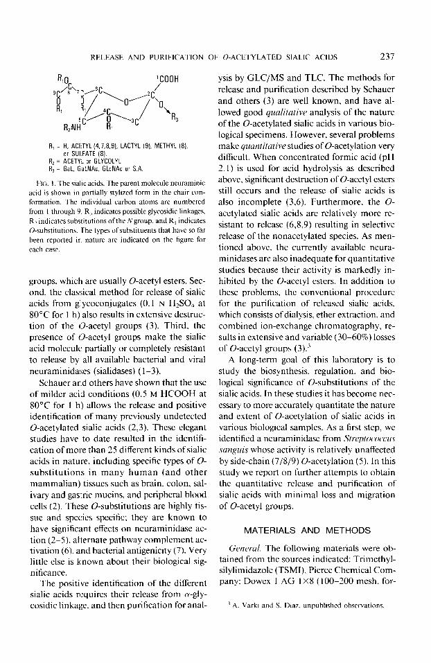
R, = H, ACETYL (4.7.8.9). LACTYL (9). METHYL (8). or SULFATE (8).
R2 = ACETYL or GLYCOLYL R3 = Gal. GaLNAc. GLcNAc or S.A
FIG. I The sialic acids. The parent molecule neuraminic acid is shown in partially stylized form in the chair con-
formation. The individual carbon atoms are numbered
from I through 9. R3 indicates possible glycosidic linkages.
Rz indicates substitutions of the Ngroup, and R, indicates O-substitutions. The types of substituents that have so far
been reported in nature are indicated on the figure for each case.
groups. which are usually 0-acetyl esters. Sec- ond, the classical method for release of sialic acids from glycoconjugates (0.1 N HzS04 at 80°C for 1 h) also results in extensive destruc- tion of the 0-acetyl groups (3). Third, the presence of 0-acetyl groups make the sialic acid molecule partially or completely resistant to release by all available bacterial and viral neuraminidases (sialidases) (l-3).
Schauer and others have shown that the use of milder acid conditions (0.5 M HCOOH at 80°C for 1 h) allows the release and positive identification of many previously undetected 0-acetylated sialic acids (2,3). These elegant studies have to date resulted in the identifi- cation of more than 25 different kinds of sialic acids in nature, including specific types of O- substitutions in many human (and other mammalian) tissues such as brain, colon, sal- ivary and gastric mucins, and peripheral blood cells (2). These O-substitutions are highly tis- sue and species specific: they are known to have significant effects on neuraminidase ac- tion (2-5). ahernate pathway complement ac- tivation (6), and bacterial antigenicity (7). Very little else is known about their biological sig- nificance.
The positive identification of the different sialic acids requires their release from a-gly- cosidic linkage. and then purification for anal-
ysis by GLC/MS and TLC. The methods for release and purification described by Schauer and others (3) are well known, and have al- lowed good qualitative analysis of the nature of the 0-acetylated sialic acids in various bio- logical specimens. However, several problems make quantitative studies of 0-acetylation very difficult. When concentrated formic acid (pH 2.1) is used for acid hydrolysis as described above, significant destruction of 0-acetyl esters still occurs and the release of sialic acids is also incomplete (3.6). Furthermore, the O- acetylated sialic acids are relatively more re- sistant to release (6,8,9) resulting in selective release of the nonacetylated species. As men- tioned above. the currently available neura- minidases are also inadequate for quantitative studies because their activity is markedly in- hibited by the 0-acetyl esters. In addition to these problems, the conventional procedure for the purification of released sialic acids, which consists of dialysis, ether extraction, and combined ion-exchange chromatography, re- sults in extensive and variable (30-60%) losses of 0-acetyl groups (3).j
A long-term goal of this laboratory is to study the biosynthesis, regulation, and bio- logical significance of O-substitutions of the sialic acids. In these studies it has become nec- essary to more accurately quantitate the nature and extent of 0-acetylation of sialic acids in various biological samples. As a first step, we identified a neuraminidase from Streptococcus sanguis whose activity is relatively unaffected by side-chain (7/8/9) 0-acetylation (5). In this study we report on further attempts to obtain the quantitative release and purification of sialic acids with minimal loss and migration of 0-acetyl groups.
MATERIALS AND METHODS
Getwral. The following materials were ob- tained from the sources indicated: Trimethyl- silylimidazole (TSMI), Pierce Chemical Com- pany: Dowex 1 AG 1 X8 (100-200 mesh, for-
’ A. Varki and S. Diaz. unpublished observations.

238 VARKI AND DIAZ
mate form), Dowex 3X4A (100-200 mesh, chloride form), and Dowex 50 AG 1 X2 ( 1 OO- 200 mesh, hydrogen form), Bio-Rad: 3% OV- 17 on Gas ChromQ ( 1 OO/ 120 mesh) and 3.8% SE-30 on CW-AW-DMCS (SO/l00 mesh). Applied Sciences Laboratories; butylated hy- droxytoluene (BHT), Sigma. The Dowex 3X4A resin was converted to the formate form, as recommended by the manufacturer. Di- azomethane in ether was kindly provided by Dr. Alan Hoffman, division of gastroenter- ology, UCSD School of Medicine. All other chemicals were of reagent grade and were pur- chased from commercial sources. The im- mersible CX- 10 reverse ultrafiltration units were from Millipore, and the Centrifree mi- cropartition units from Amicon. Unless oth- erwise indicated, all evaporations of aqueous samples were performed on a Buchler shaker- evaporator with the water bath set at 35°C or a Savant centrifuge evaporator with the heating element turned on (temperature at 3s40°C.)
Standards, substrates, and enqmes. [4-14C]- N-acetylneuraminic acid was from New En- gland Nuclear: chemically synthesized N-ace- tylneuraminic acid (NeuSAc) (>99% purity) was from Kantoishi Pharmaceutical Com- pany. Tokyo, Japan; N-glycolylneuraminic acid (NeuSGc) was from Sigma; and sialic ac- ids from bovine submaxillary mucin (a mix- ture of NeuSAc, NeuSGc. Neu5,9Ac2, Neu5,7Ac2, Neu5,7(8),9Ac3, and NeuSGc- 9Ac) were prepared as previously described (3,5). Authentic Neu5,9AcZ was kindly pro- vided by Professor Roland Schauer, Kiel, Federal Republic of Germany. Bovine sub- maxillary mucin was from U. S. Biochemical Corporation. Edible bird’s nest (Wai Tai Hong, Hong Kong) was purchased from a Chinese grocery store, and collocalia mucoid (CM) was extracted from it as previously de- scribed (5,lO). Equine submaxillary mucin (ESM) was prepared from fresh glands as pre- viously described (5). S. sangztis neuramini- dase was purified as described (5). Vihrio chol- erae and Arthrobacter ureajticiens neuramin- idases were purchased from Calbiochem.
C’lostridilrm pe~fringcws neuraminidase (Type X) was from Sigma.
.1 na!,:si.c of .sia/ic acids. For general pur- poses, the ferric-orcinol and 2-thiobarbituric acid (TBA) assays were used to assay total and free sialic acids. respectively (3,l 1). In the case of the TBA assay. analytical de-0-acetylation was always carried out (see below) to quantify the interference caused by the presence of 0-acetyl substituents at the 7, 8, or 9 positions (6,12). Also, 1 ~1 of 1% BHT in ethanol was added prior to acid hydrolysis: we have pre- viously shown that this retards lipid peroxi- dation and generation of interfering chro- mophores with absorption maximum at 532 nm (6). Purified sialic acids were analyzed by gas-liquid chromatography on 3% OV- 17 (tri- methylsilyl esters, trimethylsilyl ethers), or on 3.8% SE-30 (Methyl esters, trimethylsilyl ethers), exactly as described by Schauer (3). The total 0-acyl content of various sialo- mucins was also directly determined by the alkaline Hestrin method (3).
RESULTS
De-O-Acety~ation of ‘Siahc Acids
It is well known that 0-acetylation of the sialic acid side chain can cause considerable interference with the TBA assay, depending upon the position and extent of substitution (3,12). 0-acetyl esters were therefore hydro- lyzed in base prior to the TBA reaction to eliminate this interference (6.12). In exploring different alkaline conditions to optimize this de-0-acetylation, we found that the 0-acetyl esters on free sialic acids were significantly more labile than those on bound sialic acids (detailed data not shown). We found that de-0-acetylation of free siahc acids can be car- ried out in 2 M NH40H (pH 10.5) at room temperature for 6 h or at 60°C for I h. In the case of glycosidically bound, 0-acetylated sialic acids these conditions were inadequate, and it was necessary to use stronger base for complete de-0-acetylation. The sample was adjusted to 0.1 N NaOH (pH 13). and the

RELEASE AND PURIFICATION OF 0ACETYLATED SIALIC ACIDS 239
Neu5.9Ac~ NedGcSAc Neu5Gc7(8/Ac
I !
LlNTREATELl i-
TIME (mins)
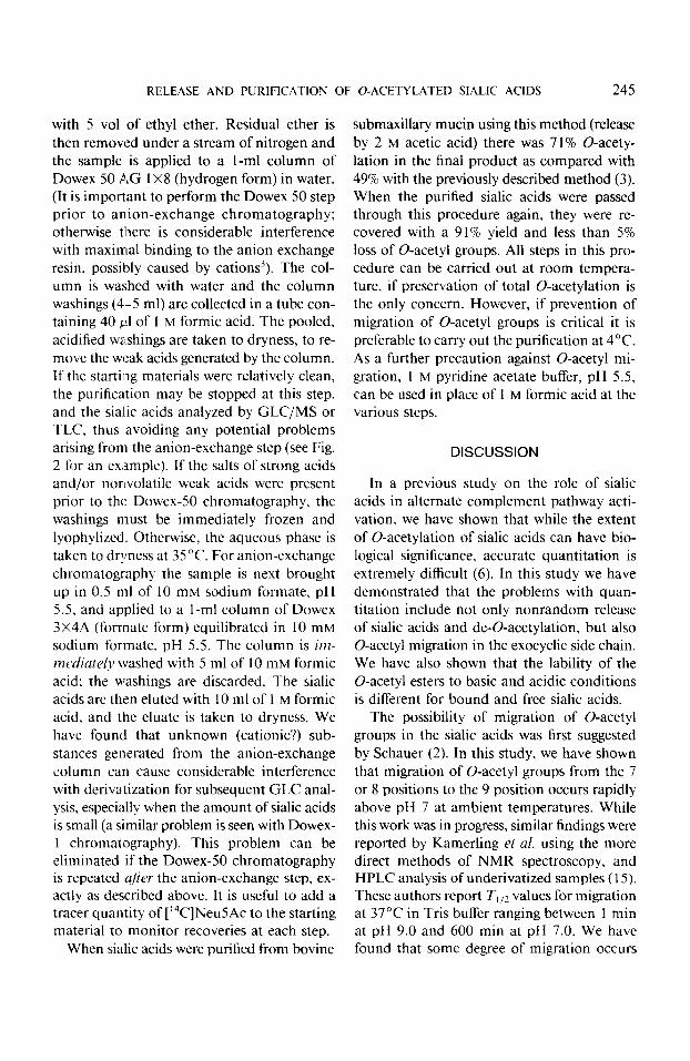
FIG. 2. Demonstration of the migration of O-acetyl groups in sialic acids. Sialic acids were released from 100 mg of bovine submaxillary mucin by treatment with 2 M
acetic acid at 80°C for 3 h (see Fig. 3). After chilling. insoluble matenal was removed by centrifugation at SO.OOOg for 30 min. The supemate was fractionated in an Amicon Centrifree micropartition system and the ul- trafiltrate was taken to dryness with a Buchler shaker- evaporator apparatus with the water bath set at 35°C. The residue was brought up in 0.5 ml of water and irn- mediaiatel~~ passed over a 1 S-ml column of Dowex 50 AC 1x2 (hydrogen form). The run-through and water washings (4 ml) were collected into a tube containing 40 ~1 of I M
formic acid, and assayed for total sialic acid content. Aliquots containing 300 nmol total sialic acids were taken to dryness and then brought up in 4 ml of pyridine acetate buffer (20 mM in pyridine) at the various pH values in- dicated (except pH 2.5. which was obtained with 4 ml of 20 mM formic acid). The samples were then incubated for I6 h at the temperatures indicated, and then taken to dryness quickly in the same tubes using the shaker-evap- orator. The samples were transferred to reactivials in 200 ~1 of 10 mM formic acid and then taken to dryness again in a Savant centrifuge evaporator. The sialic acids were then analyzed as their methyl esters, trimethylsilyl ethers on 3.8% SE-30 (exactly as described by Schauer (3). Six separate tracings are superimposed in this figure for con- venience. The various conditions of preincubation are indicated in the figure. The elution times of the various sialic acids are based on known standards (NeuSAc, Neu5,9Ac,, and NeuSGc) and on the literature (Neu5,7/ 8Ac2, NeuSGc9Ac. and Neu5Gc7/8Acz). In addition, the O-acetylated sialic acids were identified by the fact that these peaks were eliminated by prior treatment with mild
mixture placed on ice for 45 min and then neutralized (6,12).
Migration of 0-Acetyl Groups in the Sialic Acids
Bovine submaxillary mucin (BSM) contains Neu5Ac and Neu5Gc that is also 0-acetylated to a variable extent at the 7, 8, and 9 positions (2). We noticed that when sialic acids were purified from a single batch of BSM on dif- ferent occasions, the yield of the different iso- mers was quite variable. Furthermore, upon prolonged storage of a mixture of purified sialic acids from BSM, the ratio of the various iso- mers changed, without any overall loss of 0-acetyl groups. On the other hand, a sample of pure Neu5,9Ac2 remained unchanged dur- ing storage under identical conditions. We an- alyzed this phenomenon further by prolonged incubation of freshly purified BSM sialic acids at different pH values and temperatures, fol- lowed by analysis by GLC. Some typical re- sults are shown in Fig. 2. At pH values of less than 7.5, there was a partial or complete loss of the peak containing the comigrating 7 or 8 mono-0-acetylated isomers (Neu5,7/8Acl) with a corresponding increase in the peak of 9 mono-0-acetylated species (Neu5,9Ac2). This shows that 0-acetyl migration can occur from the 7 or 8 positions to the primary hy- droxyl group at the 9 position. where the 0-acetyl group is more stable. This effect was pH dependent, and increased at pH values of greater than 6 and less than 3. At pH values higher than 7.5 the 0-acetyl migration is rapid, but this is also accompanied by significant de-0-acetylation.
Release ~$Sialic Acids .fivm Gl~lcoconjl~gatcl.~
Acid hydrolysis. The commonly used con- ditions for complete release of sialic acids (0.1
alkali (2 M NH,OH. at 60°C for 60 min). with corre- sponding increase in the amounts of the Neu5Ac and NeuSGc peaks (not shown).

240 VARKI AND DIAZ
N H2S04, 8O“C, 1 h) result in extensive de- struction of 0-acetyl groups and even in some destruction of the sialic acid molecule itself (3). Milder conditions have therefore been used (0.5 N formic acid, pH 2.1, 80°C 1 h). With this method less destruction of 0-acetyl groups occurs. but the release is incomplete (40-60%) (3).3 Also, since the 0-acetylated sialic acids are relatively more resistant to release, selective release of the nonacetylated molecules occurs. We therefore investigated the effects of some other weak acids on 0-acetylated sialic acids. We found that when a mixture of previously purified sialic acids from BSM (0-acetylated at the 7, 8, or 9 positions) was heated in 2 M acetic acid (pH 2.5) at 80°C for 1 h and then reex- amined by GLC there was no detectable loss of 0-acetyl groups. When the heating period was extended to 3 h, there was only a 4% loss of 0-acetyl groups. Furthermore, when non- 0-acetylated NeuSAc was heated under iden- tical conditions for 3 h, there was no evidence of chemical 0-acetylation or of any destruc- tion of the neuraminic acid molecule (detailed data not shown). This suggested that prolonged hydrolysis under such conditions might be successful in achieving near-quantitative re- lease of bound sialic acids without excessive losses of 0-acetyl groups.
As model compounds to test this hypothesis, we used three well-known sialomucins, bovine submaxillary mucin (NeuSAc and NeuSGc with -80% 0-acetyl substitution at the 7, 8, or 9 positions), equine submaxillary mucin (NeuSAc with -80% 0-acetyl substitution at the 4 position and an unknown extent of 9- 0-lactyl substitution) (3,13), and collocalia mucoid (NeuSAc with no substitution). Shown in Fig. 3 are the effects of prolonged heating ofthese different mucins at 80°C in 2 M acetic acid. In each case, aliquots were removed at hourly intervals, lyophylized to remove the acetic acid, and then analyzed for total free sialic acid (by the TBA reaction after de-O- acetylation) and for total remaining 0-acyl esters by the alkaline Hestrin method. It can
PRIOR BASE
FIG. 3. Release of sialic acids from various mucins by concentrated acetic acid. Sialic acids were released from bovine submaxillary mucin (0) equine submaxillary mu- tin (O), and collocalia mucoid (0) with (A) or without (B) prior de-O-acetylation (0.1 N NaOH for 45 min on ice). In each case the reaction mixture was adjusted to a final concentration of 2 M acetic acid and heated at 80°C. At the various time points indicated, aliquots were removed and assayed for total free sialic acid by the TBA method. In the case of samples of that had not been previously treated with base (B) the acetic acid was removed by ly- ophilization, and de-0acetylation was carried out prior to performing the TBA assay. Aliquots from the experi- ment shown in (B) (containing about 300 nmol of siahc acid each) were also removed at the various time points indicated, lyophylized, and assayed for total O-acyl ester content by the alkaline Hestrin method. The results are shown in (C).
be seen (Fig. 3B) that under these conditions the sialic acids are gradually released over a period of hours, at different rates. In keeping with previous studies (6,8,9), 0-acetylated sialic acids of BSM and ESM are released at a slower rate than the non-0-acetylated sialic acids of CM. These differences in rates of re- lease could be due to the 0-acetylation, to differences in the sialic acid linkage, or to other nonspecific factors. To test this, the sialic acids

RELEASE AND PURIFICATION OF O-ACETYLATED SIALIC ACIDS 241
were de-0-acetylated prior to the acid hydro- lysis. When this was done, the rates of release from the three mucins became nearly identical (Fig. 3A), suggesting that the 0-acetylation was responsible for the differences. The total 0-acyl ester content of the ESM and BSM was also directly determined at several points during the hydrolysis (Fig. 3C). Unfortunately the 0-acyl content of the native, unhydrolyzed mucins could not be accurately determined by this method because of problems with tur- bidity. However. from the first hour of hy- drolysis onwards, the rate of 0-acyl loss could be followed ‘(Fig. 30. In the case of BSM the rate of loss appears to be quite low (-8% per hour between the 1 and 5 h time points). However. this is a significantly higher rate of 0-acetyl loss than predicted from the pilot experiment in which ,fiec sialic acids were heated under identical conditions (see above). This suggests that the 0-acetyl esters at 7, 8. and 9 positi’ons are somewhat more labile to acid when th:ey are on bour~ci sialic acids. With ESM, there (appears to be a more rapid initial loss of 0-acyl groups, possibly representing loss of 0-lactyl esters that are known to exist in this sialomucin ( 12.13); thereafter, the rate of loss is similar to that seen in BSM.
These data suggest that prolonged hydrolysis at 80°C in 2 M acetic acid may be a good method for obtaining maximal release of 0-acetylatecl sialic acids with relatively low losses of 0-acetyl groups. The exact hydrolysis time required will vary with different glyco- conjugates (I:.g., gangliosides are known to re- quire more prolonged hydrolysis (2,3)) and with different types of 0-acetylation. These conditions also may not be optimal for other sialic acid substitutions such as 0-lactyl groups. Although some degree of migration of 0-acetyl groups will probably occur under these conditions. it is not complete. since 7/ X mono-0-acetylated sialic acids can be found after such hydrolysis (see Fig. 1). An additional advantage of this method is that regardless of the presence of other more dilute salts and huff‘ers in the original sample to be hydrolyzed,
the adjustment to 2 M acetic acid results in a reproducible pH value in the range of 2.4 to 2.5.
Enzymatic release. We have also explored the use of neuraminidases in obtaining quan- titative release of sialic acids from glycocon- jugates. The commercially available neura- minidases from I: choferae and C. perfringens have markedly decreased activity against side- chain (7/g/9), 0-acetylated sialic acids, par- ticularly the di-0-acetylated species (5).3 We have previously described a neuraminidase of S. sanguis that is not restricted by the type of sialic acid linkage, or by the presence of 0-acetyl groups at the 7, 8, and 9 positions (5) (see Table 1). We” and others” have also recently found that the A. ureajbciens neur- aminidase (which is now commercially avail- able) is effective against many 0-acetylated sialic acids: additionally it is known to be ef- fective against the GM, linkage (14). We therefore now routinely use a mixture of the latter two enzymes when dealing with samples with unknown sialic acid composition. It should be noted that under the conditions of a prolonged enzyme reaction a significant amount of migration of 0-acetyl groups does occur.1
However, sialic acids with 4-0-acetyl groups are resistant to the action of these enzymes as they are to every other known neuraminidase (3-5). Fortunately, this type of sialic acid has only been reported so far in equine species and in Australian monotremes (3.13).
The first step in the purification of released sialic acids is their separation from macro- molecular materials. This is usually achieved by prolonged and repeated dialysis against distilled water, followed by concentration of the pooled dialysates (3). During this process, some de-0-acetylation and 0-acetyl migration
4 A. Shukla and R. Schauer. personal commumcation.

242 VARKI AND DIAZ
TABLE I
COMPARISON OF METHODS FOR RFLEASE OF SIALIC ACIL)S FROM GLYCWONJUGATES
Completeness of release
Method Non-O-
acetylated
Mono-O- acetylated
7, 8. or 9 4
Di-O- acetylated
(7, 8. 9)
Destruction of 0-acetyl
groups
Migration of 0-acetyl
groups
Acid hydrolysis 0. I N H,SO, (pH 1.0)
80°C x Ih 0.5 M HCOOH (pH 2.0)
80°C x Ih 2 M CH,COOH (pH 2.5)
80°C x 3-5 h’ Enzymatic release L’ihrio cholerae
Neuraminidase Streplococcus sanguis
Neuraminidase
>90%” >9O%‘O” >90%” >90%” >90% NA
60-80% -50% -50% (3 lo-25% + (2
>90% >90% > 90% >90% (?) e IO-20% + (7
>90% >90%< 0 0 0 +d
>90% >90% 0 >90% 0 +d
Arole. This table is a composite summary of the findings from several experiments reported in this study and in Refs. (5) and (6). The question marks (?) indicate that accurate data could not be obtained for technical reasons. NA = Not applicable. because of loss of 0-acetyl groups.
u Some destruction of the sialic acid molecule itself occurs under these conditions. ’ Time must be individualized for each glycoconjugate (see text). c Prolonged treatment with an excess of enzyme is required to achieve complete release. d The extent of migration will depend upon the pH and the duration of the enzyme reaction.
occur and the recovery is somewhat poor. We have explored two new methods to improve this process, using an Amicon Centrifree mi- cropartition system or a Millipore CX- 10 im- mersible ultrafiltration capsule. Both devices were prewashed to remove preservatives and used exactly as recommended by the respective manufacturers. Both allow the rapid collection of an ultrafiltrate with a molecular weight cut- off limit at about 10,000. The Millipore im- mersible CX-10 reverse filtration capsule is operated using a conventional vacuum source; when more than 90% of the solution has been extracted, the volume is then made up to the original level with 10 mM formic acid and the filtration is repeated. The Amicon Centrifree system simply requires a fixed-angle rotor head that can generate 16OOg for 30-60 min. These approaches were compared directly with the
conventional dialysis method by passing acid- released sialic acids from BSM containing a [ “C]Neu5Ac tracer thru each of the three sep- aration methods. Recoveries of radioactivity and TBA-positive material were monitored, and the loss or migration of 0-acetyl groups was monitored by gas-liquid chromatography. A summary of the results and an overall com- parison of the three methods are shown in Table 2. Both new methods gave superior re- coveries of the free sialic acids, and are less time consuming. Of the two, the Millipore system requires relatively more time and is performed at room temperature: it is therefore likely to result in some degree of 0-acetyl mi- gration. The Amicon system is very rapid and efficient and is carried out at 4°C; however, only small volumes (l-2 ml) can be handled by the currently available cartridges.

RELEASE AND PURIFICATION OF O-ACETYLATED SIALIC ACIDS 243
TABLE 2
CQMPARISON OFMETHODS FORTHE~EPARATION OFRELEASED SIALICACIDS FROM MACROMOLECULAR MATERIALS
Method
Maximum Recovery Recovery Destruction Migration volume of macro- of sialic of 0-acetyl of 0-acetyl Time of starting molecular
acids groups grOUPS (h) Temperature mixture materials
1. Conventional 60% O-10%” +h 24-36 4°C -10 ml 100% dialysis
2. Vacuum reverse >90% - +b 6’ Room temper- -10 ml -9oY@ dialysis ature millipore CX-IO device
3. Amicon >9oYr - - 0.5’ 4°C l-2 ml -9OYr centrifree unit
Notch. Sialic acids were released from bovine submaxillary mucin with 2 M CH,COOH at gO”C for 3 h. [?JNeuSAc (90,000 cpm) was added to monitor subsequent recovery. The hydrolysate was divided into three equal portions of 1 ml each. The released sialic acids were separated from the macromolecular material by the three methods indicated above. The low-molecular-weight fraction in each case was lyophylized to remove the acetic acid, brought up in 0.5 ml of water, and passed over a l-ml column of Dowex-50 AC 1x2 (hydrogen form). The run-through and 4 ml of water washings were collected into a tube containing 40 ~1 of 1 M HCOOH and taken to dryness. The recoveries at each step were monitored by counting a I& aliquot. The sialic acids were then analyzed for 0-acetyl loss and migration by gas-liquid chromatography.
’ Depends on the time and conditions of dialysis (see also Ref. (25)). ‘The extent of migration of 0-acetyl groups depends upon the time and the conditions of the procedures. ’ More time may be required for viscous materials.
Prohitvns with the Purijcation of’ the Rekased Sialic Acids
The currently used method for purification of released sialic acids includes dialysis (see above). ether extraction to remove lipids. and combined ion-exchange chromatography with Dowex 50 (hydrogen form) and Dowex 1 or 2 (formate form) (3). This procedure results in variable (30-60%) losses of 0-acetyl groups, with varying degrees of migration of 0-acetyl groups, and a 40-50% yield (3)” In addition, all steps must be carried out at 4°C. We an- alyzed this procedure to identify exactly which step(s) resulted in the de-0-acetylation and migration. This was done by passing previ- ously purified BSM sialic acids through the individual steps and reanalyzing them by GLC. An example from several experiments
is shown in Table 3. For the reasons men- tioned above, the conventional dialysis step was replaced by vacuum reverse dialysis fil- tration with the Millipore CX-10 device in these experiments. We found that the major step responsible for the de-0-acetylation was the coupled ion-exchange procedure during which the sialic acids have prolonged contact with the basic anion-exchange resin.
Careful buffering of the Dowex-1 column with sodium acetate buffer. pH 5.5. and min- imization of the time spent by the sialic acids on the column resulted in some improvement in the amount of de-0-acetylation. We also tried weaker anion-exchange columns such as Dowex AG 3X4A and Bio-Rex-70 (both con- verted to formate form and carefully buffered in 10 mM sodium formate. pH 5.5). Under these conditions, Dowex 3X4A. a weak tertiary

244 VARKI AND DIAZ
TABLE 3
IDENTIFICATION OF THE STEP CAUSING DEO-ACETYLATION DURING PURIFICATION OF SIALJC AUDS
Step Treatment
1. pH 2.1. 80°C. 1 h. - + + + + + 2. Vacuum reverse dialysis” - + + + + 3. Ether extraction - - + + + 4. Dowex 50 (H+ form) - - - + + 5. Dowex I (formate form) - - - - +
Percentage loss of U-acetylationh 0 <I% < 1% < I % <I% 3890
Note. Previously purified BSM sialic acids were put through various steps of the conventional purification procedure (3) as indicated. Steps 2. 3, and 4 were carried out at room temperature. When step 5 was included it was carried out as combined ion-exchange chromatography at 4°C exactly as described (3).
a Using a Millipore CX-IO vacuum reverse ultrafiltration device (see Table 2). h The repurified sialic acids were analyzed by gas-liquid chromatography. The relative peak areas were used to
calculate the percentage of O-acetylation. (Correction was not made for differences in relative detector response of the different sialic acid derivatives).
amine column, will bind all the sialic acids in a mixture from bovine submaxillary mucin. After washing the column with 10 mM formic acid, all the sialic acids could then be eluted with 1 M formic acid. Even when run at room temperature. this column caused only a 4% loss of 0-acetylation from a mixture of BSM sialic acids (data not shown).
Suggested Method jb Release and PurQication ofsialic ilcids jkrn Glycoconjugates
Based on the experiments described above, we have adopted the following general pro- cedure for the release and purification of sialic acids from glycoconjugates. The total sialic acid content of the material is determined by performing a TBA assay after de-0-acetylation and hydrolysis in 0.1 N H2S04 at 80°C for 1 h. Aliquots of the sample containing 10 nmol of total sialic acids are then heated for 3, 4, or 5 h in 2 M acetic acid at 80°C. The hy- drolysates are lyophylized, subjected to de-O- acetylation, and then analyzed for total free sialic acid. The time required for maximal release of sialic acids is thus determined, and
then applied to the entire sample. If more ac- curate quantitation of the degree of O-acet- ylation is desired, the sialic acids are released by treatment with a mixture of neuraminidases from S. sanguis and A. we&zciens. The en- zyme treatment is carried out at pH 6 in buff- ers containing only salts of weak acids, e.g., formate and acetate (this avoids generation of strong acids at the subsequent Dowex-50 step). In both cases, the adequacy of the release is checked by the TBA reaction (after de-o-acet- ylation in 0.1 N NaOH for 45 min on ice). and compared with the total content as de- termined above.
Following the release by acid or enzyme treatment, the reaction mixture is chilled, and any particulate material is removed by cen- trifugation. The released sialic acids are then separated from macromolecular material by an Amicon Centrifree micropartition system. If the volume is large, vacuum reverse dialysis with a Millipore CX- 10 device is used instead, and the level of fluid is maintained using 10 mM formic acid. To remove lipid impurities that may interfere with subsequent GLC anal- ysis, the ultrafiltrate (buffered at pH 5.5 to 6 in sodium formate) is extracted three times
RELEASE AND PURIFICATION OF 0-ACETYLATED SIALIC ACIDS 245
with 5 vol of ethyl ether. Residual ether is then removed under a stream of nitrogen and the sample is applied to a l-ml column of Dowex 50 AG 1x8 (hydrogen form) in water. (It is important to perform the Dowex 50 step prior to anion-exchange chromatography; otherwise there is considerable interference with maximal binding to the anion exchange resin, possibly caused by cations3). The col- umn is washed with water and the column washings (4-5 ml) are collected in a tube con- taining 40 btl of 1 M formic acid. The pooled, acidified washings are taken to dryness, to re- move the weak acids generated by the column. If the starting materials were relatively clean, the purification may be stopped at this step, and the sialic acids analyzed by GLC/MS or TLC. thus avoiding any potential problems arising from the anion-exchange step (see Fig. 2 for an example). If the salts of strong acids and/or nonvolatile weak acids were present prior to the Dowex-50 chromatography, the washings must be immediately frozen and lyophylized. Otherwise. the aqueous phase is taken to dryness at 35°C. For anion-exchange chromatography the sample is next brought up in 0.5 ml of 10 mM sodium formate, pH 5.5, and applied to a l-ml column of Dowex 3X4A (forrnate form) equilibrated in 10 mM sodium formate, pH 5.5. The column is irn- mt’diat~f~ washed with 5 ml of 10 mM formic acid; the washings are discarded. The sialic acids are then eluted with 10 ml of 1 M formic acid, and the eluate is taken to dryness. We have found that unknown (cationic?) sub- stances generated from the anion-exchange column can cause considerable interference with derivatization for subsequent GLC anal- ysis, especially when the amount of sialic acids is small (a similar problem is seen with Dowex- 1 chromatography). This problem can be eliminated if the Dowex-50 chromatography is repeated afier the anion-exchange step, ex- actly as described above. It is useful to add a tracer quantity of [‘4C]Neu5Ac to the starting material to monitor recoveries at each step.
When sialic acids were purified from bovine
submaxillary mucin using this method (release by 2 M acetic acid) there was 7 1% O-acety- lation in the final product as compared with 49% with the previously described method (3). When the purified sialic acids were passed through this procedure again, they were re- covered with a 9 1% yield and less than 5% loss of 0-acetyl groups. All steps in this pro- cedure can be carried out at room tempera- ture, if preservation of total 0-acetylation is the only concern. However, if prevention of migration of 0-acetyl groups is critical it is preferable to carry out the purification at 4°C. As a further precaution against 0-acetyl mi- gration, 1 M pyridine acetate buffer, pH 5.5, can be used in place of 1 M formic acid at the various steps.
DISCUSSION
In a previous study on the role of sialic acids in alternate complement pathway acti- vation we have shown that while the extent of 0-acetylation of sialic acids can have bio- logical significance, accurate quantitation is extremely difficult (6). In this study we have demonstrated that the problems with quan- titation include not only nonrandom release of sialic acids and de-0-acetylation, but also 0-acetyl migration in the exocyclic side chain. We have also shown that the lability of the 0-acetyl esters to basic and acidic conditions is different for bound and free sialic acids.
The possibility of migration of 0-acetyl groups in the sialic acids was first suggested by Schauer (2). In this study, we have shown that migration of 0-acetyl groups from the 7 or 8 positions to the 9 position occurs rapidly above pH 7 at ambient temperatures. While this work was in progress, similar findings were reported by Kamerling et al. using the more direct methods of NMR spectroscopy, and HPLC analysis of underivatized samples (15). These authors report T,,, values for migration at 37°C in Tris buffer ranging between 1 min at pH 9.0 and 600 min at pH 7.0. We have found that some degree of migration occurs

246 VARKI AND DIAZ
even between pH 3 and 6 in pyridine acetate buffer (see Fig. 1); furthermore, the rate of migration again increases when the pH goes below 3. This pattern of pH dependence is very similar to that reported for 0-acyl mi- gration in the lysophospholipids ( 16.17). It is quite possible that the mechanism of 0-acetyl migration in the sialic acids is also similar to that proposed for the lysophospholipids, with the formation of a cyclic ortho ester inter- mediate ( 17).
Significant de-0-acetylation of the sialic ac- ids does not seem to occur between the pH values of 3 and 6; the modifications in the release and purification procedures described here take this into account. Under similar conditions, 0-acetyl migration is also retarded. However, for the reasons mentioned above, complete control of this problem is very dif- ficult, no matter what precautions are em- ployed. We feel that it is therefore necessary to use independent methods to directly quan- titate 7-, 8-, and 9-0-acetylation prior to re- lease of the sialic acids from the glycoconju- gate, e.g., vinylation of free hydroxyl groups (18.19) or by assay of formaldehyde released by mild periodate (6,20).
Based upon these findings, we have devel- oped procedures for the release and purifi- cation of sialic acids that involve some new methods, and several modifications of pre- viously described methods. We emphasize, however, that we have only studied a few model compounds, and it is important to check the adequacy of these conditions for each kind of biological specimen that is stud- ied. Also, we have not studied the rarer 0-lactyl, O-methyl, and O-sulfate groups that have been reported in the sialic acids (2,13,2 l- 23). The methods we have described here allow the quantitative evaluation of the nature and extent of 0-acetylation of sialic acids in bio- logical samples, to a degree that was previously not possible. We are currently using these methods to study the biosynthesis and regu- lation of 0-acetylated sialic acids in tissue cul- ture cells and in colonic mucosal diseases.
ACKNOWLEDGMENTS
The authors would like to thank Professor Roland Schauer for helpful discussions. This work is in part sup- ported by USPHS Grants CA23100 from the National Cancer Institute. and GM 32373 from the National In- stitute of General Medical Sciences, DHHS; Institutional Grant No. IN-93K from the American Cancer Society: and a grant from the UCSD Academic Senate. A.V. is a Faculty Fellow of the John A. and George L. Hartford Foundation.
NO~P added in proof This study has concerned itself primarily with the analysis of side-chain (7/E/9) O-acet- ylation. Since this work was submitted. others have found that the 0-acetyl esters on 40acetyIated sialic acids are labile even under the mild acid conditions reported here (J. Paulson, T. Pritchett, persona1 communication). We emphasize that. our results obtained with (7/E/9) mono- 0-acetylated sialic acids should not be extrapolated to other substituted sialic acids.
REFERENCES
1. Rosenberg, A., and Schengrund, C.. eds. (1976) Bio- logical Roles of Sialic Acids, Plenum. New York.
2. Schauer. R. ( 1982) .4dvan. Carh. Chem. Biochem. 40, 131-234.
3. Schauer, R. (I 978) in Methods in Enzymology (Gins- burg, V., ed.). Vol. 50. pp. 64-89, Academic Press, New York.
4. Drzeniek, R. (1973) Hislochem. J. 5, 271-290. 5. Varki, A., and Diaz, S. (1983) J. Biol. Chem. 258,
12465-12471. 6. Varki, A., and Komfeld. S. (1980) J. E.q. h4ed. 1.52,
532-544. 7. Orskov. F.. Orskov, I., Sutton. A., Schneerson, R..
Lin, W., Egan, W., Hoff, G., and Robbins, J. ( 1979) J. E-y. Med. 149, 669-685.
8. Neuberger, A.. and Ratcliffe. W. A. (I 972) Biochem. J 129,683-693.
9. Neuberger, A.. and Ratcliffe, W. A. (1973) Biochem. J. 133,623-628.
10. Kathan, R. H., and Weeks, D. I. (1969) Arch. Em&m. Biophyx 134, 572-576.
I I. Warren, L. (1959) J. Bml. Chem 234, 1971-1975. 12. San%, A. H., and Palade, G. (I 979) J. Biol. Chem.
254,6724-6731. 13. Reuter, G., Pfeil. R.. Kamerling, .I. P.. Vliegenthart,
J. F. G.. and Schauer. R. (1980) Biochim. Biophyx Acta 630, 306-310.
14. Saito, M., Sugano. K., and Nagai. Y. (1979) J. Biol. Chem. 254, 7845-7854.
15. Kamerling. J. P., van Halbeek. H., Vliegenthart. J. F. G., Pfeil, R., Shukla. A. K., and Schauer, R.

RELEASE AND PURIFICATION OF O-ACETYLATED SIALIC ACIDS 247
(1983) in Proceedings of the 7th International Symposium on Glycoconjugates Lund-Ronneby. Sweden, July 1983. p. 160.
16. Albright. F. R., White, D. A., and Lennarz, W. J. (1973) .I. Biol. C’hem. 24, 3968-3977.
17. Pluckthun. A., and Dennis. E. A. (1982) Biochemistr?, 21, 1743-1750.
18. DeBelder. A. N.. and Norrman, B. (1968) Curhohydr. Rex 8, 1-6.
19. van Halbeek, H., Haverkamp. J., Kamerling, J. P., Vliegenthart, J. F. G., Versluis. C., and Schauer, R. (1978) Chrbohydr. Res. 60, 51-62.
20. Shukla. A. L., and Schauer, R. (1982) Hoppe-Seykr’.s Z. Phl,?iol. Chem. 363, X-262.
21. Warren, L. (1964) Biochim. Biophvs. dcta 83, 129- 132.
22. Kochetov, N. K.. Smurnova, G. P., and Chekareva, N. V. (1979) Bio<,him. BiophJ:r. Ac!a 424, 274- 283.
23. Slomiany, A., Kojima, K., Banas-Gruszka, Z.. and Slomiany. B. L. (198 I) Biochem Biophyx. Res. Commun. 100, 778-784.
24. Haverkamp. J. H.. van Halbeek, H.. Dorland, L., Vliegenthart. J. F. G., and Schauer. R. (1982) Ezrr. J. Biochem. 122, 305-3 I 1.
25. Haverkamp. J. H., Schauer, R.. Wember, M., Far- riaux, J. P., and Kamerling, J. P. ( 1976) Z. PhyGol. (‘hem. 357, 1699- 1705.
Related Documents

![Biochemical, Cellular,Physiological, and …and some cancers.[42] In the final steps of sialic acid biosynthe-sis, the primary sialic acids of vertebrates (Neu5Acand Kdn) are formed](https://static.cupdf.com/doc/110x72/5f08023a7e708231d41fdeff/biochemical-cellularphysiological-and-and-some-cancers42-in-the-final-steps.jpg)