Arq Neuropsiquiatr 2006;64(2-B):382-387 1 Laboratório de Mapeamento Cerebral e Integração Sensório-Motora, Instituto de Psiquiatria (IPUB), Universidade Federal do Rio de Janeiro, Brasil (UFRJ); 2 COPPE, UFRJ; 3 Laboratório de Neurociências (LIM-27) Faculdade de Medicina da Universidade de São Paulo SP, Brasil; 4 Coordenador Laboratório de Mapeamento Cerebral e Integração Sensório-Motora, IPUB, UFRJ; 5 Escola de Educação Física e Desportos (EEFD) - Departamento de Biociências e Atividade Física, Laboratório de Mapeamento Cerebral e Integração Sensório-Motora, IPUB-UFRJ, Professor Pesquisador Universidade Castelo Branco (PROCIMH-UCB). Received 9 September 2005, received in final form 20 February 2006. Accepted 2 March 2006. Dra. Camila Ferreira - Rua Delfina 47 / 104 - 20511-270 Rio de Janeiro RJ - Brasil. E-mail: [email protected]. THE RELATION BETWEEN EEG PREFRONTAL ASYMMETRY AND SUBJECTIVE FEELINGS OF MOOD FOLLOWING 24 HOURS OF SLEEP DEPRIVATION Camila Ferreira 1 , Andréa Deslandes 1 , Helena Moraes 1 , Maurício Cagy 2 , Luiz Fernando Basile 3 , Roberto Piedade 4 , Pedro Ribeiro 5 ABSTRACT - Several studies have investigated the relationship between asymmetrical EEG activity over the f rontal cortex and mood. This study aimed at investigating the association between state fluctuations in f rontal alpha EEG asymmetry and state changes followed by 24 h of sleep deprivation (SD). Our results show that sleep deprivation caused a significant alteration in the asymmetry values. Activation shifted from the left hemisphere, before SD, to the right hemisphere, after SD, in all frontal electrode pairs. In addition, according to the self-rating scale of SD-related mood effects, subjects became significantly less a l e rted and active, and sleepier. According to these results, increased right pre f rontal activation might be potentially associated with the negative mood states typically seen after sleep deprivation, although the causal relationship is still uncertain. However, more studies will be necessary to establish the viability of EEG asymmetry and the cerebral lateralization hypothesis to explain the SD-related affective changes. KEY WORDS: EEG prefrontal asymmetry, sleep deprivation, affective disorders. Relação entre assimetria pré-frontal no EEG e sensações subjetivas de humor após 24 horas de privação de sono RESUMO - Diversos estudos têm investigado a relação entre a atividade assimétrica do EEG no córtex frontal e mudanças no humor. Adotando tal abordagem, o presente estudo teve como objetivo investigar a asso- ciação entre os estados de oscilação na assimetria frontal de alfa e mudanças no estado emocional ou moti- vacional após 24h de privação de sono. Os resultados mostram que 24h de privação de sono ocasionaram alterações significativas nos valores de assimetria. Ativação cerebral mudou do hemisfério esquerdo, antes da privação de sono, para o hemisfério direito, após a privação de sono, em todos os pares de eletro d o s f rontais. Além disso, de acordo com a escala relacionada aos efeitos subjetivos do humor após privação de sono, os sujeitos mostraram-se significativamente menos alerta e ativos e mais sonolentos. É possível que as duas variáveis estejam associadas, embora a relação causal seja ainda incerta. Estudos serão ainda necessários para que se possa estabelecer a viabilidade da assimetria pré-frontal e da hipótese de latera- lização cerebral na elucidação de mudanças emocionais relacionadas à privação ou falta de sono. PALAVRAS-CHAVE: assimetria pré-frontal, privação de sono, desordens afetivas. Over the past years, clinical and laboratory obser- vations suggest that frontal EEG asymmetry reflects individual diff e rences in the regulation of an elicit- ed emotion, a biological marker of affective style 1-5 . A c c o rding to Petruzzello et al. 6 EEG frontal asymme- t ry reflects a diathesis that, in concurrence with an emotion elicitor stimulus, will result in a change in positive or negative affect appropriate with the emo- tion-eliciting stimulus. Even though such evidence does not involve sleep deprivation (SD), there have been many studies examining EEG activity as a con- sequence of total or partial sleep deprivation which reported decreased alpha power and concomitant reduced alertness and increased irritability 7-9 . However, up to now, no investigations have ob- served the relationship between motivational or mood changes after SD and pre f rontal asymmetry. But, given the acknowledged SD-related negative

Welcome message from author
This document is posted to help you gain knowledge. Please leave a comment to let me know what you think about it! Share it to your friends and learn new things together.
Transcript
Arq Neuropsiquiatr 2006;64(2-B):382-387
1Laboratório de Mapeamento Cerebral e Integração Sensório-Motora, Instituto de Psiquiatria (IPUB), Universidade Federal do Riode Janeiro, Brasil (UFRJ); 2COPPE, UFRJ; 3Laboratório de Neurociências (LIM-27) Faculdade de Medicina da Universidade de SãoPaulo SP, Brasil; 4C o o rdenador Laboratório de Mapeamento Cerebral e Integração Sensório-Motora, IPUB, UFRJ; 5Escola de EducaçãoFísica e Desportos (EEFD) - Departamento de Biociências e Atividade Física, Laboratório de Mapeamento Cerebral e IntegraçãoSensório-Motora, IPUB-UFRJ, Professor Pesquisador Universidade Castelo Branco (PROCIMH-UCB).
Received 9 September 2005, received in final form 20 February 2006. Accepted 2 March 2006.
Dra. Camila Ferreira - Rua Delfina 47 / 104 - 20511-270 Rio de Janeiro RJ - Brasil. E-mail: [email protected].
THE RELATION BETWEEN EEG PREFRONTAL ASYMMETRY AND SUBJECTIVE FEELINGS OF MOOD FOLLOWING 24 HOURS OF SLEEP DEPRIVATION
Camila Ferreira1, Andréa Deslandes1, Helena Moraes1, Maurício Cagy2, Luiz Fernando Basile3, Roberto Piedade4, Pedro Ribeiro5
ABSTRACT - Several studies have investigated the relationship between asymmetrical EEG activity over thef rontal cortex and mood. This study aimed at investigating the association between state fluctuations inf rontal alpha EEG asymmetry and state changes followed by 24 h of sleep deprivation (SD). Our re s u l t sshow that sleep deprivation caused a significant alteration in the asymmetry values. Activation shiftedf rom the left hemisphere, before SD, to the right hemisphere, after SD, in all frontal electrode pairs. Inaddition, according to the self-rating scale of SD-related mood effects, subjects became significantly lessa l e rted and active, and sleepier. According to these results, increased right pre f rontal activation might bepotentially associated with the negative mood states typically seen after sleep deprivation, although thecausal relationship is still uncertain. However, more studies will be necessary to establish the viability ofEEG asymmetry and the cerebral lateralization hypothesis to explain the SD-related affective changes.
KEY WORDS: EEG prefrontal asymmetry, sleep deprivation, affective disorders.
Relação entre assimetria pré-frontal no EEG e sensações subjetivas de humor após 24 horas deprivação de sono
RESUMO - Diversos estudos têm investigado a relação entre a atividade assimétrica do EEG no córtex fro n t a le mudanças no humor. Adotando tal abordagem, o presente estudo teve como objetivo investigar a asso-ciação entre os estados de oscilação na assimetria frontal de alfa e mudanças no estado emocional ou moti-vacional após 24h de privação de sono. Os resultados mostram que 24h de privação de sono ocasionaramalterações significativas nos valores de assimetria. Ativação cerebral mudou do hemisfério esquerdo, antesda privação de sono, para o hemisfério direito, após a privação de sono, em todos os pares de eletro d o sf rontais. Além disso, de acordo com a escala relacionada aos efeitos subjetivos do humor após privação desono, os sujeitos mostraram-se significativamente menos alerta e ativos e mais sonolentos. É possível queas duas variáveis estejam associadas, embora a relação causal seja ainda incerta. Estudos serão aindanecessários para que se possa estabelecer a viabilidade da assimetria pré-frontal e da hipótese de latera-lização cerebral na elucidação de mudanças emocionais relacionadas à privação ou falta de sono.
PALAVRAS-CHAVE: assimetria pré-frontal, privação de sono, desordens afetivas.
Over the past years, clinical and laboratory obser-vations suggest that frontal EEG asymmetry reflectsindividual diff e rences in the regulation of an elicit-ed emotion, a biological marker of affective style1 - 5.A c c o rding to Petruzzello et al.6 EEG frontal asymme-t ry reflects a diathesis that, in concurrence with anemotion elicitor stimulus, will result in a change inpositive or negative affect appropriate with the emo-tion-eliciting stimulus. Even though such evidence
does not involve sleep deprivation (SD), there havebeen many studies examining EEG activity as a con-sequence of total or partial sleep deprivation whichre p o rted decreased alpha power and concomitantreduced alertness and increased irritability7-9.
H o w e v e r, up to now, no investigations have ob-s e rved the relationship between motivational ormood changes after SD and pre f rontal asymmetry.But, given the acknowledged SD-related negative
Arq Neuropsiquiatr 2006;64(2-B) 383
m o o d / a ffective changes9 - 1 2 and according to the cere-bral lateralization hypothesis, we may well observ eif these variables are correlated, helping to under-stand how the brain adjusts itself when confrontedwith sleep loss or sleep deprivation.
T h e re f o re, the primary purpose of this study wasto observe alterations in EEG pre f rontal asymmetryafter sleep deprivation; and if SD-related aff e c t i v echanges are associated with such electrophysiologi-cal adjustments. We predicted that more activationin the anterior portions of the right hemisphere, re l-ative to the anterior portions of the left hemisphereshould occur as a result of sleep deprivation, and thatsuch modifications are related to enhanced negativeaffect.
METHODSubjects – The sample consisted of 11 individuals, 4 ma-
les and 7 females, with ages varying between 21 and 40years (30.8±6.0 years), weighing between 55 and 92 kg( 7 0 . 5±17.2 kg). All subjects were healthy, right-handed,non-smokers, free of cognitive deficits and were not mak-ing use of oral contraceptives or any psychoactive or psy-c h o t ropic substance at the time of the test. To ensure thatsubjects did not present any impairment of their physicaland mental health, a questionnaire was applied. The ques-t i o n n a i re also aimed at identifying food intake, body tem-p e r a t u re, fatigue, and drugs use, among others. Subjectsw e re not allowed to consume any alcoholic beverages orc a ffeine. The Edinburgh inventory1 3 was used to assess lat-erality and exclude left-handed individuals from the exper-iment. All subjects signed a consent form, where the exper-imental condition was thoroughly described. The PsychiatricInstitute’s Ethics Committee approved the experiment.
Study design and pro c e d u res – The pro c e d u res weres t a n d a rdized in two diff e rent times; pre- and post-sleepdeprivation, following the subsequent routine: subjects ar-rived at the laboratory by 8:00 p.m. , perf o rmed a baselineeight-minute EEG re c o rding (4 minutes with eyes closedand 4 minutes with eyes open) and answered to an 11-itemsleep deprivation-related mood changes questionnaire (bet-ter described below). An abbreviated form was favore dbecause of the several estimations that were made alongwhich objected a rapidly conclusion, allowing a close timecorrespondence with EEG measures night. It is relevant toremind that the questionnaire used here attempted toreflect mood changes related only to the sleep deprivatione ffects, re p roducing pleasant (active, energetic, alert e d ,satisfied) and unpleasant states (sleepy, dro w s y, tired, lazy,d e p ressed). Nevertheless, the same elicitor will produce ana rray of diff e rent emotions across subjects1 4, even in re s p o n-se to elicitors that are specifically chosen to target part i c-ular emotions. During all night long, volunteers played ga-mes, watched videos and carried out re c reational activities,and were also fed each three or four hours. Two experimen-ters were re q u i red to ensure continuous wakefulness of
the subjects monitored sleep deprivation continuously.Subjects abstained from smoking or drinking xanthine-con-taining beverages (coffee, tea, cola or soft drinks). No con-c u rrent treatment was allowed during the study. At 7:00a.m. (Time 2- after sleep deprivation) of the subsequentm o rning, all subjects repeated the same identical EEG re-c o rding and answered on more time the mood-re l a t e dchanges questionnaire.
Data acquisition – The study design respected the Inter-national Pharmaco-EEG group guidelines. During the task,all lights remained turned off to minimize visual stimulii n t e rf e rences, besides the video monitor. Individuals satc o m f o rtably in a large supported chair, in ord e r, to also mi-nimize also muscular artifacts inside this sound-light-atten-uated room, while EEG was collected from 20 monopolarderivations for eight minutes (eyes closed, alert but re s t-ing). Data were collected with eyes closed in order to ob-s e rve the cortex electrical activity without any external stim-uli, minimizing possible visual artifacts. Electrodes were po-sitioned according to the International 10 / 20 System(referred to linked earlobes with ground at FPZ). All elec-t rode impedances were kept below 5 kΩ. The signal wasamplified with a gain of 22,000, analogically filtered bet-ween 0.01 Hz (high-pass) and 100 Hz (low-pass), and sam-pled at 240 Hz using a Braintech-3000® (EMSA-Medical Ins-t ruments, Rio de Janeiro, Brazil) EEG acquisition system.The EEG was re c o rded by means of the software ERP Acqui-sition (Delphi 5.0®, Borland-Inprise), developed at the BrainMapping and Sensorimotor Integration Lab, employing thefollowing digital filters: notch (60 Hz), high-pass of 0.3 H zand low-pass of 30 Hz. Visual inspection was employed fordetection and elimination of artifacts. Eye-movement (EOG)a rtifact was monitored with a bipolar electrode montageusing two 9-mm-diameter electrodes attached superior toand on the external cantus of the right eye.
Data analysis: Alpha asymmetry – EEG containing art i-fact was marked and excluded from each EEG trial prior tof u rther analysis of the data. All art i f a c t - f ree data extract-ed from the EEG’s total re c o rd were subjected to a Matlab5 . 3® (The Mathworks Inc., Massachusetts, USA) routine top e rf o rm power spectral analysis, producing estimates ofabsolute spectral power (µV2) and asymmetry for the alphaf requency band. A Fast Fourier Tr a n s f o rmation (FFT) wasapplied, analyzing the repetitive data during regular timei n t e rvals. There f o re, through the FFT, it is possible to definehow much energy (power) exists in a given frequency band.A f t e rw a rds, a natural log transformation was applied topower density values to normalize the distribution. Powerspectral analysis had to be carried out, in order to extractthe asymmetry values. Subjects had a minimum of 25 arti-f a c t - f ree epochs for each experimental time. After the dataacquisition and storage, all statistics were computed to ex-tract the asymmetry values for the alpha frequency bandin the frontal areas of the brain cortex (Fp1-Fp2, F3-F4, andF7-F8). Although the software provides us with power spec-tral data from all electrode sites in the four fre q u e n c ybands, we have focused our analysis on the three anterior
384 Arq Neuropsiquiatr 2006;64(2-B)
e l e c t rode pairs, because previous re s e a rc h e s2 , 3 , 1 5 s h o w e drelations between brain electrophysiology at these sitesand measures of affective states. In addition, power in thealpha band (typically in the 8-13 Hz) is inversely related toactivation and has been the measure most consistentlyobtained in studies1 4 of EEG asymmetry. Alpha power hasbeen found to be more reliably related to task perf o rm-ance compared with power in other frequency bands, whenthe tasks that are compared are carefully matched on psy-chometric pro p e rties and since it is inversely related to acti-vation, blocking of or decreases in alpha are seen1 6 w h e nunderlying cortical systems engage in active pro c e s s i n g .M o re o v e r, alpha asymmetry has been found1 5 to be re l a-tively stable over a three-week interval leading1 Davidsonsuggested that this measurement reflects a trait-like ten-dency to respond diff e rentially to positive (i.e. appro a c h -related) and negative (i.e. withdrawal-related) stimuli.T h e re f o re, we expected that the SD-related affective chan-ges would differ on measures of alpha power asymmetry,especially in anterior regions. The alpha power asymmetrymay be considered a gradient of power that exists betweenthe two homologous electrodes in the pair, with the slopeof the gradient being towards the electrode with the gre a t-est amount of power in this frequency band. Computationalindices of power asymmetry provide a left-right compari-son in the alpha frequency band, as well as homologousbipolar pairs and across the specified multivariate electro d ea rrays (for example, left hemisphere versus right hemisphe-re power asymmetry). The basic mathematical formula (Ma-t l a b 5 . 3® The Mathworks Inc., Massachusetts, USA) for a s y m-m e t ry calculations is % Asymmetry = (L-R/L+R) X 100. WhereL refers to the left homologous electrode in the pair ofc o m p a re electrodes, and R is the right homologous elec-trode.
Self-rating scale of mood effects – This is an 11-itemf o u r-point self-rating scale utilized1 7 in sleep deprivation-paradigms. Motivational or affective states in this studyshould be related to sleep deprivation or sleep loss. Themost frequent emotions considered in asymmetry studies,such as happy or sad wouldn’t make sense in this paradigm.The subjective effects evaluated should be related to theemotion elicitor1 4, although the same elicited produces ana rray of unintended emotions across subjects. For example,when investigating hemispheric activation and exerc i s e - re-lated affective states, Petru z z e l l o1 8 and Landers and Petru-zzello et al.6 assessed pleasant emotions related to the elic-i t o r, exercise, such as, reduced anxiety or increased ener-getic arousal. In the present study, individuals re p o rted theirsubjective state in terms of the eleven clusters of the fol-lowing effects: alert, satisfied, nervous, sleepy, active, de-p ressed, talkative, headache, upset stomach, lazy, irr i t a b l e .
Statistical analysis – Data were expressed as mean ±s . d .The statistical software SPSS for Windows was used for alldata analysis. Asymmetry values were submitted to a non-parametric Wilcoxon analysis, in order to observe variationsin the hemispheres activation pre and post 24h of sleepdeprivation. A paired t-test was perf o rmed to compare the
q u e s t i o n n a i re scores in the two experimental times (pre -and post-sleep deprivation ). Sleep deprivation effects ona s y m m e t ry - related brain activation were examined com-paring EEG data collecte d pre sleep deprivation with thefirst post sleep deprivation measures. Significance levelswere set at p≤0.05.
RESULTSE l e c t rophysiological results – Twenty-four hours
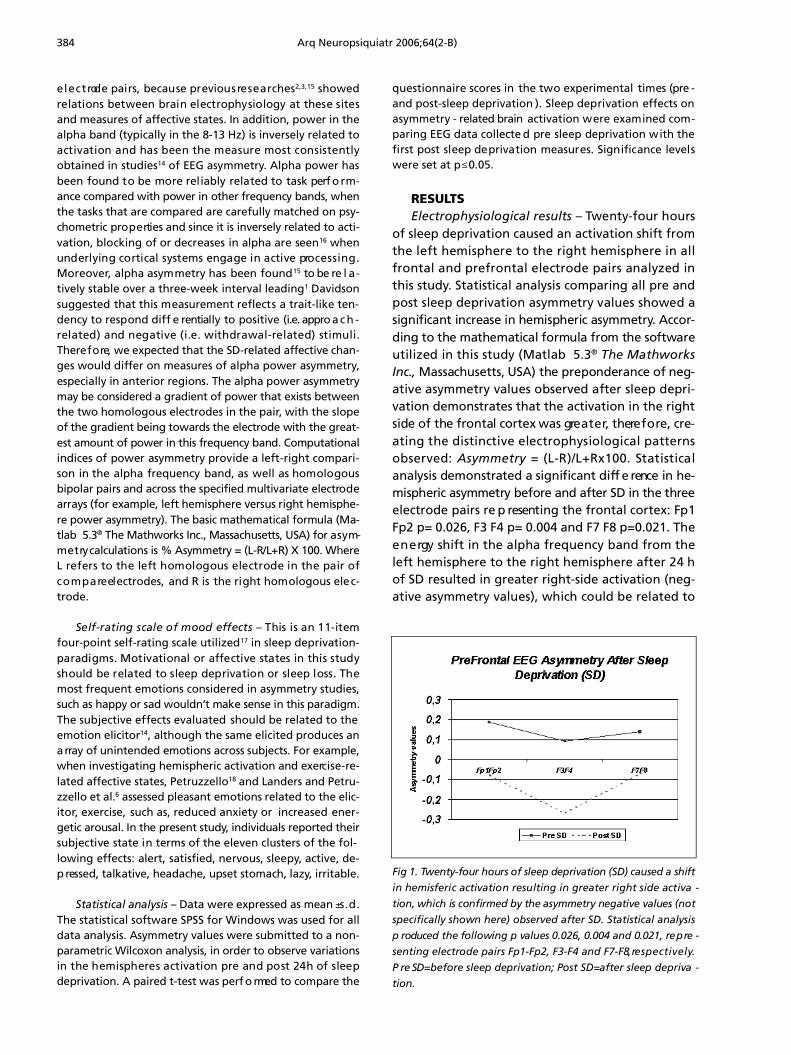
of sleep deprivation caused an activation shift fromthe left hemisphere to the right hemisphere in allf rontal and pre f rontal electrode pairs analyzed inthis study. Statistical analysis comparing all pre andpost sleep deprivation asymmetry values showed asignificant increase in hemispheric asymmetry. Accor-ding to the mathematical formula from the softwareutilized in this study (Matlab 5 . 3® The MathworksInc., Massachusetts, USA) the preponderance of neg-ative asymmetry values observed after sleep depri-vation demonstrates that the activation in the rightside of the frontal cortex was gre a t e r, there f o re, cre-ating the distinctive electrophysiological pattern so b s e rved: A s y m m e t ry = (L-R)/L+Rx100. Statisticalanalysis demonstrated a significant diff e rence in he-mispheric asymmetry before and after SD in the thre ee l e c t rode pairs re p resenting the frontal cortex: Fp1Fp2 p= 0.026, F3 F4 p= 0.004 and F7 F8 p=0.021. Thee n e rgy shift in the alpha frequency band from theleft hemisphere to the right hemisphere after 24 hof SD resulted in greater right-side activation (neg-ative asymmetry values), which could be related to
Fig 1. Twenty-four hours of sleep deprivation (SD) caused a shiftin hemisferic activation resulting in greater right side activa -tion, which is confirmed by the asymmetry negative values (notspecifically shown here) observed after SD. Statistical analysisp roduced the following p values 0.026, 0.004 and 0.021, re p re -senting electrode pairs Fp1-Fp2, F3-F4 and F7-F8, re s p e c t i v e l y.P re SD=before sleep deprivation; Post SD=after sleep depriva -t i o n .
Arq Neuropsiquiatr 2006;64(2-B) 385
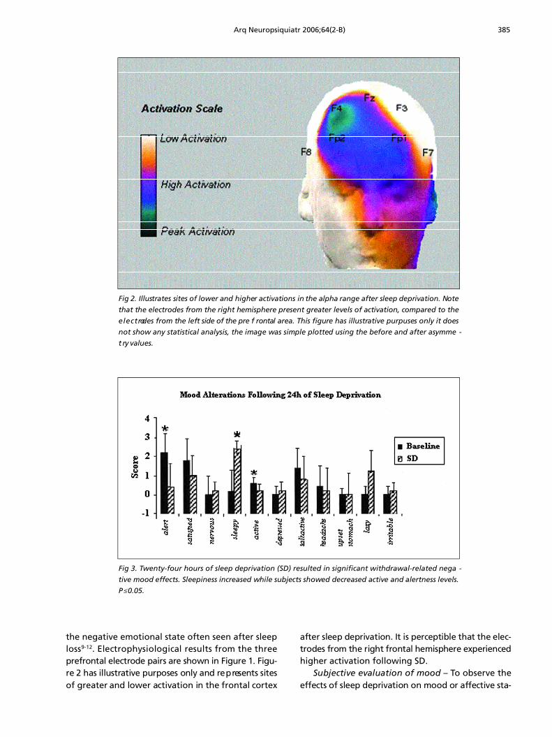
the negative emotional state often seen after sleepl o s s9 - 1 2. Electrophysiological results from the thre ep re f rontal electrode pairs are shown in Figure 1. Figu-re 2 has illustrative purposes only and re p resents sitesof greater and lower activation in the frontal cort e x
after sleep deprivation. It is perceptible that the elec-t rodes from the right frontal hemisphere experiencedhigher activation following SD.
Subjective evaluation of mood – To observe thee ffects of sleep deprivation on mood or affective sta-
Fig 2. Illustrates sites of lower and higher activations in the alpha range after sleep deprivation. Notethat the electrodes from the right hemisphere present greater levels of activation, compared to thee l e c t rodes from the left side of the pre f rontal area. This figure has illustrative purpuses only it doesnot show any statistical analysis, the image was simple plotted using the before and after asymme -t ry values.
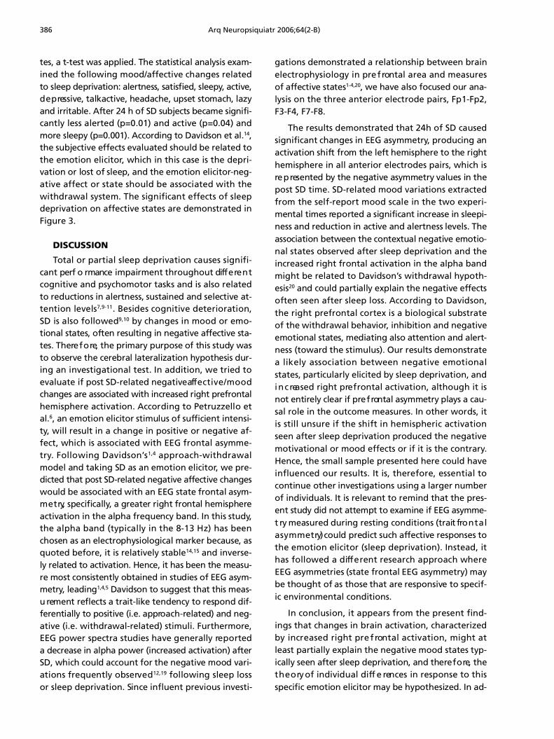
Fig 3. Twenty-four hours of sleep deprivation (SD) resulted in significant withdrawal-related nega -tive mood effects. Sleepiness increased while subjects showed decreased active and alertness levels.P≤0.05.
386 Arq Neuropsiquiatr 2006;64(2-B)
tes, a t-test was applied. The statistical analysis exam-ined the following mood/affective changes re l a t e dto sleep deprivation: alertness, satisfied, sleepy, active,d e p ressive, talkactive, headache, upset stomach, lazyand irritable. After 24 h of SD subjects became signifi-cantly less alerted (p=0.01) and active (p=0.04) andm o re sleepy (p=0.001). According to Davidson et al.1 4,the subjective effects evaluated should be related tothe emotion elicitor, which in this case is the depri-vation or lost of sleep, and the emotion elicitor- n e g-ative affect or state should be associated with thewithdrawal system. The significant effects of sleepdeprivation on affective states are demonstrated inFigure 3.
DISCUSSION
Total or partial sleep deprivation causes signifi-cant perf o rmance impairment throughout diff e re n tcognitive and psychomotor tasks and is also relatedto reductions in alertness, sustained and selective at-tention levels7 , 9 - 1 1. Besides cognitive deterioration,SD is also followed9 , 1 0 by changes in mood or emo-tional states, often resulting in negative affective sta-tes. There f o re, the primary purpose of this study wasto observe the cerebral lateralization hypothesis dur-ing an investigational test. In addition, we tried toevaluate if post SD-related negative aff e c t i v e / m o o dchanges are associated with increased right pre f ro n t a lh e m i s p h e re activation. According to Petruzzello eta l .6, an emotion elicitor stimulus of sufficient intensi-t y, will result in a change in positive or negative af-fect, which is associated with EEG frontal asymme-t ry. Following Davidson’s1 , 4 a p p ro a c h - w i t h d r a w a lmodel and taking SD as an emotion elicitor, we pre-dicted that post SD-related negative affective changeswould be associated with an EEG state frontal asym-m e t ry, specifically, a greater right frontal hemisphereactivation in the alpha frequency band. In this study,the alpha band (typically in the 8-13 Hz) has beenchosen as an electrophysiological marker because, asquoted before, it is relatively stable14,15 and inverse-ly related to activation. Hence, it has been the measu-re most consistently obtained in studies of EEG asym-m e t ry, leading1 , 4 , 5 Davidson to suggest that this meas-u rement reflects a trait-like tendency to respond dif-f e rentially to positive (i.e. appro a c h - related) and neg-ative (i.e. withdrawal-related) stimuli. Furthermore,EEG power spectra studies have generally re p o rt e da decrease in alpha power (increased activation) afterSD, which could account for the negative mood vari-ations frequently observ e d1 2 , 1 9 following sleep lossor sleep deprivation. Since influent previous investi-
gations demonstrated a relationship between braine l e c t rophysiology in pre f rontal area and measure sof affective states1 - 4 , 2 0, we have also focused our ana-lysis on the three anterior electrode pairs, Fp1-Fp2,F3-F4, F7-F8.
The results demonstrated that 24h of SD causedsignificant changes in EEG asymmetry, producing anactivation shift from the left hemisphere to the righthemisphere in all anterior electrodes pairs, which isre p resented by the negative asymmetry values in thepost SD time. SD-related mood variations extractedf rom the self-re p o rt mood scale in the two experi-mental times re p o rted a significant increase in sleepi-ness and reduction in active and alertness levels. Theassociation between the contextual negative emotio-nal states observed after sleep deprivation and theincreased right frontal activation in the alpha bandmight be related to Davidson’s withdrawal hypoth-e s i s2 0 and could partially explain the negative eff e c t soften seen after sleep loss. According to Davidson,the right pre f rontal cortex is a biological substrateof the withdrawal behavior, inhibition and negativeemotional states, mediating also attention and alert-ness (toward the stimulus). Our results demonstratea likely association between negative emotionalstates, particularly elicited by sleep deprivation, andi n c reased right pre f rontal activation, although it isnot entirely clear if pre f rontal asymmetry plays a cau-sal role in the outcome measures. In other words, itis still unsure if the shift in hemispheric activationseen after sleep deprivation produced the negativemotivational or mood effects or if it is the contrary.Hence, the small sample presented here could haveinfluenced our results. It is, there f o re, essential tocontinue other investigations using a larger numberof individuals. It is relevant to remind that the pres-ent study did not attempt to examine if EEG asymme-t ry measured during resting conditions (trait fro n t a la s y m m e t ry) could predict such affective responses tothe emotion elicitor (sleep deprivation). Instead, ithas followed a diff e rent re s e a rch approach whereEEG asymmetries (state frontal EEG asymmetry) maybe thought of as those that are responsive to specif-ic environmental conditions.
In conclusion, it appears from the present find-ings that changes in brain activation, characterizedby increased right pre f rontal activation, might atleast partially explain the negative mood states typ-ically seen after sleep deprivation, and there f o re, thet h e o ry of individual diff e rences in response to thisspecific emotion elicitor may be hypothesized. In ad-
Arq Neuropsiquiatr 2006;64(2-B) 387
dition to replicating the present findings, future workalso needs to examine SD-related affective respons-es of subjects with: 1) previously higher levels of neg-ative affect, 2) suffering from mood or anxiety dis-o rders and 3) resting EEG asymmetry as a pre d i c t o rof emotional responses to sleep deprivation or sleeploss. Measuring the diff e rences in individual re s p o n s eto SD-mood effects might be interesting and useful,given the involvement of almost all psychopatholo-gies in the sleep regulation.
REFERENCES 1. Davidson RJ. Anterior cerebral asymmetry and the nature of emotion.
Brain Cognit 1992; 20:125-151.2. Harmon-Jones E, Allen JJ. Behavioral activation sensitivity and re s t i n g
f rontal EEG asymmetry: covariation of putative indicators related torisk for mood disorders. J Abnormal Psychol 1997;106:159-163.
3. Sutton KS, Davidson RJ. Pre f rontal brain electrical asymmetry pre d i c t sthe evaluation of affective stimuli. Neuropsychologia 2000;38:1723-1733.
4. Davidson RJ. Asymmetric brain function, affective style and psycho-pathology: the role of early experience and plasticity. Develop Psy-chopathol 1994;6:741-758.
5. Sackeim HA, Gre e n b e rg MS, Weiman AL, Gur RC, Hungerbuhler JP,Geschwind N. Hemispheric asymmetry in the expression of positiveand negative emotions: neurological evidence. A rch Neurol 1982; 39:210-218.
6. P e t ruzzello SJ, Hall E, Ekkekakis P. Regional brain activation as a bio-logical marker of affective responsivity to acute exercise: influence offitness. Psychophysiology 2001;38:99-106.
7. C o r s i - C a b rera M, A rce C, Ramos J, Lorenzo I, Guevara MA. Time courseof reaction time and EEG while performing a vigilance task duringtotal sleep deprivation. Sleep 1996;19:563-569.
8. Cajochen C, Foy R, Dijk DJ. Frontal predominance of a relative incre a s ein sleep delta and theta EEG activity after sleep loss in humans. SleepRes Online 1999; 2:65-69.
9. B a b k o ff H, Caspy T, Mikulincer M. Subjective sleepiness ratings: thee ffects of sleep deprivation, circadian rhythmicity and cognitive per-formance. Sleep 1991;14:534-539.
10. Batejat D, Lagarde D. Circadian rhythm and sleep deprivation: eff e c t son psychomotor performance. Med Sci Res 1992;20:167-168.
11. Dinges DF. An overview of sleepiness and accidents. J Sleep Res 1995;4:4.12. Dijkman M, Sachs N, Levine E et al. Effects of reduced stimulation on
n e u robehavioral alertness depend on circadian phase during humansleep deprivation. Sleep Res 1997;26:265.
13. Jung P, Baumgärtner U, Bauermann T, et al. Asymmetry in the humanprimary somatosensory cortex and handedness. Neuroimage 2003;19:913-923.
14. Davidson RJ, Ekman P, Saron CD, Senulis JA, Friesen WV. Approach-withdrawal and cerebral asymmetry: emotional expression and brainphysiology I. J Pers Soc Psycho 1990; 58:330-341.
15. Tomarken AJ, Davidson RJ, Wheeler RE, Doss RC. Psychometric pro p-erties of resting anterior EEG asymmetry: temporal stability and inter-nal consistency. Psychophysiology 1992;29:576-592.
16. Davidson RJ, Chapman JP, Chapman LJ, Henriques JB. Asymmetricalbrain electrical activity discriminates between psychometrically-matched verbal and spatial cognitive tasks. Psychophysiology 1990;27:528-543.
17. James JE, Gregg ME. Hemodynamic effects of dietary caffeine sleeprestriction, and laboratory stress. Psychophysiology 2004;41:914-923.
18. P e t ruzzello SJ, Landers DM. State anxiety reduction and exercise: doeshemispheric activation reflect such changes? Med Sci Sports Exerc i s e1994;26:1028-1035.
19. Corsi-Cabrera M, Sanchéz AI, Del-Rio-Portilla Y, Villanueva Y, Pérez-G a rcí. Effect of 38h of sleep deprivation on the waking EEG in women:sex differences. Int Psychophysiol 2003;50:213-224.
20. Davidson RJ, Irwin W. The functional neuroanatomy of emotion andaffective style. Trends Cogn Sci 1991;3:11-21.
Related Documents