The Recognition of Identical Ligands by Unrelated Proteins Sarah Barelier, Teague Sterling, Matthew J. O’Meara, and Brian K. Shoichet* Department of Pharmaceutical Chemistry, University of California San Francisco, 1700 Fourth Street, Byers Hall, San Francisco, California 94158, United States * S Supporting Information ABSTRACT: The binding of drugs and reagents to off-targets is well-known. Whereas many off-targets are related to the primary target by sequence and fold, many ligands bind to unrelated pairs of proteins, and these are harder to anticipate. If the binding site in the off-target can be related to that of the primary target, this challenge resolves into aligning the two pockets. However, other cases are possible: the ligand might interact with entirely different residues and environments in the off-target, or wholly different ligand atoms may be implicated in the two complexes. To investigate these scenarios at atomic resolution, the structures of 59 ligands in 116 complexes (62 pairs in total), where the protein pairs were unrelated by fold but bound an identical ligand, were examined. In almost half of the pairs, the ligand interacted with unrelated residues in the two proteins (29 pairs), and in 14 of the pairs wholly different ligand moieties were implicated in each complex. Even in those 19 pairs of complexes that presented similar environments to the ligand, ligand superposition rarely resulted in the overlap of related residues. There appears to be no single pattern-matching “code” for identifying binding sites in unrelated proteins that bind identical ligands, though modeling suggests that there might be a limited number of different patterns that suffice to recognize different ligand functional groups. T he search for ligands specific for their receptors has dominated medicinal chemistry for a century. 1 Mean- while, a central dogma of biology has been the fidelity of information flow from gene to protein to folded structure to specific activity. Thus, when seeking “off-targets” to which drugs may bind, it has been natural to focus on proteins related in sequence and structure to the primary target. Obtaining specificity against a related human target, not involved in the disease but perhaps in an adverse reaction, or for a pathogen target while sparing the human homologue, often requires capitalizing on subtle differences in the binding sites. Such optimization can be difficult, but the nature of the challenge is well understood. More perplexing is the possibility that a ligand might modulate a protein unrelated in sequence and structure to its primary target. The advent of large ligand-target association databases 2-4 has revealed that these off-targets can bear little relationship to their primary ones. Paolini and colleagues observed that not only do molecules targeting aminergic G Protein Coupled Receptors (GPCRs) cross-react with other GPCRs but they often are active on protein kinases, while kinase inhibitors in turn have activity on ion channels and phosphodiesterases. 5 Bork and colleagues linked related side-effects to predict that raloxifene, an estrogen nuclear hormone receptor (NHR) drug, also inhibits the 5HT 1D GPCR, 6 that the proton-pump inhibitor rabeprazole also acts on the dopamine D 3 GPCR, and that the antihistamine loratadine modulates the GABA ion channel. A large-scale study found that many approved drugs act on targets unrelated to their primary efficacy targets, 7 with over a quarter crossing major target boundaries. Thus, kinase inhibitors antagonized GPCRs, GPCR ligands antagonized ion channels, ion channel modulators bound to NHRs, among others. Intriguingly, some of this polypharmacology tracks that of endogenous hormones and neurotransmitters, which also modulate multiple receptors unrelated in sequence or structure. 8 Among other examples, acetylcholine, glutamate, serotonin, and ATP all modulate both ion channels and GPCRs as primary signaling receptors, while estradiol, progesterone, and leukotrienes do the same against both NHRs and GPCRs. If mimicry of endogenous signaling molecules may suggest an origin for drug polypharmacology, 8 it does not explain its structural basis. By analogy to convergent enzyme evolution, 9 one might imagine that two binding sites recognizing the same ligand will have similar residues making similar interactions with the ligand. If true, then the problem of predicting off- targets would resolve into detecting similar binding sites in two otherwise unrelated proteins. This is the case that would most easily fit with our current target-based approaches to medicinal chemistry and chemical biology. However, we already know that similar ligand motifs can be recognized by altogether different environments (Figure 1). 10,11 We can reasonably expect that some ligands at least will bind to unrelated binding Received: August 27, 2015 Accepted: September 30, 2015 Published: September 30, 2015 Articles pubs.acs.org/acschemicalbiology © 2015 American Chemical Society 2772 DOI: 10.1021/acschembio.5b00683 ACS Chem. Biol. 2015, 10, 2772-2784 Downloaded via UNIV OF CALIFORNIA SAN FRANCISCO on October 31, 2018 at 18:44:47 (UTC). See https://pubs.acs.org/sharingguidelines for options on how to legitimately share published articles.

Welcome message from author
This document is posted to help you gain knowledge. Please leave a comment to let me know what you think about it! Share it to your friends and learn new things together.
Transcript
The Recognition of Identical Ligands by Unrelated ProteinsSarah Barelier, Teague Sterling, Matthew J. O’Meara, and Brian K. Shoichet*
Department of Pharmaceutical Chemistry, University of California San Francisco, 1700 Fourth Street, Byers Hall, San Francisco,California 94158, United States
*S Supporting Information
ABSTRACT: The binding of drugs and reagents to off-targetsis well-known. Whereas many off-targets are related to theprimary target by sequence and fold, many ligands bind tounrelated pairs of proteins, and these are harder to anticipate. Ifthe binding site in the off-target can be related to that of theprimary target, this challenge resolves into aligning the twopockets. However, other cases are possible: the ligand mightinteract with entirely different residues and environments in theoff-target, or wholly different ligand atoms may be implicated inthe two complexes. To investigate these scenarios at atomicresolution, the structures of 59 ligands in 116 complexes (62pairs in total), where the protein pairs were unrelated by foldbut bound an identical ligand, were examined. In almost half of the pairs, the ligand interacted with unrelated residues in the twoproteins (29 pairs), and in 14 of the pairs wholly different ligand moieties were implicated in each complex. Even in those 19pairs of complexes that presented similar environments to the ligand, ligand superposition rarely resulted in the overlap of relatedresidues. There appears to be no single pattern-matching “code” for identifying binding sites in unrelated proteins that bindidentical ligands, though modeling suggests that there might be a limited number of different patterns that suffice to recognizedifferent ligand functional groups.
The search for ligands specific for their receptors hasdominated medicinal chemistry for a century.1 Mean-
while, a central dogma of biology has been the fidelity ofinformation flow from gene to protein to folded structure tospecific activity. Thus, when seeking “off-targets” to whichdrugs may bind, it has been natural to focus on proteins relatedin sequence and structure to the primary target. Obtainingspecificity against a related human target, not involved in thedisease but perhaps in an adverse reaction, or for a pathogentarget while sparing the human homologue, often requirescapitalizing on subtle differences in the binding sites. Suchoptimization can be difficult, but the nature of the challenge iswell understood. More perplexing is the possibility that a ligandmight modulate a protein unrelated in sequence and structureto its primary target.The advent of large ligand-target association databases2−4 has
revealed that these off-targets can bear little relationship to theirprimary ones. Paolini and colleagues observed that not only domolecules targeting aminergic G Protein Coupled Receptors(GPCRs) cross-react with other GPCRs but they often areactive on protein kinases, while kinase inhibitors in turn haveactivity on ion channels and phosphodiesterases.5 Bork andcolleagues linked related side-effects to predict that raloxifene,an estrogen nuclear hormone receptor (NHR) drug, alsoinhibits the 5HT1D GPCR,6 that the proton-pump inhibitorrabeprazole also acts on the dopamine D3 GPCR, and that theantihistamine loratadine modulates the GABA ion channel. Alarge-scale study found that many approved drugs act on targets
unrelated to their primary efficacy targets,7 with over a quartercrossing major target boundaries. Thus, kinase inhibitorsantagonized GPCRs, GPCR ligands antagonized ion channels,ion channel modulators bound to NHRs, among others.Intriguingly, some of this polypharmacology tracks that ofendogenous hormones and neurotransmitters, which alsomodulate multiple receptors unrelated in sequence orstructure.8 Among other examples, acetylcholine, glutamate,serotonin, and ATP all modulate both ion channels and GPCRsas primary signaling receptors, while estradiol, progesterone,and leukotrienes do the same against both NHRs and GPCRs.If mimicry of endogenous signaling molecules may suggest
an origin for drug polypharmacology,8 it does not explain itsstructural basis. By analogy to convergent enzyme evolution,9
one might imagine that two binding sites recognizing the sameligand will have similar residues making similar interactionswith the ligand. If true, then the problem of predicting off-targets would resolve into detecting similar binding sites in twootherwise unrelated proteins. This is the case that would mosteasily fit with our current target-based approaches to medicinalchemistry and chemical biology. However, we already knowthat similar ligand motifs can be recognized by altogetherdifferent environments (Figure 1).10,11 We can reasonablyexpect that some ligands at least will bind to unrelated binding
Received: August 27, 2015Accepted: September 30, 2015Published: September 30, 2015
Articles
pubs.acs.org/acschemicalbiology
© 2015 American Chemical Society 2772 DOI: 10.1021/acschembio.5b00683ACS Chem. Biol. 2015, 10, 2772−2784
Dow
nloa
ded
via
UN
IV O
F C
AL
IFO
RN
IA S
AN
FR
AN
CIS
CO
on
Oct
ober
31,
201
8 at
18:
44:4
7 (U
TC
).
See
http
s://p
ubs.
acs.
org/
shar
ingg
uide
lines
for
opt
ions
on
how
to le
gitim
atel
y sh
are
publ
ishe
d ar
ticle
s.
sites. Indeed, in a study of 100 complexes involving ninecofactors, Kahraman and Thornton found that unrelatedbinding sites, in different folds, could recognize eachcofactor.12,13 For the prediction of enzyme function, a focusof that study, they found no simple mapping between ligandsand recognition pockets.Three possibilities can be distinguished for how an identical
ligand interacts with unrelated proteins (Figure 2). The first iswhere the same ligand groups make similar interactions withrelated residues in both binding sites (class A). A secondpossibility is where the same ligand groups interact withdissimilar residues and environments in the two binding sites(class B). Finally, different ligand groups may interact with theprotein, such that a different part of the ligand makes thedefining pharmacophore interactions in each complex (class C).Inspection of ligand−protein complexes reveals examples of
each of these cases (Figure 3). For instance, the anti-Alzheimer’s drug galanthamine (GNT) binds to both themainly-β distorted sandwich acetylcholine binding protein14
(PDB 2ph9) and the α/β 3-layer (aba) sandwich acetylcholine
esterase15 (PDB 1dx6; Figure 3, class A). Though superpositionof the two complexes finds few overlapping residues,recognition is dominated by cation−π interactions at theaminergic cation, and a similar mixture of nonpolar andhydrogen bond interactions on the other side of the ligand, inboth complexes. Conversely, carboxyaminoimidazole ribonu-cleotide (CAIR, ligand code C2R) finds different environmentsin its complexes with the α/β three-layer (aba) sandwich N5-CAIR mutase16 (PurE, PDB 2nsl) and the α/β two-layersandwich SAICAR synthetase17 (PurC, PDB 2gqs). CAIR bindsto each protein with similar affinities (2116,18 and 7.8 μM,19
respectively), but different protein recognition motifs areengaged (Figure 3, class B). These differences are mostprominent in the ribose-carboxyimidazole moiety, which inPurE forms a network of hydrogen bonds to main chain atomsof six different residues. In PurC, the same groups arerecognized by two catalytic magnesium ions and hydrogenbonds from an arginine and an aspartate. The imidazole ispartly exposed in PurC but is buried in PurE. Finally, themetabolite allantoin (2AL) binds both to the α-bundle 2-oxo-4-hydroxy-4-carboxy-5-ureidoimidazoline (OHCU) decarboxy-lase (PDB 2o73)20 and to the α/β-fold urate oxidase21 (PDB2fxl; Figure 3, class C). Wholly different ligand groups areimplicated in each. In the OHCU decarboxylase complex, forwhich allantoin is the reaction product, multiple hydrogenbonds are made to the imidazole-dione core, and the ligand isentirely buried. Conversely, with urate oxidase, which is twoenzymes upstream from OHCU decarboxylase in the xanthine
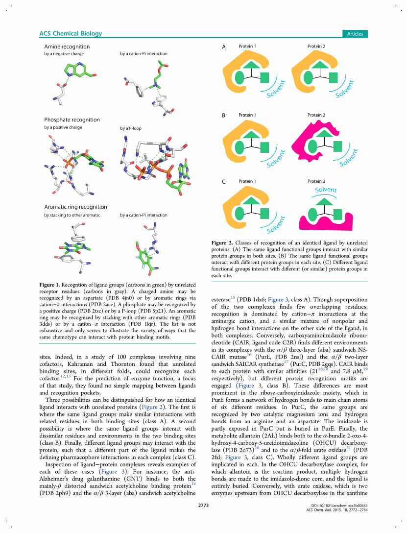
Figure 1. Recogntion of ligand groups (carbons in green) by unrelatedreceptor residues (carbons in gray). A charged amine may berecognized by an aspartate (PDB 4jn0) or by aromatic rings viacation−π interactions (PDB 2ace). A phosphate may be recognized bya positive charge (PDB 2tsc) or by a P-loop (PDB 5p21). An aromaticring may be recognized by stacking with other aromatic rings (PDB3dds) or by a cation−π interaction (PDB 1kjr). The list is notexhaustive and only serves to illustrate the variety of ways that thesame chemotype can interact with protein binding motifs.
Figure 2. Classes of recognition of an identical ligand by unrelatedproteins. (A) The same ligand functional groups interact with similarprotein groups in both sites. (B) The same ligand functional groupsinteract with different protein groups in each site. (C) Different ligandfunctional groups interact with different (or similar) protein groups ineach site.
ACS Chemical Biology Articles
DOI: 10.1021/acschembio.5b00683ACS Chem. Biol. 2015, 10, 2772−2784
2773
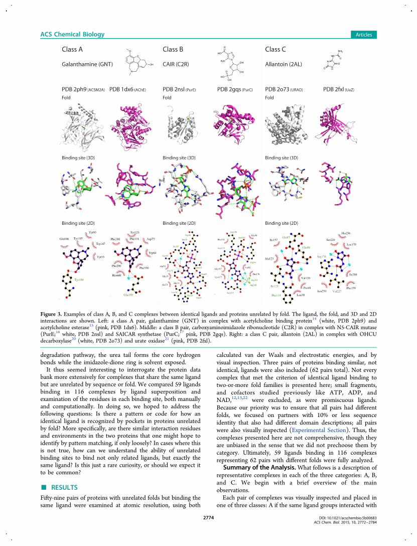
degradation pathway, the urea tail forms the core hydrogenbonds while the imidazole-dione ring is solvent exposed.It thus seemed interesting to interrogate the protein data
bank more extensively for complexes that share the same ligandbut are unrelated by sequence or fold. We compared 59 ligandsbinding in 116 complexes by ligand superposition andexamination of the residues in each binding site, both manuallyand computationally. In doing so, we hoped to address thefollowing questions: Is there a pattern or code for how anidentical ligand is recognized by pockets in proteins unrelatedby fold? More specifically, are there similar interaction residuesand environments in the two proteins that one might hope toidentify by pattern matching, if only loosely? In cases where thisis not true, how can we understand the ability of unrelatedbinding sites to bind not only related ligands, but exactly thesame ligand? Is this just a rare curiosity, or should we expect itto be common?
■ RESULTS
Fifty-nine pairs of proteins with unrelated folds but binding thesame ligand were examined at atomic resolution, using both
calculated van der Waals and electrostatic energies, and byvisual inspection. Three pairs of proteins binding similar, notidentical, ligands were also included (62 pairs total). Not everycomplex that met the criterion of identical ligand binding totwo-or-more fold families is presented here; small fragments,and cofactors studied previously like ATP, ADP, andNAD,12,13,22 were excluded, as were promiscuous ligands.Because our priority was to ensure that all pairs had differentfolds, we focused on partners with 10% or less sequenceidentity that also had different domain descriptions; all pairswere also visually inspected (Experimental Section). Thus, thecomplexes presented here are not comprehensive, though theyare unbiased in the sense that we did not prechoose them bycategory. Ultimately, 59 ligands binding in 116 complexesrepresenting 62 pairs with different folds were fully analyzed.
Summary of the Analysis.What follows is a description ofrepresentative complexes in each of the three categories: A, B,and C. We begin with a brief overview of the mainobservations.Each pair of complexes was visually inspected and placed in
one of three classes: A if the same ligand groups interacted with
Figure 3. Examples of class A, B, and C complexes between identical ligands and proteins unrelated by fold. The ligand, the fold, and 3D and 2Dinteractions are shown. Left: a class A pair, galanthamine (GNT) in complex with acetylcholine binding protein14 (white, PDB 2ph9) andacetylcholine esterase15 (pink, PDB 1dx6). Middle: a class B pair, carboxyaminoimidazole ribonucleotide (C2R) in complex with N5-CAIR mutase(PurE;16 white, PDB 2nsl) and SAICAR synthetase (PurC;17 pink, PDB 2gqs). Right: a class C pair, allantoin (2AL) in complex with OHCUdecarboxylase20 (white, PDB 2o73) and urate oxidase21 (pink, PDB 2fxl).
ACS Chemical Biology Articles
DOI: 10.1021/acschembio.5b00683ACS Chem. Biol. 2015, 10, 2772−2784
2774
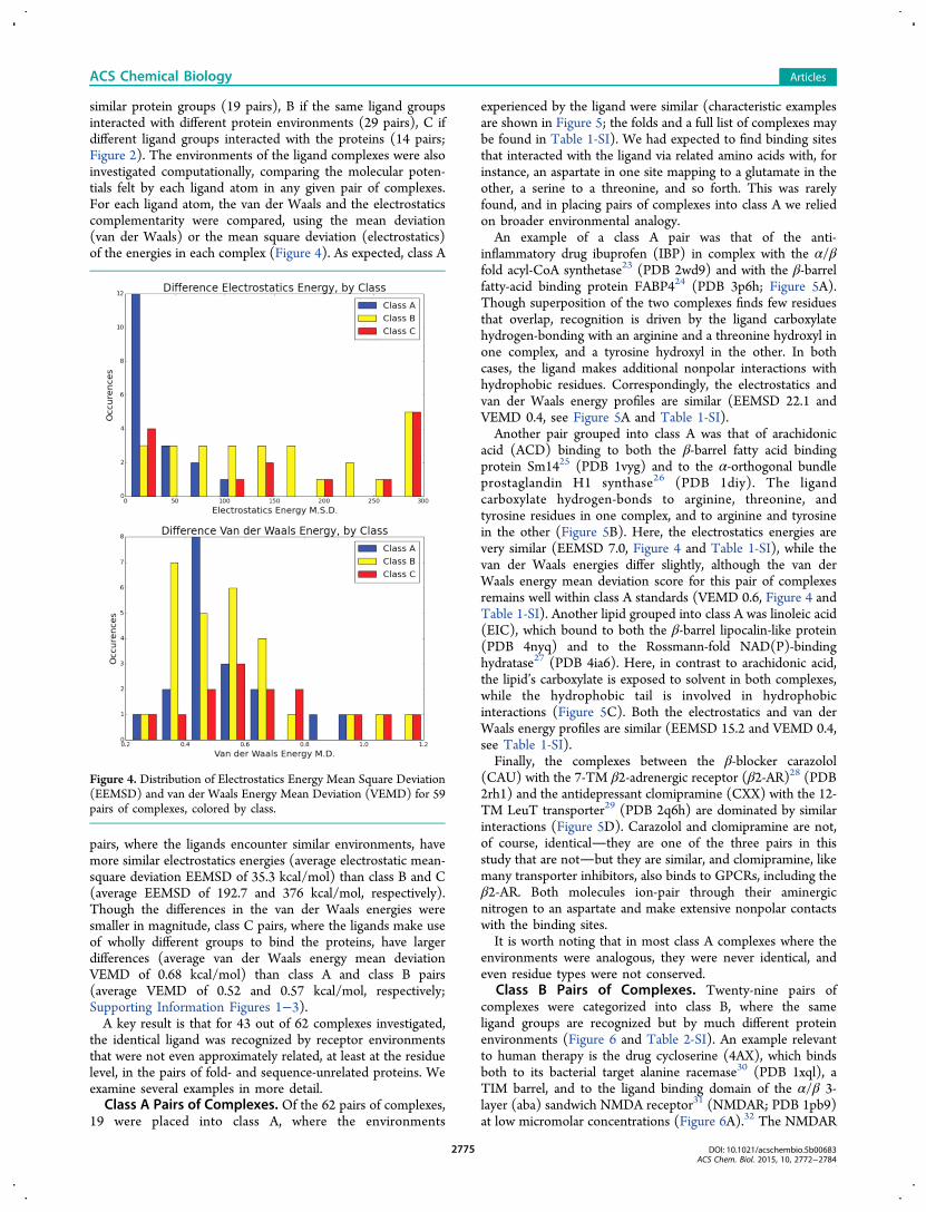
similar protein groups (19 pairs), B if the same ligand groupsinteracted with different protein environments (29 pairs), C ifdifferent ligand groups interacted with the proteins (14 pairs;Figure 2). The environments of the ligand complexes were alsoinvestigated computationally, comparing the molecular poten-tials felt by each ligand atom in any given pair of complexes.For each ligand atom, the van der Waals and the electrostaticscomplementarity were compared, using the mean deviation(van der Waals) or the mean square deviation (electrostatics)of the energies in each complex (Figure 4). As expected, class A
pairs, where the ligands encounter similar environments, havemore similar electrostatics energies (average electrostatic mean-square deviation EEMSD of 35.3 kcal/mol) than class B and C(average EEMSD of 192.7 and 376 kcal/mol, respectively).Though the differences in the van der Waals energies weresmaller in magnitude, class C pairs, where the ligands make useof wholly different groups to bind the proteins, have largerdifferences (average van der Waals energy mean deviationVEMD of 0.68 kcal/mol) than class A and class B pairs(average VEMD of 0.52 and 0.57 kcal/mol, respectively;Supporting Information Figures 1−3).A key result is that for 43 out of 62 complexes investigated,
the identical ligand was recognized by receptor environmentsthat were not even approximately related, at least at the residuelevel, in the pairs of fold- and sequence-unrelated proteins. Weexamine several examples in more detail.Class A Pairs of Complexes. Of the 62 pairs of complexes,
19 were placed into class A, where the environments
experienced by the ligand were similar (characteristic examplesare shown in Figure 5; the folds and a full list of complexes maybe found in Table 1-SI). We had expected to find binding sitesthat interacted with the ligand via related amino acids with, forinstance, an aspartate in one site mapping to a glutamate in theother, a serine to a threonine, and so forth. This was rarelyfound, and in placing pairs of complexes into class A we reliedon broader environmental analogy.An example of a class A pair was that of the anti-
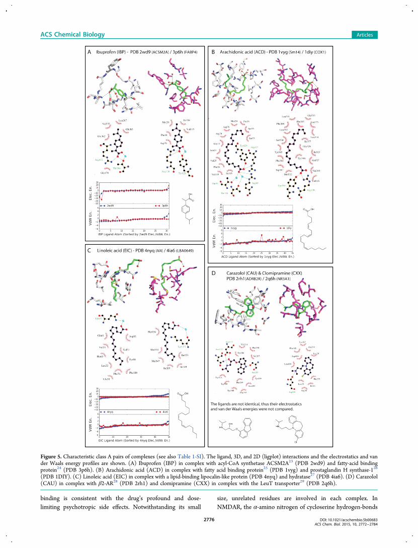
inflammatory drug ibuprofen (IBP) in complex with the α/βfold acyl-CoA synthetase23 (PDB 2wd9) and with the β-barrelfatty-acid binding protein FABP424 (PDB 3p6h; Figure 5A).Though superposition of the two complexes finds few residuesthat overlap, recognition is driven by the ligand carboxylatehydrogen-bonding with an arginine and a threonine hydroxyl inone complex, and a tyrosine hydroxyl in the other. In bothcases, the ligand makes additional nonpolar interactions withhydrophobic residues. Correspondingly, the electrostatics andvan der Waals energy profiles are similar (EEMSD 22.1 andVEMD 0.4, see Figure 5A and Table 1-SI).Another pair grouped into class A was that of arachidonic
acid (ACD) binding to both the β-barrel fatty acid bindingprotein Sm1425 (PDB 1vyg) and to the α-orthogonal bundleprostaglandin H1 synthase26 (PDB 1diy). The ligandcarboxylate hydrogen-bonds to arginine, threonine, andtyrosine residues in one complex, and to arginine and tyrosinein the other (Figure 5B). Here, the electrostatics energies arevery similar (EEMSD 7.0, Figure 4 and Table 1-SI), while thevan der Waals energies differ slightly, although the van derWaals energy mean deviation score for this pair of complexesremains well within class A standards (VEMD 0.6, Figure 4 andTable 1-SI). Another lipid grouped into class A was linoleic acid(EIC), which bound to both the β-barrel lipocalin-like protein(PDB 4nyq) and to the Rossmann-fold NAD(P)-bindinghydratase27 (PDB 4ia6). Here, in contrast to arachidonic acid,the lipid’s carboxylate is exposed to solvent in both complexes,while the hydrophobic tail is involved in hydrophobicinteractions (Figure 5C). Both the electrostatics and van derWaals energy profiles are similar (EEMSD 15.2 and VEMD 0.4,see Table 1-SI).Finally, the complexes between the β-blocker carazolol
(CAU) with the 7-TM β2-adrenergic receptor (β2-AR)28 (PDB2rh1) and the antidepressant clomipramine (CXX) with the 12-TM LeuT transporter29 (PDB 2q6h) are dominated by similarinteractions (Figure 5D). Carazolol and clomipramine are not,of course, identicalthey are one of the three pairs in thisstudy that are notbut they are similar, and clomipramine, likemany transporter inhibitors, also binds to GPCRs, including theβ2-AR. Both molecules ion-pair through their aminergicnitrogen to an aspartate and make extensive nonpolar contactswith the binding sites.It is worth noting that in most class A complexes where the
environments were analogous, they were never identical, andeven residue types were not conserved.
Class B Pairs of Complexes. Twenty-nine pairs ofcomplexes were categorized into class B, where the sameligand groups are recognized but by much different proteinenvironments (Figure 6 and Table 2-SI). An example relevantto human therapy is the drug cycloserine (4AX), which bindsboth to its bacterial target alanine racemase30 (PDB 1xql), aTIM barrel, and to the ligand binding domain of the α/β 3-layer (aba) sandwich NMDA receptor31 (NMDAR; PDB 1pb9)at low micromolar concentrations (Figure 6A).32 The NMDAR
Figure 4. Distribution of Electrostatics Energy Mean Square Deviation(EEMSD) and van der Waals Energy Mean Deviation (VEMD) for 59pairs of complexes, colored by class.
ACS Chemical Biology Articles
DOI: 10.1021/acschembio.5b00683ACS Chem. Biol. 2015, 10, 2772−2784
2775
binding is consistent with the drug’s profound and dose-limiting psychotropic side effects. Notwithstanding its small
size, unrelated residues are involved in each complex. InNMDAR, the α-amino nitrogen of cycloserine hydrogen-bonds
Figure 5. Characteristic class A pairs of complexes (see also Table 1-SI). The ligand, 3D, and 2D (ligplot) interactions and the electrostatics and vander Waals energy profiles are shown. (A) Ibuprofen (IBP) in complex with acyl-CoA synthetase ACSM2A23 (PDB 2wd9) and fatty-acid bindingprotein24 (PDB 3p6h). (B) Arachidonic acid (ACD) in complex with fatty acid binding protein25 (PDB 1vyg) and prostaglandin H synthase-126
(PDB 1DIY). (C) Linoleic acid (EIC) in complex with a lipid-binding lipocalin-like protein (PDB 4nyq) and hydratase27 (PDB 4ia6). (D) Carazolol(CAU) in complex with β2-AR28 (PDB 2rh1) and clomipramine (CXX) in complex with the LeuT transporter29 (PDB 2q6h).
ACS Chemical Biology Articles
DOI: 10.1021/acschembio.5b00683ACS Chem. Biol. 2015, 10, 2772−2784
2776

with an aspartate, a threonine, and a main chain carbonyloxygen, while in the complex with alanine racemase the same
nitrogen is uncomplemented, leaving it free to react with thepyridoxal phosphate. The ring nitrogen of the drug hydrogen-
Figure 6. Characteristic class B pairs of complexes (see also Table 2-SI). The ligand, 3D, and 2D (ligplot) interactions and the electrostatics and vander Waals energy profiles are shown. (A) Cycloserine (4AX) in complex with alanine racemase30 (PDB 1xql) and the ligand binding domain of theNMDA receptor31 (PDB 1pb9). (B) Celecoxib (CEL) in complex with COX-233 (PDB 3ln1) and carbonic anhydrase34 (PDB 1oq5). (C)Flavopiridol (CPB) in complex with glycogen phophorylase36 (PDB 3ebp) and CDK937 (PDB 3blr). (D) Biopterin (BIO) in complex withtetrahydropterine synthase38 (PDB 1b66) and sepiapterin synthase39 (PBD 1sep).
ACS Chemical Biology Articles
DOI: 10.1021/acschembio.5b00683ACS Chem. Biol. 2015, 10, 2772−2784
2777
bonds with an arginine in NMDAR but with a backbonenitrogen in alanine racemase, and the polarized ring oxygen
hydrogen-bonds to a backbone nitrogen in NMDAR butaccepts a hydrogen bond from a tyrosine in the racemase. The
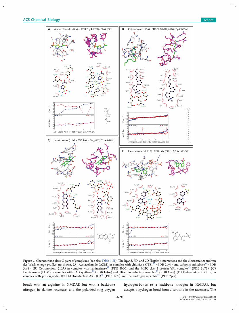
Figure 7. Characteristic class C pairs of complexes (see also Table 3-SI). The ligand, 3D, and 2D (ligplot) interactions and the electrostatics and vander Waals energy profiles are shown. (A) Acetazolamide (AZM) in complex with chitiniase CTS140 (PDB 2uy4) and carbonic anhydrase41 (PDB3hs4). (B) Cetrimonium (16A) in complex with laminarinase42 (PDB 3b00) and the MHC class I protein YF1 complex43 (PDB 3p73). (C)Lumichrome (LUM) in complex with FAD synthase44 (PDB 1s4m) and biliverdin reductase complex45 (PDB 1hes). (D) Flufenamic acid (FLF) incomplex with prostaglandin D2 11-ketoreductase AKR1C346 (PDB 1s2c) and the androgen receptor47 (PDB 2pix).
ACS Chemical Biology Articles
DOI: 10.1021/acschembio.5b00683ACS Chem. Biol. 2015, 10, 2772−2784
2778
energy profiles reflect these interactions well. While the van derWaals energies are very similar in the two complexes (VEMD0.4), as expected for a small ligand fully involved with theprotein in both pairs, the electrostatics energies are distinctlydifferent (EEMSD 300.4, Figure 6A and Table 2-SI).The anti-inflammatory drug celecoxib (CEL) finds different
environments in its complexes with the α-orthogonal bundleCOX-233 (PDB 3ln1) and the α/β roll carbonic anhydrase34
(PDB 1oq5; Figure 6B). The drug binds COX-2 with a Ki of 4nM.33 Subsequently, it was predicted and shown to bind tocarbonic anhydrase with a Ki of 21 nM.34 While the samepharmacophore is recognized in each site, the residuesdiffer.34,35 Most obviously, the drug’s sulfonamide hydrogen-bonds with an arginine, a serine, and a backbone carbonyl inCOX-2, while in carbonic anhydrase it binds as an anion to thecatalytic zinc. The same ligand group is involved in bothcomplexes, which is reflected in the van der Waals energyprofile. Except for two atoms, including the nitrogen, which isin close contact with the zinc ion, explaining a much higher vander Waals energy, the van der Waals energies are similar(VEMD 0.6, Figure 6B and Table 2-SI). The electrostaticsenergy profiles differ (EEMSD 169.1, Figure 6B and Table 2-SI), especially in the sulfonamide group, which makes keyinteractions in both complexes.The complexes of several flavonoid-like plant natural
products also fall into class B (among them, emodin (EMO),flavopiridol (CPB), and naringerin (NAR), Figure 6C andTable 2-SI). In the α/β three-layer (aba) sandwich glycogenphophorylase36 (PDB 3ebp), the flavone ring of flavopiridol(CPB) π-stacks in a sandwich with a tyrosine and aphenylalanine (Figure 6C). There are otherwise no directpolar interactions, with the cationic nitrogen and the ligandoxygens exposed to solvent. Conversely, the same ring issandwiched by nonpolar side chains in the α/β two-layersandwich CDK937 (PDB 3blr), where the ligand cation ion-pairs with an aspartate while one of the flavone carbonylsinteracts with a backbone nitrogen. We do note that both theelectrostatics and van der Waals energies are similar (EEMSD25.5 and VDW 0.4, see Table 1-SI), as expected for a large classB ligand making few electrostatics interactions.Finally, in the case of the cofactor biopterin (BIO), both the
α/β two-layer sandwich tetrahydropterine synthase38 (PDB1b66) and the α/β three-layer sandwich sepiapterin synthase39
(PBD 1sep) use a glutamate and an aspartate, respectively, torecognize the guanidine headgroup of the ligand, but the gem-diol side chain is recognized by a zinc ion in the former and byhydrogen bonds from a Ser/Tyr pair in the latter (Figure 6D).In this pair of complexes, most ligand atoms are engaged indiffering electrostatics interactions, as indicated by dissimilarelectrostatics energy profiles (EEMSD 120.0, see Figure 6D andTable 2-SI).Class C Pairs of Complexes. For 14 pairs of complexes,
not only did the protein environments differ but so did the verymoieties on the ligand that were recognized; these werecategorized as class C pairs (Figure 7 and Table 3-SI). Mostfollow the pattern set by allantoin binding to the α-bundleOHCU decarboxylase20 (PDB 2o73) and to the α/β-fold urateoxidase21 (PDB 2fxl; Figure 3). In the first of these complexes,allantoin’s imidazole-dione ring is recognized by the protein,while in the second it is exposed to solvent.In the complex between acetazolamide (AZM) and the TIM
barrel chitiniase CTS140 (PDB 2uy4), the drug interacts via itsacetamide moiety while the thiadiazole ring stacks to a
tryptophan, and the sulfonamide is solvent-exposed (Figure7A). When bound to its primary target, the α/β roll carbonicanhydrase41 (PDB 3hs4), the sulfonamide is buried andinteracts both with a threonine and a zinc ion; the thiadazolering hydrogen-bonds to a threonine, and the rest of themolecule is exposed to solvent. Correspondingly, the electro-statics energy profile suggests that the ligand atoms experiencewholly different physical environments (EEMSD 1877.8, Figure7A and Table 3-SI).Similarly, the charged amine in the antiseptic surfactant
cetrimonium (16A) is buried and ion-pairs with two glutamatesand an aspartate, and the hydrophobic chain is partly solvent-exposed in its complex with the β-sandwich laminarinase42
(PDB 3b00), whereas in the complex with the immunoglobulinC1-set domain of MHC class I protein YF143 (PDB 3p73), thehydrophobic chain is deeply buried (Figure 7B). As might beexpected, the major difference in the electrostatics energyoccurs in the amine region of the ligand, the only part that ispolar.In the case of the riboflavin catabolite lumichrome (LUM),
the ligand is partly exposed to solvent in both complexes, butthe recognition motifs vary substantially (Figure 7C). In thecomplex with the α/β three-layer (aba) sandwich FADsynthetase44 (PDB 1s4m), recognition is dominated by fourhydrogen bonds down one side of the flavin. In the complexwith the β-sandwich biliverdin reductase45 (PDB 1hes),recognition is dominated by stacking to an NADP cofactor,with only one poor geometry hydrogen bond, again to thecofactor. Although the ligand adopts the same orientation inboth binding sites (the same atoms of the ligands are buried orsolvent-exposed in both sites) and the van der Waals energiesare similar, it is put into class C because of the difference in therecognition motifs, reflected in the electrostatics energy profiles(Figure 7C).Finally, the anti-inflammatory flufenamic acid (FLF) binds to
the α/β barrel prostaglandin D2 11-ketoreductase AKR1C346
(PDB 1s2c) via networks of hydrogen bonds (histidine,tyrosine, and NADP cofactor), whereas in its complex withthe α-orthogonal bundle androgen receptor47 (PDB 2pix),recognition is exclusively driven by nonpolar contacts (Figure7D). Here, the major difference in the electrostatics energyoccurs in the carboxylate and at the linker amine regions, whilethe van der Waals energies are more similar (Figure 7D).We note that despite the only partial complementarity of
these class C complexes, affinity can be substantial, withacetazolamide binding to endochitinase in the low micromolarrange18 and to carbonic anhydrase in the low nanomolarrange,18 and flufenamic acid binding to AKR1C3 in the lowmicromolar range18 and to the androgen receptor in themidmicromolar range.47 More examples of class C complexesare available in Table 3-SI.
Effects of Energy Minimization on Complex Classi-fication. Up until now, we have compared pairs of complexesvisually, and by molecular energy potentials, essentially as theywere determined experimentally. However, some crystalstructures retain unfavorable ligand−protein contacts, oftenowing to force fields under-parametrized for ligand atoms.Accordingly, for eight pairs of complexes (16 structures), weparametrized the ligand using the GAFF procedures (http://t1.chem.umn.edu/amsol/, OEChem version 1.7.4 and reduceprogram48) and relaxed the ligand−protein complex in theAMBER force field.49−51 The resulting complexes werereinspected, and the analysis of the molecular potential energies
ACS Chemical Biology Articles
DOI: 10.1021/acschembio.5b00683ACS Chem. Biol. 2015, 10, 2772−2784
2779
was repeated (Supporting Information Figure 4-SI). Asexpected, there was only modest visual change in the complexesafter minimization. While this relaxation did lead to smoothervan der Waals energieseliminating most peaks owing tooccasional close contactsthe electrostatics energies were lessaffected, and overall no substantial changes in patterns wereseen. Thus, minimization leads to smoother energy profiles butdoes not change how we would classify pairs of complexes intoclass A, B, and C complexes (Figure 4).A Benchmarking Set for Testing Binding Site
Comparison Methods. Several methods have been recentlyintroduced to compare binding sites.34,52−55 One of these, ornewly developed ones, may be able to recognize environmentalsimilarities between pairs of targets that we see now as verydifferent. To enable such comparison, we have constructed andmade publicly available an open access benchmarking set of thepairs of fold-unrelated complexes described here (Directory ofUnrelated Complexes, DUC, http://duc.docking.org). Eachpair is organized by the single ligand they bind, with the PBBID for each complex, the structure modified for ready energycalculation, and a 2D image of the key interactions in thebinding sites. Each pair may be downloaded for comparison, ascan the entire set.Modeling How Many Different Environments May
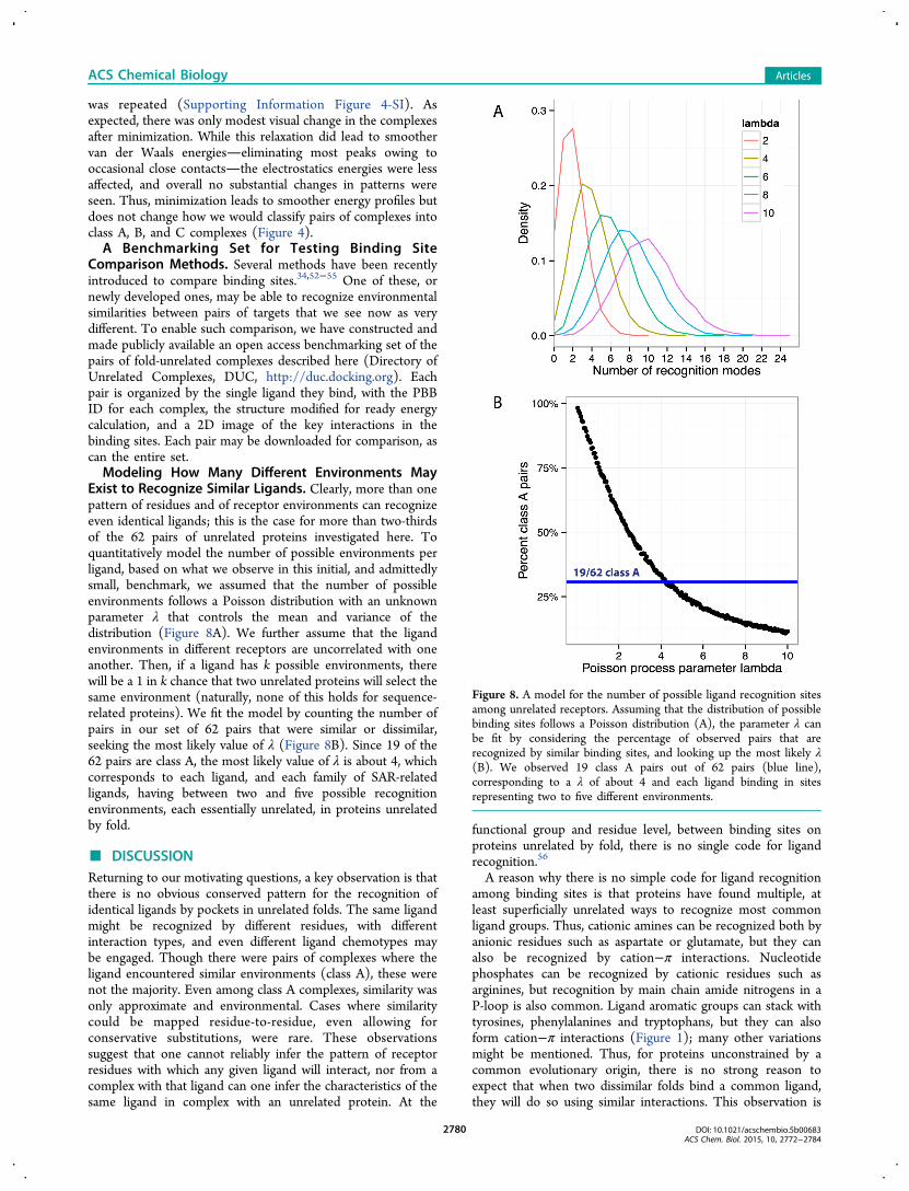
Exist to Recognize Similar Ligands. Clearly, more than onepattern of residues and of receptor environments can recognizeeven identical ligands; this is the case for more than two-thirdsof the 62 pairs of unrelated proteins investigated here. Toquantitatively model the number of possible environments perligand, based on what we observe in this initial, and admittedlysmall, benchmark, we assumed that the number of possibleenvironments follows a Poisson distribution with an unknownparameter λ that controls the mean and variance of thedistribution (Figure 8A). We further assume that the ligandenvironments in different receptors are uncorrelated with oneanother. Then, if a ligand has k possible environments, therewill be a 1 in k chance that two unrelated proteins will select thesame environment (naturally, none of this holds for sequence-related proteins). We fit the model by counting the number ofpairs in our set of 62 pairs that were similar or dissimilar,seeking the most likely value of λ (Figure 8B). Since 19 of the62 pairs are class A, the most likely value of λ is about 4, whichcorresponds to each ligand, and each family of SAR-relatedligands, having between two and five possible recognitionenvironments, each essentially unrelated, in proteins unrelatedby fold.
■ DISCUSSIONReturning to our motivating questions, a key observation is thatthere is no obvious conserved pattern for the recognition ofidentical ligands by pockets in unrelated folds. The same ligandmight be recognized by different residues, with differentinteraction types, and even different ligand chemotypes maybe engaged. Though there were pairs of complexes where theligand encountered similar environments (class A), these werenot the majority. Even among class A complexes, similarity wasonly approximate and environmental. Cases where similaritycould be mapped residue-to-residue, even allowing forconservative substitutions, were rare. These observationssuggest that one cannot reliably infer the pattern of receptorresidues with which any given ligand will interact, nor from acomplex with that ligand can one infer the characteristics of thesame ligand in complex with an unrelated protein. At the
functional group and residue level, between binding sites onproteins unrelated by fold, there is no single code for ligandrecognition.56
A reason why there is no simple code for ligand recognitionamong binding sites is that proteins have found multiple, atleast superficially unrelated ways to recognize most commonligand groups. Thus, cationic amines can be recognized both byanionic residues such as aspartate or glutamate, but they canalso be recognized by cation−π interactions. Nucleotidephosphates can be recognized by cationic residues such asarginines, but recognition by main chain amide nitrogens in aP-loop is also common. Ligand aromatic groups can stack withtyrosines, phenylalanines and tryptophans, but they can alsoform cation−π interactions (Figure 1); many other variationsmight be mentioned. Thus, for proteins unconstrained by acommon evolutionary origin, there is no strong reason toexpect that when two dissimilar folds bind a common ligand,they will do so using similar interactions. This observation is
Figure 8. A model for the number of possible ligand recognition sitesamong unrelated receptors. Assuming that the distribution of possiblebinding sites follows a Poisson distribution (A), the parameter λ canbe fit by considering the percentage of observed pairs that arerecognized by similar binding sites, and looking up the most likely λ(B). We observed 19 class A pairs out of 62 pairs (blue line),corresponding to a λ of about 4 and each ligand binding in sitesrepresenting two to five different environments.
ACS Chemical Biology Articles
DOI: 10.1021/acschembio.5b00683ACS Chem. Biol. 2015, 10, 2772−2784
2780
not restricted to protein−ligand complexes, as it has long beenknown that between repressors and operators, multiplerecognition patterns are observed, and unrelated repressorscan bind identical operators. Here too there is “no code” forrecognition.56
Naturally, we do not imply that the number of possiblereceptor environments for any family of ligands is unbounded.Working backward from the observation that only 19 of the 62pairs belong to class A, a simple model of the distribution ofenvironments suggests that most ligand families can berecognized by between two and five different receptorenvironments. Given the small size of this benchmarking set,we do not make too much of these values, other than to saythat we can reject the possibility that only a single receptorenvironment is plausible for most ligands, and that we expectthat the number of plausible dissimilar environments for aligand is substantially smaller than what full combinatorialelaboration of environments for each chemotype wouldsuggest.This study does not imply that predicting off-target binding
across fold families is impossible. Any approach that cancalculate absolute binding affinities would recognize oppor-tunities for off-target binding, irrespective of fold. Moregenerally, true biophysicalas opposed to pattern match-ingapproaches might also do so. After all, the discovery thatcelecoxib not only binds to COX-2 but also to carbonicanhydrase arose from an examination of pocket structures,34
and recently several methods have been introduced tocompare52−55,57,58 and exploit59,60 “off-target” binding sites,based on their structures. These and related methods may findrelationships among what, by pattern matching and by mappingof molecular potentials, appear to be unrelated ligand bindingsites. Thus, we have organized the 116 complexes and 62 pairsof proteins into a readily accessible set, where any given pair offold-unrelated proteins for any of the 59 ligands, or all of themtogether, may be rapidly accessed and compared byinvestigators interested in testing new methods (http://duc.docking.org).Several other caveats merit mentioning. Distinguishing
between a class A, B, or C retains a subjective aspect, and wesometimes disagreed with the calculated van der Waals andelectrostatic complementarities, with a few class A pairs havingnoticeably different van der Waals energies (high VEMD score)and a few class B and C pairs having similar electrostaticsenergies (low EEMSD score; Figure 4 and Tables S1−S3).Sometimes these discrepancies are readily explained. Forexample, berberine (BER) makes few electrostatics interactionswith either phospholipase A261 (PDB 2qvd) or BmrR62 (PDB3d6y), explaining its low EEMSD score (Figure S3 and Table 3-SI). Still, the ligand is exposed to substantially differentenvironments: its interaction with phospholipase A2 isdominated by its dioxolane ring while the rest of the ligand issolvent-exposed, whereas in BmrR it hydrogen-bonds to anordered water molecule via one of the two methoxy groups,thus falling in the class C category. Also, with only 59 ligands in116 complexes, this study does not pretend to becomprehensive, though we hope it is large and diverse enoughto be representative.These caveats should not obscure the principal observation
from this study, that when identical ligands bind to unrelatedtargets, the binding site similarity is rarely more thanapproximate, and most sites differ substantially. This reflectsthe multiple ways a protein can recognize most ligand
functional groups (Figure 1). Since ligands use multiple groupsto bind to a protein, and since each group can be recognized inseveral unrelated ways, there is little expectation thatevolutionarily unrelated proteins will use the same residuepatterns to recognize similar or even identical ligands. This isimportant because of the profound polypharmacology of smallmolecule drugs and reagents, whose biological effects can rarelybe understood through binding to only a single target. Whenoff-targets are related by sequence or structure to the primarytarget, as is often the case, such off-target activity may beoptimized against (or for), or at least accommodated.60
However, ligands frequently bind to off-targets that areunrelated by sequence or structure, and these present deeperchallenges; there is no simple pattern-matching code forprotein−ligand recognition.
■ EXPERIMENTAL SECTIONLigand-Protein Complex Identification. A mapping of all PDB
structures organized by the ligands is available from RCSB (http://ligand-expo.rcsb.org/dictionaries/cc-to-pdb.tdd). Scripts identifiedthose ligands that occurred in at least two or more complexes anddiscarded those that shared the same protein name or uniprot ID.Most ligands that had 15 or more targets were discarded, as these weretypically simple salts, precipitants, or common residue modifications.The ligands were further filtered by limiting their molecular weight to500 Da and removing most ligands with fewer than 10 non-hydrogenatoms or that were cofactors such as ATP, ADP, NAD, and othersstudied by Kahraman and co-workers.12,13 The complexes were furtherreduced by selecting pairs of proteins with at most 10% sequenceidentity, that had different PFAM, CATH, and SCOP domaindescriptions,63−65 and that were of different sizes. Finally, each pairof complexes was visually inspected. Ultimately, 59 ligands in 116complexes were selected (62 pairs in total, some ligands bind to morethan one pair of unrelated proteins). We do not pretend to havecomprehensively described all pairs of unrelated proteins that bindidentical ligands, nor unrelated proteins that bind similar ligands; wesuspect that more such pairs may be found among determinedstructures.
Classification of Ligand−Protein Complexes. The 62 pairs ofcomplexes were classified by inspection at atomic resolution.Categorization into class C, where wholly different ligand groups areengaged with each protein, was the simplest, as typically for onecomplex in the pair a key ligand group would be exposed to solvent,whereas in the second complex the same warhead would directlyinteract with the protein. Classes A and B demanded more judgment.When most ligand atoms encountered similar interaction types,irrespective of the side chains that contributed them, they wereassigned to class A. Class B complexes were those where similar ligandgroups were engaged in both complexes but the residues that theyencountered differed substantially, at least by pattern-matching. Inevery case, visual classification was compared to the per atom van derWaals (AMBER potential function) and electrostatic energies of thecrystallographic pose of the ligand, calculated with the scoreoptutility66 from DOCK 3.6,67 using the Poisson−Boltzmann methodQNIFFT to calculate electrostatic potential maps.68,69 Cofactors andions were included in the DOCK receptor preparation when they wereinvolved in ligand binding, as indicated by Ligplot70 visualization of thesite. In a few cases, manual manipulation was required to correct thepositioning of hydrogens added during the preparation. Per atomscores for both electrostatics and van der Waals energies were plottedfor each pair. The average difference for both electrostatic and van derWaals energies was calculated between the ligand atoms in each of thetwo structures. As only binding sites containing the same ligands areconsidered, atoms can be matched by name and directly compared(the similar but nonidentical ligands were excluded from this analysis).For electrostatics scores, the mean squared difference is used tohighlight major differences over a large number of small fluctuations(Electrostatics Energy Mean Square Deviation, or EEMSD). Due to a
ACS Chemical Biology Articles
DOI: 10.1021/acschembio.5b00683ACS Chem. Biol. 2015, 10, 2772−2784
2781
smaller scale, the mean difference is sufficient to compare van derWaals scores (van der Waals Energy Mean Deviation, or VEMD).
∑ = −=
x yN
x yEEMSD( , )1
( )i
N
i i1
2
where x and y are the per-ligand-atom electrostatics energies for theligand in each pair
∑ = | − |=
x yN
x yVEMD( , )1
i
N
i i1
where x and y are the per-ligand-atom van der Waals energies for theligand in each pairAtoms with high magnitude energies (electrostatics energies over 30
kcal/mol and van der Waals energies over 5 kcal/mol) were excluded.Additionally, unusually low electrostatics energies were typicallyclipped at −50 kcal/mol to prevent them from dominating theaverage difference, unless it caused two scores to appear unnaturallysimilar. Since the overall energy in two binding sites may not beequivalent, yet contributions of individual atoms may be similar, scoreswere normalized after filtering and clipping by subtracting off the meanenergy score of the ligand atoms in each structure. Per atom scores forboth electrostatics and van der Waals energies were plotted for eachpair and overall scores recorded. When constructing per atom plots,atoms in both structures were sorted by ascending energetic scores ofthe first structure to make visual inspection easier.The 3D images of the complexes were rendered with PyMOL (The
PyMOL Molecular Graphics System, Version 1.7.4; Schrodinger,LLC) and 2D images with Ligplot.70
■ ASSOCIATED CONTENT*S Supporting InformationThe Supporting Information is available free of charge on theACS Publications website at DOI: 10.1021/acschem-bio.5b00683.
Tables 1-SI to 3-SI, including the ligand codes, PDBcodes, fold, Electrostatics Energy Mean Square Deviation(EEMSD), and van der Waals Energy Mean Deviation(VEMD) for all pairs of complexes; Figures 1-SI to 4-SI,including the electrostatics and van der Waals energyprofiles and ligplot representation for all pairs ofcomplexes; and the effect of minimization on the ligandelectrostatics and van der Waals energies (PDF)
■ AUTHOR INFORMATIONCorresponding Author*Phone: +1 415 514 4126. E-mail: [email protected] authors declare no competing financial interest.
■ ACKNOWLEDGMENTSWe thank K. Sharp for a gift of QNIFFT. We thank G. Rocklin,H. Lin, J. Irwin, and T. Balius for reading this manuscript and A.Edwards, C. Arrowsmith (Univ. of Toronto), and B. Roth(UNC-Chapel Hill) for hosting sabbatical stays, during whichthis work was begun. Supported by GM71896 (to J. Irwin & B.Shoichet).
■ ABBREVIATIONSAUC, Area Under the Curve; β2-AR, β2-adrenergic receptor;CAIR, carboxyaminoimidazole ribonucleotide; EEMSD, Elec-trostatics Energy Mean Square Deviation; GPCR, G-ProteinCoupled Receptors; NHR, Nuclear Hormone Receptor;NMDAR, NMDA receptor; OHCU, 2-oxo-4-hydroxy-4-car-
boxy-5-ureidoimidazoline decarboxylase; VEMD, van der WaalsEnergy Mean Deviation
■ REFERENCES(1) Ehrlich, P. (1913) Address to the 17th International MedicalCongress.(2) Olah, M., Mracec, M., Ostopovici, L., Rad, R., Bora, A., Hadaruga,N., Olah, I., Banda, M., Simon, Z., Mracec, M., and Oprea, T. I. (2004)WOMBAT: World of Molecular Bioactivity, In Chemoinformatics inDrug Discovery (Oprea, T. I., Ed.), pp 223−239, Wiley-VCH, NewYork.(3) Warr, W. (2009) ChEMBL. An interview with John Overington,team leader, chemogenomics at the European Bioinformatics InstituteOutstation of the European Molecular Biology Laboratory (EMBL-EBI). Interview by Wendy A. Warr. J. Comput.-Aided Mol. Des. 23,195−198.(4) Roth, B. L., Sheffler, D. J., and Kroeze, W. K. (2004) Magicshotguns versus magic bullets: selectively non-selective drugs for mooddisorders and schizophrenia. Nat. Rev. Drug Discovery 3, 353−359.(5) Paolini, G. V., Shapland, R. H., van Hoorn, W. P., Mason, J. S.,and Hopkins, A. L. (2006) Global mapping of pharmacological space.Nat. Biotechnol. 24, 805−815.(6) Campillos, M., Kuhn, M., Gavin, A. C., Jensen, L. J., and Bork, P.(2008) Drug target identification using side-effect similarity. Science321, 263−266.(7) Lounkine, E., Keiser, M. J., Whitebread, S., Mikhailov, D.,Hamon, J., Jenkins, J. L., Lavan, P., Weber, E., Doak, A. K., Cote, S.,Shoichet, B. K., and Urban, L. (2012) Large-scale prediction andtesting of drug activity on side-effect targets. Nature 486, 361−367.(8) Lin, H., Sassano, M. F., Roth, B. L., and Shoichet, B. K. (2013) Apharmacological organization of G protein-coupled receptors. Nat.Methods 10, 140−146.(9) Wright, C. S., Alden, R. A., and Kraut, J. (1969) Structure ofsubtilisin BPN’ at 2.5 angstrom resolution. Nature 221, 235−242.(10) Dougherty, D. A. (1996) Cation-pi interactions in chemistryand biology: a new view of benzene, Phe, Tyr, and Trp. Science 271,163−168.(11) Hirsch, A. K., Fischer, F. R., and Diederich, F. (2007) Phosphaterecognition in structural biology. Angew. Chem., Int. Ed. 46, 338−352.(12) Kahraman, A., Morris, R. J., Laskowski, R. A., and Thornton, J.M. (2007) Shape variation in protein binding pockets and theirligands. J. Mol. Biol. 368, 283−301.(13) Kahraman, A., Morris, R. J., Laskowski, R. A., Favia, A. D., andThornton, J. M. (2010) On the diversity of physicochemicalenvironments experienced by identical ligands in binding pockets ofunrelated proteins. Proteins: Struct., Funct., Genet. 78, 1120−1136.(14) Hansen, S. B., and Taylor, P. (2007) Galanthamine and non-competitive inhibitor binding to ACh-binding protein: evidence for abinding site on non-alpha-subunit interfaces of heteromeric neuronalnicotinic receptors. J. Mol. Biol. 369, 895−901.(15) Greenblatt, H. M., Kryger, G., Lewis, T., Silman, I., andSussman, J. L. (1999) Structure of acetylcholinesterase complexed with(−)-galanthamine at 2.3 A resolution. FEBS Lett. 463, 321−326.(16) Hoskins, A. A., Morar, M., Kappock, T. J., Mathews, II, Zaugg, J.B., Barder, T. E., Peng, P., Okamoto, A., Ealick, S. E., and Stubbe, J.(2007) N5-CAIR mutase: role of a CO2 binding site and substratemovement in catalysis. Biochemistry 46, 2842−2855.(17) Ginder, N. D., Binkowski, D. J., Fromm, H. J., and Honzatko, R.B. (2006) Nucleotide complexes of Escherichia coli phosphoribosy-laminoimidazole succinocarboxamide synthetase. J. Biol. Chem. 281,20680−20688.(18) Benson, M. L., Smith, R. D., Khazanov, N. A., Dimcheff, B.,Beaver, J., Dresslar, P., Nerothin, J., and Carlson, H. A. (2007) BindingMOAD, a high-quality protein-ligand database. Nucleic Acids Res. 36,D674−D678.(19) Nelson, S. W., Binkowski, D. J., Honzatko, R. B., and Fromm, H.J. (2005) Mechanism of action of Escherichia coli phosphoribosyla-minoimidazolesuccinocarboxamide synthetase. Biochemistry 44, 766−774.
ACS Chemical Biology Articles
DOI: 10.1021/acschembio.5b00683ACS Chem. Biol. 2015, 10, 2772−2784
2782
(20) Cendron, L., Berni, R., Folli, C., Ramazzina, I., Percudani, R.,and Zanotti, G. (2007) The structure of 2-oxo-4-hydroxy-4-carboxy-5-ureidoimidazoline decarboxylase provides insights into the mechanismof uric acid degradation. J. Biol. Chem. 282, 18182−18189.(21) Gabison, L., Chiadmi, M., Colloc’h, N., Castro, B., El Hajji, M.,and Prange, T. (2006) Recapture of [S]-allantoin, the product of thetwo-step degradation of uric acid, by urate oxidase. FEBS Lett. 580,2087−2091.(22) Stegemann, B., and Klebe, G. (2012) Cofactor-binding sites inproteins of deviating sequence: comparative analysis and clustering intorsion angle, cavity, and fold space. Proteins: Struct., Funct., Genet. 80,626−648.(23) Kochan, G., Pilka, E. S., von Delft, F., Oppermann, U., and Yue,W. W. (2009) Structural snapshots for the conformation-dependentcatalysis by human medium-chain acyl-coenzyme A synthetaseACSM2A. J. Mol. Biol. 388, 997−1008.(24) Gonzalez, J. M., and Fisher, S. Z. (2015) Structural analysis ofibuprofen binding to human adipocyte fatty-acid binding protein(FABP4). Acta Crystallogr., Sect. F: Struct. Biol. Commun. 71, 163−170.(25) Angelucci, F., Johnson, K. A., Baiocco, P., Miele, A. E., Brunori,M., Valle, C., Vigorosi, F., Troiani, A. R., Liberti, P., Cioli, D., Klinkert,M. Q., and Bellelli, A. (2004) Schistosoma mansoni fatty acid bindingprotein: specificity and functional control as revealed by crystallo-graphic structure. Biochemistry 43, 13000−13011.(26) Malkowski, M. G., Ginell, S. L., Smith, W. L., and Garavito, R.M. (2000) The productive conformation of arachidonic acid bound toprostaglandin synthase. Science 289, 1933−1937.(27) Volkov, A., Khoshnevis, S., Neumann, P., Herrfurth, C.,Wohlwend, D., Ficner, R., and Feussner, I. (2013) Crystal structureanalysis of a fatty acid double-bond hydratase from Lactobacillusacidophilus. Acta Crystallogr., Sect. D: Biol. Crystallogr. 69, 648−657.(28) Rosenbaum, D. M., Cherezov, V., Hanson, M. A., Rasmussen, S.G., Thian, F. S., Kobilka, T. S., Choi, H. J., Yao, X. J., Weis, W. I.,Stevens, R. C., and Kobilka, B. K. (2007) GPCR engineering yieldshigh-resolution structural insights into beta2-adrenergic receptorfunction. Science 318, 1266−1273.(29) Singh, S. K., Yamashita, A., and Gouaux, E. (2007)Antidepressant binding site in a bacterial homologue of neuro-transmitter transporters. Nature 448, 952−956.(30) Fenn, T. D., Holyoak, T., Stamper, G. F., and Ringe, D. (2005)Effect of a Y265F mutant on the transamination-based cycloserineinactivation of alanine racemase. Biochemistry 44, 5317−5327.(31) Furukawa, H., and Gouaux, E. (2003) Mechanisms of activation,inhibition and specificity: crystal structures of the NMDA receptorNR1 ligand-binding core. EMBO J. 22, 2873−2885.(32) Leeson, P. D., and Iversen, L. L. (1994) The glycine site on theNMDA receptor: structure-activity relationships and therapeuticpotential. J. Med. Chem. 37, 4053−4067.(33) Portevin, B., Tordjman, C., Pastoureau, P., Bonnet, J., and DeNanteuil, G. (2000) 1,3-Diaryl-4,5,6,7-tetrahydro-2H-isoindole deriv-atives: a new series of potent and selective COX-2 inhibitors in whicha sulfonyl group is not a structural requisite. J. Med. Chem. 43, 4582−4593.(34) Weber, A., Casini, A., Heine, A., Kuhn, D., Supuran, C. T.,Scozzafava, A., and Klebe, G. (2004) Unexpected nanomolar inhibitionof carbonic anhydrase by COX-2-selective celecoxib: new pharmaco-logical opportunities due to related binding site recognition. J. Med.Chem. 47, 550−557.(35) Wang, J. L., Limburg, D., Graneto, M. J., Springer, J., Hamper, J.R., Liao, S., Pawlitz, J. L., Kurumbail, R. G., Maziasz, T., Talley, J. J.,Kiefer, J. R., and Carter, J. (2010) The novel benzopyran class ofselective cyclooxygenase-2 inhibitors. Part 2: the second clinicalcandidate having a shorter and favorable human half-life. Bioorg. Med.Chem. Lett. 20, 7159−7163.(36) Tsitsanou, K. E., Hayes, J. M., Keramioti, M., Mamais, M.,Oikonomakos, N. G., Kato, A., Leonidas, D. D., and Zographos, S. E.(2013) Sourcing the affinity of flavonoids for the glycogenphosphorylase inhibitor site via crystallography, kinetics and QM/
MM-PBSA binding studies: comparison of chrysin and flavopiridol.Food Chem. Toxicol. 61, 14−27.(37) Baumli, S., Lolli, G., Lowe, E. D., Troiani, S., Rusconi, L.,Bullock, A. N., Debreczeni, J. E., Knapp, S., and Johnson, L. N. (2008)The structure of P-TEFb (CDK9/cyclin T1), its complex withflavopiridol and regulation by phosphorylation. EMBO J. 27, 1907−1918.(38) Ploom, T., Thony, B., Yim, J., Lee, S., Nar, H., Leimbacher, W.,Richardson, J., Huber, R., and Auerbach, G. (1999) Crystallographicand kinetic investigations on the mechanism of 6-pyruvoyltetrahydropterin synthase. J. Mol. Biol. 286, 851−860.(39) Auerbach, G., Herrmann, A., Gutlich, M., Fischer, M., Jacob, U.,Bacher, A., and Huber, R. (1997) The 1.25 A crystal structure ofsepiapterin reductase reveals its binding mode to pterins and brainneurotransmitters. EMBO J. 16, 7219−7230.(40) Hurtado-Guerrero, R., and van Aalten, D. M. (2007) Structureof Saccharomyces cerevisiae Chitinase 1 and screening-based discoveryof potent inhibitors. Chem. Biol. 14, 589−599.(41) Sippel, K. H., Robbins, A. H., Domsic, J., Genis, C., Agbandje-McKenna, M., and McKenna, R. (2009) High-resolution structure ofhuman carbonic anhydrase II complexed with acetazolamide revealsinsights into inhibitor drug design. Acta Crystallogr., Sect. F: Struct. Biol.Cryst. Commun. 65, 992−995.(42) Jeng, W. Y., Wang, N. C., Lin, C. T., Shyur, L. F., and Wang, A.H. (2011) Crystal structures of the laminarinase catalytic domain fromThermotoga maritima MSB8 in complex with inhibitors: essentialresidues for beta-1,3- and beta-1,4-glucan selection. J. Biol. Chem. 286,45030−45040.(43) Hee, C. S., Gao, S., Loll, B., Miller, M. M., Uchanska-Ziegler, B.,Daumke, O., and Ziegler, A. (2010) Structure of a classical MHC classI molecule that binds ″non-classical″ ligands. PLoS Biol. 8, e1000557.(44) Wang, W., Kim, R., Yokota, H., and Kim, S. H. (2005) Crystalstructure of flavin binding to FAD synthetase of Thermotoga maritima.Proteins: Struct., Funct., Genet. 58, 246−248.(45) Pereira, P. J., Macedo-Ribeiro, S., Parraga, A., Perez-Luque, R.,Cunningham, O., Darcy, K., Mantle, T. J., and Coll, M. (2001)Structure of human biliverdin IXbeta reductase, an early fetal bilirubinIXbeta producing enzyme. Nat. Struct. Biol. 8, 215−220.(46) Lovering, A. L., Ride, J. P., Bunce, C. M., Desmond, J. C.,Cummings, S. M., and White, S. A. (2004) Crystal structures ofprostaglandin D(2) 11-ketoreductase (AKR1C3) in complex with thenonsteroidal anti-inflammatory drugs flufenamic acid and indometha-cin. Cancer Res. 64, 1802−1810.(47) Estebanez-Perpina, E., Arnold, L. A., Nguyen, P., Rodrigues, E.D., Mar, E., Bateman, R., Pallai, P., Shokat, K. M., Baxter, J. D., Guy, R.K., Webb, P., and Fletterick, R. J. (2007) A surface on the androgenreceptor that allosterically regulates coactivator binding. Proc. Natl.Acad. Sci. U. S. A. 104, 16074−16079.(48) Word, J. M., Lovell, S. C., Richardson, J. S., and Richardson, D.C. (1999) Asparagine and glutamine: using hydrogen atom contacts inthe choice of side-chain amide orientation. J. Mol. Biol. 285, 1735−1747.(49) Cock, P. J., Antao, T., Chang, J. T., Chapman, B. A., Cox, C. J.,Dalke, A., Friedberg, I., Hamelryck, T., Kauff, F., Wilczynski, B., and deHoon, M. J. (2009) Biopython: freely available Python tools forcomputational molecular biology and bioinformatics. Bioinformatics 25,1422−1423.(50) Pettersen, E. F., Goddard, T. D., Huang, C. C., Couch, G. S.,Greenblatt, D. M., Meng, E. C., and Ferrin, T. E. (2004) UCSFChimera–a visualization system for exploratory research and analysis. J.Comput. Chem. 25, 1605−1612.(51) Case, D. A., Berryman, J. T., Betz, R. M., Cerutti, D. S.,Cheatham, T. E., Darden, T. A., Duke, R. E., Giese, T. J., Gohlke, H.,Goetz, A. W., Homeyer, N., Izadi, S., Janowski, P., Kaus, J., Kovalenko,A., Lee, T. S., LeGrand, S., Li, P., Luchko, T., Luo, R., Madej, B., Merz,K. M., Monard, G., Needham, P., Nguyen, H., Nguyen, H. T.,Omelyan, I., Onufriev, A., Roe, D. R., Roitberg, A., Salomon-Ferrer, R.,Simmerling, C. L., Smith, W., Swails, J., Walker, R. C., Wang, J., Wolf,
ACS Chemical Biology Articles
DOI: 10.1021/acschembio.5b00683ACS Chem. Biol. 2015, 10, 2772−2784
2783
R. M., Wu, X., York, D. M., and Kollman, P. A. (2015) AMBER 2015,University of California, San Francisco.(52) Schmitt, S., Kuhn, D., and Klebe, G. (2002) A new method todetect related function among proteins independent of sequence andfold homology. J. Mol. Biol. 323, 387−406.(53) Liu, T., and Altman, R. B. (2011) Using multiple microenviron-ments to find similar ligand-binding sites: application to kinaseinhibitor binding. PLoS Comput. Biol. 7, e1002326.(54) Konc, J., and Janezic, D. (2010) ProBiS algorithm for detectionof structurally similar protein binding sites by local structuralalignment. Bioinformatics 26, 1160−1168.(55) Ito, J., Tabei, Y., Shimizu, K., Tsuda, K., and Tomii, K. (2012)PoSSuM: a database of similar protein-ligand binding and putativepockets. Nucleic Acids Res. 40, D541−548.(56) Matthews, B. W. (1988) Protein-DNA interaction. No code forrecognition. Nature 335, 294−295.(57) Krotzky, T., Rickmeyer, T., Fober, T., and Klebe, G. (2014)Extraction of protein binding pockets in close neighborhood of boundligands makes comparisons simple due to inherent shape similarity. J.Chem. Inf. Model. 54, 3229−3237.(58) Morris, R. J., Najmanovich, R. J., Kahraman, A., and Thornton, J.M. (2005) Real spherical harmonic expansion coefficients as 3D shapedescriptors for protein binding pocket and ligand comparisons.Bioinformatics 21, 2347−2355.(59) Kinnings, S. L., Liu, N., Buchmeier, N., Tonge, P. J., Xie, L., andBourne, P. E. (2009) Drug discovery using chemical systems biology:repositioning the safe medicine Comtan to treat multi-drug andextensively drug resistant tuberculosis. PLoS Comput. Biol. 5,e1000423.(60) Milletti, F., and Vulpetti, A. (2010) Predicting polypharmacol-ogy by binding site similarity: from kinases to the protein universe. J.Chem. Inf. Model. 50, 1418−1431.(61) Chandra, D. N., Prasanth, G. K., Singh, N., Kumar, S., Jithesh,O., Sadasivan, C., Sharma, S., Singh, T. P., and Haridas, M. (2011)Identification of a novel and potent inhibitor of phospholipase A(2) ina medicinal plant: crystal structure at 1.93A and Surface PlasmonResonance analysis of phospholipase A(2) complexed with berberine.Biochim. Biophys. Acta, Proteins Proteomics 1814, 657−663.(62) Newberry, K. J., Huffman, J. L., Miller, M. C., Vazquez-Laslop,N., Neyfakh, A. A., and Brennan, R. G. (2008) Structures of BmrR-drug complexes reveal a rigid multidrug binding pocket andtranscription activation through tyrosine expulsion. J. Biol. Chem.283, 26795−26804.(63) Finn, R. D., Bateman, A., Clements, J., Coggill, P., Eberhardt, R.Y., Eddy, S. R., Heger, A., Hetherington, K., Holm, L., Mistry, J.,Sonnhammer, E. L., Tate, J., and Punta, M. (2014) Pfam: the proteinfamilies database. Nucleic Acids Res. 42, D222−230.(64) Sillitoe, I., Lewis, T. E., Cuff, A., Das, S., Ashford, P., Dawson, N.L., Furnham, N., Laskowski, R. A., Lee, D., Lees, J. G., Lehtinen, S.,Studer, R. A., Thornton, J., and Orengo, C. A. (2015) CATH:comprehensive structural and functional annotations for genomesequences. Nucleic Acids Res. 43, D376−381.(65) Murzin, A. G., Brenner, S. E., Hubbard, T., and Chothia, C.(1995) SCOP: a structural classification of proteins database for theinvestigation of sequences and structures. J. Mol. Biol. 247, 536−540.(66) Lorber, D. M., and Shoichet, B. K. (2005) Hierarchical dockingof databases of multiple ligand conformations. Curr. Top. Med. Chem.5, 739−749.(67) Mysinger, M. M., and Shoichet, B. K. (2010) Rapid context-dependent ligand desolvation in molecular docking. J. Chem. Inf.Model. 50, 1561−1573.(68) Gallagher, K., and Sharp, K. (1998) Electrostatic contributionsto heat capacity changes of DNA-ligand binding. Biophys. J. 75, 769−776.(69) Sharp, K. A. (1995) Polyelectrolyte Electrostatics - SaltDependence, Entropic, and Enthalpic Contributions to Free-Energyin the Nonlinear Poisson-Boltzmann Model. Biopolymers 36, 227−243.
(70) Laskowski, R. A., and Swindells, M. B. (2011) LigPlot+:multiple ligand-protein interaction diagrams for drug discovery. J.Chem. Inf. Model. 51, 2778−2786.
ACS Chemical Biology Articles
DOI: 10.1021/acschembio.5b00683ACS Chem. Biol. 2015, 10, 2772−2784
2784
Related Documents