Original Article The rational development of CD5-targeting biepitopic CARs with fully human heavy-chain-only antigen recognition domains Zhenyu Dai, 1,3 Wei Mu, 1,3 Ya Zhao, 2 Xiangyin Jia, 2 Jianwei Liu, 2 Qiaoe Wei, 2 Taochao Tan, 2 and Jianfeng Zhou 1 1 Department of Hematology, Tongji Hospital, Tongji Medical College, Huazhong University of Science and Technology, Wuhan, Hubei 430030, China; 2 Nanjing IASO Biotherapeutics, Nanjing, Jiangsu 210000, China T cell malignancies are a group of hematologic cancers with high recurrence and mortality rates. CD5 is highly expressed in 85% of T cell malignancies, although normal expression of CD5 is restricted to thymocytes, T cells, and B1 cells. How- ever, CD5 expression on chimeric antigen receptor (CAR)-T cells leads to CAR-T cell fratricide. Once this limitation is over- come, CD5-targeting CAR-T therapy could be an attractive strategy to treat T cell malignancies. Here, we report the selec- tion of novel CD5-targeting fully human heavy-chain variable (FHV H ) domains for the development of a biepitopic CAR, termed FHV H 3/V H 1, containing FHV H 1 and FHV H 3, which were validated to bind different epitopes of the CD5 antigen. To prevent fratricide in CD5 CAR-T cells, we optimized the manufacturing procedures of a CRISPR-Cas9-based CD5 knockout (CD5KO) and lentiviral transduction of anti-CD5 CAR. In vitro and in vivo functional comparisons demon- strated that biepitopic CD5KO FHV H 3/V H 1 CAR-T cells exhibited enhanced and longer lasting efficacy; produced mod- erate levels of cytokine secretion; showed similar specificity profiles as either FHV H 1, FHV H 3, or the clinically tested H65; and is therefore suitable for further development. INTRODUCTION In recent years, advances have been made in chimeric antigen recep- tor T cell (CAR-T) therapy to target B cell malignancies, induce remission, and improve long-term relapse-free survival in patients with B cell leukemia and lymphoma. 1–5 However, the overall prog- nosis of refractory or relapse (r/r) T cell malignancies is much poorer compared to B cell malignancies, and salvage chemotherapy regimens remain the best treatment for patients with r/r T cell leukemia or lym- phoma. 6–8 Therefore, it is imperative to develop novel effective CAR- T cell therapies to fight T cell malignancies. CD5 is constitutively expressed on normal T cells and present in 85% of T cell malignancies. 9 It contains three scavenger receptor cysteine-rich (SRCR) domains, 10–12 which act as inhibitory regulators of both T cell receptor (TCR) and B cell receptor (BCR) signaling and serve as a target for evolutionary immunotherapeutic strategies against T cell malignancies. 13–19 In addition, CD5 is also frequently expressed in some B cell malignancies, with expression in normal tis- sues restricted to thymocytes, T cells, and a small subpopulation of B cells (B1 cells). 20,21 The data from a phase I clinical trial (Clinical- Trials.gov: NCT03081910) conducted by Baylor College of Medicine demonstrated that CAR-T cells incorporated a murine-derived sin- gle-chain variable fragment (scFv) H65 targeting CD5 were safe and had anti-tumor activity in patients with r/r T cell malignancies. 9 However, the patients who obtained an objective response post-infu- sion without receiving planned hematopoietic stem cell transplanta- tion relapsed with their latent CD5 + malignancy after a few weeks. 9 Clearly, improvements in the efficacy and persistence of CD5-target- ing CAR-T cell therapies for T cell malignancies are urgently needed. The limited lifespan of CAR-T cells in patients may be related to the fratricide of anti-CD5 CAR-T cells and human anti-mouse antibody responses, whereas the application of CD5 knockout (CD5KO) fully human (FH) antibody-derived CAR-T cells may extend the survival and optimal function of CAR-T cells. 22–25 Herein, we report the selection of novel FH heavy-chain variable (V H ) domains and the rational development of a fratricide-resistant, CD5- targeting biepitopic CAR-T therapy. Initially, using a high-quality FH phage display library developed in house containing 8.32 10 10 V H domains, through panning, primary screening, and identification, we obtained some V H s that specifically bind to recombinant CD5 protein and cell surface CD5. We hypothesized that the tandem use of V H domains targeting different epitopes could potentially enhance the function of CAR-T cells and minimize the risk of tumor escape due to antigen mutation. Therefore, through competitive binding fluores- cence-activated cell sorting (FACS) analysis, FHV H 1 and FHV H 3, two V H domains that bind to different epitopes of CD5, were identified. Received 13 January 2021; accepted 6 July 2021; https://doi.org/10.1016/j.ymthe.2021.07.001. 3 These authors contributed equally Correspondence: Jianfeng Zhou, MD, PhD, Department of Hematology, Tongji Hospital, Tongji Medical College, Huazhong University of Science and Technology, 1095 Jiefang Avenue, Wuhan, Hubei 430030, China. E-mail: [email protected] Correspondence: Taochao Tan, PhD, Nanjing IASO Biotherapeutics, Building D, Zhongdanyuan, 3-1 Xinjinhu Road, Jiangbei New District, Nanjing, Jiangsu 210000, China. E-mail: [email protected] Molecular Therapy Vol. 29 No 9 September 2021 ª 2021 The Author(s). 1 This is an open access article under the CC BY license (http://creativecommons.org/licenses/by/4.0/). Please cite this article in press as: Dai et al., The rational development of CD5-targeting biepitopic CARs with fully human heavy-chain-only antigen recog- nition domains, Molecular Therapy (2021), https://doi.org/10.1016/j.ymthe.2021.07.001

Welcome message from author
This document is posted to help you gain knowledge. Please leave a comment to let me know what you think about it! Share it to your friends and learn new things together.
Transcript
Please cite this article in press as: Dai et al., The rational development of CD5-targeting biepitopic CARs with fully human heavy-chain-only antigen recog-nition domains, Molecular Therapy (2021), https://doi.org/10.1016/j.ymthe.2021.07.001
Original Article
The rational development of CD5-targetingbiepitopic CARs with fully humanheavy-chain-only antigen recognition domainsZhenyu Dai,1,3 Wei Mu,1,3 Ya Zhao,2 Xiangyin Jia,2 Jianwei Liu,2 Qiaoe Wei,2 Taochao Tan,2 and Jianfeng Zhou1
1Department of Hematology, Tongji Hospital, Tongji Medical College, Huazhong University of Science and Technology, Wuhan, Hubei 430030, China; 2Nanjing IASO
Biotherapeutics, Nanjing, Jiangsu 210000, China
Received 13 January 2021; accepted 6 July 2021;https://doi.org/10.1016/j.ymthe.2021.07.001.3These authors contributed equally
Correspondence: Jianfeng Zhou, MD, PhD, Department of Hematology, TongjiHospital, Tongji Medical College, Huazhong University of Science and Technology,1095 Jiefang Avenue, Wuhan, Hubei 430030, China.E-mail: [email protected]: Taochao Tan, PhD, Nanjing IASO Biotherapeutics, Building D,Zhongdanyuan, 3-1 Xinjinhu Road, Jiangbei New District, Nanjing, Jiangsu210000, China.E-mail: [email protected]
T cell malignancies are a group of hematologic cancers withhigh recurrence and mortality rates. CD5 is highly expressedin �85% of T cell malignancies, although normal expressionof CD5 is restricted to thymocytes, T cells, and B1 cells. How-ever, CD5 expression on chimeric antigen receptor (CAR)-Tcells leads to CAR-T cell fratricide. Once this limitation is over-come, CD5-targeting CAR-T therapy could be an attractivestrategy to treat T cell malignancies. Here, we report the selec-tion of novel CD5-targeting fully human heavy-chain variable(FHVH) domains for the development of a biepitopic CAR,termed FHVH3/VH1, containing FHVH1 and FHVH3, whichwere validated to bind different epitopes of the CD5 antigen.To prevent fratricide in CD5 CAR-T cells, we optimized themanufacturing procedures of a CRISPR-Cas9-based CD5knockout (CD5KO) and lentiviral transduction of anti-CD5CAR. In vitro and in vivo functional comparisons demon-strated that biepitopic CD5KO FHVH3/VH1 CAR-T cellsexhibited enhanced and longer lasting efficacy; produced mod-erate levels of cytokine secretion; showed similar specificityprofiles as either FHVH1, FHVH3, or the clinically testedH65; and is therefore suitable for further development.
INTRODUCTIONIn recent years, advances have been made in chimeric antigen recep-tor T cell (CAR-T) therapy to target B cell malignancies, induceremission, and improve long-term relapse-free survival in patientswith B cell leukemia and lymphoma.1–5 However, the overall prog-nosis of refractory or relapse (r/r) T cell malignancies is much poorercompared to B cell malignancies, and salvage chemotherapy regimensremain the best treatment for patients with r/r T cell leukemia or lym-phoma.6–8 Therefore, it is imperative to develop novel effective CAR-T cell therapies to fight T cell malignancies.
CD5 is constitutively expressed on normal T cells and present in�85% of T cell malignancies.9 It contains three scavenger receptorcysteine-rich (SRCR) domains,10–12 which act as inhibitory regulatorsof both T cell receptor (TCR) and B cell receptor (BCR) signaling andserve as a target for evolutionary immunotherapeutic strategiesagainst T cell malignancies.13–19 In addition, CD5 is also frequentlyexpressed in some B cell malignancies, with expression in normal tis-
MolecThis is an open access article unde
sues restricted to thymocytes, T cells, and a small subpopulation of Bcells (B1 cells).20,21 The data from a phase I clinical trial (Clinical-Trials.gov: NCT03081910) conducted by Baylor College of Medicinedemonstrated that CAR-T cells incorporated a murine-derived sin-gle-chain variable fragment (scFv) H65 targeting CD5 were safeand had anti-tumor activity in patients with r/r T cell malignancies.9
However, the patients who obtained an objective response post-infu-sion without receiving planned hematopoietic stem cell transplanta-tion relapsed with their latent CD5+ malignancy after a few weeks.9
Clearly, improvements in the efficacy and persistence of CD5-target-ing CAR-T cell therapies for T cell malignancies are urgently needed.The limited lifespan of CAR-T cells in patients may be related to thefratricide of anti-CD5 CAR-T cells and human anti-mouse antibodyresponses, whereas the application of CD5 knockout (CD5KO) fullyhuman (FH) antibody-derived CAR-T cells may extend the survivaland optimal function of CAR-T cells.22–25
Herein, we report the selection of novel FH heavy-chain variable (VH)domains and the rational development of a fratricide-resistant, CD5-targeting biepitopic CAR-T therapy. Initially, using a high-quality FHphage display library developed in house containing 8.32 � 1010 VH
domains, through panning, primary screening, and identification, weobtained some VHs that specifically bind to recombinant CD5 proteinand cell surface CD5. We hypothesized that the tandem use of VH
domains targeting different epitopes could potentially enhance thefunction of CAR-T cells and minimize the risk of tumor escape dueto antigen mutation. Therefore, through competitive binding fluores-cence-activated cell sorting (FACS) analysis, FHVH1 and FHVH3, twoVH domains that bind to different epitopes of CD5, were identified.
ular Therapy Vol. 29 No 9 September 2021 ª 2021 The Author(s). 1r the CC BY license (http://creativecommons.org/licenses/by/4.0/).
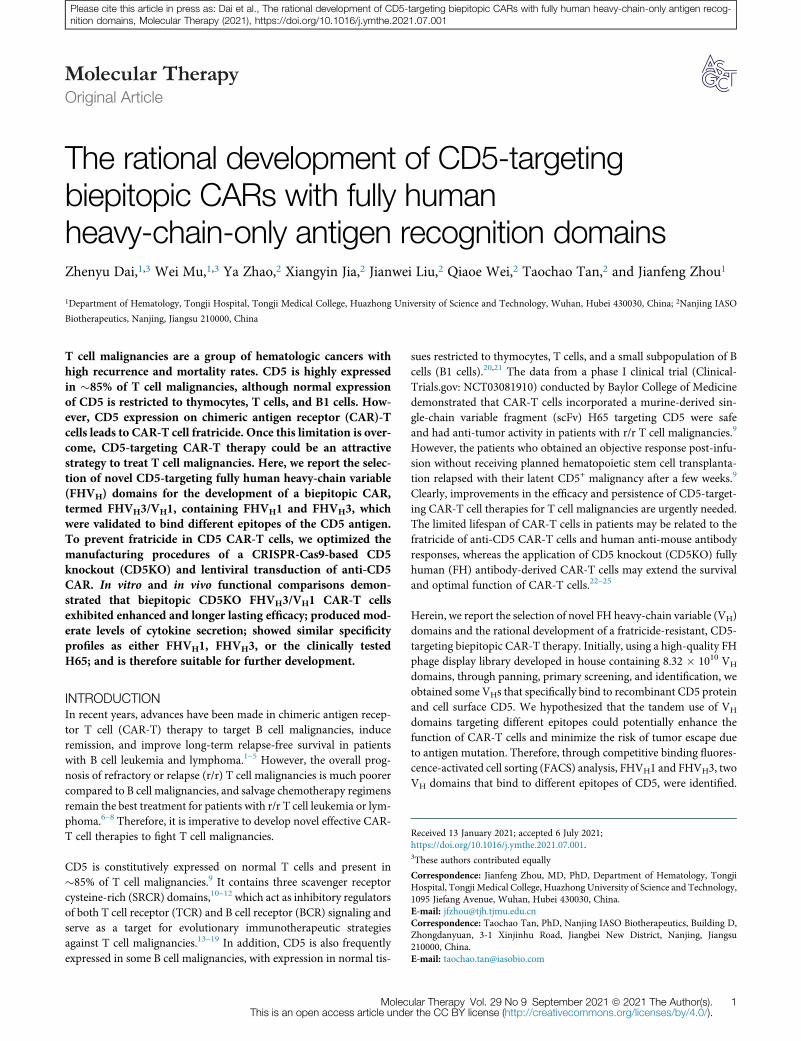
Figure 1. Phage display library screening and selection of fully human (FH) CD5-specific, heavy-chain variable domain (VH)
(A) Schematic of the CD5-specific VH discovery process. (B) Binding activities of representative phage clones and KO7 (negative control phage) are shown. Binding to
different antigens was detected after staining with mouse anti-M13 antibody and horseradish peroxidase (HRP)-goat anti-mouse IgG antibody before reading the optical
density at 450 nm. Mouse anti-CD5 and HRP-goat anti-mouse IgG antibodies were used as the positive control. (C) Phage clones FHVH1–4 bind to Jurkat and CCRF-CEM
(both CD5+) cells, but not to Raji and K562 (both CD5�) cells. Shown is analyzed using flow cytometry.
Molecular Therapy
Please cite this article in press as: Dai et al., The rational development of CD5-targeting biepitopic CARs with fully human heavy-chain-only antigen recog-nition domains, Molecular Therapy (2021), https://doi.org/10.1016/j.ymthe.2021.07.001
Next, to prolong CAR-T cell persistence, we applied a 4-1BB co-stim-ulatory domain in the design of the CAR construct that has been re-ported to possess the capacity to promote CAR-T cell survival26,27 andsubsequently optimized the CD5 knockout and lentiviral transduc-tion steps to successfully generate anti-CD5 CAR-T cells that resistfratricide. Furthermore, the functions of degranulation, cytotoxicity,cytokine release, expansion, and persistence of H65, FHVH1,FHVH3, and biepitopic FHVH3/VH1 CAR-T cells were confirmedin vitro and in vivo. FHVH3/VH1 CAR-T cells exhibited potent andlonger lasting efficacy and specificity profiles similar to those of clin-ically used H65. This biepitopic CAR is currently being prepared forfurther evaluation and development.
RESULTSScreening and selection of FH CD5-specific VH domains
A diagram of the screening and selection procedure is shown in Fig-ure 1A. The VH domains were screened from a phage display librarycomprising both naive and synthetic human VH domains by threerounds of protein panning after enrichment (Table S1). A total of92 monoclonal antibodies were selected for enzyme-linked immuno-sorbent assay (ELISA) and validated using CD5+ Jurkat and CD5�
Raji cells for FACS screening, of which 38 clones specifically bound
2 Molecular Therapy Vol. 29 No 9 September 2021
to CD5 protein, without binding to other control proteins, and therepresentative clones are shown in Figure 1B. After sequencing, 29unique clones were identified. The binding specificity of these 29clones with additional CD5+/� cell lines was further characterizedby FACS analysis. Of these, four clones (FHVH1–4) were confirmedto bind to another human-derived CD5+ cell line (CCRF-CEM; Fig-ure 1C). To further measure their binding affinities to the CD5antigen, these VH clones, along with H65, were expressed in VH/scFv-human immunoglobulin Fc fragment (hFc) or VH/scFv-rabbitimmunoglobulin Fc fragment (rFc) format and purified. Binding af-finity was determined using the Octet96e system. FHVH1 (KD =1.63 � 10�9 M), FHVH2 (KD = 8.32 � 10�9 M), and FHVH3 (KD =1.39� 10�9M) showed slightly lower but comparable binding affinityto CD5 protein compared with H65 (KD = 6.53� 10�10 M), althoughFHVH4 (KD = 2.47 � 10�8 M) exhibited substantially lower affinitythan the above clones (Figure S1).
Identification of VH domains against different epitopes of CD5
and specificity validation of anti-CD5 biepitopic antibodies
A diagram of the anti-CD5 VH domains competitive binding assay isshown (Figure 2A). H65, FHVH1, and FHVH3 CAR-T cells all bind toCD5 antigen, and interestingly, H65-hFc and FHVH3-hFc antibodies
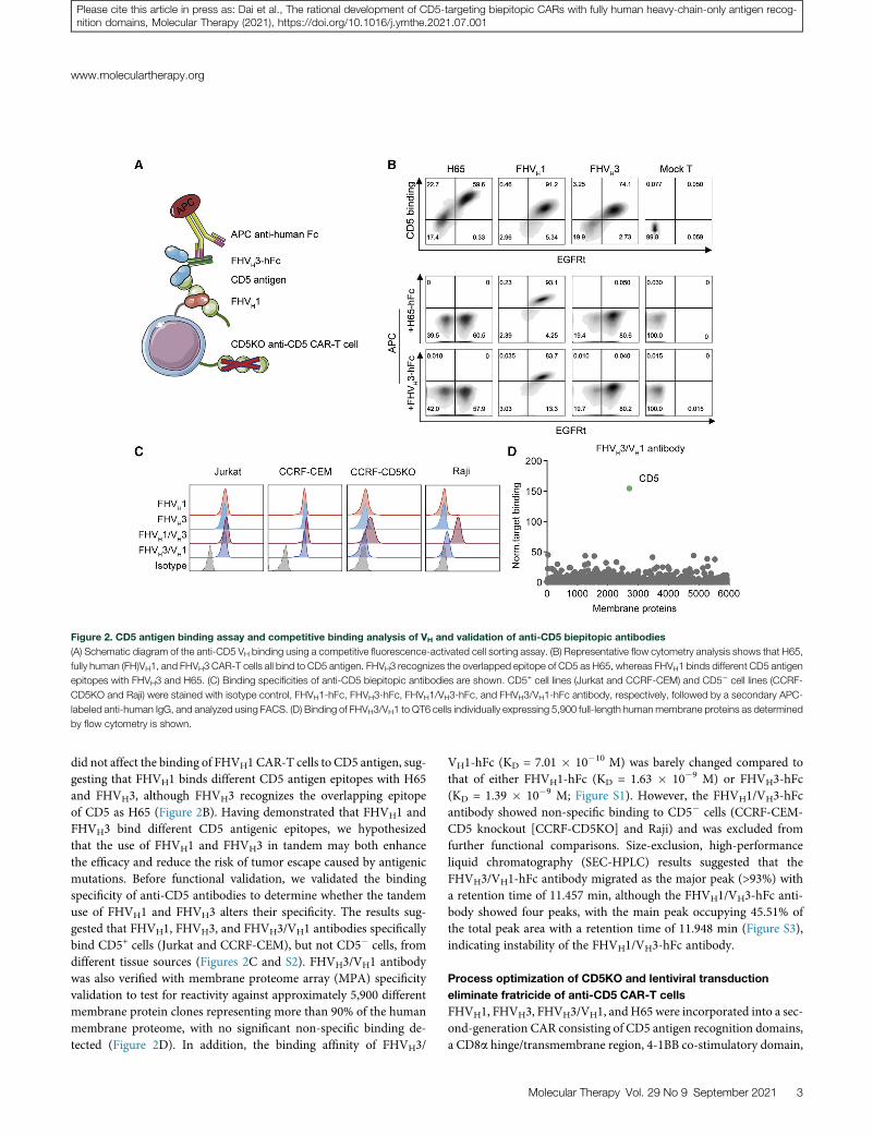
Figure 2. CD5 antigen binding assay and competitive binding analysis of VH and validation of anti-CD5 biepitopic antibodies
(A) Schematic diagram of the anti-CD5 VH binding using a competitive fluorescence-activated cell sorting assay. (B) Representative flow cytometry analysis shows that H65,
fully human (FH)VH1, and FHVH3 CAR-T cells all bind to CD5 antigen. FHVH3 recognizes the overlapped epitope of CD5 as H65, whereas FHVH1 binds different CD5 antigen
epitopes with FHVH3 and H65. (C) Binding specificities of anti-CD5 biepitopic antibodies are shown. CD5+ cell lines (Jurkat and CCRF-CEM) and CD5� cell lines (CCRF-
CD5KO and Raji) were stained with isotype control, FHVH1-hFc, FHVH3-hFc, FHVH1/VH3-hFc, and FHVH3/VH1-hFc antibody, respectively, followed by a secondary APC-
labeled anti-human IgG, and analyzed using FACS. (D) Binding of FHVH3/VH1 to QT6 cells individually expressing 5,900 full-length humanmembrane proteins as determined
by flow cytometry is shown.
www.moleculartherapy.org
Please cite this article in press as: Dai et al., The rational development of CD5-targeting biepitopic CARs with fully human heavy-chain-only antigen recog-nition domains, Molecular Therapy (2021), https://doi.org/10.1016/j.ymthe.2021.07.001
did not affect the binding of FHVH1 CAR-T cells to CD5 antigen, sug-gesting that FHVH1 binds different CD5 antigen epitopes with H65and FHVH3, although FHVH3 recognizes the overlapping epitopeof CD5 as H65 (Figure 2B). Having demonstrated that FHVH1 andFHVH3 bind different CD5 antigenic epitopes, we hypothesizedthat the use of FHVH1 and FHVH3 in tandem may both enhancethe efficacy and reduce the risk of tumor escape caused by antigenicmutations. Before functional validation, we validated the bindingspecificity of anti-CD5 antibodies to determine whether the tandemuse of FHVH1 and FHVH3 alters their specificity. The results sug-gested that FHVH1, FHVH3, and FHVH3/VH1 antibodies specificallybind CD5+ cells (Jurkat and CCRF-CEM), but not CD5� cells, fromdifferent tissue sources (Figures 2C and S2). FHVH3/VH1 antibodywas also verified with membrane proteome array (MPA) specificityvalidation to test for reactivity against approximately 5,900 differentmembrane protein clones representing more than 90% of the humanmembrane proteome, with no significant non-specific binding de-tected (Figure 2D). In addition, the binding affinity of FHVH3/
VH1-hFc (KD = 7.01 � 10�10 M) was barely changed compared tothat of either FHVH1-hFc (KD = 1.63 � 10�9 M) or FHVH3-hFc(KD = 1.39 � 10�9 M; Figure S1). However, the FHVH1/VH3-hFcantibody showed non-specific binding to CD5� cells (CCRF-CEM-CD5 knockout [CCRF-CD5KO] and Raji) and was excluded fromfurther functional comparisons. Size-exclusion, high-performanceliquid chromatography (SEC-HPLC) results suggested that theFHVH3/VH1-hFc antibody migrated as the major peak (>93%) witha retention time of 11.457 min, although the FHVH1/VH3-hFc anti-body showed four peaks, with the main peak occupying 45.51% ofthe total peak area with a retention time of 11.948 min (Figure S3),indicating instability of the FHVH1/VH3-hFc antibody.
Process optimization of CD5KO and lentiviral transduction
eliminate fratricide of anti-CD5 CAR-T cells
FHVH1, FHVH3, FHVH3/VH1, andH65 were incorporated into a sec-ond-generation CAR consisting of CD5 antigen recognition domains,a CD8a hinge/transmembrane region, 4-1BB co-stimulatory domain,
Molecular Therapy Vol. 29 No 9 September 2021 3
(legend on next page)
Molecular Therapy
4 Molecular Therapy Vol. 29 No 9 September 2021
Please cite this article in press as: Dai et al., The rational development of CD5-targeting biepitopic CARs with fully human heavy-chain-only antigen recog-nition domains, Molecular Therapy (2021), https://doi.org/10.1016/j.ymthe.2021.07.001
www.moleculartherapy.org
Please cite this article in press as: Dai et al., The rational development of CD5-targeting biepitopic CARs with fully human heavy-chain-only antigen recog-nition domains, Molecular Therapy (2021), https://doi.org/10.1016/j.ymthe.2021.07.001
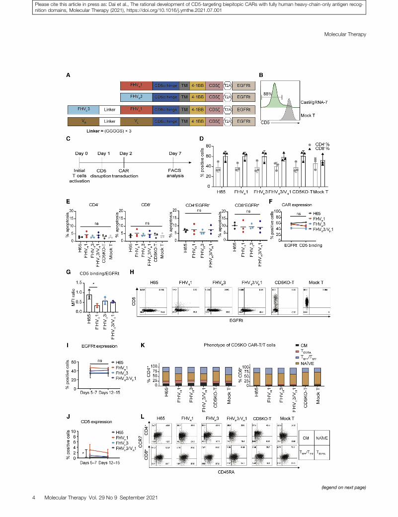
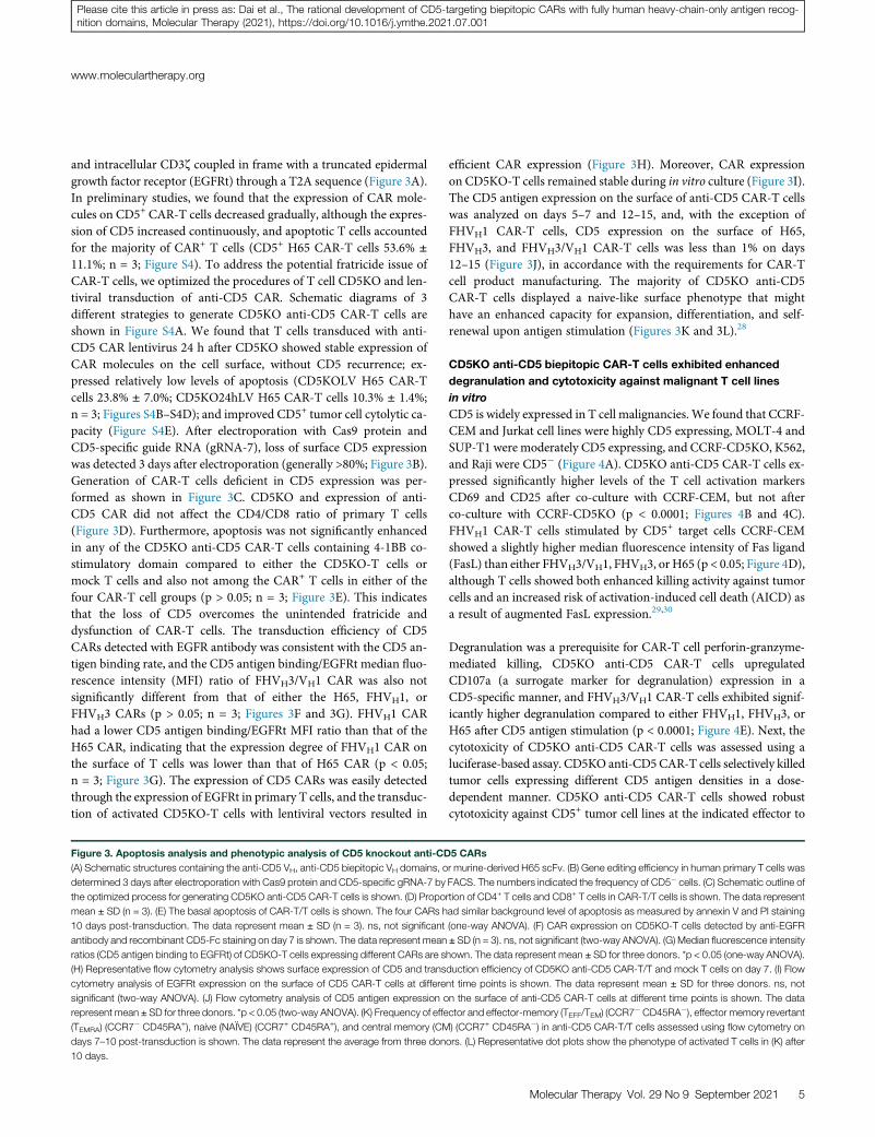
and intracellular CD3z coupled in frame with a truncated epidermalgrowth factor receptor (EGFRt) through a T2A sequence (Figure 3A).In preliminary studies, we found that the expression of CAR mole-cules on CD5+ CAR-T cells decreased gradually, although the expres-sion of CD5 increased continuously, and apoptotic T cells accountedfor the majority of CAR+ T cells (CD5+ H65 CAR-T cells 53.6% ±
11.1%; n = 3; Figure S4). To address the potential fratricide issue ofCAR-T cells, we optimized the procedures of T cell CD5KO and len-tiviral transduction of anti-CD5 CAR. Schematic diagrams of 3different strategies to generate CD5KO anti-CD5 CAR-T cells areshown in Figure S4A. We found that T cells transduced with anti-CD5 CAR lentivirus 24 h after CD5KO showed stable expression ofCAR molecules on the cell surface, without CD5 recurrence; ex-pressed relatively low levels of apoptosis (CD5KOLV H65 CAR-Tcells 23.8% ± 7.0%; CD5KO24hLV H65 CAR-T cells 10.3% ± 1.4%;n = 3; Figures S4B–S4D); and improved CD5+ tumor cell cytolytic ca-pacity (Figure S4E). After electroporation with Cas9 protein andCD5-specific guide RNA (gRNA-7), loss of surface CD5 expressionwas detected 3 days after electroporation (generally >80%; Figure 3B).Generation of CAR-T cells deficient in CD5 expression was per-formed as shown in Figure 3C. CD5KO and expression of anti-CD5 CAR did not affect the CD4/CD8 ratio of primary T cells(Figure 3D). Furthermore, apoptosis was not significantly enhancedin any of the CD5KO anti-CD5 CAR-T cells containing 4-1BB co-stimulatory domain compared to either the CD5KO-T cells ormock T cells and also not among the CAR+ T cells in either of thefour CAR-T cell groups (p > 0.05; n = 3; Figure 3E). This indicatesthat the loss of CD5 overcomes the unintended fratricide anddysfunction of CAR-T cells. The transduction efficiency of CD5CARs detected with EGFR antibody was consistent with the CD5 an-tigen binding rate, and the CD5 antigen binding/EGFRt median fluo-rescence intensity (MFI) ratio of FHVH3/VH1 CAR was also notsignificantly different from that of either the H65, FHVH1, orFHVH3 CARs (p > 0.05; n = 3; Figures 3F and 3G). FHVH1 CARhad a lower CD5 antigen binding/EGFRt MFI ratio than that of theH65 CAR, indicating that the expression degree of FHVH1 CAR onthe surface of T cells was lower than that of H65 CAR (p < 0.05;n = 3; Figure 3G). The expression of CD5 CARs was easily detectedthrough the expression of EGFRt in primary T cells, and the transduc-tion of activated CD5KO-T cells with lentiviral vectors resulted in
Figure 3. Apoptosis analysis and phenotypic analysis of CD5 knockout anti-CD
(A) Schematic structures containing the anti-CD5 VH, anti-CD5 biepitopic VH domains, o
determined 3 days after electroporation with Cas9 protein and CD5-specific gRNA-7 by
the optimized process for generating CD5KO anti-CD5 CAR-T cells is shown. (D) Propo
mean ± SD (n = 3). (E) The basal apoptosis of CAR-T/T cells is shown. The four CARs h
10 days post-transduction. The data represent mean ± SD (n = 3). ns, not significant
antibody and recombinant CD5-Fc staining on day 7 is shown. The data represent mean
ratios (CD5 antigen binding to EGFRt) of CD5KO-T cells expressing different CARs are s
(H) Representative flow cytometry analysis shows surface expression of CD5 and trans
cytometry analysis of EGFRt expression on the surface of CD5 CAR-T cells at differe
significant (two-way ANOVA). (J) Flow cytometry analysis of CD5 antigen expression o
represent mean ± SD for three donors. *p < 0.05 (two-way ANOVA). (K) Frequency of effe
(TEMRA) (CCR7� CD45RA+), naive (NAIVE) (CCR7+ CD45RA+), and central memory (CM
days 7–10 post-transduction is shown. The data represent the average from three don
10 days.
efficient CAR expression (Figure 3H). Moreover, CAR expressionon CD5KO-T cells remained stable during in vitro culture (Figure 3I).The CD5 antigen expression on the surface of anti-CD5 CAR-T cellswas analyzed on days 5–7 and 12–15, and, with the exception ofFHVH1 CAR-T cells, CD5 expression on the surface of H65,FHVH3, and FHVH3/VH1 CAR-T cells was less than 1% on days12–15 (Figure 3J), in accordance with the requirements for CAR-Tcell product manufacturing. The majority of CD5KO anti-CD5CAR-T cells displayed a naive-like surface phenotype that mighthave an enhanced capacity for expansion, differentiation, and self-renewal upon antigen stimulation (Figures 3K and 3L).28
CD5KO anti-CD5 biepitopic CAR-T cells exhibited enhanced
degranulation and cytotoxicity against malignant T cell lines
in vitro
CD5 is widely expressed in T cell malignancies. We found that CCRF-CEM and Jurkat cell lines were highly CD5 expressing, MOLT-4 andSUP-T1 were moderately CD5 expressing, and CCRF-CD5KO, K562,and Raji were CD5� (Figure 4A). CD5KO anti-CD5 CAR-T cells ex-pressed significantly higher levels of the T cell activation markersCD69 and CD25 after co-culture with CCRF-CEM, but not afterco-culture with CCRF-CD5KO (p < 0.0001; Figures 4B and 4C).FHVH1 CAR-T cells stimulated by CD5+ target cells CCRF-CEMshowed a slightly higher median fluorescence intensity of Fas ligand(FasL) than either FHVH3/VH1, FHVH3, or H65 (p < 0.05; Figure 4D),although T cells showed both enhanced killing activity against tumorcells and an increased risk of activation-induced cell death (AICD) asa result of augmented FasL expression.29,30
Degranulation was a prerequisite for CAR-T cell perforin-granzyme-mediated killing, CD5KO anti-CD5 CAR-T cells upregulatedCD107a (a surrogate marker for degranulation) expression in aCD5-specific manner, and FHVH3/VH1 CAR-T cells exhibited signif-icantly higher degranulation compared to either FHVH1, FHVH3, orH65 after CD5 antigen stimulation (p < 0.0001; Figure 4E). Next, thecytotoxicity of CD5KO anti-CD5 CAR-T cells was assessed using aluciferase-based assay. CD5KO anti-CD5CAR-T cells selectively killedtumor cells expressing different CD5 antigen densities in a dose-dependent manner. CD5KO anti-CD5 CAR-T cells showed robustcytotoxicity against CD5+ tumor cell lines at the indicated effector to
5 CARs
r murine-derived H65 scFv. (B) Gene editing efficiency in human primary T cells was
FACS. The numbers indicated the frequency of CD5� cells. (C) Schematic outline of
rtion of CD4+ T cells and CD8+ T cells in CAR-T/T cells is shown. The data represent
ad similar background level of apoptosis as measured by annexin V and PI staining
(one-way ANOVA). (F) CAR expression on CD5KO-T cells detected by anti-EGFR
± SD (n = 3). ns, not significant (two-way ANOVA). (G) Median fluorescence intensity
hown. The data represent mean ± SD for three donors. *p < 0.05 (one-way ANOVA).
duction efficiency of CD5KO anti-CD5 CAR-T/T and mock T cells on day 7. (I) Flow
nt time points is shown. The data represent mean ± SD for three donors. ns, not
n the surface of anti-CD5 CAR-T cells at different time points is shown. The data
ctor and effector-memory (TEFF/TEM) (CCR7�CD45RA�), effector memory revertant
) (CCR7+ CD45RA�) in anti-CD5 CAR-T/T cells assessed using flow cytometry on
ors. (L) Representative dot plots show the phenotype of activated T cells in (K) after
Molecular Therapy Vol. 29 No 9 September 2021 5
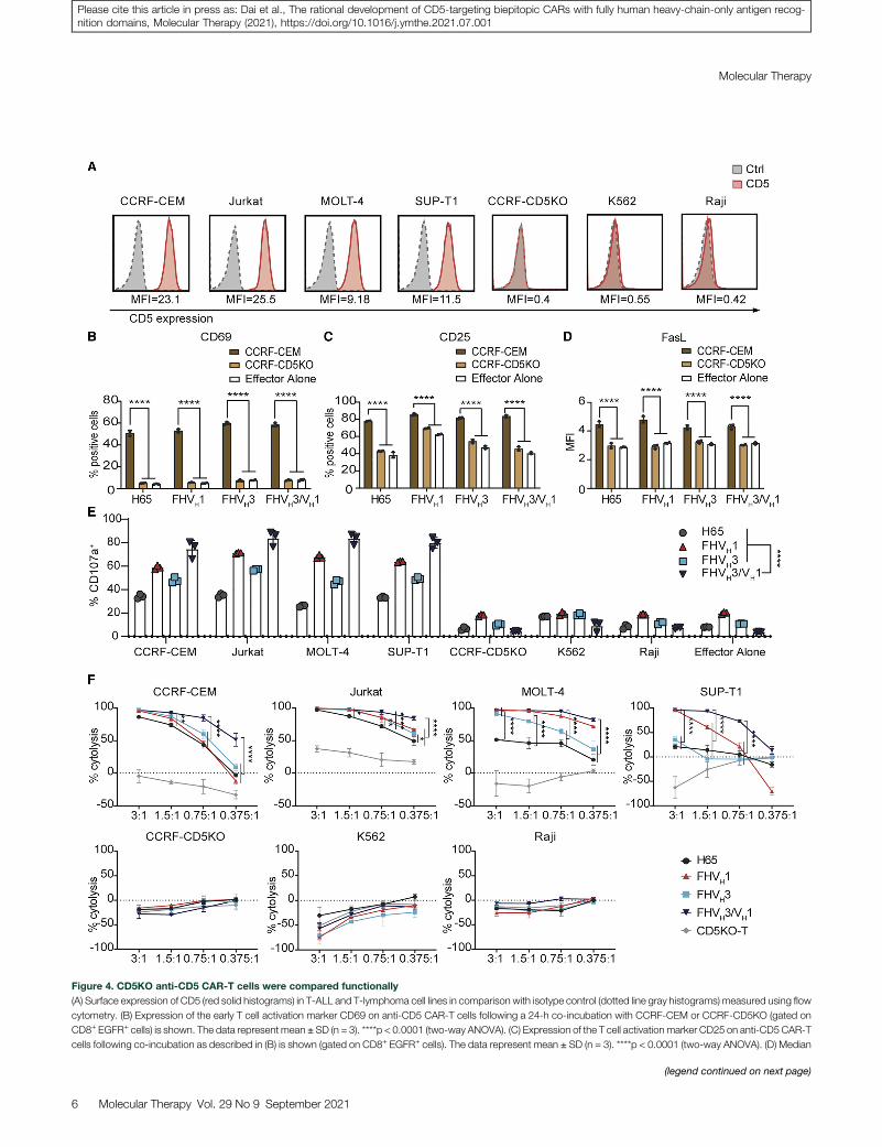
Figure 4. CD5KO anti-CD5 CAR-T cells were compared functionally
(A) Surface expression of CD5 (red solid histograms) in T-ALL and T-lymphoma cell lines in comparison with isotype control (dotted line gray histograms) measured using flow
cytometry. (B) Expression of the early T cell activation marker CD69 on anti-CD5 CAR-T cells following a 24-h co-incubation with CCRF-CEM or CCRF-CD5KO (gated on
CD8+ EGFR+ cells) is shown. The data represent mean ± SD (n = 3). ****p < 0.0001 (two-way ANOVA). (C) Expression of the T cell activation marker CD25 on anti-CD5 CAR-T
cells following co-incubation as described in (B) is shown (gated on CD8+ EGFR+ cells). The data represent mean ± SD (n = 3). ****p < 0.0001 (two-way ANOVA). (D) Median
(legend continued on next page)
Molecular Therapy
6 Molecular Therapy Vol. 29 No 9 September 2021
Please cite this article in press as: Dai et al., The rational development of CD5-targeting biepitopic CARs with fully human heavy-chain-only antigen recog-nition domains, Molecular Therapy (2021), https://doi.org/10.1016/j.ymthe.2021.07.001
www.moleculartherapy.org
Please cite this article in press as: Dai et al., The rational development of CD5-targeting biepitopic CARs with fully human heavy-chain-only antigen recog-nition domains, Molecular Therapy (2021), https://doi.org/10.1016/j.ymthe.2021.07.001
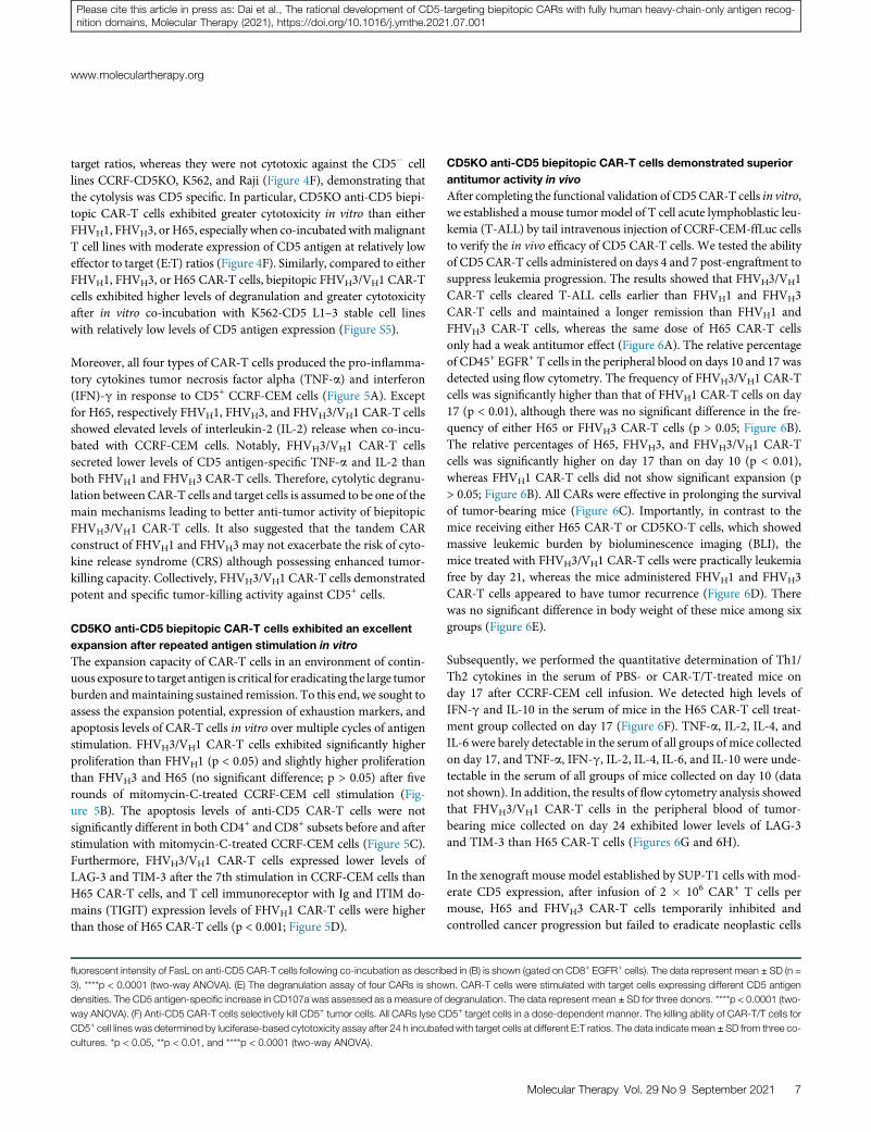
target ratios, whereas they were not cytotoxic against the CD5� celllines CCRF-CD5KO, K562, and Raji (Figure 4F), demonstrating thatthe cytolysis was CD5 specific. In particular, CD5KO anti-CD5 biepi-topic CAR-T cells exhibited greater cytotoxicity in vitro than eitherFHVH1, FHVH3, orH65, especially when co-incubated withmalignantT cell lines with moderate expression of CD5 antigen at relatively loweffector to target (E:T) ratios (Figure 4F). Similarly, compared to eitherFHVH1, FHVH3, or H65 CAR-T cells, biepitopic FHVH3/VH1 CAR-Tcells exhibited higher levels of degranulation and greater cytotoxicityafter in vitro co-incubation with K562-CD5 L1–3 stable cell lineswith relatively low levels of CD5 antigen expression (Figure S5).
Moreover, all four types of CAR-T cells produced the pro-inflamma-tory cytokines tumor necrosis factor alpha (TNF-a) and interferon(IFN)-g in response to CD5+ CCRF-CEM cells (Figure 5A). Exceptfor H65, respectively FHVH1, FHVH3, and FHVH3/VH1 CAR-T cellsshowed elevated levels of interleukin-2 (IL-2) release when co-incu-bated with CCRF-CEM cells. Notably, FHVH3/VH1 CAR-T cellssecreted lower levels of CD5 antigen-specific TNF-a and IL-2 thanboth FHVH1 and FHVH3 CAR-T cells. Therefore, cytolytic degranu-lation between CAR-T cells and target cells is assumed to be one of themain mechanisms leading to better anti-tumor activity of biepitopicFHVH3/VH1 CAR-T cells. It also suggested that the tandem CARconstruct of FHVH1 and FHVH3 may not exacerbate the risk of cyto-kine release syndrome (CRS) although possessing enhanced tumor-killing capacity. Collectively, FHVH3/VH1 CAR-T cells demonstratedpotent and specific tumor-killing activity against CD5+ cells.
CD5KO anti-CD5 biepitopic CAR-T cells exhibited an excellent
expansion after repeated antigen stimulation in vitro
The expansion capacity of CAR-T cells in an environment of contin-uous exposure to target antigen is critical for eradicating the large tumorburden andmaintaining sustained remission. To this end, we sought toassess the expansion potential, expression of exhaustion markers, andapoptosis levels of CAR-T cells in vitro over multiple cycles of antigenstimulation. FHVH3/VH1 CAR-T cells exhibited significantly higherproliferation than FHVH1 (p < 0.05) and slightly higher proliferationthan FHVH3 and H65 (no significant difference; p > 0.05) after fiverounds of mitomycin-C-treated CCRF-CEM cell stimulation (Fig-ure 5B). The apoptosis levels of anti-CD5 CAR-T cells were notsignificantly different in both CD4+ and CD8+ subsets before and afterstimulation with mitomycin-C-treated CCRF-CEM cells (Figure 5C).Furthermore, FHVH3/VH1 CAR-T cells expressed lower levels ofLAG-3 and TIM-3 after the 7th stimulation in CCRF-CEM cells thanH65 CAR-T cells, and T cell immunoreceptor with Ig and ITIM do-mains (TIGIT) expression levels of FHVH1 CAR-T cells were higherthan those of H65 CAR-T cells (p < 0.001; Figure 5D).
fluorescent intensity of FasL on anti-CD5 CAR-T cells following co-incubation as describ
3). ****p < 0.0001 (two-way ANOVA). (E) The degranulation assay of four CARs is show
densities. The CD5 antigen-specific increase in CD107a was assessed as a measure of
way ANOVA). (F) Anti-CD5 CAR-T cells selectively kill CD5+ tumor cells. All CARs lyse C
CD5+ cell lines was determined by luciferase-based cytotoxicity assay after 24 h incubate
cultures. *p < 0.05, **p < 0.01, and ****p < 0.0001 (two-way ANOVA).
CD5KO anti-CD5 biepitopic CAR-T cells demonstrated superior
antitumor activity in vivo
After completing the functional validation of CD5CAR-T cells in vitro,we established a mouse tumor model of T cell acute lymphoblastic leu-kemia (T-ALL) by tail intravenous injection of CCRF-CEM-ffLuc cellsto verify the in vivo efficacy of CD5 CAR-T cells. We tested the abilityof CD5 CAR-T cells administered on days 4 and 7 post-engraftment tosuppress leukemia progression. The results showed that FHVH3/VH1CAR-T cells cleared T-ALL cells earlier than FHVH1 and FHVH3CAR-T cells and maintained a longer remission than FHVH1 andFHVH3 CAR-T cells, whereas the same dose of H65 CAR-T cellsonly had a weak antitumor effect (Figure 6A). The relative percentageof CD45+ EGFR+ T cells in the peripheral blood on days 10 and 17 wasdetected using flow cytometry. The frequency of FHVH3/VH1 CAR-Tcells was significantly higher than that of FHVH1 CAR-T cells on day17 (p < 0.01), although there was no significant difference in the fre-quency of either H65 or FHVH3 CAR-T cells (p > 0.05; Figure 6B).The relative percentages of H65, FHVH3, and FHVH3/VH1 CAR-Tcells was significantly higher on day 17 than on day 10 (p < 0.01),whereas FHVH1 CAR-T cells did not show significant expansion (p> 0.05; Figure 6B). All CARs were effective in prolonging the survivalof tumor-bearing mice (Figure 6C). Importantly, in contrast to themice receiving either H65 CAR-T or CD5KO-T cells, which showedmassive leukemic burden by bioluminescence imaging (BLI), themice treated with FHVH3/VH1 CAR-T cells were practically leukemiafree by day 21, whereas the mice administered FHVH1 and FHVH3CAR-T cells appeared to have tumor recurrence (Figure 6D). Therewas no significant difference in body weight of these mice among sixgroups (Figure 6E).
Subsequently, we performed the quantitative determination of Th1/Th2 cytokines in the serum of PBS- or CAR-T/T-treated mice onday 17 after CCRF-CEM cell infusion. We detected high levels ofIFN-g and IL-10 in the serum of mice in the H65 CAR-T cell treat-ment group collected on day 17 (Figure 6F). TNF-a, IL-2, IL-4, andIL-6 were barely detectable in the serum of all groups of mice collectedon day 17, and TNF-a, IFN-g, IL-2, IL-4, IL-6, and IL-10 were unde-tectable in the serum of all groups of mice collected on day 10 (datanot shown). In addition, the results of flow cytometry analysis showedthat FHVH3/VH1 CAR-T cells in the peripheral blood of tumor-bearing mice collected on day 24 exhibited lower levels of LAG-3and TIM-3 than H65 CAR-T cells (Figures 6G and 6H).
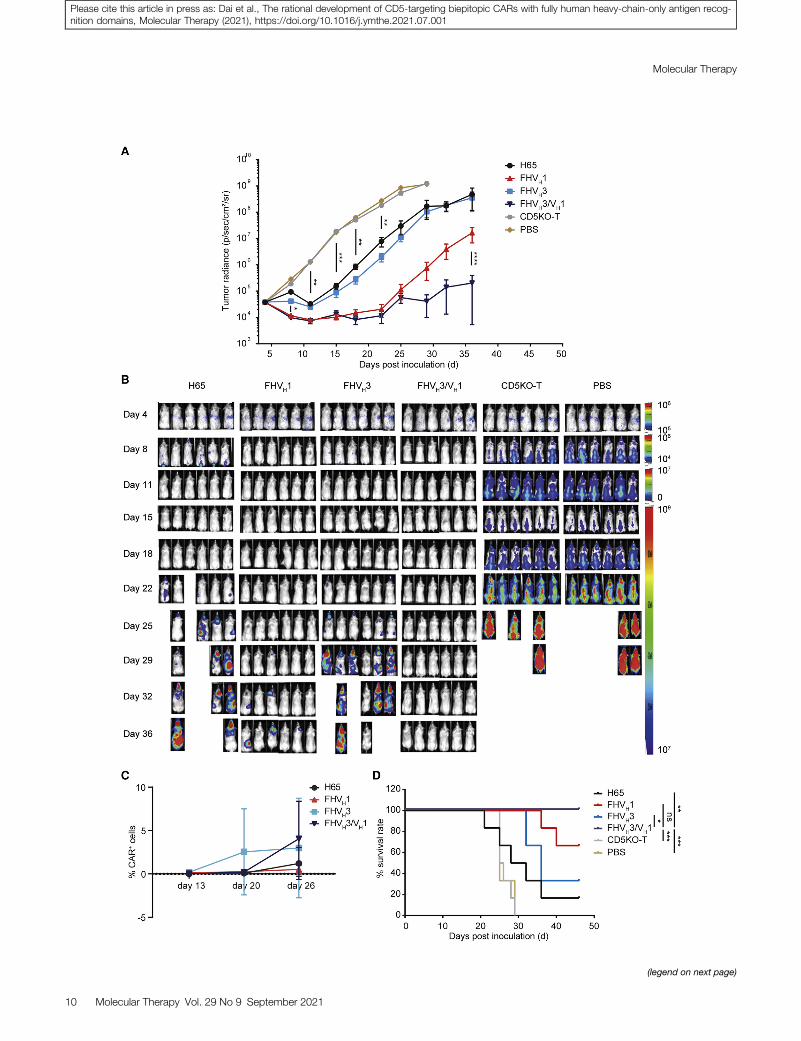
In the xenograft mouse model established by SUP-T1 cells with mod-erate CD5 expression, after infusion of 2 � 106 CAR+ T cells permouse, H65 and FHVH3 CAR-T cells temporarily inhibited andcontrolled cancer progression but failed to eradicate neoplastic cells
ed in (B) is shown (gated on CD8+ EGFR+ cells). The data represent mean ± SD (n =
n. CAR-T cells were stimulated with target cells expressing different CD5 antigen
degranulation. The data represent mean ± SD for three donors. ****p < 0.0001 (two-
D5+ target cells in a dose-dependent manner. The killing ability of CAR-T/T cells for
d with target cells at different E:T ratios. The data indicate mean ± SD from three co-
Molecular Therapy Vol. 29 No 9 September 2021 7
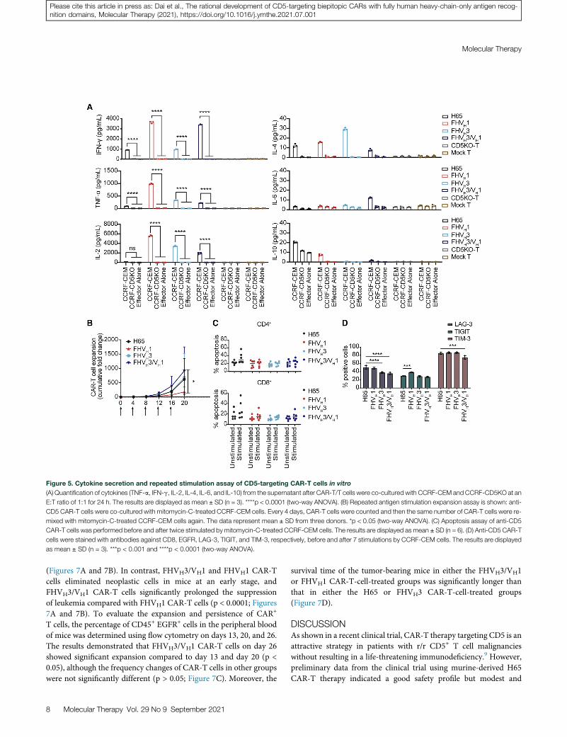
Figure 5. Cytokine secretion and repeated stimulation assay of CD5-targeting CAR-T cells in vitro
(A) Quantification of cytokines (TNF-a, IFN-g, IL-2, IL-4, IL-6, and IL-10) from the supernatant after CAR-T/T cells were co-cultured with CCRF-CEM and CCRF-CD5KO at an
E:T ratio of 1:1 for 24 h. The results are displayed as mean ± SD (n = 3). ****p < 0.0001 (two-way ANOVA). (B) Repeated antigen stimulation expansion assay is shown: anti-
CD5 CAR-T cells were co-cultured with mitomycin-C-treated CCRF-CEM cells. Every 4 days, CAR-T cells were counted and then the same number of CAR-T cells were re-
mixed with mitomycin-C-treated CCRF-CEM cells again. The data represent mean ± SD from three donors. *p < 0.05 (two-way ANOVA). (C) Apoptosis assay of anti-CD5
CAR-T cells was performed before and after twice stimulated by mitomycin-C-treated CCRF-CEM cells. The results are displayed as mean ± SD (n = 6). (D) Anti-CD5 CAR-T
cells were stained with antibodies against CD8, EGFR, LAG-3, TIGIT, and TIM-3, respectively, before and after 7 stimulations by CCRF-CEM cells. The results are displayed
as mean ± SD (n = 3). ***p < 0.001 and ****p < 0.0001 (two-way ANOVA).
Molecular Therapy
Please cite this article in press as: Dai et al., The rational development of CD5-targeting biepitopic CARs with fully human heavy-chain-only antigen recog-nition domains, Molecular Therapy (2021), https://doi.org/10.1016/j.ymthe.2021.07.001
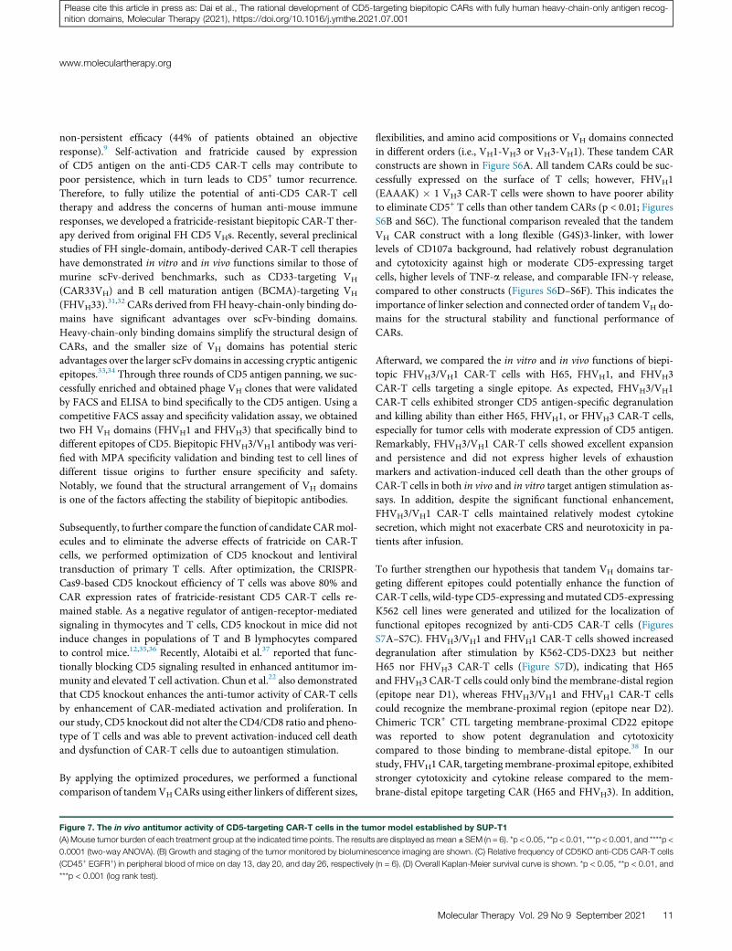
(Figures 7A and 7B). In contrast, FHVH3/VH1 and FHVH1 CAR-Tcells eliminated neoplastic cells in mice at an early stage, andFHVH3/VH1 CAR-T cells significantly prolonged the suppressionof leukemia compared with FHVH1 CAR-T cells (p < 0.0001; Figures7A and 7B). To evaluate the expansion and persistence of CAR+
T cells, the percentage of CD45+ EGFR+ cells in the peripheral bloodof mice was determined using flow cytometry on days 13, 20, and 26.The results demonstrated that FHVH3/VH1 CAR-T cells on day 26showed significant expansion compared to day 13 and day 20 (p <0.05), although the frequency changes of CAR-T cells in other groupswere not significantly different (p > 0.05; Figure 7C). Moreover, the
8 Molecular Therapy Vol. 29 No 9 September 2021
survival time of the tumor-bearing mice in either the FHVH3/VH1or FHVH1 CAR-T-cell-treated groups was significantly longer thanthat in either the H65 or FHVH3 CAR-T-cell-treated groups(Figure 7D).
DISCUSSIONAs shown in a recent clinical trial, CAR-T therapy targeting CD5 is anattractive strategy in patients with r/r CD5+ T cell malignancieswithout resulting in a life-threatening immunodeficiency.9 However,preliminary data from the clinical trial using murine-derived H65CAR-T therapy indicated a good safety profile but modest and
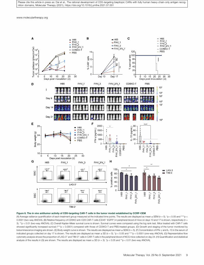
Figure 6. The in vivo antitumor activity of CD5-targeting CAR-T cells in the tumor model established by CCRF-CEM
(A) Average radiance quantification of each treatment group measured at the indicated time points. The results are displayed as mean ± SEM (n = 5). *p < 0.05 and ****p <
0.0001 (two-way ANOVA). (B) Relative frequency of CD5KO anti-CD5 CAR-T cells (CD45+ EGFR+) in peripheral blood of mice on days 10 and 17 is shown, respectively (n =
3). **p < 0.01 (two-way ANOVA). (C) Overall Kaplan-Meier survival curve is shown. Survival curves were compared using the log rank test. Mice treated with CAR-T cells
showed significantly increased survival (****p < 0.0001) compared with those of CD5KO-T and PBS-treated groups. (D) Growth and staging of the tumor monitored by
bioluminescence imaging are shown. (E) Body weight curve is shown. The results are displayed as mean ± SEM (n = 5). (F) Concentration of IFN-g and IL-10 in the serum of
indicated groups collected on day 17 is shown. The results are displayed as mean ± SD (n = 5). *p < 0.05 and ****p < 0.0001 (one-way ANOVA). (G) Representative flow
cytometry analysis shows the proportion of LAG-3+ and TIM-3+ cells in CAR-T cells in the peripheral blood of NCGmice collected on day 24. (H) Quantification and statistical
analysis of the results in (G) are shown. The results are displayed as mean ± SD (n = 3). *p < 0.05 and **p < 0.01 (two-way ANOVA).
www.moleculartherapy.org
Molecular Therapy Vol. 29 No 9 September 2021 9
Please cite this article in press as: Dai et al., The rational development of CD5-targeting biepitopic CARs with fully human heavy-chain-only antigen recog-nition domains, Molecular Therapy (2021), https://doi.org/10.1016/j.ymthe.2021.07.001
(legend on next page)
Molecular Therapy
10 Molecular Therapy Vol. 29 No 9 September 2021
Please cite this article in press as: Dai et al., The rational development of CD5-targeting biepitopic CARs with fully human heavy-chain-only antigen recog-nition domains, Molecular Therapy (2021), https://doi.org/10.1016/j.ymthe.2021.07.001
www.moleculartherapy.org
Please cite this article in press as: Dai et al., The rational development of CD5-targeting biepitopic CARs with fully human heavy-chain-only antigen recog-nition domains, Molecular Therapy (2021), https://doi.org/10.1016/j.ymthe.2021.07.001
non-persistent efficacy (44% of patients obtained an objectiveresponse).9 Self-activation and fratricide caused by expressionof CD5 antigen on the anti-CD5 CAR-T cells may contribute topoor persistence, which in turn leads to CD5+ tumor recurrence.Therefore, to fully utilize the potential of anti-CD5 CAR-T celltherapy and address the concerns of human anti-mouse immuneresponses, we developed a fratricide-resistant biepitopic CAR-T ther-apy derived from original FH CD5 VHs. Recently, several preclinicalstudies of FH single-domain, antibody-derived CAR-T cell therapieshave demonstrated in vitro and in vivo functions similar to those ofmurine scFv-derived benchmarks, such as CD33-targeting VH
(CAR33VH) and B cell maturation antigen (BCMA)-targeting VH
(FHVH33).31,32 CARs derived from FH heavy-chain-only binding do-
mains have significant advantages over scFv-binding domains.Heavy-chain-only binding domains simplify the structural design ofCARs, and the smaller size of VH domains has potential stericadvantages over the larger scFv domains in accessing cryptic antigenicepitopes.33,34 Through three rounds of CD5 antigen panning, we suc-cessfully enriched and obtained phage VH clones that were validatedby FACS and ELISA to bind specifically to the CD5 antigen. Using acompetitive FACS assay and specificity validation assay, we obtainedtwo FH VH domains (FHVH1 and FHVH3) that specifically bind todifferent epitopes of CD5. Biepitopic FHVH3/VH1 antibody was veri-fied with MPA specificity validation and binding test to cell lines ofdifferent tissue origins to further ensure specificity and safety.Notably, we found that the structural arrangement of VH domainsis one of the factors affecting the stability of biepitopic antibodies.
Subsequently, to further compare the function of candidate CARmol-ecules and to eliminate the adverse effects of fratricide on CAR-Tcells, we performed optimization of CD5 knockout and lentiviraltransduction of primary T cells. After optimization, the CRISPR-Cas9-based CD5 knockout efficiency of T cells was above 80% andCAR expression rates of fratricide-resistant CD5 CAR-T cells re-mained stable. As a negative regulator of antigen-receptor-mediatedsignaling in thymocytes and T cells, CD5 knockout in mice did notinduce changes in populations of T and B lymphocytes comparedto control mice.12,35,36 Recently, Alotaibi et al.37 reported that func-tionally blocking CD5 signaling resulted in enhanced antitumor im-munity and elevated T cell activation. Chun et al.22 also demonstratedthat CD5 knockout enhances the anti-tumor activity of CAR-T cellsby enhancement of CAR-mediated activation and proliferation. Inour study, CD5 knockout did not alter the CD4/CD8 ratio and pheno-type of T cells and was able to prevent activation-induced cell deathand dysfunction of CAR-T cells due to autoantigen stimulation.
By applying the optimized procedures, we performed a functionalcomparison of tandemVHCARs using either linkers of different sizes,
Figure 7. The in vivo antitumor activity of CD5-targeting CAR-T cells in the tum
(A) Mouse tumor burden of each treatment group at the indicated time points. The results
0.0001 (two-way ANOVA). (B) Growth and staging of the tumor monitored by biolumine
(CD45+ EGFR+) in peripheral blood of mice on day 13, day 20, and day 26, respectively
***p < 0.001 (log rank test).
flexibilities, and amino acid compositions or VH domains connectedin different orders (i.e., VH1-VH3 or VH3-VH1). These tandem CARconstructs are shown in Figure S6A. All tandem CARs could be suc-cessfully expressed on the surface of T cells; however, FHVH1(EAAAK) � 1 VH3 CAR-T cells were shown to have poorer abilityto eliminate CD5+ T cells than other tandem CARs (p < 0.01; FiguresS6B and S6C). The functional comparison revealed that the tandemVH CAR construct with a long flexible (G4S)3-linker, with lowerlevels of CD107a background, had relatively robust degranulationand cytotoxicity against high or moderate CD5-expressing targetcells, higher levels of TNF-a release, and comparable IFN-g release,compared to other constructs (Figures S6D–S6F). This indicates theimportance of linker selection and connected order of tandem VH do-mains for the structural stability and functional performance ofCARs.
Afterward, we compared the in vitro and in vivo functions of biepi-topic FHVH3/VH1 CAR-T cells with H65, FHVH1, and FHVH3CAR-T cells targeting a single epitope. As expected, FHVH3/VH1CAR-T cells exhibited stronger CD5 antigen-specific degranulationand killing ability than either H65, FHVH1, or FHVH3 CAR-T cells,especially for tumor cells with moderate expression of CD5 antigen.Remarkably, FHVH3/VH1 CAR-T cells showed excellent expansionand persistence and did not express higher levels of exhaustionmarkers and activation-induced cell death than the other groups ofCAR-T cells in both in vivo and in vitro target antigen stimulation as-says. In addition, despite the significant functional enhancement,FHVH3/VH1 CAR-T cells maintained relatively modest cytokinesecretion, which might not exacerbate CRS and neurotoxicity in pa-tients after infusion.
To further strengthen our hypothesis that tandem VH domains tar-geting different epitopes could potentially enhance the function ofCAR-T cells, wild-type CD5-expressing andmutated CD5-expressingK562 cell lines were generated and utilized for the localization offunctional epitopes recognized by anti-CD5 CAR-T cells (FiguresS7A–S7C). FHVH3/VH1 and FHVH1 CAR-T cells showed increaseddegranulation after stimulation by K562-CD5-DX23 but neitherH65 nor FHVH3 CAR-T cells (Figure S7D), indicating that H65and FHVH3 CAR-T cells could only bind the membrane-distal region(epitope near D1), whereas FHVH3/VH1 and FHVH1 CAR-T cellscould recognize the membrane-proximal region (epitope near D2).Chimeric TCR+ CTL targeting membrane-proximal CD22 epitopewas reported to show potent degranulation and cytotoxicitycompared to those binding to membrane-distal epitope.38 In ourstudy, FHVH1CAR, targetingmembrane-proximal epitope, exhibitedstronger cytotoxicity and cytokine release compared to the mem-brane-distal epitope targeting CAR (H65 and FHVH3). In addition,
or model established by SUP-T1
are displayed asmean ± SEM (n = 6). *p < 0.05, **p < 0.01, ***p < 0.001, and ****p <
scence imaging are shown. (C) Relative frequency of CD5KO anti-CD5 CAR-T cells
(n = 6). (D) Overall Kaplan-Meier survival curve is shown. *p < 0.05, **p < 0.01, and
Molecular Therapy Vol. 29 No 9 September 2021 11
Molecular Therapy
Please cite this article in press as: Dai et al., The rational development of CD5-targeting biepitopic CARs with fully human heavy-chain-only antigen recog-nition domains, Molecular Therapy (2021), https://doi.org/10.1016/j.ymthe.2021.07.001
stimulation with K562-CD5-D123 resulted in higher levels ofFHVH3/VH1 CAR-T cell degranulation compared to stimulationwith K562-CD5-DX23 (p < 0.0001; Figure S7D), reflecting a signifi-cant synergistic effect. Xu et al.39 previously reported the developmentof a biepitopic llamas-derived BCMA-targeting CAR-T therapy andapplied this system to achieve a better objective response rate, com-plete remission rate, and lower relapse rate than monovalentBCMA CAR-T therapy in clinical trials.40 With a similar conceptualbasis and excellent preclinical results, we speculate that FHVH3/VH1CAR-T cells could also demonstrate favorable efficacy and safety infuture clinical studies. Moreover, due to its superior efficacy andpotentially lower immunogenicity, the FHVH3/VH1 CAR may alsobe of interest when used for “off-the-shelf” universal anti-CD5CAR-T/CAR-NK therapy.
CAR-T therapy targeting CD5 has good clinical application prospectsbeyond just in the treatment of T cell malignancies; with high expres-sion on a subset of B cell malignancies, it can also be used to treat B cellmalignancies, such as mantle cell lymphoma, diffuse large B cell lym-phoma, and chronic lymphocytic leukemia or small-cell lymphocyticlymphoma.41–43 Furthermore, application of CAR-T therapy to targetmultiple antigens simultaneously is an attractive strategy for treatmentand prevention of antigen-loss relapses, and VH binding domainsfurthermore ease the design of multispecific CARs.44–47 The use oftwo VH domains can simplify the design of bispecific CAR constructscapable of recognizing different antigens compared to the design utiliz-ing two scFv domains. Therefore, FHVH1 and FHVH3 are suitable forfurther use in the design of bispecific or even multispecific CAR struc-tures to solve critical problems in current cancer drug development,such as clonal heterogeneity and antigen escape.
MATERIALS AND METHODSCell lines
CD5+ cell lines, Jurkat (acute T cell leukemia), CCRF-CEM (acute Tlymphoblastic leukemia), MOLT-4 (acute T lymphoblastic leukemia),SUP-T1 (T cell lymphoblastic lymphoma), and the CD5� cell lines,K562 (chronic myelogenous leukemia), Raji (Burkitt’s lymphoma),and NALM-6 (acute B-lymphocytic leukemia), were cultured inRPMI-1640 medium containing 10% fetal bovine serum (FBS)(Thermo Fisher Scientific,Waltham,MA, USA). QT6 (fibrosarcoma),HCT-116 (colorectal carcinoma), HEPG2 (hepatocellular carci-noma), MDA-MB-468 (breast adenocarcinoma), OVCAR3 (ovarianadenocarcinoma), NCI-H460 (large cell lung cancer), 293CT/293T(embryonic kidney), KATO III (gastric carcinoma), and PANC-1(pancreatic epithelioid carcinoma) were cultured in DMEM (Corn-ing, Corning, NY, USA) medium containing 10% FBS (ThermoFisher Scientific). All cell lines were verified before use. CCRF-CD5KO is a human CD5 knockout cell line generated from CCRF-CEM by CRISPR-Cas9. K562-CD5 L1–5 are stable cell lines withvarying degrees of expression of human CD5 molecules, as deter-mined by flow cytometry sorting. K562-CD5-D123 expresses all threeCD5 SRCR domains, named D1, D2, and D3. K562-CD5-DX23 dele-tionmutant lacks D1, and D1 andD2were removed fromK562-CD5-DXX3.
12 Molecular Therapy Vol. 29 No 9 September 2021
Screening for FH anti-CD5 VH domains
A FH heavy-chain-only phage display antibody library (IMARS;Nanjing IASO Biotherapeutics, Nanjing, China) was used to generateanti-CD5 VH domains by optimal protein panning. In brief, threerounds of bead panning were performed using CD5-hFc-Bio as thetarget antigen and CD19-hFc-Bio as the counterpart. After threerounds of panning, the CD5-specific phages were enriched. Thephage clones were first tested for their ability to bind to recombinantCD5 via ELISAs using plates coated with CD5-hFc-Bio (Kactus Bio-systems, Shanghai, China)/CD5-His (ACRO Biosystems, Newark,DE, USA) and streptavidin or control antigen CD19-hFc-Bio(ACRO Biosystems)/BAFFR-His-Bio (Kactus Biosystems) andstreptavidin (Pierce, Rockford, IL, USA). KO7 (M13KO7 helperphage; Invitrogen, Waltham, MA, USA) served as a negative control.The specificity of these clones to Jurkat/Raji was evaluated by flow cy-tometry. After sequencing, to validate the binding specificity ofunique clones, their binding affinities to multiple CD5+ (Jurkat andCCRF-CEM) and CD5� (Raji and K562) cell lines were furtherconfirmed using flow cytometry. Clones with good specificity toboth recombinant CD5 protein and cell lines were constructed usingCARs and immunoglobulin G (IgG) proteins for further analysis. Theanti-CD5 biepitopic CAR and IgG protein vector constructs involvedFHVH1 and FHVH3. In the IgG protein vector constructs, the twoclones were linked by a flexible (G4S)3-linker with either the VH1at the N terminus (VH1-VH3) or the VH3 at the N terminus (VH3-VH1). Tandem VH CARs used either linkers of different sizes, flexibil-ities, and amino acid compositions or VH domains connected indifferent orders (i.e., VH1-VH3 or VH3-VH1).
Generation of CD5KO anti-CD5 CAR-T cells
FH anti-CD5 VH domains and the control H65 scFv were grafted intoa second-generation CARwith a CD8a hinge/transmembrane region,4-1BB co-stimulatory domain, and intracellular CD3z, with the CD5CAR gene linked to a EGFRt by a T2A sequence for further transla-tional and clinical research.
Lentivirus was generated using Lipo3000 (Invitrogen) by transienttransduction of Lenti-X293T cells with psPAX2 and pMD2.G pack-aging plasmids. The viral supernatants were collected at either 48 hor 72 h after transduction, filtered, concentrated, aliquoted, andstored at �80�C.
Human donor peripheral blood leukocytes from healthy donors wereused for in vitro and in vivo CAR-T/T functional validation. The pro-tocol was approved by the Institutional Review Board of Tongji Hos-pital, Tongji Medical College, Huazhong University of Science andTechnology. Appropriate informed consent was obtained from all do-nors before specimen collection, following the Declaration of Helsinki.Peripheral blood mononuclear cells (PBMCs) were isolated from thecollected blood leukocytes via density gradient centrifugation using Fi-coll-Paque Plus (GEHealthcare, Boston,MA, USA). CD3+ T cells fromPBMCs were purified with CD3microbeads (Miltenyi Biotec, BergischGladbach, Germany) according to the manufacturer’s instructions.Then, T cells were cultured in X-VIVO 15 medium (Lonza, Basel,
www.moleculartherapy.org
Please cite this article in press as: Dai et al., The rational development of CD5-targeting biepitopic CARs with fully human heavy-chain-only antigen recog-nition domains, Molecular Therapy (2021), https://doi.org/10.1016/j.ymthe.2021.07.001
Switzerland) supplemented with 10% FBS (Thermo Fisher Scientific),100 U/mL IL-2 (Sigma-Aldrich, St. Louis, MO, USA), and activatedwith Dynabeads Human T-Activator CD3/CD28 (Thermo Fisher Sci-entific). After 1 day of activation, the CD5 gene was genomicallydisrupted in primary human T cells using a Celetrix electroporationsystem (Celetrix, Manassas, VA, USA) following the manufacturer’sinstructions.48 Then, CD5KO-T cells were transduced with lentivirusat a multiplicity of infection of 2–5 1 day later. Following electropora-tion, T cells were incubated in X-VIVO 15medium supplemented with10% FBS in the presence of IL-7 (40 ng/mL; Novoprotein Scientific,Summit, NJ, USA) and IL-15 (50 ng/mL; Novoprotein Scientific).
CD5 antigen binding assay and competitive binding FACS
analysis of VH domains
For the antigen binding detection, CD5KO CAR-T and mock T cells(1� 106) were harvested and incubated for 1 h at 4�C with CD5-hFc-Bio (0.4 mg/mL; Kactus Biosystems), washed twice, and then incubatedwith allophycocyanin (APC)-conjugated streptavidin (BioLegend, SanDiego, CA, USA) before subjected to flow cytometry analysis.
For the competitive binding assay, CD5KO CAR-T and mock T cells(1 � 106) were co-cultured with a pre-mixed solution of FHVH3-hFc(10 mg/mL) or H65-hFc (10 mg/mL) and CD5-His (0.4 mg/mL;ACROBiosystems) for 1 h at 4�C, respectively. After washing twice,the cells were incubated with APC-conjugated goat anti-human IgGFc antibody (Jackson ImmunoResearch Laboratories, West Grove,PA, USA), and the cells were analyzed using flow cytometry.
Specificity validation of anti-CD5 biepitopic antibodies
For specificity analysis of biepitopic antibodies, FHVH1/VH3 andFHVH3/VH1 were engineered into full-length antibody formatswith human or rabbit IgG1 Fc regions. CD5+ cell lines (1 � 106)and CD5� cell lines (1 � 106) were harvested and incubated for 1 hat 4�C with either isotype control, FHVH1, FHVH3, FHVH1/VH3,or FHVH3/VH1 antibody (10 mg/mL); washed twice; incubated witheither APC-conjugated anti-human IgG antibody (polyclonal; Jack-son ImmunoResearch Laboratories) or phycoerythrin (PE)-conju-gated anti-rabbit IgG antibody (clone: Poly4064; BioLegend); andthen analyzed using flow cytometry.
MPA specificity validation
Specificity testing of FHVH3/VH1 using MPA was accomplished byIntegral Molecular (Philadelphia, PA, USA). The MPA comprisesapproximately 5,900 different membrane protein clones, representingmore than 90% of the human membrane proteome. The binding ac-tivity across the protein library was measured on an Intellicyt iQue(Essen BioScience, Ann Arbor, MI, USA), whereas each target iden-tified with MPA screening was reaffirmed in a second flow cytometryexperiment after continuous dilutions of FHVH3/VH1 antibody, aspreviously described.49
Flow cytometry-based assays
The cell lines were stained with APC-conjugated mouse anti-humanCD5 antibody (clone: UCHT2; BD PharMingen, San Diego, CA,
USA) and isotype antibody (clone: MOPC-21; BioLegend) to deter-mine the CD5 antigen expression level.
PE or APC-conjugated EGFR antibody (clone: AY13), fluorescein iso-thiocyanate (FITC)-conjugated CD8 (clone: SK1), PE/cyanine7-conju-gated CD4 (clone: A161A1), BV421-conjugated FasL (clone: NOK-1),BV421-conjugated CCR7 (clone: G043H7), APC-conjugated CD45RA(clone: HI100), FITC-conjugated CD8 (clone: SK1), BV421-conju-gated TIM-3 (clone: F38-2E2), APC-conjugated LAG-3 (clone:7H2C65), PE/cyanine7-conjugated TIGIT (clone: A15153G), andFITC-conjugated CD45 (clone: HI30) antibodies were all purchasedfrom BioLegend. CAR-T cells (4 � 105 CAR+) were co-incubatedwith equal tumor cells for 24 h and then the activation markersCD69 and CD25 were detected with BV421-conjugated CD69 anti-body (clone: FN50; BioLegend) and FITC-conjugated CD25 antibody(clone: BC96; BioLegend) and the activation-induced surface expres-sion of FasL was also detected. For T cell exhaustion detection,CAR-T cells (1 � 105 CAR+) were co-incubated with equal CCRF-CEM cells for 48 h and then equal amounts of CCRF-CEM cellswere added again and incubated for another 48 h and repeated seventimes in total. After that, the exhaustion markers TIM-3, LAG-3,and TIGIT were detected with BV421-conjugated TIM-3, APC-conju-gated LAG-3, and PE/cyanine7-conjugated TIGIT antibodies. The datawere acquired with MACS Quant Analyzer 10 (Miltenyi Biotec) andanalyzed with FlowJo software version 10 (Tree Star, Ashland, OR,USA).
Apoptosis assays
Apoptotic cell death was analyzed by annexin V/phosphatidylinositol(PI) staining, and CD5KO CAR-T/T cells (5 � 105) were harvestedand stained with FITC Annexin V Apoptosis Detection Kit with PI(BioLegend) following the manufacturer’s instructions and then sub-jected to flow cytometry analysis to detect apoptosis.
Degranulation
For the degranulation assay, CD5KO CAR-T cells were co-incubatedwith different target cells for 4 h in the presence of 1:50 PE/cyanine7-conjugatedCD107a antibody (cloneH4A3;BioLegend) and1:500mon-ensin (BioLegend), and CD107a was detected using flow cytometry.
Cytolysis assays
To determine the cytotoxicity of CD5KO anti-CD5 CAR-T cellsagainst CD5+ cell lines, Jurkat, CCRF-CEM, MOLT-4, SUP-T1,CCRF-CD5KO, K562, and Raji cell lines were stably transducedwith firefly luciferase via lentivirus, and the monoclonal stablyffLuc-expressing cell lines were generated by limiting dilution. ForCAR-T cell killing assays, target cells (2 � 104) were plated in U-bot-tom 96-well plates in triplicate with CD5KO anti-CD5 CAR+ T/Tcells at specified E:T ratios and incubated for 24 h. Luciferase assaywas performed using the Steady-Glo Luciferase assay system (Prom-ega, Madison, WI, USA) according to the manufacturer’sinstructions.
Molecular Therapy Vol. 29 No 9 September 2021 13
Molecular Therapy
Please cite this article in press as: Dai et al., The rational development of CD5-targeting biepitopic CARs with fully human heavy-chain-only antigen recog-nition domains, Molecular Therapy (2021), https://doi.org/10.1016/j.ymthe.2021.07.001
CBA-based, cytokine-releasing assays
Cytokine-releasing assays were performed by co-incubating 2 � 105
CAR+ T/T cells with 4 � 105 target cells at a 1:2 ratio. After furtherculture for 24 h, the supernatants were collected for cytokine levelmeasurement using a Cytometric Bead Array (CBA) Human Th1/Th2 Cytokine Kit II (BD Biosciences) according to themanufacturer’sinstructions. The quantitative determination of cytokines in theserum of PBS- or CAR-T/T-treated mice was also performed usingCBA.
Repeat antigen stimulation expansion
For the repeat antigen stimulation expansion assay, on day 0, CCRF-CEM cells were plated in 6-well plates treated with mitomycin C at afinal concentration of 1 mg/mL. On day 1, mitomycin-C-treatedCCRF-CEM cells (3 � 105) were washed six times with PBS andthen mixed with 3 � 105 viable CAR-T cells in 24-well plates withX-VIVO 15 medium supplemented with IL-2. On day 4, newCCRF-CEM cells were treated as on day 0, viable CAR-T cells werecounted, and 3 � 105 CAR-T cells from the 24-well plates thatexpanded were re-mixed with 1 � 105 mitomycin-C-treated CCRF-CEM cells as on day 1. This process was repeated five times. Foldexpansion after each stimulation was calculated as (viable CAR-Tcells on day 4)/(3 � 105), whereas the cumulative expansion wascalculated by the following equation: expansion folds = (fold expan-sionn) � (fold expansionn+1).
Mouse xenograft models
Animal experiments were accomplished byGemPharmatech (Nanjing,China). The protocol and procedures involving the care and use of an-imals in this studywere reviewed and approved by the Institutional An-imal Care andUseCommittee (IACUC) ofGemPharmatech before theexperiments began. The animals were handled in accordance with theregulations of the Association for Assessment and Accreditation ofLaboratory Animal Care (AAALAC). 6-week-old female NOD-Prkdcem26Cd52Il2rgem26Cd22/Nju (NCG) mice were engrafted with 1 �106 CCRF-CEM-firefly luciferase cells via tail injection on day 0.Then, the mice were treated by infusion with 2 � 106 and 1 � 106
CAR+ T cells via tail injection on days 4 and 7 (n = 5 for each group).In the cancer model established by SUP-T1, 6-week-old female NCGmice were engrafted with 1 � 106 SUP-T1-firefly luciferase cells viatail injection on day 0. Then, the mice were treated by infusion with2� 106 CAR+ T cells via tail injection on day 5 (n = 6 for each group).The leukemic burden was evaluated using bioluminescence imaging,and body mass and survival were monitored.
Antibody affinity measurement
The binding affinity of FHVH1, FHVH3, FHVH3/VH1, and H65 anti-bodies to CD5 was measured using the Octet96e system (ForteBio,Menlo Park, CA, USA). In brief, anti-CD5 antibodies were dilutedto 20 mg/mL with loading buffer and loaded at�0.8 nM onto the bio-sensors. After a 60-s equilibration phase, the binding kinetics of theCD5 antigen were monitored at multiple antigen concentrations(12.5–400 nM). Each concentration was tested for 160 s of associationand 300 s of disassociation. The binding kinetics were analyzed using
14 Molecular Therapy Vol. 29 No 9 September 2021
a 1:1 binding site model (Biacore X100 version 2.0; Cytiva, Marlbor-ough, MA, USA).
SEC-HPLC assays
The SEC-HPLC analysis was performed using an Alliance HPLCWa-ters 2695 Separation Module attached to a Waters UV detector (Mil-ford, MA, USA). Samples were analyzed with a TSK 3000SWxl col-umn (5 mm; 300 � 7.5 � 300 mm). Each sample (30 mL) wasinjected, and separation was performed at a flow rate of 0.8 mL/min. The mobile phase consisted of 300 mM NaCl and 50 mM so-dium phosphate at pH 6.8. The total run time was 20 min, and UVdetection was performed at 280 nm. Empower 3 software (Waters)was used for the data evaluation.
Intracellular cytokine staining assays
Intracellular cytokine staining assays were performed by co-incu-bating 1 � 105 CD5KO CAR+ T/T cells with 5 � 105 indicated targetcells at a 1:1 ratio; brefeldin A (BioLegend) and monensin (Bio-Legend) were added 1 h after plating according to the manufacturer’sinstructions. After further culture for 4 h, cells were incubated withantibodies for surface markers and permeabilized for 20 min usingBD FACS Permeabilizing Solution 2, followed by staining with PE-conjugated TNF-a (clone: MAb11; BioLegend) and PE/cyanine7-conjugated IFN-g (clone: 4S.B3; BioLegend) antibodies.
Graphs and statistical analysis
Graphs and data analyses were performed using GraphPad PrismSoftware version 8.3.0. Some of these graphs were obtained andmodi-fied from Servier Medical Art. Unless otherwise stated, all data arerepresentative of at least three independent experiments. All dataare presented as mean ± SD except for mouse tumor radiance quan-tification and body weight data shown as mean ± SEM. Significantdifferences were analyzed by one-way analysis of variance, two-wayanalysis of variance, or log rank test. p values are represented as eithernot significant (ns), *p < 0.05, **p < 0.01, ***p < 0.001, or ****p <0.0001.
Data availability statement
The data that support the findings of this study are available from thecorresponding authors upon reasonable request.
SUPPLEMENTAL INFORMATIONSupplemental information can be found online at https://doi.org/10.1016/j.ymthe.2021.07.001.
ACKNOWLEDGMENTSThe study was sponsored by Nanjing IASO Biotherapeutics. The au-thors thank Guangrong Meng, Wei Cheng, and Xuefeng Wu forproviding CAR constructs and Guang Hu for excellent technicalassistance.
AUTHOR CONTRIBUTIONSJ.Z., T.T., and Z.D. designed this study. Z.D., W.M., Y.Z., X.J., J.L., andQ.W. performed the experiments and collected and analyzed the data.
www.moleculartherapy.org
Please cite this article in press as: Dai et al., The rational development of CD5-targeting biepitopic CARs with fully human heavy-chain-only antigen recog-nition domains, Molecular Therapy (2021), https://doi.org/10.1016/j.ymthe.2021.07.001
Z.D. wrote the manuscript. J.Z., T.T., Z.D., and W.M. reviewed andrevised this manuscript. All authors read and approved the finalmanuscript.
DECLARATION OF INTERESTST.T., Y.Z., X.J., J.L., and Q.W. are employees of Nanjing IASO Bio-therapeutics and held interests in the company. J.Z., T.T., Z.D.,Y.Z., X.J., J.L., and Q.W. are among inventors of patent applicationsrelated to the fully human heavy-chain-only CD5 antibodies andCARs. J.Z. is a nonpaid member of Scientific and Medical AdvisoryBoard of Nanjing IASO Biotherapeutics.
REFERENCES1. Neelapu, S.S., Locke, F.L., Bartlett, N.L., Lekakis, L.J., Miklos, D.B., Jacobson, C.A.,
Braunschweig, I., Oluwole, O.O., Siddiqi, T., Lin, Y., et al. (2017). Axicabtagene cilo-leucel CAR T-cell therapy in refractory large B-cell lymphoma. N. Engl. J. Med. 377,2531–2544.
2. Jacobson, C.A. (2019). CD19 chimeric antigen receptor therapy for refractory aggres-sive B-cell lymphoma. J. Clin. Oncol. 37, 328–335.
3. Hirayama, A.V., Gauthier, J., Hay, K.A., Voutsinas, J.M., Wu, Q., Gooley, T., Li, D.,Cherian, S., Chen, X., Pender, B.S., et al. (2019). The response to lymphodepletionimpacts PFS in aggressive non-Hodgkin lymphoma patients treated with CD19CAR T cells. Blood 133, 1876–1887.
4. Davila, M.L., Riviere, I., Wang, X., Bartido, S., Park, J., Curran, K., Chung, S.S.,Stefanski, J., Borquez-Ojeda, O., Olszewska, M., et al. (2014). Efficacy and toxicitymanagement of 19-28z CAR T cell therapy in B cell acute lymphoblastic leukemia.Sci. Transl. Med. 6, 224ra25.
5. Gill, S., Frey, N.V., Hexner, E.O., Lacey, S.F., Melenhorst, J.J., Byrd, J.C., Metzger, S.,Marcus, T., Gladney, W., Marcucci, K., et al. (2017). CD19 CAR-T cells combinedwith ibrutinib to induce complete remission in CLL. J. Clin. Oncol. 35, 7509.
6. Vose, J., Armitage, J., and Weisenburger, D.; International T-Cell Lymphoma Project(2008). International peripheral T-cell and natural killer/T-cell lymphoma study: pa-thology findings and clinical outcomes. J. Clin. Oncol. 26, 4124–4130.
7. Winter, S.S., Dunsmore, K.P., Devidas, M., Wood, B.L., Esiashvili, N., Chen, Z.,Eisenberg, N., Briegel, N., Hayashi, R.J., Gastier-Foster, J.M., et al. (2018).Improved survival for children and young adults with T-lineage acute lymphoblasticleukemia: results from the Children’s Oncology Group AALL0434 methotrexaterandomization. J. Clin. Oncol. 36, 2926–2934.
8. Jain, N., Lamb, A.V., O’Brien, S., Ravandi, F., Konopleva, M., Jabbour, E., Zuo, Z.,Jorgensen, J., Lin, P., Pierce, S., et al. (2016). Early T-cell precursor acute lympho-blastic leukemia/lymphoma (ETP-ALL/LBL) in adolescents and adults: a high-risksubtype. Blood 127, 1863–1869.
9. Hill, L.C., Rouce, R.H., Smith, T.S., Yang, L., Srinivasan, M., Zhang, H., Perconti, S.,Mehta, B., Dakhova, O., Randall, J., et al. (2019). Safety and anti-tumor activity ofCD5 CAR T-cells in patients with relapsed/refractory T-cell malignancies. Blood134, 199.
10. Tabbekh,M., Mokrani-Hammani, M., Bismuth, G., andMami-Chouaib, F. (2013). T-cell modulatory properties of CD5 and its role in antitumor immune responses.OncoImmunology 2, e22841.
11. Huang, H.J., Jones, N.H., Strominger, J.L., and Herzenberg, L.A. (1987). Molecularcloning of Ly-1, a membrane glycoprotein of mouse T lymphocytes and a subset ofB cells: molecular homology to its human counterpart Leu-1/T1 (CD5). Proc. Natl.Acad. Sci. USA 84, 204–208.
12. Perez-Villar, J.J., Whitney, G.S., Bowen, M.A., Hewgill, D.H., Aruffo, A.A., andKanner, S.B. (1999). CD5 negatively regulates the T-cell antigen receptor signal trans-duction pathway: involvement of SH2-containing phosphotyrosine phosphataseSHP-1. Mol. Cell. Biol. 19, 2903–2912.
13. Leonard, J.E., Johnson, D.E., Shawler, D.L., and Dillman, R.O. (1988). Inhibition ofhuman T-cell tumor growth by T101-ricin A-chain in an athymic mouse model.Cancer Res. 48, 4862–4867.
14. LeMaistre, C.F., Rosen, S., Frankel, A., Kornfeld, S., Saria, E., Meneghetti, C., Drajesk,J., Fishwild, D., Scannon, P., and Byers, V. (1991). Phase I trial of H65-RTA immu-noconjugate in patients with cutaneous T-cell lymphoma. Blood 78, 1173–1182.
15. Mamonkin, M., Rouce, R.H., Tashiro, H., and Brenner, M.K. (2015). A T-cell-directed chimeric antigen receptor for the selective treatment of T-cell malignancies.Blood 126, 983–992.
16. Mamonkin, M., Mukherjee, M., Srinivasan, M., Sharma, S., Gomes-Silva, D., Mo, F.,Krenciute, G., Orange, J.S., and Brenner, M.K. (2018). Reversible transgene expres-sion reduces fratricide and permits 4-1BB costimulation of CAR T cells directed toT-cell malignancies. Cancer Immunol. Res. 6, 47–58.
17. Xu, Y., Liu, Q., Zhong, M., Wang, Z., Chen, Z., Zhang, Y., Xing, H., Tian, Z., Tang, K.,Liao, X., et al. (2019). 2B4 costimulatory domain enhancing cytotoxic ability of anti-CD5 chimeric antigen receptor engineered natural killer cells against T cell malig-nancies. J. Hematol. Oncol. 12, 49.
18. Brossard, C., Semichon, M., Trautmann, A., and Bismuth, G. (2003). CD5 inhibitssignaling at the immunological synapse without impairing its formation.J. Immunol. 170, 4623–4629.
19. Freitas, C.M.T., Johnson, D.K., andWeber, K.S. (2018). T cell calcium signaling regu-lation by the co-receptor CD5. Int. J. Mol. Sci. 19, 1295.
20. Jones, N.H., Clabby, M.L., Dialynas, D.P., Huang, H.J., Herzenberg, L.A., andStrominger, J.L. (1986). Isolation of complementary DNA clones encoding the hu-man lymphocyte glycoprotein T1/Leu-1. Nature 323, 346–349.
21. Berland, R., andWortis, H.H. (2002). Origins and functions of B-1 cells with notes onthe role of CD5. Annu. Rev. Immunol. 20, 253–300.
22. Chun, I., Kim, K.H., Chiang, Y.-H., Xie, W., Lee, Y.G.G., Pajarillo, R., Rotolo, A.,Shestova, O., Hong, S.J., Abdel-Mohsen, M., et al. (2020). CRISPR-Cas9 knock outof CD5 enhances the anti-tumor activity of chimeric antigen receptor T cells.Blood 136, 51–52.
23. Raikar, S.S., Fleischer, L.C., Moot, R., Fedanov, A., Paik, N.Y., Knight, K.A., Doering,C.B., and Spencer, H.T. (2017). Development of chimeric antigen receptors targetingT-cell malignancies using two structurally different anti-CD5 antigen binding do-mains in NK and CRISPR-edited T cell lines. OncoImmunology 7, e1407898.
24. Lamers, C.H.J., Willemsen, R., van Elzakker, P., van Steenbergen-Langeveld, S.,Broertjes, M., Oosterwijk-Wakka, J., Oosterwijk, E., Sleijfer, S., Debets, R., andGratama, J.W. (2011). Immune responses to transgene and retroviral vector in pa-tients treated with ex vivo-engineered T cells. Blood 117, 72–82.
25. Turtle, C.J., Hanafi, L.-A., Berger, C., Gooley, T.A., Cherian, S., Hudecek, M.,Sommermeyer, D., Melville, K., Pender, B., Budiarto, T.M., et al. (2016). CD19CAR-T cells of defined CD4+:CD8+ composition in adult B cell ALL patients.J. Clin. Invest. 126, 2123–2138.
26. Philipson, B.I., O’Connor, R.S., May, M.J., June, C.H., Albelda, S.M., and Milone,M.C. (2020). 4-1BB costimulation promotes CAR T cell survival through noncanon-ical NF-kB signaling. Sci. Signal. 13, eaay8248.
27. Long, A.H., Haso, W.M., Shern, J.F., Wanhainen, K.M., Murgai, M., Ingaramo, M.,Smith, J.P., Walker, A.J., Kohler, M.E., Venkateshwara, V.R., et al. (2015). 4-1BB cos-timulation ameliorates T cell exhaustion induced by tonic signaling of chimeric an-tigen receptors. Nat. Med. 21, 581–590.
28. Cieri, N., Camisa, B., Cocchiarella, F., Forcato, M., Oliveira, G., Provasi, E., Bondanza,A., Bordignon, C., Peccatori, J., Ciceri, F., et al. (2013). IL-7 and IL-15 instruct thegeneration of human memory stem T cells from naive precursors. Blood 121,573–584.
29. Peter, M.E., Hadji, A., Murmann, A.E., Brockway, S., Putzbach, W., Pattanayak, A.,and Ceppi, P. (2015). The role of CD95 and CD95 ligand in cancer. Cell DeathDiffer. 22, 885–886.
30. O’Connell, J., O’Sullivan, G.C., Collins, J.K., and Shanahan, F. (1996). The Fas coun-terattack: Fas-mediated T cell killing by colon cancer cells expressing Fas ligand.J. Exp. Med. 184, 1075–1082.
31. Schneider, D., Xiong, Y., Hu, P., Wu, D., Chen, W., Ying, T., Zhu, Z., Dimitrov, D.S.,Dropulic, B., and Orentas, R.J. (2018). A unique human immunoglobulin heavy chainvariable domain-only CD33 CAR for the treatment of acute myeloid leukemia. Front.Oncol. 8, 539.
Molecular Therapy Vol. 29 No 9 September 2021 15
Molecular Therapy
Please cite this article in press as: Dai et al., The rational development of CD5-targeting biepitopic CARs with fully human heavy-chain-only antigen recog-nition domains, Molecular Therapy (2021), https://doi.org/10.1016/j.ymthe.2021.07.001
32. Lam, N., Trinklein, N.D., Buelow, B., Patterson, G.H., Ojha, N., and Kochenderfer,J.N. (2020). Anti-BCMA chimeric antigen receptors with fully human heavy-chain-only antigen recognition domains. Nat. Commun. 11, 283.
33. Holliger, P., and Hudson, P.J. (2005). Engineered antibody fragments and the rise ofsingle domains. Nat. Biotechnol. 23, 1126–1136.
34. Kumar, M., Keller, B., Makalou, N., and Sutton, R.E. (2001). Systematic determina-tion of the packaging limit of lentiviral vectors. Hum. Gene Ther. 12, 1893–1905.
35. Azzam, H.S., Grinberg, A., Lui, K., Shen, H., Shores, E.W., and Love, P.E. (1998). CD5expression is developmentally regulated by T cell receptor (TCR) signals and TCRavidity. J. Exp. Med. 188, 2301–2311.
36. Tarakhovsky, A., Müller, W., and Rajewsky, K. (1994). Lymphocyte populations andimmune responses in CD5-deficient mice. Eur. J. Immunol. 24, 1678–1684.
37. Alotaibi, F., Rytelewski, M., Figueredo, R., Zareardalan, R., Zhang, M., Ferguson, P.J.,Maleki Vareki, S., Najajreh, Y., El-Hajjar, M., Zheng, X., et al. (2020). CD5 blockadeenhances ex vivo CD8+ T cell activation and tumour cell cytotoxicity. Eur. J.Immunol. 50, 695–704.
38. James, S.E., Greenberg, P.D., Jensen, M.C., Lin, Y., Wang, J., Till, B.G., Raubitschek,A.A., Forman, S.J., and Press, O.W. (2008). Antigen sensitivity of CD22-specificchimeric TCR is modulated by target epitope distance from the cell membrane.J. Immunol. 180, 7028–7038.
39. Xu, J., Chen, L.-J., Yang, S.-S., Sun, Y., Wu, W., Liu, Y.-F., Xu, J., Zhuang, Y., Zhang,W., Weng, X.Q., et al. (2019). Exploratory trial of a biepitopic CAR T-targeting B cellmaturation antigen in relapsed/refractory multiple myeloma. Proc. Natl. Acad. Sci.USA 116, 9543–9551.
40. Fan, X., Zhuang, Q., Wang, P., Wang, L., Yang, L., Hao, J., Zhao, D., and He, X.(2020). Chimeric antigen receptors targeting bcma and methods of use thereof. USpatent EP3475307A4, filed August 10, 2017, and published August 19, 2020.
41. Miyazaki, K., Yamaguchi, M., Suzuki, R., Kobayashi, Y., Maeshima, A.M., Niitsu, N.,Ennishi, D., Tamaru, J.I., Ishizawa, K., Kashimura, M., et al. (2011). CD5-positive
16 Molecular Therapy Vol. 29 No 9 September 2021
diffuse large B-cell lymphoma: a retrospective study in 337 patients treated by chemo-therapy with or without rituximab. Ann. Oncol. 22, 1601–1607.
42. Zhao, P., Li, L., Zhou, S., Qiu, L., Qian, Z., Liu, X., Meng, B., and Zhang, H. (2019).CD5 expression correlates with inferior survival and enhances the negative effectof p53 overexpression in diffuse large B-cell lymphoma. Hematol. Oncol. 37,360–367.
43. Wang, H.-Y., and Zu, Y. (2017). Diagnostic algorithm of commonmature B-cell lym-phomas by immunohistochemistry. Arch. Pathol. Lab. Med. 141, 1236–1246.
44. Zah, E., Lin, M.Y., Silva-Benedict, A., Jensen, M.C., and Chen, Y.Y. (2016). T cells ex-pressing CD19/CD20 bispecific chimeric antigen receptors prevent antigen escape bymalignant B cells. Cancer Immunol. Res. 4, 498–508.
45. Ruella, M., Barrett, D.M., Kenderian, S.S., Shestova, O., Hofmann, T.J., Perazzelli, J.,Klichinsky, M., Aikawa, V., Nazimuddin, F., Kozlowski, M., et al. (2016). Dual CD19and CD123 targeting prevents antigen-loss relapses after CD19-directed immuno-therapies. J. Clin. Invest. 126, 3814–3826.
46. Martyniszyn, A., Krahl, A.C., André, M.C., Hombach, A.A., and Abken, H. (2017).CD20-CD19 bispecific CAR T cells for the treatment of B-cell malignancies. Hum.Gene Ther. 28, 1147–1157.
47. De Munter, S., Ingels, J., Goetgeluk, G., Bonte, S., Pille, M., Weening, K., Kerre, T.,Abken, H., and Vandekerckhove, B. (2018). Nanobody based dual specific CARs.Int. J. Mol. Sci. 19, 403.
48. Gundry, M.C., Brunetti, L., Lin, A., Mayle, A.E., Kitano, A., Wagner, D., Hsu, J.I.,Hoegenauer, K.A., Rooney, C.M., Goodell, M.A., and Nakada, D. (2016). Highly effi-cient genome editing of murine and human hematopoietic progenitor cells byCRISPR/Cas9. Cell Rep. 17, 1453–1461.
49. Dai, Z., Hu, X., Jia, X., Liu, J., Yang, Y., Niu, P., Hu, G., Tan, T., and Zhou, J. (2021).Development and functional characterization of novel fully human anti-CD19chimeric antigen receptors for T-cell therapy. J. Cell. Physiol. 236, 5832–5847.
Related Documents