The RapGEF PDZ-GEF2 is required for maturation of cell–cell junctions Nadia Dubé a, 1 , Matthijs R.H. Kooistra a , Willem-Jan Pannekoek a , Marjolein J. Vliem a , Viola Oorschot b , Judith Klumperman b , Holger Rehmann a , Johannes L. Bos a, ⁎ a Department of Physiological Chemistry, Centre for Biomedical Genetics and Cancer Genomics Centre, Utrecht, The Netherlands b Department of Cell Biology and Cell Microscopy Centre, University Medical Center, Utrecht, The Netherlands ABSTRACT ARTICLE INFO Article history: Received 23 April 2008 Accepted 11 May 2008 Available online 23 May 2008 Keywords: Rap1 PDZ-GEF2 Adherens junction E-cadherin The small G-protein Rap1 is a critical regulator of cell–cell contacts and is activated by the remodeling of adherens junctions. Here we identify the Rap1 guanine nucleotide exchange factor PDZ-GEF2 as an upstream activator of Rap1 required for the maturation of adherens junctions in the lung carcinoma cells A549. Knockdown of PDZ-GEF2 results in the persistence of adhesion zippers at cell–cell contacts. Activation of Rap1A rescues junction maturation in absence of PDZ-GEF2, demonstrating that Rap1A is downstream of PDZ-GEF2 in this process. Moreover, depletion of Rap1A, but not Rap1B, impairs adherens junction maturation. siRNA for PDZ-GEF2 also lowers the levels of E-cadherin, an effect that can be mimicked by Rap1B, but not Rap1A siRNA. Since junctions in Rap1B depleted cells have a mature appearance, these data suggest that PDZ-GEF2 activates Rap1A and Rap1B to perform different functions. Our results present the first direct evidence that PDZ-GEF2 plays a critical role in the maturation of adherens junctions. © 2008 Elsevier Inc. All rights reserved. 1. Introduction Rap1 is a small G-protein that acts as a molecular switch in many different processes, including integrin-mediated cell adhesion and epithelial cadherin (E-cadherin)-based adherens junction (AJ) formation [1]. AJ are mainly composed of cadherins that interact homophilically with cadherins of neighboring cells in a calcium (Ca 2+ )-dependent manner to mediate cell–cell adhesion. Depending on the cell type, the formation of AJ requires two neighboring cells to extend their filopodia into the membrane of the opposing cell to enhance cell–cell contacts. Cadherin–catenin complexes are recruited at the tip of these filopodial projections, also know as puncta or transmembrane clusters of AJ proteins. Dimers of cadherin–catenin complexes then reorganize to generate a zipper-like structure of assembled puncta. Further junction maturation is ensured by the lateral clustering of cadherins as well as the recruitment of a variety of junctional scaffolding proteins that promote the association of actin-binding and actin polymerization regulators, such as vinculin, zyxin, VASP and Mena [2]. Finally, actin polymerization drives the sealing of the cell–cell contacts into a linear shape by generating the mechanical forces to stabilize the junctions and by providing a connection between the E-cadherin complexes and the actin Cellular Signalling 20 (2008) 1608–1615 ⁎ Corresponding author. Department of Physiological Chemistry, Centre for Biome- dical Genetics and Cancer Genomics Centre, Universiteitsweg 100, 3584 CG, Utrecht, The Netherlands. Tel.: +31 887 568989; fax: +31 887 568101. E-mail address: [email protected] (J.L. Bos). 1 Present address: European Molecular Biology Laboratory, Cell Biology and Biophysics Unit, Meyerhofstrasse 1, 69117 Heidelberg, Germany. cytoskeleton [2–4]. Alternatively, in epithelial cells, interactions between opposing lamellipodia initiate E-cadherin clustering while a circumferential actin cable surrounds the cells. As cell–cell adhesion proceeds, E-cadherin molecules cluster into puncta within the cell–cell contact interface and associate with actin filaments. E-cadherin puncta stabilize actin filaments, leading to the dissolution of the circumfer- ential actin cables and their insertion into the cell–cell contact. At the same time, clustering of E-cadherin puncta into E-cadherin plaques at the edges of the cell–cell contacts strengthens cell–cell adhesion [5–7]. Moreover, myosin II, myosin VI, as well as microtubule dynamics are thought to be important for the establishment of E-cadherin-based junctions [8–10]. In addition to the steps and proteins mentioned above, the coordinated actions of several other processes such as trafficking and cell surface organization of E-cadherin are also involved in the formation and maturation of E-cadherin-based cell–cell contacts [11]. However, the exact molecular mechanisms involved in AJ formation and maturation remain elusive. Increasing evidence suggests that Rap1 plays a critical role in the formation of AJ. Indeed, Rap1 localizes at the junctions in epithelial and endothelial cells [12–15] and is activated at cell–cell contacts in endothelial cells [16]. In epithelial cells, disruption of AJ activates Rap1 and, inversely, restoration of cell–cell adhesion decreases Rap1 activity in an E-cadherin dependent manner [17]. Several Rap-specific guanine nucleotide exchange factors (GEFs), e.g. C3G, Epac, CalDAG-GEF, and PDZ-GEF, respond to different stimuli to activate Rap1 and are thought to be critical for the spatio- temporal regulation of Rap1 [18,19]. Interestingly, several of these GEFs were found to play a role in the formation of AJ as well [19–21]. For instance, C3G (RapGEF1) interacts with E-cadherin [13] and is 0898-6568/$ – see front matter © 2008 Elsevier Inc. All rights reserved. doi:10.1016/j.cellsig.2008.05.006 Contents lists available at ScienceDirect Cellular Signalling journal homepage: www.elsevier.com/locate/cellsig

Welcome message from author
This document is posted to help you gain knowledge. Please leave a comment to let me know what you think about it! Share it to your friends and learn new things together.
Transcript

Cellular Signalling 20 (2008) 1608–1615
Contents lists available at ScienceDirect
Cellular Signalling
j ourna l homepage: www.e lsev ie r.com/ locate /ce l l s ig
The RapGEF PDZ-GEF2 is required for maturation of cell–cell junctions
Nadia Dubé a,1, Matthijs R.H. Kooistra a, Willem-Jan Pannekoek a, Marjolein J. Vliem a, Viola Oorschot b,Judith Klumperman b, Holger Rehmann a, Johannes L. Bos a,⁎a Department of Physiological Chemistry, Centre for Biomedical Genetics and Cancer Genomics Centre, Utrecht, The Netherlandsb Department of Cell Biology and Cell Microscopy Centre, University Medical Center, Utrecht, The Netherlands
⁎ Corresponding author. Department of Physiologicaldical Genetics and Cancer Genomics Centre, UniversiteThe Netherlands. Tel.: +31 887 568989; fax: +31 887 56
E-mail address: [email protected] (J.L. Bos).1 Present address: European Molecular Biology L
Biophysics Unit, Meyerhofstrasse 1, 69117 Heidelberg, G
0898-6568/$ – see front matter © 2008 Elsevier Inc. Aldoi:10.1016/j.cellsig.2008.05.006
A B S T R A C T
A R T I C L E I N F OArticle history:
The small G-protein Rap1 i Received 23 April 2008Accepted 11 May 2008Available online 23 May 2008Keywords:Rap1PDZ-GEF2Adherens junctionE-cadherin
s a critical regulator of cell–cell contacts and is activated by the remodeling ofadherens junctions. Here we identify the Rap1 guanine nucleotide exchange factor PDZ-GEF2 as an upstreamactivator of Rap1 required for the maturation of adherens junctions in the lung carcinoma cells A549.Knockdown of PDZ-GEF2 results in the persistence of adhesion zippers at cell–cell contacts. Activation ofRap1A rescues junction maturation in absence of PDZ-GEF2, demonstrating that Rap1A is downstream ofPDZ-GEF2 in this process. Moreover, depletion of Rap1A, but not Rap1B, impairs adherens junctionmaturation. siRNA for PDZ-GEF2 also lowers the levels of E-cadherin, an effect that can be mimicked byRap1B, but not Rap1A siRNA. Since junctions in Rap1B depleted cells have a mature appearance, these datasuggest that PDZ-GEF2 activates Rap1A and Rap1B to perform different functions. Our results present thefirst direct evidence that PDZ-GEF2 plays a critical role in the maturation of adherens junctions.
© 2008 Elsevier Inc. All rights reserved.
1. Introduction
Rap1 is a small G-protein that acts as a molecular switch in manydifferent processes, including integrin-mediated cell adhesion andepithelial cadherin (E-cadherin)-based adherens junction (AJ) formation[1]. AJ are mainly composed of cadherins that interact homophilicallywith cadherins of neighboring cells in a calcium (Ca2+)-dependentmanner to mediate cell–cell adhesion. Depending on the cell type, theformation of AJ requires two neighboring cells to extend their filopodiainto the membrane of the opposing cell to enhance cell–cell contacts.Cadherin–catenin complexes are recruited at the tip of these filopodialprojections, also know as puncta or transmembrane clusters of AJproteins. Dimers of cadherin–catenin complexes then reorganize togenerate a zipper-like structure of assembled puncta. Further junctionmaturation is ensuredby the lateral clusteringof cadherins aswell as therecruitment of a variety of junctional scaffolding proteins that promotethe association of actin-binding and actin polymerization regulators,such as vinculin, zyxin, VASP andMena [2]. Finally, actin polymerizationdrives the sealing of the cell–cell contacts into a linear shape bygenerating the mechanical forces to stabilize the junctions and byproviding a connectionbetween theE-cadherin complexes and the actin
Chemistry, Centre for Biome-itsweg 100, 3584 CG, Utrecht,8101.
aboratory, Cell Biology andermany.
l rights reserved.
cytoskeleton [2–4]. Alternatively, in epithelial cells, interactionsbetween opposing lamellipodia initiate E-cadherin clustering while acircumferential actin cable surrounds the cells. As cell–cell adhesionproceeds, E-cadherin molecules cluster into puncta within the cell–cellcontact interface and associate with actin filaments. E-cadherin punctastabilize actin filaments, leading to the dissolution of the circumfer-ential actin cables and their insertion into the cell–cell contact. At thesame time, clustering of E-cadherin puncta into E-cadherin plaques atthe edges of the cell–cell contacts strengthens cell–cell adhesion [5–7].Moreover, myosin II, myosin VI, as well as microtubule dynamics arethought to be important for the establishment of E-cadherin-basedjunctions [8–10]. In addition to the steps and proteinsmentioned above,the coordinated actions of several other processes such as traffickingand cell surface organization of E-cadherin are also involved in theformation and maturation of E-cadherin-based cell–cell contacts [11].However, the exact molecular mechanisms involved in AJ formationand maturation remain elusive.
Increasing evidence suggests that Rap1 plays a critical role in theformation of AJ. Indeed, Rap1 localizes at the junctions in epithelialand endothelial cells [12–15] and is activated at cell–cell contacts inendothelial cells [16]. In epithelial cells, disruption of AJ activates Rap1and, inversely, restoration of cell–cell adhesion decreases Rap1activity in an E-cadherin dependent manner [17].
Several Rap-specific guanine nucleotide exchange factors (GEFs),e.g. C3G, Epac, CalDAG-GEF, and PDZ-GEF, respond to differentstimuli to activate Rap1 and are thought to be critical for the spatio-temporal regulation of Rap1 [18,19]. Interestingly, several of theseGEFs were found to play a role in the formation of AJ as well [19–21].For instance, C3G (RapGEF1) interacts with E-cadherin [13] and is

1609N. Dubé et al. / Cellular Signalling 20 (2008) 1608–1615
required for E-cadherin recruitment at cell–cell contacts [22]. Epac1(RapGEF3), in contrast, is not directly involved in the formation ofjunctions, but controls endothelial integrity and permeability [5,23–25]. PDZ-GEF1 (RapGEF2, RA-GEF-1) was shown to associate withthe junctional scaffolding proteins membrane-associated guanylatekinase with inverted orientation-1 and -2 (MAGI-1 and -2), and tobind and colocalize with β-catenin [16,26–29]. Genetic studiessuggest that C. elegans PDZ-GEF (pxf-1) is required for maintenanceof epithelial integrity [30]. Additionally, loss of function of theDrosophila PDZ-GEF (dPDZ-GEF, Gef26) impairs AJ formation at thenon-dividing somatic cell-germline stem cell interface, causing lossof stem cell identity [31]. Recently dPDZ-GEF was also shown tomodulate cell shape and cell plasticity in epithelial cells [32],further stressing a possible role for this GEF in junction formation.However, whether the mammalian PDZ-GEF1 or its family memberPDZ-GEF2 (RapGEF6, RA-GEF-2) plays a role in cell–cell junctionformation is currently unknown. Here we report two novel anddistinct roles of PDZ-GEF2, first in AJ maturation in epithelial andendothelial cells, and second in the regulation of E-cadherin levels.We also find that these functions are differentially regulated byRap1 isoforms.
2. Materials and methods
2.1. Cell culture
A549 cells were maintained in RPMI media (BioWhittaker) supplemented with 10%FBS (Cambrex), 1.2 mM L-Glutamine, 100 units/ml penicillin, 100 μg/ml streptomycin(BioWhittaker). Human vascular endothelial cells (HUVEC) were isolated and culturedas previously described [25]. Second to fifth passage cells were used.
2.2. Antibodies
Recombinant protein corresponding to the first atypical cyclic nucleotide bindingdomain of human PDZ-GEF1 (gene ID 9693; amino acids 1–260) and PDZ-GEF2 (gene ID51735; amino acids 1–123) were used to produce polyclonal antibodies. Otherantibodies used are anti-α-catenin, anti-β-catenin and anti-p120-catenin (BD Trans-duction Laboratories); anti-ZO-1 and anti-Occludin (Zymed); anti-E-Cadherin (HECD-1)(AbCam); anti-Tubulin (Oncogene); anti-Rap1 (Santa Cruz); anti-HA (12CA5); AlexaFluor 488 anti-human CD324 (anti-E-cadherin; clone 67A4) (BioLegend); Alexa Fluor488 Mouse IgG1, κ isotype control (clone MOPC-21) (BioLegend); anti-Epac1 (clone5D3) [21]; anti-VE-cadherin (clones TEA 1.31 and BV9) [33,34]; anti-mouse and anti-rabbit Alexa 488 and 568 (Molecular Probes).
2.3. Expression vectors and siRNA transfection
HA-Epac1 [35] and HA-V12Rap1A [36] vectors were transfected using FuGENE6(Roche Diagnostics Corporation). Small interfering RNA (siRNA) oligo duplexes wereobtained from Dharmacon and transfected using oligofectamine according to themanufacturer's instructions (Invitrogen). For HUVEC, the transfection was repeatedafter 24 h and samples were analyzed 48 h after the second round of transfection. Oligosequences: PG2 #1 (GGATCCAACTTATATAGAA); Rap1A #2 (GTGGTGTAACTGTGCCTTT +CCTGGAAGATGAGCGAGTA); Rap1B #2 (CCTAGTGCGGCAAATTAAC + GTGCGGCAAAT-TAACAGAA); on-target plus SMARTpool: PG2 #2 (L-008593); Rap1A #1 (L-003623);Rap1B #1 (L-010364); E-cadherin (L-003877); α-catenin (L-010505); β-catenin (L-003482); p120-catenin (L-012572).
2.4. Immunofluorescence
Cells were fixed in 4% formaldehyde, permeabilized in 0.1% Triton X-100 andblocked with 2% BSA in PBS. Coverslips were incubated with the indicated primaryantibodies, followed by incubation with Alexa antibodies and mounted on glass slides.Actin filaments were stained with rhodamine-phalloidin (Molecular Probes). Imageswere acquired on a DeltaVision Restoration Imaging System (Applied Precision, Seattle)with a 40X/1.42NA Plan-ApoN objective (Olympus) using SoftWorx software. Acquisi-tions in Z were at 0.2 μm intervals, and stacks were deconvoluted using SoftWorx.Alternatively, images were acquired using a Zeiss LSM 510Meta confocal microscope. 8-pCPT-2′O-Me-cAMP (007) (Biolog, Bremen, Germany) was used at a final concentrationof 50 μM.
2.5. Quantification of adhesion zippers
The percentage of adhesion zippers was determined by counting the number ofcells that displayed a straight, linear organization of E-cadherin at cell–cell contacts(mature AJ) compared to junctions where the E-cadherin staining presented a non-linear, zipper-like organization (adhesion zippers).
2.6. Electron microscopy
siRNA transfected A549 cells were replated and fixed for electron microscopy 24 hlater with a mixture of 2% glutaraldehyde, 5 mM CaCl2, 10 mM MgCl2 in 0.1 M Na-cacodylatebuffer pH 7.4 for 4 h at room temperature. Post fixation was performed in 1%OsO4, 1.5% K4Fe(Cn)6-3H2O in distilled water for 2 h at 4 °C. After ethanol dehydration,cells were flat embedded in Epon and sectioned in parallel to the substrate of growth.Ultrathin sections were stained with uranylacetate and lead citrate. To establish thetype of contacts between neighboring cells, grids were scanned along a random track,by which all encountered cell–cell interactions were ascribed to one of the followingcategories: “sealed”, where the plasma membranes of the neighboring cells were veryclose together, often running in parallel over extended distances; “intermediate”, tightregions of close contact alternating with regions in which the two plasma membraneswere at considerable distance; “loose”, where the plasma membranes of neighboringcells were mostly at a considerable distance, with very few points of contact, but noregions of membranes running in parallel. Only cell–cell interactions withinwhich bothnuclei were visible in the plane of the section were taken into account for thequantification.
2.7. E-cadherin cell surface levels
siRNA transfected A549 cells were replated and collected 24 h later in cold PBScontaining 2% FBS on ice. Cell suspensions were incubated with Alexa Fluor 488 anti-human CD324 (anti-E-cadherin; clone 67A4) or Alexa Fluor 488 Mouse IgG1, κ isotypecontrol (clone MOPC-21). Data acquisition was done on a FACSCalibur flow cytometer(Becton Dickinson) using CellQuest software, and analysis was performed usingWinMDI 2.9 software.
2.8. Western blotting
Cells were washed in ice-cold PBS and lysed in 150 mM NaCl, 50 mM Tris–HCl pH7.4, 1% NP-40 or 200 mM NaCl, 50 mM Tris–HCl pH 7.6, 1% NP-40, 10% glycerol, 2 mMMgCl2 containing protease inhibitors, sodium vanadate and sodium fluoride, cleared bycentrifugation at 14,000 ×g for 10 min at 4 °C, and subjected to Western blotting.
3. Results
3.1. PDZ-GEF2/Rap1Apathway is essential for AJmaturation in epithelial cells
To investigate the role of PDZ-GEF2 in junction formation, weintroduced siRNA in the lung carcinoma cell line A549. E-cadherinstaining was analyzed in cells treated with two distinct siRNA for PDZ-GEF2, 24 and 48 h after replating of the cells (Fig. 1A–C). The junctionsobserved in PDZ-GEF2 depleted cells were zipper-like structures(Fig. 1A and B), as previously observed in cells establishing cell–cellcontacts [2,3]. Both siRNA resulted in a 2-fold increase in the numberof adhesion zippers (Fig. 1F), demonstrating that PDZ-GEF2 is involvedin junctionmaturation. The kinetics of E-cadherin recovery at cell–cellcontacts following a Ca2+ switch was similar in control and PDZ-GEF2depleted cells (data not shown), implying that E-cadherin is recruitedat cell–cell contacts but the junctions cannot proceed to completematuration and are “trapped” in the zipper state.
Since PDZ-GEF2 is a GEF for Rap1A as well as for Rap1B, we testedisoform specific effects of Rap1A and Rap1B knockdown by siRNA on AJmaturation. Rap1A or Rap1B depletion gave an approximately 40%reduction of the total Rap1 levels (Fig. 1D) and together a furtherdecrease (Fig. 1E). This indicates that both proteins are expressed inA549 cells and that each of the isoforms is significantly depleted by itsrespective siRNA. Interestingly, Rap1A siRNA impaired AJ maturationand gave a similar increase in the number of adhesion zippers as PDZ-GEF2 depletion, whereas Rap1B depleted cells did not presentsignificant alterations in AJ maturation (Fig. 1A, B and F). Hence,Rap1A, and not Rap1B, mediates junction maturation in A549 cells.Importantly, if the A549 cells were not replated following siRNAtransfection, adhesion zippers were not observed in PDZ-GEF2 depleted(data not shown), suggesting that the Rap1 pathway is required for theestablishment rather than the maintenance of cell–cell contacts.
The structure of the adhesion zippers was further investigated byelectronmicroscopy. Control siRNA treated cells presented in general atypical “sealed” cell–cell contact phenotype, with the closely opposedadjacent plasma membranes (P1 and P2) running in parallel (Fig. 2A).In contrast, PDZ-GEF2 depleted cells presented strongly meandering

Fig. 1. PDZ-GEF2 or Rap1A depletion in epithelial cells impairs AJ maturation. A and B. A549 cells transfected with control, PDZ-GEF2 (PG2 #1, PG2 #2), Rap1A (Rap1A #1) or Rap1B(Rap1B #1) siRNA were replated on glass coverslips, fixed and immunostained with anti-E-cadherin antibodies or rhodamine-phalloidin (F-actin) after 24 (A) and 48 h (B). Similarresults were obtained with two different sets of siRNA oligos. A representative picture from at least three independent experiments is shown in each panel. Bar, 20 μm. C, D and E. Celllysates were analyzed byWestern blottingwith anti-PDZ-GEF2 or Rap1 antibodies, and anti-Tubulin antibodies were used as a loading control. F.Quantification of adhesion zippers atcell–cell contacts. The ratios were determined blindly by counting the number of junctions that displayed a straight, linear organization of E-cadherin at cell–cell contacts (mature AJ)compared to junctions where the E-cadherin staining presented a non-linear, zipper-like organization (adhesion zippers). The data is expressed as percentage of the number ofjunctions scoring as “adhesion zippers” over the total E-cadherin junction staining (total of mature AJ+adhesion zippers) of at least 200 junctions per experiment. Averages representthe mean±SEM of two to five independent experiments. t-test: ⁎ Pb0.05.
1610 N. Dubé et al. / Cellular Signalling 20 (2008) 1608–1615
cell–cell attachment sites, with frequent openings between the twomembranes and extensions crossing the gaps, supporting the presenceof adhesion zippers (Fig. 2B and C). The cell–cell contacts werephenotyped as “sealed” (Fig. 2A), “intermediate” (Fig. 2B), and “loose”(Fig. 2C). Quantification of the type of junctions revealed a dramaticincrease in intermediate and loose junctions following PDZ-GEF2knockdown, from 16% in control cells compared to 79–88% in PDZ-GEF2 depleted cells (Fig. 2D). Similarly, a strong increase inintermediate and loose cell–cell contact types was observed inRap1A depleted cells (Fig. 2D). These results indicate that the PDZ-GEF2/Rap1A pathway is a predominant mediator of AJ maturation.
3.2. Expression of activated Rap1A or Epac1 rescues AJ maturation inA549 PDZ-GEF2 depleted cells
To further corroborate that the PDZ-GEF2/Rap1A pathway isinvolved in AJ maturation, we introduced active Rap1A (V12Rap1A)
in PDZ-GEF2 depleted cells to rescue the defective AJ maturation.The linear E-cadherin organization at cell–cell contacts indicatedthat V12Rap1A restored junction maturation to levels observed incontrol cells (Fig. 3A and C). Similar results were obtained whenwe activated endogenous Rap1 by overexpressing a Rap1 GEF(Epac1), whose endogenous levels are low in A549 cells (Fig. 3Aand C).
3.3. PDZ-GEF2 knockdown in human vascular endothelial cells leads toadhesion zippers
PDZ-GEF2 depletion also impaired AJ maturation in primaryHUVEC. VE-cadherin staining of PDZ-GEF2 depleted HUVECshowed adhesion zippers (Fig. 3D and E). Activation of Rap1using the Epac1 selective agonist 8-pCPT-2′-O-Me-cAMPrestored junction maturation in PDZ-GEF2 depleted cells (Fig. 3Dand E).

Fig. 2. Electronmicrographs illustrating thedifferent typesof cell-cell contactsobserved incontrol andPDZ-GEF2depletedcells.A549cells transfectedwithcontrol, PDZ-GEF2 (PG2#1orPG2#2),orRap1A (Rap1A#1) siRNAwere replated,fixed24h later andprocessed forelctronmicroscopy.A,Typical type of cell–cell contact foundbetween twocontrol siRNAtreated cells, named “sealed”contact. The arrowspoint at twocellular junctions, probablydesmosomes.B, PDZ-GEF2depletedcells (PG2#2)displayinga cell–cell contact classifiedas “intermediate”. C,NeighboringPDZ-GEF2depleted cells (PG2 #1) showing a “loose” contact phenotype. The enlarged gap between the cells is filled with fibrillar membrane extensions (arrows). Bars, 500 nm. D, The total number of“sealed”, “intermediate” and “loose” cell–cell contacts observed in the control, PDZ-GEF2, and Rap1A depleted cells were counted and expressed as percentage of the total number of cell–cellcontacts. The number of junctions counted for each conditionwere: Control: 31; PG2 #1: 42; PG2 #2: 33; Rap1A #1: 35. P, plasma membrane.
1611N. Dubé et al. / Cellular Signalling 20 (2008) 1608–1615
3.4. The PDZ-GEF2/Rap1B pathway regulates cell surface E-cadherinlevels
To gain insight into the maturation defect observed in PDZ-GEF2and Rap1A knockdown A549 cells, we investigated whether the levelsof junctional proteins were modified by the introduction of the siRNA.E-cadherin levels were slightly reduced in PDZ-GEF2 depleted cells,whereas the other AJ or tight junction (TJ) proteins were unaffected
(Fig. 4A). The TJ proteins ZO-1 and Occludin were also normallypresent at junctional membranes in PDZ-GEF2 depleted cells (data notshown). Depletion of α-catenin, β-catenin and p120-catenin waspreviously reported to reduce E-cadherin levels in whole cell lysates[37–39], but the levels of these proteins were unchanged in PDZ-GEF2deficient cells (Fig. 4A). We next analyzed E-cadherin surface levels byflow cytometry using Alexa Fluor 488 labeled anti-E-cadherinantibodies and observed a reduction of cell surface E-cadherin in

Fig. 3. Expression of constitutively activated Rap1A or Epac1 restores AJ maturation in PDZ-GEF2 depleted cells. A. A549 cells were treated with control or PDZ-GEF2 (PG2 #1) siRNAand transfected 24 h later with HA-tagged constructs (EV, V12Rap1A or Epac1). Cells were replated on glass coverslips the next day, grown for 24 h, fixed and immunostained withanti-E-cadherin antibodies. Bar, 15 μm. B. Cell lysates were analyzed by Western blotting with anti-PDZ-GEF2 or anti-HA antibodies to assess PDZ-GEF2, Epac1 and V12Rap1Aexpression. C. E-cadherin junction staining was quantified as described in Fig. 2F. Averages represent the mean±SEM of two to six independent experiments. t-test: ⁎ Pb0.04;⁎⁎ Pb0.006. D. PDZ-GEF2 knockdown impairs adherens junction maturation in endothelial cells. HUVEC were transfected twice with control or PDZ-GEF2 (PG2 #1) siRNA andreplated on glass coverslips. When the cells formed a monolayer, they were treated with vehicle (mock) or 8-pCPT-2′-O-Me-cAMP (007) for 30 min, then fixed and immunostainedwith anti-VE-cadherin antibodies. Bar, 10 μm. E. Quantification of zipper-like cell-cell contacts. The ratios were determined has described in Fig. 2F, by counting blindly at least100 cells per condition. Averages represent the mean±SEM. t-test: ⁎ Pb0.00003; ⁎⁎ Pb0.00001. F. HUVEC lysates were analyzed byWestern blotting with anti-PDZ-GEF2 antibodies,and anti-Tubulin antibodies were used as a loading control.
1612 N. Dubé et al. / Cellular Signalling 20 (2008) 1608–1615
PDZ-GEF2 depleted cells (Fig. 4B and summarized in F). To determineif the Rap1 pathway is downstream of PDZ-GEF2 in this process, wemeasured E-cadherin levels in Rap1A and Rap1B deficient cells.Interestingly, Rap1B depletion, but not Rap1A, gave a slight reductionin E-cadherin levels (Fig. 4E). The cell surface E-cadherin levels werealso significantly lower in Rap1B deficient cells as demonstrated bythe shift in mean fluorescence measured by flow cytometry (Fig. 4Dand summarized in F). Since Rap1B knockdown had no effect on AJmaturation but decreased E-cadherin levels, our results suggest thatAJ maturation can occur in part independently of E-cadherin levels.Taken together, our findings indicate that the PDZ-GEF2/Rap1A
pathway is required for AJ maturation, whereas PDZ-GEF2/Rap1Bsignaling regulates E-cadherin levels.
4. Discussion
We show that PDZ-GEF2 depleted epithelial and endothelial cellspresent adhesion zippers, demonstrated by E-cadherin organizationat cell–cell contacts and further confirmed by electron microscopystudies, where the adjacent plasma membranes were rarely seen inparallel and cell–cell contacts presented irregular membrane invagi-nations. Since E-cadherin is still at the junctions, it is likely that they
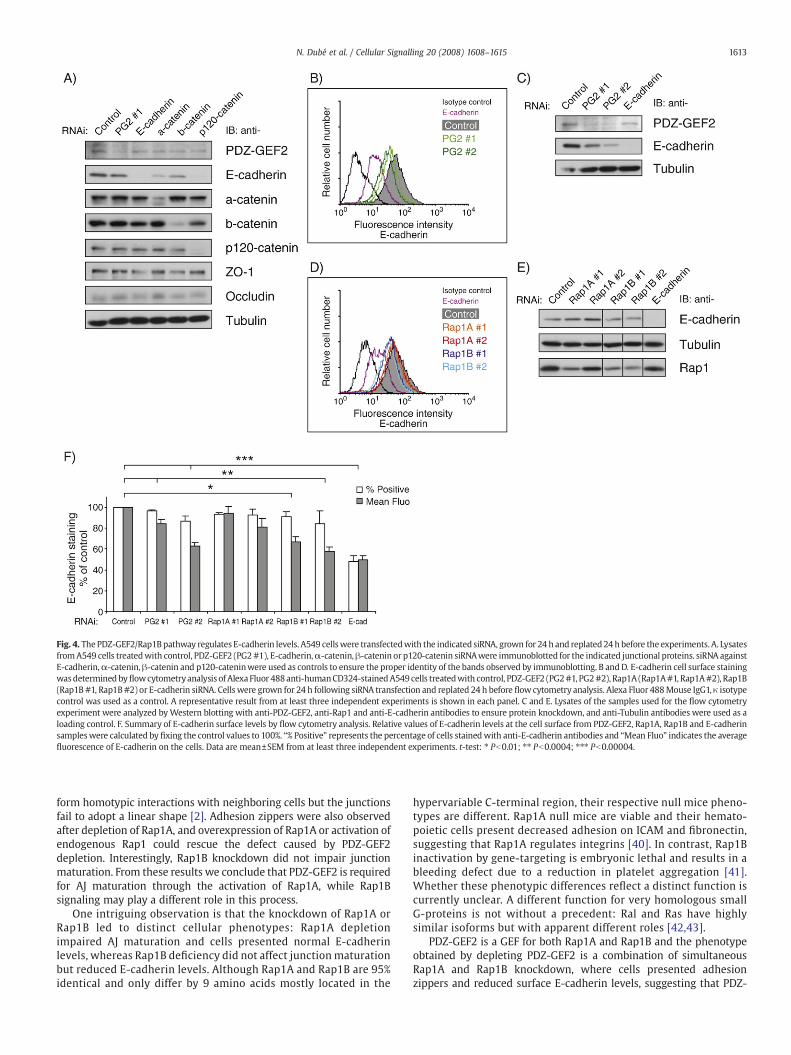
Fig. 4. ThePDZ-GEF2/Rap1Bpathway regulates E-cadherin levels. A549 cellswere transfectedwith the indicated siRNA, grown for 24 h and replated24 h before the experiments. A. LysatesfromA549 cells treatedwith control, PDZ-GEF2 (PG2#1), E-cadherin,α-catenin,β-catenin or p120-catenin siRNAwere immunoblotted for the indicated junctional proteins. siRNA againstE-cadherin,α-catenin, β-catenin and p120-cateninwere used as controls to ensure the proper identity of the bands observed by immunoblotting. B andD. E-cadherin cell surface stainingwasdetermined byflowcytometryanalysis of Alexa Fluor 488anti-humanCD324-stainedA549 cells treatedwith control, PDZ-GEF2 (PG2#1, PG2#2), Rap1A (Rap1A#1, Rap1A#2), Rap1B(Rap1B #1, Rap1B#2) or E-cadherin siRNA. Cells were grown for 24 h following siRNA transfection and replated 24 h before flow cytometry analysis. Alexa Fluor 488Mouse IgG1, κ isotypecontrol was used as a control. A representative result from at least three independent experiments is shown in each panel. C and E. Lysates of the samples used for the flow cytometryexperiment were analyzed byWestern blotting with anti-PDZ-GEF2, anti-Rap1 and anti-E-cadherin antibodies to ensure protein knockdown, and anti-Tubulin antibodies were used as aloading control. F. Summary of E-cadherin surface levels by flow cytometry analysis. Relative values of E-cadherin levels at the cell surface from PDZ-GEF2, Rap1A, Rap1B and E-cadherinsampleswere calculated by fixing the control values to 100%. “% Positive” represents the percentage of cells stainedwith anti-E-cadherin antibodies and “Mean Fluo” indicates the averagefluorescence of E-cadherin on the cells. Data are mean±SEM from at least three independent experiments. t-test: ⁎ Pb0.01; ⁎⁎ Pb0.0004; ⁎⁎⁎ Pb0.00004.
1613N. Dubé et al. / Cellular Signalling 20 (2008) 1608–1615
form homotypic interactions with neighboring cells but the junctionsfail to adopt a linear shape [2]. Adhesion zippers were also observedafter depletion of Rap1A, and overexpression of Rap1A or activation ofendogenous Rap1 could rescue the defect caused by PDZ-GEF2depletion. Interestingly, Rap1B knockdown did not impair junctionmaturation. From these results we conclude that PDZ-GEF2 is requiredfor AJ maturation through the activation of Rap1A, while Rap1Bsignaling may play a different role in this process.
One intriguing observation is that the knockdown of Rap1A orRap1B led to distinct cellular phenotypes: Rap1A depletionimpaired AJ maturation and cells presented normal E-cadherinlevels, whereas Rap1B deficiency did not affect junction maturationbut reduced E-cadherin levels. Although Rap1A and Rap1B are 95%identical and only differ by 9 amino acids mostly located in the
hypervariable C-terminal region, their respective null mice pheno-types are different. Rap1A null mice are viable and their hemato-poietic cells present decreased adhesion on ICAM and fibronectin,suggesting that Rap1A regulates integrins [40]. In contrast, Rap1Binactivation by gene-targeting is embryonic lethal and results in ableeding defect due to a reduction in platelet aggregation [41].Whether these phenotypic differences reflect a distinct function iscurrently unclear. A different function for very homologous smallG-proteins is not without a precedent: Ral and Ras have highlysimilar isoforms but with apparent different roles [42,43].
PDZ-GEF2 is a GEF for both Rap1A and Rap1B and the phenotypeobtained by depleting PDZ-GEF2 is a combination of simultaneousRap1A and Rap1B knockdown, where cells presented adhesionzippers and reduced surface E-cadherin levels, suggesting that PDZ-

1614 N. Dubé et al. / Cellular Signalling 20 (2008) 1608–1615
GEF2 activates Rap1A to mediate junction maturation, and Rap1B toregulate E-cadherin levels. Indeed, introduction of Rap1GAP, whichblocks all Rap proteins, prevents junction formation [13,21]. Since theknockdown of both Rap1A and Rap1B did not result in a phenotype assevere as Rap1GAP on AJ formation, it is possible that more Rapproteins such as Rap2 are involved in this process. However, otherexplanations are possible and we are currently investigating the roleof other Rap GEFs in further detail.
Which processes are regulated by the two Rap1 proteins in AJformation? In Rap1A depleted cells, E-cadherin levels are normal but thejunctions fail to stretch and to form tight cell–cell contacts. The extensionsobserved are decorated with E-cadherin, representing immature adhe-sions. This suggests either a defect in the linkage of E-cadherin to the actincytoskeleton or a failure to generate tension at cell–cell contacts, asactomyosin contractility and adequate cytoskeletal tension are requiredfor proper AJ assembly [8,44]. In that respect, it is important to note thatactivationofRap1can result in accumulationof cortical actin, independentof junction formation [25]. In PDZ-GEF2 depleted cells, F-actin is notproperly organized at cell–cell contacts. This phenocopies the inhibition ofactin assembly at cadherin adhesive contacts blocked by inhibiting Arp2/3activity, which is known to be necessary for the formation of cadherin-based cell junctions [45]. Furthermore, replating A549 cells in mediacontaining the ROCK inhibitor Y27632, which inhibits actin contractility[7], resulted in impairedactinorganizationat cell–cell contacts,mimickingthe PDZ-GEF2 knockdown phenotype (data not shown). An alternativeexplanation for the presence of adhesion zippers is that PDZ-GEF2depletion decreases the strength of the homotypic interaction betweencadherins. We assessed this possibility by plating A549 cells on a Fc-E-cadherin-coated surface, but we were unable to measure clear adhesive-ness, both in the control and PDZ-GEF2 depleted cells. Perhaps therelatively low levels of p120-catenin expressed in these cells areresponsible for the weak adhesive properties [46,47]. In contrast, Rap1Bmay play a role either in E-cadherin translocation or E-cadherinstabilization, but this possibility remains to be determined. In this respect,it is interesting to note that Rap nullDrosophilawing cells showa defect inDE-cadherin localization and junction integrity, but DE-cadherin is stillpresent at cell–cell contacts [12]. Similarly, in C. elegans, Rap1 nulls do notshowdefect in junction formation,whereas interferingwithboth theRap1pathway and the basolateral protein transport via the exocyst complexresults in the loss of the cadherin–catenin complex from the membrane[48].
It was previously shown that Rap1 is activated when junctions areformed [13], but also when junctions are disrupted [17]. This indicatesthat Rap1 plays multiple roles in junction formation, compatible withthe observation that both PDZ-GEF2 and the Rap1GEF C3G areinvolved in junction formation. Despite many attempts, we were thusfar unable to conclusively pinpoint the trigger that activates PDZ-GEF2, nor themechanism of activation. It was previously reported thatPDZ-GEF2 binds to the small G-protein M-Ras [49,50], resulting in thetranslocation of PDZ-GEF2 to the plasma membrane, but the stimulusthat activates M-Ras is currently unclear.
Mechanistic details of how Rap1 regulates junction formation arecurrently lacking. However, a number of proteins have been assigned asRap1 effectors, including Tiam, Vav2, AF6 andRiam, allwith connectionsto the actin cytoskeleton. Recently the Rap1 effector KRIT-1 [51] wasshown to colocalize with junctional proteins and is involved inendothelial junction integrity in a Rap1-dependent fashion [52], furthersupporting a role for Rap1 in junction formation. Clearly, theinterconnectivity between the Rap1 GEFs and the Rap1 effectorsinvolved in this process needs further investigation.
Acknowledgments
We thank the members of our laboratory for helpful discussions,Drs Johan de Rooij, Shannon M. Harper and Fried Zwartkruis forcritical reading of the manuscript, and Dr Geert Kops and members of
his laboratory for assistance with microscopy. We thank Wim vanDriel for protein purification, Jeroen van Ark and Yvonne Legtenbergfor technical help with antibodies production, Elisabetta Dejana forVE-cadherin antibodies, and Johan de Rooij for Fc-E-cadherin. N.D.was supported by an EMBO Long-term Fellowship and currently holdsa Research Fellowship of The Terry Fox Foundation through an awardfrom the National Cancer Institute of Canada. M.R.H.K. is supported bythe Netherlands Proteomics Centre, H.R. by the Chemical Sciences, andJ.L.B. by the Netherlands Genomics Initiative of the NetherlandsOrganization for Scientific Research.
References
[1] J.L. Bos, Curr. Opin. Cell Biol 17 (2) (2005) 123.[2] V. Vasioukhin, C. Bauer, M. Yin, E. Fuchs, Cell. 100 (2) (2000) 209.[3] A. Vaezi, C. Bauer, V. Vasioukhin, E. Fuchs, Dev. Cell. 3 (3) (2002) 367.[4] R.M. Mege, J. Gavard, M. Lambert, Curr. Opin. Cell Biol 18 (5) (2006) 541.[5] C.L. Adams, Y.T. Chen, S.J. Smith, W.J. Nelson, J. Cell Biol 142 (4) (1998) 1105.[6] T.D. Perez, M. Tamada, M.P. Sheetz, W.J. Nelson, J. Biol. Chem 283 (8) (2008) 5014.[7] S. Yamada, W.J. Nelson, J. Cell Biol 178 (3) (2007) 517.[8] A.M. Shewan,M.Maddugoda, A. Kraemer, S.J. Stehbens, S. Verma, E.M. Kovacs, A.S. Yap,
Mol. Biol. Cell 16 (10) (2005) 4531.[9] S.J. Stehbens, A.D. Paterson,M.S. Crampton, A.M. Shewan, C. Ferguson, A. Akhmanova,
R.G. Parton, A.S. Yap, J. Cell Sci 119 (Pt 9) (2006) 1801.[10] M.P.Maddugoda,M.S. Crampton, A.M. Shewan, A.S. Yap, J. Cell Biol 178 (3) (2007) 529.[11] A.S. Yap, M.S. Crampton, J. Hardin, Curr. Opin. Cell Biol 19 (5) (2007) 508.[12] A.L. Knox, N.H. Brown, Science 295 (5558) (2002) 1285.[13] C. Hogan, N. Serpente, P. Cogram, C.R. Hosking, C.U. Bialucha, S.M. Feller, V.M. Braga,
W. Birchmeier, Y. Fujita, Mol. Cell Biol 24 (15) (2004) 6690.[14] K.J. Mandell, B.A. Babbin, A. Nusrat, C.A. Parkos, J. Biol. Chem 280 (12) (2005) 11665.[15] E.S. Wittchen, R.A. Worthylake, P. Kelly, P.J. Casey, L.A. Quilliam, K. Burridge, J. Biol.
Chem 280 (12) (2005) 11675.[16] A. Sakurai, S. Fukuhara, A. Yamagishi, K. Sako, Y. Kamioka, M. Masuda, Y. Nakaoka,
N. Mochizuki, Mol. Biol. Cell 17 (2) (2006) 966.[17] F. Balzac, M. Avolio, S. Degani, I. Kaverina, M. Torti, L. Silengo, J.V. Small, S.F. Retta,
J. Cell Sci 118 (Pt 20) (2005) 4765.[18] S. Hisata, T. Sakisaka, T. Baba, T. Yamada, K. Aoki, M. Matsuda, Y. Takai, J. Cell Biol
178 (5) (2007) 843.[19] M.R. Kooistra, N. Dube, J.L. Bos, J. Cell Sci 120 (Pt 1) (2007) 17.[20] V. Yajnik, C. Paulding, R. Sordella, A.I. McClatchey,M. Saito, D.C.Wahrer, P. Reynolds,
D.W. Bell, R. Lake, S. van den Heuvel, J. Settleman, D.A. Haber, Cell 112 (5) (2003)673.
[21] L.S. Price, A. Hajdo-Milasinovic, J. Zhao, F.J. Zwartkruis, J.G. Collard, J.L. Bos, J. Biol.Chem 279 (34) (2004) 35127.
[22] T. Fukuyama, H. Ogita, T. Kawakatsu, T. Fukuhara, T. Yamada, T. Sato, K. Shimizu,T. Nakamura, M. Matsuda, Y. Takai, J. Biol. Chem 280 (1) (2005) 815.
[23] X. Cullere, S.K. Shaw, L. Andersson, J. Hirahashi, F.W. Luscinskas, T.N. Mayadas,Blood 105 (5) (2005) 1950.
[24] S. Fukuhara, A. Sakurai, H. Sano, A. Yamagishi, S. Somekawa, N. Takakura, Y. Saito,K. Kangawa, N. Mochizuki, Mol. Cell Biol 25 (1) (2005) 136.
[25] M.R. Kooistra, M. Corada, E. Dejana, J.L. Bos, FEBS Lett 579 (22) (2005) 4966.[26] T. Ohtsuka, Y. Hata, N. Ide, T. Yasuda, E. Inoue, T. Inoue, A. Mizoguchi, Y. Takai,
Biochem. Biophys. Res. Commun 265 (1) (1999) 38.[27] I.Y. Dobrosotskaya, G.L. James, Biochem. Biophys. Res. Commun 270 (3) (2000) 903.[28] A. Kawajiri, N. Itoh, M. Fukata, M. Nakagawa, M. Yamaga, A. Iwamatsu, K. Kaibuchi,
Biochem. Biophys. Res. Commun 273 (2) (2000) 712.[29] A. Mino, T. Ohtsuka, E. Inoue, Y. Takai, Genes Cells 5 (12) (2000) 1009.[30] W. Pellis-van Berkel, M.H. Verheijen, E. Cuppen, M. Asahina, J. de Rooij, G. Jansen,
R.H. Plasterk, J.L. Bos, F.J. Zwartkruis, Mol. Biol. Cell 16 (1) (2005) 106.[31] H. Wang, S.R. Singh, Z. Zheng, S.W. Oh, X. Chen, K. Edwards, S.X. Hou, Dev. Cell 10
(1) (2006) 117.[32] B. Boettner, L. Van Aelst, Mol. Cell Biol. (2007).[33] F. Breviario, L. Caveda, M. Corada, I. Martin-Padura, P. Navarro, J. Golay, M. Introna,
D. Gulino, M.G. Lampugnani, E. Dejana, Arterioscler. Thromb. Vasc. Biol 15 (8)(1995) 1229.
[34] M.G. Lampugnani, M. Resnati, M. Raiteri, R. Pigott, A. Pisacane, G. Houen, L.P. Ruco,E. Dejana, J. Cell Biol 118 (6) (1992) 1511.
[35] J. de Rooij, F.J. Zwartkruis, M.H. Verheijen, R.H. Cool, S.M. Nijman, A. Wittinghofer,J.L. Bos, Nature 396 (6710) (1998) 474.
[36] F.J. Zwartkruis, R.M. Wolthuis, N.M. Nabben, B. Franke, J.L. Bos, EMBO. J 17 (20)(1998) 5905.
[37] M. Torres, A. Stoykova, O. Huber, K. Chowdhury, P. Bonaldo, A. Mansouri, S. Butz,R. Kemler, P. Gruss, Proc. Natl. Acad. Sci. U S A. 94 (3) (1997) 901.
[38] H. Haegel, L. Larue, M. Ohsugi, L. Fedorov, K. Herrenknecht, R. Kemler,Development 121 (11) (1995) 3529.
[39] M.A. Davis, R.C. Ireton, A.B. Reynolds, J. Cell Biol 163 (3) (2003) 525.[40] M. Duchniewicz, T. Zemojtel, M. Kolanczyk, S. Grossmann, J.S. Scheele, F.J. Zwartkruis,
Mol. Cell Biol 26 (2) (2006) 643.[41] M. Chrzanowska-Wodnicka, S.S. Smyth, S.M. Schoenwaelder, T.H. Fischer, G.C.White II,
J. Clin. Invest. 115 (3) (2005) 680.[42] A. Ehrhardt, G.R. Ehrhardt, X. Guo, J.W. Schrader, Exp. Hematol 30 (10) (2002)
1089.

1615N. Dubé et al. / Cellular Signalling 20 (2008) 1608–1615
[43] J.H. Camonis, M.A. White, Trends Cell Biol 15 (6) (2005) 327.[44] Y. Miyake, N. Inoue, K. Nishimura, N. Kinoshita, H. Hosoya, S. Yonemura, Exp. Cell
Res 312 (9) (2006) 1637.[45] S. Verma, A.M. Shewan, J.A. Scott, F.M.Helwani, N.R. den Elzen, H.Miki, T. Takenawa,
A.S. Yap, J. Biol. Chem 279 (32) (2004) 34062.[46] M.A. Thoreson, P.Z. Anastasiadis, J.M. Daniel, R.C. Ireton, M.J. Wheelock, K.R. Johnson,
D.K. Hummingbird, A.B. Reynolds, J. Cell Biol 148 (1) (2000) 189.[47] D.T. Fox, M. Peifer, Curr. Biol 17 (1) (2007) R24.[48] E.W. Frische,W.Pellis-vanBerkel,G. vanHaaften, E. Cuppen,R.H. Plasterk,M.Tijsterman,
J.L. Bos, F.J. Zwartkruis, EMBO. J 26 (24) (2007) 5083.
[49] X. Gao, T. Satoh, Y. Liao, C. Song, C.D. Hu, K. Kariya Ki, T. Kataoka, J. Biol. Chem 276(45) (2001) 42219.
[50] Y. Yoshikawa, T. Satoh, T. Tamura, P.Wei, S.E. Bilasy, H. Edamatsu, A. Aiba, K. Katagiri,T. Kinashi, K. Nakao, T. Kataoka, Mol. Biol. Cell 18 (8) (2007) 2949.
[51] I. Serebriiskii, J. Estojak, G. Sonoda, J.R. Testa, E.A. Golemis, Oncogene 15 (9) (1997)1043.
[52] A. Glading, J. Han, R.A. Stockton, M.H. Ginsberg, J. Cell Biol 179 (2) (2007) 247.
Related Documents


