Molecular Biology of the Cell Vol. 12, 2422–2432, August 2001 The Proteasomal Substrate Stm1 Participates in Apoptosis-like Cell Death in Yeast Martin Ligr,* † Iris Velten,* † Eleonore Fro ¨ hlich, ‡ Frank Madeo, § Matthias Ledig,* Kai-Uwe Fro ¨ hlich, § Dieter H. Wolf,* and Wolfgang Hilt* *Institut fu ¨ r Biochemie, Universita ¨t Stuttgart, 70569 Stuttgart, Germany; ‡ Anatomisches Institut, Universita ¨t Tu ¨ bingen, 72074 Tu ¨ bingen, Germany; and § Physiologisch-Chemisches Institut, Universita ¨t Tu ¨ bingen, 72076 Tu ¨ bingen, Germany Submitted December 5, 2000; Revised April 27, 2001; Accepted May 22, 2001 Monitoring Editor: Martin Raff We have identified the yeast gene STM1 in an overexpression screen for new proteasomal substrates. Stm1 is unstable in wild-type cells and stabilized in cells with defective proteasomal activity and thus a bona fide substrate of the proteasome. It is localized in the perinuclear region and is required for growth in the presence of mutagens. Overexpression in cells with impaired proteasomal degradation leads to cell death accompanied with cytological markers of apoptosis: loss of plasma membrane asymmetry, chromatin condensation, and DNA cleavage. Cells lacking Stm1 display deficiency in the apoptosis-like cell death process induced by treatment with low concentrations of H 2 O 2 . We suggest that Stm1 is involved in the control of the apoptosis-like cell death in yeast. Survival is increased when Stm1 is completely missing from the cells or when inhibition of Stm1 synthesis permits proteasomal degradation to decrease its amount in the cell. Conversely, Stm1 accumulation induces cell death. In addition we identified five other genes whose overexpression in proteasomal mutants caused similar apoptotic phenotypes. INTRODUCTION Multicellular organisms are in the state of dynamic equilib- rium, sustained by the mutually opposing processes of cell division and cell death. The importance of programmed cell death to maintain the integrity of metazoans is widely ap- preciated, but is there a place for this process in the life cycle of single cell organisms? The existence of programmed cell death in bacteria is now firmly established (Engelberg-Kulka and Glaser, 1999). Re- cently we have identified a translation-dependent pro- grammed cell death process also in the unicellular eukaryote Saccharomyces cerevisiae (Fro ¨ hlich and Madeo, 2000). We ob- served that yeast cells underwent cell death due to presence of the cdc48-S565G mutation (Madeo et al., 1997), overexpres- sion of the mammalian apoptotic cell death regulator Bax (Ligr et al., 1998), or exposure to oxidative conditions (Madeo et al., 1999). This process resembled apoptosis, a form of programmed cell death indispensable for develop- ment and homeostasis of metazoan organisms (Webb et al., 1999). The occurrence of cytological markers of metazoan apoptosis in yeast, such as loss of plasma membrane asym- metry, chromatin condensation and margination, fragmen- tation of DNA, and membrane blebbing, as well as the identification of reactive oxygen species as a common regu- lator (Madeo et al., 1999), led us to suggest that the basic mechanism of apoptosis is present already in this unicellular eukaryote (Fro ¨ hlich and Madeo, 2000). This view is further supported by recent reports that the orthologues of Cdc48 regulate the apoptotic pathways of Caenorhabditis elegans (Wu et al., 1999) and humans (Shirogane et al., 1999). Cdc48 is an ATPase of the AAA family associated with a variety of cellular activities. Notably, Cdc48p is emerging as a factor involved in the regulation of the evolutionary con- served ubiquitin-proteasome system (Ghislain et al., 1996; Dai et al., 1998; Koegl et al., 1999; Meyer et al., 2000). Sub- strates to be degraded by this pathway are first covalently tagged with the small protein ubiquitin by an enzymatic cascade consisting of ubiquitin activating and conjugating enzymes, in most cases in cooperation with additional sub- strate-specific recognition elements. Polyubiquitylated pro- teins are recognized and degraded by the 26S proteasome, a multisubunit multicatalytic protease (Hilt and Wolf, 1996). In mammals, inhibition of proteasome-dependent proteoly- sis leads to either repression or induction of apoptosis, de- pending on the proliferative status of the particular cell type (Drexler, 1998). It has been suggested that in proliferating cells the proteasome continuously degrades an activator of † These authors contributed equally to the study. Corresponding author. E-mail address: [email protected]. 2422 © 2001 by The American Society for Cell Biology

Welcome message from author
This document is posted to help you gain knowledge. Please leave a comment to let me know what you think about it! Share it to your friends and learn new things together.
Transcript
Molecular Biology of the CellVol. 12, 2422–2432, August 2001
The Proteasomal Substrate Stm1 Participates inApoptosis-like Cell Death in Yeast
Martin Ligr,*† Iris Velten,*† Eleonore Frohlich,‡ Frank Madeo,§ MatthiasLedig,* Kai-Uwe Frohlich,§ Dieter H. Wolf,* and Wolfgang Hilt*�
*Institut fur Biochemie, Universitat Stuttgart, 70569 Stuttgart, Germany; ‡Anatomisches Institut,Universitat Tubingen, 72074 Tubingen, Germany; and §Physiologisch-Chemisches Institut, UniversitatTubingen, 72076 Tubingen, Germany
Submitted December 5, 2000; Revised April 27, 2001; Accepted May 22, 2001Monitoring Editor: Martin Raff
We have identified the yeast gene STM1 in an overexpression screen for new proteasomalsubstrates. Stm1 is unstable in wild-type cells and stabilized in cells with defective proteasomalactivity and thus a bona fide substrate of the proteasome. It is localized in the perinuclear regionand is required for growth in the presence of mutagens. Overexpression in cells with impairedproteasomal degradation leads to cell death accompanied with cytological markers of apoptosis:loss of plasma membrane asymmetry, chromatin condensation, and DNA cleavage. Cells lackingStm1 display deficiency in the apoptosis-like cell death process induced by treatment with lowconcentrations of H2O2. We suggest that Stm1 is involved in the control of the apoptosis-like celldeath in yeast. Survival is increased when Stm1 is completely missing from the cells or wheninhibition of Stm1 synthesis permits proteasomal degradation to decrease its amount in the cell.Conversely, Stm1 accumulation induces cell death. In addition we identified five other geneswhose overexpression in proteasomal mutants caused similar apoptotic phenotypes.
INTRODUCTION
Multicellular organisms are in the state of dynamic equilib-rium, sustained by the mutually opposing processes of celldivision and cell death. The importance of programmed celldeath to maintain the integrity of metazoans is widely ap-preciated, but is there a place for this process in the life cycleof single cell organisms?
The existence of programmed cell death in bacteria is nowfirmly established (Engelberg-Kulka and Glaser, 1999). Re-cently we have identified a translation-dependent pro-grammed cell death process also in the unicellular eukaryoteSaccharomyces cerevisiae (Frohlich and Madeo, 2000). We ob-served that yeast cells underwent cell death due to presenceof the cdc48-S565G mutation (Madeo et al., 1997), overexpres-sion of the mammalian apoptotic cell death regulator Bax(Ligr et al., 1998), or exposure to oxidative conditions(Madeo et al., 1999). This process resembled apoptosis, aform of programmed cell death indispensable for develop-ment and homeostasis of metazoan organisms (Webb et al.,1999). The occurrence of cytological markers of metazoanapoptosis in yeast, such as loss of plasma membrane asym-
metry, chromatin condensation and margination, fragmen-tation of DNA, and membrane blebbing, as well as theidentification of reactive oxygen species as a common regu-lator (Madeo et al., 1999), led us to suggest that the basicmechanism of apoptosis is present already in this unicellulareukaryote (Frohlich and Madeo, 2000). This view is furthersupported by recent reports that the orthologues of Cdc48regulate the apoptotic pathways of Caenorhabditis elegans(Wu et al., 1999) and humans (Shirogane et al., 1999).
Cdc48 is an ATPase of the AAA family associated with avariety of cellular activities. Notably, Cdc48p is emerging asa factor involved in the regulation of the evolutionary con-served ubiquitin-proteasome system (Ghislain et al., 1996;Dai et al., 1998; Koegl et al., 1999; Meyer et al., 2000). Sub-strates to be degraded by this pathway are first covalentlytagged with the small protein ubiquitin by an enzymaticcascade consisting of ubiquitin activating and conjugatingenzymes, in most cases in cooperation with additional sub-strate-specific recognition elements. Polyubiquitylated pro-teins are recognized and degraded by the 26S proteasome, amultisubunit multicatalytic protease (Hilt and Wolf, 1996).In mammals, inhibition of proteasome-dependent proteoly-sis leads to either repression or induction of apoptosis, de-pending on the proliferative status of the particular cell type(Drexler, 1998). It has been suggested that in proliferatingcells the proteasome continuously degrades an activator of
† These authors contributed equally to the study.� Corresponding author. E-mail address: [email protected].
2422 © 2001 by The American Society for Cell Biology

apoptosis. Curbing proteasomal activity is thought to resultin accumulation of this hypothetical regulator and therebyactivation of the apoptotic cell death cascade (Drexler, 1997).
Does proteasomal degradation play a similar role in theapoptosis-like cell death process in yeast? To answer thisquestion, we screened for genes that cause this type of deathwhen overexpressed in cells with defective proteasomes.
MATERIALS AND METHODS
Yeast Strains, Plasmids, and MediaTo construct plasmid pML1, a PRE1-containing BamHI-XhoI frag-ment of p13/PRE1 (a gift of W. Heinemeyer) was ligated intoBamHI-XhoI sites of pRS318 (CYH2 LEU2 CEN6; Sikorski and Boeke,1991). The integrative plasmid pL090 was assembled from the NheI-MluI fragment of pYES2 (Invitrogen, San Diego, CA), a polymerasechain reaction (PCR) fragment of the STM1 terminator (flanked bySphI and MluI sites), and the STM1 open reading frame (ORF)flanked by NheI at the 5�-end and the IRS sequence (Luo et al., 1996)followed by SphI site at the 3�-end. The STM1 terminator region wasamplified from yeast chromosomal DNA with the use of primersAAAAGCATGCAAGCCTTATATATGAATAATTCCAACTG andAAAAACGCGTCGAACGGAAGAAGTGAATGG. The STM1 ORFwas amplified with the use of primers AAAAGCTAGCATGTC-CAACCCATTTGATTTG and AAAAGCATGCCTAAGAACGAA-TATAACGAGCCAAAGATGGCAAGTTAG, with an STM1 cDNAlibrary plasmid as the template. Plasmid pL092 (PGAL1::STM1::IRSURA3 2�) was made by inserting the NcoI-XbaI fragment of pYES2(containing PGAL1 and 2� sequences) between NcoI and NheI sites ofpL090. All PCR and molecular cloning steps were done understandard conditions (Ausubel et al., 1989).
S. cerevisiae strains used in this study are listed in Table 1. Thestrains YML1 and YML2 were constructed in two steps. First,YHI29-1 and YHI29-14 were selected for spontaneous mutations inthe CYH2 gene on YPD plates containing 10 �g�cm�3 cycloheximide(Sikorski and Boeke, 1991). Cyhr clones were isolated and trans-formed with plasmid pML1, yielding strains YML1, and YML2,respectively. Complementation of the pre1-1 mutation was con-firmed by the restoration of proteasomal chymotrypsin-like activity,assayed by a substrate overlay test as described previously (Hilt andWolf, 1999). Strains YL280 and YL286 were generated by pop-in/pop-out allele replacement with the use of plasmid pL090 linearizedwith ClaI. The growth of YL280 was indistinguishable from wildtype on YPD plates supplemented with 12 mM caffeine or 10�g�cm�3 bleomycin, proving that the Stm1-IRS construct was fullyfunctional.
Yeast cells were grown at 30°C if not stated otherwise and liquidcultures were agitated at 200 rpm. Rich growth medium (YPD)contained 1% yeast extract, 2% Bacto-peptone, and 2% d-glucose.Synthetic complete (SC) medium (0.67% nitrogen base withoutamino acids and nucleotide bases) was lacking the appropriateauxotrophic factors for selection and contained either 2% glucose or2% galactose as required. Yeast transformations were carried out asdescribed previously (Gietz et al., 1995).
High Expression Lethality ScreenA pYES2-based cDNA library (Espinet et al., 1995) was transformedinto YML1 and YML2 strains pregrown on YPD. Transformantswere selected on SC glucose medium lacking leucine and uracil (SCura� leu�). After 3 d of growth, colonies were replica plated onto SCglucose medium lacking uracil (SC ura�) to enable loss of plasmidpML1. After an additional 2 d of growth the colonies were replicaplated onto SC glucose medium lacking uracil supplemented with10 �g�cm�3 cycloheximide (SC ura� cyh�). This step was repeatedafter 2 d of growth to ensure that colonies consisted of cells that hadlost the plasmid pML1 complementing the pre1-1 mutation. Loss ofplasmid pML1 carrying PRE1 was further confirmed by test forabsence of the chymotrypsin-like activity (Hilt, unpublished re-sults). Two days later, the colonies were replica plated onto SCgalactose medium lacking uracil (SCgal ura�). At the same time theoriginal colonies from SC ura� leu� plates (“wild type”) were alsoreplica plated onto the SCgal ura� medium to induce expression ofthe library genes. After 2 d the two sets of plates were compared andscreened for clones able to grow on galactose in the presence but notin the absence of plasmid pML1. To confirm the phenotype, candi-dates showing such features were picked from the original plates(SC glucose ura� leu� or SC glucose ura� cyh�) onto SCgal ura�.Plasmid DNA from positive clones (cured of pML1) was isolatedand a restriction analysis was performed to ensure homogeneity ofthe colonies and to estimate the size of the cDNA inserts. Plasmidsobtained by these means were transformed into the strainsWCG4/a, YHI29–1, and YHI29–14 and retested for the ability oftheir encoded cDNAs to cause high expression growth arrest in cellswith impaired proteasome by streaking onto SCgal ura� plates.
Gene DisruptionThe STM1 ORF was disrupted with a PCR-mediated method withthe use of the kanamycin resistance gene as a selection marker(Guldener et al., 1996). PCR was performed with the use of plasmidpUG6 as a template and primers designed to amplify the kanamycincassette flanked by 40 base sequences corresponding to immediatedown- and upstream region of the STM1 ORF. Yeast cells weretransformed with the PCR product and integrants were selected onYPD plates containing geneticin G418 (Life Technologies, Rockville,MD) at 0.2 mg�cm�3. Correct integration was confirmed by Southernblotting with the kanamycin cassette as a probe.
Analysis of DNASequencing was performed with the use of dideoxy sequencing (T7Sequencing Kit; Pharmacia Biotech, Uppsala, Sweden) and the Se-qui-Gen GT Nucleic Acid Electrophoresis Cell (Bio-Rad, Hercules,CA). For Southern blotting the semidry system and the SouthernGen Image kit (Amersham Pharmacia Biotech, Piscataway, NJ) wereused.
Immunofluorescence MicroscopyCells growing in logarithmical phase were fixed for 30 min (3.7%formaldehyde, 0.1 M PO4
3�, pH 6.5) and then washed three times inSP buffer (1.2 M sorbitol, 0.1 M PO4
3�, pH 6.5). The cell wall wasdigested with 15 U�cm�3 Zymolyase 100T (Seikagaku, Tokyo, Japan)in 1.2 M sorbitol, 20 mM �-mercaptoethanol, 0.1 M PO4
3�, pH 6.5, at30°C for 30 min. After washing three times in SP buffer spheroplasts
Table 1. Yeast strains
Strain Genotype Reference/Source
WCG4/a MATa his3-11,15 leu2-3,112 ura3 Heinemeyer et al.,1993
YHI29-1 MATa pre1-1 Heinemeyer et al.,1991
YHI29-14 MATa pre1-1 pre4-1 Hilt et al., 1993YML1 MATa pre1-1 cyh2 [pML1] This studyYML2 MATa pre1-1 pre4-1 cyh2 [pML1] This studyYIV2 MATa stm1-�1::kanMX This studyYL280 MATa STM1::IRS This studyYL286 MATa STM1::IRS pre1-1 pre4-1 This study
All strains are isogenic with WCG4/a.
Proteasome and Apoptosis in Yeast
Vol. 12, August 2001 2423
were bound on poly-l-lysine-coated slides, washed three times withphosphate-buffered saline (PBS; 53 mM NaH2HPO4, 13 mMNaH2PO4, 75 mM NaCl), and then incubated for 20 min at roomtemperature in PBT (1% bovine serum albumin, 0.1% Triton X-100 inPBS). The IRS-specific monoclonal antibody (BabCO, Richmond,CA) was diluted 1:100 in PBT and applied to the samples for 2 h atroom temperature in a humid chamber. The slides were washed fivetimes in PBT and incubated with goat anti-mouse immunoglobulinG-AlexaFluor 594 conjugate (Molecular Probes, Eugene, OR) diluted1:250 in PBT for 90 min in a dark humid chamber. The antibody wasremoved and the samples were washed five times with PBT and fivetimes with PBS. A coverslip was mounted with 90% glycerol and22.5 ng�cm�3 4�,6-diamidino-2-phenylindole in PBS.
Chromosome SpreadsImmunostaining of spread chromosomes was performed as de-scribed earlier (Bishop, 1994) with modifications. Spheroplasts wereprepared (see the previous section) and resuspended in ice-cold 0.1M 2-(N-morpholino)ethanesulfonic acid, 1 mM EDTA, 0.5 mMMgCl2, and 1 M sorbitol. Twenty microliters of this suspension wereplaced on a glass slide and mixed with 40 �l of 4% paraformalde-hyde in 3.4% sucrose. Afterward 80 �l of 1% Lipsol were added, andthen after a few seconds 80 �l of 4% paraformaldehyde in 3.4%sucrose were added. The mixture was spread over the slide with aglass rod and allowed to dry overnight. The slide was submerged inPBS for 10 min and blocked for 10 min in 1% bovine serum albuminin PBS. The reaction with antibodies and mounting of the slides wasperformed as described above.
Annexin V AssayExternalization of phosphatidylserine was detected essentially asdescribed previously (Ligr et al., 1998). Cells were resuspended indigestion buffer (1.2 M sorbitol, 0.5 mM MgCl2, 35 mM PO4
3�, pH6.8) and incubated for 2 h at 30°C with 15 U�cm�3 Zymolyase 100T(Seikagaku) and 5.5% Glusulase (NEN, Boston, MA). After cell walldigestion the cells were washed in binding buffer containing sorbi-tol (1.2 M sorbitol, 10 mM HEPES/NaOH, pH 7.4, 140 mM NaCl, 2.5mM CaCl2). The protoplasts were resuspended in 38 �l of bindingbuffer and incubated with 2 �l of green fluorescence protein (GFP)-annexin V (Clontech, Palo Alto, CA) and 2 �l of propidium iodide(50 �g�cm�3) in the dark for 20 min at room temperature. The cellswere mounted on a slide and examined under the fluorescencemicroscope.
Terminal Deoxynucleotidyl Transferase-mediateddUTP Nick End-labeling (TUNEL)The TUNEL assay for detection of fragmented nuclear DNA in yeastwas used as previously described (Ligr et al., 1998). Cells were fixedin 3.7% formaldehyde for 1 h and the cell walls were removed asdescribed above. The protoplasts were then applied to polylysine-coated slides. The In Situ Cell Death Detection Kit POD (BoehringerMannheim, Mannheim, Germany) was used according to the man-ufacturer’s instructions. After mounting a coverslip with a drop ofKaiser’s glycerol gelatin (Merck, Darmstadt, Germany) the cellswere examined under the light microscope.
Electron MicroscopyYeast cells were fixed with phosphate-buffered glutardialdehyde,the cell walls were removed, and the cells were postfixed withosmium tetroxide and uranyl acetate and dehydrated as describedfor stationary-phase cells (Byers and Goetsch, 1991). After the 100%ethanol washes, the cells were washed with 100% acetone, infil-trated with 50% acetone/50% Epon for 30 min and with 100% Eponfor 20 h. The cells were transferred to fresh 100% Epon, incubated at
56°C for 48 h, and thereafter cut into thin sections and stained withlead acetate.
Promoter Shut-off and Cycloheximide-ChaseAnalysis and Western BlottingStrains expressing plasmid-encoded IRS-tagged STM1 under thecontrol of GAL1 promoter were grown on SC glucose medium untilA600 � 1 and then transferred to SC galactose to the final density ofA600 � 0.5. After the culture reached A600 � 1.5 glucose and cyclo-heximide were added to the final concentration of 2% and 0.5mg�cm�3, respectively. Strains expressing Stm1-IRS from chromo-some were grown on YPD until A600 � 1.5, and cycloheximide wasadded to the final concentration of 0.5 mg�cm�3. The cells (5 A600 U)were harvested and lysed in 0.25 M NaOH and 1% �-mercatoetha-nol. The proteins were precipitated with 5.8% trichloroacetic acid,pelleted, and washed with acetone. The dry pellet was resuspendedin urea buffer (8 M urea, 5% SDS, 0.1 M EDTA, 0.02% bromphenolblue, 1% �-mercaptoethanol, 40 mM Tris/HCl, pH 6.8). The proteinswere resolved by SDS-PAGE and transferred onto a nitrocellulosemembrane. IRS-tagged Stm1 was detected with monoclonal anti-IRSantibody and the ECL kit (Amersham).
RESULTS
A Screen for Yeast cDNAs That Causes GrowthArrest when Overexpressed in Cells with ImpairedProteasome-mediated ProteolysisWe developed a screen to search for proteins whose degra-dation by the ubiquitin-proteasome system is required forviability or growth. We reasoned that overexpression ofsuch a protein should cause little effect in wild-type cellswith fully functioning proteasomes but cause a growth de-fect in cells in which proteasomal function is impaired.Proteasomal activity could not be eliminated completely,because knock-outs of proteasomal subunits are lethal. Cellswith the pre1-1 mutation residing in a �-type subunit of theproteasome are defective in chymotrypsin-like activity andshow a significant defect in growth but only slightly im-paired protein degradation. The pre4-1 mutation locating inanother �-type subunit causes loss of the PGPH-like activityof the proteasome, but cells otherwise behave phenotypi-cally like wild type. When pre1-1 and pre4-1 mutations arecombined, proteasomal protein degradation is significantlyslowed down, and cells grow at a reduced rate (Hilt et al.,1993). A pre1-1 pre4-1 strain was selected for spontaneousrecessive mutations in the CYH2 locus conferring cyclohex-imide resistance (Sikorski and Boeke, 1991). Plasmid pML1carrying wild-type PRE1 and CYH2 genes was introducedinto this strain to complement the defect in the chymotryp-sin-like activity of the proteasome. The resulting strain,which was phenotypically wild type concerning protea-some-dependent proteolysis and cycloheximide sensitivebecause of the presence of CYH2, was transformed with a2�-based cDNA library under the control of the GAL1 pro-moter (Espinet et al., 1995). Transformants were plated onselective medium with glucose and replicas were made oncycloheximide-containing plates to select for cells that hadlost the PRE1-encoding plasmid pML1. Original plates con-taining wild type cells and their copies containing cloneswith a pre1-1 pre4-1 background were then replica platedonto medium containing galactose to induce expression ofthe plasmid-encoded cDNAs. Library plasmids that causedgrowth arrest in pre1-1 pre4-1 mutants but not in the PRE1
Ligr et al.
Molecular Biology of the Cell2424
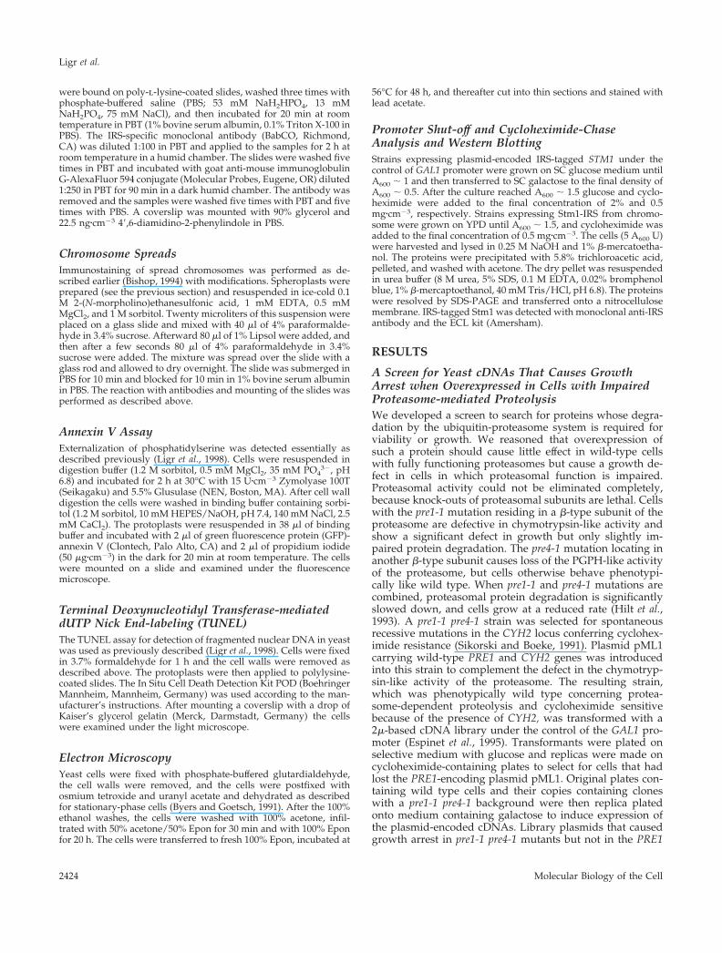
pre4-1 background (wild type) were isolated and their abilityto induce growth arrest in cells with defective proteasomeswas confirmed after retransformation of the isolated plas-mids into the wild type, pre1-1, and pre1-1 pre4-1 cells (Fig-ure 1A). Library plasmids (n � 125) conferring the expectedphenotype were isolated and sequenced, revealing 62 indi-vidual ORFs causing high expression lethality (HEL genes).
Overexpression of Distinct HEL Genes Causes CellDeath and Apoptotic Phenotypes in ProteasomalMutantsWe noticed that overexpression of some HEL genes didnot only halt growth but also led to decreased survival.Therefore, we examined all isolated cDNAs for their abil-ity to induce apoptosis-like cell death in pre1-1 pre4-1mutants.
In both S. cerevisiae and mammalian cells, 90% of phos-phatidylserine is found under normal conditions in the innerleaflet of the plasma membrane, facing the cytosol (Cerbonand Calderon, 1991). Early during apoptosis in mammals(Martin et al., 1995) and during the apoptosis-like cell deathprocess in yeast (Madeo et al., 1997; Ligr et al., 1998) theasymmetric distribution of phosphatidylserine is lost. Thiseffect can be detected by binding of annexin V to the cellsurface. We observed GFP-annexin V binding to yeast pro-
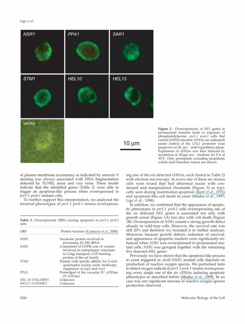
toplasts derived from pre1-1 pre4-1 cells that overexpressedsix of the 62 HEL genes identified in the screen (Figure 2).Integrity of protoplasts was assessed by counterstainingwith propidium iodide to exclude cells with GFP-annexin Vbound to the cytosolic face of the plasma membrane (notshown). The highest rates of staining (�40% of the cells)were observed in strains that overexpressed the PPA1 or theYOR309C gene. Lower but significant rates of annexin stain-ing (15–20% of the cells) were found for pre1-1 pre4-1 clonesoverexpressing NSR1, SAR1, STM1, or YNL208W. No stain-ing was observed in pre1-1 pre4-1 cells carrying an emptyvector.
Another hallmark of apoptosis is DNA fragmentation(Collins et al., 1997) that can be detected in situ by theTUNEL test. This assay detects the increased presence of free3�-ends of DNA generated by fragmentation of chromo-somes and visualizes them via attachment of labeled nucle-otides by terminal deoxynucleotidyl transferase. The 62cDNAs identified in the high expression lethality screenwere analyzed for their capacity to cause DNA fragmenta-tion when overexpressed in pre1-1 pre4-1 cells. Six cDNAswere detected that induced TUNEL staining in a significantfraction of nuclei in the respective cells (Figure 1B). Signifi-cantly, these were the same HEL genes that were identifiedto cause a positive signal in the annexin V test. Thus, the loss
Figure 1. Several HEL genes cause growtharrest and cell death when overexpressed inproteasomal mutants. Wild-type and pre1-1pre4-1 cells carrying pYES2-encoded, PGAL1-driven cDNAs of HEL genes (as indicated)were cultivated overnight in SC ura�. Cellswere harvested, and washed in water. (A) Totest for growth arrest, 10-fold serial dilutionswere spotted on SCgal ura� plates. Plateswere incubated for 3 d at 30°C. (B) After 8 h ofinduction in SCgal ura� liquid medium, celldeath rate was scored as 100% � (platingefficiency on YPD medium). Frequency ofcells with TUNEL positive nuclei was deter-mined by microscopic inspection. Resultswere averaged from two experiments.
Proteasome and Apoptosis in Yeast
Vol. 12, August 2001 2425
of plasma membrane asymmetry as indicated by annexin Vstaining was always associated with DNA fragmentationdetected by TUNEL assay and vice versa. These resultsindicate that the identified genes (Table 2) were able totrigger an apoptosis-like process when overexpressed inpre1-1 pre4-1 mutant cells.
To further support this interpretation, we analyzed theterminal phenotypes of pre1-1 pre4-1 strains overexpress-
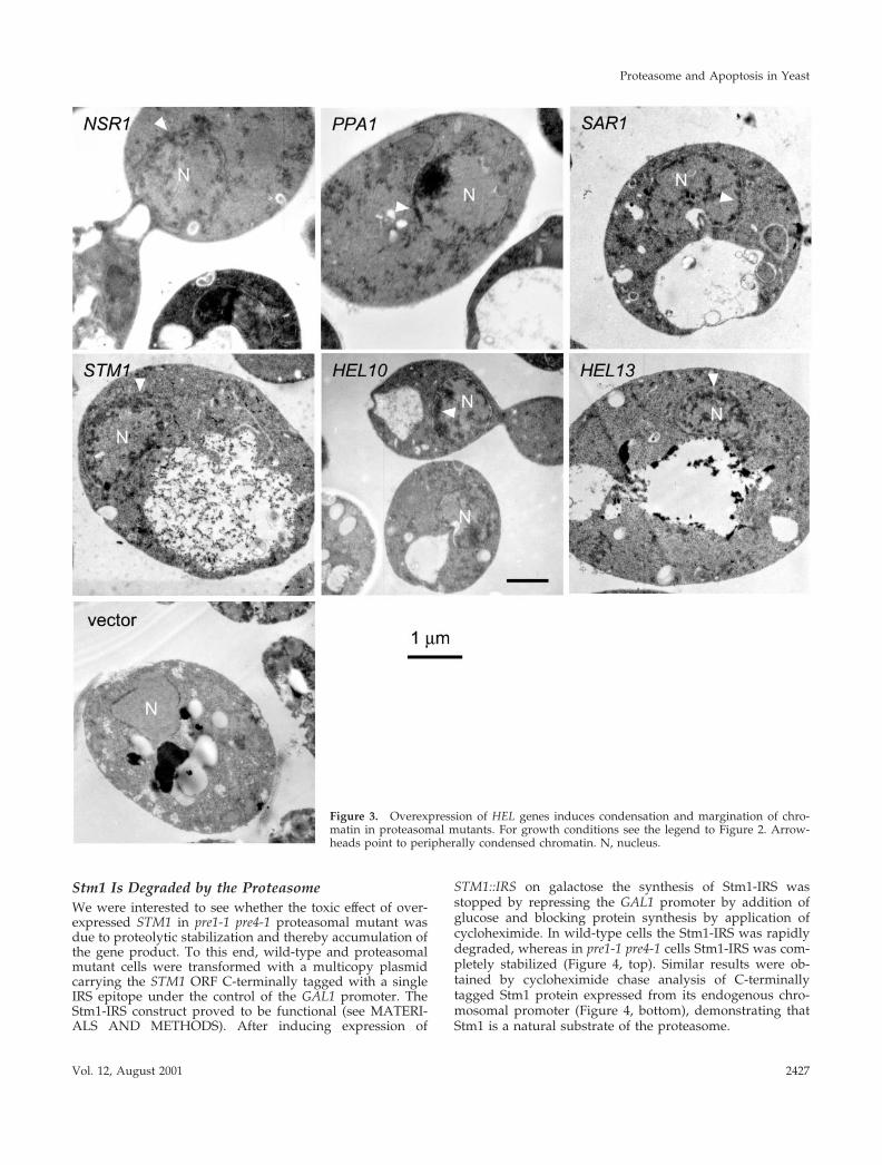
ing one of the six detected cDNAs, each (listed in Table 2)with electron microscopy. In every one of these six strainscells were found that had abnormal nuclei with con-densed and marginalized chromatin (Figure 3) as typi-cally seen during mammalian apoptosis (Kerr et al., 1972)and apoptosis-like cell death in yeast (Madeo et al., 1997;Ligr et al., 1998).
In addition, we confirmed that the appearance of apopto-tic phenotypes in pre1-1 pre4-1 cells overexpressing one ofthe six detected HEL genes is associated not only withgrowth arrest (Figure 1A) but also with cell death (Figure1B). Overexpression of NSR1 caused a strong growth defectalready in wild-type cells. However, the survival rate wasstill 20% and therefore we included it in further analyses.Moreover, because growth defects, reduction of survival,and appearance of apoptotic markers were significantly en-hanced when NSR1 was overexpressed in proteasomal mu-tant cells, NSR1 was grouped together with the remainingfive detected HEL genes.
Previously we have shown that the apoptosis-like processin yeast triggered in cdc48-S565G mutant cells depends onproduction of reactive oxygen species. We performed teststo detect oxygen radicals in pre1-1 pre4-1 strains overexpress-ing every single one of the six cDNAs inducing apoptoticphenotypes as described before (Madeo et al., 1999). In nocase was any significant increase in reactive oxygen speciesproduction observed.
Figure 2. Overexpression of HEL genes inproteasomal mutants leads to exposure ofphosphatidylserine. pre1-1 pre4-1 cells thatcarried pYES2-encoded cDNAs (as indicated)under control of the GAL1 promoter werepregrown on SC ura� until logarithmic phase.Expression of cDNAs was then induced byincubation in SCgal ura� medium for 8 h at30°C. Only protoplasts excluding propidiumiodide (and therefore intact) are shown.
Table 2. Overexpressed ORFs causing apoptosis in pre1-1 pre4-1cells
ORF Protein function (Costanzo et al., 2000)
NSR1 Nucleolar protein involved inprocessing 20–18S rRNA
SAR1 Component of COPII coat of vesiclesinvolved in endoplasmic reticulumto Golgi transport; GTP-bindingprotein of the arf family
STM1 Protein with specific affinity for G-richquadruplex nucleic acids; multicopysuppressor of pop2 and tom1
PPA1 Proteolipid of the vacuolar H�-ATPase(V-ATPase)
HEL 10 (YNL208W) UnknownHEL13 (YOR309C) Unknown
Ligr et al.
Molecular Biology of the Cell2426
Stm1 Is Degraded by the ProteasomeWe were interested to see whether the toxic effect of over-expressed STM1 in pre1-1 pre4-1 proteasomal mutant wasdue to proteolytic stabilization and thereby accumulation ofthe gene product. To this end, wild-type and proteasomalmutant cells were transformed with a multicopy plasmidcarrying the STM1 ORF C-terminally tagged with a singleIRS epitope under the control of the GAL1 promoter. TheStm1-IRS construct proved to be functional (see MATERI-ALS AND METHODS). After inducing expression of
STM1::IRS on galactose the synthesis of Stm1-IRS wasstopped by repressing the GAL1 promoter by addition ofglucose and blocking protein synthesis by application ofcycloheximide. In wild-type cells the Stm1-IRS was rapidlydegraded, whereas in pre1-1 pre4-1 cells Stm1-IRS was com-pletely stabilized (Figure 4, top). Similar results were ob-tained by cycloheximide chase analysis of C-terminallytagged Stm1 protein expressed from its endogenous chro-mosomal promoter (Figure 4, bottom), demonstrating thatStm1 is a natural substrate of the proteasome.
Figure 3. Overexpression of HEL genes induces condensation and margination of chro-matin in proteasomal mutants. For growth conditions see the legend to Figure 2. Arrow-heads point to peripherally condensed chromatin. N, nucleus.
Proteasome and Apoptosis in Yeast
Vol. 12, August 2001 2427
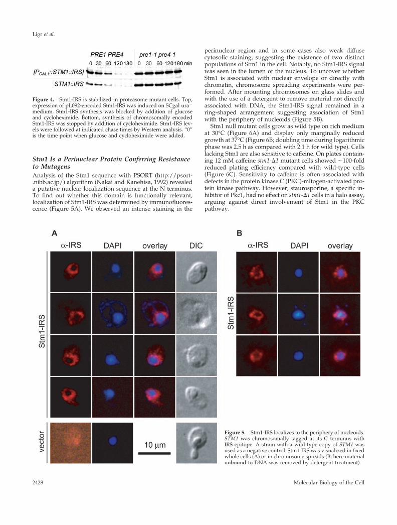
Stm1 Is a Perinuclear Protein Conferring Resistanceto MutagensAnalysis of the Stm1 sequence with PSORT (http://psort-.nibb.ac.jp/) algorithm (Nakai and Kanehisa, 1992) revealeda putative nuclear localization sequence at the N terminus.To find out whether this domain is functionally relevant,localization of Stm1-IRS was determined by immunofluores-cence (Figure 5A). We observed an intense staining in the
perinuclear region and in some cases also weak diffusecytosolic staining, suggesting the existence of two distinctpopulations of Stm1 in the cell. Notably, no Stm1-IRS signalwas seen in the lumen of the nucleus. To uncover whetherStm1 is associated with nuclear envelope or directly withchromatin, chromosome spreading experiments were per-formed. After mounting chromosomes on glass slides andwith the use of a detergent to remove material not directlyassociated with DNA, the Stm1-IRS signal remained in aring-shaped arrangement suggesting association of Stm1with the periphery of nucleoids (Figure 5B).
Stm1 null mutant cells grow as wild type on rich mediumat 30°C (Figure 6A) and display only marginally reducedgrowth at 37°C (Figure 6B; doubling time during logarithmicphase was 2.5 h as compared with 2.1 h for wild type). Cellslacking Stm1 are also sensitive to caffeine. On plates contain-ing 12 mM caffeine stm1-�1 mutant cells showed �100-foldreduced plating efficiency compared with wild-type cells(Figure 6C). Sensitivity to caffeine is often associated withdefects in the protein kinase C (PKC)-mitogen-activated pro-tein kinase pathway. However, staurosporine, a specific in-hibitor of Pkc1, had no effect on stm1-�1 cells in a halo assay,arguing against direct involvement of Stm1 in the PKCpathway.
Figure 4. Stm1-IRS is stabilized in proteasome mutant cells. Top,expression of pL092-encoded Stm1-IRS was induced on SCgal ura�
medium. Stm1-IRS synthesis was blocked by addition of glucoseand cycloheximide. Bottom, synthesis of chromosomally encodedStm1-IRS was stopped by addition of cycloheximide. Stm1-IRS lev-els were followed at indicated chase times by Western analysis. “0”is the time point when glucose and cycloheximide were added.
Figure 5. Stm1-IRS localizes to the periphery of nucleoids.STM1 was chromosomally tagged at its C terminus withIRS epitope. A strain with a wild-type copy of STM1 wasused as a negative control. Stm1-IRS was visualized in fixedwhole cells (A) or in chromosome spreads (B; here materialunbound to DNA was removed by detergent treatment).
Ligr et al.
Molecular Biology of the Cell2428

Given that caffeine is a purine analogue, we explored thepossibility that the sensitivity of stm1-�1 cells to this sub-stance may reflect a role of Stm1 in nucleic acid metabolism.We observed that mutant cells show 10-fold enhanced UVsensitivity as compared with wild-type cells (Figure 6D).Bleomycin is a radiomimetic drug that induces single- anddouble-strand breaks through the production of free radicals(Hampsey, 1997). stm1-�1 mutant cells displayed only aslight growth defect on YPD plates containing 10 �g�cm�3
bleomycin at 30°C (not shown), but their plating efficiencyon the same medium dropped �10-fold at 37°C comparedwith wild type (Figure 6E). In contrast, an alkylating agent,methyl methanesulfonate (MMS), did not cause any differ-ential effect in wild-type and stm1-�1 strains in a halo assay
(Ligr and Hilt, unpublished results) or in a test on YPDplates containing 0.02% MMS, either at 30 or 37°C (Figure6F).
Immunofluorescence experiments were performed tocheck whether treatment with caffeine or bleomycin leads toan alteration of Stm1 localization within the cell. No changein the localization pattern of Stm1-IRS was observed after2.5 h growth of cells on YPD in the presence of 12 mMcaffeine or 750 �g�cm�3 bleomycin at 30 and 37°C as com-pared with cells grown on YPD at 30°C.
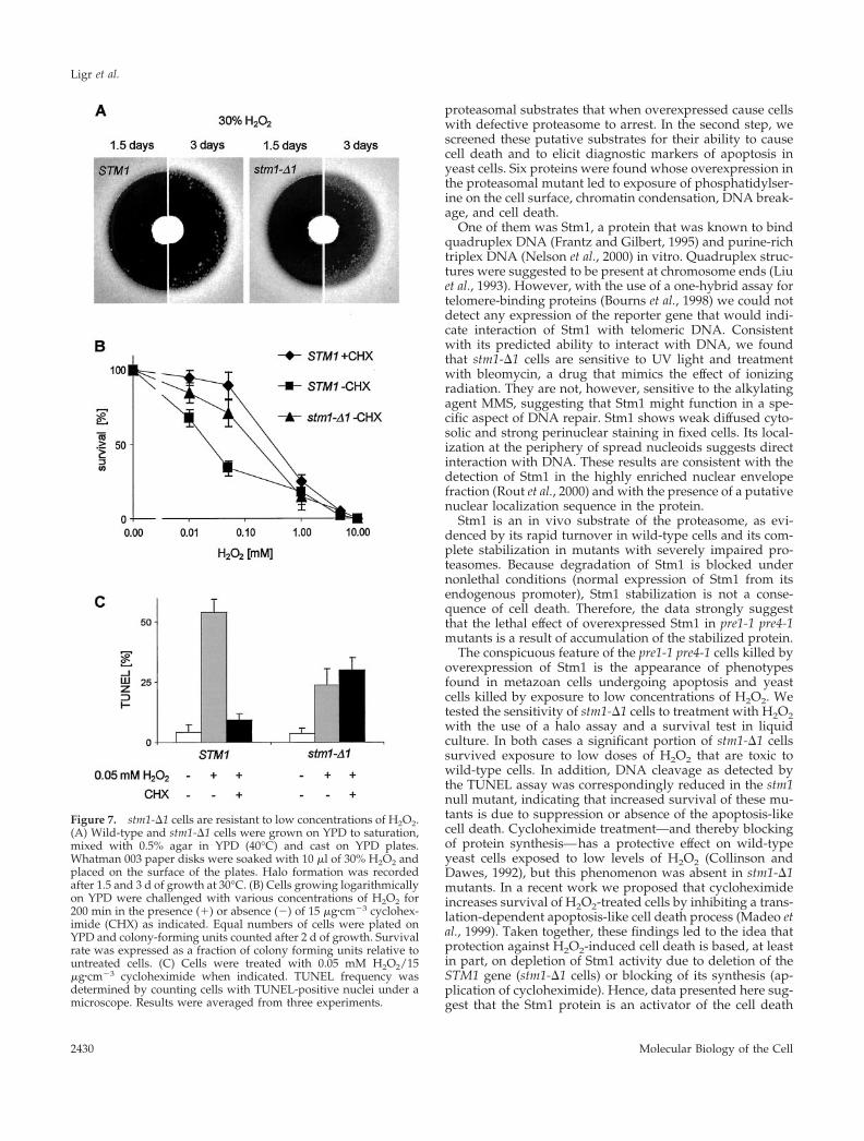
Cells Lacking Stm1 Can Recover from H2O2TreatmentAs described in a previous section, accumulation of Stm1leads to apoptosis-like cell death. Apoptotic phenotypes canalso be induced in yeast by treatment with low concentra-tions of H2O2 (Madeo et al., 1999). Therefore the questionarose whether stm1-�1 cells are as sensitive to H2O2 treat-ment as wild type. In a halo assay, both strains displayed thesame level of sensitivity after incubation for 1.5 d. However,after 3 d stm1-�1 cells started populating the zone that wasup to that point devoid of any growth, thereby decreasingthe size of the halo. In contrast, wild-type cells did notextend their growth significantly toward the center of thehalo (Figure 7A). To address the possibility that the stm1-�1cells growing in the halo were suppressor mutants, severalof them were isolated and tested again for H2O2 sensitivitywith the use of the halo assay. No increase in H2O2 resis-tance relative to the original stm1-�1 strain was observed,thereby excluding the appearance of suppressor mutations.A possible explanation for the recovery of stm1-�1 in thehalo zone is that a portion of stm1-�1 cells survived theotherwise lethal level of H2O2 and resumed their growthafter the decrease of H2O2 concentration (by diffusion/reac-tion with the components of the media). Therefore we ana-lyzed plating efficiency of wild-type and stm1-�1 cells afterexposure to various concentrations of H2O2 in liquid cul-tures. This experiment showed that, compared with wildtype, stm1-�1 cells are slightly, but significantly, more resis-tant to treatment with low concentrations of H2O2, whereashigher concentrations of H2O2 (�1 mM) are lethal to bothmutant and wild-type cells (Figure 7B). Quantification ofTUNEL staining showed that the increase in survival rate ofstm1-�1 cells treated with a low dose of H2O2 (0.05 mM) isaccompanied by a decrease in the number of cells showing anapoptotic phenotype (Figure 7C).
As previously shown, cycloheximide treatment leads toincreased survival of yeast cells after exposure to low con-centrations of H2O2 (Collinson and Dawes, 1992) by pre-venting apoptosis-like cell death (Madeo et al., 1999). How-ever, cycloheximide treatment did not further increaseresistance of stm1-�1 cells to apoptosis-like cell deathbrought about by low concentrations of H2O2 (Figure 7C).
DISCUSSION
The ubiquitin-proteasome system has been proposed to con-trol mammalian apoptosis by degrading a short-lived pro-apoptotic protein (Drexler, 1997). To find out if the ubiquitinsystem plays a similar role in yeast we performed a two-layer screen. In the first step we looked for potential yeast
Figure 6. stm1-�1 cells are sensitive to caffeine and DNA-damag-ing agents. Cells were grown on liquid YPD to saturation and 5 �lof 10-fold serial dilutions were spotted on agar medium. Cells onYPD plates were incubated at 30°C (A) and 37°C (B) for 2 d. (C) Cellswere spotted on YPD plates containing 12 mM caffeine and incu-bated at 30°C for 5 d. (D) Cells plated on YPD were exposed to UVlight for 45 s and incubated in dark for 1.5 d at 30°C. (E) Cells wereincubated for 4 d at 37°C on YPD plates containing 10 �g�cm�3
bleomycin. (F) Cells were grown for 5 d at 30°C on YPD platescontaining 0.02% MMS.
Proteasome and Apoptosis in Yeast
Vol. 12, August 2001 2429
proteasomal substrates that when overexpressed cause cellswith defective proteasome to arrest. In the second step, wescreened these putative substrates for their ability to causecell death and to elicit diagnostic markers of apoptosis inyeast cells. Six proteins were found whose overexpression inthe proteasomal mutant led to exposure of phosphatidylser-ine on the cell surface, chromatin condensation, DNA break-age, and cell death.
One of them was Stm1, a protein that was known to bindquadruplex DNA (Frantz and Gilbert, 1995) and purine-richtriplex DNA (Nelson et al., 2000) in vitro. Quadruplex struc-tures were suggested to be present at chromosome ends (Liuet al., 1993). However, with the use of a one-hybrid assay fortelomere-binding proteins (Bourns et al., 1998) we could notdetect any expression of the reporter gene that would indi-cate interaction of Stm1 with telomeric DNA. Consistentwith its predicted ability to interact with DNA, we foundthat stm1-�1 cells are sensitive to UV light and treatmentwith bleomycin, a drug that mimics the effect of ionizingradiation. They are not, however, sensitive to the alkylatingagent MMS, suggesting that Stm1 might function in a spe-cific aspect of DNA repair. Stm1 shows weak diffused cyto-solic and strong perinuclear staining in fixed cells. Its local-ization at the periphery of spread nucleoids suggests directinteraction with DNA. These results are consistent with thedetection of Stm1 in the highly enriched nuclear envelopefraction (Rout et al., 2000) and with the presence of a putativenuclear localization sequence in the protein.
Stm1 is an in vivo substrate of the proteasome, as evi-denced by its rapid turnover in wild-type cells and its com-plete stabilization in mutants with severely impaired pro-teasomes. Because degradation of Stm1 is blocked undernonlethal conditions (normal expression of Stm1 from itsendogenous promoter), Stm1 stabilization is not a conse-quence of cell death. Therefore, the data strongly suggestthat the lethal effect of overexpressed Stm1 in pre1-1 pre4-1mutants is a result of accumulation of the stabilized protein.
The conspicuous feature of the pre1-1 pre4-1 cells killed byoverexpression of Stm1 is the appearance of phenotypesfound in metazoan cells undergoing apoptosis and yeastcells killed by exposure to low concentrations of H2O2. Wetested the sensitivity of stm1-�1 cells to treatment with H2O2with the use of a halo assay and a survival test in liquidculture. In both cases a significant portion of stm1-�1 cellssurvived exposure to low doses of H2O2 that are toxic towild-type cells. In addition, DNA cleavage as detected bythe TUNEL assay was correspondingly reduced in the stm1null mutant, indicating that increased survival of these mu-tants is due to suppression or absence of the apoptosis-likecell death. Cycloheximide treatment—and thereby blockingof protein synthesis—has a protective effect on wild-typeyeast cells exposed to low levels of H2O2 (Collinson andDawes, 1992), but this phenomenon was absent in stm1-�1mutants. In a recent work we proposed that cycloheximideincreases survival of H2O2-treated cells by inhibiting a trans-lation-dependent apoptosis-like cell death process (Madeo etal., 1999). Taken together, these findings led to the idea thatprotection against H2O2-induced cell death is based, at leastin part, on depletion of Stm1 activity due to deletion of theSTM1 gene (stm1-�1 cells) or blocking of its synthesis (ap-plication of cycloheximide). Hence, data presented here sug-gest that the Stm1 protein is an activator of the cell death
Figure 7. stm1-�1 cells are resistant to low concentrations of H2O2.(A) Wild-type and stm1-�1 cells were grown on YPD to saturation,mixed with 0.5% agar in YPD (40°C) and cast on YPD plates.Whatman 003 paper disks were soaked with 10 �l of 30% H2O2 andplaced on the surface of the plates. Halo formation was recordedafter 1.5 and 3 d of growth at 30°C. (B) Cells growing logarithmicallyon YPD were challenged with various concentrations of H2O2 for200 min in the presence (�) or absence (�) of 15 �g�cm�3 cyclohex-imide (CHX) as indicated. Equal numbers of cells were plated onYPD and colony-forming units counted after 2 d of growth. Survivalrate was expressed as a fraction of colony forming units relative tountreated cells. (C) Cells were treated with 0.05 mM H2O2/15�g�cm�3 cycloheximide when indicated. TUNEL frequency wasdetermined by counting cells with TUNEL-positive nuclei under amicroscope. Results were averaged from three experiments.
Ligr et al.
Molecular Biology of the Cell2430
process triggered by exposure of cells to low concentrationsof H2O2. Control of its synthesis and/or degradation may beregulatory steps of H2O2-induced apoptosis-like cell deathin yeast.
STM1 was originally identified as a multicopy suppressorof tom1, htr1, and pop2 mutations, each of them being in-volved in an aspect of cell cycle control (for a summary, seeNelson et al., 2000). In a genome-wide two-hybrid screen,Stm1 was found to interact with a product of a predictedgene YJR072C (Uetz et al., 2000), which has conserved ortho-logues in C. elegans and humans. Stm1 itself has a highlyconserved orthologue in Schizosaccharomyces pombe and aputative orthologue in Drosophila melanogaster (Nelson et al.,2000). This hints that Stm1 may regulate or participate in anevolutionarily conserved process.
Apoptosis in mammalian cells has been tightly linked toactivation of caspases (Rich et al., 1999), which are missing inyeast. However, recent reports suggest that the appearanceof apoptotic morphology can proceed in the absence ofcaspases, albeit in a less efficient manner (Borner and Mon-ney, 1999). Notable is the role of reactive oxygen species asmediators of—in many cases—caspase-independent apo-ptosis in mammalian cells (Xiang et al., 1996; Carmody andCotter, 2000). Recently, a mammalian apoptosis-inducingfactor, AIF, was identified that has closely related ortho-logues in all phyla (Susin et al., 1999; Lorenzo et al., 1999).Consequent to its translocation from mitochondria to thenucleus, this factor triggers a cell death process with all ofthe cytological hallmarks of apoptosis but without the acti-vation of caspases. Thus, the target compartment of AIF andthe major place of localization of Stm1 is the same—thenucleus—and both share similar function—induction ofcaspase-independent cell death resembling apoptosis. Thetempting hypothesis to be tested is that AIF, Stm1, and theirrespective orthologues participate in the same caspase-inde-pendent pathway in yeast and mammals.
ACKNOWLEDGMENTS
We thank Jochen Strayle, Sibylle Jager, and Richard Plemper forstimulating discussions, Dragica Kapucija for excellent technicalsupport, and Rachel E. Patton for the linguistic correction of themanuscript. We acknowledge J. Hegemann, W. Heinemeyer, and E.Herrero for providing us with plasmids. This research was sup-ported by the Fonds der Chemischen Industrie, Frankfurt, the EU-TMR network on the ubiquitin-proteasome system, and the Deut-sche Forschungsgemeinschaft.
REFERENCES
Ausubel, F.M., Brent, R., Kingston, R., Moore, D., Seidmann, J.G.,Smith, J.A., and Struhl, K. (1989). Current Protocols in MolecularBiology, New York: Wiley.
Bishop, D.K. (1994). RecA homologs Dmc1 and Rad51 interact toform multiple nuclear complexes prior to meiotic chromosome syn-apsis. Cell 79, 1081–1092.
Borner, C., and Monney, L. (1999). Apoptosis without caspases: aninefficient molecular guillotine? Cell Death Differ. 6, 497–507.
Bourns, B.D., Alexander, M.K., Smith, A.M., and Zakian, V.A.(1998). Sir proteins, Rif proteins, and Cdc13p bind Saccharomycestelomeres in vivo. Mol. Cell. Biol. 18, 5600–5608.
Byers, B., and Goetsch, L. (1991). Preparation of yeast cells forthin-section electron microscopy. Methods Enzymol. 194, 602–608.
Carmody, R.J., and Cotter, T.G. (2000). Oxidative stress inducescaspase-independent retinal apoptosis in vitro. Cell Death Differ. 7,282–291.
Cerbon, J., and Calderon, V. (1991). Changes of the compositionalasymmetry of phospholipids associated to the increment in themembrane surface potential. Biochim. Biophys. Acta 1067, 139–144.
Collins, J.A., Schandi, C.A., Young, K.K., Vesely, J., and Willingham,M.C. (1997). Major DNA fragmentation is a late event in apoptosis.J. Histochem. Cytochem. 45, 923–934.
Collinson, L.P., and Dawes, I.W. (1992). Inducibility of the responseof yeast cells to peroxide stress. J. Gen. Microbiol. 138, 329–335.
Costanzo, M.C., Hogan, J.D., Cusick, M.E., Davis, B.P., Fancher,A.M., Hodges, P.E., Kondu, P., Lengieza, C., Lew-Smith, J.E., Ling-ner, C., Roberg-Perez, K.J., Tillberg, M., Brooks, J.E., and Garrels, J.I.(2000). The yeast proteome database (YPD) and Caenorhabditis el-egans proteome database (WormPD): comprehensive resources forthe organization and comparison of model organism protein infor-mation. Nucleic Acids Res. 28, 73–76.
Dai, R.M., Chen, E., Longo, D.L., Gorbea, C.M., and Li, C.C.H.(1998). Involvement of valosin-containing protein, an ATPase co-purified with I�B� and 26S proteasome, in ubiquitin-proteasomemediated degradation of I�B�. J. Biol. Chem. 273, 3562–3573.
Drexler, H.C. (1997). Activation of the cell death program by inhibitionof proteasome function. Proc. Natl. Acad. Sci. USA 94, 855–860.
Drexler, H.C.A. (1998). Programmed cell death and the proteasome.Apoptosis 3, 1–7.
Engelberg-Kulka, H., and Glaser, G. (1999). Addiction modules andprogrammed cell death and antideath in bacterial cultures. Annu.Rev. Microbiol. 53, 43–70.
Espinet, C., Angeles de Tore, M., Aldea, M., and Herrero, E. (1995).An efficient method to isolate yeast genes causing overexpression-mediated growth-arrest. Yeast 11, 25–32.
Frantz, J.D., and Gilbert, W. (1995). A yeast gene product, G4p2,with a specific affinity for quadruplex nucleic acids. J. Biol. Chem.270, 9413–9419.
Frohlich, K.-U., and Madeo, F. (2000). Apoptosis in yeast: a mono-cellular organism exhibits altruistic behavior. FEBS Lett. 473, 6–9.
Ghislain, M., Dohmen, R.J., Levy, F., and Varshavsky, A. (1996).Cdc48p interacts with Ufd3p, a WD repeat protein required forubiquitin-mediated proteolysis in Saccharomyces cerevisiae. EMBO J.15, 4884–4899.
Gietz, R.D., Schiestl, R.H., Willems, A.R., and Woods, R.A. (1995).Studies on the transformation of intact yeast cells by the LiAc/SS-DNA/PEG procedure. Yeast 11, 355–360.
Guldener, U., Heck, S., Fiedler, T., Beinhauer, J., and Hegemann,J.H. (1996). A new efficient gene disruption cassette for repeated usein budding yeast. Nucleic Acids Res. 24, 2519–2524.
Hampsey, M. (1997). A review of phenotypes in Saccharomyces cer-evisiae. Yeast 13, 1099–1133.
Heinemeyer, W., Gruhler, A., Mohrle, V., Mahe, Y., and Wolf, D.H.(1993). PRE2, highly homologous to the human major histocompat-ibility complex-linked RING10 gene, codes for a yeast proteasomesubunit necessary for chymotryptic activity and degradation ofubiquitinated proteins. J. Biol. Chem. 268, 5115–5120.
Heinemeyer, W., Kleinschmidt, J.A., Saidowsky, J., Escher, C., andWolf, D.H. (1991). Proteinase yscE, the yeast proteasome/multicata-lytic-multifunctional proteinase: mutants unravel its function instress induced proteolysis and uncover its necessity for cell survival.EMBO J. 10, 555–562.
Proteasome and Apoptosis in Yeast
Vol. 12, August 2001 2431
Hilt, W., Enenkel, C., Gruhler, A., Singer, T., and Wolf, D.H. (1993).The PRE4 gene codes for a subunit of the yeast proteasome neces-sary for peptidylglutamyl-peptide-hydrolyzing activity: mutationslink the proteasome to stress- and ubiquitin-dependent proteolysis.J. Biol. Chem. 268, 3479–3486.
Hilt, W., and Wolf, D.H. (1996). Proteasomes: destruction as aprogram. Trends Biochem. Sci. 21, 96–102.
Hilt, W., and Wolf, D.H. (1999). Protein degradation and proteinasesin yeast. In: Posttranslational Processing: A Practical Approach, ed.S.J. Higgins and B.D. Hames, Oxford, U.K.: Oxford University Press,263–302.
Kerr, J.F.R., Wyllie, A.H., and Currie, A.R. (1972). Apoptosis: a basicbiological phenomenon with wide-ranging implications in tissuekinetics. Br. J. Cancer 26, 239–257.
Koegl, M., Hoppe, T., Schlenker, S., Ulrich, H.D., Mayer, T.U., andJentsch, S. (1999). A novel ubiquitination factor, E4, is involved inmultiubiquitin chain assembly. Cell 96, 635–644.
Ligr, M., Madeo, F., Frohlich, E., Hilt, W., Frohlich, K.-U., and Wolf,D.H. (1998). Mammalian Bax triggers apoptotic changes in yeast.FEBS Lett. 438, 61–65.
Liu, Z., Frantz, J.D., Gilbert, W., and Tye, B.K. (1993). Identificationand characterization of a nuclease activity specific for G4 tetra-stranded DNA. Proc. Natl. Acad. Sci. USA 90, 3157–3161.
Lorenzo, H.K., Susin, S.A., Penninger, J., and Kroemer, G. (1999).Apoptosis inducing factor (AIF): a phylogenetically old, caspase-independent effector of cell death. Cell Death Differ. 6, 516–524.
Luo, W., Liang, T.C., Li, J.M., Hsieh, J.T., and Lin, S.H. (1996). Auniversal tag for recombinant proteins. Arch. Biochem. Biophys.329, 215–220.
Madeo, F., Frohlich, E., and Frohlich, K.-U. (1997). A yeast mutantshowing diagnostic markers of early and late apoptosis. J. Cell Biol.139, 729–734.
Madeo, F., Frohlich, E., Ligr, M., Sigrist, S., Gray, M., Wolf, D.H.,and Frohlich, K.-U. (1999). Oxygen stress: a regulator of apoptosis inyeast. J. Cell Biol. 145, 757–767.
Martin, S.J., Reutelingsperger, C.P.M., McGahon, A.J., Rader, J., vanSchie, C.A.A., LaFace, D.M., and Green, D.R. (1995). Early redistri-bution of plasma membrane phosphatidylserine is a general featureof apoptosis regardless of the initiating stimulus: inhibition byoverexpression of Bcl-2 and AB1. J. Exp. Med. 182, 1545–1557.
Meyer, H.H., Shorter, J.G., Seemann, J., Pappin, D., and Warren, G.(2000). A complex of mammalian Ufd1 and Npl4 links the AAA-
ATPase, p97, to ubiquitin and nuclear transport pathways. EMBO J.19, 2181–2192.
Nakai, K., and Kanehisa, M. (1992). A knowledge base for predictingprotein localization sites in eukaryotic cells. Genomics 4, 897–911.
Nelson, L.D., Musso, M., and van Dyke, M.W. (2000). The yeastSTM1 gene encodes a purine motif triple helical DNA-bindingprotein. J. Biol. Chem. 275, 5573–5581.
Rich, T., Watson, C.J., and Wyllie, A. (1999). Apoptosis: the germs ofdeath. Nat. Cell Biol. 1, E69–E71.
Rout, M.P., Aitchison, J.D., Suprapto, A., Hjertaas, K., Zhao, Y., andChait, B.T. (2000). The yeast nuclear pore complex: composition,architecture, and transport mechanism. J. Cell Biol. 148, 635–651.
Shirogane, T., Fukada, T., Muller, J.M., Shima, D.T., Hibi, M., andHirano, T. (1999). Synergistic roles for Pim-1 and c-Myc in STAT3-mediated cell cycle progression and antiapoptosis. Immunity 11,709–719.
Sikorski, R.S., and Boeke, J.D. (1991). In vitro mutagenesis andplasmid shuffling: from cloned gene to mutant yeast. MethodsEnzymol. 194, 302–318.
Susin, S.A., Lorenzo, H.K., Zamzami, N., Marzo, I., Snow, B.E.,Brothers, G.M., Mangion, J., Jacotot, E., Costantini, P., Loeffler, M.,Larochette, N., Goodlett, D.R., Aebersold, R., Siderovski, D.P., Pen-ninger, J.M., and Kroemer, G. (1999). Molecular characterization ofmitochondrial apoptosis-inducing factor. Nature 397, 441–446.
Uetz, P., Giot, L., Cagney, G., Mansfield, T.A., Judson, R.S., Knight,J.R., Lockshon, D., Narayan, V., Srinivasan, M., Pochart, P., Qureshi-Emili, A., Li, Y., Godwin, B., Conover, D., Kalbfleisch, T., Vijayad-amodar, G., Yang, M., Johnston, M., Fields, S., and Rothberg, J.M.(2000). A comprehensive analysis of protein-protein interactions inSaccharomyces cerevisiae. Nature 403, 623–627.
Webb, S.J., Harrison, D.J., and Wyllie, A.H. (1999). Apoptosis: anoverview of the process and its relevance in disease. In: Advances inPharmacology, ed. S.H. Kaufmann, San Diego, CA: Academic Press,1–34.
Wu, D., Chen, P.J., Chen, S., Hu, Y., Nunez, G., and Ellis, R.E. (1999).C. elegans MAC-1, an essential member of the AAA family ofATPases, can bind CED-4 and prevent cell death. Development 126,2021–2031.
Xiang, J., Chao, D.T., and Korsmeyer, S.J. (1996). BAX-induced celldeath may not require interleukin 1�-converting enzyme-like pro-teases. Proc. Natl. Acad. Sci. USA 93, 14559–14563.
Ligr et al.
Molecular Biology of the Cell2432
Related Documents