The prospects of fetal electrocardiography during pregnancy and labour Citation for published version (APA): Verdurmen, K. M. J. (2017). The prospects of fetal electrocardiography during pregnancy and labour. [Phd Thesis 1 (Research TU/e / Graduation TU/e), Electrical Engineering]. Technische Universiteit Eindhoven. Document status and date: Published: 05/07/2017 Document Version: Publisher’s PDF, also known as Version of Record (includes final page, issue and volume numbers) Please check the document version of this publication: • A submitted manuscript is the version of the article upon submission and before peer-review. There can be important differences between the submitted version and the official published version of record. People interested in the research are advised to contact the author for the final version of the publication, or visit the DOI to the publisher's website. • The final author version and the galley proof are versions of the publication after peer review. • The final published version features the final layout of the paper including the volume, issue and page numbers. Link to publication General rights Copyright and moral rights for the publications made accessible in the public portal are retained by the authors and/or other copyright owners and it is a condition of accessing publications that users recognise and abide by the legal requirements associated with these rights. • Users may download and print one copy of any publication from the public portal for the purpose of private study or research. • You may not further distribute the material or use it for any profit-making activity or commercial gain • You may freely distribute the URL identifying the publication in the public portal. If the publication is distributed under the terms of Article 25fa of the Dutch Copyright Act, indicated by the “Taverne” license above, please follow below link for the End User Agreement: www.tue.nl/taverne Take down policy If you believe that this document breaches copyright please contact us at: [email protected] providing details and we will investigate your claim. Download date: 21. Jul. 2022

Welcome message from author
This document is posted to help you gain knowledge. Please leave a comment to let me know what you think about it! Share it to your friends and learn new things together.
Transcript
The prospects of fetal electrocardiography during pregnancyand labourCitation for published version (APA):Verdurmen, K. M. J. (2017). The prospects of fetal electrocardiography during pregnancy and labour. [PhdThesis 1 (Research TU/e / Graduation TU/e), Electrical Engineering]. Technische Universiteit Eindhoven.
Document status and date:Published: 05/07/2017
Document Version:Publisher’s PDF, also known as Version of Record (includes final page, issue and volume numbers)
Please check the document version of this publication:
• A submitted manuscript is the version of the article upon submission and before peer-review. There can beimportant differences between the submitted version and the official published version of record. Peopleinterested in the research are advised to contact the author for the final version of the publication, or visit theDOI to the publisher's website.• The final author version and the galley proof are versions of the publication after peer review.• The final published version features the final layout of the paper including the volume, issue and pagenumbers.Link to publication
General rightsCopyright and moral rights for the publications made accessible in the public portal are retained by the authors and/or other copyright ownersand it is a condition of accessing publications that users recognise and abide by the legal requirements associated with these rights.
• Users may download and print one copy of any publication from the public portal for the purpose of private study or research. • You may not further distribute the material or use it for any profit-making activity or commercial gain • You may freely distribute the URL identifying the publication in the public portal.
If the publication is distributed under the terms of Article 25fa of the Dutch Copyright Act, indicated by the “Taverne” license above, pleasefollow below link for the End User Agreement:www.tue.nl/taverne
Take down policyIf you believe that this document breaches copyright please contact us at:[email protected] details and we will investigate your claim.
Download date: 21. Jul. 2022


The prospects of fetal electrocardiography during pregnancy and labour
Kim Verdurmen
Cover design and lay-out by Kleine Kunstjes
Printed by GVO drukkers & vormgevers B.V.
ISBN: 978-94-6332-194-5
A catalogue record is available from the Eindhoven University of Technology Library.
© Copyright 2017, Kim M.J. Verdurmen
All rights reserved. No part of this book may be reproduced in any form by any means, without prior
permission of the author.
Financial support for the publication of this thesis has been kindly provided by Bridea Medical B.V.,
ChipSoft, Gedeon Richter, GrafiMedics B.V., Nemo Healthcare, Memidis Pharma B.V., Vakblad Vroeg,
Noldus Information Technology B.V., Stichting de Wijerhorst.
A part of the research described in this thesis is performed within the IMPULS perinatology framework.
The prospects of fetal electrocardiography during pregnancy and labour
PROEFSCHRIFT
ter verkrijging van de graad van doctor aan de Technische Universiteit Eindhoven, op gezag van de rector magnificus prof. dr. ir. F.P.T. Baaijens, voor
een commissie aangewezen door het College voor Promoties, in het openbaar te verdedigen op woensdag 5 juli 2017 om 16:00 uur
door
Kim Margriet Johannes Verdurmen
geboren te Terneuzen

Dit proefschrift is goedgekeurd door de promotor en de copromotores.
De samenstelling van de promotiecommissie is als volgt:
Voorzitter: prof. dr. ir. A.B. Smolders
Promotor: prof. dr. S.G. Oei
1e copromotor: dr. J.O.E.H. van Laar Máxima Medisch Centrum, Veldhoven
2e copromotor: dr. ir. R. Vullings
Leden:
dr. M.C. Haak Leids Universitair Medisch Centrum
prof. dr. J.G. Nijhuis Maastricht Universitair Medisch Centrum
prof. dr. G.H.A. Visser Universitair Medisch Centrum Utrecht
prof. dr. ir. P.F.F. Wijn
prof. dr. ir. J.W.M. Bergmans
Het onderzoek dat in dit proefschrift wordt beschreven is uitgevoerd in overeenstemming met
de TU/e Gedragscode Wetenschapsbeoefening.
Summary
The prospects of fetal electrocardiography during pregnancy and labour Fetal electrocardiography (ECG) is a relatively new and still evolving technique in fetal monitoring. During pregnancy it can be registered non-invasively via electrodes on the maternal abdomen and during labour it can be obtained via a fetal scalp electrode. In this thesis, several prospects of fetal ECG are described.
In Part I of this thesis we describe that fetal ECG could be valuable in diagnosing congenital heart disease (CHD) in fetuses. Since CHD is the most common severe congenital anomaly worldwide, an adequate and timely diagnosis is important. Nowadays, screening for CHD is performed during the fetal anomaly scan around 20 weeks of gestation. This ultrasound has a detection rate of approximately 65-80%. The fetal ECG can be obtained non-invasively from 18 weeks of gestation onwards. It reflects the intimate relation between the conduction system and the structural morphology of the heart, and it is particularly helpful in detecting the electrophysiological effects of cardiac anatomical defects (e.g. hypotrophy, hypertrophy and conduction interruption). Therefore, it seems to be a promising diagnostic tool to comple-ment ultrasonography in the screening for CHD. However, the normal values and ranges of amplitudes and segment intervals of the fetal ECG in a healthy fetus should be established first, before we are able to detect CHD. In this thesis, the study design for a prospective cohort study that will provide these normal values and ranges is described.
In Part II of this thesis we study the effect of drugs that are administered during threatened preterm birth on heart rate frequency and variability. Corticosteroids are administered in order to expedite fetal lung maturation, and are known to decrease neonatal morbidity and mortality. Tocolytics are administered to attenuate uterine contractions, and therefore postpone preterm delivery. Since heart rate variability is one of the most important features when assessing fetal wellbeing, it is important to bear in mind the “side-effects” of adminis-tered drugs on heart rate variability. As corticosteroids and some tocolytic drugs can cause a decrease in fetal heart rate variability and in fetal movements, clinicians need to be aware
of the risk of iatrogenic preterm birth when patients receive these drugs. By analysing the fetal ECG, we found that the influence of the autonomic nervous system is minor following administration of betamethasone (a corticosteroid). This indicates that the reduced fetal heart rate variability is not a sign of fetal distress, but rather a consequence of a reduction in fetal movements in the first days following corticosteroid administration.
In Part III of this thesis we focus on ST monitoring during labour. In ST monitoring, the fetal ECG is obtained invasively via a fetal scalp electrode. The T/QRS baseline is measured early during delivery and serves as a benchmark for successive T/QRS ratios. The amplitude of the T wave, and therefore the T/QRS ratio, is influenced by hypoxia in the fetal myocard.T/QRS ratios that exceed the baseline value can cause an ST alarm, hence warning for possible fetal hypoxia. However, in current ST monitoring false alarms are encountered frequently. We hypothesised that this might be due to variation in orientation of the fetal electrical heart axis. We demonstrated that there is major variation in orientation of the electrical heart axis between fetuses. This variation in orientation yields variation in shape and amplitude of the ECG, and therefore in height of the T/QRS ratio. We further hypothesised that the orientation of the electrical heart axis is related to the occurrence of ST alarms. In a retrospective study we confirmed our hypothesis and found that there was a significant increment in ST alarms with increasing height of the T/QRS baseline, irrespective of the fetal condition at birth. As a solution for these false alarms we studied “relative” ST analysis, which analyses the T/QRS rise as a percentage from baseline. In a small retrospective case-control study we found the same sensitivity of conventional and relative ST analysis, and a significant increase in specificity of relative ST analysis. This first explorative study therefore shows that relative ST analysis is a promising alternative for detecting imminent fetal distress.
The results of the fundamental research reported in this thesis show that fetal ECG has multiple promising prospects, both during pregnancy and labour. Fetal ECG measurements can provide additional and objective information, amongst others in detecting congenital heart disease, measuring fetal heart rate variability, describing autonomic modulation and detecting fetal distress. Further improvement of the technologies described in this thesis will aid clinicians in more accurate diagnosis of the fetal condition, and will therefore improve perinatal outcome in the future.
Summary
Table of Contents
Chapter 1 General introduction 9
Chapter 2 Physiological background 23
Chapter 3 Technical background 35
Chapter 4 A systematic review of prenatal screening for congenital heart disease by fetal electrocardiography. Int J Gynaecol Obstet. 2016;135(2):129-134
53
Chapter 5 Normal ranges for fetal electrocardiogram values for the healthy fetus of 18-24 weeks of gestation: a prospective cohort study. BMC Pregnancy Childbirth. 2016;16:227
71
Chapter 6 The influence of corticosteroids on fetal heart rate variability: a systematic review of the literature. Obstet Gynecol Surv. 2013;68(12):811-824
87
Chapter 7 Effect of tocolytic drugs on fetal heart rate variability: a systematic review. J Matern Fetal Neonatal Med. 2016;1-8
113
Chapter 8 The influence of betamethasone on fetal heart rate variability, obtained by non-invasive fetal electrocardiogram recordings. Revision pending
131
Contents
Part I: fetal ECG and congenital heart disease
Part II: fetal ECG in preterm labour
Chapter 9 Orientation of the electrical heart axis in mid-term pregnancy. Eur J Obstet Gynecol Reprod Biol. 2016;207:243-246 Letter to the Editor – Brief communication
153
Chapter 10 The electrical heart axis and ST events in fetal monitoring: a post-hoc analysis following a multicentre randomised controlled trial. PLoS One. 2017;12(4):e0175823
161
Chapter 11 Relative versus absolute rises in T/QRS ratio: a case-control study.Submitted for publication
175
Chapter 12 General discussion 189
Part III: fetal ECG to prevent asphyxia
Appendices
List of abbreviations 206
List of publications 208
Nederlandse samenvatting 210
Dankwoord 215
Curriculum vitae 220

11
1Chapter 1
General introduction
12
Introduction
Pregnancy and delivery are life changing events, and it is the task of obstetric caregivers to make sure both are completed as safely as possible. The World Health Organisation reports that of the 130 million babies born worldwide every year, there are over 6.3 million perinatal deaths1. The description “perinatal mortality” includes deaths that might be related to obstetric events, such as stillbirths and neonatal deaths in the first week of life. Perinatal mortality is six times more common in low-income countries, in comparison to high-income countries1. Despite the relatively low occurrence of perinatal mortality in high-income countries, the major part of these deaths is preventable. If all high-income countries achieved stillbirth rates equal to the best performing countries, almost 20.000 stillbirths beyond 28 weeks of gestation could have been avoided in 20152. In the Netherlands, the perinatal mortality rates are relatively high compared to other European countries3. In 85% of the cases, perinatal mortality is preceded by at least one of the “Big Four”4:
• Congenital anomalies• Preterm labour • Birth asphyxia • Fetal growth restriction
In addition to perinatal mortality, it is also important to take perinatal morbidity into account. The “Big Four” mentioned above can also lead to perinatal morbidity, possibly resulting in major impact on, for instance, neurological and cognitive development. Moreover, there are associations with chronic diseases such as diabetes, cardiovascular disease and chronic lung disease5. Therefore, it is important that obstetricians keep seeking for new methods that can aid in identifying possible threats in pregnancy or during labour.
This thesis is subdivided into three parts, that apply to the first three items of the “Big Four”.In Part I, we focus on identifying congenital heart disease (CHD) early in gestation. CHD is the most common congenital anomaly worldwide. Next, in Part II we describe the effects of medication commonly used in threatened preterm labour on fetal heart rate parameters. By knowing the exact effect of these drugs, misinterpretation of fetal heart rate tracings and consequent unnecessary iatrogenic preterm delivery can be prevented. Finally, in Part III we focus on false alarms in fetal monitoring during labour; a method introduced to warn in case of fetal hypoxia. We explain and investigate our hypothesis regarding the orientation of the fetal electrical heart axis and these false alarms. Fetal electrocardiography (ECG) is a promising and still evolving technique that can be used for multiple purposes during pregnancy and labour. All studies described in this thesis use fetal ECG to detect possible threats during pregnancy and labour.
Chapter 1
13
1General introduction
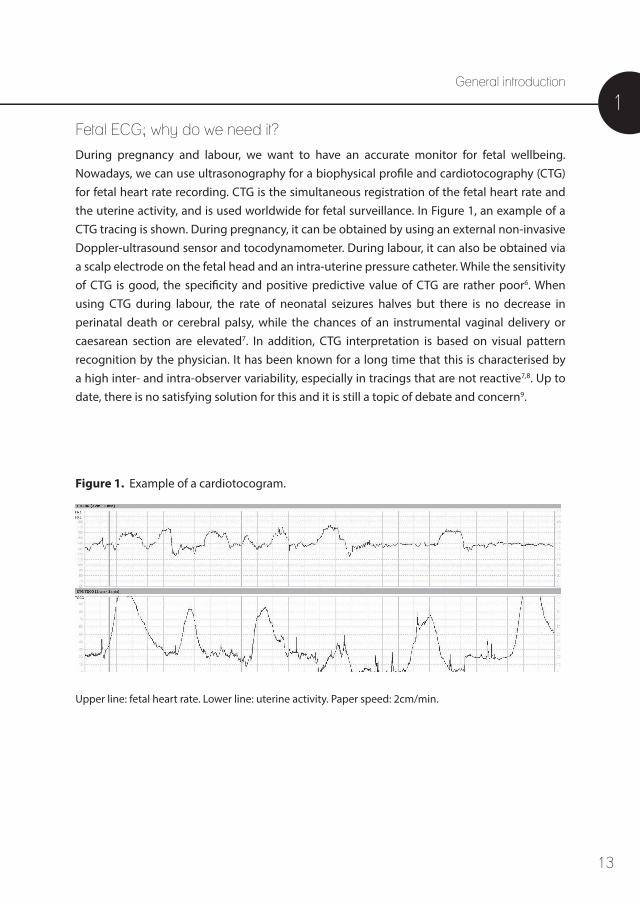
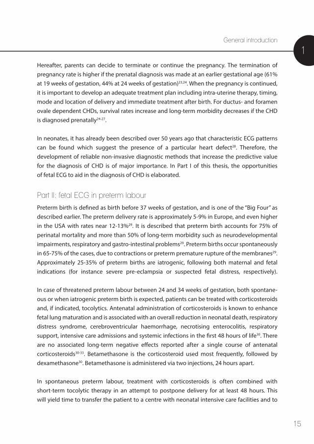
Upper line: fetal heart rate. Lower line: uterine activity. Paper speed: 2cm/min.
Fetal ECG; why do we need it? During pregnancy and labour, we want to have an accurate monitor for fetal wellbeing. Nowadays, we can use ultrasonography for a biophysical profile and cardiotocography (CTG) for fetal heart rate recording. CTG is the simultaneous registration of the fetal heart rate and the uterine activity, and is used worldwide for fetal surveillance. In Figure 1, an example of a CTG tracing is shown. During pregnancy, it can be obtained by using an external non-invasive Doppler-ultrasound sensor and tocodynamometer. During labour, it can also be obtained via a scalp electrode on the fetal head and an intra-uterine pressure catheter. While the sensitivity of CTG is good, the specificity and positive predictive value of CTG are rather poor6. When using CTG during labour, the rate of neonatal seizures halves but there is no decrease in perinatal death or cerebral palsy, while the chances of an instrumental vaginal delivery or caesarean section are elevated7. In addition, CTG interpretation is based on visual pattern recognition by the physician. It has been known for a long time that this is characterised by a high inter- and intra-observer variability, especially in tracings that are not reactive7,8. Up to date, there is no satisfying solution for this and it is still a topic of debate and concern9.
Figure 1. Example of a cardiotocogram.
14
Chapter 1
During pregnancy, there are no complementary diagnostics like ST analysis or fetal scalp blood sampling that can be used to objectify fetal wellbeing10. Moreover, these complementary diagnostics are not applicable in case of prematurity. Therefore, there is need for a non-invasive method that provides more reliable information concerning the fetal condition than CTG alone can provide. The fetal electrocardiogram (ECG) can be measured by direct registration via a scalp electrode on the fetal head during labour, or antepartum via indirect measurements with skin elec-trodes on the maternal abdomen. Fetal ECG recordings obtain beat-to-beat heart rate information and spectral analysis can be performed on these recordings. This gives detailed information regarding heart rate variability, which is a reliable marker for fetal wellbeing11,12. Spectral analysis can quantify rather small changes in fetal heart rate variability, that can remain undetected with visual interpretation of the fetal heart rate tracing13. In addition, the shape and amplitude of the fetal ECG can be assessed. More details concerning fetal ECG measurements and spectral analysis can be found in chapter 3 – technical background.
Part I: fetal ECG and congenital heart disease
CHD is the most common severe congenital anomaly worldwide14. It is estimated that CHD has an incidence of 6-12 per 1000 live births15, of which 4 per 1000 are major forms of CHD that are lethal or require intervention16. CHD is a major health problem, being six times more common than chromosomal anomalies and four times more common than neural tube defects16,17.
CHD is classically diagnosed by echocardiography, which is part of the congenital anomaly scan offered to all pregnant women around 20 weeks of gestation. However, the detection rate of CHD with this ultrasound is rather low and varies from 65-81%16,18-20. Specific echocar-diography has a higher detection rate, with a sensitivity of 90% and a specificity of 98%21. This specific ultrasound is only performed in fetuses with a risk factor for CHD; amongst others family history, maternal diabetes, exposure to teratogens and infections, abnormal nuchal translucency, aneuploidies and other known congenital anomalies. However, up to 90% of all CHD occur in the low risk population17. About 25% of the neonates with major CHD is described to be discharged from the hospital undiagnosed22.
It is important to diagnose CHD early in pregnancy for multiple reasons. First, it enables the identification of associated extracardiac and chromosomal anomalies, that occur in respec-tively 29% and 26% of the fetus with CHD23. Both influence the fetal and postnatal prognosis, and should be included in prenatal and genetic counselling that is offered to parents.
15
1Hereafter, parents can decide to terminate or continue the pregnancy. The termination of pregnancy rate is higher if the prenatal diagnosis was made at an earlier gestational age (61% at 19 weeks of gestation, 44% at 24 weeks of gestation)23,24. When the pregnancy is continued, it is important to develop an adequate treatment plan including intra-uterine therapy, timing, mode and location of delivery and immediate treatment after birth. For ductus- and foramen ovale dependent CHDs, survival rates increase and long-term morbidity decreases if the CHD is diagnosed prenatally24-27.
In neonates, it has already been described over 50 years ago that characteristic ECG patterns can be found which suggest the presence of a particular heart defect28. Therefore, the development of reliable non-invasive diagnostic methods that increase the predictive value for the diagnosis of CHD is of major importance. In Part I of this thesis, the opportunitiesof fetal ECG to aid in the diagnosis of CHD is elaborated.
Part II: fetal ECG in preterm labour Preterm birth is defined as birth before 37 weeks of gestation, and is one of the “Big Four” as described earlier. The preterm delivery rate is approximately 5-9% in Europe, and even higher in the USA with rates near 12-13%29. It is described that preterm birth accounts for 75% of perinatal mortality and more than 50% of long-term morbidity such as neurodevelopmental impairments, respiratory and gastro-intestinal problems29. Preterm births occur spontaneously in 65-75% of the cases, due to contractions or preterm premature rupture of the membranes29. Approximately 25-35% of preterm births are iatrogenic, following both maternal and fetal indications (for instance severe pre-eclampsia or suspected fetal distress, respectively).
In case of threatened preterm labour between 24 and 34 weeks of gestation, both spontane-ous or when iatrogenic preterm birth is expected, patients can be treated with corticosteroids and, if indicated, tocolytics. Antenatal administration of corticosteroids is known to enhance fetal lung maturation and is associated with an overall reduction in neonatal death, respiratory distress syndrome, cerebroventricular haemorrhage, necrotising enterocolitis, respiratory support, intensive care admissions and systemic infections in the first 48 hours of life30. There are no associated long-term negative effects reported after a single course of antenatal corticosteroids30-33. Betamethasone is the corticosteroid used most frequently, followed by dexamethasone30. Betamethasone is administered via two injections, 24 hours apart.
In spontaneous preterm labour, treatment with corticosteroids is often combined with short-term tocolytic therapy in an attempt to postpone delivery for at least 48 hours. This will yield time to transfer the patient to a centre with neonatal intensive care facilities and to
General introduction
16
await maximal beneficial effect of corticosteroids. There are multiple tocolytic agents, and the tocolytic agent of first choice is still a topic of debate and varies considerably in different parts of the world34. The tocolytics most commonly used in clinical practice nowadays are nife-dipine, magnesium sulphate, atosiban, indomethacin, fenoterol and ritodrine. Maintaining tocolytic therapy with nifedipine for more than 48 hours does not improve perinatal outcome, neither is it effective to prolong pregnancy35,36. Both corticosteroids and most tocolytics are known to have influence on fetal heart rate parameters. Since they are administered to a highly vulnerable population, fetuses at risk for preterm birth, it is of the utmost importance to know the exact effects of these drugs. Only then, iatrogenic preterm birth due to misinterpretation of fetal heart rate tracing, caused by therapeutic side-effects, can be avoided. In Part II of this thesis, the effects of corticosteroids and tocolytics on fetal heart rate tracings are studied.
Part III: fetal ECG to prevent asphyxia The World Health Organisation estimated that 4 million neonatal deaths occur every year due to birth asphyxia, representing 38% of deaths of children under 5 years of age37. As described above, fetal monitoring with CTG alone has some shortcomings and additional information regarding the fetal condition is often desired during labour. Fetal blood sampling can offer information regarding the acid-base balance of the fetus. In some parts of the world it is used frequently in clinical practice, but it can only be performed during labour, when the membranes are ruptured and there is enough dilation. In addition, the relation between relative changes of acid-base balance in the various subcutaneous, cerebral and blood levels during fetal hypoxia are not yet fully understood38. The Cochrane review considering electronic fetal monitoring reported more instrumental deliveries, but less neonatal acidosis following fetal blood sampling (p = 0.04 for both) in a subgroup analysis7. However, the access to fetal blood sampling did not influence the difference in neonatal seizures or any other outcomes. Although the NICE guideline recommends fetal blood sampling as an additional test during labour39, this received criticism since fetal blood sampling was never validated and the scientific evidence for its use is questionable40. It is important to realise that fetal blood sampling only gives information regarding the fetal condition at the time of blood collection and sometimes has to be repeated multiple times during labour. There are rare but serious complications such as leakage of cerebral spinal fluid, haemorrhage and sepsis reported following fetal blood sampling41. Therefore, advantages and disadvantages should be consid-ered carefully before performing fetal blood sampling.
Chapter 1

17
1ST analysis is a second source for additional information regarding the fetal condition during labour. The ST segment of the fetal ECG is analysed, following registration via an invasive scalp electrode. More technical details considering ST analysis can be found in chapter 3 – technical background. In combination with CTG, ST analysis was reported to significantly lower the rates of metabolic acidosis42 and operative delivery42,43. Subsequent multicentre trials were performed, including the most recent and largest randomised trial in the USA44. These trials could not reproduce the initial findings and showed no additional benefit for perinatal outcome, besides a reduction in the need for fetal blood sampling44-47. Conflicting results regarding the decrease in metabolic acidosis are reported in recent meta-analysis, indicating the need for more research48-52. In addition, ST analysis gives as many alarms in cases of proven uncompromised fetal condition as in cases of deteriorating fetal condition53. The guidelines that apply to ST analysis state that alarms must be ignored when CTG shows a reassuring pattern. However, taking the high inter-observer variability and low specificity of CTG into account, one can wonder if this is a proper solution for the false alarms encoun-tered. Classifying between a reassuring or non-reassuring CTG determines whether or not to ignore an alarm, making the success of ST monitoring dependent on CTG assessment54. In term fetuses during labour, the orientation of the fetal electrical heart axis can vary between +90 and +180 degrees55. Similar inter-person variations in orientation of the elec-trical heart axis are present in neonates and adults28,56-58. This orientation of the electrical heart axis influences the shape and amplitude of the ECG. We hypothesise that the alignment between the scalp lead and the electrical heart axis, a normal variation in human physiology, can make the difference between ST alarms and no ST alarms. In current ST analysis, this is not taken into account properly. In Part III of this thesis, we will discuss the orientation of the fetal electrical heart axis and its effect on false ST alarms.
General introduction
18
Outline of the thesis This thesis concerns fetal electrocardiography and its applicability. This thesis aims to answer the following questions:
1. Is fetal electrocardiography valuable in diagnosing congenital heart disease in fetuses?
2. What is the influence of corticosteroids and tocolytics on fetal heart rate variability?
3. Are the changes in fetal heart rate variability following corticosteroid administration in the time-domain (obtained by Doppler ultrasound cardiotocography) comparable to the changes in fetal heart rate variability in the frequency-domain (obtained by non-invasive fetal electrocardiography recordings)?
4. Is the variation in orientation of the fetal electrical heart axis in premature fetuses comparable to the variation seen in term fetuses?
5. Is variation in orientation of the electrical heart axis the cause of false ST events in ST analysis during labour?
6. Can we improve the method of ST analysis for fetal monitoring during labour?
To answer these questions we performed several literature and clinical studies, which are described below. The results of these studies are described in this thesis.
Chapter 2 provides physiological background information considering the fetal heart. Chapter 3 provides technical background information considering CTG recordings, non-invasive transabdominal fetal ECG measurements, calculating fetal heart rate variability by means of spectral analysis and the reasoning and technology behind ST analysis.
Part I: fetal ECG and congenital heart disease
Chapter 4 reviews the possibilities of fetal ECG as a screening tool for the detection of CHD in fetuses.
Chapter 5 describes the study protocol of a prospective cohort study, in which the normal ranges for fetal ECG values for the healthy fetus of 18-24 weeks of gestation are established.
Chapter 1
19
1
Part II: fetal ECG in preterm labour
Chapter 6 gives an overview of the literature regarding the influence of the corticosteroids betamethasone and dexamethasone on fetal heart rate parameters, in particular heart rate variability, and fetal behaviour.
Chapter 7 gives an overview of the literature regarding the influence of the tocolytics nifedipine, magnesium sulphate, atosiban, indomethacin, fenoterol and ritodrine on fetal heart rate parameters, in particular heart rate variability.
Chapter 8 presents the results of a prospective cohort study that describes the influence of betamethasone on fetal heart rate variability. Measurements were obtained by non-invasive fetal ECG recordings and spectral analysis was used to calculate fetal heart rate variability.
Part III: fetal ECG to prevent asphyxia Chapter 9 describes the inter-fetal variation and the main orientation of the electrical heart axis in premature fetuses. These results are compared with what is known for term fetuses and adults.
Chapter 10 reveals the results from a post-hoc analysis following the Dutch multicentre randomised controlled ST analysis trial. This study describes the relation between the fetal electrical heart axis and the number of ST alarms that were encountered in fetal monitoring. Chapter 11 presents the results of a case-control study, in which a new method for ST analysis is proposed; relative ST analysis is compared to regular “absolute” ST analysisin intrapartum fetal monitoring.
Chapter 12 provides a summary and general discussion considering the data presented in this thesis. In addition, suggestions for future research are included.
Chapters 4 to 11 are either published or submitted for publication. Therefore, these chaptersare written to be self-contained which causes some overlap between these chapters.
General introduction
20
References
1. World Health Organisation. Neonatal and Perinatal Mortality. 2006; Available at: http://apps.who.int/iris/bitstream/10665/43444/1/9241563206_eng.pdf.
2. Flenady V, Wojcieszek AM, Middleton P, Ellwood D, Erwich JJ, Coory M, et al. Stillbirths: recall to action in high-income countries. Lancet 2016 Feb 13;387(10019):691-702.
3. Vos AA, Bonsel GJ, Steegers EA. Foetal and neonatal mortality in a European perspective: improvement of perinatal health care in the Netherlands still necessary. Ned Tijdschr Geneeskd 2014;158:A7594.
4. van der Kooy J, Poeran J, de Graaf JP, Birnie E, Denktass S, Steegers EA, et al. Planned home compared with planned hospital births in the Netherlands: intrapartum and early neonatal death in low-risk pregnancies. Obstet Gynecol 2011 Nov;118(5):1037-1046.
5. Moss W, Darmstadt GL, Marsh DR, Black RE, Santosham M. Research priorities for the reduction of perinatal and neonatal morbidity and mortality in developing country communities. J Perinatol 2002 Sep;22(6):484-495.
6. Ayres-de-Campos D, Spong CY, Chandraharan E, FIGO Intrapartum Fetal Monitoring Expert Consensus Panel. FIGO consensus guidelines on intrapartum fetal monitoring: Cardiotocography. Int J Gynaecol Obstet 2015 Oct;131(1):13-24.
7. Alfirevic Z, Devane D, Gyte GM. Continuous cardiotocography (CTG) as a form of electronic fetal monitoring (EFM) for fetal assessment during labour. Cochrane Database Syst Rev 2013 May 31;(5):CD006066.
8. Ayres-de-Campos D, Bernardes J, Costa-Pereira A, Pereira-Leite L. Inconsistencies in classification by experts of cardiotocograms and subsequent clinical decision. Br J Obstet Gynaecol 1999 Dec;106(12):1307-1310.
9. Hruban L, Spilka J, Chudacek V, Janku P, Huptych M, Bursa M, et al. Agreement on intrapartum cardiotocogram recordings between expert obstetricians. J Eval Clin Pract 2015 Aug;21(4):694-702.
10. Visser GH, Ayres-de-Campos D, FIGO Intrapartum Fetal Monitoring Expert Consensus Panel. FIGO consensus guidelines on intrapartum fetal monitoring: Adjunctive technologies. Int J Gynaecol Obstet 2015 Oct;131(1):25-29.
11. Williams KP, Galerneau F. Intrapartum fetal heart rate patterns in the prediction of neonatal acidemia. Am J Obstet Gynecol 2003 Mar;188(3):820-823.
12. Anotayanonth S, Subhedar Nimish V, Neilson James P, Harigopal S. Betamimetics for inhibiting preterm labour. Cochrane Database Syst Rev. 2004 Oct 18;(4):CD004352.
13. Siira SM, Ojala TH, Vahlberg TJ, Jalonen JO, Valimaki IA, Rosen KG, et al. Marked fetal acidosis and specific changes in power spectrum analysis of fetal heart rate variability recorded during the last hour of labour. BJOG 2005 Apr;112(4):418-423.
14. van der Linde D, Konings EE, Slager MA, Witsenburg M, Helbing WA, Takkenberg JJ, et al. Birth prevalence of congenital heart disease worldwide: a systematic review and meta-analysis. J Am Coll Cardiol 2011 Nov 15;58(21):2241-2247.
15. Donofrio MT, Moon-Grady AJ, Hornberger LK, Copel JA, Sklansky MS, Abuhamad A, et al. Diagnosis and treatment of fetal cardiac disease: a scientific statement from the American Heart Association. Circulation 2014 May 27;129(21):2183-2242.
Chapter 1
21
1
16. Carvalho JS, Mavrides E, Shinebourne EA, Campbell S, Thilaganathan B. Improving the effectiveness of routine prenatal screening for major congenital heart defects. Heart 2002 Oct;88(4):387-391.
17. Simpson LL. Screening for congenital heart disease. Obstet Gynecol Clin North Am 2004 Mar;31(1):51-59.
18. Ogge G, Gaglioti P, Maccanti S, Faggiano F, Todros T. Prenatal screening for congenital heart disease with four-chamber and outflow-tract views: a multicenter study. Ultrasound Obstet Gynecol 2006 Nov;28(6):779-784.
19. Kirk JS, Riggs TW, Comstock CH, Lee W, Yang SS, Weinhouse E. Prenatal screening for cardiac anomalies: the value of routine addition of the aortic root to the four-chamber view. Obstet Gynecol 1994 Sep;84(3):427-431.
20. Wu Q, Li M, Ju L, Zhang W, Yang X, Yan Y, et al. Application of the 3-vessel view in routine prenatal sonographic screening for congenital heart disease. J Ultrasound Med 2009 Oct;28(10):1319-1324.
21. Cohen EH, Rein AJ. Antenatal diagnosis of cardiac malformation: a structural study. Fetal Diagn Ther 2000 Jan-Feb;15(1):54-60.
22. Sharland G. Fetal cardiac screening and variation in prenatal detection rates of congenital heart disease: why bother with screening at all? Future Cardiol 2012 Mar;8(2):189-202.
23. Clur SA, Van Brussel PM, Mathijssen IB, Pajkrt E, Ottenkamp J, Bilardo CM. Audit of 10 years of referrals for fetal echocardiography. Prenat Diagn 2011 Dec;31(12):1134-1140.
24. Trines J, Fruitman D, Zuo KJ, Smallhorn JF, Hornberger LK, Mackie AS. Effectiveness of prenatal screening for congenital heart disease: assessment in a jurisdiction with universal access to health care. Can J Cardiol 2013 Jul;29(7):879-885.
25. Brick DH, Allan LD. Outcome of prenatally diagnosed congenital heart disease: an update. Pediatr Cardiol 2002 Jul-Aug;23(4):449-453.
26. Hunter LE, Simpson JM. Prenatal screening for structural congenital heart disease. Nat Rev Cardiol 2014 Jun;11(6):323-334.
27. Brown KL, Ridout DA, Hoskote A, Verhulst L, Ricci M, Bull C. Delayed diagnosis of congenital heart disease worsens preoperative condition and outcome of surgery in neonates. Heart 2006 Sep;92(9):1298-1302.
28. Depasquale NP, Burch GE. The Electrocardiogram, Ventricular Gradient and Spatial Vectorcardio-gram during the First Week of Life. Am J Cardiol 1963 Oct;12:482-493.
29. Goldenberg RL, Culhane JF, Iams JD, Romero R. Epidemiology and causes of preterm birth. Lancet 2008 Jan 5;371(9606):75-84.
30. Roberts D, Dalziel S. Antenatal corticosteroids for accelerating fetal lung maturation for women at risk of preterm birth. Cochrane Database Syst Rev 2006 Jul 19;(3):CD004454.
31. Dalziel SR, Lim VK, Lambert A, McCarthy D, Parag V, Rodgers A, et al. Antenatal exposure to beta-methasone: psychological functioning and health related quality of life 31 years after inclusion in randomised controlled trial. BMJ 2005 Sep 24;331(7518):665.
32. Dessens AB, Haas HS, Koppe JG. Twenty-year follow-up of antenatal corticosteroid treatment. Pedi-atrics 2000 Jun;105(6):E77.
General introduction
22
33. Mariotti V, Marconi AM, Pardi G. Undesired effects of steroids during pregnancy. J Matern Fetal Neonatal Med 2004 Nov;16 Suppl 2:5-7.
34. Crowther CA, Brown J, McKinlay CJ, Middleton P. Magnesium sulphate for preventing preterm birth in threatened preterm labour. Cochrane Database Syst Rev 2014 Aug 15;8:CD001060.
35. Roos C, Spaanderman ME, Schuit E, Bloemenkamp KW, Bolte AC, Cornette J, et al. Effect of mainte-nance tocolysis with nifedipine in threatened preterm labor on perinatal outcomes: a randomized controlled trial. JAMA 2013 Jan 2;309(1):41-47.
36. Roos C, Vis JY, Scheepers HC, Bloemenkamp KW, Duvekot HJ, van Eyck J, et al. Fetal fibronectin status and cervical length in women with threatened preterm labor and the effectiveness of main-tenance tocolysis. J Matern Fetal Neonatal Med 2016;29(10):1556-1561.
37. World Health Organisation. Birth Asphyxia - Summary of the previous meeting and protocol overview. 2007; Available at: http://www.curoservice.com/health_ professionals/news/pdf/10-09-2007_birth_asphyxia02.pdf.
38. Amer-Wahlin I, Nord A, Bottalico B, Hansson SR, Ley D, Marsal K, et al. Fetal cerebral energy metab-olism and electrocardiogram during experimental umbilical cord occlusion and resuscitation. J Matern Fetal Neonatal Med 2010 Feb;23(2):158-166.
39. National Institute of Clinical Excellence. Intrapartum care: care of healthy women and their babies during labour. NICE Clinical Guideline. December 2014.
40. Chandraharan E. Should national guidelines continue to recommend fetal scalp blood sampling during labor? J Matern Fetal Neonatal Med 2016 Feb 24:1-4.
41. Schaap TP, Moormann KA, Becker JH, Westerhuis ME, Evers A, Brouwers HA, et al. Cerebrospinal fluid leakage, an uncommon complication of fetal blood sampling: a case report and review of the literature. Obstet Gynecol Surv 2011 Jan;66(1):42-46.
42. Amer-Wahlin I, Hellsten C, Noren H, Hagberg H, Herbst A, Kjellmer I, et al. Cardiotocography only versus cardiotocography plus ST analysis of fetal electrocardiogram for intrapartum fetal monitor-ing: a Swedish randomised controlled trial. Lancet 2001 Aug 18;358(9281):534-538.
43. Westgate J, Harris M, Curnow JS, Greene KR. Plymouth randomized trial of cardiotocogram only versus ST waveform plus cardiotocogram for intrapartum monitoring in 2400 cases. Am J Obstet Gynecol 1993 Nov;169(5):1151-1160.
44. Belfort MA, Saade GR, Thom E, Blackwell SC, Reddy UM, Thorp JM,Jr, et al. A Randomized Trial of Intrapartum Fetal ECG ST-Segment Analysis. N Engl J Med 2015 Aug 13;373(7):632-641.
45. Ojala K, Vaarasmaki M, Makikallio K, Valkama M, Tekay A. A comparison of intrapartum automated fetal electrocardiography and conventional cardiotocography--a randomised controlled study. BJOG 2006 Apr;113(4):419-423.
46. Vayssiere C, David E, Meyer N, Haberstich R, Sebahoun V, Roth E, et al. A French randomized controlled trial of ST-segment analysis in a population with abnormal cardiotocograms during labor. Am J Obstet Gynecol 2007 Sep;197(3):299.e1-299.e6.
47. Westerhuis ME, Visser GH, Moons KG, Zuithoff N, Mol BW, Kwee A. Cardiotocography plus ST analysis of fetal electrocardiogram compared with cardiotocography only for intrapartum monitor-ing: a randomized controlled trial. Obstet Gynecol 2011 Feb;117(2 Pt 1):406-407.
Chapter 1

23
148. Schuit E, Amer-Wahlin I, Ojala K, Vayssiere C, Westerhuis ME, Marsal K, et al. Effectiveness of elec-
tronic fetal monitoring with additional ST analysis in vertex singleton pregnancies at >36 weeks of gestation: an individual participant data metaanalysis. Am J Obstet Gynecol 2013 Mar;208(3):187.e1-187.e13.
49. Blix E, Brurberg KG, Reierth E, Reinar LM, Oian P. ST waveform analysis versus cardiotocography alone for intrapartum fetal monitoring: a systematic review and meta-analysis of randomized trials. Acta Obstet Gynecol Scand 2016 Jan;95(1):16-27.
50. Neilson JP. Fetal electrocardiogram (ECG) for fetal monitoring during labour. Cochrane Database Syst Rev 2015 Dec 21;(12):CD000116.
51. Saccone G, Schuit E, Amer-Wahlin I, Xodo S, Berghella V. Electrocardiogram ST Analysis During Labor: A Systematic Review and Meta-analysis of Randomized Controlled Trials. Obstet Gynecol 2016 Jan;127(1):127-135.
52. Vayssiere C, Ehlinger V, Paret L, Arnaud C. Is STAN monitoring associated with a significant decrease in metabolic acidosis at birth compared with cardiotocography alone? Review of the three meta-analyses that included the recent US trial. Acta Obstet Gynecol Scand 2016 Oct;95(10):1190-1191.
53. Kwee A, Dekkers AH, van Wijk HP, van der Hoorn-van den Beld CW, Visser GH. Occurrence of ST-changes recorded with the STAN S21-monitor during normal and abnormal fetal heart rate patterns during labour. Eur J Obstet Gynecol Reprod Biol 2007 Nov;135(1):28-34.
54. Amer-Wahlin I, Arulkumaran S, Hagberg H, Marsal K, Visser GH. Fetal electrocardiogram: ST waveform analysis in intrapartum surveillance. BJOG 2007 Oct;114(10):1191-1193.
55. Larks SD. Estimation of the Electrical Axis of the Fetal Heart. Am J Obstet Gynecol 1965 Jan 1;91:46-55.
56. Wagner GS, Strauss DG. Marriott’s Practical Electrocardiography. 12th edition ed. Philadelphia: Lippincott Williams & Wilkins; 2014.
57. Goodacre S, McLeod K. ABC of clinical electrocardiography: Paediatric electrocardiography. BMJ 2002 Jun 8;324(7350):1382-1385.
58. Schaffer AI, Beinfield WH. The vectorcardiogram of the newborn infant. Am Heart J 1952 Jul;44(1):89-94.
General introduction

24

25
Chapter 2
2
Physiological background
26
The fetal heart
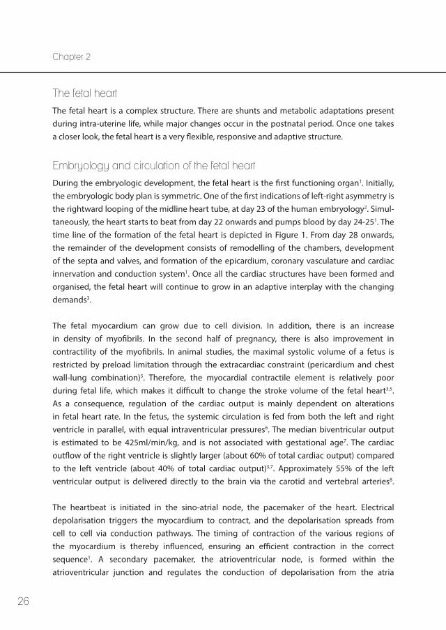
The fetal heart is a complex structure. There are shunts and metabolic adaptations present during intra-uterine life, while major changes occur in the postnatal period. Once one takes a closer look, the fetal heart is a very flexible, responsive and adaptive structure.
Embryology and circulation of the fetal heart
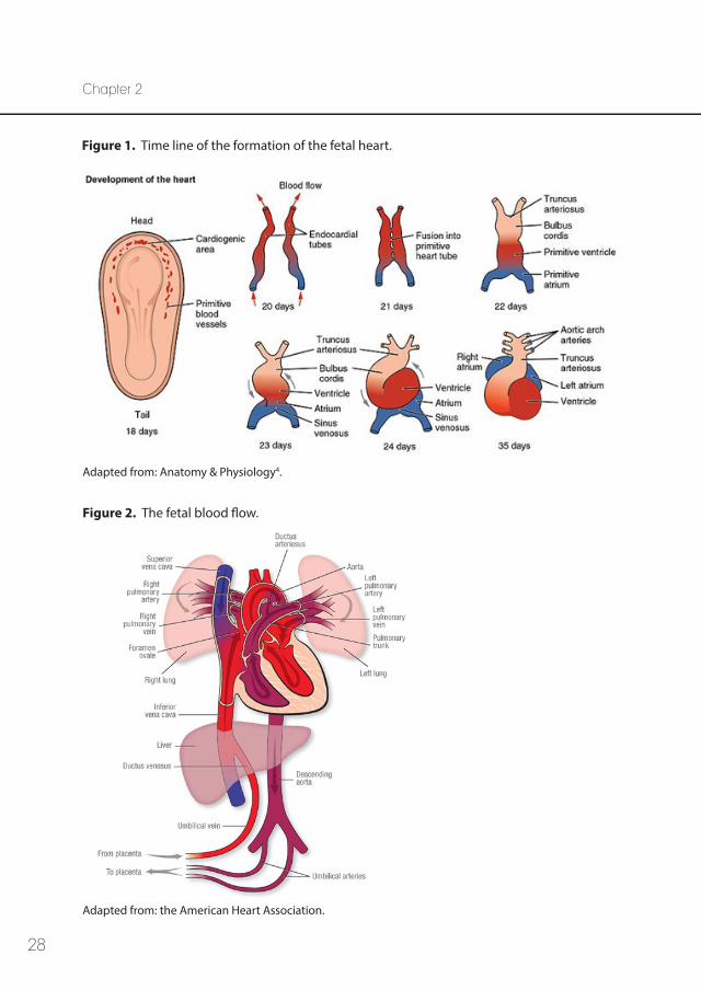
During the embryologic development, the fetal heart is the first functioning organ1. Initially, the embryologic body plan is symmetric. One of the first indications of left-right asymmetry is the rightward looping of the midline heart tube, at day 23 of the human embryology2. Simul-taneously, the heart starts to beat from day 22 onwards and pumps blood by day 24-251. The time line of the formation of the fetal heart is depicted in Figure 1. From day 28 onwards, the remainder of the development consists of remodelling of the chambers, development of the septa and valves, and formation of the epicardium, coronary vasculature and cardiac innervation and conduction system1. Once all the cardiac structures have been formed and organised, the fetal heart will continue to grow in an adaptive interplay with the changing demands3.
The fetal myocardium can grow due to cell division. In addition, there is an increase in density of myofibrils. In the second half of pregnancy, there is also improvement in contractility of the myofibrils. In animal studies, the maximal systolic volume of a fetus is restricted by preload limitation through the extracardiac constraint (pericardium and chest wall-lung combination)5. Therefore, the myocardial contractile element is relatively poor during fetal life, which makes it difficult to change the stroke volume of the fetal heart3,5. As a consequence, regulation of the cardiac output is mainly dependent on alterations in fetal heart rate. In the fetus, the systemic circulation is fed from both the left and right ventricle in parallel, with equal intraventricular pressures6. The median biventricular output is estimated to be 425ml/min/kg, and is not associated with gestational age7. The cardiac outflow of the right ventricle is slightly larger (about 60% of total cardiac output) compared to the left ventricle (about 40% of total cardiac output)3,7. Approximately 55% of the left ventricular output is delivered directly to the brain via the carotid and vertebral arteries8.
The heartbeat is initiated in the sino-atrial node, the pacemaker of the heart. Electrical depolarisation triggers the myocardium to contract, and the depolarisation spreads from cell to cell via conduction pathways. The timing of contraction of the various regions of the myocardium is thereby influenced, ensuring an efficient contraction in the correct sequence1. A secondary pacemaker, the atrioventricular node, is formed within the atrioventricular junction and regulates the conduction of depolarisation from the atria
Chapter 2
27
2to the ventricles. From there, the depolarisation is transmitted through both ventricles via the bundle of His. Following neural input of the sympathetic and parasympathetic branches of the autonomic nervous system, the heart rate can be modified.
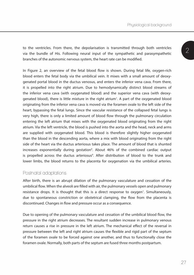
In Figure 2, an overview of the fetal blood flow is shown. During fetal life, oxygen-rich blood enters the fetal body via the umbilical vein. It mixes with a small amount of deoxy-genated portal blood in the ductus venosus, and enters the inferior vena cava. From there, it is propelled into the right atrium. Due to hemodynamically distinct blood streams of the inferior vena cava (with oxygenated blood) and the superior vena cava (with deoxy-genated blood), there is little mixture in the right atrium1. A part of the oxygenated blood originating from the inferior vena cava is moved via the foramen ovale to the left side of the heart, bypassing the fetal lungs. Since the vascular resistance of the collapsed fetal lungs is very high, there is only a limited amount of blood flow through the pulmonary circulation entering the left atrium that mixes with the oxygenated blood originating from the right atrium. Via the left ventricle, the blood is pushed into the aorta and the head, neck and arms are supplied with oxygenated blood. This blood is therefore slightly higher oxygenated than the blood in the descending aorta, where a mix with blood originating from the right side of the heart via the ductus arteriosus takes place. The amount of blood that is shunted increases exponentially during gestation9. About 46% of the combined cardiac output is propelled across the ductus arteriosus9. After distribution of blood to the trunk and lower limbs, the blood returns to the placenta for oxygenation via the umbilical arteries.
Physiological background
Postnatal adaptations
After birth, there is an abrupt dilation of the pulmonary vasculature and cessation of the umbilical flow. When the alveoli are filled with air, the pulmonary vessels open and pulmonary resistance drops. It is thought that this is a direct response to oxygen1. Simultaneously, due to spontaneous constriction or obstetrical clamping, the flow from the placenta is discontinued. Changes in flow and pressure occur as a consequence.
Due to opening of the pulmonary vasculature and cessation of the umbilical blood flow, the pressure in the right atrium decreases. The resultant sudden increase in pulmonary venous return causes a rise in pressure in the left atrium. The mechanical effect of the reversal in pressure between the left and right atrium causes the flexible and rigid part of the septum of the foramen ovale to be forced against one another, and thus to functionally close the foramen ovale. Normally, both parts of the septum are fused three months postpartum.
28
Figure 2. The fetal blood flow.
Adapted from: the American Heart Association.
Chapter 2
Adapted from: Anatomy & Physiology4.
Figure 1. Time line of the formation of the fetal heart.
29
2
The decrease in pressure in the pulmonary trunk resulting from opening of the pulmonary circulation, is thought to cause a slight reverse flow of oxygenated aortic blood through the ductus arteriosus1. It is thought that the oxygen tension might locally induce the vascular smooth muscle cells to contract and therefore restrict the blood flow. However, the exact mechanism for closure of the ductus arteriosus is not clear yet1. At term, constriction of the ductus arteriosus normally occurs within 72 hours after birth10.
In addition, the ductus venosus closes soon after birth as no blood is flowing through theumbilical vein anymore1. Hereafter, the portal circulation replaces the hepatic blood flow from the placenta.
Hypoxia and the fetal heart
Hypoxemia is defined as an abnormally low level of oxygen in the arterial blood. In case of the human fetus, the natural environment is hypoxemic (but not pathologi-cally hypoxic)11. The fetus can thrive under these conditions due to several adaptation mechanisms, amongst others fetal haemoglobin that has a high affinity for oxygen and allows easy diffusion from the maternal to the fetal circulation, the organisation of the fetal circulation as described above and the higher rate of tissue perfusion in the fetus compared to adults. These mechanisms compensate for the lower oxygen levels in the fetal blood, and make sure that oxygen supply is sufficient at tissue level.
In case of hypoxia, the oxygen supply at tissue level is inadequate and tissue damage can arise. Even so, the fetus can compensate through variabele mechanisms such as redistri-bution of blood flow and activation of the anaerobic metabolism. Progressive metabolic acidosis occurs following hypoxia if these mechanisms are not sufficient in compensating the oxygen shortage or in case of longlasting activation of the anaerobic metabolism. When there is acidosis in combination with organ damage, this is referred to as asphyxia.
In the late 1900s, multiple animal studies have been performed regarding the effect of hypoxia on the fetal heart. As summarised by Widmark et al.12, these studies showed that when the maternal-placental blood flow is reduced, and therefore an acute hypoxic event is induced, both the chemo- and baroreceptor reflexes are activated. Initially, the chemoreceptor reflex increases both sympathetic and parasympathetic tone. In the fetal heart, parasympathetic influences predominate resulting in fetal heart rate bradycardia. During this reduced fetal heart rate the end-diastolic filling time is prolonged, which increases the end-diastolic volume. Therefore, cardiac output and perfusion pressure are both maintained despite fetal heart rate bradycardia13. Sympathetic activation causes
Physiological background
30
peripheral vasoconstriction, realising blood flow redistribution favouring the brain, heart and adrenals13,14. An increase in mean arterial blood pressure activates the baroreceptor reflex, and causes a secondary (inhibitory) effect on the fetal heart rate. Once initiated, the peripheral vasoconstriction is maintained by release of vasoactive agents14. These can be measured 15 minutes from the onset of acute hypoxia13. In addition, it is thought that impulse conduction through the atrioventricular node is directly inhibited (parasympa-thetic activation, baroreceptor reflexes) or directly facilitated (sympathetic activation)12.
After reoxygenation, there is a baroreceptor reflex-enhanced parasympathetic tone that causes a negative chronotropic effect. This might exert a protective effect on the cardi-ovascular system, as this is still affected by an enhanced sympathetic tone due to circulat-ing humoral agents12. Therefore, late decelerations may be interpreted as an adaptive mechanism to excessive sympathetic influence after a hypoxic event12. Because of the increased levels of catecholamines and other constrictor agents in the fetal circulation, the peripheral vasoconstrictor response and redistribution of blood flow is maintained longer13.
A previous study by Amer-Wåhlin et al.15 showed that cardiac effects following hypoxia (changes in the electrocardiogram) precede cerebral damage, and can therefore been seen as a marker for fetal distress. In the fetal heart, a high amount of glycogen is stored in case anaerobic metabolism needs to set in when oxygen supply is inade-quate16. As described by Rosén and Isaksson16, in the initial less severe states of hypoxia both liver and cerebral glycogen stores are well maintained and the fetus maintains a normal cardiac rhythm. When hypoxia becomes more severe, cerebral glycogen is still unaffected while there is depletion of liver glycogen stores. In 50% of these cases, fetal heart rate bradycardia was present. In case of severe hypoxia, both brain and liver glycogen stores were depleted and all cases showed fetal heart rate bradycardia. In addition, a correlation between changes in the ST segment of the electrocardio-gram and depletion of glycogen in the fetal heart was found in animal experiments16.
Fetal heart rate variability & the autonomic nervous system
Variability in the fetal heart rate is a resultant from the counteracting autonomic influences of the sympathetic and parasympathetic nervous system17,18. These systems do not simply interact as a “push-pull” system, but complex interactions exist between them18. Amongst others, intrinsic variability of the heart, the baroreceptor reflex, humoral agents (for instancecirculating catecholamines), respiration and thermoregulation have their influence on heart rate variability18,19.
Chapter 2
31
2
When fetal heart rate variability is normal, this is a reliable indicator of fetal wellbeing, irre-spective of the fetal heart rate pattern20. Decreased fetal heart rate variability is associated with fetal acidosis, low Apgar score and perinatal death20. Therefore, fetal heart rate varia-bility is one of the most important factors to assess in fetal monitoring. Specific heart rate variability parameters that are mentioned below (for instance high-frequency (HF)-energy and low-frequency (LF)-energy) are described in detail in chapter 3 – technical background.
Multiple confounding factors can influence fetal heart rate variability. Several drugs that are commonly used in obstetric care may alter the fetal heart rate, heart rate variability, or the central nervous system. Two categories of these drugs will be described in this thesis in chapter 6 (betamethasone, a corticosteroid) and chapter 7 (various tocolytic drugs). Another confounder is birthweight; a study by Siira et al.21 revealed that HF-power is negatively related to birthweight in case of fetal growth restriction. In addition, in cases where pregnancy is complicated by pregnancy induced hypertension or pre-eclampsia higher absolute LF- and HF-power was found in comparison to a control group, and greater instability of the fetal heart rate was seen in this group22. Changes in fetal behavioural states and diurnal rhythm can also influence fetal heart rate variability23,24. Below, the influences of gestational age and oxygen depletion on fetal heart rate variability are described in more detail.
When gestational age advances, there is evolution towards a more stable and mature autonomic nervous system25. By means of spectral analysis of fetal heart rate variability, the functional state of the autonomic nervous system can be assessed. This is described in more detail in chapter 3 – technical background. Prior research has shown there is an increase in absolute LF- and HF-power with increase in gestational age26-28. The sympathetic nervous system is effective from mid-gestation onwards, while the parasympathetic nervous system matures later18,29. The typical parasympathetic reflex responses are first seen during term gestation and reach adult levels after birth29. As gestational age progresses the parasym-pathetic modulation seems to increase, while the sympathetic modulation decreases30.
As described previously in this chapter, the autonomic nervous system is activated in case of oxygen shortage. Subsequently, the beat-to-beat heart rate is modulated17. Initially, in case of mild fetal distress, there is a rise in fetal heart rate variability (total power, LF- and HF-power, LF/HF ratio)21,31,32. This is a reflection of increased sympathetic activity. Thereafter, in case of fetal hypoxia or acidemia, there is a decrease in fetal heart rate variability. More specifically, the absolute LF-power seems to be decreased33. In prior research, normalised LF-power was found to be negatively associated with fetal pH, while normalised HF-power was positively
Physiological background
32
associated with fetal pH34,35. This can be explained by increased sympathetic and decreased parasympathetic cardiac modulation, as the internal pH value decreases. This is in line with the increased LF/HF ratio that is found in acidotic fetuses, which indicates a shift towards sympathetic predominance21. It is suggested that both circulating catecholamines and sympathetic neural activity cause the decreased heart rate variability that is seen in case of severe fetal distress35. Eventually, in case of major fetal compromise, loss of central autonomic cardiovascular control and cardiovascular decompensation is likely21,33.
In adult cardiology, heart rate variability already plays a role in determining prognosis and guiding therapy36. Almost 25 years ago, it was suggested that analysis of fetal heart rate variability might be able to improve the diagnosis of pathological conditions25. Since then, advances have been made regarding defining, monitoring and interpreting fetal heart rate variability.
Chapter 2

33
2References 1. Schoenwolf GC, Bleyl SB, Brauer PR, Francis-West PH. Chapter 12: Development of the heart,
Chapter 13: Development of the vasculature. Larsen’s Human Embryology. Fourth edition ed. Phila-delphia: Churchill Livingstone Elsevier; 2009. p. 337-433.
2. Kathiriya IS, Srivastava D. Left-right asymmetry and cardiac looping: implications for cardiac devel-opment and congenital heart disease. Am J Med Genet 2000 Winter;97(4):271-279.
3. Kiserud T, Acharya G. The fetal circulation. Prenat Diagn 2004 Dec 30;24(13):1049-1059.
4. OpenStax. Anatomy & Physiology. Chapter 19.5: Development of the Heart. Available at: http://philschatz.com/anatomy-book/contents/m46673.html.
5. Grant DA, Fauchere JC, Eede KJ, Tyberg JV, Walker AM. Left ventricular stroke volume in the fetal sheep is limited by extracardiac constraint and arterial pressure. J Physiol 2001 Aug 15;535(Pt 1):231-239.
6. Johnson P, Maxwell DJ, Tynan MJ, Allan LD. Intracardiac pressures in the human fetus. Heart 2000 Jul;84(1):59-63.
7. Mielke G, Benda N. Cardiac output and central distribution of blood flow in the human fetus. Circu-lation 2001 Mar 27;103(12):1662-1668.
8. Artman M, Mahony L, Teitel DF. Perinatal Cardiovascular Physiology. Neonatal cardiology. Second Edition ed.: McGraw-Hill Professional; 2010. p. 45-60.
9. Winberg P, Jansson M, Marions L, Lundell BP. Left ventricular output during postnatal circulatory adaptation in healthy infants born at full term. Arch Dis Child 1989 Oct;64(10 Spec No):1374-1378.
10. Dice JE, Bhatia J. Patent ductus arteriosus: an overview. J Pediatr Pharmacol Ther 2007 Jul;12(3):138-146.
11. Rainaldi MA, Perlman JM. Pathophysiology of Birth Asphyxia. Clin Perinatol 2016 Sep;43(3):409-422.
12. Widmark C, Lindecrantz K, Murray H, Rosen KG. Changes in the PR, RR intervals and ST waveform of the fetal lamb electrocardiogram with acute hypoxemia. J Dev Physiol 1992 Sep;18(3):99-103.
13. Giussani DA. The fetal brain sparing response to hypoxia: physiological mechanisms. J Physiol 2016 Mar 1;594(5):1215-1230.
14. Thakor AS, Giussani DA. Effects of acute acidemia on the fetal cardiovascular defense to acute hypoxemia. Am J Physiol Regul Integr Comp Physiol 2009 Jan;296(1):R90-9.
15. Amer-Wahlin I, Nord A, Bottalico B, Hansson SR, Ley D, Marsal K, et al. Fetal cerebral energy metab-olism and electrocardiogram during experimental umbilical cord occlusion and resuscitation. J Matern Fetal Neonatal Med 2010 Feb;23(2):158-166.
16. Rosén K, Isaksson O. Alterations in Fetal Heart Rate and ECG Correlated to Glycogen, Creatine Phosphate and ATP Levels during Graded Hypoxia. Biol Neonate 1976;30:17-24.
17. Van Ravenswaaij-Arts C, Kollee L, Hopman J, Stoelinga G, van Geijn H. Heart rate variabilitiy. Ann Intern Med 1993;118:436-447.
18. Dalton KJ, Dawes GS, Patrick JE. The autonomic nervous system and fetal heart rate variability. Am J Obstet Gynecol 1983 Jun 15;146(4):456-462.
Physiological background
34
19. Jongen GJ, van der Hout-van der Jagt,MB, Oei SG, van de Vosse FN, Bovendeerd PH. Simulation of fetal heart rate variability with a mathematical model. Med Eng Phys 2017 Feb 11.
20. Paul RH, Suidan AK, Yeh S, Schifrin BS, Hon EH. Clinical fetal monitoring. VII. The evaluation and significance of intrapartum baseline FHR variability. Am J Obstet Gynecol 1975 Sep 15;123(2):206-210.
21. Siira SM, Ojala TH, Vahlberg TJ, Jalonen JO, Valimaki IA, Rosen KG, et al. Marked fetal acidosis and specific changes in power spectrum analysis of fetal heart rate variability recorded during the last hour of labour. BJOG 2005 Apr;112(4):418-423.
22. Yum M, Kim C, Park E, Kim J. Instability and frequency-domain variability of heart rates in fetusus with or without growth restriction affected bij severe preeclampsia. Physiol. Meas. 2004;25:1105-1113.
23. Davidson SR, Rankin JH, Martin CB,Jr, Reid DL. Fetal heart rate variability and behavioral state: analysis by power spectrum. Am J Obstet Gynecol 1992 Sep;167(3):717-722.
24. Suzuki T, Kimura Y, Murotsuki J, Murakami T, Uehara S, Okamura K. Detection of a biorhythm of human fetal autonomic nervous activity by a power spectral analysis. Am J Obstet Gynecol 2001 Nov;185(5):1247-1252.
25. Karin J, Hirsch M, Akselrod S. An estimate of fetal autonomic state by spectral analysis of fetal heart rate fluctuations. Pediatr Res 1993 Aug;34(2):134-138.
26. Van Leeuwen P, Geue D, Lange S, Hatzmann W, Gronemeyer D. Changes in the frequency power spectrum of fetal heart rate in the course of pregnancy. Prenat Diagn 2003;23:909-916.
27. David M, Hirsch M, Karin J, Toledo E, Akselrod S. An estimate of fetal autonomic state by time-fre-quency analysis of fetal heart rate variability. J Appl Physiol (1985) 2007 Mar;102(3):1057-1064.
28. van Laar JO, Warmerdam GJ, Verdurmen KM, Vullings R, Peters CH, Houterman S, et al. Fetal heart rate variability during pregnancy, obtained from non-invasive electrocardiogram recordings. Acta Obstet Gynecol Scand 2014 Jan;93(1):93-101.
29. Assali NS, Brinkman CR,3rd, Woods JR,Jr, Dandavino A, Nuwayhid B. Development of neurohumoral control of fetal, neonatal, and adult cardiovascular functions. Am J Obstet Gynecol 1977 Dec 1;129(7):748-759.
30. van Laar JO, Peters CH, Vullings R, Houterman S, Oei SG. Power spectrum analysis of fetal heart rate variability at near term and post term gestation during active sleep and quiet sleep. Early Hum Dev 2009 Dec;85(12):795-798.
31. Dalton KJ, Dawes GS, Patrick JE. Diurnal, respiratory, and other rhythms of fetal heart rate in lambs. Am J Obstet Gynecol 1977 Feb 15;127(4):414-424.
32. Min SW, Ko H, Kim CS. Power spectral analysis of heart rate variability during acute hypoxia in fetal lambs. Acta Obstet Gynecol Scand 2002 Nov;81(11):1001-1005.
33. Van Laar JO, Porath MM, Peters CH, Oei SG. Spectral analysis of fetal heart rate variability for fetal surveillance: review of the literature. Acta Obstet Gynecol Scand 2008;87(3):300-306.
34. van Laar JO, Peters CH, Houterman S, Wijn PF, Kwee A, Oei SG. Normalized spectral power of fetal heart rate variability is associated with fetal scalp blood pH. Early Hum Dev 2011 Apr;87(4):259-263.
Chapter 2

35
2
Physiological background
35. van Laar JO, Peters CH, Vullings R, Houterman S, Bergmans JW, Oei SG. Fetal autonomic response to severe acidaemia during labour. BJOG 2010 Mar;117(4):429-437.
36. Rosenstock EG, Cassuto Y, Zmora E. Heart rate variability in the neonate and infant: analytical methods, physiological and clinical observations. Acta Paediatr 1999 May;88(5):477-482.

36

37
3
Chapter 3
Technical background
38
Chapter 3
Introduction
In this thesis, various studies considering fetal monitoring are described in which multiple technologies were used. In this chapter, the technological background regarding these techniques will be explained. First, the cardiotocogram (CTG) will be discussed. This technique is mainly used in prior studies regarding fetal heart rate parameters, as described in chapters 6 and 7. The fetal electrocardiogram (ECG) is a technique that is still developing. In chapter 5 and chapter 9, fetal ECG measurements are used to establish the normal fetal ECG in mid-term pregnancy and to calculate the orientation of the electrical heart axis in premature fetuses. Once fetal ECG recordings have been performed, these can be used to apply spectral analysis and hence give more reliable estimates considering fetal heart rate variability, as described in chapter 8. During labour, fetal ECG measurements are required to be able to perform ST analysis. Chapters 10 and 11 relate to ST analysis.
The cardiotocogram
External cardiotocography (CTG) is a Doppler ultrasound-based system, detecting heartbeats as a reflection from all moving parts of the fetal heart. Simultaneously, uterine contractions are recorded by means of tocodynamometry. External registration of the fetal heart rate is prone to signal loss, inadvertent maternal heart rate monitoring, signal artefacts such as double-counting or half-counting and it might not record fetal arrhythmias accurately1. In addition, this technology is not able to provide beat-to-beat interval registration, since some of these irradiated ultrasound waves are reflected by the valves instead of the walls of the heart, causing inaccuracies. Moreover, the autocorrelation techniques used cause averaging of two to three subsequent cardiac cycles. Therefore, it cannot follow fast changes in fetal heart rate signal. This has a significant influence on the values of some variability indices; mainly on the indices that describe parasympathetic activity. As studied by Jezewski et al.2, this introduces an average error in the RR-intervals of 0.42 ms, compared to the fetal ECG. For visual interpretation of fetal heart rate variability this has little influence, given the limited resolution of the human eye. However, in automated analysis this can have a significant effect on values of variability indices2. Therefore, CTG is an imprecise method for acquiring varia-bility of the fetal heart rate. By means of fetal ECG measurements, QRS complex detection enables precise registration of fetal beat-to-beat heart rate variability. This technique considers the full shape of the analysed signal; an example is shown in Figure 1. Definitions for quantitative evaluation of fetal heart rate variability (long- and short-term variability) were originally proposed for the direct fetal ECG signal2. However, they have been applied for ultra-sound-based registration without any adaptation.
39
3
Technical background
Besides the shortcomings of CTG as listed above, the specificity and positive predictive value of CTG are rather poor as previously described in chapter 1 – introduction1. This indicates the need for a technology that enables more accurate and reliable monitoring of fetal wellbeing.
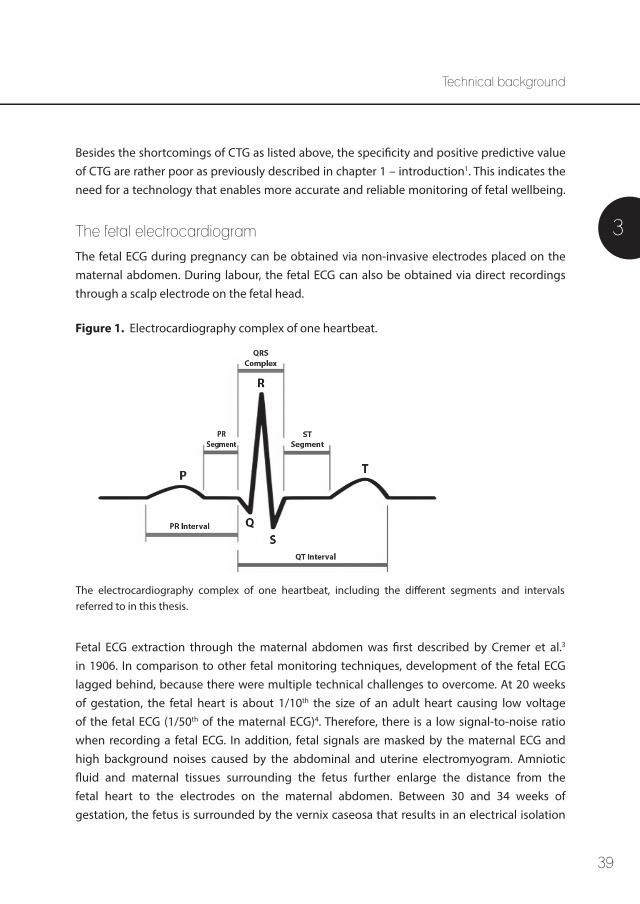
The fetal electrocardiogram The fetal ECG during pregnancy can be obtained via non-invasive electrodes placed on thematernal abdomen. During labour, the fetal ECG can also be obtained via direct recordings through a scalp electrode on the fetal head.
Fetal ECG extraction through the maternal abdomen was first described by Cremer et al.3 in 1906. In comparison to other fetal monitoring techniques, development of the fetal ECG lagged behind, because there were multiple technical challenges to overcome. At 20 weeks of gestation, the fetal heart is about 1/10th the size of an adult heart causing low voltage of the fetal ECG (1/50th of the maternal ECG)4. Therefore, there is a low signal-to-noise ratio when recording a fetal ECG. In addition, fetal signals are masked by the maternal ECG and high background noises caused by the abdominal and uterine electromyogram. Amniotic fluid and maternal tissues surrounding the fetus further enlarge the distance from the fetal heart to the electrodes on the maternal abdomen. Between 30 and 34 weeks of gestation, the fetus is surrounded by the vernix caseosa that results in an electrical isolation
The electrocardiography complex of one heartbeat, including the different segments and intervals referred to in this thesis.
Figure 1. Electrocardiography complex of one heartbeat.
40
which diminishes the signal amplitude further, and which is the main cause of the poor signal-to-noise ratio in this period5,6. Furthermore, the complex three-dimensional shape of the fetal ECG alters with changes in the fetal position with reference to the electrodes that are placed in a fixed configuration on the maternal abdomen. Despite these challenges, fetal ECG technique improved and it has previously been demonstrated that fetal heart rate recordings obtained by non-invasive fetal ECG measurements correlate very well with fetal ECG signals obtained directly via a scalp electrode7.
Apart from the technical difficulties encountered when performing a fetal ECG, the inter-pretation is also challenging. This is mainly caused by the marked differences in prenatal and postnatal circulation, which are elaborated in chapter 2 – physiological background.
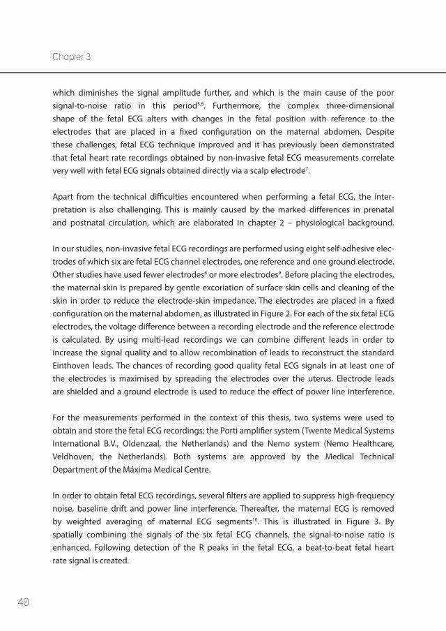
In our studies, non-invasive fetal ECG recordings are performed using eight self-adhesive elec-trodes of which six are fetal ECG channel electrodes, one reference and one ground electrode. Other studies have used fewer electrodes8 or more electrodes9. Before placing the electrodes, the maternal skin is prepared by gentle excoriation of surface skin cells and cleaning of the skin in order to reduce the electrode-skin impedance. The electrodes are placed in a fixed configuration on the maternal abdomen, as illustrated in Figure 2. For each of the six fetal ECG electrodes, the voltage difference between a recording electrode and the reference electrode is calculated. By using multi-lead recordings we can combine different leads in order to increase the signal quality and to allow recombination of leads to reconstruct the standard Einthoven leads. The chances of recording good quality fetal ECG signals in at least one of the electrodes is maximised by spreading the electrodes over the uterus. Electrode leads are shielded and a ground electrode is used to reduce the effect of power line interference.
For the measurements performed in the context of this thesis, two systems were used to obtain and store the fetal ECG recordings; the Porti amplifier system (Twente Medical Systems International B.V., Oldenzaal, the Netherlands) and the Nemo system (Nemo Healthcare, Veldhoven, the Netherlands). Both systems are approved by the Medical Technical Department of the Máxima Medical Centre.
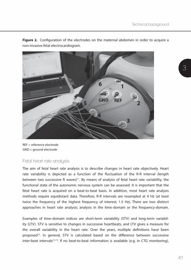
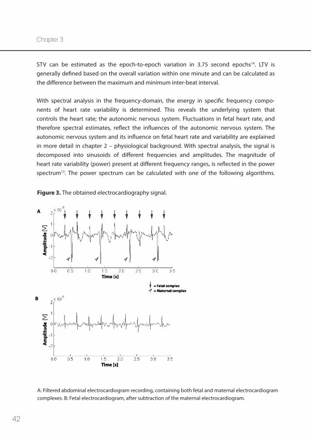
In order to obtain fetal ECG recordings, several filters are applied to suppress high-frequency noise, baseline drift and power line interference. Thereafter, the maternal ECG is removed by weighted averaging of maternal ECG segments10. This is illustrated in Figure 3. By spatially combining the signals of the six fetal ECG channels, the signal-to-noise ratio is enhanced. Following detection of the R peaks in the fetal ECG, a beat-to-beat fetal heart rate signal is created.
Chapter 3
41
3
Technical background
REF = reference electrodeGND = ground electrode
Figure 2. Configuration of the electrodes on the maternal abdomen in order to acquire a non-invasive fetal electrocardiogram.
Fetal heart rate analysis
The aim of fetal heart rate analysis is to describe changes in heart rate objectively. Heart rate variability is depicted as a function of the fluctuation of the R-R interval (length between two successive R waves)11. By means of analysis of fetal heart rate variability, the functional state of the autonomic nervous system can be assessed. It is important that the fetal heart rate is acquired on a beat-to-beat basis. In addition, most heart rate analysis methods require equidistant data. Therefore, R-R intervals are resampled at 4 Hz (at least twice the frequency of the highest frequency of interest; 1.5 Hz). There are two distinct approaches in heart rate analysis; analysis in the time-domain or the frequency-domain.
Examples of time-domain indices are short-term variability (STV) and long-term variabil-ity (LTV). STV is sensitive to changes in successive heartbeats, and LTV gives a measure for the overall variability in the heart rate. Over the years, multiple definitions have been proposed12. In general, STV is calculated based on the difference between successive inter-beat intervals12,13. If no beat-to-beat information is available (e.g. in CTG monitoring),
42
STV can be estimated as the epoch-to-epoch variation in 3.75 second epochs14. LTV is generally defined based on the overall variation within one minute and can be calculated as the difference between the maximum and minimum inter-beat interval.
With spectral analysis in the frequency-domain, the energy in specific frequency compo-nents of heart rate variability is determined. This reveals the underlying system that controls the heart rate; the autonomic nervous system. Fluctuations in fetal heart rate, and therefore spectral estimates, reflect the influences of the autonomic nervous system. The autonomic nervous system and its influence on fetal heart rate and variability are explained in more detail in chapter 2 – physiological background. With spectral analysis, the signal is decomposed into sinusoids of different frequencies and amplitudes. The magnitude of heart rate variability (power) present at different frequency ranges, is reflected in the power spectrum13. The power spectrum can be calculated with one of the following algorithms.
= Fetal complex= Maternal complex
A
B
Am
plit
ude
Am
plit
ude
Time [s]
Time [s]
A: Filtered abdominal electrocardiogram recording, containing both fetal and maternal electrocardiogram complexes. B: Fetal electrocardiogram, after subtraction of the maternal electrocardiogram.
Figure 3. The obtained electrocardiography signal.
Chapter 3
43
3
Technical background
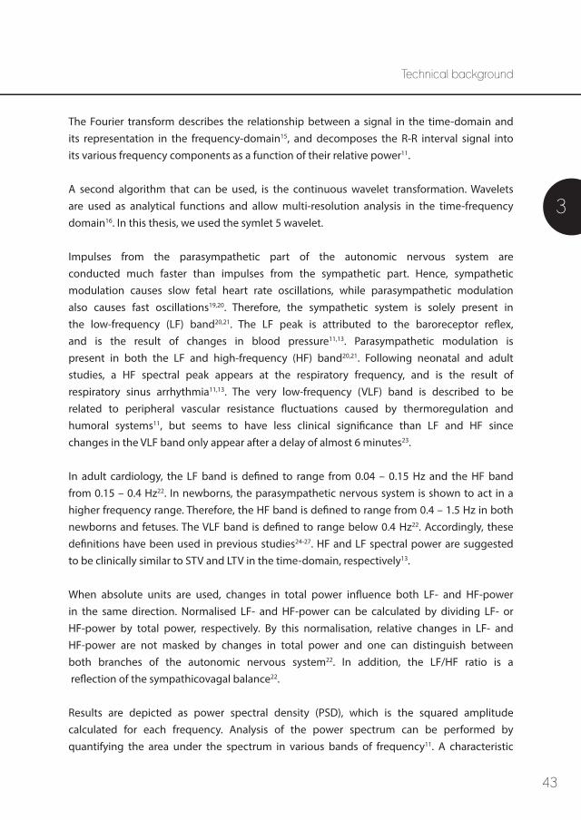
The Fourier transform describes the relationship between a signal in the time-domain and its representation in the frequency-domain15, and decomposes the R-R interval signal into its various frequency components as a function of their relative power11.
A second algorithm that can be used, is the continuous wavelet transformation. Wavelets are used as analytical functions and allow multi-resolution analysis in the time-frequency domain16. In this thesis, we used the symlet 5 wavelet.
Impulses from the parasympathetic part of the autonomic nervous system are conducted much faster than impulses from the sympathetic part. Hence, sympathetic modulation causes slow fetal heart rate oscillations, while parasympathetic modulation also causes fast oscillations19,20. Therefore, the sympathetic system is solely present in the low-frequency (LF) band20,21. The LF peak is attributed to the baroreceptor reflex, and is the result of changes in blood pressure11,13. Parasympathetic modulation is present in both the LF and high-frequency (HF) band20,21. Following neonatal and adult studies, a HF spectral peak appears at the respiratory frequency, and is the result of respiratory sinus arrhythmia11,13. The very low-frequency (VLF) band is described to be related to peripheral vascular resistance fluctuations caused by thermoregulation and humoral systems11, but seems to have less clinical significance than LF and HF since changes in the VLF band only appear after a delay of almost 6 minutes23.
In adult cardiology, the LF band is defined to range from 0.04 – 0.15 Hz and the HF band from 0.15 – 0.4 Hz22. In newborns, the parasympathetic nervous system is shown to act in a higher frequency range. Therefore, the HF band is defined to range from 0.4 – 1.5 Hz in both newborns and fetuses. The VLF band is defined to range below 0.4 Hz22. Accordingly, these definitions have been used in previous studies24-27. HF and LF spectral power are suggested to be clinically similar to STV and LTV in the time-domain, respectively13.
When absolute units are used, changes in total power influence both LF- and HF-power in the same direction. Normalised LF- and HF-power can be calculated by dividing LF- or HF-power by total power, respectively. By this normalisation, relative changes in LF- and HF-power are not masked by changes in total power and one can distinguish between both branches of the autonomic nervous system22. In addition, the LF/HF ratio is a reflection of the sympathicovagal balance22.
Results are depicted as power spectral density (PSD), which is the squared amplitude calculated for each frequency. Analysis of the power spectrum can be performed by quantifying the area under the spectrum in various bands of frequency11. A characteristic
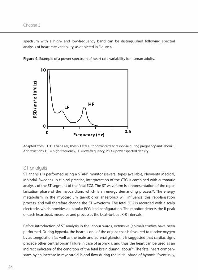
44
spectrum with a high- and low-frequency band can be distinguished following spectral analysis of heart rate variability, as depicted in Figure 4.
Adapted from: J.O.E.H. van Laar, Thesis: Fetal autonomic cardiac response during pregnancy and labour15. Abbreviations: HF = high-frequency, LF = low-frequency, PSD = power spectral density.
Figure 4. Example of a power spectrum of heart rate variability for human adults.
ST analysis ST analysis is performed using a STAN® monitor (several types available, Neoventa Medical, Mölndal, Sweden). In clinical practice, interpretation of the CTG is combined with automatic analysis of the ST segment of the fetal ECG. The ST waveform is a representation of the repo-larisation phase of the myocardium, which is an energy demanding process28. The energy metabolism in the myocardium (aerobic or anaerobic) will influence this repolarisation process, and will therefore change the ST waveform. The fetal ECG is recorded with a scalp electrode, which provides a unipolar ECG lead configuration. The monitor detects the R peak of each heartbeat, measures and processes the beat-to-beat R-R intervals.
Before introduction of ST analysis in the labour wards, extensive (animal) studies have been performed. During hypoxia, the heart is one of the organs that is favoured to receive oxygen by autoregulation (as well as the brain and adrenal glands). It is suggested that cardiac signs precede other central organ failure in case of asphyxia, and thus the heart can be used as an indirect indicator of the condition of the fetal brain during labour28. The fetal heart compen-sates by an increase in myocardial blood flow during the initial phase of hypoxia. Eventually,
Chapter 3
45
3
Technical background
sustained deprivation of oxygen is followed by enhanced β-adrenoreceptor activity and an adrenalin surge, to switch from an aerobic to an anaerobic cardiac metabolism with glycog-enolysis29,30. This is accompanied by an increase of potassium ions in the myocard31, which mainly affects the relaxation phase of the cardiac cycle and leads to an increase in T wave amplitude of the fetal ECG32. The oxygen content can fall more than 50% before the anaerobic metabolism is activated, and ST waveform changes occur30. A direct correlation between the increase in ST waveform and the rate of myocardial glycogenolysis (lactate production) is seen in fetal lambs29, as well as a direct correlation between the T/QRS ratio and adrenalin levels30. In acidotic fetuses, the increase in T/QRS ratio coincides with the increase in LF-power33.
In ST analysis, the hypoxia-related rise in T wave amplitude is analysed via a three-step protocol;
1. Of 30 heart cycles, an average ECG signal is rendered. The amplitude of the T wave is normalised against the amplitude of the QRS complex, yielding a T/QRS value.
2. A baseline T/QRS value is determined; this is the median value of at least 20 T/QRS values within a time frame of 20 minutes at the start of the recording. This baseline needs to be determined when there is a normal CTG (normal variability, accelerations) or the fetal status should be verified by fetal blood sampling. The baseline resets if it becomes lower or after three hours of recording.
3. New T/QRS values are compared to this baseline value.
An increase in T/QRS ratio is known to correlate with a surge in catecholamines, activation of β-adrenoreceptors, myocardial glycogenolysis and metabolic acidosis34. The STAN® monitor gives an alarm (“event”) in case any of the three following changes occur; a rise in baseline T/QRS (during >10 minutes), an episodic rise in T/QRS (during <10 minutes) or a biphasic ST segment. However, there is limited evidence regarding the cut-offs, specificity and sensitivity of these individual items35. In addition, dynamic changes in the ST waveform have been reported during repeated brief hypoxic insults34.
Biphasic ST segments can occur when there is acute hypoxic stress in the fetal heart with no time to respond to the hypoxia, or in case of chronic stress with a reduced capacity to respond in case of lack of resources or already used resources34,36. A biphasic ST segment is also associated with disturbances in the function of the heart muscle, infection or congenital malformations36. A recent study suggested that biphasic ST events have no additional value in detecting hypoxia35. ST depression can occur in case of myocardial ischemia, accompanied by severe fetal acidosis and hypotension34. It is possible that the
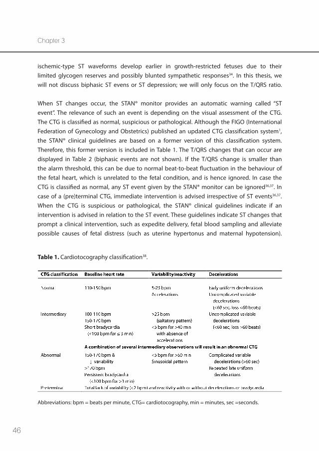
46
ischemic-type ST waveforms develop earlier in growth-restricted fetuses due to their limited glycogen reserves and possibly blunted sympathetic responses34. In this thesis, we will not discuss biphasic ST evens or ST depression; we will only focus on the T/QRS ratio.
When ST changes occur, the STAN® monitor provides an automatic warning called “ST event”. The relevance of such an event is depending on the visual assessment of the CTG. The CTG is classified as normal, suspicious or pathological. Although the FIGO (International Federation of Gynecology and Obstetrics) published an updated CTG classification system1, the STAN® clinical guidelines are based on a former version of this classification system. Therefore, this former version is included in Table 1. The T/QRS changes that can occur are displayed in Table 2 (biphasic events are not shown). If the T/QRS change is smaller than the alarm threshold, this can be due to normal beat-to-beat fluctuation in the behaviour of the fetal heart, which is unrelated to the fetal condition, and is hence ignored. In case the CTG is classified as normal, any ST event given by the STAN® monitor can be ignored36,37. In case of a (pre)terminal CTG, immediate intervention is advised irrespective of ST events36,37. When the CTG is suspicious or pathological, the STAN® clinical guidelines indicate if an intervention is advised in relation to the ST event. These guidelines indicate ST changes that prompt a clinical intervention, such as expedite delivery, fetal blood sampling and alleviate possible causes of fetal distress (such as uterine hypertonus and maternal hypotension).
Abbreviations: bpm = beats per minute, CTG= cardiotocography, min = minutes, sec =seconds.
Table 1. Cardiotocography classification38.
Chapter 3
↓

47
3
Technical background
Abbreviations: CTG = cardiotocography, min = minutes.
Table 2. STAN® clinical guidelines38.
48
References 1. Ayres-de-Campos D, Spong CY, Chandraharan E, FIGO Intrapartum Fetal Monitoring Expert
Consensus Panel. FIGO consensus guidelines on intrapartum fetal monitoring: Cardiotocography. Int J Gynaecol Obstet 2015 Oct;131(1):13-24.
2. Jezewski J, Wrobel J, Horoba K. Comparison of doppler ultrasound and direct electrocardiography acquisition techniques for quantification of fetal heart rate variability. IEEE Trans Biomed Eng 2006 May;53(5):855-864.
3. Cremer M. Über die direkte Ableitung der Aktionsströme des menschlichen Herzens vom Oesopha-gus und über das Elektrokardiogramm des Fötus. Münch Med Wschr 1906;53:811-813.
4. Kimura Y, Sato N, Sugawara J, Velayo C, Hoshiai T, Nagase S, et al. Recent Advances in Fetal Electro-cardiography. The Open Medical Devices Journal 2012;4:7-12.
5. van Laar JO, Warmerdam GJ, Verdurmen KM, Vullings R, Peters CH, Houterman S, et al. Fetal heart rate variability during pregnancy, obtained from non-invasive electrocardiogram recordings. Acta Obstet Gynecol Scand 2014 Jan;93(1):93-101.
6. Oostendorp TF, van Oosterom A, Jongsma HW. The fetal ECG throughout the second half of gestation. Clin Phys Physiol Meas 1989 May;10(2):147-160.
7. Vullings R, Peters C, Andriessen P, Oei S, Wijn P. Monitoring the Fetal Heart Rate and Fetal Electro-cardiogram: Abdominal Recordings Are As Good As Direct Ecg Measurements. Pediatric Research 2005;58(2):242.
8. Reinhard J, Hayes-Gill BR, Schiermeier S, Hatzmann H, Heinrich TM, Louwen F. Intrapartum heart rate ambiguity: a comparison of cardiotocogram and abdominal fetal electrocardiogram with maternal electrocardiogram. Gynecol Obstet Invest 2013;75(2):101-108.
9. Clifford G, Sameni R, Ward J, Robinson J, Wolfberg AJ. Clinically accurate fetal ECG parameters acquired from maternal abdominal sensors. Am J Obstet Gynecol 2011 Jul;205(1):47.e1-47.e5.
10. Vullings R. Non-invasive fetal electrocardiogram: analysis and interpretation, PhD thesis. Eindhoven: Eindhoven University of Technology; 2010.
11. Rosenstock EG, Cassuto Y, Zmora E. Heart rate variability in the neonate and infant: analytical methods, physiological and clinical observations. Acta Paediatr 1999 May;88(5):477-482.
12. Cesarelli M, Romano M, Bifulco P. Comparison of short term variability indexes in cardiotocographic foetal monitoring. Comput Biol Med 2009 Feb;39(2):106-118.
13. Van Ravenswaaij-Arts C, Kollee L, Hopman J, Stoelinga G, van Geijn H. Heart rate variabilitiy. Ann Intern Med 1993;118:436-447.
14. Dawes GS, Lobb M, Moulden M, Redman CW, Wheeler T. Antenatal cardiotocogram quality and interpretation using computers. BJOG 2014 Dec;121 Suppl 7:2-8.
15. van Laar JO. Fetal autonomic cardiac response during pregnancy and labour; PhD thesis. Eindhoven: Eindhoven University of Technology; 2012.
16. Warmerdam GJ, Vullings R, Bergmans JW, Oei SG. Reliability of spectral analysis of fetal heart rate variability. Conf Proc IEEE Eng Med Biol Soc 2014;2014:2817-2820.
Chapter 3

49
3
Technical background
17. Peters CH, ten Broeke ED, Andriessen P, Vermeulen B, Berendsen RC, Wijn PF, et al. Beat-to-beat detection of fetal heart rate: Doppler ultrasound cardiotocography compared to direct ECG cardiot-ocography in time and frequency domain. Physiol Meas 2004 Apr;25(2):585-593.
18. Peters C, Vullings R, Bergmans J, Oei G, Wijn P. The effect of artifact correction on spectral estimates of heart rate variability. Conf Proc IEEE Eng Med Biol Soc 2008;2008:2669-2672.
19. van Laar JO, Peters CH, Vullings R, Houterman S, Bergmans JW, Oei SG. Fetal autonomic response to severe acidaemia during labour. BJOG 2010 Mar;117(4):429-437.
20. Akselrod S, Gordon D, Ubel FA, Shannon DC, Berger AC, Cohen RJ. Power spectrum analysis of heart rate fluctuation: a quantitative probe of beat-to-beat cardiovascular control. Science 1981 Jul 10;213(4504):220-222.
21. Dalton KJ, Dawes GS, Patrick JE. The autonomic nervous system and fetal heart rate variability. Am J Obstet Gynecol 1983 Jun 15;146(4):456-462.
22. Task Force of the European Society of Cardiology and the North American Society of Pacing and Electrophysiology. Heart rate variability: standards of measurement, physiological interpretation and clinical use. Circulation 1996 Mar 1;93(5):1043-1065.
23. Van Laar JO, Porath MM, Peters CH, Oei SG. Spectral analysis of fetal heart rate variability for fetal surveillance: review of the literature. Acta Obstet Gynecol Scand 2008;87(3):300-306.
24. van Laar JO, Peters CH, Houterman S, Wijn PF, Kwee A, Oei SG. Normalized spectral power of fetal heart rate variability is associated with fetal scalp blood pH. Early Hum Dev 2011 Apr;87(4):259-263.
25. van Laar JO, Peters CH, Vullings R, Houterman S, Oei SG. Power spectrum analysis of fetal heart rate variability at near term and post term gestation during active sleep and quiet sleep. Early Hum Dev 2009 Dec;85(12):795-798.
26. De Beer N, Andriessen P, Berendsen R, Oei S, Wijn P, Bambang Oetomo S. Customized spectral band analysis compared with conventional Fourier analysis of herat rate variability in neonates. Physiol Meas. 2004;25:1385-1395.
27. Min SW, Ko H, Kim CS. Power spectral analysis of heart rate variability during acute hypoxia in fetal lambs. Acta Obstet Gynecol Scand 2002 Nov;81(11):1001-1005.
28. Amer-Wahlin I, Nord A, Bottalico B, Hansson SR, Ley D, Marsal K, et al. Fetal cerebral energy metab-olism and electrocardiogram during experimental umbilical cord occlusion and resuscitation. J Matern Fetal Neonatal Med 2010 Feb;23(2):158-166.
29. Greene KR, Dawes GS, Lilja H, Rosen KG. Changes in the ST waveform of the fetal lamb electrocardi-ogram with hypoxemia. Am J Obstet Gynecol 1982 Dec 15;144(8):950-958.
30. Rosen KG, Dagbjartsson A, Henriksson BA, Lagercrantz H, Kjellmer I. The relationship between circulating catecholamines and ST waveform in the fetal lamb electrocardiogram during hypoxia. Am J Obstet Gynecol 1984 May 15;149(2):190-195.
31. Fenn W. The deposition of potassium and phosphate with glycogen in rat livers. J Biol Chem 1939;128:297-308.

50
32. Rosén K, Isaksson O. Alterations in Fetal Heart Rate and ECG Correlated to Glycogen, Creatine Phosphate and ATP Levels during Graded Hypoxia. Biol Neonate 1976;30:17-24.
33. Siira SM, Ojala TH, Vahlberg TJ, Jalonen JO, Valimaki IA, Rosen KG, et al. Marked fetal acidosis and specific changes in power spectrum analysis of fetal heart rate variability recorded during the last hour of labour. BJOG 2005 Apr;112(4):418-423.
34. Westgate JA, Bennet L, Brabyn C, Williams CE, Gunn AJ. ST waveform changes during repeated umbilical cord occlusions in near-term fetal sheep. Am J Obstet Gynecol 2001 Mar;184(4):743-751.
35. Becker JH, Krikhaar A, Schuit E, Martendal A, Marsal K, Kwee A, et al. The added predictive value of biphasic events in ST analysis of the fetal electrocardiogram for intrapartum fetal monitoring. Acta Obstet Gynecol Scand 2015 Feb;94(2):175-182.
36. Amer-Wahlin I, Kwee A. Combined cardiotocographic and ST event analysis: A review. Best Pract Res Clin Obstet Gynaecol 2016 Jan;30:48-61.
37. Visser GH, Ayres-de-Campos D, FIGO Intrapartum Fetal Monitoring Expert Consensus Panel. FIGO consensus guidelines on intrapartum fetal monitoring: Adjunctive technologies. Int J Gynaecol Obstet 2015 Oct;131(1):25-29.
38. Amer-Wahlin I, Arulkumaran S, Hagberg H, Marsal K, Visser GH. Fetal electrocardiogram: ST waveform analysis in intrapartum surveillance. BJOG 2007 Oct;114(10):1191-1193.
Chapter 3

51
3
Technical background


Part I
Fetal ECG and congenital heart disease

54
55
Chapter 4
4
A systematic review of prenatal screening for congenital heart disease by fetal
electrocardiography
Kim M.J. Verdurmen, Noortje B. Eijsvoogel, Carlijn Lempersz, Rik Vullings, Christian Schroer, Judith O.E.H. van Laar, S. Guid Oei
Published: International Journal of Gynecology and Obstetrics. 2016;135(2):129-134
56
Chapter 4
AbstractBackgroundCongenital heart disease is the most common severe congenital anomaly worldwide. Diagnosing early in pregnancy is important, but the detection rate by two-dimensional ultrasonography is only 65%-81%.
ObjectivesTo evaluate existing data on congenital heart disease and non-invasive abdominal fetal electrocardiography.
Search strategyA systematic review was performed through a search of the Cochrane Library, PubMed and Embase for studies published up to April 2016 using the terms “congenital heart disease”, “fetal electrocardiogram”, and other similar keywords.
Selection criteriaPrimary articles that described changes in fetal electrocardiography among fetuses with congenital heart disease published in English were included.
Data collection and analysisOutcomes of interest were changes in fetal electrocardiography parameters observed for fetuses with congenital heart disease. Findings were reported descriptively.
Main resultsOnly five studies described changes observed in the fetal electrocardiogram for fetuses with congenital heart disease, including heart rate, heart rate variability, and PR, QRS and QT intervals. Fetal electrocardiography reflects the intimate relation between the cardiac nerve conduction system and the structural morphology of the heart. It seems particularly helpful in detecting the electrophysiological effects of cardiac anatomical defects (e.g. hypotrophy, hypertrophy, and conduction interruption).
ConclusionsFetal electrocardiography might be a promising clinical tool to complement ultrasonography in the screening programme for congenital heart disease.
57
Review: screening for congenital heart disease with fetal ECG
Introduction
Congenital heart disease (CHD) is the most common severe congenital anomaly worldwide1. CHD is defined as “a gross structural abnormality of the heart or intra-thoracic large vessels, that is actually or potentially of functional significance”2. Major CHD is usually defined as a form of CHD that is lethal or requires intervention in the first year of life. The incidence of CHD is estimated at 6-12 per 1000 live births (4 cases of major CHD per 1000 live births), which makes this disorder six times more common than chromosomal anomalies and four times more common than neural tube defects3-5. In Europe, the overall rate of mortality due to CHD (both perinatal deaths and termination of pregnancy) was 0.7 per 1000 births in 2000-20056. Of the fetuses affected by CHD, 4.5% die in utero and 21.1% die after birth7.
Diagnosing CHD early in pregnancy enables the identification of associated extracardiac anomalies (present in 29% of cases) and chromosomal anomalies (26% of cases) that have an effect on fetal and postnatal prognosis8. Prenatal and genetic counselling by experts can be offered to parents. Thereafter, parents can decide to terminate or continue with the pregnancy. Studies have shown that the frequency of pregnancy termination is higher if prenatal diagnosis is made at an earlier gestational age (61% and 44% at 19 and 24 weeks of pregnancy, respectively)8,9. If pregnancy is continued, an adequate plan of management can be developed, including intra-uterine therapy, timing, mode and location of delivery, and immediate treatment after birth. It has been demonstrated that prenatal diagnosis of CHD increases survival rates and decreases long-term morbidity in both ductus-dependent and foramen ovale-dependent CHD9-12. As Yates et al.13 has pointed out, however, prenatally diagnosed CHD often has a worse prognosis because it is more likely to be severe (i.e. easier to detect by ultrasonography) or associated with extracardiac or chromosomal anomalies.
Fetal cardiac screening during the second trimester was standardised in 200614. The detection rate of CHD varies widely, from 65% to 81%15-18. The challenges encoun-tered include the complex anatomy of the fetal heart, its motion, and small size. Specific echocardiography is performed for fetuses with risk factors for CHD, and this technique has a higher detection rate (sensitivity 90%, specificity 98%)19. However, up to 90% of all cases of CHD occur in the low-risk population, indicating the necessity of an effective screening procedure that is available to all pregnant women3,4,20-22.
Therefore, there is need for a reliable non-invasive diagnostic method with improved predic-tive value for the diagnosis of CHD. Non-invasive transabdominal fetal electrocardiography (ECG) is a new field that is being investigated. This technique can be used early in pregnancy (from 18 gestational weeks), is safe to use, and easy to apply23. A big advantage
4
58
is that fetal ECG is a potentially non-expensive long-term diagnostic tool, and raw data can be forwarded for evaluation elsewhere.
Extraction of fetal ECG data was first described in 1906 by Cremer et al.24, and the approach was first reviewed in 1986 by Pardi et al.25. Despite this early documentation, the develop-ment of fetal ECG has lagged behind other techniques for fetal monitoring, partly because of technical challenges. The fetal signal has low amplitude (2-50 microvolts, 1/50th of the maternal ECG), and is masked by both the maternal ECG and background noises (maternal electromyogram), resulting in a low signal-to-noise ratio25,26. The fetus is surrounded by amniotic fluid and maternal tissues, which enlarge the distance to the electrodes and cause a non-homogenous tissue conduction that interferes with signal quality. Additionally, the vernix caseosa is electrically isolating and a main cause of the poor signal-to-noise ratio from 30 to 34 gestational weeks23,27. Other challenging factors are the complex three dimensional form of the fetal ECG and the movements of the fetus, which makes it difficult to evaluate the heart from one direction. Furthermore, at 20 gestational weeks, the fetal heart is approximate-ly one-tenth of the size of an adult heart and the fetal heart rate is two to three times faster than the adult heart rate28. With improvements in technology and knowledge of information theory, however, fetal ECG is becoming more and more attractive.
In addition to the challenges in the conduction of fetal ECG, it is also difficult to interpret the data. By contrast with postnatal life, the systemic circulation in the fetus is fed from the left and right ventricle in parallel with equal intraventricular pressure29. The right ventricular outflow is slightly larger than the left ventricular outflow. The ductus arteriosus propels 40% of the combined cardiac output during the second trimester. Right-sided obstructive lesions (e.g. tetralogy of Fallot or pulmonary stenosis) with a dominance of the right ventricle are difficult to diagnose in utero; however, they are often accompanied by septal defects or by left-side obstructive lesions (e.g. aortic stenosis or coarctation of the aorta), which can be detected more easily. Owing to the fetal circulation in utero, fetuses affected by CHD do not always show overt signs of cardiac failure, because one side of the heart can compensate for an abnormality on the other side. At present, the changes in the fetal ECG amplitudes, segment intervals, and heart axis that are characteristic of CHD are not known. Although the changes due to CHD seen on neonatal ECG are documented, these data are not likely to correspond with those of fetal ECG because the circulation changes markedly directly after birth. The aim of the present review was to evaluate the existing data on CHD and non-invasive abdominal fetal ECG.
Chapter 4
59
Materials and Methods
As part of a systematic review, the Cochrane Library (2016, Issue 4), PubMed and Embase elec-tronic databases were searched to identify all studies published on fetal ECG and CHD up to April 30, 2016. The following keywords were used: “congenital heart disease”, “congenital heart defects”, “fetal electrocardiogram”, “fetal electrocardiography”, and “fetal ECG”. The outcomes of interest were changes seen in fetal ECG parameters, such as ECG intervals, ECG segments, and the electrical heart axis among fetuses with CHD.
Primary articles that described the changes in fetal ECG among fetuses with CHD were selected. The reference lists of the selected articles were also searched. The study language was restricted to English. Review articles and studies describing diagnostic tools other than non-invasive abdominal fetal ECG were excluded. Articles that solely described fetal arrhyth-mia were excluded because only few arrhythmias are associated with CHD.
The search and selection of articles were performed independently by two authors (K.M.J.V. and N.B.E.). The guidelines and quality assessment forms of the Dutch Cochrane Centre were used to evaluate the quality of the studies. The findings were reported descriptively and no statistical analysis was performed.
Results
The search and selection of articles is summarised in Figure 1. In total, five articles met the inclusion criteria and were reviewed, including case reports by Hamilton et al.30 and Brambati and Bonsignore31. Three articles by Siddiqui et al.32, Velayo et al.33, and Yilmaz et al.34 were prospective cohort studies, including normal fetuses and cases of CHD. The five studies were published between 1977 and 2016.
Owing to the low number of fetuses, the variation in outcome measures described, and the differences in signal processing techniques used in the five studies, it was not possible to directly compare or pool the results. The basic characteristics and a quality assessment of the two case reports are given in Table 1, whereas the basic characteristics and a quality assess-ment of the prospective studies are given in Tables 2 and 3. Table 4 presents an overview of the fetal ECG parameters of the fetuses with CHD included in this review.
Review: screening for congenital heart disease with fetal ECG
4
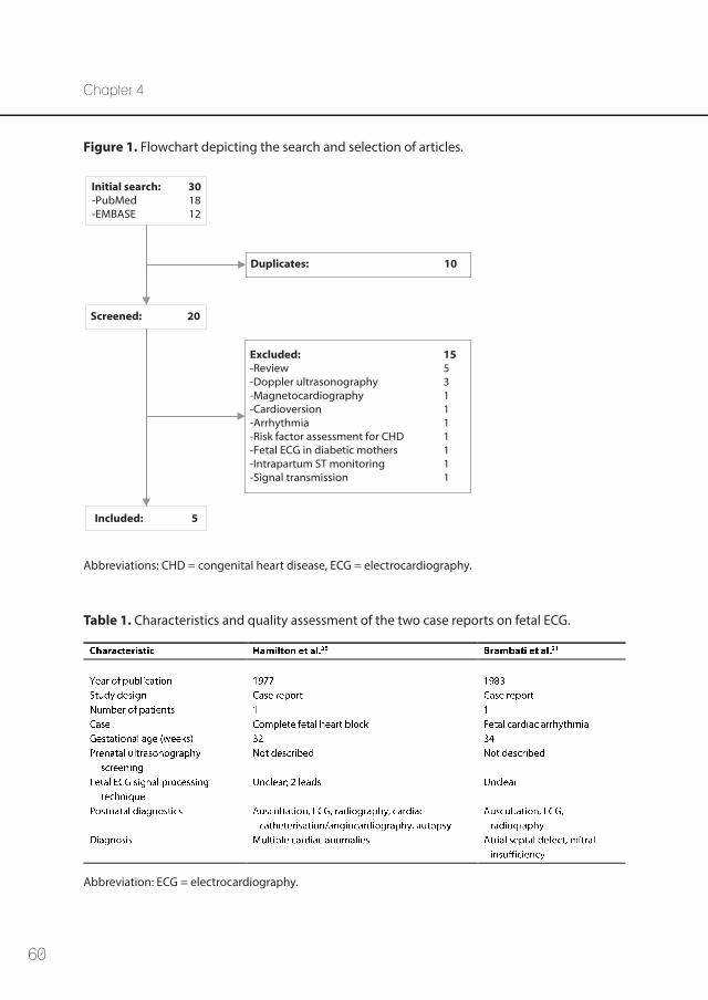
60
Chapter 4
Initial search: 30-PubMed 18-EMBASE 12
Screened: 20
Included: 5
Duplicates: 10
Excluded: 15-Review 5-Doppler ultrasonography 3-Magnetocardiography 1-Cardioversion 1-Arrhythmia 1-Risk factor assessment for CHD 1-Fetal ECG in diabetic mothers 1-Intrapartum ST monitoring 1-Signal transmission 1
Abbreviations: CHD = congenital heart disease, ECG = electrocardiography.
Figure 1. Flowchart depicting the search and selection of articles.
Table 1. Characteristics and quality assessment of the two case reports on fetal ECG.
Abbreviation: ECG = electrocardiography.
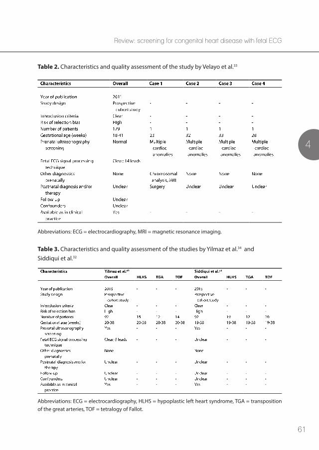
61
Review: screening for congenital heart disease with fetal ECG
4
Table 2. Characteristics and quality assessment of the study by Velayo et al.33
Abbreviations: ECG = electrocardiography, MRI = magnetic resonance imaging.
Table 3. Characteristics and quality assessment of the studies by Yilmaz et al.34 and Siddiqui et al.32
Abbreviations: ECG = electrocardiography, HLHS = hypoplastic left heart syndrome, TGA = transposition of the great arteries, TOF = tetralogy of Fallot.
62
Chapter 4
Hamilton et al.30 described a case of complex CHD, in which a complete heart block was seen in 1977. They used a cardiotocograph with the capacity to process fetal phonocar-diographic and abdominal fetal ECG signals. The bizarre QRS complexes found on fetal ECG (not otherwise specified) suggest that the pacemaker was distal to the bundle of His, with a fetal heart rate of 50 beats per minute. After delivery, cardiac catheterisation and angiocardiography were performed to confirm the existence of complex CHD (Table 4).
Seven years later, Brambati et al.31 described a case of an atrioventricular septal defect in which cardiac arrhythmia was seen. The signal processing method is not extensively described, but data extraction was mainly performed manually and a median fetal ECG constituting 50 heartbeats was generated. Extrasystoles without a preceding P wave were found, suggestive of ventricular origin. Additionally, a prolonged QRS time was found, which was stated to be suspicious of cardiac enlargement and/or cardiac anomaly. After delivery, ventricular extrasystoles, left axis deviation and right ventricular hypertrophy were found on ECG. The neonate was diagnosed with an atrioventricular septal defect.
Velayo et al.33 performed a prospective cohort study using simultaneous fetal ECG and cardiotocography (Doppler ultrasonography) recordings. The fetal heart rate information derived from Doppler ultrasonography was used to filter the fetal ECG35. Overall, 179 women were prospectively screened from a low-risk population. Twelve (7%) showed an abnormal fetal ECG; of these, 8 (4%) were excluded from further analysis because the fetus had no underlying structural heart defects, whereas 4 (2%) were confirmed to have CHD by an ultrasonography examination performed after inclusion in the study. The remaining 167 women (93%) with normal fetal ECG and no CHD or other anomalies on ultrasonogra-phy evaluation were used for standardisation of normal parameters. The fetal ECG was found to have a sensitivity of 100%, specificity of 95%, positive predictive value of 33%, and negative predictive value of 100% for the detection of CHD in a low-risk population.
Velayo et al.33 described the four cases of CHD that they found in their study population in detail (Table 4). They stated that premature ventricular contractions, as seen in cases one and three, might be caused by a primary developmental anomaly of the conduction system in the presence of endocardial cushion defects or an underlying genetic defect affecting intrinsic myocardial cell physiology. Prolongation of the QT interval, as seen in cases two and four, might be explained by repolarisation dysfunction (alterations in action potential duration), caused by ventricular aberrations33. This is also seen in cases of cardiac remodelling due to disease progression. In case four, the various prolonged intervals might have been late signs of the critical condition of the fetus, supported by the cardiomegaly with signs of heart failure.

63
Review: screening for congenital heart disease with fetal ECG
4
Yilmaz et al.34 and Siddiqui et al.32 both performed a prospective cohort study of the same set of 92 participants: 41 healthy controls and 51 fetuses with CHD, confirmed by fetal echocardiography. The Monica AN24 fetal electrocardiographic monitor was used to perform measurements at three gestational ages (19-27, 28-33 and 34-38 gestational weeks). Tracing quality was analysed in both studies.
Yilmaz et al.34 calculated the PR, QRS, and QT intervals during gestation. They showed that the PR and QRS intervals both lengthen as gestational age increases among normal fetuses. Among fetuses with CHD, this lengthening during gestation was not seen, but longer PR and QRS intervals were seen at all gestational ages as compared with normal values (Table 4). T waves seemed to be difficult to detect, and were therefore not included in the analysis.
Siddiqui et al.32 calculated fetal heart rate and fetal heart rate variability during gestation. Among the control fetuses, heart rate decreased during gestation, whereas heart rate variability increased. Fetuses with CHD generally had a lower fetal heart rate than healthy fetuses, but no differences in heart rate variability at 34-38 weeks were observed between the controls and cases, except for fetuses with hypoplastic left heart syndrome (which showed significantly lower heart rate variability during the active fetal state).
Discussion Overall, the present systematic review revealed that little research has been published regarding the changes seen in fetal ECG parameters observed for fetuses with CHD. The methods of conducting the fetal ECG measurements were different in most studies and evolved with time. Additionally, the fetal ECG parameters described in the studies varied. All the studies included show that fetal ECG can be a valuable tool to diagnose CHD early in utero. However, data on the fetal ECG changes observed among fetuses with CHD remain limited.
Both the case reports by Brambati et al.31 and Hamilton et al.30 are from another era, and are therefore not comparable with the signal processing techniques in current use. It is not likely that the two pregnancies described received prenatal ultrasonography screening to detect CHD. Nevertheless, both studies do indicate the potential of fetal ECG in diagnosing CHD and fetal arrhythmias.
Velayo et al.33 reported a promising sensitivity and specificity of 100% and 95%, respectively; however, the positive predictive value of fetal ECG was only 33%. The low positive predictive value can be explained by the low probability that fetuses in the general population will have CHD. The eight cases with an abnormal ECG that were excluded had other abnormalities,
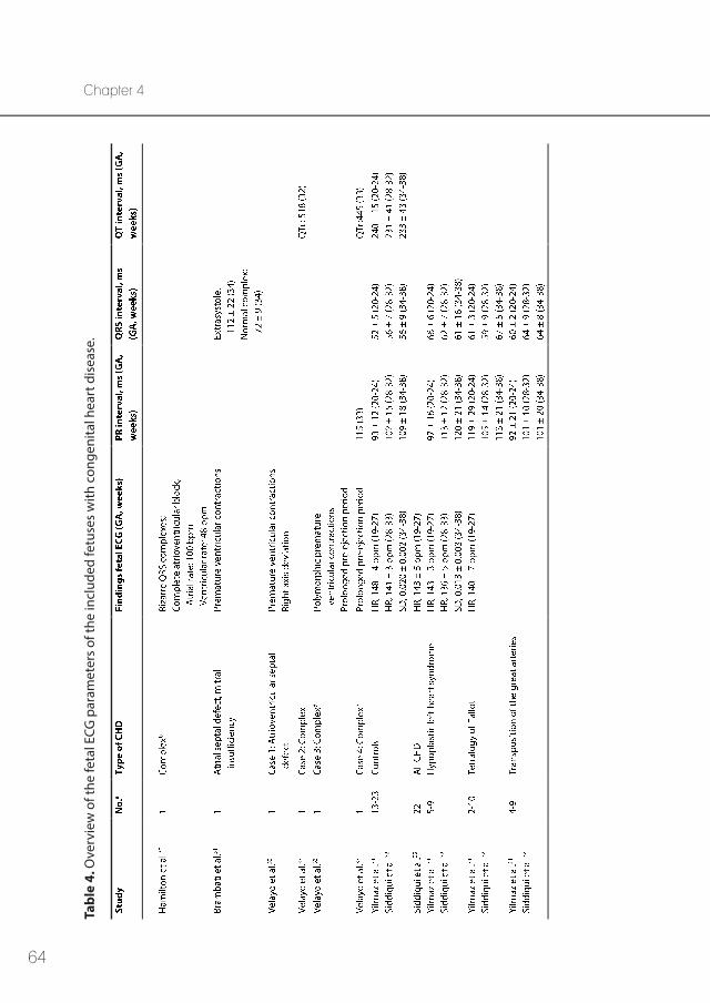
64
Chapter 4Ta
ble
4. O
verv
iew
of t
he fe
tal E
CG p
aram
eter
s of
the
incl
uded
fetu
ses
with
con
geni
tal h
eart
dis
ease
.

65
Review: screening for congenital heart disease with fetal ECG
4
a In th
e st
udie
s of
Yilm
az e
t al.34
and
Sid
diqu
i et a
l.32, t
he n
umbe
r of f
etus
es in
clud
ed v
arie
d by
ges
tatio
nal a
ge; t
here
fore
, the
min
imum
and
max
imum
nu
mbe
r of f
etus
es p
er g
roup
is in
dica
ted.
b
Com
mon
atr
ium
, com
plet
e at
riove
ntric
ular
can
al, d
oubl
e ou
tlet
right
ven
tric
le, p
ulm
onic
ste
nosi
s, an
eury
smal
rig
ht a
ortic
arc
h w
ith m
irror
-imag
e br
anch
ing,
inte
rrup
ted
left
-sid
ed v
ena
cava
with
azy
gous
con
tinua
tion.
c T
otal
ano
mal
ous
pulm
onar
y ve
nous
con
nect
ion,
pul
mon
ic s
teno
sis,
syst
emic
righ
t ven
tric
ular
dys
func
tion,
com
mon
atr
iove
ntric
ular
val
ve, b
ilate
ral
supe
rior v
ena
cava
, pul
mon
ary
veno
us re
turn
ano
mal
y.
d Tet
ralo
gy o
f Fal
lot,
vent
ricul
ar s
epta
l def
ect,
pulm
onic
ste
nosi
s, do
uble
out
let r
ight
ven
tric
le, t
rans
posi
tion
of th
e gr
eat a
rter
ies,
mul
ti-ao
rtop
ulm
o-na
ry c
olla
tera
l art
erie
s.e D
ilate
d ca
rdio
myo
path
y, v
entr
icul
ar s
epta
l def
ect,
cong
estiv
e he
art f
ailu
re.
Abb
revi
atio
ns: b
pm =
bea
ts p
er m
inut
e, C
HD
= c
onge
nita
l hea
rt d
isea
se, E
CG =
ele
ctro
card
iogr
aphy
, GA
= g
esta
tiona
l age
, HR
= he
art
rate
, ms
= m
illis
econ
d, N
o. =
num
ber o
f inc
lude
d pa
tient
s, SD
= s
tand
ard
devi
atio
n of
hea
rt ra
te d
urin
g ac
tive
feta
l sta
te (i
.e. h
eart
rate
var
iabi
lity)
, QTc
= fe
tal Q
T in
terv
al c
orre
cted
for h
eart
rate
(QT
divi
ded
by th
e sq
uare
root
of R
R).
66
Chapter 4
including non-immune hydrops fetalis, hypoxemia, or arrhythmia (not otherwise specified). Although CHD was not detected in this group, it is nonetheless important to identify this subset of fetuses that are possibly at risk of fetal distress36.
Yilmaz et al.34 and Siddiqui et al.32 describe the largest case series of fetal ECG measurements among fetuses with CHD published so far. However, only three types of CHD – which were commonly diagnosed and with “distinct anatomical and physiological features that could potentially impact ECG intervals”34 – were included in those studies. As a result, there is a high risk of selection bias in both studies. Additionally, identification of the P, QRS and T waves is not extensively described by Yilmaz et al.34. Siddiqui et al.32 calculated heart rate variability in three different ways: interquartile range of the fetal heart rate, standard deviation of the fetal heart rate, and root mean square of the standard deviation of the heart rate. Unfortunately, exact values are not given for each type of CHD separately for either fetal heart rate or varia-bility. A new trial with a greater sample population and the inclusion of more types of CHD is needed.
The fetal ECG reflects the intimate relationship between the cardiac nerve conduction system and the structural morphology of the heart33. In accordance with Yilmaz et al.34, other studies also found that the duration of the P wave, QRS complex, and PR interval increases progressively from 18 gestational weeks until term for fetuses with normal cardiac structures34,37. This reflects the anatomical development of the atria and ventri-cles during pregnancy, with gain in the weight and mass of the fetal heart. The increase in PR interval indicates the development of the atrioventricular conducting tissue38.
Yilmaz et al.34 showed that, among fetuses with CHD, the normal lengthening in PR and QRS intervals during pregnancy is absent. Additionally, most CHDs are associated with an increased or decreased ventricular mass or cardiac arrhythmias25,30,31,33. In case of a severe endocardial cushion defect (atrioventricular septum defect), the abnormal atrioventricular connection affects the conduction system, and this is reflected in a longer PR interval and left axis deviation25,31,33,39.
The aim of Siddiqui et al.32 was to characterise autonomic regulation in fetuses with CHD, and to study whether autonomic development is altered in comparison with healthy controls. Although it was not the aim, the study seems to indicate that heart rate variability might not be a good screening tool to detect CHD because heart rate variability was lower only among fetuses with hypoplastic left heart syndrome and only in the measurement at 34-38 gesta-tional weeks. Moreover, there were only minor differences in fetal heart rate between controls and fetuses with CHD.
67
Review: screening for congenital heart disease with fetal ECG
4
The rate of prenatal detection by two-dimensional ultrasonography is mainly influenced by the experience of the sonographer. It is relatively difficult to interpret the anatomy of the fetal heart correctly because it is a dynamic, constantly moving structure that rhythmically beats approximately more than twice per second. In addition, some diagnoses (e.g. coarctation of the aorta and CHDs that develop/progress during pregnancy such as pulmonary stenosis, aortic stenosis, ventricular hypoplasia, and cardiomyopathy) remain challenging to diagnose even in experienced centres. Given the fetal circulation, it is difficult to define lesion severity in the presence of the unique fetal shunts that permit redistribution of ventricular preload and output to the contralateral ventricle or great artery3.
Computerised evaluation of the fetal ECG might eliminate some of the abovementioned problems. First, performer variability – as occurs in the case of ultrasonography – would be absent because the data are analysed by computerised algorithms. Second, the equipment is cheaper and smaller by comparison with ultrasonography equipment. Third, application of the fetal ECG system involves minimum training. Last, at the time of the fetal anomaly scan, CHDs that evolve during gestation might present no anomalies by ultrasonography, but might show fetal ECG characteristics.
The present review has some limitations. Because little research has been published, the amount of ECG recordings that was available per type of CHD was limited. Only two case reports and three prospective trials were found. The methods of conduction the fetal ECG measurements were different in the five studies and evolved during time. Additionally, the fetal ECG parameters that were described varied in every study. Nevertheless, all the studies included show that fetal ECG can be a valuable tool for diagnosing CHD early in utero.
In conclusion, fetal ECG is a promising clinical tool that complements ultrasonography in the screening programme for CHD. It is particularly suitable for the detection of secondary effects due to CHD (i.e. hypotrophy, hypertrophy, and conduction interruption). However, more research establishing normal fetal ECG values and studies concerning the true incidence of fetal ECG anomalies in CHD are needed.
Acknowledgements
The research was performed within the IMPULS Perinatology framework (Netherlands).

68
Chapter 4
References 1. van der Linde D, Konings EE, Slager MA, Witsenburg M, Helbing WA, Takkenberg JJ, et al. Birth
prevalence of congenital heart disease worldwide: a systematic review and meta-analysis. J Am Coll Cardiol 2011 Nov 15;58(21):2241-2247.
2. Mitchell SC, Korones SB, Berendes HW. Congenital heart disease in 56,109 births. Incidence and natural history. Circulation 1971 Mar;43(3):323-332.
3. Donofrio MT, Moon-Grady AJ, Hornberger LK, Copel JA, Sklansky MS, Abuhamad A, et al. Diagnosis and treatment of fetal cardiac disease: a scientific statement from the American Heart Association. Circulation 2014 May 27;129(21):2183-2242.
4. Simpson LL. Screening for congenital heart disease. Obstet Gynecol Clin North Am 2004 Mar;31(1):51-59.
5. Gardiner HM. Keeping abreast of advances in fetal cardiology. Early Hum Dev 2006 Jun;82(6):415-419.
6. Dolk H, Loane M, Garne E, European Surveillance of Congenital Anomalies (EUROCAT) Working Group. Congenital heart defects in Europe: prevalence and perinatal mortality, 2000 to 2005. Circu-lation 2011 Mar 1;123(8):841-849.
7. Fesslova’ V, Brankovic J, Boschetto C, Masini A, Prandstraller D, Perolo A, et al. Changed outcomes of fetuses with congenital heart disease: new Italian Multicentre study. J Cardiovasc Med (Hagers-town) 2014 Jun 13.
8. Clur SA, Van Brussel PM, Mathijssen IB, Pajkrt E, Ottenkamp J, Bilardo CM. Audit of 10 years of referrals for fetal echocardiography. Prenat Diagn 2011 Dec;31(12):1134-1140.
9. Trines J, Fruitman D, Zuo KJ, Smallhorn JF, Hornberger LK, Mackie AS. Effectiveness of prenatal screening for congenital heart disease: assessment in a jurisdiction with universal access to health care. Can J Cardiol 2013 Jul;29(7):879-885.
10. Brick DH, Allan LD. Outcome of prenatally diagnosed congenital heart disease: an update. Pediatr Cardiol 2002 Jul-Aug;23(4):449-453.
11. Hunter LE, Simpson JM. Prenatal screening for structural congenital heart disease. Nat Rev Cardiol 2014 Jun;11(6):323-334.
12. Brown KL, Ridout DA, Hoskote A, Verhulst L, Ricci M, Bull C. Delayed diagnosis of congenital heart disease worsens preoperative condition and outcome of surgery in neonates. Heart 2006 Sep;92(9):1298-1302.
13. Yates RS. The influence of prenatal diagnosis on postnatal outcome in patients with structural congenital heart disease. Prenat Diagn 2004 Dec 30;24(13):1143-1149.
14. International Society of Ultrasound in Obstetrics & Gynecology. Cardiac screening examination of the fetus: guidelines for performing the ‘basic’ and ‘extended basic’ cardiac scan. Ultrasound Obstet Gynecol 2006 Jan;27(1):107-113.
15. Carvalho JS, Mavrides E, Shinebourne EA, Campbell S, Thilaganathan B. Improving the effectiveness of routine prenatal screening for major congenital heart defects. Heart 2002 Oct;88(4):387-391.
16. Kirk JS, Riggs TW, Comstock CH, Lee W, Yang SS, Weinhouse E. Prenatal screening for cardiac anomalies: the value of routine addition of the aortic root to the four-chamber view. Obstet Gynecol 1994 Sep;84(3):427-431.
69
Review: screening for congenital heart disease with fetal ECG
4
17. Ogge G, Gaglioti P, Maccanti S, Faggiano F, Todros T. Prenatal screening for congenital heart disease with four-chamber and outflow-tract views: a multicenter study. Ultrasound Obstet Gynecol 2006 Nov;28(6):779-784.
18. Wu Q, Li M, Ju L, Zhang W, Yang X, Yan Y, et al. Application of the 3-vessel view in routine prenatal sonographic screening for congenital heart disease. J Ultrasound Med 2009 Oct;28(10):1319-1324.
19. Cohen EH, Rein AJ. Antenatal diagnosis of cardiac malformation: a structural study. Fetal Diagn Ther 2000 Jan-Feb;15(1):54-60.
20. Galindo A, Herraiz I, Escribano D, Lora D, Melchor JC, de la Cruz J. Prenatal detection of congenital heart defects: a survey on clinical practice in Spain. Fetal Diagn Ther 2011;29(4):287-295.
21. Rocha LA, Araujo Junior E, Nardozza LM, Moron AF. Screening of fetal congenital heart disease: the challenge continues. Rev Bras Cir Cardiovasc 2013 Jul-Sep;28(3):V-VII.
22. Achiron R, Glaser J, Gelernter I, Hegesh J, Yagel S. Extended fetal echocardiographic examination for detecting cardiac malformations in low risk pregnancies. BMJ 1992 Mar 14;304(6828):671-674.
23. van Laar JO, Warmerdam GJ, Verdurmen KM, Vullings R, Peters CH, Houterman S, et al. Fetal heart rate variability during pregnancy, obtained from non-invasive electrocardiogram recordings. Acta Obstet Gynecol Scand 2014 Jan;93(1):93-101.
24. Cremer M. Über die direkte Ableitung der Aktionsströme des menschlichen Herzens vom Oesopha-gus und über das Elektrokardiogramm des Fötus. Münch Med Wschr 1906;53:811-813.
25. Pardi G, Ferrazzi E, Cetin I, Rampello S, Baselli G, Cerutti S, et al. The clinical relevance of the abdominal fetal electrocardiogram. J Perinat Med 1986;14(6):371-377.
26. Kimura Y, Sato N, Sugawara J, Velayo C, Hoshiai T, Nagase S, et al. Recent Advances in Fetal Electro-cardiography. The Open Medical Devices Journal 2012;4:7-12.
27. Oostendorp TF, van Oosterom A, Jongsma HW. The fetal ECG throughout the second half of gestation. Clin Phys Physiol Meas 1989 May;10(2):147-160.
28. Van Mieghem T, DeKoninck P, Steenhaut P, Deprest J. Methods for prenatal assessment of fetal cardiac function. Prenat Diagn 2009 Dec;29(13):1193-1203.
29. Kiserud T, Acharya G. The fetal circulation. Prenat Diagn 2004 Dec 30;24(13):1049-1059.
30. Hamilton LA,Jr, Fisher E, Horn C, DuBrow I, Vidyasagar D. A new prenatal cardiac diagnostic device for congenital heart disease. Obstet Gynecol 1977 Oct;50(4):491-494.
31. Brambati B, Bonsignore L. The significance of indirect electrocardiography in fetal cardiac arrhyth-mias. Eur J Obstet Gynecol Reprod Biol 1983 Mar;14(6):371-373.
32. Siddiqui S, Wilpers A, Myers M, Nugent JD, Fifer WP, Williams IA. Autonomic regulation in fetuses with congenital heart disease. Early Hum Dev 2015 Mar;91(3):195-198.
33. Velayo C, Sato N, Ito T, Chisaka H, Yaegashi N, Okamura K, et al. Understanding congenital heart defects through abdominal fetal electrocardiography: case reports and clinical implications. J Obstet Gynaecol Res 2011 May;37(5):428-435.
34. Yilmaz B, Narayan HK, Wilpers A, Wiess C, Fifer WP, Williams IA. Electrocardiographic intervals in foetuses with CHD. Cardiol Young 2016 Jan;26(1):84-89.
70
Chapter 4
35. Sato M, Kimura Y, Chida S, Ito T, Katayama N, Okamura K, et al. A novel extraction method of fetal electrocardiogram from the composite abdominal signal. IEEE Trans Biomed Eng 2007 Jan;54(1):49-58.
36. Sato N, Hoshiai T, Ito T, Owada K, Chisaka H, Aoyagi A, et al. Successful detection of the fetal electrocardiogram waveform changes during various states of singletons. Tohoku J Exp Med 2011;225(2):89-94.
37. Chia EL, Ho TF, Rauff M, Yip WC. Cardiac time intervals of normal fetuses using noninvasive fetal electrocardiography. Prenat Diagn 2005 Jul;25(7):546-552.
38. Pardi G, Marconi A, Ferrazzi E. The intraventricular conduction time of fetal heart in pregnancies with suspected fetal growth retardation. Br J Obstet Gynaecol 1986 Mar;93(3):250-254.
39. Barnes N, Archer N. Understanding congenital heart disease. Current Paediatrics 2005(15):421-428.

71
Review: screening for congenital heart disease with fetal ECG
4

72
73
5Normal ranges for fetal electrocardiogram
values for the healthy fetus of 18-24 weeks of gestation: a prospective cohort study
Kim M.J. Verdurmen, Carlijn Lempersz, Rik Vullings, Christian Schroer, Tammo Delhaas, Judith O.E.H. van Laar, S. Guid Oei
Published: BMC Pregnancy and Childbirth. 2016;16:227
Chapter 5
74
Chapter 5
Abstract
BackgroundThe fetal anomaly ultrasound only detects 65-81% of the patients with congenital heart disease, making it the most common structural fetal anomaly of which a significant part is missed during prenatal life. Therefore, we need a reliable non-invasive diagnostic method which improves the predictive value for congenital heart diseases early in pregnancy. Fetal electrocardiography could be this desired diagnostic method. There are multiple technical challenges to overcome in the conduction of the fetal electrocardiogram. In addition, interpretation is difficult due to the organisation of the fetal circulation in utero. We want to establish the normal ranges and values of the fetal electrocardiogram parameters in healthy fetuses of 18 to 24 weeks of gestation.
Methods/DesignWomen with an uneventful singleton pregnancy between 18 and 24 weeks of gestation are asked to participate in this prospective cohort study. A certified and experienced sonogra-phist performs the fetal anomaly scan. Subsequently, a fetal electrocardiogram recording is performed using dedicated signal processing methods. Measurements are performed at two institutes. We will include 300 participants to determine the normal values and 95% confi-dence intervals of the fetal electrocardiogram parameters in a healthy fetus. We will evaluate the fetal heart rate, segment intervals, normalised amplitude and the fetal heart axis. Three months postpartum, we will evaluate if a newborn is healthy through a questionnaire.
DiscussionFetal electrocardiography could be a promising tool in the screening programme for congen-ital heart diseases. The electrocardiogram is a depiction of the intimate relationship between the cardiac nerve conduction pathways and the structural morphology of the fetal heart, and therefore particularly suitable for the detection of secondary effects due to a congenital heart disease (hypotrophy, hypertrophy and conduction interruption).
75
5
Normal fetal ECG ranges: design of a prospective cohort study
Background
During pregnancy, the condition of the fetus is assessed with different techniques. One of these techniques is ultrasound examination. Between week 18 and 22 of gestation the fetal anomaly ultrasound is performed. During this examination, the fetus is screened for all kind of possible congenital anomalies, including congenital heart disease (CHD). CHD is defined as a “gross structural abnormality of the heart or intra-thoracic large vessels, (possibly) with functional significance”1. CHD is the most common severe congenital anomaly worldwide2; the incidence is estimated at 6-12 per 1000 live births3-5. CHD is six times more common than chromosomal anomalies and four times more common than neural tube defects4,6.
The fetal anomaly ultrasound, including planes of the ventricular outflow tracts and the three-vessel view, only detects 65-81% of the patients with CHD6-9. That makes CHD the most common structural fetal abnormality of which a significant part is missed during prenatal life. Therefore, we need a reliable non-invasive diagnostic method which improves the predictive value for the diagnosis CHD. This diagnostic technique should be able to diagnose CHD early in pregnancy for multiple reasons. First, we get the opportunity to identify associated extracardiac and chromosomal anomalies that affect fetal and postnatal prognosis. Second, parents get the chance to opt for termination of pregnancy in case of a severe CHD. Third, one can develop an adequate treatment plan including intra-uterine therapy, timing, mode and location of delivery and planning of immediate treatment after birth. In ductus- and foramen ovale dependent CHDs, it is demonstrated that prenatal diagnosis increases the survival rates and decreases long term morbidity10-13.
The currently used two-dimensional ultrasonography provides multiple anatomic planes, relying on the sonographists mental reconstruction of these planes to define the fetal cardiac anatomy. The antenatal diagnostic value is therefore to a great extent depending on the experience of the performer. As stated by Gardiner5; “you only see what you look for and identify what you already know”. Three- and four-dimensional ultrasonography gives a more fluid and representative image of the fetal cardiac structures, and therefore aids in this mental reconstruction14. Spatio-temporal image correlation (STIC) is a new modality using automated volume acquisition of the fetal heart with one sweep of the probe, further facilitating the examination of the fetal heart. Disadvantages of these ultrasound modalities are that they are extremely expensive and only applicable in centres with experienced personnel.
The non-invasive fetal electrocardiogram (ECG) could be a valuable tool for the detection of CHD early in pregnancy. In 1906, Cremer and colleagues15 were the first to describe fetal ECG extraction through the maternal abdomen and 80 years later, Pardi and colleagues16
76
were the first to write a review considering fetal ECG and, amongst others, CHD. Compared to other techniques for fetal monitoring, the development of the fetal ECG lagged behind. This is mainly because there are multiple technical challenges to overcome. First, at a gestational age of 20 weeks, the fetal heart is about 1/10th of the size of an adult heart. Due to the low voltage of the fetal ECG (1/50th of the maternal ECG), there is a low signal-to-noise ratio. In addition, identifying the fetal signals is challenging due to masking by the maternal ECG and high background noises caused by the maternal electromyogram. The amniotic fluid and maternal tissues that surround the fetus enlarge the distance to the electrodes, and cause a non-homogenous tissue conduction that interferes with signal quality. In addition, the vernix caseosa causes electrical isolation and further diminishes the signal amplitude17. This is the main cause of the poor signal-to-noise ratio from 30 to 34 weeks of gestation18,19. Second, the fetal ECG has a complex three-dimensional shape, alternating with changes in fetal presenta-tion. Following fetal movements, the electrical signal from each electrode changes frequently. Another challenging factor is the speed of the fetal heart rate, which is two to three times faster compared to the adult heart rate20.
Besides the technical difficulties encountered when conducting a fetal ECG, it is also chal-lenging to interpret the fetal ECG. In contrast with postnatal life, the systemic circulation in the fetus is fed from both the left and right ventricle in parallel, with equal intraventricular pressures21. The outflow in the right ventricle is slightly larger compared to the outflow in the left ventricle, and increases during gestation; 53% vs 47% at 20 weeks of gestation, 57% vs 43% at 30 weeks of gestation and 60% vs 40% at 38 weeks of gestation21. In utero, the O2-rich blood flows from the umbilical vein to the right atrium. There, the formen ovale propels a major part of the O2-rich blood to the left side of the heart and into the systemic circulation, bypassing the fetal lungs. In addition, in the second trimester the ductus arteriosus propels 40% of the combined cardiac output. Because of these major differences between the systemic circulation in utero and postpartum, it is difficult to predict what a normal fetal ECG looks like. Furthermore, due to this organisation of the fetal circulation in utero, in case of a CHD one side of the heart can compensate for an abnormality on the other site, and fetuses affected by a CHD do not always show signs of cardiac failure. However, before we are able to detect CHD with the fetal ECG, we need to establish the normal range and values of amplitudes and segment intervals of the fetal ECG in a healthy fetus.
Chapter 5
77
5
Normal fetal ECG ranges: design of a prospective cohort study
Methods/Design
AimThe aim of this study is to establish the normal ranges and values (mean with 95% confidence intervals) of fetal ECG parameters in a healthy fetus of 18 to 24 weeks of gestation.
Study designWe will perform a prospective cohort study. The study protocol is approved by the medical ethical committee of the Máxima Medical Centre, Veldhoven, the Netherlands (NL48535.015.14).
SettingMeasurements are performed at the Máxima Medical Centre, Veldhoven, the Netherlands and “Diagnostiek voor U” (DvU), Eindhoven, the Netherlands. The Máxima Medical Centre is a tertiary care teaching hospital for obstetrics. DvU is a diagnostic centre which, amongst others, performs blood tests and ultrasounds. Measurements are performed directly before or after the sonographist performed the fetal anomaly scan. The fetal anomaly ultrasound is performed by a certified and experienced sonographist.
Participants Patients with an uneventful pregnancy, carrying a singleton fetus with a gestational age between 18 and 24 weeks, are included in the study after written informed consent. At the Máxima Medical Centre, this will be patients who visit the outpatient clinic for an appoint-ment. At DvU, this will be patients who visit the centre for their fetal anomaly ultrasound. These patients are generally seen by a midwife or by a doctor at the Máxima Medical Centre for their obstetrical care. Pregnant women must be aged older than 18 years. If any of the fetuses turn out to have a form of CHD, they are excluded from the cohort. Other exclusion criteria are multiple pregnancies, insufficient understanding of the Dutch language, and any known fetal congenital anomalies.
Three months postpartum we will evaluate if the newborn is healthy, which is defined as absence of CHD, through a questionnaire. If the neonate turns out to have a CHD, which was missed at the time of the structural anomaly ultrasound, we will exclude the patient from the cohort.
Procedures The fetal ECG is a non-invasive, transabdominal research method. The pregnant women is lying down in a semi-recumbent position to prevent aortocaval compression. The fetal ECG
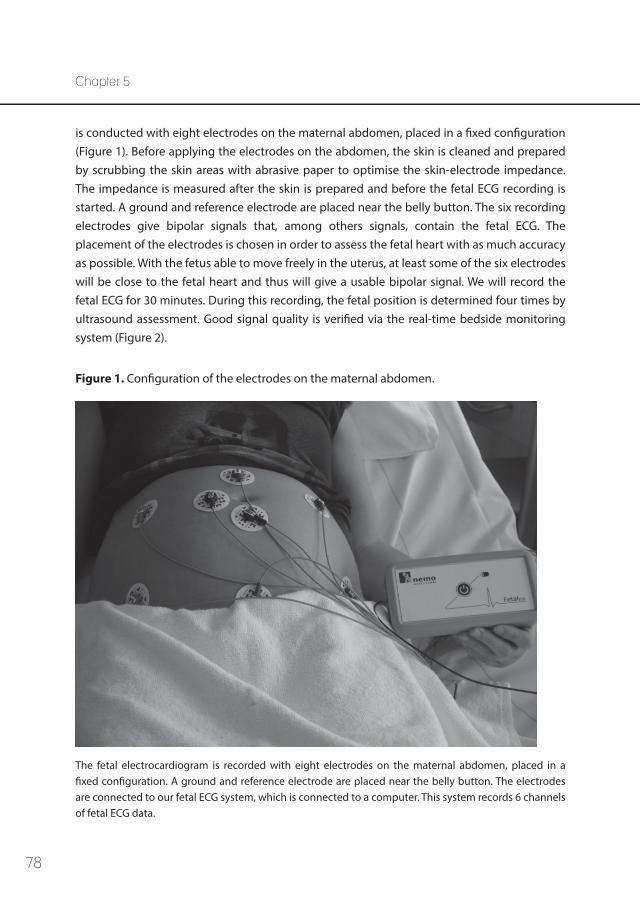
78
is conducted with eight electrodes on the maternal abdomen, placed in a fixed configuration (Figure 1). Before applying the electrodes on the abdomen, the skin is cleaned and prepared by scrubbing the skin areas with abrasive paper to optimise the skin-electrode impedance. The impedance is measured after the skin is prepared and before the fetal ECG recording is started. A ground and reference electrode are placed near the belly button. The six recording electrodes give bipolar signals that, among others signals, contain the fetal ECG. The placement of the electrodes is chosen in order to assess the fetal heart with as much accuracy as possible. With the fetus able to move freely in the uterus, at least some of the six electrodes will be close to the fetal heart and thus will give a usable bipolar signal. We will record the fetal ECG for 30 minutes. During this recording, the fetal position is determined four times by ultrasound assessment. Good signal quality is verified via the real-time bedside monitoring system (Figure 2).
Figure 1. Configuration of the electrodes on the maternal abdomen.
The fetal electrocardiogram is recorded with eight electrodes on the maternal abdomen, placed in a fixed configuration. A ground and reference electrode are placed near the belly button. The electrodes are connected to our fetal ECG system, which is connected to a computer. This system records 6 channels of fetal ECG data.
Chapter 5
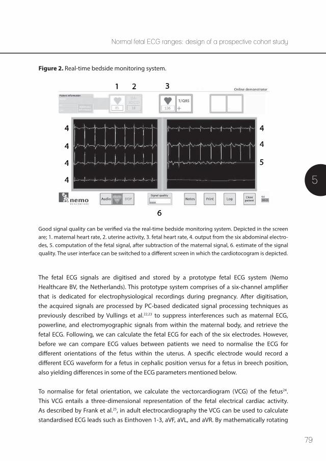
79
5
Normal fetal ECG ranges: design of a prospective cohort study
Figure 2. Real-time bedside monitoring system.
4 4
4
5
4
4
4
1 3
6
2
Good signal quality can be verified via the real-time bedside monitoring system. Depicted in the screen are; 1. maternal heart rate, 2. uterine activity, 3. fetal heart rate, 4. output from the six abdominal electro-des, 5. computation of the fetal signal, after subtraction of the maternal signal, 6. estimate of the signal quality. The user interface can be switched to a different screen in which the cardiotocogram is depicted.
The fetal ECG signals are digitised and stored by a prototype fetal ECG system (Nemo Healthcare BV, the Netherlands). This prototype system comprises of a six-channel amplifier that is dedicated for electrophysiological recordings during pregnancy. After digitisation, the acquired signals are processed by PC-based dedicated signal processing techniques as previously described by Vullings et al.22,23 to suppress interferences such as maternal ECG, powerline, and electromyographic signals from within the maternal body, and retrieve the fetal ECG. Following, we can calculate the fetal ECG for each of the six electrodes. However, before we can compare ECG values between patients we need to normalise the ECG for different orientations of the fetus within the uterus. A specific electrode would record a different ECG waveform for a fetus in cephalic position versus for a fetus in breech position, also yielding differences in some of the ECG parameters mentioned below.
To normalise for fetal orientation, we calculate the vectorcardiogram (VCG) of the fetus24. This VCG entails a three-dimensional representation of the fetal electrical cardiac activity. As described by Frank et al.25, in adult electrocardiography the VCG can be used to calculate standardised ECG leads such as Einthoven 1-3, aVF, aVL, and aVR. By mathematically rotating
80
the fetal VCG prior to calculating the ECG, we can create standardised fetal ECG leads. The amount of rotation required is determined based on a simultaneously performed ultrasound assessment of the fetal presentation. Via these mathematical rotations, we are also capable of detecting and correcting for fetal movements in between the ultrasound assessments, as described previously by Vullings et al.26. The four ultrasound assessments during the measurements are used to correct for cumulative errors in this correction method and to determine the initial orientation of the fetus. To enhance the signal quality of the measure-ments, the fetal ECG is filtered further (amongst others by averaging of the ECG waveforms). The detection of segments and intervals is performed semi-automatically. The detection of fetal ECG complexes is computerised, while marking of the fetal ECG intervals (P top, QRS complex, T top) is performed manually by two independent researchers following a protocol that is verified by an experienced paediatric cardiologist. We will calculate the inter-observer variability between the two researchers.
Normal heart rhythm is assumed to show variations in heartbeat intervals smaller than 20% between consecutive beats. In case these variations are larger, this is assumed to be the result of either fetal arrhythmia or erroneous detection of the heartbeat interval, e.g. because of poor signal quality. Assessment of erroneous detection is based on energy of the ECG signal and correlations between consecutive ECG waveforms. The ECG is a quasi-periodic signal, meaning that consecutive ECG waveforms have a similar appearance and similar amplitude/energy. In case of poor ECG signal quality, the energy of the ECG signals is expected to differ from the energy during good quality recordings. Present artefacts or noise cause the energy of the ECG to increase beyond physiologically plausible ranges. Likewise, correlations between consecutive ECG waveforms are reduced in the presence of poor signal quality.
It has to be noted here, that fetal arrhythmia can also cause poor correlation between ECG waveforms. Some arrhythmias are hence expected to be incorrectly classified as poor signal quality. This misclassification affects the detection of fetal arrhythmia, but will not have any impact on other study parameters as these are determined only during normal rhythm and good signal quality. The recording must contain a minimum of 200 ECG complexes that were assessed to have good signal quality and that were corrected for fetal movement26.
Chapter 5
81
5
Normal fetal ECG ranges: design of a prospective cohort study
Study parametersMultiple outcome values are evaluated: • Fetal heart rate; mean, standard deviation, 95% confidence intervals and heart rate arrhythmia
• Segment intervals (PQ, QRS, ST etc.); mean, standard deviation and 95% confidence intervals
• Normalised amplitudes (P, QRS, T); mean, standard deviation and 95% confidence intervals
• Fetal electrical heart axis
• % of total patients in which the recording contains the required amount of data to perform the analysis Heart rate arrhythmia is defined based on heuristic rules that dictate that during normal rhythm subsequent heartbeat intervals cannot differ more than 20%. Any rhythm not complying with this rule, and assessed to not be caused by erroneous detection of heart-beats, e.g. as a result of poor signal quality, is labelled as a fetal arrhythmia.
Sample size There are previously published studies (see Discussion for more details) that describe fetal ECG parameters. However, these studies use different methods for obtaining the fetal signal and do not correct for the fetal position in the uterus. Therefore they are not able to calculate the fetal electrical heart axis. Moreover, all studies describe another parameter of the fetal ECG. Statistical experts calculated that we need a study population of 200 pregnant patients in order to determine normal values and 95% confidence intervals of a healthy fetus27. Antic-ipating on loss to follow-up and insufficient data quality, we will include 300 patients in the initial cohort.
Statistical analysis The collected data is analysed through SPSS. With the collected data, we perform several analyses. We calculate the normal values and ranges of the fetal heart rate, segment intervals (PQ, QRS, ST etc.), normalised amplitudes (P, QRS and T) and the fetal heart axis. Initially, we will calculate the values and ranges for all included patients as one group (18 to 24 weeks of gestational age). Thereafter, we will perform a subanalysis for every group per week of gestational age.
82
Discussion
Previous studies have been published regarding the normal values and ranges of the fetal ECG. In their review, Pardi et al.16 summarised the normal evolution of the cardiac cycle during gestation. From the 17th week of gestation up to term, the duration of the P wave increases progressively. This reflects the anatomical development of the atria during pregnancy. Similar, the duration of the QRS complex increases progressively, parallel with the weight gain of the fetal heart and in particular with the gain in ventricular mass. In fetal life, the intraventricular conduction is delayed compared to adult values, most likely due to anatomical differences of the ventricular conduction tissue. There is a slight increase in PR interval during pregnancy, indicating development of the atrioventricular conduction tissue.
Recently, longitudinal studies followed pregnancies from 14 to 41 weeks of pregnancy and performed fetal ECG measurements during different stages of gestation18,28,29. In the 1960s, Larks and colleagues30-33 described the orientation of the electrical fetal heart axis. All mentioned studies performed fetal ECG recordings with different conduction and analysis techniques. The amount of electrodes on the maternal abdomen varied from three to sixteen. Average fetal ECG complexes were generated from segments of 60 seconds up to 2.5 minutes. Analyses were performed manually or by computerised signal processing programs. These studies did not take the fetal position in the uterus into account.
In a group around 20 weeks of gestation, the following mean values were found by Chia and Taylor28,29, respectively: P wave length 43.9 ms, PR interval 102.1/91.7 ms, QRS duration 47.2/40.7 ms, QT interval 224.0/242.3 ms and T wave duration 123.8 ms. Larks33 found a normal range of the fetal heart axis between +100 and +160 degrees, with a mean value of +134 degrees in term fetuses during labour. Due to the lack of correcting for the fetal position in utero, fetuses in breech position showed a negative electrical heart axis (-180 to 0 degrees)32. In fact, due to the lack of correcting for fetal position, also findings for fetuses in vertex position were unreliable. In their analysis, Larks implicitly assumed that every fetus was facing the frontal plane. In cases where this assumption was incorrect, the measured heart axis must have been incorrect as a consequence. For example, a fetus with an electrical heart axis at +135 degrees will indeed be measured as +135 degrees when facing the frontal plane. When this same fetus, still in vertex position, rotates to face the sagittal plane, the measured heart axis will be +90 degrees. When opposing the frontal plane, the measured heart axis will be +45 degrees.
Chapter 5
83
5
Normal fetal ECG ranges: design of a prospective cohort study
Up to our knowledge our study is the first to calculate the fetal electrical heart axis, taken the fetal position into account. A reliable calculation of the electrical heart axis is important in interpreting the fetal ECG. In addition, changes in the orientation of the fetal electrical heart axis might be able to aid in the diagnosis of CHD in the future.
The fetal ECG can be used from early gestation, it is non-invasive, easy to apply and safe to use18. One of the big advantages of the fetal ECG is that it potentially is a non-expensive diagnostic test in the long term. In addition, it creates the opportunity to perform meas-urements anywhere in the world and transmit the raw ECG data to be evaluated elsewhere. The equipment is smaller in comparison to ultrasound machines. Moreover, the fetal ECG is evaluated by semi-computerised algorithms, taking away some of the performer-dependent variability in diagnostic value. The fetal ECG system takes minimum training to be applied.
The fetal ECG could be a promising clinical tool in the screening programme for CHD. It is a depiction of the intimate relationship between the cardiac nerve conduction pathways and the structural morphology of the fetal heart8,34. The fetal ECG is likely to be particularly suitable for the detection of secondary effects due to a CHD; hypotrophy, hypertrophy and conduction interruption.
84
References 1. Mitchell SC, Korones SB, Berendes HW. Congenital heart disease in 56,109 births. Incidence and
natural history. Circulation 1971 Mar;43(3):323-332.
2. van der Linde D, Konings EE, Slager MA, Witsenburg M, Helbing WA, Takkenberg JJ, et al. Birth prevalence of congenital heart disease worldwide: a systematic review and meta-analysis. J Am Coll Cardiol 2011 Nov 15;58(21):2241-2247.
3. Donofrio MT, Moon-Grady AJ, Hornberger LK, Copel JA, Sklansky MS, Abuhamad A, et al. Diagnosis and treatment of fetal cardiac disease: a scientific statement from the American Heart Association. Circulation 2014 May 27;129(21):2183-2242.
4. Simpson LL. Screening for congenital heart disease. Obstet Gynecol Clin North Am 2004 Mar;31(1):51-59.
5. Gardiner HM. Keeping abreast of advances in fetal cardiology. Early Hum Dev 2006 Jun;82(6):415-419.
6. Carvalho JS, Mavrides E, Shinebourne EA, Campbell S, Thilaganathan B. Improving the effectiveness of routine prenatal screening for major congenital heart defects. Heart 2002 Oct;88(4):387-391.
7. Kirk JS, Riggs TW, Comstock CH, Lee W, Yang SS, Weinhouse E. Prenatal screening for cardiac anomalies: the value of routine addition of the aortic root to the four-chamber view. Obstet Gynecol 1994 Sep;84(3):427-431.
8. Ogge G, Gaglioti P, Maccanti S, Faggiano F, Todros T. Prenatal screening for congenital heart disease with four-chamber and outflow-tract views: a multicenter study. Ultrasound Obstet Gynecol 2006 Nov;28(6):779-784.
9. Wu Q, Li M, Ju L, Zhang W, Yang X, Yan Y, et al. Application of the 3-vessel view in routine prenatal sonographic screening for congenital heart disease. J Ultrasound Med 2009 Oct;28(10):1319-1324.
10. Brick DH, Allan LD. Outcome of prenatally diagnosed congenital heart disease: an update. Pediatr Cardiol 2002 Jul-Aug;23(4):449-453.
11. Hunter LE, Simpson JM. Prenatal screening for structural congenital heart disease. Nat Rev Cardiol 2014 Jun;11(6):323-334.
12. Brown KL, Ridout DA, Hoskote A, Verhulst L, Ricci M, Bull C. Delayed diagnosis of congenital heart disease worsens preoperative condition and outcome of surgery in neonates. Heart 2006 Sep;92(9):1298-1302.
13. Trines J, Fruitman D, Zuo KJ, Smallhorn JF, Hornberger LK, Mackie AS. Effectiveness of prenatal screening for congenital heart disease: assessment in a jurisdiction with universal access to health care. Can J Cardiol 2013 Jul;29(7):879-885.
14. Rogers L, Li J, Liu L, Balluz R, Rychik J, Ge S. Advances in fetal echocardiography: early imaging, three/four dimensional imaging, and role of fetal echocardiography in guiding early postnatal management of congenital heart disease. Echocardiography 2013 Apr;30(4):428-438.
15. Cremer M. Über die direkte Ableitung der Aktionsströme des menschlichen Herzens vom Oesopha-gus und über das Elektrokardiogramm des Fötus. Münch Med Wschr 1906;53:811-813.
16. Pardi G, Ferrazzi E, Cetin I, Rampello S, Baselli G, Cerutti S, et al. The clinical relevance of the abdominal fetal electrocardiogram. J Perinat Med 1986;14(6):371-377.
Chapter 5

85
5
Normal fetal ECG ranges: design of a prospective cohort study
17. Kimura Y, Sato N, Sugawara J, Velayo C, Hoshiai T, Nagase S, et al. Recent Advances in Fetal Electro-cardiography. The Open Medical Devices Journal 2012;4:7-12.
18. van Laar JO, Warmerdam GJ, Verdurmen KM, Vullings R, Peters CH, Houterman S, et al. Fetal heart rate variability during pregnancy, obtained from non-invasive electrocardiogram recordings. Acta Obstet Gynecol Scand 2014 Jan;93(1):93-101.
19. Oostendorp TF, van Oosterom A, Jongsma HW. The fetal ECG throughout the second half of gestation. Clin Phys Physiol Meas 1989 May;10(2):147-160.
20. Van Mieghem T, DeKoninck P, Steenhaut P, Deprest J. Methods for prenatal assessment of fetal cardiac function. Prenat Diagn 2009 Dec;29(13):1193-1203.
21. Kiserud T, Acharya G. The fetal circulation. Prenat Diagn 2004 Dec 30;24(13):1049-1059.
22. Vullings R, Peters CH, Sluijter RJ, Mischi M, Oei SG, Bergmans JW. Dynamic segmentation and linear prediction for maternal ECG removal in antenatal abdominal recordings. Physiol Meas 2009 Mar;30(3):291-307.
23. Vullings R, de Vries B, Bergmans JW. An adaptive Kalman filter for ECG signal enhancement. IEEE Trans Biomed Eng 2011 Apr;58(4):1094-1103.
24. Vullings R, Peters CH, Mossavat I, Oei SG, Bergmans JW. Bayesian approach to patient-tailored vectorcardiography. IEEE Trans Biomed Eng 2010 Mar;57(3):586-595.
25. Frank E. General theory of heat-vector projection. Circ Res 1954 May;2(3):258-270.
26. Vullings R, Mischi M, Oei SG, Bergmans JW. Novel Bayesian vectorcardiographic loop alignment for improved monitoring of ECG and fetal movement. IEEE Trans Biomed Eng 2013 Jun;60(6):1580-1588.
27. Altman DG. Practical Statistics for Medical Research. London: Chapman&Hall/CRC; 1990.
28. Chia EL, Ho TF, Rauff M, Yip WC. Cardiac time intervals of normal fetuses using noninvasive fetal electrocardiography. Prenat Diagn 2005 Jul;25(7):546-552.
29. Taylor MJ, Smith MJ, Thomas M, Green AR, Cheng F, Oseku-Afful S, et al. Non-invasive fetal electro-cardiography in singleton and multiple pregnancies. BJOG 2003 Jul;110(7):668-678.
30. Larks SD, Larks GG. Components of the fetal electrocardiogram and intrauterine electrical axis: quantitative data. Biol Neonat 1966;10(3):140-152.
31. Larks SD, Larks GG. Comparative aspects of the fetal and newborn electrocardiograms. Evidence for the validity of the method for calculation of the electrical axis of the fetal heart. Am J Obstet Gynecol 1966 Oct 15;96(4):553-555.
32. Larks SD. Estimation of the Electrical Axis of the Fetal Heart. Am J Obstet Gynecol 1965 Jan 1;91:46-55.
33. Larks SD, Larks GG. The electrical axis of the fetal heart: a new criterion for fetal well-being or distress. Am J Obstet Gynecol 1965 Dec 1;93(7):975-983.
34. Velayo C, Sato N, Ito T, Chisaka H, Yaegashi N, Okamura K, et al. Understanding congenital heart defects through abdominal fetal electrocardiography: case reports and clinical implications. J Obstet Gynaecol Res 2011 May;37(5):428-435.


Part II
Fetal ECG in preterm labour

88

89
6
The influence of corticosteroids on fetal heart rate variability: a systematic review
of the literature
Kim M.J. Verdurmen, Joris Renckens, Judith O.E.H. van Laar, S. Guid Oei
Published: Obstetrical and Gynecological Survey. 2013; 68(12):811-824
Chapter 6
90
Chapter 6
Abstract
Corticosteroids play an important role in the clinical management of threatened preterm delivery between 24 and 34 weeks of gestational age. It is known that corticosteroids have a direct, transient effect on fetal heart rate parameters. Fetal heart rate variability is a reflection of autonomic nervous system activity and a useful marker for fetal wellbeing. Therefore, it is important to interpret the changes that occur in fetal heart rate parameters during cortico-steroid treatment correctly, to avoid unnecessary iatrogenic preterm delivery. We performed a systematic review of the literature in CENTRAL, PubMed, and EMBASE, including 15 articles. In this review, we discuss the influence of corticosteroids on fetal heart rate parameters, in particular fetal heart rate variability, and fetal behaviour. Furthermore, we explain possible mechanisms of action and confounding factors.
91
6
Review: corticosteroids and fetal heart rate variability
Background
Antenatal corticosteroids are administered to enhance fetal lung maturation in cases of threatened preterm delivery between 24 and 34 weeks’ gestational age (GA)1-3. In 1972, Liggins and Howie4 first described this breakthrough in obstetric care. A significant reduction in neonatal mortality and morbidity, due to a reduction in respiratory distress syndrome, respiratory support/intensive care admissions, cerebroventricular hemorrhage and necrotis-ing enterocolitis, has been demonstrated in the 2006 Cochrane review by Roberts and Dalziel5.
As elucidated by van Runnard Heimel et al.6, endogenous corticosteroids are converted into their inactive metabolites by the 11β-hydroxysteroid dehydrogenase (HSD) enzyme. This enzyme limits the fetal exposure to prednisone and methylprednisolone, making them ideal for maternal treatment (e.g. treatment of preterm HELLP [hemolysis, elevated liver enzymes, and low platelet count] syndrome). In contrast, betamethasone and dexamethasone are not converted into inactive metabolites by the 11β-HSD enzyme and can therefore easily cross the placenta in active form6-8. This makes them specifically useful for fetal treatment in case of threatened preterm delivery. Confirming this, Blanford and Murphy9 demonstrated that cortisol and prednisolone were significantly converted into their inactive metabolites in an in vitro study, whereas the conversion of betamethasone and dexamethasone into inactive metabolites was low or negligible.
After administration of a single course of antenatal corticosteroids, no associated long-term negative effects have been reported10-12. However, it is known that corticosteroids have a direct, transient, effect on fetal heart rate (HR) and fetal heart rate variability (HRV)13.
In cases of threatened preterm delivery, objective information concerning the fetal condition is important for clinical decision making. During hypoxia, the autonomic nervous system (ANS) is activated and modulates the beat-to-beat fetal HR14. A fetus is not able to adapt its single stroke volume because of the small size of the fetal heart. Therefore, fetal HR is the primary variable for controlling cardiac stability and is primarily regulated by the ANS15. It is known that fetal HRV is a reliable marker of fetal wellbeing16,17. In 1963, Hon and Lee18 observed that, in cases of fetal distress, changes in beat-to-beat heart rate interval occur before changes in heart rate itself. Assessing the variability of the fetal HR is therefore useful in fetal monitoring and determining fetal wellbeing.
Autonomic modulation and therefore fetal HRV are influenced by several medications commonly used in obstetric care19. With regard to corticosteroids, it is known that a transient
92
Chapter 6
but significant decrease in fetal HRV occurs20. It is important to understand and appreciate the exact effects of corticosteroids on fetal HR parameters so that these changes are not misinter-preted as non-reassuring fetal status, with iatrogenic preterm delivery as a consequence.
In this review, we will focus on literature describing the effect on fetal HRV of corticosteroids administered in the setting of potential preterm delivery. We will describe the changes in fetal HR parameters and discuss the proposed mechanisms of action.
Material and Methods
Inclusion and exclusion criteriaPublished studies that describe the influence of corticosteroids (betamethasone and dexa-methasone) on fetal HRV, assessed by computerised analysis, were included in this review. Review articles were excluded. Participants of interest were human fetuses at risk for preterm delivery, receiving corticosteroids.
Search We performed a systematic search in the electronic databases CENTRAL (the Cochrane Library; 2013, Issue 3), PubMed, and EMBASE through June 2013. The study language was restricted to English. The following keywords were used: fetal HRV, fetal HR variation, betamethasone, dexamethasone and corticosteroid. In addition, references of selected and related articles were searched.
Two authors (K.M.J.V. and J.R.) independently abstracted the data. There were no discrepan-cies regarding inclusion or data extraction of the reviewed articles. Included studies were critically assessed using the review guidelines for cohort studies and randomised trials from the Dutch Cochrane Centre.
A quality assessment of the included cohort studies was performed. The risk of selection bias was determined taking into account the study type, description of inclusion and exclusion criteria, and the methods for recruitment of the study population and is described as “high” or “low”. The duration of follow-up was scored as “adequate” when follow-up lasted at least 4 days after the first dose of the corticosteroid, to assess for both short- and longer-term direct effects on fetal HR parameters. Important confounders included administration of other drugs, intra-uterine growth restriction (IUGR), GA, and the influence of diurnal rhythm. In the column “confounding variables described”, the number of confounding factors addressed in the study is given.
93
6
Review: corticosteroids and fetal heart rate variability
A quality assessment of the included randomised trials was also performed. We described blinding of randomisation and compared the description of baseline characteristics of the different treatment groups. Baseline characteristics included demographic characteristics, reason for hospitalisation and fetal HR parameters at baseline. Comparability was described as the combination of co-interventions, contamination, and compliance. This was scored as “yes”, “partially” or “no”, with the number of factors described in parentheses. The final conclu-sion concerning the overall quality of the included studies was based on internal validity and clinical applicability of the study. The internal validity was a summary of the previously assessed factors.
The outcome measures of interest were fetal HR and variability of the fetal HR, assessed by computerised analysis. Mean fetal HR (mfHR) is expressed in beats per minute. Long-term variation (LTV) is calculated as the average of 1-minute pulse-interval differences, while short-term variation (STV) is calculated as the average of sequential 1/16th minute (3.75 seconds) pulse-interval differences in all studies.
Results
Our search in the Cochrane Database revealed no reports applicable for this review. In Pub-Med, we found 35 reports. Of these, 20 articles were excluded because they did not meet our inclusion criteria. Among the remainder, seven articles described other effects rather than fetal HRV, three articles described effects of other drugs rather than corticosteroids, two articles as-sessed only fetal HRV in fetuses at term or in newborns who were previously treated with cor-ticosteroids, and two articles described visual analysis of fetal HRV. In EMBASE, two additional articles were found that were not present in PubMed. Both of these articles did not meet our inclusion criteria; one article was a conference abstract, and one article described the effect of a congenital heart block on fetal HR patterns. Eventually, 15 articles were included in this review. A quality assessment of the included studies is shown in Tables 1 and 2. Characteristics and basic fetal HR parameters of the included studies are shown in Table 3.
In all studies, day 0 is defined as the “control day”, before administration of the first dose of corticosteroids. Subsequently, day 1 is defined as the next day after the first dose of cortico-steroids (approximately 24 hours later), day 2 is the second day (approximately 48 hours later), and so on. All values are compared with the baseline value, measured on day 0.
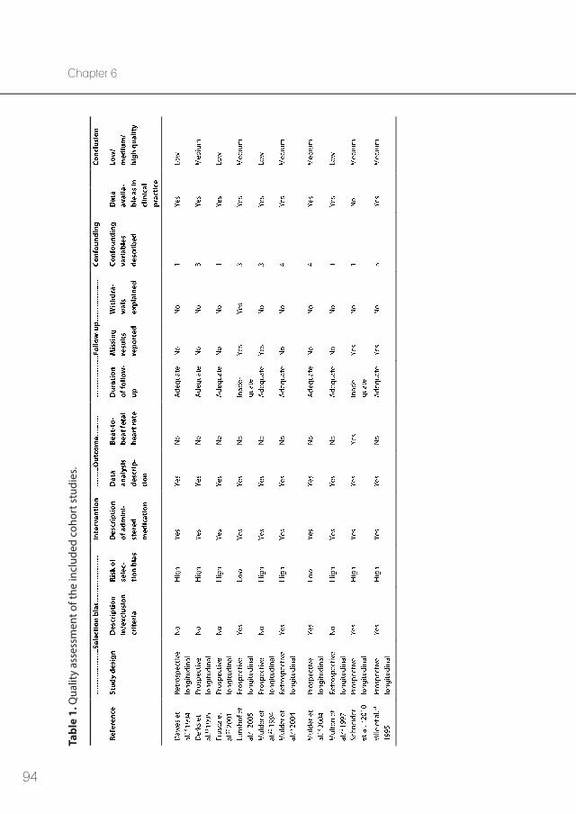
94
Chapter 6Ta
ble
1. Q
ualit
y as
sess
men
t of t
he in
clud
ed c
ohor
t stu
dies
.
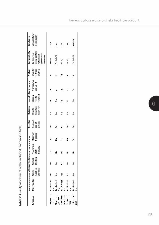
95
6
Review: corticosteroids and fetal heart rate variabilityTa
ble
2. Q
ualit
y as
sess
men
t of t
he in
clud
ed ra
ndom
ised
tria
ls.
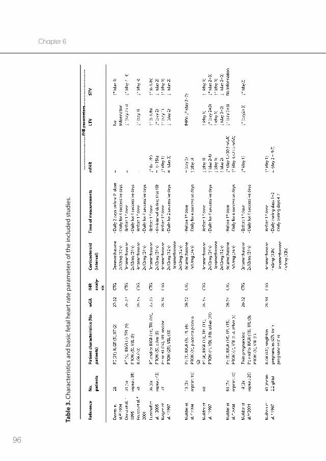
96
Chapter 6
Tabl
e 3.
Cha
ract
eris
tics
and
basi
c fe
tal h
eart
rate
par
amet
ers
of th
e in
clud
ed s
tudi
es.
↑
↓↓
↓↓
↓↑ ↓
↑ ↓↓
↑ ↓↑ ↓
↑↓
↓ ↑↑ ↓
↑ ↓↓ ↑
↑ ↓↑ ↓
↓ ↑↓
↓↓
↓
↑
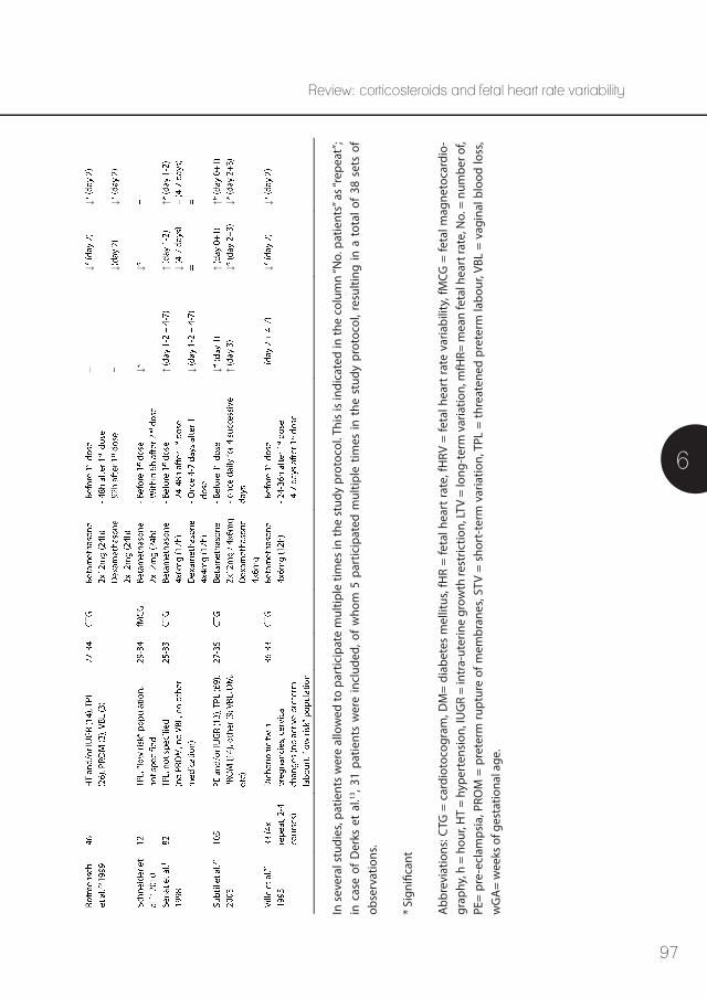
97
6
Review: corticosteroids and fetal heart rate variability
In s
ever
al s
tudi
es, p
atie
nts
wer
e al
low
ed to
par
ticip
ate
mul
tiple
tim
es in
the
stud
y pr
otoc
ol. T
his
is in
dica
ted
in th
e co
lum
n “N
o. p
atie
nts”
as “
repe
at”;
in c
ase
of D
erks
et
al.13
, 31
patie
nts
wer
e in
clud
ed, o
f w
hom
5 p
artic
ipat
ed m
ultip
le t
imes
in t
he s
tudy
pro
toco
l, re
sulti
ng in
a t
otal
of
38 s
ets
of
obse
rvat
ions
.
* Si
gnifi
cant
Abb
revi
atio
ns: C
TG =
car
diot
ocog
ram
, DM
= di
abet
es m
ellit
us, f
HR
= fe
tal h
eart
rate
, fH
RV =
feta
l hea
rt ra
te v
aria
bilit
y, fM
CG =
feta
l mag
neto
card
io-
grap
hy, h
= h
our,
HT
= hy
pert
ensi
on, I
UG
R =
intr
a-ut
erin
e gr
owth
rest
rictio
n, LT
V =
long
-ter
m v
aria
tion,
mfH
R= m
ean
feta
l hea
rt ra
te, N
o. =
num
ber o
f, PE
= pr
e-ec
lam
psia
, PRO
M =
pre
term
rupt
ure
of m
embr
anes
, STV
= s
hort
-ter
m v
aria
tion,
TPL
= th
reat
ened
pre
term
labo
ur, V
BL =
vag
inal
blo
od lo
ss,
wG
A=
wee
ks o
f ges
tatio
nal a
ge.
↓↓
↓↓
↓↓
↑↑ ↓
↑
↓ ↓ ↑↑ ↓
↑ ↓
↓↓

98
Chapter 6
In several studies, patients were allowed to participate multiple times in the study protocol if corticosteroids were administered again after a 10- to 14-day interval. This is indicated in the column “number of patients” in Table 3 as “repeat”. In the study by Derks et al.13, 31 patients were included, of whom 5 participated multiple times in the study protocol, resulting in a total of 38 sets of observations. All included studies were published between 1994 and 2010. A total of 538 fetuses (range; 8-105) were described.
BetamethasoneAs shown in Table 3, a decrease in mfHR was found after betamethasone administration on day 1 in eight of the included studies15,21-27, of which seven showed a significant result. However, this effect was relatively small, with a 3-5% decrease in mfHR21,27. On days 2 and 3, a trend was seen towards an increase in mfHR in multiple studies20,24,27. This trend was signifi-cant only in fetuses younger than 27.5 weeks’ GA25. Other studies showed no effect on mfHR after corticosteroid administration during the study period13,28-31.
All studies observing the effects of corticosteroids on fetal HRV during day 1 showed an increase in both LTV and STV (Table 3)1,21,22,24,27,28. In addition, nearly all studies showed a decrease in both LTV and STV during days 2 to 31,13,15,20-25,27,29-31. Both Mulder et al.23 and Ville et al.31 demonstrated a similar overall response in twin pregnancies compared with singleton pregnancies. There were no significant intertwin differences reported. In IUGR fetuses with redistribution of blood flow (cerebral vasodilation), these modifications were even more profound30. In the study reported by Lunshof et al.21 fetal HRV was decreased by 10% during day 2. The decrease in fetal HRV was also present in more than 80% of the fetuses studied by Mulder et al.27. Likewise, Senat et al.1 demonstrated a significant decrease in both LTV and STV when comparing measurements during days 1 to 2 with measurements during days 4 to 7. Fetal HRV fell below the normal range (30 milliseconds at 30 weeks’ GA) in 46% of the cases; however, no fetal HR decelerations occurred20.
Mulder et al.20,27 also studied movement patterns of the included fetuses with ultrasound, with activity expressed as a percentage of total observation time. All medians were compared with the fetal HR recording during day 0. Body movements were significantly reduced during days 1 and 2 (p <0.01). After both doses of betamethasone, the incidence of body movements was about 50% of the incidence during day 0 (p <0.01). Breathing movements were decreased by 90% during days 2 and 3 (p <0.01); the incidence of hiccups did not change. The total fetal activity (the sum of body- and breathing movements and hiccups) showed a trend with a minimum during day 2 (median incidence falls from 47.7% during day 0 to 8.3% during day 2). A decrease in body- and breathing movements was demonstrated in 88.5% of all fetuses.
99
6
Review: corticosteroids and fetal heart rate variability
For all parameters measured, there were no significant differences between the subgroups (fetuses with IUGR, preterm onset of labour, pre-eclampsia, placenta praevia, and a miscel-laneous group) in all of the included studies. All values returned to baseline during day 420,24.
Dexamethasone Dexamethasone is less well-studied in comparison with betamethasone. Most studies found the same effect of dexamethasone on fetal HR parameters as with betamethasone22,24,27,29. Both Senat et al.1 and Multon et al.26 concluded no significant changes were present after administration of dexamethasone. Concerning mfHR, Dawes et al.28 described no significant changes, whereas Mulder et al.27 found a trend towards decreasing mfHR on day 1 in more than 50% of fetuses following two doses of dexamethasone. STV was increased significantly during day 1 in the reports of both Dawes et al.28 and Mulder et al.27, and this increase was present in more than 80% of the fetuses. In addition, Rotmensch et al.29 found a reduction in fetal HRV during day 2. However, the magnitude of this reduction was less after exposure to dexamethasone as compared with betamethasone. In addition, fetuses exposed to betamethasone had a significantly slower return to baseline LTV as compared with the dexamethasone group (p <0.001). In contrast to reports of betamethasone, no significant differences in fetal movement patterns were observed after the administration of dexamethasone27.
Discussion Quality assessment of the included studies A summary of the quality of the included studies can be found in Tables 1 and 2. In the included cohort studies, a high risk for selection bias was found. This can be attributed to an unclear definition of inclusion and exclusion criteria, the retrospective nature of some studies, and the selection procedure of participants. In the three retrospective studies, only complete data sets were selected for analysis, resulting in a risk for selection bias23,26,28. This risk for selection bias is not clearly reported by most authors. However, the consequences of this possible bias are limited, because every fetus is regarded as its own control in these studies.
Most included patients were at high risk for preterm delivery, and in most studies, it takes up to five days to complete the study protocol. Therefore, a considerable loss of participants can be expected, mainly caused by preterm delivery or discharge before completing the final measurements. Unfortunately, this is not described properly in some of the included studies. It is not clear how, or if, this might impact the results of these studies.
100
Chapter 6
Multon et al.27 used both betamethasone and dexamethasone for fetal lung maturation in fetuses with IUGR; however, this was not in a randomised fashion. Fetuses received 1-7 (mean; 2.2) successive weekly courses of corticosteroids, and six fetuses received both drugs alterna-tively at weekly intervals. Therefore, this study is different in study design from other included studies.
Randomisation was blinded in all randomised trials, whereas observer blinding was performed in three of five studies. In this type of research, observer blinding is less important because of computerised analysis of the data. In general, the description of baseline characteristics of the study population and fetal HR parameters was sufficient. None of the studies conducted an intention-to-treat analysis, because there was no contamination between betamethasone and dexamethasone in clinical practice. Co-interventions were not clearly described in all studies, except for the study by Magee et al.22. The most important co-intervention is the administration of other drugs in addition to corticosteroids.
A point of concern is the small population size in all included studies. Magee et al.22 performed a power analysis but failed to include enough patients in their final analysis. Subtil et al.24 used the same values to calculate power and included just enough patients to fulfill their power criteria. A likely challenge to recruitment is that these studies were conducted in tertiary care hospitals, where a substantial number of the preterm patients are transferred from other care settings. In most cases, tocolysis and corticosteroid administration are started before transport, which makes it impossible to conduct a baseline measurement prior to the first dose of corticosteroids.
Another point to consider is the difference in nature and duration of corticosteroid admin-istration in the various studies. Because of the heterogeneous dosing regimens, a direct comparison by pooling the study results is impossible.
A major drawback of all but one of the included studies is that the recordings used for analysis of fetal HRV were all derived from a Doppler ultrasound-based computerised cardiotocogram (CTG), making it impossible to achieve true beat-to-beat interval registration. With a known beat-to-beat interval, fetal HRV can be calculated more accurately. In addition, spectral energy in the high- and low-frequency bands can be calculated, reflecting both branches of the ANS. Schneider et al.15 used fetal magnetocardiography to obtain beat-to-beat fetal HR parameters non-invasively. Unfortunately, this reliable method is not applicable in daily clinical practice. Direct fetal electrocardiogram (ECG) measurements could be a valuable tool to overcome this problem. Therefore, we suggest further studies on the influence of commonly used medicines in obstetric care on fetal HRV, to focus on electrophysiology of the fetal HR tracings. The
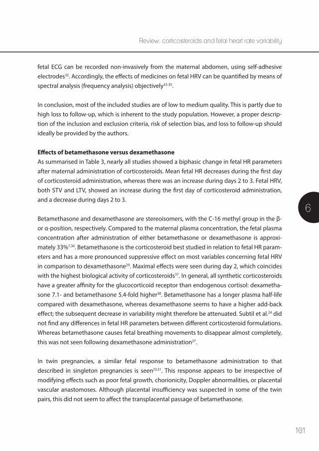
101
6
Review: corticosteroids and fetal heart rate variability
fetal ECG can be recorded non-invasively from the maternal abdomen, using self-adhesive
electrodes32. Accordingly, the effects of medicines on fetal HRV can be quantified by means of spectral analysis (frequency analysis) objectively33-35.
In conclusion, most of the included studies are of low to medium quality. This is partly due to high loss to follow-up, which is inherent to the study population. However, a proper descrip-tion of the inclusion and exclusion criteria, risk of selection bias, and loss to follow-up should ideally be provided by the authors.
Effects of betamethasone versus dexamethasone As summarised in Table 3, nearly all studies showed a biphasic change in fetal HR parameters after maternal administration of corticosteroids. Mean fetal HR decreases during the first day of corticosteroid administration, whereas there was an increase during days 2 to 3. Fetal HRV, both STV and LTV, showed an increase during the first day of corticosteroid administration, and a decrease during days 2 to 3.
Betamethasone and dexamethasone are stereoisomers, with the C-16 methyl group in the β- or α-position, respectively. Compared to the maternal plasma concentration, the fetal plasma concentration after administration of either betamethasone or dexamethasone is approxi-mately 33%7,36. Betamethasone is the corticosteroid best studied in relation to fetal HR param-eters and has a more pronounced suppressive effect on most variables concerning fetal HRV in comparison to dexamethasone29. Maximal effects were seen during day 2, which coincides with the highest biological activity of corticosteroids37. In general, all synthetic corticosteroids have a greater affinity for the glucocorticoid receptor than endogenous cortisol: dexametha-sone 7.1- and betamethasone 5.4-fold higher38. Betamethasone has a longer plasma half-life compared with dexamethasone, whereas dexamethasone seems to have a higher add-back effect; the subsequent decrease in variability might therefore be attenuated. Subtil et al.24 did not find any differences in fetal HR parameters between different corticosteroid formulations. Whereas betamethasone causes fetal breathing movements to disappear almost completely, this was not seen following dexamethasone administration27.
In twin pregnancies, a similar fetal response to betamethasone administration to that described in singleton pregnancies is seen23,31. This response appears to be irrespective of modifying effects such as poor fetal growth, chorionicity, Doppler abnormalities, or placental vascular anastomoses. Although placental insufficiency was suspected in some of the twin pairs, this did not seem to affect the transplacental passage of betamethasone.
102
Chapter 6
Proposed mechanisms of action The exact mechanism of alteration of fetal HRV and fetal behaviour by corticosteroids is unknown. One hypothesis involves the high affinity of corticosteroids for the gonadotropin receptor. Gonadotropin receptors are widely expressed in the central nervous system, including in the brain stem nuclei. They show high affinity to synthetic corticosteroids and suppress neuronal activity when occupied, thus reducing physical activity39. Corticosteroids enter the brain slowly and involve cytosolic gonadotropin receptors, accounting for the prolonged range of action from hours to days40.
The decrease in fetal HR is most likely associated with an increase in fetal systemic blood pressure. Corticosteroids can cause hypertension via augmentation of vascular tone by potentiating the actions of vasoconstrictor hormones and by direct actions on vascular smooth muscle cells40. This yields a vagally-mediated baroreceptor response, which triggers a reflex inhibition of the sympathetic branch and activation of the parasympathetic branch and results in a decreased fetal HR.
The increase in fetal HRV can be explained in part by the known inverse relationship between basal fetal HR and its variation. According to Nijhuis et al.41, about 50% of the differences in fetal HRV can be explained by differences in fetal HR. Furthermore, Bennet et al.42 found a positive correlation between sympathetic tone and short-term heart rate variation, which is attributed to transiently increased local levels of catecholamines (cardiac or neural), and/or to an increased sensitivity to normal circulating levels of catecholamines. ANS regulation of fetal HRV proves to be complex, because both vagal and sympathetic activation seems to cause an increase in fetal HRV. Accordingly, Frasch et al.43 described that sympathetic and vagal influences might be superimposed nonlinearly on fetal HRV and can act synergistically. The transient decrease in fetal HRV during days 2 to 4 cannot solely be explained by an increase in fetal HR, and is likely to be related to the reduction in fetal body- and breathing movements20,27. This may be caused by occupation of the gonadotropin receptors of the responsible fetal brain areas. The raphe nuclei and locus coeruleus are, among others, thought to control motor activity in the third trimester. Neurons of the nucleus of the solitary tract in the medulla are known to direct respiratory activity. Thus, one can hypothesise that complete gonadotropin receptor occupancy of these fetal brain areas after corticosteroid administration reduces both body- and breathing movements. The reduction in fetal movements and fetal HRV, both of which are associated with fetal wellbeing, can be misinterpreted as deterioration of fetal status and can therefore potentially lead to unnecessary iatrogenic preterm delivery13,24. The suppression in fetal HR parameters

103
6
Review: corticosteroids and fetal heart rate variability
is not only visible with computerised analysis, but also detected by visual analysis of CTG tracings by clinicians44. In Figures 1 and 2, the decrease in fetal HRV after corticosteroid administration is illustrated in the case of a 29-week fetus before and 2 days after betametha-sone administration. Shenhav et al.45 demonstrated that if women delivered within 48 hours after betamethasone administration, the reduced fetal HRV was not related to the fetal acid-base balance at birth. Moreover, no changes have been observed in Doppler flow velocity waveforms of uterine and umbilical arteries - brain-sparing was absent, and no decelerations or reduction in fetal eye movements have occurred after corticosteroid administration13,46. Furthermore, there are no signs of any association of steroids with uterine contractions1,24,27. In animal studies, no signs of fetal asphyxia were observed following maternal corticosteroid administration42. In addition, all parameters returned to normal during day 4. These findings all indicate no fetal deterioration during the course of the study period.
Figure 1. Cardiotocogram, one day before betamethasone administration.
Fetal age: 29 weeks + 2 days of gestation. Paper speed: 2cm/minute.
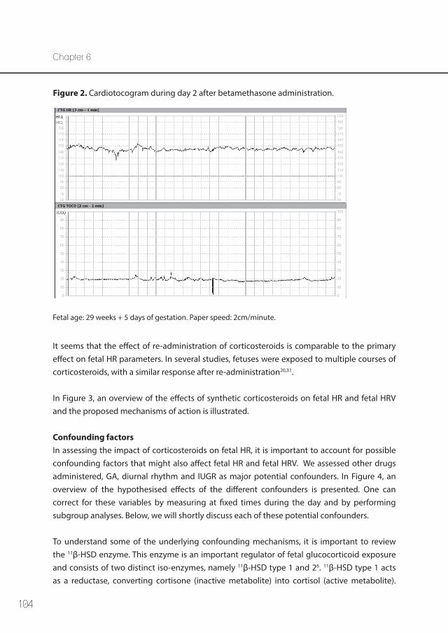
104
It seems that the effect of re-administration of corticosteroids is comparable to the primary effect on fetal HR parameters. In several studies, fetuses were exposed to multiple courses of corticosteroids, with a similar response after re-administration20,31.
In Figure 3, an overview of the effects of synthetic corticosteroids on fetal HR and fetal HRV and the proposed mechanisms of action is illustrated.
Confounding factors In assessing the impact of corticosteroids on fetal HR, it is important to account for possible confounding factors that might also affect fetal HR and fetal HRV. We assessed other drugs administered, GA, diurnal rhythm and IUGR as major potential confounders. In Figure 4, an overview of the hypothesised effects of the different confounders is presented. One can correct for these variables by measuring at fixed times during the day and by performing subgroup analyses. Below, we will shortly discuss each of these potential confounders.
To understand some of the underlying confounding mechanisms, it is important to review the 11β-HSD enzyme. This enzyme is an important regulator of fetal glucocorticoid exposure and consists of two distinct iso-enzymes, namely 11β-HSD type 1 and 26. 11β-HSD type 1 acts as a reductase, converting cortisone (inactive metabolite) into cortisol (active metabolite).
Figure 2. Cardiotocogram during day 2 after betamethasone administration.
Fetal age: 29 weeks + 5 days of gestation. Paper speed: 2cm/minute.
Chapter 6
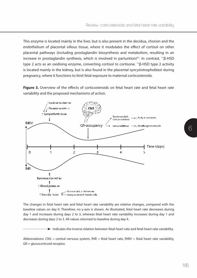
105
This enzyme is located mainly in the liver, but is also present in the decidua, chorion and the endothelium of placental villous tissue, where it modulates the effect of cortisol on other placental pathways (including prostaglandin biosynthesis and metabolism, resulting in an increase in prostaglandin synthesis, which is involved in parturition)47. In contrast, 11β-HSD type 2 acts as an oxidising enzyme, converting cortisol to cortisone. 11β-HSD type 2 activity is located mainly in the kidney, but is also found in the placental syncytiotrophoblast during pregnancy, where it functions to limit fetal exposure to maternal corticosteroids.
↑
↑
↑
↑
↑
↓
↓
Figure 3. Overview of the effects of corticosteroids on fetal heart rate and fetal heart rate variability and the proposed mechanisms of action.
The changes in fetal heart rate and fetal heart rate variability are relative changes, compared with the baseline values on day 0. Therefore, no y-axis is shown. As illustrated, fetal heart rate decreases during day 1 and increases during days 2 to 3, whereas fetal heart rate variability increases during day 1 and decreases during days 2 to 3. All values returned to baseline during day 4.
Indicates the inverse relation between fetal heart rate and fetal heart rate variability.
Abbreviations: CNS = central nervous system, fHR = fetal heart rate, fHRV = fetal heart rate variability, GR = glucocorticoid receptor.
6
Review: corticosteroids and fetal heart rate variability
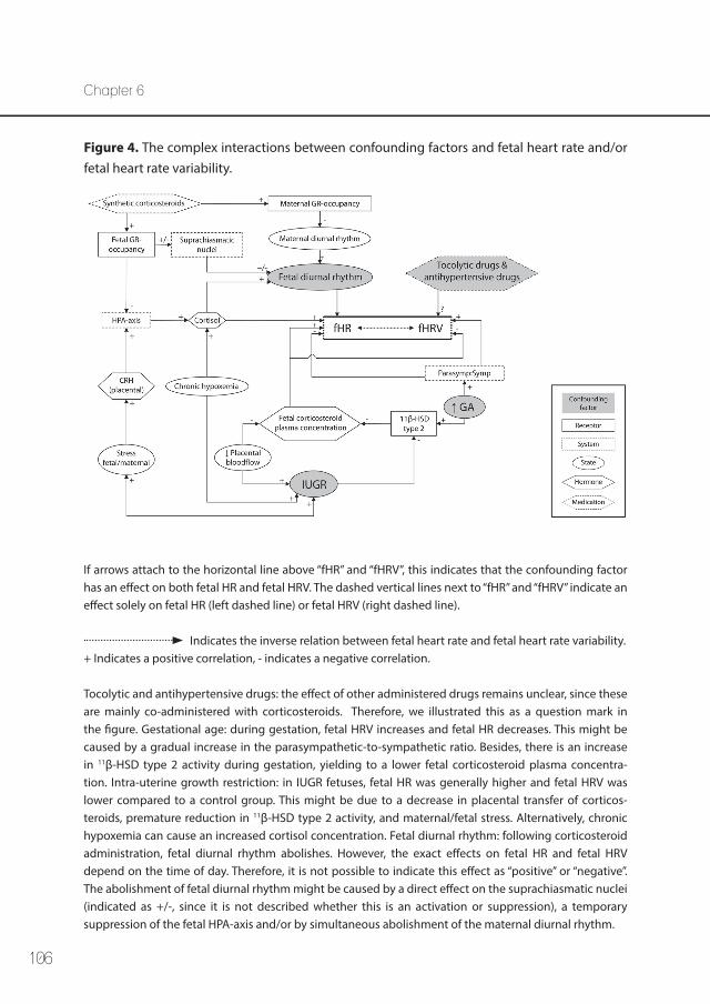
106
Figure 4. The complex interactions between confounding factors and fetal heart rate and/or fetal heart rate variability.
If arrows attach to the horizontal line above “fHR” and “fHRV”, this indicates that the confounding factor has an effect on both fetal HR and fetal HRV. The dashed vertical lines next to “fHR” and “fHRV” indicate an effect solely on fetal HR (left dashed line) or fetal HRV (right dashed line).
Indicates the inverse relation between fetal heart rate and fetal heart rate variability. + Indicates a positive correlation, - indicates a negative correlation.
Tocolytic and antihypertensive drugs: the effect of other administered drugs remains unclear, since these are mainly co-administered with corticosteroids. Therefore, we illustrated this as a question mark in the figure. Gestational age: during gestation, fetal HRV increases and fetal HR decreases. This might be caused by a gradual increase in the parasympathetic-to-sympathetic ratio. Besides, there is an increase in 11β-HSD type 2 activity during gestation, yielding to a lower fetal corticosteroid plasma concentra-tion. Intra-uterine growth restriction: in IUGR fetuses, fetal HR was generally higher and fetal HRV was lower compared to a control group. This might be due to a decrease in placental transfer of corticos-teroids, premature reduction in 11β-HSD type 2 activity, and maternal/fetal stress. Alternatively, chronic hypoxemia can cause an increased cortisol concentration. Fetal diurnal rhythm: following corticosteroid administration, fetal diurnal rhythm abolishes. However, the exact effects on fetal HR and fetal HRV depend on the time of day. Therefore, it is not possible to indicate this effect as “positive” or “negative”. The abolishment of fetal diurnal rhythm might be caused by a direct effect on the suprachiasmatic nuclei (indicated as +/-, since it is not described whether this is an activation or suppression), a temporary suppression of the fetal HPA-axis and/or by simultaneous abolishment of the maternal diurnal rhythm.
Chapter 6
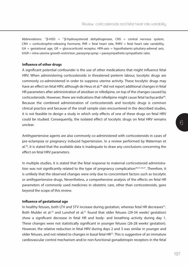
107
Abbreviations: 11β-HSD = 11β-hydroxysteroid dehydrogenase, CNS = central nervous system, CRH = corticotrophin-releasing hormone, fHR = fetal heart rate, fHRV = fetal heart rate variability, GA = gestational age, GR = glucocorticoid receptor, HPA-axis = hypothalamic-pituitary-adrenal axis, IUGR = intra-uterine growth restriction, parasymp:symp = parasympathetic:sympathetic ratio.
Influence of other drugs A significant potential confounder is the use of other medications that might influence fetal HRV. When administering corticosteroids in threatened preterm labour, tocolytic drugs are commonly co-administered in order to suppress uterine activity. These tocolytic drugs may have an effect on fetal HRV, although de Heus et al.48 did not report additional changes in fetal HR parameters after administration of atosiban or nifedipine, on top of the changes caused by corticosteroids. However, there are indications that nifedipine might cause fetal tachycardia49. Because the combined administration of corticosteroids and tocolytic drugs is common clinical practice and because of the small sample sizes encountered in the described studies, it is not feasible to design a study in which only effects of one of these drugs on fetal HRV could be studied. Consequently, the isolated effect of tocolytic drugs on fetal HRV remains unclear.
Antihypertensive agents are also commonly co-administered with corticosteroids in cases of pre-eclampsia or pregnancy induced hypertension. In a review performed by Waterman et al.50, it is stated that the available data is inadequate to draw any conclusions concerning the effect on fetal HRV parameters.
In multiple studies, it is stated that the fetal response to maternal corticosteroid administra-tion was not significantly related to the type of pregnancy complication20,21,23,27. Therefore, it is unlikely that the observed changes were only due to concomitant factors such as tocolytic or antihypertensive drugs. Nevertheless, a comprehensive analysis of the effects on fetal HR parameters of commonly used medicines in obstetric care, other than corticosteroids, goes beyond the scope of this review.
Influence of gestational age In healthy fetuses, both LTV and STV increase during gestation, whereas fetal HR decreases41. Both Mulder et al.25 and Lunshof et al.21 found that older fetuses (29-34 weeks’ gestation) show a significant decrease in fetal HR and body- and breathing activity during day 1. These changes were not statistically significant in younger fetuses (26-28 weeks’ gestation). However, the relative reduction in fetal HRV during days 2 and 3 was similar in younger and older fetuses, and not related to changes in basal fetal HR23. This is suggestive of an immature cardiovascular control mechanism and/or non-functional gonadotropin receptors in the fetal
6
Review: corticosteroids and fetal heart rate variability
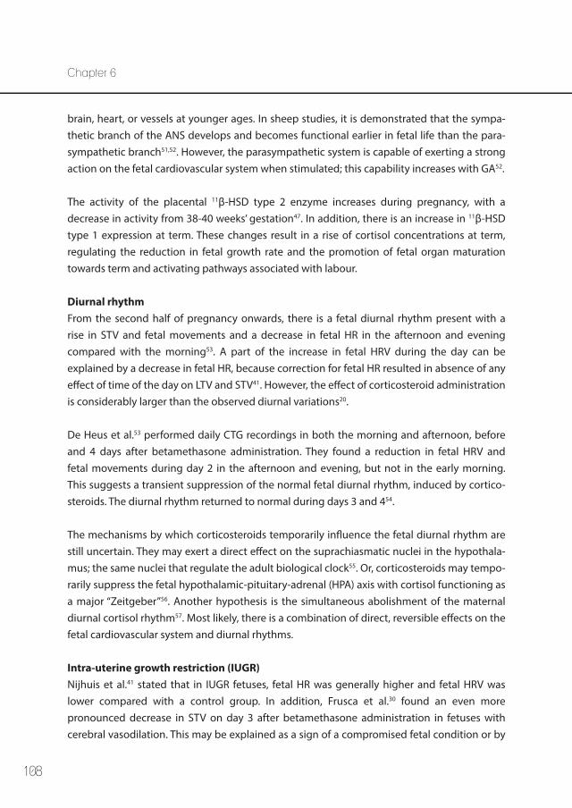
108
brain, heart, or vessels at younger ages. In sheep studies, it is demonstrated that the sympa-thetic branch of the ANS develops and becomes functional earlier in fetal life than the para-sympathetic branch51,52. However, the parasympathetic system is capable of exerting a strong action on the fetal cardiovascular system when stimulated; this capability increases with GA52.
The activity of the placental 11β-HSD type 2 enzyme increases during pregnancy, with a decrease in activity from 38-40 weeks’ gestation47. In addition, there is an increase in 11β-HSD type 1 expression at term. These changes result in a rise of cortisol concentrations at term, regulating the reduction in fetal growth rate and the promotion of fetal organ maturation towards term and activating pathways associated with labour.
Diurnal rhythm From the second half of pregnancy onwards, there is a fetal diurnal rhythm present with a rise in STV and fetal movements and a decrease in fetal HR in the afternoon and evening compared with the morning53. A part of the increase in fetal HRV during the day can be explained by a decrease in fetal HR, because correction for fetal HR resulted in absence of any effect of time of the day on LTV and STV41. However, the effect of corticosteroid administration is considerably larger than the observed diurnal variations20.
De Heus et al.53 performed daily CTG recordings in both the morning and afternoon, before and 4 days after betamethasone administration. They found a reduction in fetal HRV and fetal movements during day 2 in the afternoon and evening, but not in the early morning. This suggests a transient suppression of the normal fetal diurnal rhythm, induced by cortico-steroids. The diurnal rhythm returned to normal during days 3 and 454.
The mechanisms by which corticosteroids temporarily influence the fetal diurnal rhythm are still uncertain. They may exert a direct effect on the suprachiasmatic nuclei in the hypothala-mus; the same nuclei that regulate the adult biological clock55. Or, corticosteroids may tempo-rarily suppress the fetal hypothalamic-pituitary-adrenal (HPA) axis with cortisol functioning as a major “Zeitgeber”56. Another hypothesis is the simultaneous abolishment of the maternal diurnal cortisol rhythm57. Most likely, there is a combination of direct, reversible effects on the fetal cardiovascular system and diurnal rhythms.
Intra-uterine growth restriction (IUGR)Nijhuis et al.41 stated that in IUGR fetuses, fetal HR was generally higher and fetal HRV was lower compared with a control group. In addition, Frusca et al.30 found an even more pronounced decrease in STV on day 3 after betamethasone administration in fetuses with cerebral vasodilation. This may be explained as a sign of a compromised fetal condition or by
Chapter 6
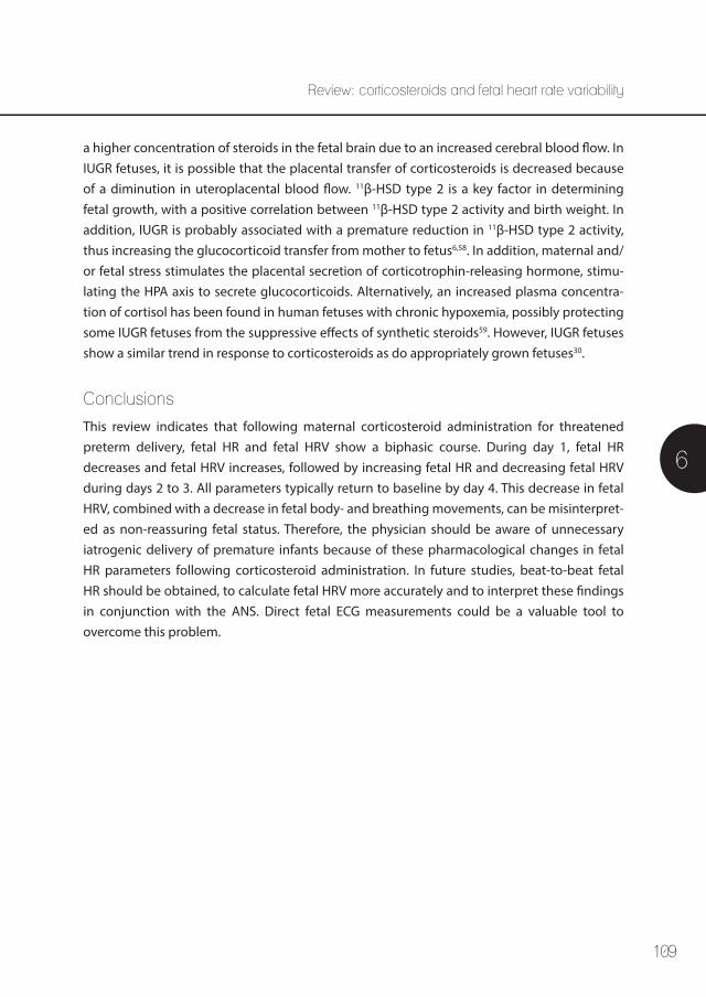
109
a higher concentration of steroids in the fetal brain due to an increased cerebral blood flow. In IUGR fetuses, it is possible that the placental transfer of corticosteroids is decreased because of a diminution in uteroplacental blood flow. 11β-HSD type 2 is a key factor in determining fetal growth, with a positive correlation between 11β-HSD type 2 activity and birth weight. In addition, IUGR is probably associated with a premature reduction in 11β-HSD type 2 activity, thus increasing the glucocorticoid transfer from mother to fetus6,58. In addition, maternal and/or fetal stress stimulates the placental secretion of corticotrophin-releasing hormone, stimu-lating the HPA axis to secrete glucocorticoids. Alternatively, an increased plasma concentra-tion of cortisol has been found in human fetuses with chronic hypoxemia, possibly protecting some IUGR fetuses from the suppressive effects of synthetic steroids59. However, IUGR fetuses show a similar trend in response to corticosteroids as do appropriately grown fetuses30.
Conclusions
This review indicates that following maternal corticosteroid administration for threatened preterm delivery, fetal HR and fetal HRV show a biphasic course. During day 1, fetal HR decreases and fetal HRV increases, followed by increasing fetal HR and decreasing fetal HRV during days 2 to 3. All parameters typically return to baseline by day 4. This decrease in fetal HRV, combined with a decrease in fetal body- and breathing movements, can be misinterpret-ed as non-reassuring fetal status. Therefore, the physician should be aware of unnecessary iatrogenic delivery of premature infants because of these pharmacological changes in fetal HR parameters following corticosteroid administration. In future studies, beat-to-beat fetal HR should be obtained, to calculate fetal HRV more accurately and to interpret these findings in conjunction with the ANS. Direct fetal ECG measurements could be a valuable tool to overcome this problem.
6
Review: corticosteroids and fetal heart rate variability
110
References1. Senat MV, Minoui S, Multon O, Fernandez H, Frydman R, Ville Y. Effect of dexamethasone and
betamethasone on fetal heart rate variability in preterm labour: a randomised study. Br J Obstet Gynaecol 1998 Jul;105(7):749-755.
2. Effect of corticosteroids for fetal maturation on perinatal outcomes. NIH Consensus Development Panel on the Effect of Corticosteroids for Fetal Maturation on Perinatal Outcomes. JAMA 1995 Feb 1;273(5):413-418.
3. Crowley PA. Antenatal corticosteroid therapy: a meta-analysis of the randomized trials, 1972 to 1994. Am J Obstet Gynecol 1995 Jul;173(1):322-335.
4. Liggins GC, Howie RN. A controlled trial of antepartum glucocorticoid treatment for prevention of the respiratory distress syndrome in premature infants. Pediatrics 1972 Oct;50(4):515-525.
5. Roberts D, Dalziel S. Antenatal corticosteroids for accelerating fetal lung maturation for women at risk of preterm birth. Cochrane Database Syst Rev 2006 Jul 19;(3):CD004454.
6. van Runnard Heimel PJ, Franx A, Schobben AF, Huisjes AJ, Derks JB, Bruinse HW. Corticosteroids, pregnancy, and HELLP syndrome: a review. Obstet Gynecol Surv 2005 Jan;60(1):57-70; quiz 73-4.
7. Petersen MC, Nation RL, Ashley JJ, McBride WG. The placental transfer of betamethasone. Eur J Clin Pharmacol 1980 Oct;18(3):245-247.
8. Reynolds F, Knott C. Pharmacokinetics in pregnancy and placental drug transfer. Oxf Rev Reprod Biol 1989;11:389-449.
9. Blanford AT, Murphy BE. In vitro metabolism of prednisolone, dexamethasone, betamethasone, and cortisol by the human placenta. Am J Obstet Gynecol 1977 Feb 1;127(3):264-267.
10. Dalziel SR, Lim VK, Lambert A, McCarthy D, Parag V, Rodgers A, et al. Antenatal exposure to beta-methasone: psychological functioning and health related quality of life 31 years after inclusion in randomised controlled trial. BMJ 2005 Sep 24;331(7518):665.
11. Dessens AB, Haas HS, Koppe JG. Twenty-year follow-up of antenatal corticosteroid treatment. Pedi-atrics 2000 Jun;105(6):E77.
12. Mariotti V, Marconi AM, Pardi G. Undesired effects of steroids during pregnancy. J Matern Fetal Neonatal Med 2004 Nov;16 Suppl 2:5-7.
13. Derks JB, Mulder EJ, Visser GH. The effects of maternal betamethasone administration on the fetus. Br J Obstet Gynaecol 1995 Jan;102(1):40-46.
14. Van Ravenswaaij-Arts C, Kollee L, Hopman J, Stoelinga G, van Geijn H. Heart rate variabilitiy. Ann Intern Med 1993;118:436-447.
15. Schneider U, Fiedler A, Schroder B, Jaekel S, Stacke A, Hoyer D, et al. The effect of antenatal steroid treatment on fetal autonomic heart rate regulation revealed by fetal magnetocardiography (fMCG). Early Hum Dev 2010 May;86(5):319-325.
16. van Laar JO, Peters CH, Vullings R, Houterman S, Oei SG. Power spectrum analysis of fetal heart rate variability at near term and post term gestation during active sleep and quiet sleep. Early Hum Dev 2009 Dec;85(12):795-798.
Chapter 6
111
17. Dawes GS, Lobb M, Moulden M, Redman CW, Wheeler T. Antenatal cardiotocogram quality and interpretation using computers. Br J Obstet Gynaecol 1992 Oct;99(10):791-797.
18. Hon EH, Lee ST. Electronic Evaluation of the Fetal Heart Rate. Viii. Patterns Preceding Fetal Death, further Observations. Am J Obstet Gynecol 1963 Nov 15;87:814-826.
19. van Laar JO, Peters CH, Vullings R, Houterman S, Bergmans JW, Oei SG. Fetal autonomic response to severe acidaemia during labour. BJOG 2010 Mar;117(4):429-437.
20. Mulder EJ, Derks JB, Zonneveld MF, Bruinse HW, Visser GH. Transient reduction in fetal activity and heart rate variation after maternal betamethasone administration. Early Hum Dev 1994 Jan;36(1):49-60.
21. Lunshof MS, Boer K, Wolf H, Koppen S, Velderman JK, Mulder EJ. Short-term (0-48 h) effects of maternal betamethasone administration on fetal heart rate and its variability. Pediatr Res 2005 Apr;57(4):545-549.
22. Magee LA, Dawes GS, Moulden M, Redman CW. A randomised controlled comparison of betamethasone with dexamethasone: effects on the antenatal fetal heart rate. Br J Obstet Gynaecol 1997 Nov;104(11):1233-1238.
23. Mulder EJ, Derks JB, Visser GH. Effects of antenatal betamethasone administration on fetal heart rate and behavior in twin pregnancy. Pediatr Res 2004 Jul;56(1):35-39.
24. Subtil D, Tiberghien P, Devos P, Therby D, Leclerc G, Vaast P, et al. Immediate and delayed effects of antenatal corticosteroids on fetal heart rate: a randomized trial that compares betamethasone acetate and phosphate, betamethasone phosphate, and dexamethasone. Am J Obstet Gynecol 2003 Feb;188(2):524-531.
25. Mulder EJ, Koenen SV, Blom I, Visser GH. The effects of antenatal betamethasone administration on fetal heart rate and behaviour depend on gestational age. Early Hum Dev 2004 Jan;76(1):65-77.
26. Multon O, Senat MV, Minoui S, Hue MV, Frydman R, Ville Y. Effect of antenatal betamethasone and dexamethasone administration on fetal heart rate variability in growth-retarded fetuses. Fetal Diagn Ther 1997 May-Jun;12(3):170-177.
27. Mulder EJ, Derks JB, Visser GH. Antenatal corticosteroid therapy and fetal behaviour: a randomised study of the effects of betamethasone and dexamethasone. Br J Obstet Gynaecol 1997 Nov;104(11):1239-1247.
28. Dawes GS, Serra-Serra V, Moulden M, Redman CW. Dexamethasone and fetal heart rate variation. Br J Obstet Gynaecol 1994 Aug;101(8):675-679.
29. Rotmensch S, Liberati M, Vishne TH, Celentano C, Ben-Rafael Z, Bellati U. The effect of betamethasone and dexamethasone on fetal heart rate patterns and biophysical activities. A prospective randomized trial. Acta Obstet Gynecol Scand 1999 Jul;78(6):493-500.
30. Frusca T, Soregaroli M, Valcamonico A, Scalvi L, Bonera R, Bianchi U. Effect of betamethasone on computerized cardiotocographic parameters in preterm growth-restricted fetuses with and without cerebral vasodilation. Gynecol Obstet Invest 2001;52(3):194-197.
6
Review: corticosteroids and fetal heart rate variability
112
31. Ville Y, Vincent Y, Tordjman N, Hue MV, Fernandez H, Frydman R. Effect of betamethasone on the fetal heart rate pattern assessed by computerized cardiotocography in normal twin pregnancies. Fetal Diagn Ther 1995 Sep-Oct;10(5):301-306.
32. Vullings R, Peters C, Andriessen P, Oei S, Wijn P. Monitoring the Fetal Heart Rate and Fetal Electro-cardiogram: Abdominal Recordings Are As Good As Direct Ecg Measurements. Pediatric Research 2005;58(2):242.
33. Siira SM, Ojala TH, Vahlberg TJ, Jalonen JO, Valimaki IA, Rosen KG, et al. Marked fetal acidosis and specific changes in power spectrum analysis of fetal heart rate variability recorded during the last hour of labour. BJOG 2005 Apr;112(4):418-423.
34. Van Laar JO, Porath MM, Peters CH, Oei SG. Spectral analysis of fetal heart rate variability for fetal surveillance: review of the literature. Acta Obstet Gynecol Scand 2008;87(3):300-306.
35. van Laar JO, Peters CH, Houterman S, Wijn PF, Kwee A, Oei SG. Normalized spectral power of fetal heart rate variability is associated with fetal scalp blood pH. Early Hum Dev 2011 Apr;87(4):259-263.
36. Kream J, Mulay S, Fukushima DK, Solomon S. Determination of plasma dexamethasone in the mother and the newborn after administration of the hormone in a clinical trial. J Clin Endocrinol Metab 1983 Jan;56(1):127-133.
37. Haynes RC. Adrenocorticotropic hormone, adrenocortical steroids and their synthetic analogs, inhibitors of adrenal steroids biosynthesis. In: Goodman Gilman A, Rall TW, Nies AS, Taylor P, editors. The Pharmacological Basis of Therapeutics. 8th ed.: Pergamon Press, New York; 1990. p. 1431-1462.
38. Ballard PL, Ballard RA. Scientific basis and therapeutic regimens for use of antenatal glucocorti-coids. Am J Obstet Gynecol 1995 Jul;173(1):254-262.
39. de Kloet ER, Reul JM, Sutanto W. Corticosteroids and the brain. J Steroid Biochem Mol Biol 1990 Nov 20;37(3):387-394.
40. Ullian ME. The role of corticosteriods in the regulation of vascular tone. Cardiovasc Res 1999 Jan;41(1):55-64.
41. Nijhuis IJ, ten Hof J, Mulder EJ, Nijhuis JG, Narayan H, Taylor DJ, et al. Fetal heart rate in relation to its variation in normal and growth retarded fetuses. Eur J Obstet Gynecol Reprod Biol 2000 Mar;89(1):27-33.
42. Bennet L, Kozuma S, McGarrigle HH, Hanson MA. Temporal changes in fetal cardiovascular, behavioural, metabolic and endocrine responses to maternally administered dexamethasone in the late gestation fetal sheep. Br J Obstet Gynaecol 1999 Apr;106(4):331-339.
43. Frasch MG, Muller T, Hoyer D, Weiss C, Schubert H, Schwab M. Nonlinear properties of vagal and sympathetic modulations of heart rate variability in ovine fetus near term. Am J Physiol Regul Integr Comp Physiol 2009 Mar;296(3):R702-7.
44. Rotmensch S, Lev S, Kovo M, Efrat Z, Zahavi Z, Lev N, et al. Effect of betamethasone administration on fetal heart rate tracing: a blinded longitudinal study. Fetal Diagn Ther 2005 Sep-Oct;20(5):371-376.
45. Shenhav S, Volodarsky M, Anteby EY, Gemer O. Fetal acid-base balance after betamethasone administration: relation to fetal heart rate variability. Arch Gynecol Obstet 2008 Oct;278(4):333-336.
Chapter 6
113
46. Cohlen BJ, Stigter RH, Derks JB, Mulder EJ, Visser GH. Absence of significant hemodynamic changes in the fetus following maternal betamethasone administration. Ultrasound Obstet Gynecol 1996 Oct;8(4):252-255.
47. Murphy VE, Clifton VL. Alterations in human placental 11beta-hydroxysteroid dehydrogenase type 1 and 2 with gestational age and labour. Placenta 2003 Aug;24(7):739-744.
48. De Heus R, Mulder E, Derks J, Visser G. The effects of the tocolytics atosiban and nifedipine on fetal movements, heart rate and blood flow. The Journal of Maternal-Fetal and Neonatal Medicine 2009;22(6):485-490.
49. Chan LW, Sahota DS, Yeung SY, Leung TY, Fung TY, Lau TK, et al. Side-effect and vital sign profile of nifedipine as a tocolytic for preterm labour. Hong Kong Med J 2008 Aug;14(4):267-272.
50. Waterman E, Magee L, Lim K, Skoll A, Rurak D, Phil D, et al. Do Commonly Used Oral Antihyper-tensives Alter Fetal or Neonatal Heart Rate Characteristics? A Systematic Review. Hypertension in pregnancy 2004;23(2):155-169.
51. Assali NS, Brinkman CR,3rd, Woods JR,Jr, Dandavino A, Nuwayhid B. Development of neurohumoral control of fetal, neonatal, and adult cardiovascular functions. Am J Obstet Gynecol 1977 Dec 1;129(7):748-759.
52. Nuwayhid B, Brinkman CR,3rd, Su C, Bevan JA, Assali NS. Development of autonomic control of fetal circulation. Am J Physiol 1975 Feb;228(2):337-344.
53. de Heus R, Mulder EJ, Derks JB, Koenen SV, Visser GH. Differential effects of betamethasone on the fetus between morning and afternoon recordings. J Matern Fetal Neonatal Med 2008 Aug;21(8):549-554.
54. Koenen SV, Mulder EJ, Wijnberger LD, Visser GH. Transient loss of the diurnal rhythms of fetal movements, heart rate, and its variation after maternal betamethasone administration. Pediatr Res 2005 May;57(5 Pt 1):662-666.
55. Rietveld WJ. The central control and ontogeny of circadian rhythmicity. Eur J Morphol 1990;28(2-4):301-307.
56. Dickmeis T, Lahiri K, Nica G, Vallone D, Santoriello C, Neumann CJ, et al. Glucocorticoids play a key role in circadian cell cycle rhythms. PLoS Biol 2007 Apr;5(4):e78.
57. Helal KJ, Gordon MC, Lightner CR, Barth WH,Jr. Adrenal suppression induced by betamethasone in women at risk for premature delivery. Obstet Gynecol 2000 Aug;96(2):287-290.
58. Edwards CR, Benediktsson R, Lindsay RS, Seckl JR. 11 beta-Hydroxysteroid dehydrogenases: key enzymes in determining tissue-specific glucocorticoid effects. Steroids 1996 Apr;61(4):263-269.
59. Economides DL, Nicolaides KH, Linton EA, Perry LA, Chard T. Plasma cortisol and adrenocorticotro-pin in appropriate and small for gestational age fetuses. Fetal Ther 1988;3(3):158-164.
6
Review: corticosteroids and fetal heart rate variability

114
115
7
Effect of tocolytic drugs on fetal heart rate variability: a systematic review
Kim M.J. Verdurmen, Alexandra D.J. Hulsenboom, Judith O.E.H. van Laar, S. Guid Oei
Published: The Journal of Maternal-Fetal & Neonatal Medicine. 2016:1-8
Chapter 7
116
Chapter 7
Abstract
IntroductionTocolytics may cause changes in fetal heart rate pattern, while fetal heart rate variability is an important marker of fetal wellbeing. We aim to systematically review the literature on how tocolytic drugs affect fetal heart rate variability.
Material and MethodsWe searched CENTRAL, PubMed, and EMBASE up to June 2016. Studies published in English, using computerised or visual analysis to describe the effect of tocolytics on heart rate varia-bility in human fetuses were included. Studies describing tocolytics during labour, external cephalic version, pre-eclampsia and infection were excluded. Eventually, we included six studies, describing 169 pregnant women.
ResultsNifedipine, atosiban and indomethacin administration show no clinically important effect on fetal heart rate variability. Following administration of magnesium sulphate, decreased variability and cases of bradycardia are described. Fenoterol administration results in a slight increase in fetal heart rate with no changes in variability. After ritodrine administration increased fetal heart rate and decreased variability is seen. The effect of co-administration of corticosteroids should be taken into account.
ConclusionIn order to prevent iatrogenic preterm labour, the effects of tocolytic drugs on fetal heart rate variability should be taken into account when monitoring these fetuses.
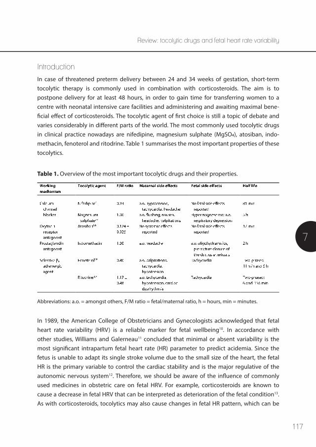
117
7
Review: tocolytic drugs and fetal heart rate variability
Introduction
In case of threatened preterm delivery between 24 and 34 weeks of gestation, short-term tocolytic therapy is commonly used in combination with corticosteroids. The aim is to postpone delivery for at least 48 hours, in order to gain time for transferring women to a centre with neonatal intensive care facilities and administering and awaiting maximal bene-ficial effect of corticosteroids. The tocolytic agent of first choice is still a topic of debate and varies considerably in different parts of the world. The most commonly used tocolytic drugs in clinical practice nowadays are nifedipine, magnesium sulphate (MgSO4), atosiban, indo-methacin, fenoterol and ritodrine. Table 1 summarises the most important properties of these tocolytics.
Table 1. Overview of the most important tocolytic drugs and their properties.
Abbreviations: a.o. = amongst others, F/M ratio = fetal/maternal ratio, h = hours, min = minutes.
In 1989, the American College of Obstetricians and Gynecologists acknowledged that fetal heart rate variability (HRV) is a reliable marker for fetal wellbeing10. In accordance with other studies, Williams and Galerneau11 concluded that minimal or absent variability is the most significant intrapartum fetal heart rate (HR) parameter to predict acidemia. Since the fetus is unable to adapt its single stroke volume due to the small size of the heart, the fetal HR is the primary variable to control the cardiac stability and is the major regulative of the autonomic nervous system12. Therefore, we should be aware of the influence of commonly used medicines in obstetric care on fetal HRV. For example, corticosteroids are known to cause a decrease in fetal HRV that can be interpreted as deterioration of the fetal condition13. As with corticosteroids, tocolytics may also cause changes in fetal HR pattern, which can be
118
Chapter 7
erroneously interpreted as a sign of fetal distress. This could lead to iatrogenic preterm delivery. In this review, we will focus on the influence of tocolytic drugs on fetal HRV in human fetuses.
Material and Methods
We performed a systematic search in the electronic databases CENTRAL (the Cochrane Library, 2016, Issue 6), PubMed, and EMBASE up to June 2016. The search strategy is attached in the Appendix.
All published studies that describe the influence of tocolytic drugs on HRV of the human fetus, assessed by computerised or visual analysis, were included in this review. The outcome measure of interest was variability of the fetal HR. There were no restrictions on publication dates. We excluded review studies, studies that described pre-eclamptic women or women with an intra-uterine infection and studies that performed measurements during labour or during external cephalic version. The study language was restricted to English.
Two reviewers (KV and AH) independently performed the search and abstracted the data. Any discrepancies were resolved by discussion. Articles were initially screened by title and abstract, when appropriate a full-text evaluation was performed. Qualitative analysis, including description of the women enrolled, description of the intervention and outcomes, and possible risks of bias, were based on the review guidelines of the Dutch Cochrane Centre.
Results
The screening and inclusion process is depicted in Figure 1. Eventually, six out of 72 articles were included in this review (169 women). Study characteristics and basic fetal HR parameters are shown in Table 2.1 (computerised analysis of fetal HR parameters) and Table 2.2 (visual analysis of fetal HR parameters). Three of the included studies are randomised controlled trials, two are case reports and one is a prospective cohort study. Table 3 includes the quality assessment of the included randomised trials. We considered the trials by de Heus et al.14 and Neri et al.15 as medium quality evidence, as blinding was not appropriate or not described. The trial by Hallak et al.16 was considered high-quality evidence. Table 4 includes the quality assessment of the included prospective cohort study by Wright et al.17. This study was consid-ered low-quality evidence since there was no description of exclusion criteria and there was a high risk of selection bias: the tocolytic agent used was the choice of the individual physician and co-administration of corticosteroids was not described.
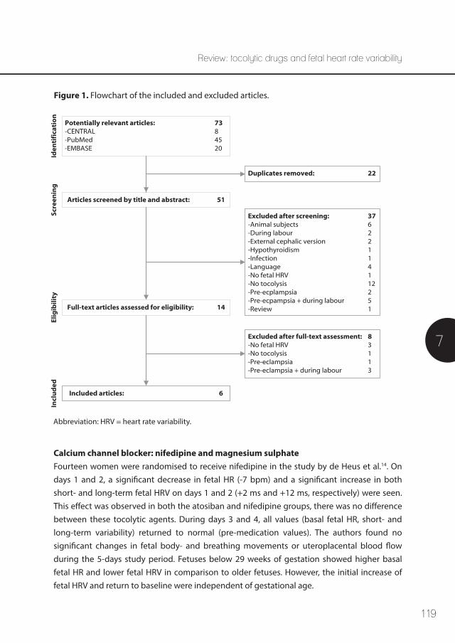
119
7
Review: tocolytic drugs and fetal heart rate variability
Figure 1. Flowchart of the included and excluded articles.
Abbreviation: HRV = heart rate variability.
Potentially relevant articles: 73-CENTRAL 8-PubMed 45-EMBASE 20
Articles screened by title and abstract: 51
Full-text articles assessed for eligibility: 14
Duplicates removed: 22
Excluded after screening: 37-Animal subjects 6-During labour 2-External cephalic version 2-Hypothyroidism 1-Infection 1-Language 4-No fetal HRV 1-No tocolysis 12-Pre-ecplampsia 2-Pre-ecpampsia + during labour 5-Review 1
Included articles: 6
Excluded after full-text assessment: 8-No fetal HRV 3-No tocolysis 1-Pre-eclampsia 1-Pre-eclampsia + during labour 3
Iden
tifi
cati
on
Scre
enin
gEl
igib
ility
Incl
ud
ed
Calcium channel blocker: nifedipine and magnesium sulphateFourteen women were randomised to receive nifedipine in the study by de Heus et al.14. On days 1 and 2, a significant decrease in fetal HR (-7 bpm) and a significant increase in both short- and long-term fetal HRV on days 1 and 2 (+2 ms and +12 ms, respectively) were seen. This effect was observed in both the atosiban and nifedipine groups, there was no difference between these tocolytic agents. During days 3 and 4, all values (basal fetal HR, short- and long-term variability) returned to normal (pre-medication values). The authors found no significant changes in fetal body- and breathing movements or uteroplacental blood flow during the 5-days study period. Fetuses below 29 weeks of gestation showed higher basal fetal HR and lower fetal HRV in comparison to older fetuses. However, the initial increase of fetal HRV and return to baseline were independent of gestational age.
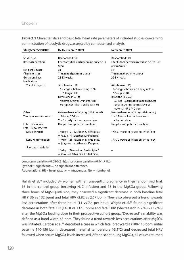
120
Chapter 7
Table 2.1 Characteristics and basic fetal heart rate parameters of included studies concerning administration of tocolytic drugs, assessed by computerised analysis.
Long-term variation (0.08-0.2 Hz), short-term variation (0.4-1.7 Hz). Symbol: *, significant; =, no significant difference.Abbreviations: HR = heart rate, i.v. = intravenous, No. = number of.
↓ ↑
↑ ↓
↑
Hallak et al.16 included 34 women with an uneventful pregnancy in their randomised trial; 16 in the control group (receiving NaCl-infusion) and 18 in the MgSO4-group. Following three hours of MgSO4-infusion, they observed a significant decrease in both baseline fetal HR (136 vs 132 bpm) and fetal HRV (2.82 vs 2.67 bpm). They also observed a trend towards less accelerations after three hours (11 vs 7.4 per hour). Wright et al.17 found a significant decrease in both fetal HR (140.8 vs 137.3 bpm) and fetal HRV (“decreased” in 2/48 vs 12/48) after the MgSO4 loading dose in their prospective cohort group. “Decreased” variability was defined as a band width ≤5 bpm. They found a trend towards less accelerations after MgSO4 was initiated. Cardosi et al.18 described a case in which fetal bradycardia (100-110 bpm, initial baseline 140-150 bpm), decreased maternal temperature (-3.1°C) and decreased fetal HRV followed when serum MgSO4 levels increased. After discontinuing MgSO4, all values returned
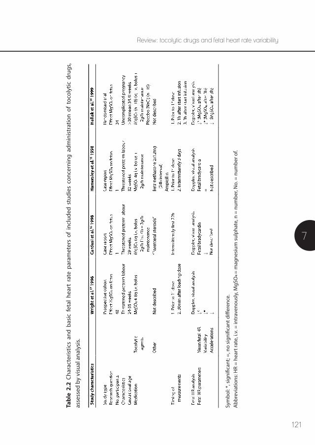
121
7
Review: tocolytic drugs and fetal heart rate variability
Tabl
e 2.
2 Ch
arac
teris
tics
and
basi
c fe
tal
hear
t ra
te p
aram
eter
s of
inc
lude
d st
udie
s co
ncer
ning
adm
inis
trat
ion
of t
ocol
ytic
dru
gs,
asse
ssed
by
visu
al a
naly
sis.
Sym
bol:
*, s
igni
fican
t; =,
no
sign
ifica
nt d
iffer
ence
. A
bbre
viat
ions
: HR
= he
art r
ate,
i.v.
= in
trav
enou
sly,
MgS
O4 =
mag
nesi
um s
ulph
ate,
n =
num
ber,
No.
= n
umbe
r of.
↓ ↓ ↓↓
↓ ↓ ↓
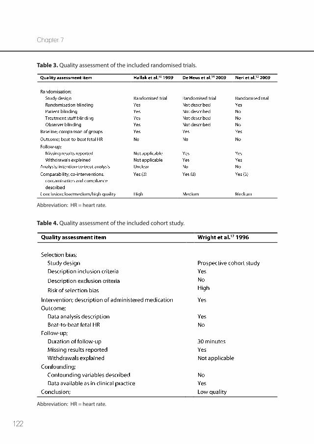
122
Chapter 7
Table 3. Quality assessment of the included randomised trials.
Table 4. Quality assessment of the included cohort study.
Abbreviation: HR = heart rate.
Abbreviation: HR = heart rate.
123
7
Review: tocolytic drugs and fetal heart rate variability
Oxytocin receptor antagonist: atosibanA total of 46 included women with threatened preterm delivery received atosiban in combi-nation with betamethasone. De Heus et al.14 randomised 17 women to the atosiban group and found no difference in changes between the atosiban and the nifedipine group: on days 1 and 2, both short- and long-term fetal HRV increased significantly, and returned to premed-ication values on days 3 and 4. Fetal HR showed a transient significant decrease during days 1 and 2, that returned to premedication values on days 3 and 4.
Neri et al.15 performed their measurements at least twelve hours after the last corticosteroid administration. They found no significant differences between the atosiban group (n=29) and the ritodrine group considering fetal HR (148.5 vs 152.5 bpm), long-term variability (18.3 vs 18.9), and short-term variability (5 vs 5.1).
Prostaglandin synthesis inhibitor: indomethacin We did not find any studies describing the effect of indomethacin on fetal HRV that matched our inclusion criteria.
Selective β2-adrengergic agent: fenoterol and ritodrine We did not find any studies describing the effect of fenoterol on fetal HRV that matched our inclusion criteria.Neri et al.15 observed no differences between 25 women who received ritodrine and those who received atosiban. Subgroup analysis showed that fetuses before 30 weeks of gestation treated with ritodrine had a significant higher fetal HR (154 vs 148 bpm), lower long-term fetal HRV (15.7 vs 21.6 ms) and lower low-frequency/high-frequency ratio (18.5 vs 29.9) than those treated with atosiban.
Discussion
General CommentsFirst of all, our search revealed that little research has been published regarding the influence of tocolytic drugs on fetal HRV. Tocolysis is nearly always combined with corticosteroids that are known to have transient but considerable effects on fetal HRV13. This makes the interpre-tation of the sole effect of tocolytics difficult. However, the combination of these medicines
to pre-MgSO4 levels within 24 hours. Hamersley et al.19 also described a case of fetal brady-cardia where baseline dropped from 140 bpm to 110 bpm following 30 minutes of MgSO4 infusion and to 100-105 bpm following 90 minutes. The fetal HRV remained good in this case. After discontinuing MgSO4-infusion on the third day, fetal HR baseline returned to 140 bpm.
124
Chapter 7
is daily clinical practice and therefore we chose to include studies that describe the effect of tocolytics, regardless of the co-administration of corticosteroids. We chose to include studies that used visual interpretation of fetal HR tracings, despite the well-known inter- and intra-observer variations in visual fetal HR tracing interpretation. Those study results are valuable, since visual analysis is common clinical practice and computerised analysis is only used for research so far.
All included studies used Doppler ultrasound-based cardiotocogram recordings for fetal HRV analysis (visual or computerised). To perform spectral analysis and obtain more complete and reliable information on fetal HRV, the fetal HR must be acquired on a beat-to-beat basis. Standard Doppler recordings cannot derive the fetal HR on a beat-to-beat basis, but produce an average fetal HR. It is feasible to use Doppler ultrasound for beat-to-beat fetal HR meas-urements, although this is technically challenging and requires special algorithms20. These are not described in the included studies. We recommend future studies to focus on beat-to-beat analysis, to calculate fetal HRV more accurately and to interpret the findings in conjunction with the autonomic nervous system.
We excluded women during labour, since the stress of labour is known to influence fetal HRV (the sympathicovagal balance)21 and a woman is likely to receive other pharmacologic agents in addition to tocolytics which can interfere with the fetal HR. In addition, we did not include women with pre-eclampsia. This pathological state with abnormal placental-fetal circulation and hemodynamical fetal stress is known to influence fetal HR tracings due to changes in cardiovascular regulation22. Women with (suspicion of ) intra-uterine infection were excluded as well, since it is demonstrated in fetuses that reduced HRV and transient decelerations are an important physiomarker and can precede clinical signs of sepsis23,24. We also chose to exclude studies that performed measurements during or after external cephalic version, since the effect of the version may interfere with the observed fetal HR parameters25-27.
Calcium channel blocker: nifedipine and magnesium sulphate Nifedipine crosses the placenta easily and contradictory results have been reported regarding changes in uteroplacental blood flow. In the study by de Heus et al.14, the number of participants steadily decreased during the study period due to preterm delivery. On day 4, 77% (24/31 women) remained available for analysis. Due to this small amount of women, study results were depicted as the influence of both atosiban and nifedipine together. A subanalysis showed no differences in fetal parameters between the two tocolytics, but details are not shown. The decrease in fetal HR and increase in short- and long-term fetal HRV on days 1 and 2 were attributed to the known effect of corticosteroids by the authors. However, our review considering the effects of corticosteroids on fetal HRV showed a decrease in fetal
125
7
Review: tocolytic drugs and fetal heart rate variability
HR and increase in fetal HRV during day 1, and an increase in fetal HR and decrease in fetal HRV during days 2 and 313. This difference might be attributable to the low number of women included in the study by de Heus et al.14. The nifedipine administration was “progressively decreased” after 48 hours. However, the authors did not exactly describe in what time course or dosages this was performed.
In summary, it seems that nifedipine has no clinically important effect on fetal HRV.
MgSO4 crosses the placenta rapidly, and reaches an equilibrium after two hours of admin-istration between maternal and fetal plasma levels4. Moreover, fetal levels seem to increase proportionally with maternal levels16,28. It is assumed that MgSO4 crosses the fetal blood-brain barrier, as it does in the mother. It suppresses the central nervous system and inhibits cardio-accelerating pathways via the cerebral cortex, hypothalamus and the cardioaccelerator center in the medulla oblongata29.
Hallak et al.16 studied healthy pregnant women, without any indication for treatment with MgSO4 in their randomised trial. Therefore, their measurements were not influenced by conditions like pre-eclampsia, premature contractions or co-administration of corticosteroids. Besides decreased fetal HR and fetal HRV, they found that the positive correlation between gestational age and the number of accelerations observed in the control group (treated with NaCl) was missing in women treated with MgSO4. An animal study confirmed a significant decrease in both baseline fetal HR as well as long- and short-term variability four hours after MgSO4 administration, compared to the control group (5% glucose infusion)29. The included prospective study by Wright et al.17 found similar results regarding fetal HR and fetal HRV. They also state that a lower gestational age increases the likelihood of decreased variability (odds ratio 0.75, p = 0.03). Cardosi and Hamersley both described a case report with fetal bradycardia following MgSO4-infusion in a preterm fetus. Cardosi et al.18 state that the effect on fetal HR might indirectly be related to the hypothermic effect of MgSO4. Hamersley et al.19 suggested a pharmacological depression of the central nervous system. The decrease in fetal HRV can be interpreted as a sign of compromised fetal condition and expedite iatrogenic preterm delivery, while Duffy et al.30 showed that the decreased variability is associated with exposure to MgSO4, since they excluded adverse fetal outcomes. This indicates a transient medication effect (central nervous system depression) rather than a marker of deteriorating fetal condition, as confirmed by other studies31,32.
In summary, decreased fetal HRV and cases of fetal bradycardia are described after MgSO4 administration.
126
Chapter 7
Oxytocin receptor antagonist: atosiban Atosiban is known to be highly specific for the oxytocin receptors in the uterus, with low placental passage and few maternal and fetal side effects. Both de Heus et al.14 and Neri et al.15 found no other changes than the changes known to be caused by corticosteroids.
Neri et al.15 performed one measurement, at least 12 hours after the last corticosteroid administration. They state this would avoid the adverse effects on fetal behaviour following corticosteroid administration. However, it can take up to 96 hours for fetal HR parameters to return to baseline values following corticosteroid administration13. Therefore, the effect of corticosteroids is still likely to overwhelm the effect of atosiban and ritodrine. No baseline (premedication) measurements were performed, but two medication groups were compared at one moment. A direct comparison between two drugs was performed, instead of compar-ison with a placebo or pre-medication group. Thus, potential changes in fetal HR parameters could be overlooked and it is difficult to conclude if atosiban or ritodrine have any effect on fetal HRV, based on this study.
A sheep study demonstrated that atosiban does not influence fetal or maternal cardiovas-cular parameters33. These findings also agree with the study by Valenzuela et al.5, who found no differences in arterial umbilical cord blood gases in women who did or did not receive atosiban intravenously before elective caesarean section.
In summary, it seems that atosiban has no clinically important effect on fetal HRV.
Prostaglandin antagonist: indomethacin We did not find any studies describing the effect of indomethacin on fetal HRV that matched our inclusion criteria. In a sheep study, there were no significant changes in maternal and fetal HR or blood pressure following administration of indomethacin34. In summary, animal studies show no effect on fetal HRV.
Selective β2-adrenergic agent: fenoterol and ritodrine β-adrenergic receptors are divided in three subtypes; β1-receptors cause positive chrono-tropic, dromotropic and inotropic effects. β2-receptors cause smooth muscle relaxation and β3-receptors cause relaxation of the detrusor muscle in the bladder. In fetuses, the density of β-adrenoreceptors is low compared to adult values, resulting in a less powerful response. The higher the affinity for the β2-adrenergic receptor, the less cardiac effects are seen.
127
7
Review: tocolytic drugs and fetal heart rate variability
Fenoterol is a selective β2-adrenergic receptor agonist, which causes receptor desensiti-sation in the myometrium resulting in relaxation35. It has negligible α-adrenergic activity and minimal β1-adrenergic activity36. We did not find any studies that describe the effect of fenoterol on fetal HRV that matched our inclusion criteria. Based on visual cardiotocography, a slight increase in fetal HR (up to 10 bpm) with no changes in fetal HRV was found37. In accordance, Kast and Hermer38 found an increase in fetal HR of approximately 10% (maximum 20%) following fenoterol administration in their review. This can be explained by the low affinity for the β1-adrenergic receptor, as well as by the low placental transfer of fenoterol (fetal-maternal ratio 0.40). Overall, little research is published so far concerning the effect of fenoterol on fetal HRV.
In summary, the abovementioned studies show a slight increase in fetal HR, with no changes in fetal HRV.
Ritodrine is a potent β2-adrenergic receptor agonist which causes direct relaxation of the myometrium. The β2-specificity of ritodrine is dose-related39. At higher dosages, other β-sympathicomimetic effects occur including cardiac stimulation and vasodilation. Fetal tachycardia, in combination with reduced fetal HRV, was observed by Neri et al.15 in fetuses below 30 weeks of gestational age. These fetuses were considered in a subgroup analysis, as baseline fetal HR decreases and parasympathetic activity increases in the second and third trimester. In other studies ritodrine was associated with persistent fetal tachycardia40, which tended to persist in the newborn for several days after delivery10. Moreover, a concentration-dependent relation between ritodrine and the fetal HR rise was found, although there was a clear inter-patient variation41. It is hypothesised that the immaturity of the nervous system induces an exaggerated response through heart receptor stimulation15. Another explanation could be that the sympathetic branch of the autonomic nervous system matures earlier than the parasympathetic branch, resulting in an exaggerated sympathetic reflex with little parasympathetic suppression42. In addition, ritodrine can freely pass the placenta and there is even accumulation of ritodrine in the fetal blood (fetal-maternal ratio 1.17). This can explain the more pronounced fetal tachycardia following ritodrine administration compared to fenoterol.
In summary, ritodrine seems to cause an increase in fetal HR and a decrease in fetal HRV.
128
Chapter 7
Conclusions Our search revealed that little research has been published regarding the influence of tocolytic drugs on fetal HRV. The limited available evidence indicates that both nifedipine and atosiban have no clinically important effect on fetal HRV. A decrease in fetal HRV and cases of fetal bradycardia are described following MgSO4 administration. Animal studies show no effect on fetal HR parameters after indomethacin administration. Studies considering fenoterol show a slight increase in fetal HR, with no changes in fetal HRV. Ritodrine seems to cause an increase in fetal HR and a decrease in fetal HRV. Since most women treated with tocolytics also receive corticosteroids, the known profound effects of corticosteroids on fetal HR parameters should be taken into account and could overwhelm the effect of tocolytics in clinical practice. As with corticosteroids, the changes caused by several tocolytics can be erroneously interpreted as a sign of fetal distress. Therefore, physicians should be aware of these effects.
Acknowledgements This research was performed within the framework of IMPULS Perinatology.
129
7
Review: tocolytic drugs and fetal heart rate variability
References
1. Ferguson JE,2nd, Schutz T, Pershe R, Stevenson DK, Blaschke T. Nifedipine pharmacokinetics during preterm labor tocolysis. Am J Obstet Gynecol 1989 Dec;161(6 Pt 1):1485-1490.
2. Crowther CA, Brown J, McKinlay CJ, Middleton P. Magnesium sulphate for preventing preterm birth in threatened preterm labour. Cochrane Database Syst Rev 2014 Aug 15;8:CD001060.
3. Tsatsaris V, Cabrol D, Carbonne B. Pharmacokinetics of tocolytic agents. Clin Pharmacokinet 2004;43(13):833-844.
4. Lu JF, Nightingale CH. Magnesium sulfate in eclampsia and pre-eclampsia: pharmacokinetic princi-ples. Clin Pharmacokinet 2000 Apr;38(4):305-314.
5. Valenzuela GJ, Craig J, Bernhardt MD, Holland ML. Placental passage of the oxytocin antagonist atosiban. Am J Obstet Gynecol 1995 Apr;172(4 Pt 1):1304-1306.
6. Goodwin TM, Millar L, North L, Abrams LS, Weglein RC, Holland ML. The pharmacokinetics of the oxytocin antagonist atosiban in pregnant women with preterm uterine contractions. Am J Obstet Gynecol 1995 Sep;173(3 Pt 1):913-917.
7. von Mandach U, Boni R, Danko J, Huch R, Huch A. Pharmacokinetics of fenoterol in pregnant women. Arzneimittelforschung 1995 Feb;45(2):186-189.
8. von Mandach U, Huch A, Huch R. Pharmacokinetic studies on fenoterol in maternal and cord blood. Am J Perinatol 1989 Apr;6(2):209-213.
9. Gross TL, Kuhnert BR, Kuhnert PM, Rosen MG, Kazzi NJ. Maternal and fetal plasma concentrations of ritodrine. Obstet Gynecol 1985 Jun;65(6):793-797.
10. Neilson JP, West HM, Dowswell T. Betamimetics for inhibiting preterm labour. Cochrane Database Syst Rev 2014 Feb 5;(2):CD004352.
11. Williams KP, Galerneau F. Intrapartum fetal heart rate patterns in the prediction of neonatal acidemia. Am J Obstet Gynecol 2003 Mar;188(3):820-823.
12. Schneider U, Fiedler A, Schroder B, Jaekel S, Stacke A, Hoyer D, et al. The effect of antenatal steroid treatment on fetal autonomic heart rate regulation revealed by fetal magnetocardiography (fMCG). Early Hum Dev 2010 May;86(5):319-325.
13. Verdurmen KMJ, Renckens J, van Laar JOEH, Oei SG. The Influence of Corticosteroids on Fetal Heart Rate Variability: A Systematic Review of the Literature. Obstetrical & Gynecological Survey 2013;68(12):811-812-824.
14. de Heus R, Mulder EJ, Derks JB, Visser GH. The effects of the tocolytics atosiban and nifedipine on fetal movements, heart rate and blood flow. J Matern Fetal Neonatal Med 2009 Jun;22(6):485-490.
15. Neri I, Monari F, Valensise H, Vasapollo B, Facchinetti F, Volpe A. Computerized evaluation of fetal heart rate during tocolytic treatment: comparison between atosiban and ritodrine. Am J Perinatol 2009 Apr;26(4):259-263.
16. Hallak M, Martinez-Poyer J, Kruger ML, Hassan S, Blackwell SC, Sorokin Y. The effect of magnesium sulfate on fetal heart rate parameters: A randomized, placebo-controlled trial. Am J Obstet Gynecol 1999 Nov;181(5 Pt 1):1122-1127.
17. Wright JW, Ridgway LE, Wright BD, Covington DL, Bobitt JR. Effect of MgSO4 on heart rate monitor-ing in the preterm fetus. J Reprod Med 1996 Aug;41(8):605-608.

130
Chapter 7
18. Cardosi RJ, Chez RA. Magnesium sulfate, maternal hypothermia, and fetal bradycardia with loss of heart rate variability. Obstet Gynecol 1998 Oct;92(4 Pt 2):691-693.
19. Hamersley SL, Landy HJ, O’Sullivan MJ. Fetal bradycardia secondary to magnesium sulfate therapy for preterm labor. A case report. J Reprod Med 1998 Mar;43(3):206-210.
20. Peters CH, ten Broeke ED, Andriessen P, Vermeulen B, Berendsen RC, Wijn PF, et al. Beat-to-beat detection of fetal heart rate: Doppler ultrasound cardiotocography compared to direct ECG cardiotocography in time and frequency domain. Physiol Meas 2004 Apr;25(2):585-593.
21. van Laar JO, Peters CH, Vullings R, Houterman S, Bergmans JW, Oei SG. Fetal autonomic response to severe acidaemia during labour. BJOG 2010 Mar;117(4):429-437.
22. Yum MK, Kim CR, Park EY, Kim JH. Instability and frequency-domain variability of heart rates in fetuses with or without growth restriction affected by severe preeclampsia. Physiol Meas 2004 Oct;25(5):1105-1113.
23. Salafia CM, Ghidini A, Sherer DM, Pezzullo JC. Abnormalities of the fetal heart rate in preterm deliveries are associated with acute intra-amniotic infection. J Soc Gynecol Investig 1998 Jul-Aug;5(4):188-191.
24. Moorman JR, Lake DE, Griffin MP. Heart rate characteristics monitoring for neonatal sepsis. IEEE Trans Biomed Eng 2006 Jan;53(1):126-132.
25. Rabinovici J, Barkai G, Shalev J, Mashiach S. Fetal heart rate changes following external cephalic version under tocolysis near term. Int J Gynaecol Obstet 1987 Aug;25(4):277-281.
26. Phelan JP, Stine LE, Mueller E, McCart D, Yeh S. Observations of fetal heart rate characteristics related to external cephalic version and tocolysis. Am J Obstet Gynecol 1984 Jul 15;149(6):658-661.
27. Lau TK, Leung TY, Lo KW, Fok WY, Rogers MS. Effect of external cephalic version at term on fetal circulation. Am J Obstet Gynecol 2000 May;182(5):1239-1242.
28. Hallak M, Berry SM, Madincea F, Romero R, Evans MI, Cotton DB. Fetal serum and amniotic fluid magnesium concentrations with maternal treatment. Obstet Gynecol 1993 Feb;81(2):185-188.
29. Sameshima H, Ikenoue T, Kamitomo M, Sakamoto H. Effects of 4 hours magnesium sulfate infusion on fetal heart rate variability and reactivity in a goat model. Am J Perinatol 1998;15(9):535-538.
30. Duffy CR, Odibo AO, Roehl KA, Macones GA, Cahill AG. Effect of magnesium sulfate on fetal heart rate patterns in the second stage of labor. Obstet Gynecol 2012 Jun;119(6):1129-1136.
31. Lin CC, Pielet BW, Poon E, Sun G. Effect of magnesium sulfate on fetal heart rate variability in preeclamptic patients during labor. Am J Perinatol 1988 Jul;5(3):208-213.
32. Babaknia A, Niebyl JR. The effect of magnesium sulfate on fetal heart rate baseline variability. Obstet Gynecol 1978 Jan;51(1 Suppl):2s-4s.
33. Greig PC, Massmann GA, Demarest KT, Weglein RC, Holland ML, Figueroa JP. Maternal and fetal cardiovascular effects and placental transfer of the oxytocin antagonist atosiban in late-gestation pregnant sheep. Am J Obstet Gynecol 1993 Oct;169(4):897-902.
34. Skarsgard ED, VanderWall KJ, Morris JA, Roman C, Heymann MA, Harrison MR. Effects of nitroglycerin and indomethacin on fetal-maternal circulation and on fetal cerebral blood flow and metabolism in sheep. Am J Obstet Gynecol 1999 Aug;181(2):440-445.
131
7
Review: tocolytic drugs and fetal heart rate variability
35. Engelhardt S, Zieger W, Kassubek J, Michel MC, Lohse MJ, Brodde OE. Tocolytic therapy with fenoterol induces selective down-regulation of beta-adrenergic receptors in human myometrium. J Clin Endocrinol Metab 1997 Apr;82(4):1235-1242.
36. Kundig H. Preliminary pharmacological and toxicological studies in the baboon (papis ursinus) on a new beta2-adrenergic stimulant, fenoterol (Berotec). Med Proceed 1972;18(9).
37. Oddoy A, Joschko K. Effects of fenoterol on blood pressure, heart rate, and cardiotocogram of hypertensive and normotensive women in advanced pregnancy (author’s transl). Zentralbl Gynakol 1982;104(7):415-421.
38. Kast A, Hermer M. Beta-adrenoceptor tocolysis and effects on the heart of fetus and neonate. A review. J Perinat Med 1993;21(2):97-106.
39. Renaud R, Irrmann M, Gandar R, Flynn MJ. The use of ritodrine in the treatment of premature labour. J Obstet Gynaecol Br Commonw 1974 Mar;81(3):182-186.
40. Moutquin JM, Sherman D, Cohen H, Mohide PT, Hochner-Celnikier D, Fejgin M, et al. Double-blind, randomized, controlled trial of atosiban and ritodrine in the treatment of preterm labor: a multi-center effectiveness and safety study. Am J Obstet Gynecol 2000 May;182(5):1191-1199.
41. Caritis SN, Lin LS, Toig G, Wong LK. Pharmacodynamics of ritodrine in pregnant women during preterm labor. Am J Obstet Gynecol 1983 Dec 1;147(7):752-759.
42. van Laar JO, Warmerdam GJ, Verdurmen KM, Vullings R, Peters CH, Houterman S, et al. Fetal heart rate variability during pregnancy, obtained from non-invasive electrocardiogram recordings. Acta Obstet Gynecol Scand 2014 Jan;93(1):93-101.

132
133
8
The influence of betamethasone on fetal heart rate variability, obtained by non-invasive fetal
electrocardiogram recordings
Kim M.J. Verdurmen, Guy J.J. Warmerdam, Carlijn Lempersz, Alexandra D.J. Hulsenboom, Joris Renckens, Jeanne P. Dieleman,
Rik Vullings, Judith O.E.H. van Laar, S. Guid Oei
Revision pending
Chapter 8
134
Chapter 8
Abstract
IntroductionBetamethasone is widely used to enhance fetal lung maturation in case of threatened preterm labour. Fetal heart rate variability is one of the most important parameters to assess in fetal monitoring, since it is a reliable indicator for fetal distress. Spectral analysis of fetal heart rate variability can quantify the modulation of the autonomic nervous system. High-frequency power is parasympathetically mediated, while low-frequency power is both sympathetically and parasympathetically mediated. In this study, we examined the influence of betametha-sone on spectral values.
Materials & MethodsWe performed a prospective cohort study in a tertiary care teaching hospital. Inclusion: patients with a gestational age from 24 weeks onwards who required betamethasone. Exclusion: maternal age <18 years, fetal congenital malformation and fetal growth restriction. We performed daily non-invasive fetal electrocardiogram measurements, from hospitalisation until delivery, discharge or day 5. Recordings were analysed offline and spectral analysis was performed on extracted fetal heart rates using a continuous wavelet transform. Measure-ments during days 1, 2 and 3 were compared to a reference measurement (the median of day 0, and/or day 4, and/or day 5).
ResultsFollowing 68 inclusions, 12 patients remained with complete series of measurements and sufficient data quality. Due to the low number of patients remaining, we mainly used descrip-tive statistics. During day 1, an increase in absolute fetal heart rate variability values (low- and high-frequency power, short- and long-term variability) was seen. During day 2, a non-signifi-cant decrease in these values was seen. All trends indicate to return to pre-medication values on day 3. Normalised high- and low-frequency power show little changes during the study period. During days 1 and 2 the number of segments in quiet state increased.
ConclusionsThe changes in fetal heart rate variability following betamethasone administration show the same pattern when calculated by spectral analysis of the fetal electrocardiogram, as when calculated by cardiotocography. The change in absolute spectral values is likely to correspond to the change in quiet and active state of the fetus. Since normalised spectral values show little changes, the influence of autonomic modulation is minor.
135
Betamethasone and fetal heart rate variability, assessed by fetal ECG
8
Introduction
Cardiotocography (CTG) is used for fetal monitoring worldwide. One of the most important parameters to assess in CTG monitoring is fetal heart rate variability (HRV). Normal fetal HRV is a reliable indicator of fetal wellbeing, while decreased fetal HRV is associated with poor neonatal outcome (acidosis, low Apgar score and death)1. The fetal heart rate (HR), and thus HRV, is regulated by a complex interplay of the sympathetic and parasympathetic branches of the autonomic nervous system2. Spectral analysis (frequency analysis) of fetal HRV can be used to quantify these changes in autonomic regulation3-8. The low-frequency (LF)-compo-nent reflects baroreceptor reflex activity, and is both sympathetically and parasympathetical-ly mediated9. The high-frequency (HF)-component is associated with fetal respiration, and is solely parasympathetically mediated9.
Antenatal betamethasone administration plays an important role in the clinical management of threatened preterm delivery between 24 and 34 weeks of gestation (wG). It enhances fetal lung maturation and results in a significant reduction in, amongst others, neonatal mortality and respiratory distress syndrome10. However, betamethasone can easily cross the placenta and influence fetal autonomic modulation and thus fetal HRV11. Since fetal HRV is an important marker for fetal distress, knowledge on the influence of betamethasone on autonomic regulation is needed to avoid misinterpretation of changes in fetal HRV following betamethasone administration, and therefore prevent unnecessary iatrogenic preterm delivery.
Results of previous studies describing the effect of betamethasone on fetal HRV indicate that fetal HRV increases during the first day, followed by a decrease during days 2-312. Values returned to baseline during day 4. However, these studies were performed using CTG and measured the fetal HR by Doppler-ultrasound. With CTG, the fetal HR is averaged over several heartbeats and therefore beat-to-beat information is lacking. As a consequence, it is not possible to perform reliable spectral analysis.
The aim of this study is to quantify the effects of maternally administered betamethasone on spectral values of fetal HRV. To perform a reliable calculation of LF- and HF-power, we extracted beat-to-beat fetal HR information from non-invasive abdominal fetal ECG record-ings.
136
Chapter 8
Materials and Methods
We performed a prospective, longitudinal cohort study at the Máxima Medical Centre, Veldhoven, the Netherlands. This is a tertiary care teaching hospital for obstetrics. The study protocol was approved by the Medical Ethical Committee of the Máxima Medical Centre. Participants were included after written informed consent.
Study population As described in our study protocol, we aimed for at least 50 inclusions and expected to end with 10-20 complete sets of measurements due to the anticipated loss to follow-up in this study group. From March 2013 until July 2016, women with a singleton pregnancy, at risk for preterm delivery and admitted to the Obstetric High Care unit were asked to participate in this study. All women requiring betamethasone (Celestone Chrondose®, Schering AG, Berlin, Germany; 2 doses of 12mg intramuscularly, 24 hours apart) as part of standard clinical management were eligible to participate. In case of threatened preterm labour, co-administration of tocolytic drugs was allowed. Nifedipine was used to attenuate uterine contractions, occasionally complemented by indomethacin in case of continuous uterine contractions when betamethasone administration was not yet completed. In case of preterm prelabour rupture of membranes, patients also received antibiotics (erythromycin 250mg 4 times daily during 10 days) as part of the standard treatment protocol. Women were excluded in case of maternal age <18 years, multiple pregnancy, fetuses with a known congenital malformation or fetal growth restriction (defined as the estimated weight of the fetus below the 5th percentile for gestational age).
The following data was gathered prospectively: maternal gravidity and parity, indication for betamethasone administration, obstetrical and general medical history, gestational age at inclusion and administered medication during the study period. Follow-up measurements of study participants lasted from the date of informed consent until five days after the first measurement, discharge or delivery, whichever occurred first. Postpartum, neonatal charts were checked for any indications of congenital anomalies that might have influenced the measurements and for missed cases of growth restriction defined as birth weight below the 5th percentile (corrected for gestational age, parity and sex of the neonate).
Outcome measuresThe primary outcome was fetal HRV, which was quantified using both time-domain features (short-term variability [STV] and long-term variability [LTV]) and frequency-domain features (LF- and HF-power). As secondary outcomes, we calculated the fetal HR (in beats per minute, bpm). In addition, based on the HR variance, segments were classified into periods of
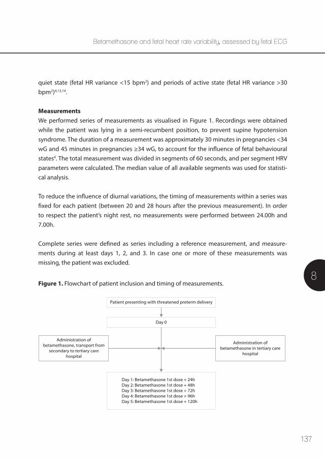
137
8
quiet state (fetal HR variance <15 bpm2) and periods of active state (fetal HR variance >30 bpm2)4,13,14.
MeasurementsWe performed series of measurements as visualised in Figure 1. Recordings were obtained while the patient was lying in a semi-recumbent position, to prevent supine hypotension syndrome. The duration of a measurement was approximately 30 minutes in pregnancies <34 wG and 45 minutes in pregnancies ≥34 wG, to account for the influence of fetal behavioural states4. The total measurement was divided in segments of 60 seconds, and per segment HRV parameters were calculated. The median value of all available segments was used for statisti-cal analysis.
To reduce the influence of diurnal variations, the timing of measurements within a series was fixed for each patient (between 20 and 28 hours after the previous measurement). In order to respect the patient’s night rest, no measurements were performed between 24.00h and 7.00h.
Complete series were defined as series including a reference measurement, and measure- ments during at least days 1, 2, and 3. In case one or more of these measurements was missing, the patient was excluded.
Patient presenting with threatened preterm delivery
Day 0
Day 1: Betamethasone 1st dose + 24hDay 2: Betamethasone 1st dose + 48hDay 3: Betamethasone 1st dose + 72hDay 4: Betamethasone 1st dose + 96hDay 5: Betamethasone 1st dose + 120h
Administration of betamethasone in tertiary care
hospital
Administration of betamethasone, transport from
secondary to tertiary care hospital
Figure 1. Flowchart of patient inclusion and timing of measurements.
Betamethasone and fetal heart rate variability, assessed by fetal ECG
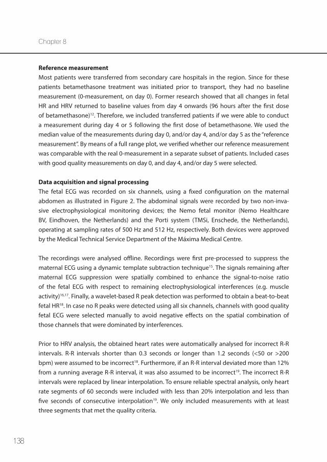
138
Chapter 8
Reference measurement Most patients were transferred from secondary care hospitals in the region. Since for these patients betamethasone treatment was initiated prior to transport, they had no baseline measurement (0-measurement, on day 0). Former research showed that all changes in fetal HR and HRV returned to baseline values from day 4 onwards (96 hours after the first dose of betamethasone)12. Therefore, we included transferred patients if we were able to conduct a measurement during day 4 or 5 following the first dose of betamethasone. We used the median value of the measurements during day 0, and/or day 4, and/or day 5 as the “reference measurement”. By means of a full range plot, we verified whether our reference measurement was comparable with the real 0-measurement in a separate subset of patients. Included cases with good quality measurements on day 0, and day 4, and/or day 5 were selected.
Data acquisition and signal processing The fetal ECG was recorded on six channels, using a fixed configuration on the maternal abdomen as illustrated in Figure 2. The abdominal signals were recorded by two non-inva-sive electrophysiological monitoring devices; the Nemo fetal monitor (Nemo Healthcare BV, Eindhoven, the Netherlands) and the Porti system (TMSi, Enschede, the Netherlands), operating at sampling rates of 500 Hz and 512 Hz, respectively. Both devices were approved by the Medical Technical Service Department of the Máxima Medical Centre.
The recordings were analysed offline. Recordings were first pre-processed to suppress the maternal ECG using a dynamic template subtraction technique15. The signals remaining after maternal ECG suppression were spatially combined to enhance the signal-to-noise ratio of the fetal ECG with respect to remaining electrophysiological interferences (e.g. muscle activity)16,17. Finally, a wavelet-based R peak detection was performed to obtain a beat-to-beat fetal HR18. In case no R peaks were detected using all six channels, channels with good quality fetal ECG were selected manually to avoid negative effects on the spatial combination of those channels that were dominated by interferences.
Prior to HRV analysis, the obtained heart rates were automatically analysed for incorrect R-R intervals. R-R intervals shorter than 0.3 seconds or longer than 1.2 seconds (<50 or >200 bpm) were assumed to be incorrect18. Furthermore, if an R-R interval deviated more than 12% from a running average R-R interval, it was also assumed to be incorrect19. The incorrect R-R intervals were replaced by linear interpolation. To ensure reliable spectral analysis, only heart rate segments of 60 seconds were included with less than 20% interpolation and less than five seconds of consecutive interpolation19. We only included measurements with at least three segments that met the quality criteria.
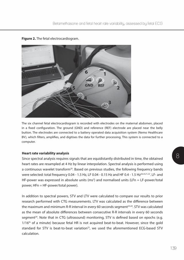
139
8Heart rate variability analysisSince spectral analysis requires signals that are equidistantly distributed in time, the obtained heart rates are resampled at 4 Hz by linear interpolation. Spectral analysis is performed using a continuous wavelet transform20. Based on previous studies, the following frequency bands were selected: total frequency 0.04 - 1.5 Hz, LF 0.04 - 0.15 Hz and HF 0.4 - 1.5 Hz4,6,9,21,22. LF- and HF-power was expressed in absolute units (ms2) and normalised units (LFn = LF-power/total power, HFn = HF-power/total power).
In addition to spectral powers, STV and LTV were calculated to compare our results to prior research performed with CTG measurements. LTV was calculated as the difference between the maximum and minimum R-R interval in every 60 seconds segment23,24. STV was calculated as the mean of absolute differences between consecutive R-R intervals in every 60 seconds segment24. Note that in CTG (ultrasound) monitoring, STV is defined based on epochs (e.g. 1/16th of a minute) because fetal HR is not acquired beat-to-beat. However, since the gold standard for STV is beat-to-beat variation23, we used the aforementioned ECG-based STV calculation.
Figure 2. The fetal electrocardiogram.
The six channel fetal electrocardiogram is recorded with electrodes on the maternal abdomen, placed in a fixed configuration. The ground (GND) and reference (REF) electrode are placed near the belly button. The electrodes are connected to a battery operated data acquisition system (Nemo Healthcare BV), which filters, amplifies, and digitises the data for further processing. This system is connected to a computer.
Betamethasone and fetal heart rate variability, assessed by fetal ECG
140
Chapter 8
Statistical analysisDescriptive statistics were used to describe the study population. Statistical analysis was performed using SPSS software version 23 (IBM Corp., Armonk, NY, USA). For each fetus a different number of segments were available during the study period, mainly due to variations in ECG signal quality. Median values and interquartile ranges were calculated for HR, LTV, STV and the different spectral values (LF, HF, LFn, HFn). The results were plotted over the four day measurement period. The result on each day was compared to the reference measurement. Statistical analysis was performed using the Wilcoxon signed-rank test. Statistical significance was assumed at the two-sided p-value of <0.05.
Results
Initially, 68 women were included in this study. The inclusion process is depicted in Figure 3. Three patients requested withdrawal from the study because of poor prognosis for an extreme premature child (1), technical issues (1) and inconvenient timing of measurements for the patient (1). In one patient unexpected intra-uterine fetal death occurred during the study period. Extensive evaluation revealed no evident cause. In 28 patients we were able to obtain a complete set of measurements, of which 16 were excluded due to insufficient data quality (fewer than three good quality segments per measurement) in one or more of the measurements. Eventually, 12 patients with a complete set of sufficient data quality were included for analysis. Table 1 shows the patient characteristics of the 12 included cases.
Reference measurementMeasurements during day 0, and/or day 4, and/or day 5 were compared in five patients. In Figure 4, absolute LF, HF, LFn and HFn are displayed. As expected, the absolute fetal HRV values showed some inter- and intra-patient variation, which can mainly be explained by variation in the segments that were recorded during active and quiet states and by variation in gestational age of the fetuses. LFn and HFn showed rather good comparability during day 0, 4, and 5.
Primary outcomeOur primary outcome was fetal HRV. Figure 5 shows the changes in LTV, STV, absolute LF and HF and normalised LFn and HFn. Only the increase in HF-power during day 1, compared to the reference measurement (day 0) was statistically significant (p = 0.025). All other trends were not statistically significant.
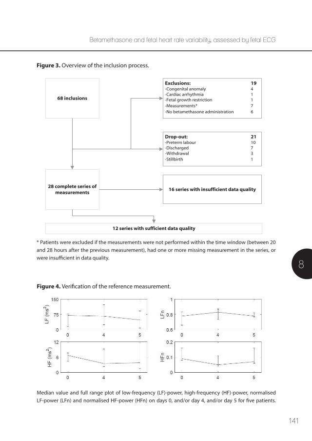
141
8
68 inclusions
28 complete series of measurements
12 series with sufficient data quality
Drop-out: 21-Preterm labour 10-Discharged 7-Withdrawal 3-Stillbirth 1
Exclusions: 19-Congenital anomaly 4-Cardiac arrhythmia 1-Fetal growth restriction 1-Measurements* 7-No betamethasone administration 6
16 series with insufficient data quality
Figure 3. Overview of the inclusion process.
* Patients were excluded if the measurements were not performed within the time window (between 20 and 28 hours after the previous measurement), had one or more missing measurement in the series, or were insufficient in data quality.
Figure 4. Verification of the reference measurement.
Median value and full range plot of low-frequency (LF)-power, high-frequency (HF)-power, normalised LF-power (LFn) and normalised HF-power (HFn) on days 0, and/or day 4, and/or day 5 for five patients.
Betamethasone and fetal heart rate variability, assessed by fetal ECG
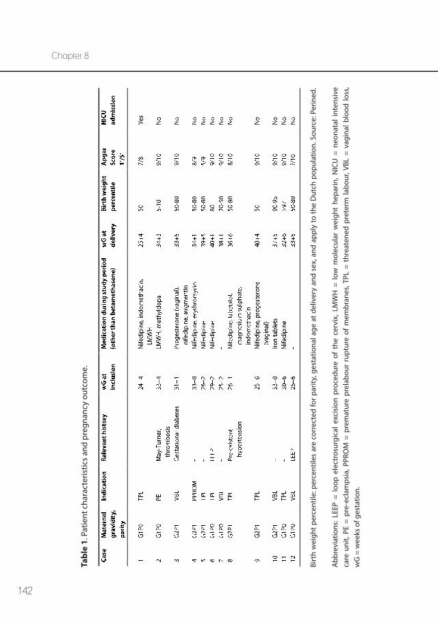
142
Chapter 8
Tabl
e 1.
Pat
ient
cha
ract
eris
tics
and
preg
nanc
y ou
tcom
e.
Birt
h w
eigh
t per
cent
ile: p
erce
ntile
s ar
e co
rrec
ted
for p
arity
, ges
tatio
nal a
ge a
t del
iver
y an
d se
x, a
nd a
pply
to th
e D
utch
pop
ulat
ion.
Sou
rce:
Per
ined
.
Abb
revi
atio
ns: L
EEP
= lo
op e
lect
rosu
rgic
al e
xcis
ion
proc
edur
e of
the
cer
vix,
LM
WH
= l
ow m
olec
ular
wei
ght
hepa
rin, N
ICU
= n
eona
tal
inte
nsiv
e ca
re u
nit,
PE =
pre
-ecl
amps
ia, P
PRO
M =
pre
mat
ure
prel
abou
r ru
ptur
e of
mem
bran
es, T
PL =
thr
eate
ned
pret
erm
labo
ur, V
BL =
vag
inal
blo
od lo
ss,
wG
= w
eeks
of g
esta
tion.
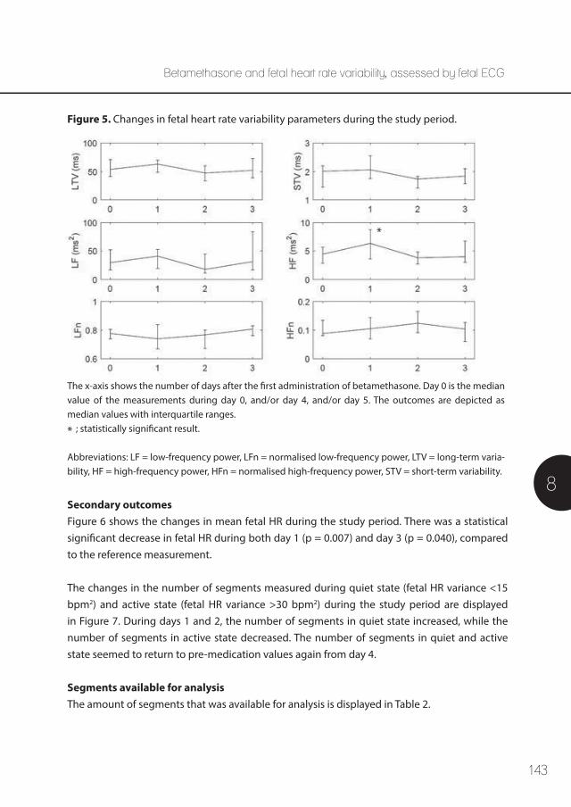
143
8
Figure 5. Changes in fetal heart rate variability parameters during the study period.
The x-axis shows the number of days after the first administration of betamethasone. Day 0 is the median value of the measurements during day 0, and/or day 4, and/or day 5. The outcomes are depicted as median values with interquartile ranges. ; statistically significant result.
Abbreviations: LF = low-frequency power, LFn = normalised low-frequency power, LTV = long-term varia-bility, HF = high-frequency power, HFn = normalised high-frequency power, STV = short-term variability.
Secondary outcomesFigure 6 shows the changes in mean fetal HR during the study period. There was a statistical significant decrease in fetal HR during both day 1 (p = 0.007) and day 3 (p = 0.040), compared to the reference measurement.
The changes in the number of segments measured during quiet state (fetal HR variance <15 bpm2) and active state (fetal HR variance >30 bpm2) during the study period are displayed in Figure 7. During days 1 and 2, the number of segments in quiet state increased, while the number of segments in active state decreased. The number of segments in quiet and active state seemed to return to pre-medication values again from day 4.
Segments available for analysis The amount of segments that was available for analysis is displayed in Table 2.
Betamethasone and fetal heart rate variability, assessed by fetal ECG
*
*
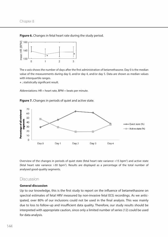
144
Chapter 8
Figure 6. Changes in fetal heart rate during the study period.
Figure 7. Changes in periods of quiet and active state.
The x-axis shows the number of days after the first administration of betamethasone. Day 0 is the median value of the measurements during day 0, and/or day 4, and/or day 5. Data are shown as median values with interquartile ranges. ; statistically significant result.
Abbreviations: HR = heart rate, BPM = beats per minute.
Overview of the changes in periods of quiet state (fetal heart rate variance <15 bpm2) and active state (fetal heart rate variance >30 bpm2). Results are displayed as a percentage of the total number of analysed good-quality segments.
Perc
enta
ge o
f all
retr
ieve
d se
gmen
ts
*
* *
Discussion
General discussion Up to our knowledge, this is the first study to report on the influence of betamethasone on spectral estimates of fetal HRV measured by non-invasive fetal ECG recordings. As we antic-ipated, over 80% of our inclusions could not be used in the final analysis. This was mainly due to loss to follow-up and insufficient data quality. Therefore, our study results should be interpreted with appropriate caution, since only a limited number of series (12) could be used for data analysis.
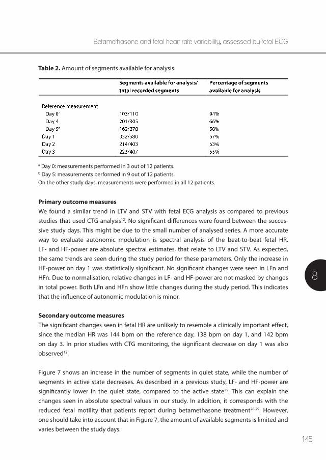
145
8
Table 2. Amount of segments available for analysis.
a Day 0: measurements performed in 3 out of 12 patients. b Day 5: measurements performed in 9 out of 12 patients. On the other study days, measurements were performed in all 12 patients.
Primary outcome measures We found a similar trend in LTV and STV with fetal ECG analysis as compared to previous studies that used CTG analysis12. No significant differences were found between the succes-sive study days. This might be due to the small number of analysed series. A more accurate way to evaluate autonomic modulation is spectral analysis of the beat-to-beat fetal HR. LF- and HF-power are absolute spectral estimates, that relate to LTV and STV. As expected, the same trends are seen during the study period for these parameters. Only the increase in HF-power on day 1 was statistically significant. No significant changes were seen in LFn and HFn. Due to normalisation, relative changes in LF- and HF-power are not masked by changes in total power. Both LFn and HFn show little changes during the study period. This indicates that the influence of autonomic modulation is minor.
Secondary outcome measuresThe significant changes seen in fetal HR are unlikely to resemble a clinically important effect, since the median HR was 144 bpm on the reference day, 138 bpm on day 1, and 142 bpm on day 3. In prior studies with CTG monitoring, the significant decrease on day 1 was also observed12.
Figure 7 shows an increase in the number of segments in quiet state, while the number of segments in active state decreases. As described in a previous study, LF- and HF-power are significantly lower in the quiet state, compared to the active state25. This can explain the changes seen in absolute spectral values in our study. In addition, it corresponds with the reduced fetal motility that patients report during betamethasone treatment26-29. However, one should take into account that in Figure 7, the amount of available segments is limited and varies between the study days.
Betamethasone and fetal heart rate variability, assessed by fetal ECG
146
Chapter 8
Segments available for analysis As shown in Table 2, the mean available amount of segments per day was always more than 50%. One has to bear in mind that this is in selected series with sufficient quality of the performed measurements. There are no obvious differences between the measurement days; the amount of available segments is steady between 52% and 57%. Only the measurements performed on day 0 show a higher percentage of available segments. However, only 3 out of 12 patients were eligible for a measurement during day 0.
Considerations We defined the reference measurement as the median value of the measurements during day 0, and/or day 4, and/or day 5. Although good comparability was seen for fetal HRV values, this remains second best with regard to a true baseline measurement. The high number of measurements that had to be excluded due to poor signal quality, can mainly be explained by presence of vernix caseosa. This fatty layer surrounds the fetus and results in an electrical isolation, which diminishes the signal amplitude of the fetal ECG. Especially between 30 and 34 wG, this layer causes a poor signal-to-noise ratio25,30.
By including patients receiving other pregnancy-related drugs rather than betamethasone, we aimed to obtain information that is applicable in daily clinical practice. Nifedipine and indomethacin seem to have no clinically important effect on fetal HRV, while magnesium sulphate can cause decreased fetal HRV and cases of bradycardia have been described31. In one case, magnesium sulphate was administered during days 1, 2 and 3 of the study period. Since magnesium sulphate was not administered during the reference measurement, this might have had some influence on the study results. In one case labetalol was administered to the patient; this was already started prior to the measurements and no changes in dosage occurred during the study period. Therefore, it is not likely that this had major influence on the study results. It is unlikely for augmentin, low-molecular weight heparin, methyldopa and progesterone to have any influence on fetal HRV parameters due to their mechanism of action.
Apart from pathological conditions, two major factors that one should consider when assessing fetal HR patterns are gestational age and fetal behavioural states32. Previous studies show that gestational age significantly affects the fetal HRV power spectrum, with a gradual increase in LF- and HF-power during gestation13,25,33. In this study, the included fetuses had a gestational age varying from 24 to 33 wG. Since we studied fetuses on successive days and were interested in relative changes in fetal HRV parameters, the influence of the increase in LF- and HF-power during gestation is likely to be minor. This study demonstrated an increase
147
8
in time spent in the quiet state following betamethasone administration. This might be caused by disturbance of the maternal glucose metabolism34, which is a known side effect of corticosteroids.
Fetal HRV and fetal movements are two parameters associated with fetal wellbeing. The reduction in both, due to betamethasone administration, can be misinterpreted as fetal deterioration and can therefore possibly lead to unnecessary iatrogenic preterm delivery35,36. No other signs of fetal hypoxia, like decelerations or abnormalities in Doppler flow velocity waveforms, have occurred after corticosteroid administration29,35-38. In addition, Shenhav et al.39 demonstrated that reduced fetal HRV was not related to the fetal acid-base balance at birth when delivery occurred <48 hours following betamethasone administration. This study confirms these results, since the influence of autonomic modulation was found to be minor (reflected as no evident changes in normalised spectral powers during the study period). Therefore, administration of betamethasone is not related to fetal distress. However, in fetal monitoring it is important to be aware of the side-effects, such as reduced fetal HRV and fetal movements.
Conclusion The changes in fetal HRV following betamethasone administration show the same pattern when calculated by spectral analysis of the fetal ECG, as when calculated by Doppler-ultra-sound CTG. The change in absolute spectral values is likely to correspond to the change in quiet and active state of the fetus. Since normalised spectral values show little changes, the influence of autonomic modulation is minor.
Acknowledgements
This research was performed within the framework of IMPULS perinatology. We would like to acknowledge Davide Aben, Jasmijn Drinkwaard, Noortje Eijsvoogel, Beatrijs van der Hout-van der Jagt, Carlijn van den Oord, Marlot Sengers, Kirsten Thijssen and Mariëlle van Wierst for their contributions in performing the measurements.
Betamethasone and fetal heart rate variability, assessed by fetal ECG
148
Chapter 8
References
1. Paul RH, Suidan AK, Yeh S, Schifrin BS, Hon EH. Clinical fetal monitoring. VII. The evaluation and significance of intrapartum baseline FHR variability. Am J Obstet Gynecol 1975 Sep 15;123(2):206-210.
2. Van Ravenswaaij-Arts C, Kollee L, Hopman J, Stoelinga G, van Geijn H. Heart rate variabilitiy. Ann Intern Med 1993;118:436-447.
3. Dawes GS, Moulden M, Redman CW. Improvements in computerized fetal heart rate analysis antepartum. J Perinat Med 1996;24(1):25-36.
4. van Laar JO, Peters CH, Vullings R, Houterman S, Oei SG. Power spectrum analysis of fetal heart rate variability at near term and post term gestation during active sleep and quiet sleep. Early Hum Dev 2009 Dec;85(12):795-798.
5. van Laar JO, Peters CH, Vullings R, Houterman S, Bergmans JW, Oei SG. Fetal autonomic response to severe acidaemia during labour. BJOG 2010 Mar;117(4):429-437.
6. van Laar JO, Peters CH, Houterman S, Wijn PF, Kwee A, Oei SG. Normalized spectral power of fetal heart rate variability is associated with fetal scalp blood pH. Early Hum Dev 2011 Apr;87(4):259-263.
7. Vullings R, Peters C, Andriessen P, Oei S, Wijn P. Monitoring the Fetal Heart Rate and Fetal Electro-cardiogram: Abdominal Recordings Are As Good As Direct Ecg Measurements. Pediatric Research 2005;58(2):242.
8. Siira SM, Ojala TH, Vahlberg TJ, Jalonen JO, Valimaki IA, Rosen KG, et al. Marked fetal acidosis and specific changes in power spectrum analysis of fetal heart rate variability recorded during the last hour of labour. BJOG 2005 Apr;112(4):418-423.
9. Heart rate variability: standards of measurement, physiological interpretation and clinical use. Task Force of the European Society of Cardiology and the North American Society of Pacing and Electro-physiology. Circulation 1996 Mar 1;93(5):1043-1065.
10. Roberts D, Dalziel S. Antenatal corticosteroids for accelerating fetal lung maturation for women at risk of preterm birth. Cochrane Database Syst Rev 2006 Jul 19;(3):CD004454.
11. Petersen MC, Nation RL, Ashley JJ, McBride WG. The placental transfer of betamethasone. Eur J Clin Pharmacol 1980 Oct;18(3):245-247.
12. Verdurmen KM, Renckens J, van Laar JO, Oei SG. The influence of corticosteroids on fetal heart rate variability: a systematic review of the literature. Obstet Gynecol Surv 2013 Dec;68(12):811-824.
13. Karin J, Hirsch M, Akselrod S. An estimate of fetal autonomic state by spectral analysis of fetal heart rate fluctuations. Pediatr Res 1993 Aug;34(2):134-138.
14. Nijhuis JG, Prechtl HF, Martin CB,Jr, Bots RS. Are there behavioural states in the human fetus? Early Hum Dev 1982 Apr;6(2):177-195.
15. Vullings R, Peters CH, Sluijter RJ, Mischi M, Oei SG, Bergmans JW. Dynamic segmentation and linear prediction for maternal ECG removal in antenatal abdominal recordings. Physiol Meas 2009 Mar;30(3):291-307.
16. Vullings R, Peters C, Hermans M, Wijn P, Oei S, Bergmans J. A robust physiology-based source sepa-ration method for QRS detection in low amplitude fetal ECG recordings. Physiol Meas 2010;31:935-951.
149
8
17. Warmerdam G, Vullings R, Van Pul C, Andriessen P, Oei SG, Wijn P. QRS classification and spatial combination for robust heart rate detection in low-quality fetal ECG recordings. Conf Proc IEEE Eng Med Biol Soc 2013;2013:2004-2007.
18. Rooijakkers MJ, Rabotti C, Oei SG, Mischi M. Low-complexity R-peak detection for ambulatory fetal monitoring. Physiol Meas 2012 Jul;33(7):1135-1150.
19. Peters C, Vullings R, Bergmans J, Oei G, Wijn P. The effect of artifact correction on spectral estimates of heart rate variability. Conf Proc IEEE Eng Med Biol Soc 2008;2008:2669-2672.
20. Peters CH, Vullings R, Rooijakkers MJ, Bergmans JW, Oei SG, Wijn PF. A continuous wavelet trans-form-based method for time-frequency analysis of artefact-corrected heart rate variability data. Physiol Meas 2011 Oct;32(10):1517-1527.
21. De Beer N, Andriessen P, Berendsen R, Oei S, Wijn P, Bambang Oetomo S. Customized spectral band analysis compared with conventional Fourier analysis of heart rate variability in neonates. Physiol Meas. 2004;25:1385-1395.
22. Min SW, Ko H, Kim CS. Power spectral analysis of heart rate variability during acute hypoxia in fetal lambs. Acta Obstet Gynecol Scand 2002 Nov;81(11):1001-1005.
23. Pardey J, Moulden M, Redman CW. A computer system for the numerical analysis of nonstress tests. Am J Obstet Gynecol 2002 May;186(5):1095-1103.
24. Magenes G, Signorini MG, Arduini D. Classification of cardiotocographic records by neural networks. Proc IEEE -INNS-ENNS International Joint Conference on Neural Networks IJCNN 2000;3:637-641.
25. van Laar JO, Warmerdam GJ, Verdurmen KM, Vullings R, Peters CH, Houterman S, et al. Fetal heart rate variability during pregnancy, obtained from non-invasive electrocardiogram recordings. Acta Obstet Gynecol Scand 2014 Jan;93(1):93-101.
26. Koenen SV, Mulder EJ, Wijnberger LD, Visser GH. Transient loss of the diurnal rhythms of fetal movements, heart rate, and its variation after maternal betamethasone administration. Pediatr Res 2005 May;57(5 Pt 1):662-666.
27. Rotmensch S, Liberati M, Vishne TH, Celentano C, Ben-Rafael Z, Bellati U. The effect of betamethasone and dexamethasone on fetal heart rate patterns and biophysical activities. A prospective randomized trial. Acta Obstet Gynecol Scand 1999 Jul;78(6):493-500.
28. Mulder EJ, Derks JB, Zonneveld MF, Bruinse HW, Visser GH. Transient reduction in fetal activity and heart rate variation after maternal betamethasone administration. Early Hum Dev 1994 Jan;36(1):49-60.
29. Mulder EJ, Derks JB, Visser GH. Antenatal corticosteroid therapy and fetal behaviour: a randomised study of the effects of betamethasone and dexamethasone. Br J Obstet Gynaecol 1997 Nov;104(11):1239-1247.
30. Oostendorp TF, van Oosterom A, Jongsma HW. The fetal ECG throughout the second half of gestation. Clin Phys Physiol Meas 1989 May;10(2):147-160.
31. Verdurmen KM, Hulsenboom AD, van Laar JO, Oei SG. Effect of tocolytic drugs on fetal heart rate variability: a systematic review. J Matern Fetal Neonatal Med 2016 Nov 8:1-8.
Betamethasone and fetal heart rate variability, assessed by fetal ECG
150
Chapter 8
32. Schneider U, Schleussner E, Fiedler A, Jaekel S, Liehr M, Haueisen J, et al. Fetal heart rate variability reveals differential dynamics in the intrauterine development of the sympathetic and parasympa-thetic branches of the autonomic nervous system. Physiol Meas 2009 Feb;30(2):215-226.
33. David M, Hirsch M, Karin J, Toledo E, Akselrod S. An estimate of fetal autonomic state by time-frequency analysis of fetal heart rate variability. J Appl Physiol (1985) 2007 Mar;102(3):1057-1064.
34. Michaan N, Baruch Y, Topilsky M, Amzalag S, Iaskov I, Many A, et al. The effect of glucose administration on perceived fetal movements in women with decreased fetal movement, a double-blinded placebo-controlled trial. J Perinatol 2016 Aug;36(8):598-600.
35. Derks JB, Mulder EJ, Visser GH. The effects of maternal betamethasone administration on the fetus. Br J Obstet Gynaecol 1995 Jan;102(1):40-46.
36. Subtil D, Tiberghien P, Devos P, Therby D, Leclerc G, Vaast P, et al. Immediate and delayed effects of antenatal corticosteroids on fetal heart rate: a randomized trial that compares betamethasone acetate and phosphate, betamethasone phosphate, and dexamethasone. Am J Obstet Gynecol 2003 Feb;188(2):524-531.
37. Cohlen BJ, Stigter RH, Derks JB, Mulder EJ, Visser GH. Absence of significant hemodynamic changes in the fetus following maternal betamethasone administration. Ultrasound Obstet Gynecol 1996 Oct;8(4):252-255.
38. Senat MV, Minoui S, Multon O, Fernandez H, Frydman R, Ville Y. Effect of dexamethasone and betamethasone on fetal heart rate variability in preterm labour: a randomised study. Br J Obstet Gynaecol 1998 Jul;105(7):749-755.
39. Shenhav S, Volodarsky M, Anteby EY, Gemer O. Fetal acid-base balance after betamethasone administration: relation to fetal heart rate variability. Arch Gynecol Obstet 2008 Oct;278(4):333-336.

151
8
Betamethasone and fetal heart rate variability, assessed by fetal ECG


Part III
Fetal ECG to prevent asphyxia

154
155
Orientation of the electrical heart axis in mid-term pregnancy
Kim M.J. Verdurmen, Alexandra D.J. Hulsenboom, Judith O.E.H. van Laar, Pieter F.F. Wijn, Rik Vullings, S. Guid Oei
Published: European Journal of Obstetrics & Gynecology and Reproductive Biology. 2016;207:243-246
Letter to the Editor – Brief communication
Chapter 9
9
156
Chapter 9
Introduction The unique fetal shunting system causes an increased cardiac muscle mass in the right ventricle1. Cardiac currents initiating each contraction are measured from the outside as the electrocardiogram (ECG), the main direction of propagation is referred to as the electrical heart axis. Ventricular depolarisation involves the largest cell mass, yielding the largest ECG signal. Therefore, the QRS segment amplitude mainly defines the electrical heart axis. Due to the increased right ventricular mass in fetuses, the electrical heart axis is expected to point towards the right. This has been confirmed in term fetuses during labour and in neonates directly postpartum2,3. In contrast, the electrical heart axis of adults points towards the left4. However, it is well-known that in both adults and newborns the orientation of the electrical heart axis can vary widely from person to person2.
For term fetuses during labour, the orientation of the electrical heart axis has been described by Larks et al.3 in the 1960s. Their recording techniques are outdated, and they did not take the fetal orientation into account. Despite these shortcomings, they already acknowledged the possibilities of the electrical heart axis to contribute in distinguishing between normal intra-uterine development, congenital heart disease and fetal distress.
The orientation of the fetal electrical heart axis during gestation has never been described. The relevance of the electrical heart axis increased, since fetal ECG is used to study fetal wellbeing more often. The aim of this study is to determine the direction of the fetal electrical heart axis in mid-term pregnancy.
Materials and Methods
We performed a post-hoc analysis on data of two (prospective) studies, both approved by the ethical committee of the Máxima Medical Centre5. Informed consent was obtained in both studies. Women between 18 and 29 weeks of pregnancy, carrying a singleton fetus were included. Exclusion criteria were maternal age <18 years, multiple pregnancy, any known fetal congenital anomaly, fetal growth restriction (birth weight <p10 for gestational age) and women receiving medication known to have any cardiac side effects. The absence of severe cardiac malformations was confirmed after birth by review of medical charts.
We conducted a single fetal ECG recording of approximately 30 minutes with eight adhesive electrodes on the maternal abdomen, placed in a fixed configuration (Figure 1). Recordings were performed using a fetal ECG data acquisition system (Nemo Healthcare BV, the Nether-lands), operating at a sampling frequency of 1 kHz. The fetal ECG was obtained and analysed from the abdominal recordings to yield a vectorcardiogram that is normalised for the fetal
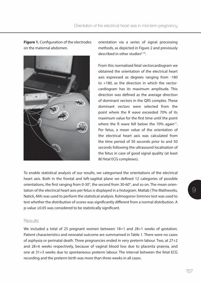
157
Orientation of the electrical heart axis in mid-term pregnancy
9
Figure 1. Configuration of the electrodes on the maternal abdomen.
orientation via a series of signal processing methods, as depicted in Figure 2 and previously described in other studies6-10.
From this normalised fetal vectorcardiogram we obtained the orientation of the electrical heart axis expressed as degrees ranging from -180 to +180, as the direction in which the vector-cardiogram has its maximum amplitude. This direction was defined as the average direction of dominant vectors in the QRS complex. These dominant vectors were selected from the point where the R wave exceeded 70% of its maximum value for the first time until the point where the R wave fell below the 70% again11. Per fetus, a mean value of the orientation of the electrical heart axis was calculated from the time period of 50 seconds prior to and 50 seconds following the ultrasound localisation of the fetus in case of good signal quality (at least 80 fetal ECG complexes).
To enable statistical analysis of our results, we categorised the orientations of the electrical heart axis. Both in the frontal and left-sagittal plane we defined 12 categories of possible orientations, the first ranging from 0-30°, the second from 30-60°, and so on. The mean orien-tation of the electrical heart axis per fetus is displayed in a histogram. Matlab (The Mathworks, Natick, MA) was used to perform the statistical analysis. Kolmogorov-Smirnov test was used to test whether the distribution of scores was significantly different from a normal distribution. A p-value ≤0.05 was considered to be statistically significant.
Results
We included a total of 25 pregnant women between 18+1 and 28+1 weeks of gestation. Patient characteristics and neonatal outcome are summarised in Table 1. There were no cases of asphyxia or perinatal death. Three pregnancies ended in very preterm labour. Two, at 27+2 and 28+6 weeks respectively, because of vaginal blood loss due to placenta praevia, and one at 31+3 weeks due to spontaneous preterm labour. The interval between the fetal ECG recording and the preterm birth was more than three weeks in all cases.
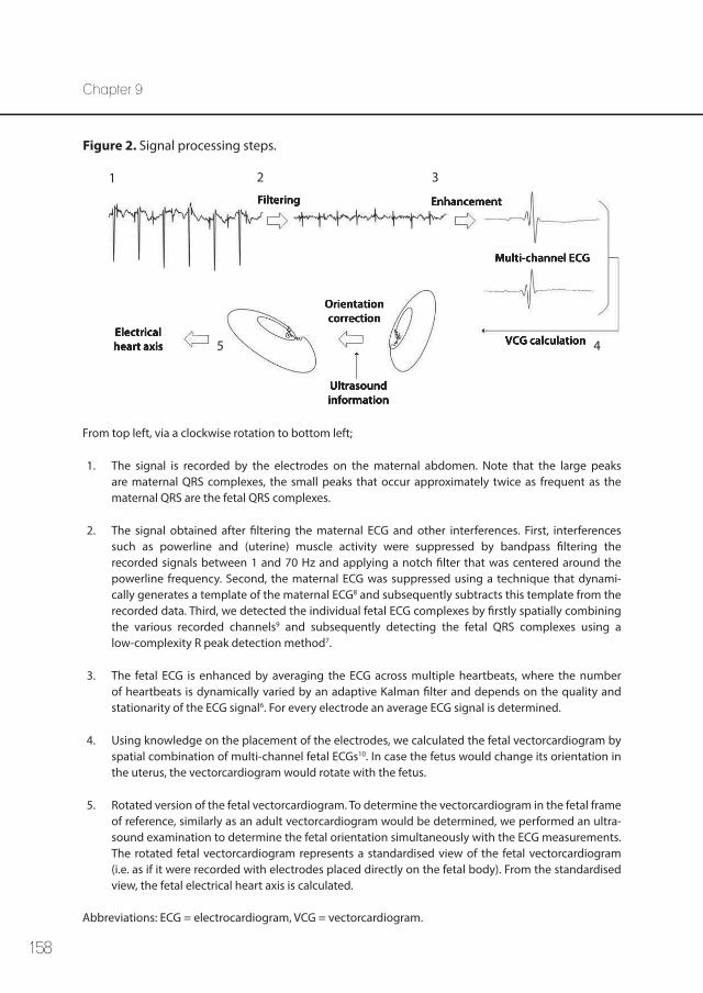
158
Chapter 9
Figure 2. Signal processing steps.
From top left, via a clockwise rotation to bottom left;
1. The signal is recorded by the electrodes on the maternal abdomen. Note that the large peaks are maternal QRS complexes, the small peaks that occur approximately twice as frequent as the maternal QRS are the fetal QRS complexes.
2. The signal obtained after filtering the maternal ECG and other interferences. First, interferences such as powerline and (uterine) muscle activity were suppressed by bandpass filtering the recorded signals between 1 and 70 Hz and applying a notch filter that was centered around the powerline frequency. Second, the maternal ECG was suppressed using a technique that dynami-cally generates a template of the maternal ECG8 and subsequently subtracts this template from the recorded data. Third, we detected the individual fetal ECG complexes by firstly spatially combining the various recorded channels9 and subsequently detecting the fetal QRS complexes using a low-complexity R peak detection method7.
3. The fetal ECG is enhanced by averaging the ECG across multiple heartbeats, where the number of heartbeats is dynamically varied by an adaptive Kalman filter and depends on the quality and stationarity of the ECG signal6. For every electrode an average ECG signal is determined.
4. Using knowledge on the placement of the electrodes, we calculated the fetal vectorcardiogram by spatial combination of multi-channel fetal ECGs10. In case the fetus would change its orientation in the uterus, the vectorcardiogram would rotate with the fetus.
5. Rotated version of the fetal vectorcardiogram. To determine the vectorcardiogram in the fetal frame of reference, similarly as an adult vectorcardiogram would be determined, we performed an ultra-sound examination to determine the fetal orientation simultaneously with the ECG measurements. The rotated fetal vectorcardiogram represents a standardised view of the fetal vectorcardiogram (i.e. as if it were recorded with electrodes placed directly on the fetal body). From the standardised view, the fetal electrical heart axis is calculated.
Abbreviations: ECG = electrocardiogram, VCG = vectorcardiogram.
Filtering Enhancement
Multi-channel ECG
VCG calculation
Orientation correction
Ultrasound information
Electrical heart axis
1 2 3
45
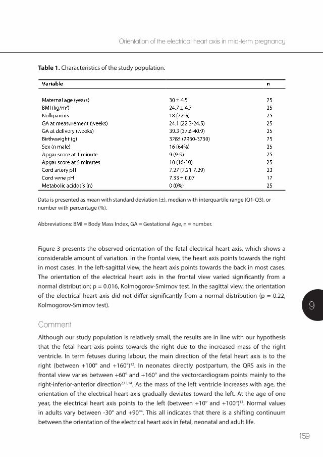
159
Figure 3 presents the observed orientation of the fetal electrical heart axis, which shows a considerable amount of variation. In the frontal view, the heart axis points towards the right in most cases. In the left-sagittal view, the heart axis points towards the back in most cases. The orientation of the electrical heart axis in the frontal view varied significantly from a normal distribution; p = 0.016, Kolmogorov-Smirnov test. In the sagittal view, the orientation of the electrical heart axis did not differ significantly from a normal distribution (p = 0.22, Kolmogorov-Smirnov test).
Orientation of the electrical heart axis in mid-term pregnancy
9
Table 1. Characteristics of the study population.
Data is presented as mean with standard deviation (±), median with interquartile range (Q1-Q3), or number with percentage (%). Abbreviations: BMI = Body Mass Index, GA = Gestational Age, n = number.
Comment
Although our study population is relatively small, the results are in line with our hypothesis that the fetal heart axis points towards the right due to the increased mass of the right ventricle. In term fetuses during labour, the main direction of the fetal heart axis is to the right (between +100° and +160°)12. In neonates directly postpartum, the QRS axis in the frontal view varies between +60° and +160° and the vectorcardiogram points mainly to the right-inferior-anterior direction2,13,14. As the mass of the left ventricle increases with age, the orientation of the electrical heart axis gradually deviates toward the left. At the age of one year, the electrical heart axis points to the left (between +10° and +100°)13. Normal values in adults vary between -30° and +90°4. This all indicates that there is a shifting continuum between the orientation of the electrical heart axis in fetal, neonatal and adult life.
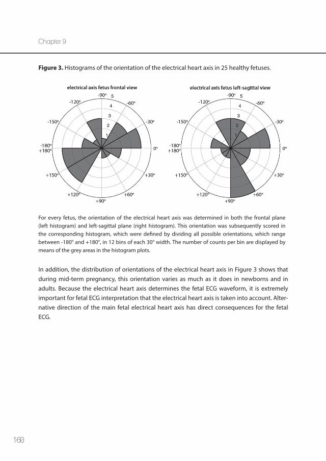
160
Chapter 9
Figure 3. Histograms of the orientation of the electrical heart axis in 25 healthy fetuses.
For every fetus, the orientation of the electrical heart axis was determined in both the frontal plane (left histogram) and left-sagittal plane (right histogram). This orientation was subsequently scored in the corresponding histogram, which were defined by dividing all possible orientations, which range between -180° and +180°, in 12 bins of each 30° width. The number of counts per bin are displayed by means of the grey areas in the histogram plots.
In addition, the distribution of orientations of the electrical heart axis in Figure 3 shows that during mid-term pregnancy, this orientation varies as much as it does in newborns and in adults. Because the electrical heart axis determines the fetal ECG waveform, it is extremely important for fetal ECG interpretation that the electrical heart axis is taken into account. Alter-native direction of the main fetal electrical heart axis has direct consequences for the fetal ECG.

161
Orientation of the electrical heart axis in mid-term pregnancy
9
References
1. Kiserud T, Acharya G. The fetal circulation. Prenat Diagn 2004 Dec 30;24(13):1049-1059.
2. Depasquale NP, Burch GE. The Electrocardiogram, Ventricular Gradient and Spatial Vectorcardio-gram during the First Week of Life. Am J Cardiol 1963 Oct;12:482-493.
3. Larks SD. Estimation of the Electrical Axis of the Fetal Heart. Am J Obstet Gynecol 1965 Jan 1;91:46-55.
4. Wagner GS, Strauss DG. Marriott’s Practical Electrocardiography. 12th edition ed. Philadelphia: Lippincott Williams & Wilkins; 2014.
5. van Laar JO, Warmerdam GJ, Verdurmen KM, Vullings R, Peters CH, Houterman S, et al. Fetal heart rate variability during pregnancy, obtained from non-invasive electrocardiogram recordings. Acta Obstet Gynecol Scand 2014 Jan;93(1):93-101.
6. Vullings R, de Vries B, Bergmans JW. An adaptive Kalman filter for ECG signal enhancement. IEEE Trans Biomed Eng 2011 Apr;58(4):1094-1103.
7. Rooijakkers MJ, Rabotti C, Oei SG, Mischi M. Low-complexity R-peak detection for ambulatory fetal monitoring. Physiol Meas 2012 Jul;33(7):1135-1150.
8. Vullings R, Peters CH, Sluijter RJ, Mischi M, Oei SG, Bergmans JW. Dynamic segmentation and linear prediction for maternal ECG removal in antenatal abdominal recordings. Physiol Meas 2009 Mar;30(3):291-307.
9. Vullings R, Peters C, Hermans M, Wijn P, Oei S, Bergmans J. A robust physiology-based source sepa-ration method for QRS detection in low amplitude fetal ECG recordings. Physiol Meas 2010;31:935-951.
10. Vullings R, Peters CH, Mossavat I, Oei SG, Bergmans JW. Bayesian approach to patient-tailored vectorcardiography. IEEE Trans Biomed Eng 2010 Mar;57(3):586-595.
11. Lipponen J,A., Gladwell VF, Kinnunen H, Karjalainen PA, Tarvainen MP. The correlation of vector-cardiographic changes to blood lactate concentration during an exercise test. Biomedical Signal Processing and Control 2013;8(6):491-499.
12. Larks SD, Larks GG. The electrical axis of the fetal heart: a new criterion for fetal well-being or distress. Am J Obstet Gynecol 1965 Dec 1;93(7):975-983.
13. Goodacre S, McLeod K. ABC of clinical electrocardiography: Paediatric electrocardiography. BMJ 2002 Jun 8;324(7350):1382-1385.
14. Schaffer AI, Beinfield WH. The vectorcardiogram of the newborn infant. Am Heart J 1952 Jul;44(1):89-94.

162
163
The electrical heart axis and ST events in fetal monitoring: a post-hoc analysis following
a multicentre randomised controlled trial
Rik Vullings, Kim M.J. Verdurmen, Alexandra D.J. Hulsenboom,
Stephanie Scheffer, Hinke de Lau, Anneke Kwee, Pieter F.F. Wijn, Isis Amer-Wåhlin, Judith O.E.H. van Laar, S. Guid Oei
Published: PLoS One. 2017;12(4):e0175823
10
Chapter 10
164
Chapter 10
Abstract
ObjectiveReducing perinatal morbidity and mortality is one of the major challenges in modern health care. Analysing the ST segment of the fetal electrocardiogram was thought to be the break-through in fetal monitoring during labour. However, its implementation in clinical practice yields many false alarms and ST monitoring is highly dependent on cardiotocogram assess-ment, limiting its value for the prediction of fetal distress during labour. This study aims to evaluate the relation between physiological variations in the orientation of the fetal electrical heart axis and the occurrence of ST events.
MethodsA post-hoc analysis was performed following a multicentre randomised controlled trial, including 1097 patients from two participating centres. All women were monitored with ST analysis during labour. Cases of fetal metabolic acidosis, poor signal quality, missing blood gas analysis, and congenital heart disease were excluded. The orientation of the fetal electri-cal heart axis affects the height of the initial T/QRS baseline, and therefore the incidence of ST events. We grouped tracings with the same initial baseline T/QRS value. We depicted the number of ST events as a function of the initial baseline T/QRS value with a linear regression model.
ResultsA significant increment of ST events was observed with increasing height of the initial T/QRS baseline, irrespective of the fetal condition; correlation coefficient 0.63, p <0.001. The most frequent T/QRS baseline is 0.12.
ConclusionThe orientation of the fetal electrical heart axis and accordingly the height of the initial T/QRS baseline should be taken into account in fetal monitoring with ST analysis.
165
The electrical heart axis and ST events
10
Introduction
Fetal asphyxia is associated with severe perinatal morbidity and mortality. The cardioto-cogram, a simultaneous recording of the fetal heart rate and uterine contractions, is used worldwide for fetal surveillance. However, the poor specificity of this method has resulted in increased rates of operative deliveries without a decrease in perinatal mortality or cerebral palsy1. ST analysis (STAN®, Neoventa Medical AB, Mölndal, Sweden) was introduced in 1992 as a promising technique that analyses the ST segment of the fetal electrocardiogram (ECG), acquired using an invasive scalp electrode2. ST analysis combined with cardiotocography was reported to significantly lower the rates of metabolic acidosis3 and operative delivery in two randomised controlled trials3,4. However, subsequent multicentre trials, including the recently published large American ST analysis trial, could not reproduce these initial findings5-9. Recent meta-analyses show conflicting results regarding the decrease in metabolic acidosis, which indicates the need for more research8,10-13. Meanwhile, Kwee et al.14 reported that the STAN® monitor gives as many ST events in cases of proven uncompromised fetal condition as in situations with deteriorating fetal condition. This is countered by the STAN® guidelines that state that ST events must be ignored when cardiotocography shows a reassuring pattern. However, the high inter-observer variability in cardiotocogram interpretation makes this a highly unsatisfying strategy15. The correct interpretation of a method as subjective as the cardiotocogram determines whether or not to ignore the ST event or to act upon the alarm, making the success of ST monitoring dependent on cardiotocogram assessment.
Background information and hypothesis
Prior to the introduction of ST analysis, the diagnostic value of the fetal ST segment was clearly demonstrated in animal studies16-18. Sustained deprivation of oxygen is followed by a surge of adrenalin to induce glycogenolysis, which is accompanied by an increase of potassium ions in the myocardial cells19. These potassium ions mainly affect the relaxation phase of the cardiac cycle and lead to an increase in the T wave amplitude of the fetal ECG20.
STAN® uses this hypoxia-related rise in T wave amplitude in a three-step protocol;
1. The T wave amplitude is normalised against the amplitude of the QRS complex (mean of 30 ECG complexes), yielding a T/QRS value.
2. A baseline T/QRS value is determined (median of at least 20 T/QRS values within 20 minutes) to gauge future T/QRS values.
3. New T/QRS values are compared to the baseline.
166
Chapter 10
In case a T/QRS value exceeds the baseline by 0.05, a baseline ST event is reported. Smaller exceedings of the baseline can be due to normal beat-to-beat fluctuation in the behaviour of the heart, which is unrelated to the fetal condition. With regard to the detection of rises in T wave amplitude due to oxygen deprivation, this alarm protocol seems plausible.
The ECG recorded from the fetal scalp electrode is a one-dimensional presentation of the electrical activity of the heart. However, the propagation of electrical currents over the cardiac muscle occurs in all three spatial dimensions. The main direction of this propagation is referred to as the electrical axis of the heart. The orientation of the electrical heart axis with respect to the fetal scalp electrode hence affects the shape and amplitude of the recorded ECG. Similarly, (adult) ECG signals recorded at different locations yield different shapes and amplitudes, as already demonstrated many years ago21.
It is known that the orientation of the fetal electrical heart axis can vary between +100° and +160° in mid-term fetuses22 and between +90° and +180° in term fetuses during labour23. Similar inter-person variation in the orientation of the electrical heart axis is present in neonates and adults24-27. The STAN® monitor attempts to correct for the orientation of the electrical heart axis with the first step in its protocol (normalisation). However, the propa-gation of the electrical currents during the contraction (QRS) phase of the cardiac cycle has a different orientation than during the relaxation (T) phase. Consequently, normalisation cannot fully compensate for inter-patient differences in the orientation of the electrical heart axis. As a result, fetuses for whom the scalp lead is almost perpendicular to the direction of propagation in the relaxation phase have a very small T wave amplitude, and typically also low T/QRS values and T/QRS baseline. Similarly, fetuses for whom the electrical heart axis is oriented in a manner creating a propagation during relaxation almost aligned with the scalp lead, typically have a high T/QRS value and T/QRS baseline.
When the hypoxia-induced release of potassium ions affects the electrical current in the relaxation phase in the fetuses with a low T/QRS baseline value, the absolute effect in T wave amplitude will only be marginal as we look at it from an almost perpendicular direction. In fetuses with high T/QRS values, the rise in T wave amplitude will be relatively large. Based on this, we hypothesise that normal fluctuations in the electrophysiological behaviour of the heart can stay below the 0.05 threshold, in case the scalp lead is oriented more perpen-dicular to the relaxation currents. Similarly, the hypoxia-related fluctuations in the electrical behaviour can more easily exceed the 0.05 threshold, when the alignment between the elec-trical heart axis and scalp lead is axial. We explain this phenomenon in Figure 1. Previously, Becker et al.28 described that the initial T/QRS baseline is not related to the fetal condition. The incidence of ST events was stated to be related to the fetal condition3, and therefore not
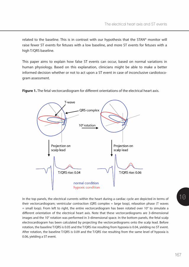
167
The electrical heart axis and ST events
10
related to the baseline. This is in contrast with our hypothesis that the STAN® monitor will raise fewer ST events for fetuses with a low baseline, and more ST events for fetuses with a high T/QRS baseline.
This paper aims to explain how false ST events can occur, based on normal variations in human physiology. Based on this explanation, clinicians might be able to make a better informed decision whether or not to act upon a ST event in case of inconclusive cardiotoco-gram assessment.
Figure 1. The fetal vectorcardiogram for different orientations of the electrical heart axis.
In the top panels, the electrical currents within the heart during a cardiac cycle are depicted in terms of their vectorcardiogram; ventricular contraction (QRS complex = large loop), relaxation phase (T waves = small loop). From left to right, the entire vectorcardiogram has been rotated over 10° to simulate a different orientation of the electrical heart axis. Note that these vectorcardiograms are 3-dimensional images and the 10° rotation was performed in 3-dimensional space. In the bottom panels, the fetal scalp electrocardiogram has been calculated by projecting the vectorcardiograms onto the scalp lead. Before rotation, the baseline T/QRS is 0.05 and the T/QRS rise resulting from hypoxia is 0.04, yielding no ST event. After rotation, the baseline T/QRS is 0.09 and the T/QRS rise resulting from the same level of hypoxia is 0.06, yielding a ST event.
168
Chapter 10
Materials and Methods
We performed a post-hoc analysis with data derived from a large multicentre randomised controlled trial, the Dutch ST analysis trial7. The initial study was approved by the Institutional Review Board of the University Medical Centre Utrecht and was performed between January 2006 and July 2008. After written informed consent, women were randomised to the index group with ST monitoring (STAN® S21 or S31 monitor) or to the control group, using a conven-tional fetal heart rate monitor (cardiotocography). The randomisation was performed on a 1:1 basis, web-based with stratification for centre and parity. Since the trial was pragmatic in nature, there was no blinding of patients or caregivers. All gynaecologists, residents and midwives in the participating centres were trained and certified as STAN®-users, and decisions were made following the STAN® clinical guidelines. Fetal blood sampling was allowed, yet restricted to specific scenarios in the index group. Inclusion criteria were maternal age over 18 years, singleton pregnancy, cephalic presentation, gestational age beyond 36 weeks and an indication for internal electronic fetal monitoring. The included women were assigned to a “high-risk pregnancy” group, since they all received secondary care. In the Netherlands, “low-risk pregnancies” are monitored by midwives or general practitioners (primary care). In both groups, the umbilical cord was double clamped immediately after birth, in order to sample both arterial and venous cord blood.
For this post-hoc analysis, we included data from two tertiary hospitals: the University Medical Centre Utrecht and the Máxima Medical Centre Veldhoven, both participating in the multicentre randomised controlled trial. Anonymised information from the initial database was used for this analysis. Following consultation of the Medical Ethical Department in the Máxima Medical Centre, no separate ethical approval was warranted for this study. We only included patients from the index group (with ST monitoring). We excluded patients in whom no STAN® registration was performed or no T/QRS baseline value could be determined, cases of metabolic acidosis, cases in which no blood gas analysis was performed postpartum and registrations performed in fetuses with congenital heart disease. Metabolic acidosis was defined as umbilical cord artery blood pH <7.05 and base deficit of the extracellular fluid compartment >12 mmol/l in two blood samples with a minimal pH difference of 0.03. In cases of only one blood sample or smaller differences between samples, metabolic acidosis was set as cord blood pH <7.10 and base deficit of extracellular fluid >12 mmol/l.
The initial baseline T/QRS value was determined the same way as done in the STAN® monitor; as the median of all T/QRS values recorded within the first 20-minute window of the recording, that contained a minimum of 20 T/QRS values. We counted the incidence of ST events throughout the entire registration. Patients were excluded in case a STAN® registration
169
The electrical heart axis and ST events
10
was temporarily stopped and more than one STAN® file was stored for the patient. For each initial baseline T/QRS value encountered in our data set, we counted the number of patients with that particular baseline. We grouped women with the same initial baseline T/QRS value. Hereafter, we calculated the relative incidence of ST events (defined as the number of ST events per 1000 T/QRS values) as a function of the initial baseline value.
Additionally, we calculated the mean pH and mean base deficit of the extracellular fluid for all women with the same initial baseline T/QRS value. Even though our dataset entails a subset of the data used by Becker et al.28, it needs to be confirmed that the conclusions from this study, that the height of the initial baseline is not related to fetal outcome, apply to our dataset as well.
Matlab (The Mathworks, Natick, MA) was used to perform the statistical analysis. For analysis of the baseline characteristics, mean, median, standard deviation and interquartile ranges were calculated using IBM SPSS statistics 22.0 for Mac (IBM corp. Armonk, NY, USA). A linear regression model was used to calculate the correlation coefficient for the relation between the number of ST events and the baseline T/QRS value.
Results and Discussion
Initially, 1401 patients were screened; in 273 cases ST information was missing, more than one STAN® file was available for the same patient, or no T/QRS baseline value had been deter-mined due to short duration of the measurement or poor quality of the data. These cases were therefore excluded. In addition, we excluded 11 cases of fetal metabolic acidosis. Further, no blood sample was available in 12 patients, whom were therefore excluded. In addition, six women gave birth to neonates with congenital heart disease, one labouring woman younger than 18 years and one prior to 36 weeks of gestation during labour were excluded. Eventually, we analysed the number of ST events in 1097 women. In this group, a total of 1.027.054 T/QRS ratios and 2066 ST events were reported.
The baseline characteristics of the included women are summarised in Table 1.
Figure 2 shows the distribution of patients across the various initial baseline T/QRS ratios. In Figure 3, we present the number of ST events as a function of the initial baseline T/QRS value. The results show an average increment of 1.42 ST events per 1000 T/QRS values for a rise of 0.1 of the initial baseline T/QRS. The correlation coefficient between data points and fit was 0.63 (p <0.001), as calculated with the linear regression model.
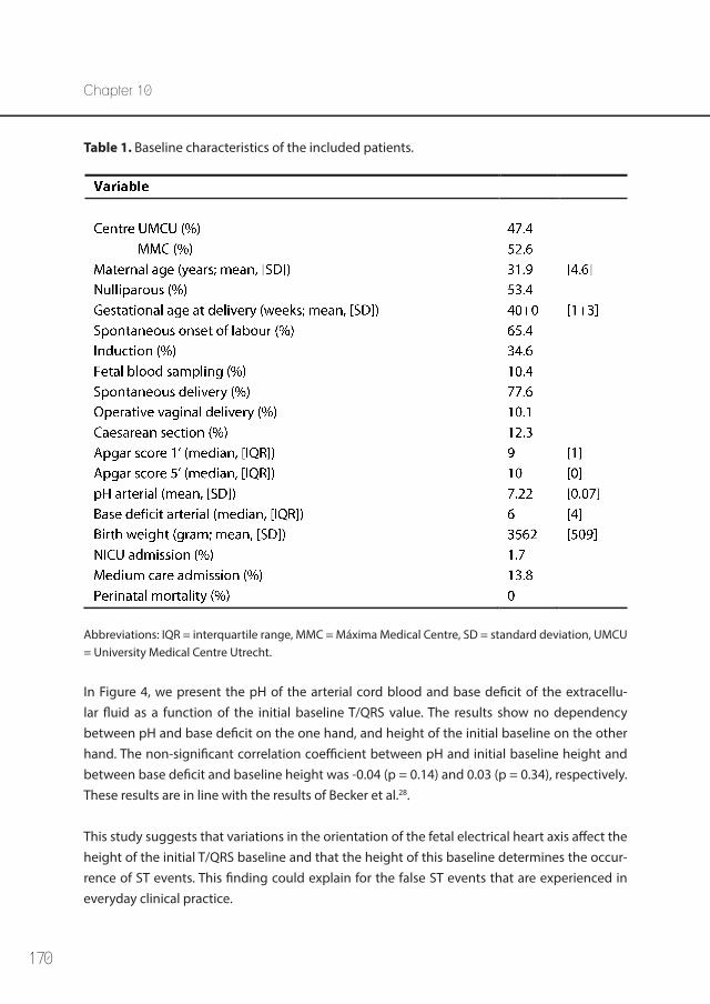
170
Chapter 10
In Figure 4, we present the pH of the arterial cord blood and base deficit of the extracellu-lar fluid as a function of the initial baseline T/QRS value. The results show no dependency between pH and base deficit on the one hand, and height of the initial baseline on the other hand. The non-significant correlation coefficient between pH and initial baseline height and between base deficit and baseline height was -0.04 (p = 0.14) and 0.03 (p = 0.34), respectively. These results are in line with the results of Becker et al.28.
This study suggests that variations in the orientation of the fetal electrical heart axis affect the height of the initial T/QRS baseline and that the height of this baseline determines the occur-rence of ST events. This finding could explain for the false ST events that are experienced in everyday clinical practice.
Table 1. Baseline characteristics of the included patients.
Abbreviations: IQR = interquartile range, MMC = Máxima Medical Centre, SD = standard deviation, UMCU = University Medical Centre Utrecht.
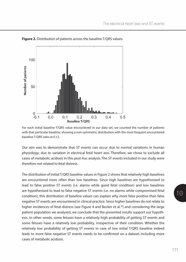
171
The electrical heart axis and ST events
10
Figure 2. Distribution of patients across the baseline T/QRS values.
For each initial baseline T/QRS value encountered in our data set, we counted the number of patients with that particular baseline, showing a non-symmetric distribution with the most frequent encountered baseline T/QRS ratio at 0.12.
Our aim was to demonstrate that ST events can occur due to normal variations in human physiology; due to variation in electrical fetal heart axis. Therefore, we chose to exclude all cases of metabolic acidosis in this post-hoc analysis. The ST events included in our study were therefore not related to fetal distress.
The distribution of initial T/QRS baseline values in Figure 2 shows that relatively high baselines are encountered more often than low baselines. Since high baselines are hypothesised to lead to false positive ST events (i.e. alarms while good fetal condition) and low baselines are hypothesised to lead to false negative ST events (i.e. no alarms while compromised fetal condition), this distribution of baseline values can explain why more false positive than false negative ST events are encountered in clinical practice. Since higher baselines do not relate to higher incidences of fetal distress (see Figure 4 and Becker et al.28) and considering the large patient population we analysed, we conclude that the presented results support our hypoth-esis. In other words, some fetuses have a relatively high probability of getting ST events and some fetuses have a relatively low probability, irrespective of their condition. Whether the relatively low probability of getting ST events in case of low initial T/QRS baseline indeed leads to more false negative ST events needs to be confirmed on a dataset including more cases of metabolic acidosis.
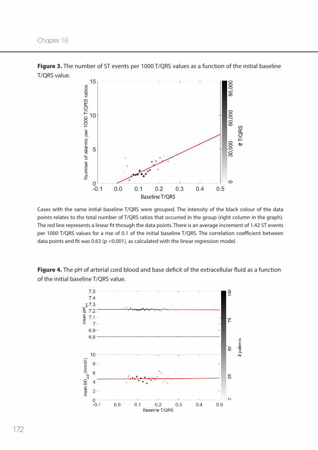
172
Chapter 10
Figure 3. The number of ST events per 1000 T/QRS values as a function of the initial baseline T/QRS value.
Figure 4. The pH of arterial cord blood and base deficit of the extracellular fluid as a function of the initial baseline T/QRS value.
Cases with the same initial baseline T/QRS were grouped. The intensity of the black colour of the data points relates to the total number of T/QRS ratios that occurred in the group (right column in the graph). The red line represents a linear fit through the data points. There is an average increment of 1.42 ST events per 1000 T/QRS values for a rise of 0.1 of the initial baseline T/QRS. The correlation coefficient between data points and fit was 0.63 (p <0.001), as calculated with the linear regression model.
173
The electrical heart axis and ST events
10
Cases with the same initial baseline T/QRS value were grouped. The intensity of the black colour of the data points relates to the total number of patients that were represented in the group (right column in the graph). The red line represents a linear fit through the data points. The fit suggests a reduction in the pH of 0.0009 and an increase in the base deficit of 0.03 for a rise in 0.1 of the initial baseline T/QRS. The respective correlation coefficients between data points and fit are -0.04 (p = 0.14) and 0.03 (p = 0.34), as calculated with the linear regression model.
Abbreviations: BDecf = base deficit in the extracellular fluid, pHart = pH of the arterial cord blood.
In addition, we propose that ST events are unreliable in case of high or low baseline T/QRS values. In case of an average T/QRS baseline value, the incidence of false ST events will be lower. When using the STAN® monitor in clinical practice, clinicians should be aware of this limitation. In case of inconclusive cardiotocogram assessment in combination with a high or low baseline T/QRS, fetal blood sampling can be used for complementary diagnostic information. However, the additional value of fetal blood sampling is uncertain and repeated fetal blood sampling is an independent risk factor for caesarean delivery29. In case of average baseline T/QRS, ST events can be considered more reliable and could be considered as complementary diagnostic information. ST analysis based on relative elevations of the T/QRS ratio with respect to the baseline or standardised non-invasive fetal ECG recordings30 might be feasible solutions that warrant further research. In addition, the relation between signal quality and T/QRS reliability needs to be explored in future research, including analysis of the effects of signal quality of small variations in the ECG that are caused by e.g. rotation of the fetal head at the end of labour.
Conclusions
This study showed a significant increment of ST events with increasing height of the initial T/QRS baseline; correlation coefficient 0.63, p <0.001. The orientation of the fetal electrical heart axis affects the height of the T/QRS baseline, and therefore the incidence of ST events. This should be taken into account in fetal monitoring with ST analysis.
Acknowledgements
We would like to thank Professor K.G. Rosén for his valuable input.

174
Chapter 10
References
1. Alfirevic Z, Devane D, Gyte GM. Continuous cardiotocography (CTG) as a form of electronic fetal monitoring (EFM) for fetal assessment during labour. Cochrane Database Syst Rev 2013 May 31;(5):CD006066.
2. Westgate J, Harris M, Curnow JS, Greene KR. Randomised trial of cardiotocography alone or with ST waveform analysis for intrapartum monitoring. Lancet 1992 Jul 25;340(8813):194-198.
3. Amer-Wahlin I, Hellsten C, Noren H, Hagberg H, Herbst A, Kjellmer I, et al. Cardiotocography only versus cardiotocography plus ST analysis of fetal electrocardiogram for intrapartum fetal monitoring: a Swedish randomised controlled trial. Lancet 2001 Aug 18;358(9281):534-538.
4. Westgate J, Harris M, Curnow JS, Greene KR. Plymouth randomized trial of cardiotocogram only versus ST waveform plus cardiotocogram for intrapartum monitoring in 2400 cases. Am J Obstet Gynecol 1993 Nov;169(5):1151-1160.
5. Ojala K, Vaarasmaki M, Makikallio K, Valkama M, Tekay A. A comparison of intrapartum automated fetal electrocardiography and conventional cardiotocography--a randomised controlled study. BJOG 2006 Apr;113(4):419-423.
6. Vayssiere C, David E, Meyer N, Haberstich R, Sebahoun V, Roth E, et al. A French randomized controlled trial of ST-segment analysis in a population with abnormal cardiotocograms during labor. Am J Obstet Gynecol 2007 Sep;197(3):299.e1-299.e6.
7. Westerhuis ME, Visser GH, Moons KG, Zuithoff N, Mol BW, Kwee A. Cardiotocography plus ST analysis of fetal electrocardiogram compared with cardiotocography only for intrapartum monitoring: a randomized controlled trial. Obstet Gynecol 2011 Feb;117(2 Pt 1):406-407.
8. Schuit E, Amer-Wahlin I, Ojala K, Vayssiere C, Westerhuis ME, Marsal K, et al. Effectiveness of elec-tronic fetal monitoring with additional ST analysis in vertex singleton pregnancies at >36 weeks of gestation: an individual participant data metaanalysis. Am J Obstet Gynecol 2013 Mar;208(3):187.e1-187.e13.
9. Belfort MA, Saade GR, Thom E, Blackwell SC, Reddy UM, Thorp JM,Jr, et al. A Randomized Trial of Intrapartum Fetal ECG ST-Segment Analysis. N Engl J Med 2015 Aug 13;373(7):632-641.
10. Blix E, Brurberg KG, Reierth E, Reinar LM, Oian P. ST waveform analysis versus cardiotocography alone for intrapartum fetal monitoring: a systematic review and meta-analysis of randomized trials. Acta Obstet Gynecol Scand 2016 Jan;95(1):16-27.
11. Neilson JP. Fetal electrocardiogram (ECG) for fetal monitoring during labour. Cochrane Database Syst Rev 2015 Dec 21;(12):CD000116.
12. Saccone G, Schuit E, Amer-Wahlin I, Xodo S, Berghella V. Electrocardiogram ST Analysis During Labor: A Systematic Review and Meta-analysis of Randomized Controlled Trials. Obstet Gynecol 2016 Jan;127(1):127-135.
13. Vayssiere C, Ehlinger V, Paret L, Arnaud C. Is STAN monitoring associated with a significant decrease in metabolic acidosis at birth compared with cardiotocography alone? Review of the three meta-analyses that included the recent US trial. Acta Obstet Gynecol Scand 2016 Oct;95(10):1190-1191.
14. Kwee A, Dekkers AH, van Wijk HP, van der Hoorn-van den Beld CW, Visser GH. Occurrence of ST-changes recorded with the STAN S21-monitor during normal and abnormal fetal heart rate patterns during labour. Eur J Obstet Gynecol Reprod Biol 2007 Nov;135(1):28-34.

175
The electrical heart axis and ST events
10
15. Westerhuis ME, van Horen E, Kwee A, van der Tweel I, Visser GH, Moons KG. Inter- and intra-observer agreement of intrapartum ST analysis of the fetal electrocardiogram in women monitored by STAN. BJOG 2009 Mar;116(4):545-551.
16. Greene KR, Dawes GS, Lilja H, Rosen KG. Changes in the ST waveform of the fetal lamb electrocardi-ogram with hypoxemia. Am J Obstet Gynecol 1982 Dec 15;144(8):950-958.
17. Widmark C, Lindecrantz K, Murray H, Rosen KG. Changes in the PR, RR intervals and ST waveform of the fetal lamb electrocardiogram with acute hypoxemia. J Dev Physiol 1992 Sep;18(3):99-103.
18. Westgate JA, Bennet L, Brabyn C, Williams CE, Gunn AJ. ST waveform changes during repeated umbilical cord occlusions in near-term fetal sheep. Am J Obstet Gynecol 2001 Mar;184(4):743-751.
19. Fenn W. The deposition of potassium and phosphate with glycogen in rat livers. J Biol Chem 1939;128:297-308.
20. Rosén K, Isaksson O. Alterations in Fetal Heart Rate and ECG Correlated to Glycogen, Creatine Phosphate and ATP Levels during Graded Hypoxia. Biol Neonate 1976;30:17-24.
21. Einthoven W, Fahr G, De Waart A. On the direction and manifest size of the variations of potential in the human heart and on the influence of the position of the heart on the form of the electrocardio-gram. Am Heart J 1950 Aug;40(2):163-211.
22. Verdurmen KMJ, Hulsenboom ADJ, van Laar JOEH, Wijn PFF, Vullings R, Oei SG. Orientation of the electrical heart axis in mid-term pregnancy. Eur J Obstet Gynecol Reprod Biol 2016.
23. Larks SD. Estimation of the Electrical Axis of the Fetal Heart. Am J Obstet Gynecol 1965 Jan 1;91:46-55.
24. Wagner GS, Strauss DG. Marriott’s Practical Electrocardiography. 12th edition ed. Philadelphia: Lippincott Williams & Wilkins; 2014.
25. Goodacre S, McLeod K. ABC of clinical electrocardiography: Paediatric electrocardiography. BMJ 2002 Jun 8;324(7350):1382-1385.
26. Depasquale NP, Burch GE. The Electrocardiogram, Ventricular Gradient and Spatial Vectorcardio-gram during the First Week of Life. Am J Cardiol 1963 Oct;12:482-493.
27. Schaffer AI, Beinfield WH. The vectorcardiogram of the newborn infant. Am Heart J 1952 Jul;44(1):89-94.
28. Becker JH, Kuipers LJ, Schuit E, Visser GH, Van Den Akker ES, Van Beek E, et al. Predictive value of the baseline T-QRS ratio of the fetal electrocardiogram in intrapartum fetal monitoring: a prospective cohort study. Acta Obstet Gynecol Scand 2012 Feb;91(2):189-197.
29. Holzmann M, Wretler S, Cnattingius S, Nordstrom L. Neonatal outcome and delivery mode in labors with repetitive fetal scalp blood sampling. Eur J Obstet Gynecol Reprod Biol 2015 Jan;184:97-102.
30. van Laar JO, Warmerdam GJ, Verdurmen KM, Vullings R, Peters CH, Houterman S, et al. Fetal heart rate variability during pregnancy, obtained from non-invasive electrocardiogram recordings. Acta Obstet Gynecol Scand 2014 Jan;93(1):93-101.

176
177
11
Chapter 11
Relative versus absolute rises in T/QRS ratio: a case-control study
Alexandra D.J. Hulsenboom, Kim M.J. Verdurmen, Rik Vullings, M. Beatrijs van der Hout – van der Jagt, Anneke Kwee,
Judith O.E.H. van Laar, S. Guid Oei
Submitted for publication
178
Chapter 11
Abstract
Introduction Conflicting results have been reported regarding the additional value of ST analysis during labour. In ST analysis, a T/QRS baseline value is calculated from the fetal electrocardiogram and successive T/QRS ratios are compared to this baseline. Variation in the orientation of the electrical heart axis between fetuses yields different T/QRS baseline values. In case of a higher T/QRS baseline value more ST events are encountered, irrespective of perinatal outcome. We hypothesised that we can partly correct for this effect by analysing T/QRS rises as a percent-age from baseline (relative ST analysis). This study aims to explore whether relative ST analysis improves the diagnostic characteristics of ST analysis to detect metabolic acidosis.
Material and Methods A case-control study was performed in 20 term human fetuses during labour; 10 cases (cord artery pH <7.05) and 10 controls (cord artery pH >7.20) were included. The fetal electrocardi-ogram was recorded using a STAN® monitor. We extracted all baseline and episodic ST alarms, T/QRS values, and calculated the relative T/QRS changes. The cut-off for relative ST alarms was determined in a receiver operator characteristic (ROC) curve at optimal specificity. Parameters of interest were area under the curve of the ROC curve for relative ST alarms and test charac-teristics of both conventional and relative ST analysis.
ResultsRelative ST analysis showed an area under the curve of 0.99 (95% confidence interval 0.96-1.00). The optimal cut-off value for relative T/QRS rise was determined at 0.70. Relative vs conventional ST analysis showed a specificity of 100% vs 40% (p = 0.031); sensitivity 90% vs 90%; positive likelihood ratio infinity vs 1.5; negative likelihood ratio 0.10 vs 0.25, respectively.
ConclusionIn this study relative ST analysis provides a good distinctive value for metabolic acidosis, with better specificity and comparable sensitivity in comparison to conventional ST analysis. Relative ST analysis seems to be a promising method to detect metabolic acidosis during labour. However, further research and validation are highly recommended.
179
Relative versus absolute rises in T/QRS ratio
11
Introduction Due to poor specificity of cardiotocography (CTG), which is used for fetal monitoring during labour, unnecessary caesarean deliveries are performed without improvement in long-term neonatal outcome1. ST analysis of the fetal electrocardiogram (ECG) was introduced in the 1990s as a promising technique to accurately detect metabolic acidosis and improve perinatal outcome2. Two large randomised trials (RCTs), both comparing CTG monitoring to CTG moni-toring plus ST analysis, showed promising results with a decrease in metabolic acidosis3 and operative deliveries2,3. Subsequent RCTs did not confirm these results4-7, and meta-analyses report conflicting results8-12.
The physiological basis for ST analysis was found in sheep studies. Rosén et al.13-15 found that hypoxia in fetal lambs leads to an adrenalin surge in the fetal heart, resulting in local glycogenolysis and potassium release. This local increase in potassium ions leads to an increase in T wave amplitude in the fetal ECG13,15,16. In ST analysis (STAN®, Neoventa Medical AB, Mölndal, Sweden), the T wave amplitude is normalised against the QRS amplitude, resulting in the T/QRS ratio. In case there is a rise in T/QRS ratio, an alarm is generated. The STAN® method discriminates three types of events (alarms): episodic, baseline, and biphasic events. Biphasic ST events will not be considered in this paper.
The relevance of a ST event depends on the visual assessment of the CTG. In case the CTG is classified as normal, all ST events given by the STAN® monitor can be ignored17-19. In this case, the ST event can be classified as a false event. In clinical practice, these false events are encountered frequently20. In case the CTG is classified as suboptimal or abnormal, ST events are considered to be significant and a clinical intervention should be prompted19. The dependence on subjective CTG interpretation, which is known to have a large inter- and intra-observer variability1,21, has a negative impact on the practical value of ST analysis.
Previously, we described a physiological explanation for false ST events. We demonstrated that the orientation of the fetal electrical heart axis varies considerably between fetuses22. The relation between the orientation of the electrical heart axis and the orientation of the scalp electrode, yields different T/QRS baseline values, due to differences in ECG morphology. We found that fetuses with a higher T/QRS baseline are more prone to ST events, independent of the fetal condition23. In contrast, we found that fetuses with a lower T/QRS baseline are less prone to exceed the threshold for a ST event. Becker et al.24 described that higher T/QRS baseline values are not related to poor neonatal outcome or hypoxia. In other words, it is expected that a high initial T/QRS baseline increases the incidence of false positive ST events and that a low initial T/QRS baseline increases the incidence of false negative ST events.
180
Chapter 11
Both the T/QRS baseline value and the rise in T wave amplitude are affected by the orientation of the electrical heart axis. We hypothesise that analysing relative, rather than the conventional, “absolute” T/QRS rises from baseline, could improve the diagnostic value of ST analysis. In case of relative ST analysis (T/QRS rises as a percentage from baseline), there is a correction for the “ease” of increment of the T wave amplitude. In conventional ST analysis, an absolute rise in T/QRS value will lead to an event; irrespective of the height of the T/QRS baseline. This study aims to explore whether relative ST analysis has a distinctive value in detecting metabolic acidosis. In addition, we aim to compare the test characteristics of both relative and absolute ST analysis. This is the first study that describes relative ST analysis. In order to directly compare both methods, we only focussed on objective information. Therefore, we omitted subjective CTG interpretations.
Material and Methods
Patient inclusion We performed a case-control study on a dataset collected by van Laar et al.25. We included fetuses of at least 36 weeks of gestation with intrapartum fetal ECG recordings, whose arterial and venous umbilical cord blood gasses were determined directly after birth. The included mothers were healthy, had an uncomplicated pregnancy and did not use any medication except oxytocin or epidural analgesia. Only good quality fetal ECG recordings were included, defined as absence of ectopic beats and no missing data in the last 10 minutes before birth. We excluded fetuses with fetal growth restriction (defined as birth weight below the 10th
percentile) and congenital heart disease.
Cases had an arterial cord pH <7.05 and controls had an arterial cord pH >7.20. Cases were consecutively selected between January 2006 and December 2007 in the Máxima Medical Centre, Veldhoven, the Netherlands. As a result of the strict inclusion and exclusion criteria, only five fetuses with acidemia could be included. In addition, five fetuses with acidemia from the University Medical Centre Utrecht, the Netherlands were selected between January 2001 and July 2002. Both hospitals are tertiary-care teaching hospitals. The ten controls were consecutively selected between January 2007 and August 2007 in the Máxima Medical Centre, Veldhoven, the Netherlands. Data acquisition and signal processingFetal ECG recordings were obtained during labour with a single helix scalp electrode (Goldtrace™), a maternal skin electrode, and a STAN S31® monitor (Neoventa Medical AB, Mölndal, Sweden). For every heartbeat, the STAN® detects an ECG complex. After 30 good quality ECG complexes, an average ECG complex (aECG) is calculated. This aECG is used to
181
Relative versus absolute rises in T/QRS ratio
11
determine the amplitudes of the T wave and QRS complex which, in turn, are used to calculate the T/QRS ratio. For every registration, we extracted all T/QRS ratios.
In absolute ST analysis, first a T/QRS baseline is determined. Following T/QRS ratios are compared to this baseline and in case they exceed the baseline by 0.05 for at least 10 minutes16, a baseline ST event is generated. When the T/QRS ratio exceeds the baseline by 0.10 within 10 minutes, an episodic ST event is generated. We used the “event log” window in the STAN® viewer software (Neoventa) to determine whether baseline and episodic ST events occurred. For relative ST analysis, instead of assessing the difference between T/QRS ratios and the baseline, we calculated the quotient. We defined the baseline via a two-step procedure, which is similar to the STAN® method. First, for every T/QRS ratio the median over the last 20 preceding T/QRS ratios was calculated. Second, the T/QRS baseline was defined as the minimum value of this median T/QRS ratio within a three hour window preceding the current T/QRS ratio. In case the 20 preceding T/QRS ratios did not fall within a 20-minute window from the current T/QRS ratio, we classified the signal as low quality and did not update the baseline. As mentioned previously, we want to assess the test characteristics of absolute and relative ST analysis in a cohort of 10 cases and 10 controls. Ideally, for cases both methods give at least one ST event. For controls, both methods should give no ST events. Hence, in this study we analysed whether the largest rise from baseline exceeds the absolute and/or the relative threshold for generating an event. In case the largest rise does not exceed the threshold, none of the other T/QRS ratios will trigger an event. In case the largest rise exceeds the threshold, at least one event was triggered. Based on whether the patient was a case or a control, we classified the event as true or false. Outcome measuresThe primary outcome was neonatal acidosis, defined as umbilical artery pH <7.05. Secondary outcomes were neonatal intensive care admission and 5 minute Apgar scores. Relative and absolute ST analysis were compared with respect to sensitivity (Sn), specificity (Sp), positive likelihood ratio (LR+) and negative likelihood ratio (LR-). Statistical analysis Baseline characteristics were compared with a Mann-Whitney U test for continuous variables and a Fisher exact test for categorical variables. To determine the optimal threshold for relative ST analysis, the largest rise from baseline per patient was plotted in a receiver operating characteristic curve (ROC curve) in SPSS 22 for Mac (IBM corp. Armonk, NY, USA). The threshold to predict neonatal acidosis was determined at the optimal cut-off in the ROC
182
Chapter 11
curve (the point closest to 0.1). This threshold was subsequently used to compute other test characteristics. We used a McNemar test to compare true positives and true negatives. A p-value <0.05 was considered statistically significant.
Results
Baseline characteristics For all 20 fetuses, good quality ECG data were available until nine minutes before birth. In nine fetuses (four cases, five controls), fetal ECG data could be obtained until the last minute before birth. Table 1 shows the baseline characteristics of the included patients. Significant differences were found between both groups regarding the frequency of fetal blood sampling, Apgar score after 1 minute, Apgar score after 5 minutes, umbilical cord artery and venous pH and umbilical artery base excess. Relative versus absolute ST analysis Figure 1 shows the ROC curve for relative ST analysis. The area under the ROC curve for relative T/QRS ratio changes in this population was 0.99 (p <0.001; 95% CI 0.96-1.00), with a standard error of 0.016. The cut-off value was determined at a relative T/QRS rise of 0.70.
We compared the test characteristics of absolute and relative ST analysis in our study popu-lation (Table 2). Sensitivity was equal for both methods, while specificity, LR+, and LR- were better for relative ST analysis.
Table 3 shows the number of correctly defined patients. When using a cut-off value of 0.70 as threshold for relative ST analysis, the detection of healthy fetuses is significantly better in comparison to absolute ST analysis (p = 0.031). We did not find significant differences between both methods to discriminate in neonatal hospital admission or Apgar score <7 at 5 minutes.
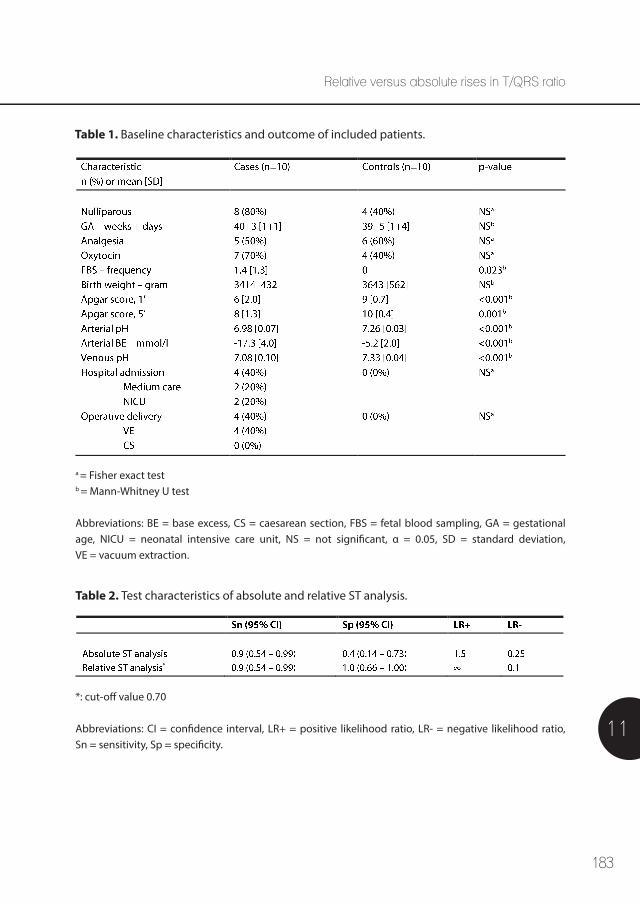
183
Relative versus absolute rises in T/QRS ratio
11
Table 1. Baseline characteristics and outcome of included patients.
Table 2. Test characteristics of absolute and relative ST analysis.
a = Fisher exact testb = Mann-Whitney U test
Abbreviations: BE = base excess, CS = caesarean section, FBS = fetal blood sampling, GA = gestational age, NICU = neonatal intensive care unit, NS = not significant, α = 0.05, SD = standard deviation, VE = vacuum extraction.
*: cut-off value 0.70
Abbreviations: CI = confidence interval, LR+ = positive likelihood ratio, LR- = negative likelihood ratio, Sn = sensitivity, Sp = specificity.
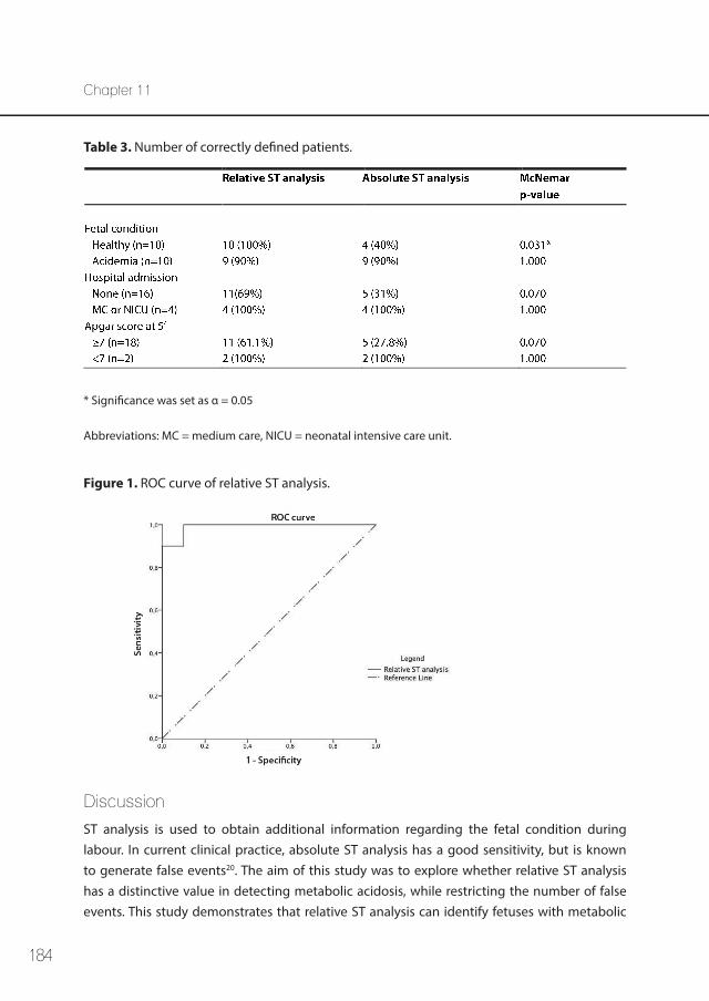
184
Chapter 11
Figure 1. ROC curve of relative ST analysis.
Table 3. Number of correctly defined patients.
* Significance was set as α = 0.05
Abbreviations: MC = medium care, NICU = neonatal intensive care unit.
Discussion
ST analysis is used to obtain additional information regarding the fetal condition during labour. In current clinical practice, absolute ST analysis has a good sensitivity, but is known to generate false events20. The aim of this study was to explore whether relative ST analysis has a distinctive value in detecting metabolic acidosis, while restricting the number of false events. This study demonstrates that relative ST analysis can identify fetuses with metabolic
185
Relative versus absolute rises in T/QRS ratio
11
acidosis at birth, as evidenced by the high AUC in the ROC curve. To explore whether relative ST analysis could be a solution for the false events encountered currently, we compared the test characteristics of both relative and absolute ST analysis. In the studied population, relative ST analysis showed to be superior in specificity, LR+ and LR-. Sensitivity was equal in both methods.
Neonatal hospital admission and low 5 minute Apgar score prevalence were low in our popu-lation. Nevertheless, we can observe a non-significant trend that relative ST analysis is more accurate in detecting false events; relative ST analysis is better in correctly defining patients with no hospital admission and a good Apgar score (>7 at 5 minutes). However, hospital admission and low Apgar score can both be caused by numerous other factors that are not necessarily related to acidosis, such as neonatal infection, respiratory insufficiency, meconium aspiration, and so on. Therefore, these factors should be assessed taking the abovementioned in consideration.
In absolute ST analysis, false events are encountered regularly20. This could lead to alarm fatigue, which can have life threatening consequences in the labour room. This study shows that ST monitoring is capable of identifying fetuses with acidosis without many false events, once one corrects for the orientation of the electrical heart axis.
In clinical practice, ST analysis is used in addition to CTG and therefore, the effect of false events can be limited by strict CTG interpretation. However, CTG interpretation is known to have a high intra- and inter-observer variability1,21. This can lead to different decisions regarding obstetric management following the same objective information.
As previously described, biphasic events were not evaluated in this study. The aim of our study was to compare baseline dependent ST events, since these are known to be related to the orientation of the electrical heart axis23. In addition, a recent study showed that biphasic events do not discriminate in the prediction of fetal distress or adverse outcome26.
The difference between absolute and relative ST analysis is based on different analyses of the same raw data. In both methods the fetal ECG is acquired by a fetal scalp electrode, and thus invasiveness of the technique is equal for patients. In addition, there is no difference in application or work load for the obstetric caregiver. Although the results from this first study describing relative ST analysis are promising, there are some important limitations. This study was designed as a case-control study, to determine if relative ST analysis could detect metabolic acidosis with a high sensitivity and low number
186
Chapter 11
of false events (high specificity). As a result of this study design, we did not evaluate the full spectrum of patients. Only cases with evident metabolic acidosis and controls with normal blood gas values at birth were included. A middle group with a pH between 7.05 and 7.20 is missing, while this group represents the majority of neonates born in clinical practice. In addition, the studied sample size was small; 10 cases and 10 controls. This can be explained by the low prevalence of metabolic acidosis (0.1-1%)3-7, combined with our strict inclusion criteria (availability of good quality fetal ECG recordings until at least 10 minutes before birth and availability of umbilical cord blood gasses).
Taken the limitations of our study into account, validity of relative ST analysis should be evaluated in a larger patient group, including the full spectrum of perinatal outcome. This allows to determine a more reliable and representative threshold value for relative ST analysis. Subsequently, this threshold needs to be validated in a different group of patients. We expect that the test characteristics of relative ST analysis determined in the latter group will be slightly less optimistic as those presented in the current work. In addition, only patients with good quality fetal ECG recordings and available umbilical cord blood gasses were included in this study. In following studies these conditions are likely to be less optimal, which might influence the test characteristics. Future studies could also focus on non-invasive abdominal registration of the fetal ECG, since this can also be used for fetal monitoring during pregnancy, before the onset of labour.
Conclusion
This study shows that relative T/QRS analysis provides a good distinctive value to detect metabolic acidosis during labour. In comparison to conventional absolute ST analysis, relative ST analysis shows better specificity with a comparable sensitivity. Therefore, relative ST analysis is a promising method for monitoring of fetal wellbeing during labour and needs to be studied in a larger population.

187
Relative versus absolute rises in T/QRS ratio
11
References
1. Alfirevic Z, Devane D, Gyte GM. Continuous cardiotocography (CTG) as a form of electronic fetal monitoring (EFM) for fetal assessment during labour. Cochrane Database Syst Rev 2013 May 31;(5):CD006066.
2. Westgate J, Harris M, Curnow JS, Greene KR. Plymouth randomized trial of cardiotocogram only versus ST waveform plus cardiotocogram for intrapartum monitoring in 2400 cases. Am J Obstet Gynecol 1993 Nov;169(5):1151-1160.
3. Amer-Wåhlin I, Hellsten C, Noren H, e.a. Intrapartum fetal monitoring: cardiotocography versus cardiotocography plus ST analysis of the fetal ECG: a Swedish randomised controlled trial. Lancet 2001;358:534– 538.
4. Belfort MA, Saade GR, Thom E, Blackwell SC, Reddy UM, Thorp JM,Jr, et al. A Randomized Trial of Intrapartum Fetal ECG ST-Segment Analysis. N Engl J Med 2015 Aug 13;373(7):632-641.
5. Ojala K, Vaarasmaki M, Makikallio K, Valkama M, Tekay A. A comparison of intrapartum automated fetal electrocardiography and conventional cardiotocography--a randomised controlled study. BJOG 2006 Apr;113(4):419-423.
6. Vayssiere C, David E, Meyer N, Haberstich R, Sebahoun V, Roth E, et al. A French randomized controlled trial of ST-segment analysis in a population with abnormal cardiotocograms during labor. Am J Obstet Gynecol 2007 Sep;197(3):299.e1-299.e6.
7. Westerhuis ME, Visser GH, Moons KG, Zuithoff N, Mol BW, Kwee A. Cardiotocography plus ST analysis of fetal electrocardiogram compared with cardiotocography only for intrapartum monitoring: a randomized controlled trial. Obstet Gynecol 2011 Feb;117(2 Pt 1):406-407.
8. Schuit E, Amer-Wahlin I, Ojala K, Vayssiere C, Westerhuis ME, Marsal K, et al. Effectiveness of elec-tronic fetal monitoring with additional ST analysis in vertex singleton pregnancies at >36 weeks of gestation: an individual participant data metaanalysis. Am J Obstet Gynecol 2013 Mar;208(3):187.e1-187.e13.
9. Blix E, Brurberg KG, Reierth E, Reinar LM, Oian P. ST waveform analysis versus cardiotocography alone for intrapartum fetal monitoring: a systematic review and meta-analysis of randomized trials. Acta Obstet Gynecol Scand 2016 Jan;95(1):16-27.
10. Neilson JP. Fetal electrocardiogram (ECG) for fetal monitoring during labour. Cochrane Database Syst Rev 2015 Dec 21;(12):CD000116.
11. Saccone G, Schuit E, Amer-Wahlin I, Xodo S, Berghella V. Electrocardiogram ST Analysis During Labor: A Systematic Review and Meta-analysis of Randomized Controlled Trials. Obstet Gynecol 2016 Jan;127(1):127-135.
12. Vayssiere C, Ehlinger V, Paret L, Arnaud C. Is STAN monitoring associated with a significant decrease in metabolic acidosis at birth compared with cardiotocography alone? Review of the three meta-analyses that included the recent US trial. Acta Obstet Gynecol Scand 2016 Oct;95(10):1190-1191.
13. Rosen KG, Dagbjartsson A, Henriksson BA, Lagercrantz H, Kjellmer I. The relationship between circulating catecholamines and ST waveform in the fetal lamb electrocardiogram during hypoxia. Am J Obstet Gynecol 1984 May 15;149(2):190-195.
188
Chapter 11
14. Rosén K, Isaksson O. Alterations in Fetal Heart Rate and ECG Correlated to Glycogen, Creatine Phosphate and ATP Levels during Graded Hypoxia. Biol Neonate 1976;30:17-24.
15. Greene KR, Dawes GS, Lilja H, Rosen KG. Changes in the ST waveform of the fetal lamb electrocardi-ogram with hypoxemia. Am J Obstet Gynecol 1982 Dec 15;144(8):950-958.
16. Sundström A, Rosén D, Rosén K. Fetal Surveillance. Tech. Rep., Neoventa Medical AB. 2006.
17. Amer-Wahlin I, Kwee A. Combined cardiotocographic and ST event analysis: A review. Best Pract Res Clin Obstet Gynaecol 2016 Jan;30:48-61.
18. Visser GH, Ayres-de-Campos D, FIGO Intrapartum Fetal Monitoring Expert Consensus Panel. FIGO consensus guidelines on intrapartum fetal monitoring: Adjunctive technologies. Int J Gynaecol Obstet 2015 Oct;131(1):25-29.
19. Amer-Wahlin I, Arulkumaran S, Hagberg H, Marsal K, Visser GH. Fetal electrocardiogram: ST waveform analysis in intrapartum surveillance. BJOG 2007 Oct;114(10):1191-1193.
20. Kwee A, Dekkers AH, van Wijk HP, van der Hoorn-van den Beld CW, Visser GH. Occurrence of ST-changes recorded with the STAN S21-monitor during normal and abnormal fetal heart rate patterns during labour. Eur J Obstet Gynecol Reprod Biol 2007 Nov;135(1):28-34.
21. Ayres-de-Campos D, Bernardes J, Costa-Pereira A, Pereira-Leite L. Inconsistencies in classification by experts of cardiotocograms and subsequent clinical decision. Br J Obstet Gynaecol 1999 Dec;106(12):1307-1310.
22. Verdurmen KMJ, Hulsenboom ADJ, van Laar JOEH, Wijn PFF, Vullings R, Oei SG. Orientation of the electrical heart axis in mid-term pregnancy. Eur J Obstet Gynecol Reprod Biol 2016.
23. Vullings R, Verdurmen KMJ, Hulsenboom ADJ, Scheffer S, de Lau H, Kwee A, et al. The electrical heart axis and ST events in fetal monitoring: a post-hoc analysis following a multicentre randomised controlled trial. PLoS One. 2017;12(4):e0175823. doi: 10.1371/journal.pone.
24. Becker JH, Kuipers LJ, Schuit E, Visser GH, Van Den Akker ES, Van Beek E, et al. Predictive value of the baseline T-QRS ratio of the fetal electrocardiogram in intrapartum fetal monitoring: a prospective cohort study. Acta Obstet Gynecol Scand 2012 Feb;91(2):189-197.
25. van Laar JO, Peters CH, Vullings R, Houterman S, Bergmans JW, Oei SG. Fetal autonomic response to severe acidaemia during labour. BJOG 2010 Mar;117(4):429-437.
26. Becker JH, Krikhaar A, Schuit E, Martendal A, Marsal K, Kwee A, et al. The added predictive value of biphasic events in ST analysis of the fetal electrocardiogram for intrapartum fetal monitoring. Acta Obstet Gynecol Scand 2015 Feb;94(2):175-182.

189
Relative versus absolute rises in T/QRS ratio
11

190

191
General discussion
12
Chapter 12
192
Chapter 12
In this thesis, the prospects of fetal electrocardiography (ECG) during pregnancy and labour are described. Fetal ECG is a technique that is still evolving. It can be applied non-invasively via electrodes on the maternal abdomen during pregnancy, or invasively with a fetal scalp electrode during labour.
Congenital heart disease (CHD) is the most common severe congenital anomaly worldwide1. The detection rate following fetal cardiac screening during the anomaly ultrasound around 20 weeks of gestation, varies between 65% and 81%2. Diagnosing CHD early in pregnancy is important, and therefore there is need for a reliable non-invasive diagnostic method that can complement ultrasonography.
Nowadays, we mainly use cardiotocography (CTG) to assess the fetal condition. However, it is known that the specificity and positive predictive value of CTG are rather poor3 and no improvement in long-term neonatal outcome is seen4. During pregnancy, besides a biophysical profile and Doppler ultrasonography there are no complementary diagnostics to give us information concerning the fetal condition. During labour, fetal scalp blood sampling and ST analysis of the fetal ECG can be performed to give additional information. Yet, these methods are both invasive and not applicable in case of prematurity. Therefore, there is need for a non-invasive method that provides more reliable information concerning the fetal condition.
Chapter 1 discusses the abovementioned issues considering fetal monitoring, that are encountered in daily clinical practice. This thesis aims to answer the following questions;
1. Is fetal electrocardiography valuable in diagnosing congenital heart disease in fetuses?
2. What is the influence of corticosteroids and tocolytics on fetal heart rate variability?
3. Are the changes in fetal heart rate variability following corticosteroid administration in the time-domain (obtained by Doppler ultrasound cardiotocography) comparable to the changes in fetal heart rate variability in the frequency-domain (obtained by non-invasive fetal electrocardiogram recordings)?
4. Is the variation in orientation of the fetal electrical heart axis in premature fetuses comparable to the variation seen in term fetuses?
5. Is variation in orientation of the electrical heart axis the cause of false ST events in ST analysis during labour?
6. Can we improve the method of ST analysis for fetal monitoring during labour?
193
12
General discussion
Background information considering the physiology of the fetal heart is provided in chapter 2. The embryology, circulation of the fetal heart and the postnatal changes in circu-lation are described. In response to hypoxia, chemoreceptors and baroreceptors influence the autonomic nervous system. It is known that changes in the ECG that are seen following hypoxia precede cerebral damage, and can therefore be seen as a marker for fetal distress5. The counteracting autonomic influences of the sympathetic and parasympathetic nervous system cause the variability in the fetal heart rate6.
In chapter 3, technical background information is provided regarding the CTG, the fetal ECG, spectral analysis of fetal heart rate variability, and ST analysis. The transabdominal CTG derives fetal heart rate information via Doppler ultrasound. The fetal heart rate is averaged over several heartbeats and beat-to-beat information is not available. From fetal ECG recordings, beat-to-beat fetal heart rate information can be extracted and therefore accurate spectral analysis (frequency analysis) can be performed with these recordings. By means of spectral analysis, changes in the autonomic regulation can be quantified7-12. The low-frequency (LF)-component reflects baroreceptor reflex activity, and is both sympathetically and parasympathetically mediated13. The high-frequency (HF)-component is associated with fetal respiration, and is solely parasympathetically mediated13.
In case of sustained hypoxia, there is a switch from aerobic to anaerobic metabolism with glycogenolysis. This is accompanied by an increase in potassium ions in the myocard14, causing an increase in T wave amplitude in the fetal ECG15. The consequent rise in T/QRS ratio is the basis of ST analysis.
This thesis is divided in three parts, that all describe one prospect of fetal ECG.
Part I: fetal ECG and congenital heart disease CHD is six times more common than chromosomal anomalies and four times more common than neural tube defects16,17. The detection rate following the congenital anomaly scan that is performed around 20 weeks of gestation varies from 65-81%16,18-20. Diagnosing CHD in pregnancy is important, since it enables the identification of associated anomalies, it gives parents the option to decide for a termination of pregnancy, and an adequate treatment plan can be developed, which is known to increase the survival rates and decrease long-term morbidity21.
In chapter 4, the possibilities of fetal ECG as a screening tool for the detection of CHD are reviewed. Not much research has been performed in this field; only five studies could be

194
Chapter 12
included. Fetal ECG is described to reflect the intimate relation between the cardiac nerve conduction system and the structural morphology of the heart22. In case of CHD, arrhythmias and changes in PR, QRS and QT interval have been described. Fetal ECG is particularly helpful in detecting the electrophysiological effects of cardiac anatomical defects (e.g. hypotrophy, hypertrophy, and conduction interruption) and it seems to be a promising clinical tool to complement ultrasonography in the screening for CHD. However, before we can detect CHD with fetal ECG we need to establish the normal range and values of amplitudes and segment intervals of the fetal ECG in the healthy fetus. Therefore, we designed a prospec-tive cohort study, including patients with an uneventful pregnancy. Non-invasive fetal ECG measurements were performed between 18 and 24 weeks of gestation. The study protocol is described in chapter 5 of this thesis.
Part II: fetal ECG in preterm labour
Preterm birth, defined as birth before 37 weeks of gestation, is one of the major causes of perinatal mortality and morbidity worldwide. In Europe, the preterm delivery rate is approx-imately 5-9%23. In case of threatened preterm labour between 24 and 34 weeks of gestation, fetal lung maturation can be enhanced by maternal administration of corticosteroids. This is associated with a reduction in neonatal mortality and morbidity24. In case of spontaneous preterm labour, tocolytics can be administered in order to attempt to postpone delivery for at least 48 hours. This will gain time to transfer the patient to a centre with neonatal intensive care facilities and to await maximal beneficial effect of corticosteroids.
Both corticosteroids and most tocolytics are known to have influence on fetal heart rate and fetal heart rate variability. It is known that fetal heart rate variability is one of the most important markers to assess fetal wellbeing25. Therefore, it is important to know the exact effects of corticosteroids and tocolytics on fetal heart rate parameters. Only then we can prevent iatrogenic preterm delivery due to misinterpretation of the CTG.
In chapter 6, the influence of corticosteroids (betamethasone and dexamethasone) on fetal heart rate parameters is described. We performed a review of the literature, eventually including 15 articles. During the first day after corticosteroid administration, there was a decrease in fetal heart rate, and an increase in fetal heart rate variability. During days 2 and 3, fetal heart rate increased while fetal heart rate variability decreased. All parameters returned to baseline by day 4. We also described confounding factors like the influence of other drugs, gestational age, diurnal rhythm, and fetal growth restriction. Since the administration of corti-costeroids is often accompanied by the administration of tocolytics, their influence on fetal heart rate parameters is described in chapter 7. In this review, it was found that nifedipine,
195
12
General discussion
atosiban and indomethacin administration show no clinically important effect on fetal heart rate variability. Magnesium sulphate can result in a decrease in fetal heart rate variability and cases of bradycardia were described. Fenoterol causes a slight increase in heart rate, with no changes in fetal heart rate variability. Following ritodrine, an increase in fetal heart rate and a decrease in variability was seen.
The measurements in the studies regarding the influence of corticosteroids on fetal heart rate variability, as described in chapter 6, used CTG (Doppler ultrasound). As a consequence, it is not possible to perform reliable spectral analysis with these measurements. We designed a prospective cohort study, including patients that required betamethasone during pregnancy. During five successive days, non-invasive abdominal fetal ECG recordings were performed. The aim of this study was to quantify the effects of maternally administered betamethasone on spectral analysis of fetal heart rate variability. In chapter 8, the results of this study are described. The changes in fetal heart rate variability following betamethasone administration show the same pattern when calculated by spectral analysis of the fetal ECG, as when calcu-lated by CTG. The change in absolute spectral values is likely to correspond to the change in quiet and active state of the fetus. Since normalised spectral values show little changes, the influence of autonomic modulation is minor.
Part III: fetal ECG to prevent asphyxia
Analysis of the ST segment of the fetal ECG complex can give additional information regarding fetal hypoxia during labour. In chapter 3, the technical details considering ST analysis are described. Initially, the combination of CTG and ST analysis was described to significantly lower the rates of metabolic acidosis5 and operative delivery5,26. However, subse-quent trials could not reproduce these findings27-30 and conflicting results have been reported in recent meta-analyses31-35. In addition, false positive alarms are often encountered in clinical practice36.
In this thesis we hypothesise that the abovementioned issues might be related to the orien-tation of the fetal electrical heart axis, since this orientation alters the shape and amplitude of the ECG. It is known that in term fetuses, neonates, and adults, there can be a significant inter-person variation in the orientation of the electrical heart axis37-41. The orientation of the fetal electrical heart axis during mid-pregnancy has never been described before. As elaborated in chapter 9, we performed a post-hoc analysis containing inclusions from two prospective cohort studies. This study showed that the main direction of the electrical heart axis is towards the right in mid-pregnancy fetuses, and that the variation in this orientation varies significantly from a normal distribution (p = 0.016).
196
Chapter 12
The orientation of the electrical heart axis can cause a relatively high, or a relatively low T/QRS baseline value. In case of fetal hypoxia, the efflux of potassium ions in the myocard causes a rise in amplitude of the T wave. This results in a rise in T/QRS value, which is repetitively compared to the baseline value. In case of an initial high baseline value (due to the orienta-tion of the electrical heart axis), the subsequent rise in T wave amplitude following hypoxia will be higher in comparison to a case where the initial baseline value is low, where the same amount of hypoxia will cause a smaller rise in T wave amplitude. In ST analysis, an alarm is generated following an absolute rise in T/QRS value above the baseline value. Therefore, in case of a high baseline the threshold for an alarm will be exceeded more often compared to a lower baseline where the T/QRS value rise is smaller.
In chapter 10, we test our hypothesis that the orientation of the electrical heart axis (and thus the height of the T/QRS baseline) is related to the occurrence of ST events. We performed a post-hoc analysis following a randomised trial. Only healthy fetuses, born in good condition without metabolic acidosis (defined as cord artery pH <7.05 and base deficit >12 mmol/l) were included. We found that there was a significant increment in ST events with increasing height of the T/QRS baseline; p <0.0001, which was irrespective of the fetal condition. Following this finding, we hypothesised that we can correct for the effect of the electrical heart axis by analysing the T/QRS rise following hypoxia as a percentage from baseline (yielding a relative ST event). A retrospective case-control study was performed, including 20 term fetuses during labour. Ten fetuses were cases with a cord artery pH <7.05 and ten were controls with a cord artery pH >7.20. As described in chapter 11, in this highly contrasting study population the optimal cut-off value for relative T/QRS ratio rises was determined at 0.70. The specificity of relative and absolute ST analysis was 100% and 40% (p = 0.031) respectively, while the sensitivity was 90% for both methods. Following this first explorative study, relative ST analysis seems to be promising.
Clinical implications and future directions
The results of the fundamental research reported in this thesis show that fetal ECG has multiple promising prospects, both during pregnancy and labour. Although the first studies describing fetal ECG measurements date back to 190642, the development of fetal ECG lagged behind in comparison to other techniques for fetal monitoring. Multiple technical challenges had to be overcome, as described in chapter 3. In 1942, it was already suggested that the effect of drugs, hypoxia, and labour could be conveniently studied by fetal ECG43. Nowadays, it is feasible to conduct a fetal ECG relatively easy, and it has been demonstrated that the recordings obtained by non-invasive measurements correlate very well with fetal ECG signals obtained directly via a scalp electrode11. With the fetal ECG technique being available, more
197
12
General discussion
sophisticated, easy to apply and safe to use, more and more research with this technique has been performed. Not all possible applications of fetal ECG are yet known, and researchers from all over the world are currently performing studies to elaborate these possible applica-tions44-46.
In our review as described in chapter 4, it was confirmed that fetal ECG can be a promising tool to complement ultrasonography in diagnosing CHD. This is intuitively logical, since it has been long known in children and adults that ECG can aid in the diagnosis of heart diseases. In particular, it seems that secondary effects due to CHD can be detected; hypotrophy, hyper-trophy and conduction interruption. However, the fetal circulation is markedly different in comparison to the neonatal and adult circulation, as described in chapter 2. In order to distin-guish abnormal fetal ECGs from normal ECGs, it is first required that we know the normal ECG in healthy fetuses. In chapter 5, the study protocol is described that will investigate the normal ranges for fetal ECG values around 20 weeks of gestation.
The collected data regarding the changes observed in fetal ECG in case of CHD is still limited. There is need for future trials that include multiple types of CHDs and have a reasonable sample size per type of CHD. In order to achieve this, these studies will most likely have to be performed as a collaboration between multiple expert centres. The ECG characteristics found in case of CHD should be compared to the characteristics found in healthy fetuses. When conducting these trials, one should take the gestational age of the fetus into account. The duration of the P wave, QRS complex and PR interval increase progressively from 18 weeks of gestation onwards until term47,48.
If pregnancy is complicated by threatened preterm labour, multiple drugs are administered. This is done in a highly vulnerable population, since preterm fetuses can tolerate contractions and labour less well compared to term fetuses. In addition, only non-invasive fetal monitoring is available which can be complicated due to movements of the fetus in-utero. Further, it is important to be aware of the effects that the administered medication can have on fetal heart rate parameters, to prevent iatrogenic preterm delivery. In this thesis, the effects of cortico-steroids and tocolytics were studied. However, in clinical practice some patients receive other drugs like antihypertensive agents or antibiotics. The effects of these drugs on fetal heart rate parameters should be studied as well. In addition, other confounding factors should be taken into account when studying the preterm fetus, such as gestational age, diurnal rhythm and fetal growth restriction. A challenging factor in studies including fetuses at risk for preterm labour is time of inclusion (most patients are transferred from secondary care hospitals, where medication is already started) and loss of study participants, e.g. due to preterm labour or discharge before the end of the study period. Nearly all studies considering this topic
198
Chapter 12
published so far used CTG (Doppler ultrasound) to perform measurements and evaluate fetal heart rate parameters. However, as previously described in this thesis, CTG measurements do not acquire fetal heart rate information on a beat-to-beat basis. Future studies should perform measurements using fetal ECG equipment that can extract the fetal heart rate on a beat-to-beat basis, since only then reliable spectral analysis can be performed and more reliable estimates for fetal heart rate variability can be calculated.
During term labour, ST analysis of the fetal ECG was developed to provide additional infor-mation regarding fetal wellbeing. However, in clinical practice it appeared that false positive events were encountered regularly36. This made some clinicians and researchers wonder whether the STAN® monitor was indeed a reliable method for fetal monitoring. In this thesis, we described that our hypothesis that false alarms can be caused by the variation in orienta-tion of the electrical heart axis indeed holds true. In addition, we provide a possible solution; relative ST analysis. Our study results should be verified in a study with a larger sample size, including the whole patient spectrum. In this group a reliable cut-off value for relative ST analysis can be defined, which should be validated in a second study. If the results of relative ST analysis are still promising, a prospective or randomised controlled trial can be performed. In addition, the clinical value of relative ST analysis in conjunction with CTG should be evaluated.
What can be learned from the above, is that when a new technique is introduced in clinical practice, we should not take this technology for granted. When unexpected findings arise, one should go back to basic physiology and anatomy and wonder if some influence might have been overlooked.
With multi-lead fetal ECG recordings being available, the applicability of antepartum ST analysis in preterm deliveries should be a field of future studies49.
Advantages of fetal ECG
A major advantage of fetal ECG measurements is that they can provide additional and objective information; e.g. ECG segment intervals, values for spectral analysis, and ST analysis. This objective information can aid current technologies. Thereby, the influence of, for instance, performer variability (in ultrasonography) and inter- and intra-observer variability (in CTG assessment) might be decreased. Spectral analysis can objectively quantify the changes in fetal heart rate variability that can be obscured in visual interpretation of fetal heart rate tracings. Spectral analysis might also be added to ST analysis; it has been described that a relative change in LF/HF ratio >30%, in combination with a significant ST event is a better predictor for metabolic acidosis in the arterial cord blood than a significant ST event alone50.
199
12
General discussion
Another advantage is that fetal ECG has the potential to be widely versatile. A fetal ECG is easy to conduct, by placing electrodes on the maternal abdomen. The electrodes are attached to small and mobile equipment, which is likely to be relatively cheap in the future. Application of the fetal ECG system requires minimum training. In addition, the raw information can be analysed by computerised algorithms or can be sent for analysis elsewhere.
Since the fetal ECG can be conducted both invasively (with a fetal scalp electrode) and non-invasively (with electrodes on the maternal abdomen), the fetal ECG is applicable from early pregnancy (from 18 weeks of gestation onwards) until delivery. Since it can be applied non-invasively and can provide additional information such as spectral estimates, it could be used for fetal monitoring in high-risk pregnancies; e.g. in case of extreme prematurity, fetal growth restriction and fetuses from mothers with diabetes. In addition, home monitoring with non-invasive fetal ECG measurements in high-risk pregnancies is an additional field that has been studied recently51. When this would be feasible, it would lead to a significant reduction in health care costs due to less hospitalisation.
Disadvantages of fetal ECG
Despite all efforts to enhance the signal quality in fetal ECG measurements, there is still a lot to gain in this area. Therefore, future research should keep focussing on further progress in signal acquisition and processing. The signal quality is reduced due to presence of vernix caseosa on the fetal skin. This fatty layer surrounds the fetus and results in an electrical isolation, which diminishes the signal amplitude of the fetal ECG. Especially between 30 and 34 weeks of gestation, this layer causes a poor signal-to-noise ratio46,52.
As with all new techniques, differences in acquiring and processing of the data exist. It is highly recommended that all future studies regarding fetal ECG measurements and spectral analysis use a standardised method. This will enable comparability, reproducibility, reliable physiological interpretation and clinical applicability.
In addition, every study regarding fetal ECG should consider the influence of the orientation of the electrical heart axis on their study results. For instance in case of ST monitoring, as described in this thesis, the physiological variation in orientation of the electrical heart axis might be responsible for the encountered false alarms. When calculating the electrical heart axis, it is important to take the fetal position in the uterus into account.
In the studies described in this thesis, data processing and calculation of spectral estimates, the electrical heart axis, etc. was performed off-line. In order to be clinically applicable, real-time monitoring should become available in the future.

200
Chapter 12
In conclusion, this thesis describes the prospects of fetal ECG during pregnancy and labour. Although non-invasive fetal ECG is not yet clinically applied, this thesis shows that fetal ECG is promising in providing complementary diagnostic information. The additional value of fetal ECG in multiple fields has been addressed, and it has been shown that it can be used both during pregnancy and during labour.
201
12
General discussion
References
1. van der Linde D, Konings EE, Slager MA, Witsenburg M, Helbing WA, Takkenberg JJ, et al. Birth prevalence of congenital heart disease worldwide: a systematic review and meta-analysis. J Am Coll Cardiol 2011 Nov 15;58(21):2241-2247.
2. Verdurmen KM, Eijsvoogel NB, Lempersz C, Vullings R, Schroer C, van Laar JO, et al. A systematic review of prenatal screening for congenital heart disease by fetal electrocardiography. Int J Gynaecol Obstet 2016 Nov;135(2):129-134.
3. Ayres-de-Campos D, Spong CY, Chandraharan E, FIGO Intrapartum Fetal Monitoring Expert Consensus Panel. FIGO consensus guidelines on intrapartum fetal monitoring: Cardiotocography. Int J Gynaecol Obstet 2015 Oct;131(1):13-24.
4. Alfirevic Z, Devane D, Gyte GM. Continuous cardiotocography (CTG) as a form of electronic fetal monitoring (EFM) for fetal assessment during labour. Cochrane Database Syst Rev 2013 May 31;(5):CD006066.
5. Amer-Wahlin I, Nord A, Bottalico B, Hansson SR, Ley D, Marsal K, et al. Fetal cerebral energy metabolism and electrocardiogram during experimental umbilical cord occlusion and resuscitation. J Matern Fetal Neonatal Med 2010 Feb;23(2):158-166.
6. Van Ravenswaaij-Arts C, Kollee L, Hopman J, Stoelinga G, van Geijn H. Heart rate variabilitiy. Ann Intern Med 1993;118:436-447.
7. Dawes GS, Moulden M, Redman CW. Improvements in computerized fetal heart rate analysis antepartum. J Perinat Med 1996;24(1):25-36.
8. van Laar JO, Peters CH, Vullings R, Houterman S, Oei SG. Power spectrum analysis of fetal heart rate variability at near term and post term gestation during active sleep and quiet sleep. Early Hum Dev 2009 Dec;85(12):795-798.
9. van Laar JO, Peters CH, Vullings R, Houterman S, Bergmans JW, Oei SG. Fetal autonomic response to severe acidaemia during labour. BJOG 2010 Mar;117(4):429-437.
10. van Laar JO, Peters CH, Houterman S, Wijn PF, Kwee A, Oei SG. Normalized spectral power of fetal heart rate variability is associated with fetal scalp blood pH. Early Hum Dev 2011 Apr;87(4):259-263.
11. Vullings R, Peters C, Andriessen P, Oei S, Wijn P. Monitoring the Fetal Heart Rate and Fetal Electro-cardiogram: Abdominal Recordings Are As Good As Direct Ecg Measurements. Pediatric Research 2005;58(2):242.
12. Siira SM, Ojala TH, Vahlberg TJ, Jalonen JO, Valimaki IA, Rosen KG, et al. Marked fetal acidosis and specific changes in power spectrum analysis of fetal heart rate variability recorded during the last hour of labour. BJOG 2005 Apr;112(4):418-423.
13. Heart rate variability: standards of measurement, physiological interpretation and clinical use. Task Force of the European Society of Cardiology and the North American Society of Pacing and Electro-physiology. Circulation 1996 Mar 1;93(5):1043-1065.
14. Fenn W. The deposition of potassium and phosphate with glycogen in rat livers. J Biol Chem 1939;128:297-308.
15. Rosén K, Isaksson O. Alterations in Fetal Heart Rate and ECG Correlated to Glycogen, Creatine Phosphate and ATP Levels during Graded Hypoxia. Biol Neonate 1976;30:17-24.
202
Chapter 12
16. Carvalho JS, Mavrides E, Shinebourne EA, Campbell S, Thilaganathan B. Improving the effectiveness of routine prenatal screening for major congenital heart defects. Heart 2002 Oct;88(4):387-391.
17. Simpson LL. Screening for congenital heart disease. Obstet Gynecol Clin North Am 2004 Mar;31(1):51-59.
18. Ogge G, Gaglioti P, Maccanti S, Faggiano F, Todros T. Prenatal screening for congenital heart disease with four-chamber and outflow-tract views: a multicenter study. Ultrasound Obstet Gynecol 2006 Nov;28(6):779-784.
19. Kirk JS, Riggs TW, Comstock CH, Lee W, Yang SS, Weinhouse E. Prenatal screening for cardiac anomalies: the value of routine addition of the aortic root to the four-chamber view. Obstet Gynecol 1994 Sep;84(3):427-431.
20. Wu Q, Li M, Ju L, Zhang W, Yang X, Yan Y, et al. Application of the 3-vessel view in routine prenatal sonographic screening for congenital heart disease. J Ultrasound Med 2009 Oct;28(10):1319-1324.
21. Brown KL, Ridout DA, Hoskote A, Verhulst L, Ricci M, Bull C. Delayed diagnosis of congenital heart disease worsens preoperative condition and outcome of surgery in neonates. Heart 2006 Sep;92(9):1298-1302.
22. Velayo C, Sato N, Ito T, Chisaka H, Yaegashi N, Okamura K, et al. Understanding congenital heart defects through abdominal fetal electrocardiography: case reports and clinical implications. J Obstet Gynaecol Res 2011 May;37(5):428-435.
23. Goldenberg RL, Culhane JF, Iams JD, Romero R. Epidemiology and causes of preterm birth. Lancet 2008 Jan 5;371(9606):75-84.
24. Roberts D, Dalziel S. Antenatal corticosteroids for accelerating fetal lung maturation for women at risk of preterm birth. Cochrane Database Syst Rev 2006 Jul 19;(3):CD004454.
25. Paul RH, Suidan AK, Yeh S, Schifrin BS, Hon EH. Clinical fetal monitoring. VII. The evaluation and significance of intrapartum baseline FHR variability. Am J Obstet Gynecol 1975 Sep 15;123(2):206-210.
26. Westgate J, Harris M, Curnow JS, Greene KR. Plymouth randomized trial of cardiotocogram only versus ST waveform plus cardiotocogram for intrapartum monitoring in 2400 cases. Am J Obstet Gynecol 1993 Nov;169(5):1151-1160.
27. Belfort MA, Saade GR, Thom E, Blackwell SC, Reddy UM, Thorp JM,Jr, et al. A Randomized Trial of Intrapartum Fetal ECG ST-Segment Analysis. N Engl J Med 2015 Aug 13;373(7):632-641.
28. Ojala K, Vaarasmaki M, Makikallio K, Valkama M, Tekay A. A comparison of intrapartum automated fetal electrocardiography and conventional cardiotocography--a randomised controlled study. BJOG 2006 Apr;113(4):419-423.
29. Vayssiere C, David E, Meyer N, Haberstich R, Sebahoun V, Roth E, et al. A French randomized controlled trial of ST-segment analysis in a population with abnormal cardiotocograms during labor. Am J Obstet Gynecol 2007 Sep;197(3):299.e1-299.e6.
30. Westerhuis ME, Visser GH, Moons KG, Zuithoff N, Mol BW, Kwee A. Cardiotocography plus ST analysis of fetal electrocardiogram compared with cardiotocography only for intrapartum monitoring: a randomized controlled trial. Obstet Gynecol 2011 Feb;117(2 Pt 1):406-407.

203
12
General discussion
31. Schuit E, Amer-Wahlin I, Ojala K, Vayssiere C, Westerhuis ME, Marsal K, et al. Effectiveness of elec-tronic fetal monitoring with additional ST analysis in vertex singleton pregnancies at >36 weeks of gestation: an individual participant data metaanalysis. Am J Obstet Gynecol 2013 Mar;208(3):187.e1-187.e13.
32. Blix E, Brurberg KG, Reierth E, Reinar LM, Oian P. ST waveform analysis versus cardiotocography alone for intrapartum fetal monitoring: a systematic review and meta-analysis of randomized trials. Acta Obstet Gynecol Scand 2016 Jan;95(1):16-27.
33. Neilson JP. Fetal electrocardiogram (ECG) for fetal monitoring during labour. Cochrane Database Syst Rev 2015 Dec 21;(12):CD000116.
34. Saccone G, Schuit E, Amer-Wahlin I, Xodo S, Berghella V. Electrocardiogram ST Analysis During Labor: A Systematic Review and Meta-analysis of Randomized Controlled Trials. Obstet Gynecol 2016 Jan;127(1):127-135.
35. Vayssiere C, Ehlinger V, Paret L, Arnaud C. Is STAN monitoring associated with a significant decrease in metabolic acidosis at birth compared with cardiotocography alone? Review of the three meta-analyses that included the recent US trial. Acta Obstet Gynecol Scand 2016 Oct;95(10):1190-1191.
36. Kwee A, Dekkers AH, van Wijk HP, van der Hoorn-van den Beld,C.W., Visser GH. Occurrence of ST-changes recorded with the STAN S21-monitor during normal and abnormal fetal heart rate patterns during labour. Eur J Obstet Gynecol Reprod Biol 2007 Nov;135(1):28-34.
37. Larks SD. Estimation of the Electrical Axis of the Fetal Heart. Am J Obstet Gynecol 1965 Jan 1;91:46-55.
38. Depasquale NP, Burch GE. The Electrocardiogram, Ventricular Gradient and Spatial Vectorcardio-gram during the First Week of Life. Am J Cardiol 1963 Oct;12:482-493.
39. Wagner GS, Strauss DG. Marriott’s Practical Electrocardiography. 12th edition ed. Philadelphia: Lippincott Williams & Wilkins; 2014.
40. Goodacre S, McLeod K. ABC of clinical electrocardiography: Paediatric electrocardiography. BMJ 2002 Jun 8;324(7350):1382-1385.
41. Schaffer AI, Beinfield WH. The vectorcardiogram of the newborn infant. Am Heart J 1952 Jul;44(1):89-94.
42. Cremer M. Über die direkte Ableitung der Aktionsströme des menschlichen Herzens vom Oesopha-gus und über das Elektrokardiogramm des Fötus. Münch Med Wschr 1906;53:811-813.
43. Goodyer AV, Geiger AJ, Monroe WM. Clinical Fetal Electrocardiography. Yale J Biol Med 1942 Oct;15(1):1-20.7.
44. Fuchs T, Grobelak K, Pomorski M, Zimmer M. Fetal Heart Rate Monitoring Using Maternal Abdominal Surface Electrodes in Third Trimester: Can We Obtain Additional Information Other than CTG Trace? Adv Clin Exp Med 2016 Mar-Apr;25(2):309-316.
45. Lakhno I. Fetal Non-invasive Electrocardiography Contributes to Better Diagnostics of Fetal Distress: A Cross-sectional Study Among Patients with Pre-eclampsia. Ann Acad Med Singapore 2015 Nov;44(11):519-523.

204
Chapter 12
46. van Laar JO, Warmerdam GJ, Verdurmen KM, Vullings R, Peters CH, Houterman S, et al. Fetal heart rate variability during pregnancy, obtained from non-invasive electrocardiogram recordings. Acta Obstet Gynecol Scand 2014 Jan;93(1):93-101.
47. Yilmaz B, Narayan HK, Wilpers A, Wiess C, Fifer WP, Williams IA. Electrocardiographic intervals in foetuses with CHD. Cardiol Young 2016 Jan;26(1):84-89.
48. Chia EL, Ho TF, Rauff M, Yip WC. Cardiac time intervals of normal fetuses using noninvasive fetal electrocardiography. Prenat Diagn 2005 Jul;25(7):546-552.
49. Hruban L, Spilka J, Chudacek V, Janku P, Huptych M, Bursa M, et al. Agreement on intrapartum cardiotocogram recordings between expert obstetricians. J Eval Clin Pract 2015 Aug;21(4):694-702.
50. Siira S, Ojala T, Ekholm E, Vahlberg T, Blad S, Rosen KG. Change in heart rate variability in relation to a significant ST-event associates with newborn metabolic acidosis. BJOG 2007 Jul;114(7):819-823.
51. Vermeulen-Giovagnoli B, Peters C, van der Hout-van der Jagt,M.B., Mischi M, van Pul C, Cottaar EJ, et al. The development of an obstetric tele-monitoring system. Conf Proc IEEE Eng Med Biol Soc 2015;2015:177-180.
52. Oostendorp TF, van Oosterom A, Jongsma HW. The fetal ECG throughout the second half of gestation. Clin Phys Physiol Meas 1989 May;10(2):147-160.

205
12
General discussion

206

207
Appendices
List of abbreviationsList of publications
Nederlandse samenvattingDankwoord
Curriculum vitae
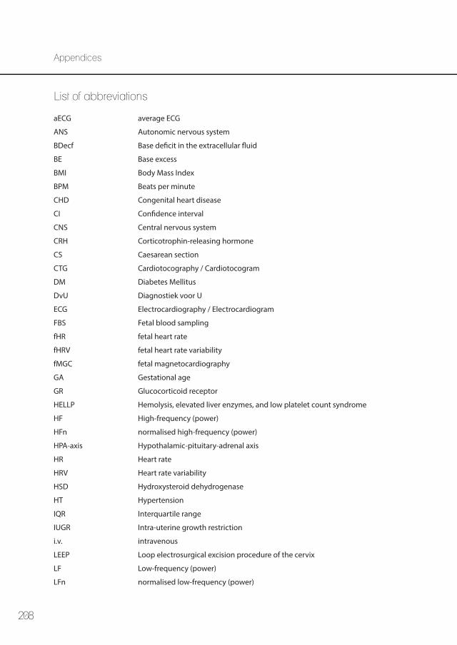
208
aECG average ECG
ANS Autonomic nervous system
BDecf Base deficit in the extracellular fluid
BE Base excess
BMI Body Mass Index
BPM Beats per minute
CHD Congenital heart disease
CI Confidence interval
CNS Central nervous system
CRH Corticotrophin-releasing hormone
CS Caesarean section
CTG Cardiotocography / Cardiotocogram
DM Diabetes Mellitus
DvU Diagnostiek voor U
ECG Electrocardiography / Electrocardiogram
FBS Fetal blood sampling
fHR fetal heart rate
fHRV fetal heart rate variability
fMGC fetal magnetocardiography
GA Gestational age
GR Glucocorticoid receptor
HELLP Hemolysis, elevated liver enzymes, and low platelet count syndrome
HF High-frequency (power)
HFn normalised high-frequency (power)
HPA-axis Hypothalamic-pituitary-adrenal axis
HR Heart rate
HRV Heart rate variability
HSD Hydroxysteroid dehydrogenase
HT Hypertension
IQR Interquartile range
IUGR Intra-uterine growth restriction
i.v. intravenous
LEEP Loop electrosurgical excision procedure of the cervix
LF Low-frequency (power)
LFn normalised low-frequency (power)
List of abbreviations
Appendices
209
List of abbreviations
LMWH Low molecular weight heparin
LR + Positive likelihood ratio
LR - Negative likelihood ratio
LTV Long-term variability
MC Medium care
mFHR mean fetal heart rate
MgSO4 Magnesium sulphate
NICU Neonatal intensive care unit
NS Not significant
PE Pre-eclampsia
pHart pH of the arterial cord blood
PPROM Premature prelabour rupture of membranes
PROM Preterm rupture of membranes
PSD Power spectral density
QTc Fetal QT interval corrected for heart rate
RCT Randomised controlled trial
ROC Receiver operator characteristic
SD Standard deviation
Sn Sensitivity
Sp Specificity
STAN ST analysis
STV Short-term variability
TPL Threatened preterm labour
VBL Vaginal blood loss
VCG Vectorcardiogram
VE Vacuum extraction
VLF Very low-frequency (power)
wG weeks of gestation
wGA weeks of gestational age
210
Appendices
List of publications Journal papers
K.M.J. Verdurmen, J. Renckens, J.O.E.H. van Laar, S.G. Oei. The influence of corticosteroids on fetal heart rate variability: a systematic review of the literature. Obstet Gynecol Surv. 2013;68(12):811-24. doi: 10.1097/OGX.
J.O.E.H. van Laar, G.J.J. Warmerdam, K.M.J. Verdurmen, R. Vullings, C.H.L. Peters, S. Houterman, P.F.F. Wijn, P. Andriessen, C. van Pul, S.G. Oei. Fetal heart rate variability during pregnancy, obtained from non-invasive electrocardiogram recordings. Acta Obstet Gynecol Scand. 2014;93(1):93-101. doi: 10.1111/aogs.
K.M.J. Verdurmen, C. Lempersz, R. Vullings, C. Schroer, T. Delhaas, J.O.E.H. van Laar, S.G. Oei. Normal ranges for fetal electrocardiogram values for the healthy fetus of 18-24 weeks of gestation: a prospective cohort study. BMC Pregnancy Childbirth. 2016;16:227. doi: 10.1186/s12884-016-1021-x.
K.M.J. Verdurmen, N.B. Eijsvoogel, C. Lempersz, R. Vullings, C. Schroer, J.O.E.H. van Laar, S.G. Oei. A systematic review of prenatal screening for congenital heart disease by fetal electrocardiography. Int J Gynaecol Obstet. 2016;135(2):129-134. doi: 10.1016/j.ijgo.
K.M.J. Verdurmen, A.D.J. Hulsenboom, J.O.E.H. van Laar, S.G. Oei. Effect of tocolytic drugs on fetal heart rate variability: a systematic review. J Matern Fetal Neonatal Med. 2016:1-8. doi: 10.1080/14767058.
K.M.J. Verdurmen, A.D.J. Hulsenboom, J.O.E.H. van Laar, P.F.F. Wijn, R. Vullings, S.G. Oei. Orientation of the electrical heart axis in mid-term pregnancy. Eur J Obstet Gynecol Reprod Biol. 2016;207:243-246. doi: 10.1016/j.ejogrb.
R. Vullings, K.M.J. Verdurmen, A.D.J. Hulsenboom, S. Scheffer, H. de Lau, A. Kwee, P.F.F. Wijn, I. Amer-Wåhlin, J.O.E.H. van Laar, S.G. Oei. The electrical heart axis and ST events in fetal monitoring: a post-hoc analysis following a multicentre randomised controlled trial. PLoS One. 2017;12(4):e0175823. doi: 10.1371/journal.pone.
K.M.J. Verdurmen, G.J.J. Warmerdam, C. Lempersz, A.D.J. Hulsenboom, J. Renckens, J.P. Dieleman, R. Vullings, J.O.E.H. van Laar, S.G. Oei. The influence of betamethasone on fetal heart rate variability, obtained by non-invasive fetal electrocardiogram recordings. Revision pending.
A.D.J. Hulsenboom, K.M.J. Verdurmen, R. Vullings, M.B. van der Hout-van der Jagt, A. Kwee, J.O.E.H. van Laar, S.G. Oei. Relative versus absolute rises in T/QRS ratio: a case-control study. Manuscript submitted for publication.
R. Vullings, C. Lempersz, S.A.B. Clur, K.M.J. Verdurmen, G.J.J. Warmerdam, J. van der Post, N.A. Blom, T. Delhaas, J.O.E.H. van Laar, S.G. Oei. The normal fetal electrocardiogram in mid-pregnancy, an additional tool for the prenatal detection of congenital heart disease. Manuscript submitted for publication.

211
Conference presentations
K.M.J. Verdurmen, J.O.E.H. van Laar, S.G. Oei. “Corticosteroids and fetal heart rate variability.” European Congress of Perinatal Medicine, Florence, 4-7 June, 2014.
K.M.J. Verdurmen, A.D.J. Hulsenboom, R. Vullings, J.O.E.H. van Laar, A. Kwee, S.G. Oei. “Improving STAN fetal monitoring”. European Congress on Intrapartum Care, Porto, 21-23 May 2015. Invited speaker at the pre-congress Intrapartum Fetal Monitoring meeting.
K.M.J. Verdurmen, R. Vullings. A.D.J. Hulsenboom, J.O.E.H. van Laar, A. Kwee, S.G. Oei. “De denkfout van de STAN”. Gynaecongres, Amersfoort, 28-29 May 2015. Wim Schellekensprice; 1st price in the category “Talent in Onderzoek”.
K.M.J. Verdurmen. “De relatieve STAN bij foetale asfyxie: wat zegt het?” Gynaecongres, Eindhoven, 19-20 May 2016. Invited speaker.
Conference posters
K.M.J. Verdurmen, J.O.E.H. van Laar, G.J.J. Warmerdam, S.G. Oei. Fetal heart rate variability during pregnancy. Symposium on Advances in Perinatal Monitoring, Eindhoven, 24 April 2013.
K.M.J. Verdurmen, J.O.E.H. van Laar, G.J.J. Warmerdam, S.G. Oei. Fetal heart rate variability during pregnancy. European Congress of Perinatal Medicine, Florence, 4-7 June, 2014.
K.M.J. Verdurmen, A.D.J. Hulsenboom, R. Vullings, J.O.E.H. van Laar, A. Kwee, S.G. Oei. Fetal ST monitoring, towards improved perinatal outcome; a three-phase study. European Congress on Intrapartum Care, Porto, 21-23 May 2015.
K.M.J. Verdurmen, G.J.J. Warmerdam, C. Lempersz, A.D.J. Hulsenboom, J. Renckens, J.P. Dieleman, R. Vullings, J.O.E.H. van Laar, S.G. Oei. The influence of betamethasone on fetal heart rate variability, obtained by non-invasive fetal electrocardiogram recordings. European Congress on Intrapartum Care, Stockholm, 25-27 May 2017.
List of publications
212
Appendices
Nederlandse samenvatting In dit proefschrift worden drie verschillende toepassingsmogelijkheden van het foetale electrocardiogram (ECG) tijdens de zwangerschap en de bevalling beschreven. Ten eerste wordt het foetale ECG als screeningsmethode bij aangeboren hartafwijkingen bestudeerd. Ten tweede onderzoeken we wat het effect van medicatie bij een dreigende vroeggeboorte is op het foetale hartritme, en ten derde toetsen we onze ideeën over valse alarmen bij het gebruik van foetaal ECG tijdens de bevalling (ST-analyse). Het foetale ECG is een techniek die nog in ontwikkeling is. Het kan niet-invasief verkregen worden met behulp van elektroden op de maternale buikhuid tijdens de zwangerschap, en invasief middels een foetale schedelelektrode tijdens de bevalling. Bij gebruik van foetaal ECG wordt slag-tot-slag informatie verkregen van het foetale hartritme. Hierdoor is het mogelijk om spectraalanalyse (frequentie analyse) te verrichten op het hartritme. Door middel van deze spectraalanalyse kunnen veranderingen in het autonome zenuwstelsel (sympathische en parasympathische activiteit) gekwantificeerd worden.
Aangeboren hartafwijkingenWereldwijd zijn aangeboren hartafwijkingen de meest voorkomende ernstige aangeboren afwijkingen. Deze hartafwijkingen komen zes keer vaker voor dan chromosomale afwijkingen (bijvoorbeeld syndroom van Down) en vier keer vaker dan neurale buisdefecten (bijvoorbeeld open rug). Momenteel wordt op aangeboren hartafwijkingen gescreend tijdens de 20-weken echo. Echter, deze echo spoort slechts 65-81% van de aangeboren hartafwijkingen op. Het diagnosticeren van een aangeboren hartafwijking vroeg in de zwangerschap is belangrijk; er kunnen extra onderzoeken plaatsvinden naar bijkomende aandoeningen, ouders hebben de mogelijkheid om te opteren voor een zwangerschapsafbreking en er kan voor gekozen worden de zwangere in een gespecialiseerd ziekenhuis te laten bevallen. Daarom is het van belang dat er onderzoek gedaan wordt naar een aanvullende, betrouwbare, niet-invasieve methode om aangeboren hartafwijkingen op te sporen.
We hebben een literatuurstudie uitgevoerd, waaruit bleek dat er tot nu toe weinig onderzoek verricht is naar het foetale ECG als screeningsmethode voor aangeboren hartafwijkingen. Wel wordt beschreven dat het foetale ECG de nauwe relatie tussen het cardiale geleidings-systeem en de structurele morfologie van het hart weergeeft. In het geval van een aange-boren hartafwijking worden onder andere hartritmestoornissen en veranderingen in PR, QRS en QT interval beschreven. Het foetale ECG is geschikt voor het opsporen van elektrofysio-logische effecten van hartafwijkingen; hypotrofie of hypertrofie van de hartspier en onder-breking in het geleidingssysteem. Het lijkt er dus op dat het foetale ECG een veelbelovende aanvullende methode zou kunnen zijn voor het opsporen van aangeboren hartafwijkingen.
213
Nederlandse samenvatting
Voordat we aangeboren hartafwijkingen met behulp van het foetale ECG kunnen opsporen, is het belangrijk dat we een goed beeld hebben van de normaalwaarden van, onder andere, intervallen en amplitudes van het foetale ECG. In dit proefschrift wordt het studieprotocol beschreven van een prospectieve cohortstudie, waarin zwangeren met een ongecompliceer-de zwangerschap geïncludeerd worden. Bij deze zwangeren wordt een niet-invasief foetaal ECG gemaakt tussen de 18 en 24 weken zwangerschap. De resultaten van deze studie zijn nog niet bekend.
Dreigende vroeggeboorte Op dit moment wordt de conditie van het ongeboren kind tijdens de zwangerschap bewaakt met behulp van het cardiotocogram (CTG). Tijdens de zwangerschap kan dit transabdo-minaal, via de buik van de moeder, verkregen worden met behulp van een echo-Doppler signaal. De foetale hartslag wordt hierbij gemiddeld over een aantal opeenvolgende hart-slagen, waardoor er bij deze registratie geen slag-tot-slag informatie beschikbaar is. Het is bekend dat de specificiteit en positief voorspellende waarde van het CTG beperkt is. Tijdens de zwangerschap zijn er naast echografie geen aanvullende onderzoeken die verricht kunnen worden om ons meer informatie te geven over de foetale conditie.
Een vroeggeboorte, gedefinieerd als een geboorte voor de 37 weken zwangerschap, is één van de belangrijkste oorzaken van perinatale mortaliteit en morbiditeit wereldwijd. In Europa is circa 5-9% van alle bevallingen een vroeggeboorte. Bij een dreigende vroeggeboorte tussen de 24 en 34 weken zwangerschap kan de foetale longrijping bevorderd worden door middel van het toedienen van corticosteroïden aan de moeder. Dit resulteert in een signifi-cante afname in neonatale mortaliteit en morbiditeit. Bij een spontane vroeggeboorte wordt dit vaak gecombineerd met het toedienen van weeënremmers; tocolyse. Hiermee wordt geprobeerd de bevalling tenminste 48 uur uit te stellen, zodat een patiënte overgeplaatst kan worden naar een centrum met een neonatale intensive care en om de corticosteroïden te laten inwerken.
Zowel corticosteroïden als weeënremmers hebben invloed op het foetale hartritme en de hartritmevariabiliteit. Uit eerder onderzoek is gebleken dat hartritmevariabiliteit één van de belangrijkste indicatoren is voor een goede foetale conditie. Daarom is het belangrijk om exact te weten wat het effect is van bijvoorbeeld corticosteroïden en weeënremmers op verschillende foetale hartritme parameters. Alleen dan kunnen we een iatrogene vroeg-geboorte door misinterpretatie van het CTG voorkomen.

214
We hebben twee literatuurstudies uitgevoerd waarin zowel het effect van corticosteroïden als dat van weeënremmers op het foetale hartritme wordt beschreven. Na toediening van corticosteroïden is op de eerste dag een afname van het foetale hartritme zichtbaar, terwijl de hartritmevariabiliteit toeneemt. Op dag 2 en 3 zien we een stijging in hartritme en een daling in hartritmevariabiliteit. Op dag 4 zijn alle waarden weer vergelijkbaar met de waarden voor toediening van medicatie. Echter, er zijn ook andere factoren die invloed hebben op deze waarden, zoals het toedienen van andere medicatie, de zwangerschapsduur, het slaap-waak ritme en een eventuele foetale groeivertraging. Als we kijken naar weeënremmers blijkt uit de bestaande literatuur dat nifedipine, atosiban en indometacine geen duidelijk effect hebben op de hartritmevariabiliteit. Magnesiumsulfaat kan een vermindering van hartritmevariabili-teit veroorzaken, en er zijn ook casus met langdurige bradycardie beschreven. Na toediening van fenoterol wordt een geringe toename van het hartritme gezien, zonder veranderingen in variabiliteit. Bij ritodrine wordt een toename in hartritme en een afname in variabiliteit gezien.
Alle studies die tot nu toe zijn verricht, hebben gebruik gemaakt van CTG (echo-Doppler). Daarom is het niet mogelijk om betrouwbaar spectraalanalyse van hartritmevariabiliteit uit te voeren aan de hand van deze metingen. Om het effect van corticosteroïden op het autonome zenuwstelsel meer in detail te kunnen bestuderen, hebben we een prospectieve cohortstudie uitgevoerd waarbij we gedurende vijf dagen een niet-invasief foetaal ECG gemaakt hebben bij zwangeren die corticosteroïden toegediend kregen in verband met een dreigende vroeggeboorte. We zien dat de veranderingen die we meten met behulp van spectraalanalyse overeenkomen met de veranderingen die eerder beschreven zijn bij studies die met CTG verricht zijn. Daarnaast zien we dat genormaliseerde waarden weinig verandering laten zien gedurende de vijfdaagse studieperiode; dit wijst op weinig invloed vanuit het autonome zenuwstelsel. Duidelijk zichtbaar is dat foetussen na toediening van corticosteroïden meer tijd doorbrengen in een rustige gedragstoestand, in vergelijking met de actieve gedragstoestand. Na vier dagen is de verdeling tussen de gedragstoestanden weer zoals voor toediening van de corticosteroïden. We zien dus dat foetussen rustiger zijn en minder hartritmevariabiliteit laten zien, maar dat er geen sprake is van foetale stress (geen veranderingen in autonome modulatie).
Foetale nood tijdens de bevalling Tijdens de bevalling kan de foetale hartslag bewaakt worden met behulp van een foetale schedelelektrode, waarbij invasief een foetaal ECG verkregen wordt. Deze registratie geeft slag-tot-slag informatie over het foetale hartritme. Tijdens de bevalling kan een micro-bloedonderzoek en/of ST-analyse van het foetale ECG aanvullende informatie geven. Echter, beide methoden zijn invasief en niet toe te passen in het geval van prematuriteit. Daarnaast
Appendices
215
is het zo dat ST-analyse in de praktijk veel valse alarmen geeft en uit eerdere studies is niet evident duidelijk geworden dat ST-analyse een positieve invloed heeft op neonatale uitkomsten.
In het geval van (aanhoudend) zuurstoftekort, schakelt de foetus van het aërobe metabolisme over naar het anaërobe metabolisme, waarbij glycogenolyse plaatsvindt. Als gevolg hiervan is er een toename van kalium-ionen in de hartspier. Dit veroorzaakt een stijging in T-top amplitude in het foetale ECG. De stijging in T/QRS-ratio die hierop volgt is de basis van ST-analyse. Er vindt ook activatie van chemo- en baroreceptoren plaats, met als gevolg modulatie van het autonome zenuwstelsel. Deze modulatie, die invloed heeft op het parasympathische en sympathische zenuwstelsel, zorgt voor veranderingen in hartritmevariabiliteit. Het is bekend dat de veranderingen die in het foetale ECG gezien worden in geval van hypoxie vóórlopen op cerebrale schade; daarom kan het foetale ECG gezien worden als een diagnosticum voor foetale stress.
Een deel van de problemen die we ervaren met ST-analyse zou verklaard kunnen worden door de inter-patiënt variatie in oriëntatie, ofwel de richting, van de elektrische hartas. Deze oriëntatie heeft namelijk invloed op de vorm en amplitude van het ECG-complex. Uit eerdere studies is bekend dat de oriëntatie van de elektrische hartas sterk kan variëren tussen personen, zowel bij à terme foetussen, neonaten als volwassenen. We hebben een post-hoc analyse verricht op data van twee prospectieve cohortstudies, om te onderzoeken of deze variatie ook bestaat bij premature foetussen. Hierbij zagen we dat de gemiddelde oriëntatie van de elektrische hartas bij foetussen tussen de 18 en 29 weken zwangerschap richting rechts is, en dat de variatie in deze oriëntatie significant varieert van een normale verdeling (p = 0.016).
De oriëntatie van de elektrische hartas kan een relatief hoge of juist relatief lage T/QRS-basiswaarde veroorzaken. Ook de T-top stijging die plaatsvindt in geval van hypoxie kan hoger danwel lager zijn door deze oriëntatie, bij een zelfde mate van zuurstoftekort. Bij een initieel hoge T/QRS-basiswaarde zal de T-top stijging in het geval van hypoxie hoger zijn, in vergelijking met een foetus waarbij de initiële T/QRS-basiswaarde lager is. Op dit moment wordt bij ST-analyse gekeken naar een absolute stijging in T/QRS-waarde ten opzichte van de basiswaarde. Onze hypothese is dat er bij een hogere initiële basiswaarde sneller een alarm gegeven wordt dan bij een lagere initiële basiswaarde, onafhankelijk van de foetale conditie.
Om onze hypothese te onderzoeken hebben we een post-hoc analyse uitgevoerd met data van de gerandomiseerde, Nederlandse ST-analyse studie. Alleen gezonde foetussen die in goede conditie geboren zijn zonder metabole acidose (gedefinieerd als navelstreng pH
Nederlandse samenvatting
216
arterieel <7.05 en base deficit >12 mmol/l) werden geïncludeerd. We vonden een significante toename in ST-alarmen bij een toename van de hoogte van de initiële T/QRS-basiswaarde (p <0.0001), welke dus onafhankelijk was van de foetale conditie.
Om deze valse alarmen in de toekomst te kunnen voorkomen, was onze volgende hypothese dat we voor de inter-patiënt variatie in oriëntatie van de elektrische hartas kunnen corrigeren tijdens ST-analyse door de T/QRS stijging als gevolg van hypoxie te kwantificeren als een percentuele stijging vanaf de basiswaarde; “relatieve ST-analyse”. We hebben een retrospectieve case-controle studie uitgevoerd met 20 à terme foetussen, waarbij invasieve ECG monitoring was tijdens de bevalling. Tien foetussen hadden tekenen van zuurstoftekort bij de bevalling (een arteriële navelstreng pH <7.05) en de tien controles hadden een goede foetale conditie (arteriële navelstreng pH >7.20). In deze specifieke groep bleek de optimale afkapwaarde voor een ST-alarm een relatieve T/QRS stijging van 70% te zijn. De specificiteit van relatieve en absolute ST-analyse in deze groep was 100% en 40% (p = 0.031), respectievelijk. De sensitiviteit van beide methoden was 90%. Aan de hand van deze eerste exploratieve studie kunnen we dus stellen dat relatieve ST-analyse een veelbelovende techniek is om het aantal valse alarmen te reduceren.
ConclusieConcluderend beschrijft dit proefschrift verschillende toepassingsmogelijkheden van het foetale ECG, zowel tijdens de zwangerschap als tijdens de bevalling. Hoewel het niet-invasieve foetale ECG nog niet klinisch toepasbaar is, laat dit proefschrift duidelijk zien dat het een veelbelovende manier is om aanvullende informatie te verkrijgen over de foetale conditie.
Appendices

217
Dankwoord
Dankwoord
Lange tijd heb ik toegewerkt naar het schrijven van dit dankwoord; het slotstuk van mijn proefschrift. Een proefschrift afronden in vier jaar, met daarnaast een fulltime baan als arts-assistent, al wonende in een caravan is niet makkelijk. Dat lukt alleen met hulp en steun van veel mensen. Nu ligt mijn proefschrift voor u, en is het tijd om iedereen die hier aan heeft bijgedragen te bedanken.
Allereerst wil ik de vrouwen en hun ongeboren baby’s bedanken die het onderzoek in dit proefschrift mogelijk hebben gemaakt.
Prof. dr. Oei, beste Guid. Het begon allemaal na een poli-ochtend tijdens mijn coschap gynaecologie in het Máxima Medisch Centrum. Mijn wetenschapsstage was eigenlijk al elders geregeld, maar gedurende het coschap zorgde jij ervoor dat ik steeds enthousiaster werd over de mogelijkheden binnen jouw onderzoeksgroep. Daarna is alles in een snel-treinvaart gegaan; het opzetten van studies, mijn eerste publicatie en voor ik het wist een kleurrijk plan voor mijn proefschrift. Bedankt voor alle kansen die je mij gegeven hebt. Het was heel bijzonder om in 2015 uit jouw handen de Wim Schellekens prijs te ontvangen op het Gynaecongres. Niet alleen op wetenschappelijk gebied, maar ook als gynaecoloog heb ik ontzettend veel van je geleerd. Het is inspirerend om met iemand als jij samen te werken.
Dr. van Laar, lieve Judith. Ik mocht jouw “kindje”, het foetaal ECG-onderzoek, overnemen. Je hebt me ingewijd in de wereld van wetenschappelijk onderzoek, en met jouw gave om zowel te stimuleren als kritische feedback te geven weet je ieder manuscript weer te verbeteren. Daarnaast ben je mijn maatje in de kliniek, eerst als oudstejaars assistent en later als gynae-coloog. We hebben samen veel mooie dingen kunnen doen, zoals een keizersnede bij een drieling. Maar ook als het even moeilijk is, sta je altijd voor me klaar. Lieve Judith, bedankt voor al je begeleiding. Het is een eer dat ik de eerste mag zijn van wie jij copromotor bent.
Dr. ir. Vullings, beste Rik. Zonder jou als copromotor was het me nooit gelukt om op zo’n technisch onderwerp te promoveren. Ik sta er altijd van versteld hoe jij iets technisch kan uitleggen aan een “leek”, waardoor het ineens begrijpelijk wordt. Daarnaast voel je als technicus haarfijn aan wat er in de kliniek leeft. Je kunt je verplaatsen in een klinisch probleem en gaat altijd terug naar de fysiologie om vanuit daar een oplossing te bedenken. Bedankt voor je begeleiding, ik heb veel van je geleerd.

218
Prof. dr. ir. Wijn, prof. dr. ir. Bergmans, prof. dr. Nijhuis, prof. dr. Visser en dr. Haak, bedankt voor het beoordelen van mijn manuscript en het zitting nemen in mijn promotiecommissie.
Dank aan alle onderzoekers en semi-artsen die hebben geholpen met het uitvoeren van de ontelbare metingen. Het was soms frustrerend en niet altijd makkelijk, maar dankzij jullie hulp hebben we de prospectieve studie goed kunnen voltooien. Daarnaast heel veel dank aan Guy Warmerdam voor het analyseren van deze metingen. Hoofdstuk 8 is het mooie resultaat van onze samenwerking.
Jeanne Dieleman en andere medewerkers van het Wetenschapsbureau in het Máxima Medisch Centrum. Bedankt voor alle hulp in de afgelopen jaren. Jeanne, je bent altijd bereid om mee te denken over (statistische) problemen en betrokken bij al het onderzoek in het Máxima. Het is heel fijn om jou als aanspreekpunt te hebben. Bart en Eugenie van het Kennis & Informatiecentrum in het Máxima Medisch Centrum, bedankt voor jullie hulp met het opsporen van oude en niet-inzichtelijke artikelen. Jullie hebben indirect enorm bijgedragen aan mijn review-artikelen.
Gynaecologen van het Máxima Medisch Centrum. Wat ben ik blij dat ik na mijn weten-schapsstage mocht blijven als arts-assistent. Ik heb ongelooflijk veel geleerd in de jaren die volgden. Het is erg motiverend om in een groep te werken die zo enthousiast is. Mede door jullie stimulans en de ruimte voor onderzoek die ik heb gekregen, heb ik mijn proefschrift zo snel kunnen afronden. Jullie zijn een echt team en ik voelde me altijd erg gewaardeerd. De persoonlijke betrokkenheid die jullie uitstralen is uniek. Bedankt voor al jullie steun en vertrouwen.
Joggem, bedankt dat je mijn mentor bent. We zien elkaar misschien niet vaak, maar ik weet dat ik altijd bij je terecht kan. Door de vragen die je stelt weet je me iedere keer aan het denken te zetten, en komen de antwoorden na verloop van tijd vanzelf.
Verloskundigen, verpleging, POK- en polimedewerksters en secretaresses van het Máxima Medisch Centrum. Het voelt als een warm bad als ik de afdeling op loop. Jullie hebben mij zien “opgroeien” en mede gemaakt tot de arts die ik nu ben. Bedankt voor jullie hulp en geduld, maar ook voor jullie betrokkenheid en passie voor het werk.
Gynaecologen en alle gynaecologie medewerkers van het Maastricht UMC+. Dat ik mijn tijd in Maastricht mocht beginnen met een maand schrijftijd heeft ervoor gezorgd dat ik de eind-sprint in kon zetten. Na een periode van wennen kan ik nu zeggen dat ik op m’n plek ben in het MUMC+. Ik kijk er naar uit om nog een jaar veel van jullie te leren.
Appendices

219
Lieve collega-AIOS, wat hebben we een leuke groep! Ook al is het vaak hard werken, vele handen maken licht werk. Het is fijn dat er altijd iemand is bij wie je even je verhaal kwijt kan. Daarnaast is er gelukkig ook veel gezelligheid. Bedankt voor het overnemen van mijn taken tijdens mijn schrijftijd en bij een acute aanval van promotie-stress.
Lieve collega-onderzoekers uit de FUN-groep, wat hebben we een gevarieerde en inspireren-de onderzoeksgroep! Het is uniek om als onderzoeker zo laagdrempelig gebruik te kunnen maken van alle expertise die binnen deze groep aanwezig is. De FUN-meetings en verschil-lende congressen zijn ideaal om even bij te kletsen met elkaar. Bedankt voor al jullie feedback en kritische vragen; het onderzoek en mijn presentaties zijn hier stuk voor stuk beter van geworden. Nanette, dank voor de gastvrijheid!
Mijn carpoolmaatjes Marion, Loes, Patty en Anne en interim-carpoolmaatjes Josien en Anne-Lotte. De dagelijkse reis naar Maastricht zou een stuk langer zijn zonder jullie! Naast alle gezelligheid is er ook altijd een luisterend oor; enorm belangrijk in ons vak. Loes en Josien, bedankt voor alle tips als gepromoveerde ervaringsdeskundigen! Marion, Patty, Anne en Anne-Lotte; met onze tips moet het bij jullie zeker goedkomen!
Mijn vriendinnen “thuis”; Joke, Judith, Katrijn, Nastasja en Mariska. De meeste van jullie ken ik al vanaf de basisschool, en het voelt altijd als thuiskomen bij jullie. Ook al zie ik jullie veel te weinig en is de afstand naar Zeeuws-Vlaanderen soms net iets te groot, ik voel me altijd welkom en kan altijd op jullie rekenen. Bedankt voor de gezellige avonden en ons jaarlijkse weekendje weg, dat waren heerlijke uitjes in deze drukke tijden. Ik ga nu écht beginnen aan het boek “Nooit meer te druk” dat ik vorig jaar voor mijn verjaardag heb gekregen. Bedankt voor al jullie steun.
Jan & Tessa, Stephan & Naomi, Tijs & Yasmina, Kevin & Mandy, Stefan & Máire, Eugene & Irene en Quinten. Bedankt voor jullie vriendschap! De gezellige avonden met jullie zorgde voor de nodige ontspanning tussen het schrijven en werken door. Lieve Máire, zonder jou was dit boekje nooit zo mooi geworden! Heel knap hoe je mijn wensen hebt omgezet in een ontwerp, waar heel veel symboliek in schuil gaat. Lieve Quinten, alvast bedankt voor de foto’s; ze worden vast prachtig!
Mijn studie-vriendinnen; Désirée, Evelien, Nicolle, Marca en Zoë. Bedankt voor de gezellige studententijd! We hadden het er vaak over; voor welk specialisme zouden we kiezen, en waar zouden we terechtkomen? Nu heeft ieder van ons z’n plekje gevonden. Jeske, per toeval kwamen we elkaar tegen in een onderwijsgroep en bleken we allebei Zeeuws-Vlamingen te zijn. Dat was genoeg bodem om een dierbare vriendschap op te bouwen. Het is altijd heerlijk om even bij te kletsen met een hapje en een drankje erbij.
Dankwoord

220
Lieve Marion. Je bent niet alleen mijn carpoolmaatje, maar ook mijn onderzoeksmaatje en opleidingsmaatje. Ik ben enorm blij dat “paranimf” nu aan dit rijtje toegevoegd mag worden! Het is altijd gezellig en vaak hebben we aan één woord genoeg. Hopelijk gaan we samen nog veel meer congressen en cursussen volgen. Ik kijk al uit naar jouw promotie!
Lieve Marieke. Dat ik jou graag naast me wil hebben op deze bijzondere dag is een inkop-pertje. Wat hebben we al veel bijzonders meegemaakt samen. Buurmeisjes vanaf ons 8e jaar en altijd vriendinnen gebleven sindsdien. Later werd onze vriendschap steeds hechter, vooral toen we gingen studeren. Ik mocht getuige zijn op jouw huwelijk met Ermano, en Stijn was ceremoniemeester. Een jaar geleden had ik de eer om jullie te mogen begeleiden tijdens de geboorte van Samuel. Ik ben nog steeds zó trots op je! Bedankt voor jullie dierbare vriend-schap.
Naast zoveel lieve vrienden heb ik ook het geluk een hechte familie te hebben. Lieve oma, ik ben erg dankbaar dat je deze dag mee kunt maken in goede gezondheid. We hebben altijd veel tijd bij jullie doorgebracht toen we klein waren, bedankt voor al die fijne momenten! Lieve familie Verdurmen en Janssens en schoonfamilie Taelman, de Bakker en Burm, het is altijd fijn om met jullie samen te zijn. Bedankt voor de betrokkenheid bij mijn onderzoek, bij mij en Stijn als personen en bij de bouw van ons huis.
Tiny & Frank, Jojanneke & Ralph & Hiske en Ynske & Laurens. Wat heb ik het getroffen met jullie als schoonouders, –zussen, en –broers. Ook jullie hebben mij zien groeien sinds de middelbare school, via de universiteit naar de persoon die ik nu ben. Bedankt voor al jullie steun de afgelopen jaren. Mannen, en Frank in het bijzonder, bedankt voor al jullie hulp bij de bouw.
Lieve papa, mama en Thomas. Wat een cliché, maar zonder jullie zou ik nooit de persoon zijn die ik nu ben. Papa & mama, jullie hebben ons altijd gestimuleerd en gesteund waar dat mogelijk was. Nog altijd staan jullie dag en nacht voor ons klaar. Papa, jij leerde me analytisch te denken en door te pakken. Daarnaast leerde je me dromen na te jagen, zoals je zelf ook hebt gedaan in Qatar. Mama, van jou heb ik zeker dat zorgzame karakter geërfd. Ik bewonder het enorm hoe je nog steeds voor mij en Thomas zorgt; dankzij jouw hulp kon ik vaak in het weekend nog een paar uur extra schrijven, of een keer iets leuks met Stijn gaan doen. We horen elkaar bijna dagelijks, en deze gesprekken zijn enorm waardevol voor mij. Het is heerlijk om te zien dat jullie na al die jaren nog steeds gek op elkaar zijn. Jullie zijn een voorbeeld voor ons.
Appendices

221
Thomas, hoe anders kunnen een broer en een zus zijn? We hebben dezelfde opvoeding gehad, maar door andere levenservaringen zijn we heel verschillende personen geworden. Je hebt een hart van goud. Ik ben trots op je en bewonder je om de persoon die je bent.
Lieve Stijn, wat een tijd hebben we achter de rug. We zijn ondertussen meer dan 12 jaar samen en hebben altijd onze dromen nagejaagd. Ik ging studeren in Maastricht, jij in Zwolle, ik ging een tijdje naar India, jij naar China. Onze relatie is hierin nooit een beperkende factor geweest. Toen ik een baan kreeg als arts-assistent hebben we dan ook niet lang moeten nadenken; het was tijd voor onze volgende droom. Een oude boerderij opknappen in een klein dorpje, helemaal zelfstandig. En wat ben ik trots op je; het wordt prachtig! Daarnaast ben ik je enorm dankbaar voor de vrijheid die je mij geeft, om ook mijn eigen dromen na te jagen. Ik werk veel en hard, en jij zult nooit klagen als ik weer eens een avond achter de laptop doorbreng. Je bent mijn rots in de branding. Hopelijk kunnen we nog veel van onze dromen werkelijkheid laten worden. Ik hou van je.
Dankwoord

222
Curriculum vitae
Kim Verdurmen werd op 17 juli 1989 geboren in Terneuzen. In 2007 haalde zij haar VWO- diploma aan het Reynaertcollege te Hulst. Na aanvankelijk uitgeloot te zijn, kon ze alsnog in hetzelfde jaar starten met de opleiding geneeskunde aan de Universiteit Maastricht. Haar keuzecoschap verloskunde volgde ze in het Mahatma Gandhi Institute of Medical Sciences te Sevagram, India. Hierna volgde haar eerste kennismaking met wetenschappelijk onderzoek tijdens haar wetenschapsstage in het Máxima Medisch Centrum te Veldhoven, onder leiding van prof. dr. Guid Oei. Hierdoor raakte ze betrokken bij de onderzoeksgroep “fundamenteel perinatologisch onderzoek” en
Appendices
werd de basis gelegd voor wat later zou uitmonden in dit proefschrift. In 2013 ging Kim na het behalen van haar diploma als basisarts aan het werk als arts-assistent niet in opleiding bij de afdeling gynaecologie & obstetrie in het Máxima Medisch Centrum. In 2015 startte ze met de opleiding tot gynaecoloog in cluster Maastricht. Haar eerste opleidingsjaar doorliep ze in het Máxima Medisch Centrum. Haar opleiders waren hier prof. dr. M.Y. Bongers en dr. J.W. Maas. Ze is nu aan het eind van haar tweede opleidingsjaar, dat ze doorliep in het MUMC+, met als opleiders prof. dr. R.F.P.M. Kruitwagen en dr. T. van Gorp.
Kim woont in Soerendonk met Stijn Taelman, waar ze samen bouwen aan hun droomhuis.


Related Documents