MOLECULAR AND CELLULAR BIOLOGY, 0270-7306/00/$04.0010 Feb. 2000, p. 825–833 Vol. 20, No. 3 Copyright © 2000, American Society for Microbiology. All Rights Reserved. The Properties of a tRNA-Specific Adenosine Deaminase from Drosophila melanogaster Support an Evolutionary Link between Pre-mRNA Editing and tRNA Modification LIAM P. KEEGAN, 1 ANDRE ´ P. GERBER, 2 JIM BRINDLE, 1 RONNY LEEMANS, 3 ANGELA GALLO, 1 WALTER KELLER, 2 AND MARY A. O’CONNELL 1 * MRC Human Genetics Unit, Western General Hospital, Edinburgh EH4 2XU, United Kingdom, 1 and Department of Cell Biology, Biozentrum of the University of Basel, CH-4056 Basel, 2 and Zoological Institute, University of Basel, CH-4051 Basel, 3 Switzerland Received 16 August 1999/Returned for modification 30 September 1999/Accepted 1 November 1999 Pre-mRNA editing involving the conversion of adenosine to inosine is mediated by adenosine deaminases that act on RNA (ADAR1 and ADAR2). ADARs contain multiple double-stranded RNA(dsRNA)-binding domains in addition to an adenosine deaminase domain. An adenosine deaminase acting on tRNAs, scTad1p (also known as scADAT1), cloned from Saccharomyces cerevisiae has a deaminase domain related to the ADARs but lacks dsRNA-binding domains. We have identified a gene homologous to scADAT1 in the region of Drosophila melanogaster Adh chromosome II. Recombinant Drosophila ADAT1 (dADAT1) has been expressed in the yeast Pichia pastoris and purified. The enzyme has no activity on dsRNA substrates but is a tRNA deaminase with specificity for adenosine 37 of insect alanine tRNA. dADAT1 shows greater similarity to vertebrate ADARs than to yeast Tad1p, supporting the hypothesis of a common evolutionary origin for ADARs and ADATs. dAdat1 transcripts are maternally supplied in the egg. Zygotic expression is widespread initially and later concentrates in the central nervous system. Adenosine and cytosine have exocyclic amino groups that participate in Watson-Crick base pairing during transcription and translation. RNA editing enzymes have been discovered that deaminate specific adenosine residues to inosine (4, 32, 38, 40) or specific cytosine residues to uridine in RNA mole- cules (11, 39). This can result in the incorporation of different amino acids at edited positions or the formation of a smaller protein due to the generation of a stop codon. Two closely related adenosine deaminases acting on RNA (ADARs) have been identified in vertebrates (3) that catalyze the deamination of specific adenosine residues to inosine in pre-mRNAs (for a review, see reference 22). These enzymes have homologous adenosine deaminase domains (32) and also contain multiple double-stranded RNA(dsRNA)-binding domains (47). ADARs recognize and deaminate specific adenosines within exons that form duplexes with flanking intronic sequences in pre-mRNA (20, 32). ADAR activity is ubiquitous and has been found in all metazoans tested and in most tissues (52). The abundance of inosine in polyA 1 RNA has been estimated to be 1 in 17,000 nucleotides in brain and less in other mammalian tissues, cor- relating with ADAR expression levels (35). Inosine in edited transcripts directs the incorporation of cytosine during first- strand synthesis of cDNA (5), and RNA editing events have usually been identified in cDNA sequences in which guanosine replaces a genomically encoded adenosine (8, 45). Pre-mRNAs encoding subunits of the glutamate-gated ion channels (for a review, see reference 42) and the G protein-coupled serotonin 2C receptor (8) undergo RNA editing of their sequences by this mechanism. Proteins encoded by edited mRNAs often have functional properties that differ from the genomically encoded versions. Inosine was first observed as a noncanonical base occurring in a number of tRNAs (21). Inosine in tRNAs is generated by deamination of genomically encoded adenosine (2). Inosine occurs at position 34 in the anticodon in a number of different tRNAs (for a review, see reference 17). Eukaryotic alanine tRNA (tRNA Ala ), containing the anticodon IGC, is the only class of tRNA that undergoes deamination of adenosine to inosine at position 37 adjacent to the anticodon (17). This inosine is subsequently methylated by an as-yet-uncharacter- ized enzyme (2). Recently, it has been found that inosine at position 34 and methylinosine at position 37 are major epitopes for anti-PL-12 myositis autoantibodies (6). An adenosine deaminase acting on tRNA (ADAT1) catalyzing the site-spe- cific deamination of adenosine at position 37 in yeast tRNA Ala has been cloned and characterized (15). This ADAT1 from Saccharomyces cerevisiae, named scTad1p (or scADAT1) and encoded by the TAD1 gene, is homologous to the ADARs throughout its sequence and has three characteristic adenosine deaminase motifs containing residues thought to chelate zinc and contribute to catalysis in the active site of the enzymes (9). Gerber et al. (15) have proposed that ADARs involved in pre-mRNA editing have a common evolutionary origin with ADATs involved in the modification of tRNA Ala at position 37. These adenosine deaminase domains are also related to a cytosine deaminase involved in mRNA editing (APOBEC) and to cytidine deaminases involved in the deamination of free nucleosides (9). Embryonic nuclear extracts from Drosophila melanogaster contain an ADAR-like activity that converts adenosine to ino- sine in the antigenome RNA of hepatitis D virus (10). Editing of mRNAs has been proposed to occur in Drosophila, based on the detection of variant cDNAs having adenosine-to-guanosine substitutions in mRNAs encoded by the 4f-rnp gene (37), the cacophony (cac) gene (36, 44), and the paralytic (para) gene (18). We describe here the cloning and characterization of a gene encoding an adenosine deaminase from D. melanogaster * Corresponding author. Mailing address: MRC Human Genetics Unit, Western General Hospital, Crewe Road, Edinburgh EH4 2XU, United Kingdom. Phone: 44-131-467 8417. Fax: 44-131-343 2620. E- mail: Mary.O’[email protected]. 825

Welcome message from author
This document is posted to help you gain knowledge. Please leave a comment to let me know what you think about it! Share it to your friends and learn new things together.
Transcript
MOLECULAR AND CELLULAR BIOLOGY,0270-7306/00/$04.0010
Feb. 2000, p. 825–833 Vol. 20, No. 3
Copyright © 2000, American Society for Microbiology. All Rights Reserved.
The Properties of a tRNA-Specific Adenosine Deaminase fromDrosophila melanogaster Support an Evolutionary Link between
Pre-mRNA Editing and tRNA ModificationLIAM P. KEEGAN,1 ANDRE P. GERBER,2 JIM BRINDLE,1 RONNY LEEMANS,3 ANGELA GALLO,1
WALTER KELLER,2 AND MARY A. O’CONNELL1*
MRC Human Genetics Unit, Western General Hospital, Edinburgh EH4 2XU, United Kingdom,1 and Department ofCell Biology, Biozentrum of the University of Basel, CH-4056 Basel,2 and Zoological Institute, University of Basel,
CH-4051 Basel,3 Switzerland
Received 16 August 1999/Returned for modification 30 September 1999/Accepted 1 November 1999
Pre-mRNA editing involving the conversion of adenosine to inosine is mediated by adenosine deaminasesthat act on RNA (ADAR1 and ADAR2). ADARs contain multiple double-stranded RNA(dsRNA)-bindingdomains in addition to an adenosine deaminase domain. An adenosine deaminase acting on tRNAs, scTad1p(also known as scADAT1), cloned from Saccharomyces cerevisiae has a deaminase domain related to the ADARsbut lacks dsRNA-binding domains. We have identified a gene homologous to scADAT1 in the region ofDrosophila melanogaster Adh chromosome II. Recombinant Drosophila ADAT1 (dADAT1) has been expressed inthe yeast Pichia pastoris and purified. The enzyme has no activity on dsRNA substrates but is a tRNAdeaminase with specificity for adenosine 37 of insect alanine tRNA. dADAT1 shows greater similarity tovertebrate ADARs than to yeast Tad1p, supporting the hypothesis of a common evolutionary origin for ADARsand ADATs. dAdat1 transcripts are maternally supplied in the egg. Zygotic expression is widespread initiallyand later concentrates in the central nervous system.
Adenosine and cytosine have exocyclic amino groups thatparticipate in Watson-Crick base pairing during transcriptionand translation. RNA editing enzymes have been discoveredthat deaminate specific adenosine residues to inosine (4, 32,38, 40) or specific cytosine residues to uridine in RNA mole-cules (11, 39). This can result in the incorporation of differentamino acids at edited positions or the formation of a smallerprotein due to the generation of a stop codon. Two closelyrelated adenosine deaminases acting on RNA (ADARs) havebeen identified in vertebrates (3) that catalyze the deaminationof specific adenosine residues to inosine in pre-mRNAs (for areview, see reference 22). These enzymes have homologousadenosine deaminase domains (32) and also contain multipledouble-stranded RNA(dsRNA)-binding domains (47). ADARsrecognize and deaminate specific adenosines within exons thatform duplexes with flanking intronic sequences in pre-mRNA(20, 32). ADAR activity is ubiquitous and has been found in allmetazoans tested and in most tissues (52). The abundance ofinosine in polyA1 RNA has been estimated to be 1 in 17,000nucleotides in brain and less in other mammalian tissues, cor-relating with ADAR expression levels (35). Inosine in editedtranscripts directs the incorporation of cytosine during first-strand synthesis of cDNA (5), and RNA editing events haveusually been identified in cDNA sequences in which guanosinereplaces a genomically encoded adenosine (8, 45). Pre-mRNAsencoding subunits of the glutamate-gated ion channels (for areview, see reference 42) and the G protein-coupled serotonin2C receptor (8) undergo RNA editing of their sequences bythis mechanism. Proteins encoded by edited mRNAs oftenhave functional properties that differ from the genomicallyencoded versions.
Inosine was first observed as a noncanonical base occurringin a number of tRNAs (21). Inosine in tRNAs is generated bydeamination of genomically encoded adenosine (2). Inosineoccurs at position 34 in the anticodon in a number of differenttRNAs (for a review, see reference 17). Eukaryotic alaninetRNA (tRNAAla), containing the anticodon IGC, is the onlyclass of tRNA that undergoes deamination of adenosine toinosine at position 37 adjacent to the anticodon (17). Thisinosine is subsequently methylated by an as-yet-uncharacter-ized enzyme (2). Recently, it has been found that inosine atposition 34 and methylinosine at position 37 are major epitopesfor anti-PL-12 myositis autoantibodies (6). An adenosinedeaminase acting on tRNA (ADAT1) catalyzing the site-spe-cific deamination of adenosine at position 37 in yeast tRNAAla
has been cloned and characterized (15). This ADAT1 fromSaccharomyces cerevisiae, named scTad1p (or scADAT1) andencoded by the TAD1 gene, is homologous to the ADARsthroughout its sequence and has three characteristic adenosinedeaminase motifs containing residues thought to chelate zincand contribute to catalysis in the active site of the enzymes (9).Gerber et al. (15) have proposed that ADARs involved inpre-mRNA editing have a common evolutionary origin withADATs involved in the modification of tRNAAla at position37. These adenosine deaminase domains are also related to acytosine deaminase involved in mRNA editing (APOBEC) andto cytidine deaminases involved in the deamination of freenucleosides (9).
Embryonic nuclear extracts from Drosophila melanogastercontain an ADAR-like activity that converts adenosine to ino-sine in the antigenome RNA of hepatitis D virus (10). Editingof mRNAs has been proposed to occur in Drosophila, based onthe detection of variant cDNAs having adenosine-to-guanosinesubstitutions in mRNAs encoded by the 4f-rnp gene (37), thecacophony (cac) gene (36, 44), and the paralytic (para) gene(18). We describe here the cloning and characterization of agene encoding an adenosine deaminase from D. melanogaster
* Corresponding author. Mailing address: MRC Human GeneticsUnit, Western General Hospital, Crewe Road, Edinburgh EH4 2XU,United Kingdom. Phone: 44-131-467 8417. Fax: 44-131-343 2620. E-mail: Mary.O’[email protected].
825

that is similar to vertebrate ADARs but lacks dsRNA-bindingdomains. Characterization of a purified recombinant proteinshows that this protein is a tRNA-specific adenosine deami-nase. This protein, dADAT1, has a higher degree of aminoacid sequence homology to the ADARs than the previouslycharacterized yeast ADAT1. These findings support an evolu-tionary relationship between pre-mRNA editing and tRNAmodification.
MATERIALS AND METHODS
Oligonucleotides used in this study. The oligonucleotides used in this studyare as follows: DRSB (59 GGATCCGGAACAAAGTGCATTG 39), DRSH (59AAGCTTAAATGTCCTACAATCGA 39), hADAT181R (59 CGTTCCATCGGGCCATCTTGTCAC 39), hADAT585R (59 TCTGGAATAATCTGAAGAGTCCAC 39), lgt10 (59 AGCAAGTTCAGCCTGGTTAAGT 39), lgt10d (59 CGAGCTGCTCTATAGACTGCTG 39), DRS2 (59 CCGCAATTTCCTTAACAG39), DRS3 (59 CGGCATGGGAATCATTCAGGATGA 39), ADAT1 (59 CTTTGTTCCGCATCCAAGCG 39), and (dC)13 adapter (59 GACTCGAGTCGACATCGCCCCCCCCCCCCC 39).
Isolation of cDNA clones and sequencing. The P1 clone DS00941, sequencedby the Berkeley Drosophila Genome Project (BDGP), carries an open readingframe (ORF) (BG:DS00941.2) with homology to the deaminase domains ofmammalian ADAR1 and ADAR2. A 600-nucleotide fragment encoding thishomologous domain was amplified from genomic DNA by PCR. The PCRprimers (DRSB and DRSH) were designed to introduce the restriction sitesBamHI and HindIII at the 59 and 39 ends, respectively, of the PCR product whichwas subcloned into the polylinker of pBluescript KS(2) (Stratagene) at thesesites (see Fig. 1). This 600-nucleotide fragment was sequenced with a DyeTerminator Cycle Sequencing Ready Reaction kit (Perkin-Elmer) and used toscreen a lgt10 (3- to 12-h embryo) cDNA library. Nitrocellulose filters werehybridized overnight at 65°C as previously described (50) with minor modifica-tions. The hybridization buffer contained 63 SSC (13 SSC is 0.15 M NaCl plus0.015 M sodium citrate), 53 Denhardt’s solution, 0.5% sodium dodecyl sulfate(SDS), sonicated salmon sperm DNA (100 mg/ml), and approximately 2 3 105
cpm of denatured probe per ml. The filters were washed twice for 15 min in 23SSC and 0.1% SDS at room temperature, followed by two 15-min washes at 68°Cin 0.1% SSC and 0.5% SDS. One full-length positive clone (clone 12) wasobtained (see Fig. 1). A second shorter clone (clone H) isolated from a lZAP (4-to 8-h embryo) cDNA library (Stratagene) was also analyzed (see Fig. 1). Bothclones were sequenced with a Dye Terminator Cycle Sequencing Ready Reac-tion kit on an Applied Biosystems 373A sequencer.
A human homologue to the dAdat1 gene was identified in the WashingtonUniversity expressed sequence tag (EST) database in a BLAST search with S.cerevisiae ADAT1. This clone (embl/AA161179/HSAA61179), isolated from aStratagene human neuron library, was obtained from the IMAGE consortium(26). Further sequencing of this 1.9-kb cDNA clone indicated that the sequencein the database was from the 59 end of the clone and that the region encoding theamino terminus was missing. In order to obtain additional 59 coding sequence,PCR amplification was performed on a human fetal brain cDNA library (Clon-tech) with lgt10d and an hADAT reverse primer, hADAT585R. Individualgel-purified PCR products from the first round of amplification were amplifiedagain with a nested vector primer, lgt10, and an hADAT-specific primer,hADAT181R. The resulting PCR products were sequenced directly to completethe coding sequence. A BLAST search with this new sequence identified threenew overlapping ESTs (embl/AI417361/AI41, emnew/AI598171/AI5, and embl/AA0854484/HSAA).
5* end mapping of dAdat1. Rapid amplification of cDNA ends was performedas previously described (14). One microgram of embryo poly(A)1 mRNA fromD. melanogaster (Clontech) was mixed with DRS3, which is a dAdat1-specificreverse primer, and reverse transcribed with 200 U of SuperScript II (RNaseH2) reverse transcriptase (Gibco BRL) at 42°C for 50 min. Excess primer wasthen removed with a QIAquick nucleotide removal kit (Qiagen) and a poly(G)tail was added to the 39 end of the first cDNA strand with 40 U of terminaldeoxynucleotide transferase (Pharmacia) for 15 min at 37°C. The tailed cDNAwas amplified with a (dC)13 adapter primer and the reverse primer ADAT1. Afurther round of amplification was carried out with the primer DRS2 and theadapter primer. The final PCR product was subcloned in the T/A cloning vectorpGEM-Teasy (Promega) and sequenced with a Dye Terminator Cycle Sequenc-ing Ready Reaction kit on an Applied Biosystems 373A sequencer with vector-specific primers.
Expression of epitope-tagged recombinant dADAT1 protein in Pichia pastorisand protein purification. The coding sequence of dAdat1 missing the first me-thionine and the stop codon (2 to 394 amino acids) was amplified by PCR fromclone 12 with primers containing NheI restriction sites at their 59 termini. ThePCR product was subsequently subcloned in the T/A cloning vector pGEM-Teasy (Promega). The resulting clone, pTE-ADAT, was sequenced, digestedwith NheI, and subcloned into the SpeI site in pSK-FLIS6 (33) to express arecombinant protein with the FLAG epitope tag (Sigma) at the N terminus anda histidine hexamer at the C terminus. This subclone was digested with NotI and
used for gene replacement of the AOX1 locus in P. pastoris GS115 (Invitrogen).Twenty-five-milliliter cultures of His1 transformants were grown in bufferedminimal methanol medium and used to make small-scale liquid nitrogen extractsfor expression monitoring. Expression of recombinant dADAT1 was monitoredby immunoblot analysis with anti-dADAT1 polyclonal antiserum (1:1,000) raisedagainst the deaminase domain of dADAT1 and with an a-FLAG M2 monoclonalantibody (1:5,000) (Sigma).
For protein purification, all manipulations were carried out at 4°C. The large-scale protein preparation was performed as previously described (16, 33). Thebuffer used was buffer A-80 (50 mM Tris-HCl [pH 7.9], 80 mM KCl, 10%glycerol, 0.1 mM dithiothreitol, 0.5 mM phenylmethylsulfonyl fluoride (PMSF),0.7 mg of pepstatin per ml, and 0.4 mg of leupeptin per ml). Twenty-five milli-liters of extract supernatant containing 5 to 10 mg of soluble protein per ml wasmixed with Ni21-nitrolotriacetic acid agarose (Ni21-NTA) (Qiagen) that hadbeen preequilibrated with buffer A-80 according to the manufacturer’s instruc-tions, poured into a column, washed, and eluted with buffer A-80 containing 250mM imidazole. Immunoblot analysis of the column fractions with an a-FLAGM2 monoclonal antibody revealed a band of 48 kDa.
Fractions containing recombinant dADAT1 from the Ni21-NTA column werepooled, further purified by chromatography on an a-FLAG M2 antibody matrix(Sigma), and eluted with FLAG peptide (Sigma). A 150-ml bed volume ofanti-FLAG M2 matrix was poured in a minicolumn (diameter, 0.7 cm) andequilibrated with TKG-150 buffer (50 mM Tris-HCl [pH 7.9], 150 mM KCl, 10%glycerol, 0.1 mM EDTA [pH 8.0], 0.02% NP-40, 0.1 mM dithiothreitol, 0.5 mMPMSF, 0.7 mg of pepstatin per ml, and 0.4 mg of leupeptin per ml). The columnwas washed with 5 ml of TKG-250 (TKG buffer with 250 mM KCl) and 4 ml ofTKG-50 (TKG buffer with 50 mM KCl). The protein was eluted with TKG-50containing 50 mg of FLAG-peptide per ml, and 200-ml fractions were collected.Fractions were frozen in liquid nitrogen and stored at 270°C. Ten microliters ofthe fractions was analyzed by electrophoresis on an SDS–12% polyacrylamidegel, and proteins were stained with Coomassie blue R-250 (Bio-Rad) (see Fig.3A). For immunoblot analysis with the rabbit anti-dADAT1 serum, 5 ml of thefractions was electrophoresed on an SDS–10% polyacrylamide gel (see Fig. 3B).
Antiserum preparation. The 600-nucleotide PCR fragment used to screencDNA libraries encoded the deaminase domain of dADAT1. This PCR fragmentwas also subcloned into the histidine tag expression vector pTrcHisB (Invitrogen)with the BamHI and HindIII restriction sites (pTrcHisB-DM) to generate afusion protein with six histidines at the N terminus. Escherichia coli containingplasmid pTrcHisB-DM was grown in Superbroth and induced at an opticaldensity at 600 nm of 1 with 1 mM isopropyl-b-D-thiogalactopyranoside (IPTG)and allowed to grow for an additional 4 h at 37°C. The bacteria were harvestedby centrifugation, and the overexpressed protein was purified under denaturingconditions as described by the manufacturer (Qiagen) and chromatographedover an Ni21-NTA affinity column. The fusion protein was eluted in 5 ml ofbuffer C (0.1 M sodium phosphate, 0.01 M Tris-HCl [pH 6.3], 250 mM imidazole,and 1 M urea). Aliquots of the fractions from the Ni21-NTA affinity column wereanalyzed by electrophoresis on an SDS–12% polyacrylamide gel, and proteinswere stained with Coomassie blue. The fusion protein had a molecular mass ofapproximately 30 kDa.
A New Zealand White rabbit was injected intradermally with approximately100 mg of the recombinant fusion protein. The antigen was emulsified withSpecol adjuvant (Central Veterinary Institute, Lelystad, The Netherlands). Therabbit was given a boost every 4 weeks, and blood was collected from the dorsalear vein 2 weeks after each boost.
tRNA adenosine deaminase assay. tRNA was transcribed in vitro with T7RNA polymerase and [a-33P]ATP and purified as previously described (2, 15).The tRNA adenosine deaminase assays were performed at 30°C for 1 h withrecombinant dADAT1. The assay was carried out under optimized conditions aspreviously described (16); in particular, 200 fmol of a-33P-labelled tRNAs wasincubated with 1 ml of purified recombinant dADAT1 for 45 min at 30°C. Onenanogram of scTad1p was used as a positive control. To measure specific activityon Bombyx or yeast substrates, 200 fmol of tRNA (4 nM) was used in a 50-mlreaction mixture with 1 ng of dADAT1 (0.04 nM). The activity unit is as definedby Auxilien et al. (2) and 1 U of enzyme activity converts 1026 mol of adenosineto inosine per h under these conditions.
UV cross-linking experiments. The cross-linking experiments were performedas previously described (30). The reaction mixture was the same as for theactivity assays. Forty-five nanograms of purified recombinant dADAT1 was in-cubated with 200 fmol of substrate tRNA for 15 min at room temperature in a10-ml final volume. The reactions were irradiated to 250 mJ in a Stratalinker-1800 (Stratagene) and subsequently digested with 250 ng of RNase A for 30 minat 37°C. A total of 2.5 ml of 43 SDS loading buffer (24) was added directly to thesamples. Samples were electrophoresed on an SDS–12% polyacrylamide gel.Gels were fixed, dried, and exposed overnight on a PhosphorImager screen(Molecular Dynamics). For cross-linking studies with recombinant scTad1p, ei-ther 50 or 100 ng of protein was incubated with the tRNAs.
tRNA genes. Yeast and Bombyx mori substrates are described by Gerber et al.(15). mut 5 was a gift from Henri Grosjean and is as previously described (2).
Whole-mount in situ hybridization. Digoxigenin-labelled sense (T3 RNA poly-merase and antisense (T7 RNA polymerase) RNA transcripts of dAdat1 fromclone 12 were generated with a Boehringer Mannheim Dig-labelling kit accord-ing to the manufacturer’s instructions and hybridized to Drosophila embryos
826 KEEGAN ET AL. MOL. CELL. BIOL.
overnight following standard protocols (49). Hybridized transcripts were de-tected with an alkaline phosphatase-conjugated anti-digoxigenin Fab fragment(Boehringer Mannheim), with nitroblue tetrazolium (Sigma) and 5-bromo-4-chloro-3-indolylphosphate (BCIP) (Sigma) as chromogenic substrates. Embryoswere whole mounted in Canada balsam (Serva) and photographed with anOlympus 35-mm camera on an Olympus Provus microscope with differentialinterference contrast optics.
Nucleotide sequence accession numbers. The nucleotide sequence of dAdat1encoded by clone 12 has been submitted to the GenBank database; the accessionnumber is AF192530. The hADAT sequence was submitted previously (28), andthe accession number is AF125188.
RESULTS
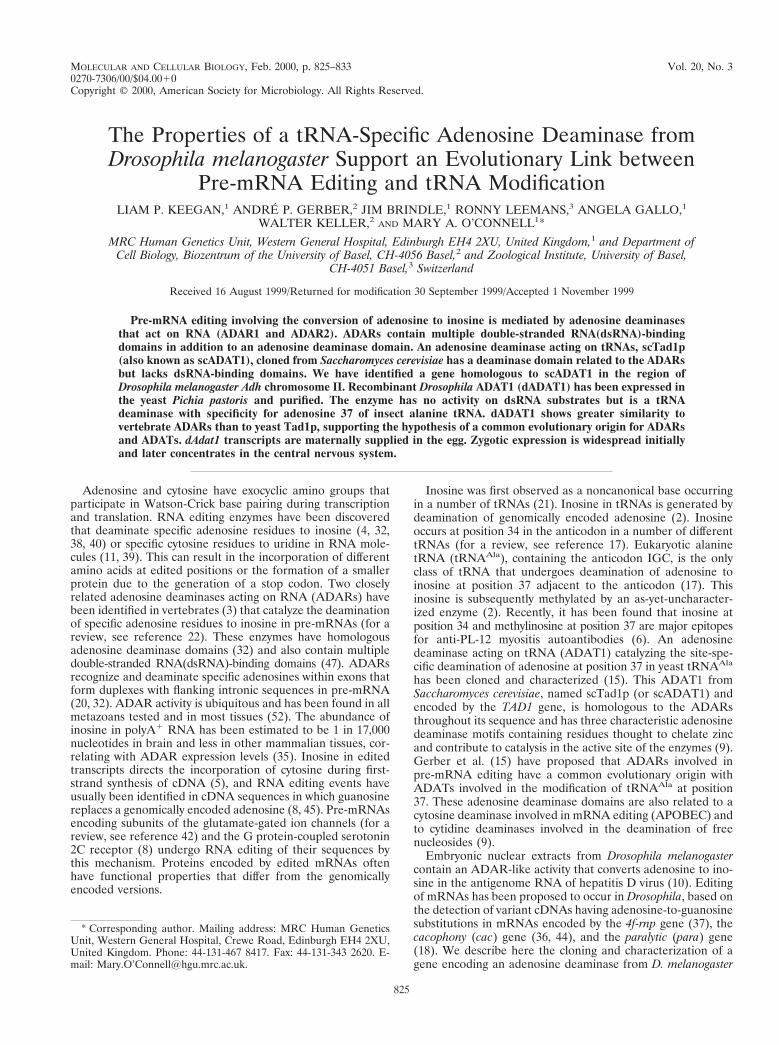
Characterization of a Drosophila gene encoding an adeno-sine deaminase domain. Homology searches with hADAR1identified an ORF encoding an ADAR-like adenosine deami-nase domain in a 3-kb subclone of D. melanogaster P1 genomicclone DS00941 that had been sequenced by the BDGP (Fig. 1).The sequence conservation in the predicted adenosine deami-nase domain included the three motifs containing a histidineand two cysteine residues, proposed to be involved in zinccoordination at the active site, as well as a conserved glutamateresidue in motif I which is also believed to participate in thedeamination reaction (23, 34). The coding sequence inDS00941 did not contain any potential dsRNA-binding domain(47).
The genomic sequence from DS00941 was used to designPCR primers to amplify a 600-bp DNA fragment encoding thisputative adenosine deaminase sequence from genomic DNA(Fig. 1), which was then used to screen embryonic cDNAlibraries. Low-stringency Southern blots with this fragmentindicated that the gene is single copy (results not shown). Onelong cDNA clone (clone 12) and another shorter clone (cloneH) were isolated, confirming that this ORF is part of a tran-scribed sequence.
The P1 clone DS00941 is now fully sequenced by the BDGPand is part of a 2.9-Mb stretch of contiguous sequence in theAdh region on the left arm of Drosophila chromosome II. Thegene encoding the adenosine deaminase domain is the secondpredicted gene product from the centromere distal end of this82-kb P1 clone and is referred to as BG:DS00941.2 (1). Therelationship between the isolated cDNAs and the structure ofthe BG:DS00941.2 transcript as predicted from the genomicsequence by Drosophila GRAIL and Genefinder programs areindicated in Fig. 1, together with the 600-nucleotide fragmentthat was used as a probe to isolate the cDNAs. Comparison ofcDNA sequences with the genomic DNA sequence revealedthat the cDNA clones are interrupted by a short intron of 57 bpwithin the protein coding sequence and by a second shortintron of 54 bp within the 39 untranslated region of the longercDNA clone. Clone H begins at base pair 574 of clone 12 andterminates at a more proximal polyadenylation site. Two dif-ferent polyadenylation sites are used, one located 30 bp afterthe ORF and a second site located 1.8 kb more 39, whichproduces a transcript with a long 39 region in which no addi-tional ORFs can be identified. Both polyadenylation sites arepreceded by a polyadenylation consensus sequence.
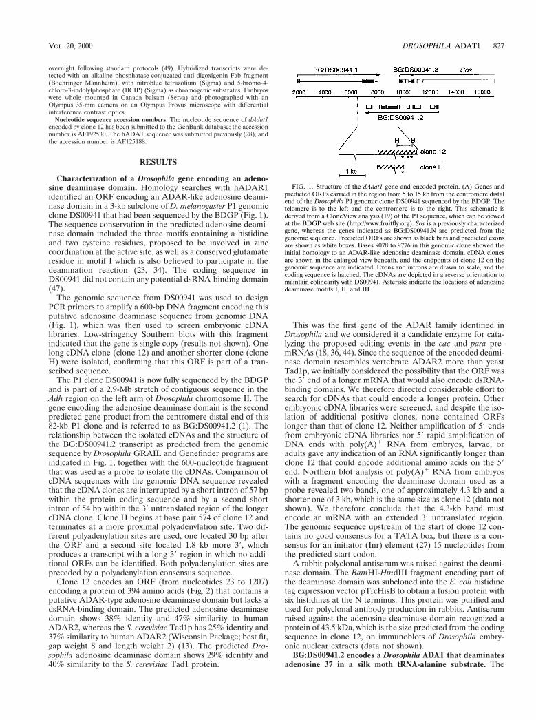
Clone 12 encodes an ORF (from nucleotides 23 to 1207)encoding a protein of 394 amino acids (Fig. 2) that contains aputative ADAR-type adenosine deaminase domain but lacks adsRNA-binding domain. The predicted adenosine deaminasedomain shows 38% identity and 47% similarity to humanADAR2, whereas the S. cerevisiae Tad1p has 25% identity and37% similarity to human ADAR2 (Wisconsin Package; best fit,gap weight 8 and length weight 2) (13). The predicted Dro-sophila adenosine deaminase domain shows 29% identity and40% similarity to the S. cerevisiae Tad1 protein.
This was the first gene of the ADAR family identified inDrosophila and we considered it a candidate enzyme for cata-lyzing the proposed editing events in the cac and para pre-mRNAs (18, 36, 44). Since the sequence of the encoded deami-nase domain resembles vertebrate ADAR2 more than yeastTad1p, we initially considered the possibility that the ORF wasthe 39 end of a longer mRNA that would also encode dsRNA-binding domains. We therefore directed considerable effort tosearch for cDNAs that could encode a longer protein. Otherembryonic cDNA libraries were screened, and despite the iso-lation of additional positive clones, none contained ORFslonger than that of clone 12. Neither amplification of 59 endsfrom embryonic cDNA libraries nor 59 rapid amplification ofDNA ends with poly(A)1 RNA from embryos, larvae, oradults gave any indication of an RNA significantly longer thanclone 12 that could encode additional amino acids on the 59end. Northern blot analysis of poly(A)1 RNA from embryoswith a fragment encoding the deaminase domain used as aprobe revealed two bands, one of approximately 4.3 kb and ashorter one of 3 kb, which is the same size as clone 12 (data notshown). We therefore conclude that the 4.3-kb band mustencode an mRNA with an extended 39 untranslated region.The genomic sequence upstream of the start of clone 12 con-tains no good consensus for a TATA box, but there is a con-sensus for an initiator (Inr) element (27) 15 nucleotides fromthe predicted start codon.
A rabbit polyclonal antiserum was raised against the deami-nase domain. The BamHI-HindIII fragment encoding part ofthe deaminase domain was subcloned into the E. coli histidinetag expression vector pTrcHisB to obtain a fusion protein withsix histidines at the N terminus. This protein was purified andused for polyclonal antibody production in rabbits. Antiserumraised against the adenosine deaminase domain recognized aprotein of 43.5 kDa, which is the size predicted from the codingsequence in clone 12, on immunoblots of Drosophila embry-onic nuclear extracts (data not shown).
BG:DS00941.2 encodes a Drosophila ADAT that deaminatesadenosine 37 in a silk moth tRNA-alanine substrate. The
FIG. 1. Structure of the dAdat1 gene and encoded protein. (A) Genes andpredicted ORFs carried in the region from 5 to 15 kb from the centromere distalend of the Drosophila P1 genomic clone DS00941 sequenced by the BDGP. Thetelomere is to the left and the centromere is to the right. This schematic isderived from a CloneView analysis (19) of the P1 sequence, which can be viewedat the BDGP web site (http://www.fruitfly.org). Sos is a previously characterizedgene, whereas the genes indicated as BG:DS00941.N are predicted from thegenomic sequence. Predicted ORFs are shown as black bars and predicted exonsare shown as white boxes. Bases 9078 to 9776 in this genomic clone showed theinitial homology to an ADAR-like adenosine deaminase domain. cDNA clonesare shown in the enlarged view beneath, and the endpoints of clone 12 on thegenomic sequence are indicated. Exons and introns are drawn to scale, and thecoding sequence is hatched. The cDNAs are depicted in a reverse orientation tomaintain colinearity with DS00941. Asterisks indicate the locations of adenosinedeaminase motifs I, II, and III.
VOL. 20, 2000 DROSOPHILA ADAT1 827
FIG. 2. Multiple sequence alignment showing the Drosophila adenosine deaminase encoded by clone 12, compared to vertebrate ADARs and yeast scTad1p andspTad1p. The adenosine deaminase motifs containing cysteine and histidine residues that are proposed zinc ligands (#) and a glutamate residue (D) that is thought
828 KEEGAN ET AL. MOL. CELL. BIOL.
methylotrophic yeast P. pastoris was used to overexpress theprotein encoded by clone 12 so as to determine its function.Previous work has shown this yeast to be excellent for theproduction of active ADARs and ADATs (15, 16, 33). Thecoding sequence of clone 12 was subcloned into the P. pastorisexpression vector pSK-FLIS6 to express a fusion protein witha FLAG epitope at the amino terminus and six histidine resi-dues at the carboxy terminus. This construct was transformedinto P. pastoris by standard methods. Extracts from trans-formed cells contain a protein with a molecular mass of 48 kDadetectable on immunoblots with a-FLAG M2 antibody. Thismolecular mass is consistent with the length of the codingsequence of clone 12 and the N- and C-terminal epitope tags(results not shown).
Extracts were made from 300-ml cultures grown in methanolmedium. Soluble extract was mixed with Ni21-NTA, pouredinto a column, and eluted with 250 mM imidazole. This eluatewas applied to an affinity column bearing the a-FLAG M2monoclonal antibody, and proteins were eluted with FLAGpeptide. Figure 3A shows SDS-polyacrylamide gel electro-phoresis of fractions from a FLAG affinity column in which theproteins have been stained with Coomassie blue. A protein of48 kDa is present in the eluate fractions, and this protein isrecognized by the antibody raised against the deaminase do-main (Fig. 3B). Approximately 40 mg of pure dADAT1 wasobtained per 300 ml of starting culture.
Peak fractions containing the 48-kDa protein from theFLAG affinity column were tested for adenosine deaminaseactivity on in vitro-synthesized [a-33P]ATP-labelled tRNAAla
from the insect B. mori (2, 46, 51) or from S. cerevisiae.scTad1p has been shown to specifically deaminate the adeno-sine at position 37 in yeast and B. mori tRNAAla (15). Therecombinant 48-kDa Drosophila protein converts adenosine toinosine in tRNAAla from B. mori with a specific activity of 8.4U/mg (Fig. 4A, lane 5). Mutated B. mori tRNAAla in which theadenosine at position 34 has been changed to guanosine hasthe same level of adenosine-to-inosine conversion as the wild-type substrate (lane 6). However, a change from adenosine toguanosine at position 37 (lane 7) or a double change of cyto-sine 36 to uridine and adenosine 37 to guanosine (lane 8)eliminates all adenosine-to-inosine conversion in the BombyxtRNA substrate, indicating that it is adenosine 37 and notadenosine 34 that is deaminated. In contrast, yeast tRNAAla isnot an efficient substrate for the Drosophila enzyme that has aspecific activity of 0.08 U/mg with this substrate (Fig. 4A, lane3). The location of the inosine formed in the tRNAAla sub-strates was confirmed by performing reverse transcription PCRon the reaction products and sequencing the resulting cDNAs(data not shown). Therefore, the Drosophila clone encodes aprotein that is clearly a functional homolog of scTad1p, and wehave named it Drosophila ADAT1 (dADAT1).
The Drosophila enzyme cannot bind efficiently to the yeasttRNAAla. UV cross-linking of dADAT1 to [a-33P]ATP-labelledtRNAAla substrates from B. mori or yeast confirm thatdADAT1 binds much more efficiently to the tRNA from B.mori (Fig. 4B, lanes 1 and 3). Surprisingly, dADAT1 also bindsto a Bombyx tRNA substrate in which positions 36 and 37 are
mutated (Fig. 4B, lane 2) although less tightly than to the wildtype. The scTad1 protein deaminates both the yeast tRNA(Fig. 4A) and the B. mori tRNA efficiently (15), and UVcross-linking experiments confirm that scTad1p binds effi-ciently to both tRNAAla from B. mori (Fig. 4B, lane 7) andfrom yeast (lane 9).
We also tested whether purified recombinant dADAT1could deaminate adenosine to inosine on extended dsRNA,but no conversion was observed (results not shown), suggestingthat dADAT1 has no role in pre-mRNA editing.
Expression of dAdat1 transcripts in Drosophila embryos.Digoxigenin-labelled sense or antisense dAdat1 RNA probesderived from clone 12 were hybridized to Drosophila embryos.As expected, a signal could be visualized only with the anti-sense dAdat1 probe. The expression patterns observed withantisense probes from clone H and clone 12 were identical, andthose obtained with clone 12 are shown in Fig. 5. A strongoverall staining was detected at the blastoderm stage (Fig. 5A),suggesting that dAdat1 mRNA is maternally provided, since nozygotic transcripts are expressed at this early stage. Zygoticexpression was very strong during germ band extension, espe-cially in the mesoderm and neuroectoderm (Fig. 5B). Fromgerm band retraction onwards, high levels of transcripts wereconfined to the central nervous system and transcript levelsremained high in the entire brain and ventral nerve cordthroughout late embryogenesis (Fig. 5C).
Isolation of a human ADAT gene and comparison with Dro-sophila Adat1 reveals metazoan ADATs with adenosine deami-nase domains surprisingly similar to ADARs involved in pre-mRNA editing. Database searches with S. cerevisiae ADAT1identified a partial sequence of a potential human homolog inthe Washington University EST database. The clone fromwhich this sequence derived was obtained, and additional se-quence information for the amino-terminal half of this proteinwas determined (see Materials and Methods). The resultingsequence revealed an ORF with homology over its entire cod-ing sequence to Drosophila Adat1, which is not surprising con-sidering that tRNAAla species from B. mori and humans arevery similar (6). While this paper was in preparation, Maas etal. (28) reported the characterization of the human ADAT1and showed that this protein deaminates adenosine 37 of hu-man tRNAAla. The amino acid sequence of this human ADATis included in the multiple sequence alignment shown in Fig. 2.Both human and Drosophila ADATs are more similar to aden-osine deaminase domains of vertebrate ADARs than to theyeast ADATs.
To examine phylogenetic relationships among adenosinedeaminase domains, ADAR and ADAT sequences were re-covered from databases and multiple sequence alignmentswere performed with the Wisconsin Genetics Computer Group(GCG) Pileup program (13). The most consistent alignmentsinvolve residues close to the histidine and cysteine residues ofdeaminase motifs I, II, and III (Fig. 2). All ADAT proteins areapproximately the same size and there is homology among theproteins over the full length of the sequences that is observedwhen lower gap insertion and extension penalties are used inthe Pileup program. Aligned sequences were truncated to re-
to be involved in proton transfer in the deaminase active site are indicated. A potential RNA recognition region flanking the third cysteine of the deaminase motif isunderlined. The human ADAT (hADAT) has 93 residues more than dADAT1 between deaminase motifs II and III, and this number of amino acids has been removedfrom hADAT between the sequences EDQP and SAKV after residue 157 (p). The DDBJ, EMBL, and GenBank accession numbers of the sequences are as follows:human hADAR1, U10439; human hADAR2a (a shorter protein lacking an Alu element inserted between deaminase motifs II and III), U82120; S. cerevisiae scTad1p,AJ007297; S. pombe spTad1p, AL021748; dADAT1, AF192530; and hADAT, AF125188. The alignment was generated with the Wisconsin GCG Pileup program witha gap penalty of 3 and a gap extension penalty of 1 and edited by using Lineup. The order in which sequences are presented is determined by the program accordingto their similarity to each other.
VOL. 20, 2000 DROSOPHILA ADAT1 829

move dsRNA-binding domain sequences from the ADARgenes, and multiple sequence alignments of adenosine deami-nase domains were used to generate phylogenetic trees withthe PAUP (phylogenetic analysis using parsimony) (48) pro-gram in the Wisconsin GCG program package (13).
Many adenosine deaminase domain trees generated fromPileup alignments made with different gap insertion and ex-tension penalties have topologies similar to that shown in Fig.6. Human ADAT and Drosophila ADAT1 are closely relatedand their relationship to S. cerevisiae Tad1p and Schizosaccha-romyces pombe Tad1p pair is distant. The ADAR genes arealways found to branch from the ADAT line. Among theADARs, human ADAR2 and the putative Drosophila ADARN35H14 (accession no. AL035207) are closely related, whilethe branching order of the other two ADARs is less consistent.In order to obtain the best possible alignment with fewer gapsand with aligned residues representing true homologies, a re-gion around the conserved deaminase motifs was selected andaligned more stringently to produce the tree shown in Fig. 6.
DISCUSSION
An enzymatic activity capable of converting adenosine toinosine in dsRNA is present in D. melanogaster (10). To find agene encoding this activity, we performed a homology searchwith hADAR1. An ORF was found that had high sequencehomology to the deaminase domain of hADAR1. We isolatedthis gene and showed that it encoded an adenosine deaminasethat specifically converts adenosine to inosine at position 37,adjacent to the anticodon in B. mori tRNAAla(IGC). We havenamed this gene dAdat1 because of its homology to the yeastscADAT1.
The dAdat1 gene is located 2 to 7 kb centromere distally toSon of sevenless (Sos) (7, 43) in the Adh region on the left armof chromosome II. It is within a 2.9-Mb stretch of contiguoussequence that has been correlated with extensive sets of dele-tions and point mutations (1). dAdat1 is flanked centromeredistally by a predicted carbonate dehydratase gene (BG:
DS00941.1) and centromere proximally by a predicted protea-some subunit gene (BG:DS00941.3). The predicted carbonatedehydratase coding sequence is fully contained within the 39untranslated region of the longer dAdat1 cDNA clone 12.dAdat1 and the predicted proteasome subunit gene are bothpresent on genomic fragments that contained Sos and 10 kb ofupstream DNA that was transformed into Drosophila to iden-tify the correct mRNA for Sos by genetic rescue (7, 43).
Drosophila ADAT1 binds and deaminates tRNAAla from B.mori 100-fold more efficiently than the tRNAAla from S. cer-evisiae (data not shown). The yeast Tad1p deaminates A37more effectively in yeast tRNAAla than in the tRNAAla from B.mori (15). Each ADAT shows preference for tRNAAla fromthe same or more closely related organisms.
The tRNAAla from B. mori (Fig. 3C) that was used as asubstrate for dADAT1 in this study is a tRNA expressed spe-cifically in the silk moth salivary gland, where alanine is incor-porated into the silk fibroin protein (46, 51). Wild-type andmutant forms of this tRNA substrate have been used in earlierstudies of adenosine-to-inosine conversion and other modifi-cations in tRNAAla (2, 17). This salivary gland-specifictRNAAla differs by one base at position 40 in the anticodonarm from the constitutive silk moth tRNAAla, which also hasan inosine at position 37 (Fig. 4C). One gene encoding atRNAAla has been characterized in Drosophila (12), but it isnot known whether some Drosophila tRNAs have tissue-spe-cific expression similar to that found for the silk moth tRNAAla
genes (46). It is not known if the conversion of adenosine toinosine at position 37 in tRNAAla is always complete, and thedegree of conversion could vary in different tissues.
The significance of adenosine-to-inosine conversion adja-cent to the anticodon in tRNAAla(IGC) is not known. Yeasttad1 mutants are viable and contain unmodified adenosine 37in their tRNAAla species (15). No deletion in dAdat1 is avail-able that only removes dAdat1 and not neighboring genes.Almost all the lethal mutations that have been found near Soshave been assigned to genes that were previously known orthat have been identified in the genomic sequence (1). None ofthe known lethal mutations are in dAdat1. The effect of loss offunction in Drosophila Adat1 therefore cannot yet be assessed.
Transcripts of dAdat1 are produced by nurse cells andloaded into the egg before fertilization (Fig. 5). Many mRNAsencoding proteins that will be required for transcription andtranslation during the early stages of embryonic developmentare provided in this way. Zygotic cells throughout the embryocontain dAdat1 mRNA in germ band-extended embryos, butmost of the expression in later germ band-retracted embryos isin the central nervous system. Much of the mesoderm andectoderm is larval tissue that will be histolyzed at metamor-phosis; thus, it is possible that early dAdat1 expression is suf-ficient for the remaining lifetime of these cells. The high levelof expression in the nervous system is intriguing. It is possiblethat dAdat1 activity is ubiquitous, even though the transcriptexpression pattern observed by in situ hybridization is concen-trated in the nervous system. Considering the high degree ofhomology between dADAT1 and the ADAR enzymes, it iscurious that the embryonic expression of dAdat1 is found in thecentral nervous system and is reminiscent of rRED2 expres-sion, which is confined to the brain (31).
It is surprising that Drosophila ADAT1 shows a greatersimilarity to ADARs involved in pre-mRNA editing than tothe yeast Tad1 proteins, considering that these proteins havevery different substrates (Fig. 2). Based on functional group-ings, dADAT1 might have been expected to resemble S. cer-evisiae Tad1p more than vertebrate ADARs. A human ADATgene also exhibits similarity to dAdat1 and to vertebrate
FIG. 3. Purification of recombinant Drosophila adenosine deaminase from P.pastoris. (A) Fractions from a FLAG affinity column eluted with a FLAG peptidewere electrophoresed on an SDS–10% polyacrylamide gel, and proteins werestained with Coomassie blue. L, load; FT, flowthrough, W, wash. Molecularmasses are indicated on the left. (B) Immunoblot analysis of fractions from theFLAG affinity column with antiserum raised against Drosophila ADAT1. Mo-lecular masses are indicated on the left. Numbers at the top of the lanes areeluate fraction numbers; numbers at the bottom are lane numbers.
830 KEEGAN ET AL. MOL. CELL. BIOL.

ADARs. It is possible that this similarity between dADAT1and ADARs reflects a role for dADAT1 in editing pre-mRNA.However, such an activity is unlikely, since recombinantdADAT1 protein cannot convert adenosine to inosine indsRNA substrates. An ADAR-like gene (N35H14) with twodsRNA-binding domains has been sequenced by the EuropeanDrosophila Genome Project and independently cloned fromDrosophila (R. Reenan, personal communication). The proteinencoded by this gene converts adenosine to inosine in dsRNAsubstrates (M. O’Connell and R. Reenan, unpublished results)and is a strong candidate for catalyzing editing in the cac andpara pre-mRNAs (18, 36, 44).
One major question is how the ADAR family of enzymesrecognize their target adenosines in pre-mRNA. The dsRNA-binding domains show little sequence specificity, recognizingcontinuous dsRNA structures (41). With the emergence of thehomologous ADAT family of enzymes having a deaminasedomain that binds to specific tRNA, it seems probable that theadenosine deaminase domain of ADARs contributes morethan was previously anticipated to RNA binding and hence tosubstrate recognition. It has previously been shown that aden-osine deaminase domains alone are not sufficient for editing byADARs in vitro (25, 29), but it has not been determined if thedeaminase domain alone can bind to RNA. A domain ofADAR corresponding to the region of ADAT homology cannow be expressed in recombinant form. Detailed RNA-bindingstudies can then be performed in vitro to determine if indeedthe deaminase domain alone can bind to pre-mRNA substratesand contribute to the target specificity of these enzymes.
ADATs are likely to have a higher affinity for RNA sub-strates than an isolated ADAR adenosine deaminase domain,since ADARs probably depend on their dsRNA-binding do-mains for RNA affinity. It is interesting therefore to compareADAR and ADAT sequences around the putative zinc-chelat-ing residues. The number of residues between deaminase mo-tifs I and II is well conserved, whereas the number of residuesbetween deaminase motifs II and III differs (22). Structuraladjustments near deaminase motif III could be associated withchanges in the target specificity of adenosine deaminase do-mains. A cluster of positively charged residues is well con-served between dADAT1, hADAT1, and the yeast ADATs inthe region amino terminal to deaminase motif III (Fig. 2) (28).This cluster of positively charged residues could be a region ofthe protein that contacts RNA.
This region of sequence similarity among ADATs has alsobeen noted by Maas et al. (28) in an alignment in which allhADAT residues between deaminase motifs II and III wereincluded. Due to the variability in the number of residuesbetween motifs II and III, sequence alignments that includethis region can have different gaps and therefore highlightdifferent blocks of homology. In addition to the ADAT signa-ture sequence near deaminase motif III, Maas et al. (28) ob-served an ADAR specific signature sequence 19 residuesamino terminal to the ADAT signature sequence. However,
FIG. 4. Recombinant Drosophila adenosine deaminase binds to and modifiestRNAAla. (A) tRNA-specific adenosine deaminase assay with peak fractionsfrom an a-FLAG M2 antibody column. Different tRNAAla species were labelledwith [a-33P]ATP. The extracts used were recombinant scTad1p (lanes 1 and 2)and recombinant dADAT1 (lanes 3 to 8). The substrates used are indicatedabove each lane. Abbreviations: y, yeast; B.m., B. mori; wt, wild-type tRNAAla;
A34G, G34 instead of A34; A37G, G37 instead of A37; CA3637UG, substrate hasa double change of C36 to U36 and A37 to G37. (B) UV cross-linking of dADAT1and scTad1p to [a-33P]ATP-labelled tRNA. The reaction mixtures were irradi-ated, digested with RNase A, and electrophoresed on an SDS–12% polyacryl-amide gel. Lanes 1 to 5 each contained 45 ng of recombinant dADAT1, and lanes7 to 11 each contained 100 ng of recombinant scTad1p. Lane 6 had no protein.The abbreviations for the substrates are the same as in panel A. Ser, tRNASer;mut5, yeast tRNAAsp with tRNAArg anticodon loop (2). (C) Sequence differ-ences between tRNAAla from yeast and B. mori. The 19-base differences betweenthe two tRNAAla species are indicated. The silk gland-specific tRNAAla from B.mori contains U instead of C at position 40.
VOL. 20, 2000 DROSOPHILA ADAT1 831

visual inspection of many different computer-generated align-ments suggests that this ADAR-specific signature could be avariant of the ADAT signature sequence which has beenmoved 19 residues further from deaminase motif III. Residues178 to 189 of hADAR1 (LRTKvenGEgTi) (Fig. 2), which areconserved among ADARs, resemble residues 176 to 187 ofdADAT1 (LRTKpgrGErT1), which are conserved amongADATs. It is possible that the evolution of ADATs to ADARsinvolved an insertion close to motif III that could have affectedtarget sequence recognition.
It is possible that an ADAT signature sequence is only vis-ible close to the active site of the enzyme, as other tRNA-protein contacts may involve multiple weakly conserved sitesover the entire protein and therefore be difficult to recognize.One or more of the 19 differences between tRNAAla from S.cerevisiae and B. mori have a more significant effect on theactivity of dADAT1 than on Tad1p, suggesting that these pro-teins may not make all RNA contacts in precisely the samemanner.
Pre-mRNA editing may have evolved when an originalADAT acquired dsRNA-binding domains and a new set oftargets in pre-mRNA. An interesting interpretation of se-quence homologies and of the phylogenetic tree shown in Fig.6 would be the following. (i) Tad1p does not encode an essen-tial function in yeast, and the sequence conservation withmetazoan ADATs is correspondingly low and focused on thezinc-chelating residues in the deaminase domain. (ii) ADARsevolved from ADATs after the divergence of fungal and meta-zoan lines. (iii) Double-stranded RNA-binding domains mayhave been added to an ADAT gene after a gene duplicationevent, and such a protein might have retained ADAT functionfor a period until it evolved to recognize a more ADAR-likerange of RNA targets. Other evolutionary scenarios can beenvisaged, however; Drosophila and human ADAT genes ap-pear to be more closely related than yeast TAD1 to a precursorgene that gave rise to the ADAR family. Studies on the rec-ognition of tRNA substrates by Drosophila or human ADAT1should help to explain how pre-mRNA editing evolved andhow target sequences are recognized in pre-mRNA editing.
ACKNOWLEDGMENTS
We are grateful to Hyouta Himeno, Hirosaki University, for yeasttRNA-Ser; to H. Grosjean for tRNA substrates; to M. Affolter and A.Jarman for cDNA libraries; and to R. Reenan for sharing unpublishedresults.
This work was supported by the Medical Research Council, theUniversity of Basel, the Swiss National Science Foundation, and theLouis-Jeantet Foundation for Medicine. A.P.G. was the recipient of apredoctoral fellowship from the Boehringer Ingelheim Fonds, andR.L. was supported by a grant from the European Union (via theBundesamt fur Bildung und Wissenschaft, Bern, Switzerland).
REFERENCES1. Ashburner, M., S. Misra, J. Roote, S. E. Lewis, R. Blazej, T. Davis, C. Doyle,
R. Galle, R. George, N. Harris, G. Hartzell, D. Harvey, L. Hong, K. Houston,R. Hoskins, G. Johnson, C. Martin, A. Moshrefi, M. Palazzolo, M. G. Reese,A. Spradling, G. Tsang, K. Wan, K. Whitelaw, B. Kimmel, et al. 1999. Anexploration of the sequence of a 2.9-Mb region of the genome of Drosophilamelanogaster. The Adh region. Genetics 153:179–219.
2. Auxilien, S., P. F. Crain, R. W. Trewyn, and H. Grosjean. 1996. Mechanism,specificity and general properties of the yeast enzyme catalysing the forma-tion of inosine 34 in the anticodon of transfer RNA. J. Mol. Biol. 262:437–458.
3. Bass, B. L., K. Nishikura, W. Keller, P. H. Seeburg, R. B. Emeson, M. A.O’Connell, C. E. Samuel, and A. Herbert. 1997. A standardized nomencla-ture for adenosine deaminases that act on RNA. RNA 3:947–949.
4. Bass, B. L., and H. Weintraub. 1987. A developmentally regulated activitythat unwinds RNA duplexes. Cell 48:607–613.
5. Bass, B. L., H. Weintraub, R. Cattaneo, and M. A. Billeter. 1989. Biasedhypermutation of viral RNA genomes could be due to unwinding/modifica-tion of double-stranded RNA. Cell 56:331.
6. Becker, H. F., Y. Corda, M. B. Mathews, J. L. Fourrey, and H. Grosjean.
FIG. 5. Expression of dAdat1 transcripts in Drosophila embryos. Embryoswere hybridized with digoxigenin-labelled antisense probe to dADAT1 clone 12.(A) Embryo at cellular blastoderm formation, showing maternally supplied tran-script. (B) Ventral view of an embryo during germ band retraction, showingwidespread expression of dAdat1 transcripts in mesoderm, ectoderm, ventralnerve cord, and supraesophageal ganglion. (C) Lateral view of a later-stageembryo, showing expression confined to the ventral nerve cord and supraesopha-geal ganglion. (D) Lateral view of late embryo hybridized with sense probe. Allembryos have their anterior to the left, and photographs were taken at 360magnification.
FIG. 6. Phylogenetic tree of ADAR and ADAT adenosine deaminase do-main sequences. The sequence region from the TGXKC block before deaminasemotif I to the PIYL conservation after deaminase motif III (see Fig. 2) was takenfor each sequence and used to generate a more-stringent Pileup alignment (gapinsertion penalty, 8; extension penalty, 2). This alignment was used to generatethe tree and bootstrap values shown with PAUP in the Wisconsin GCG softwarepackage (48). GenBank database accession numbers for sequences used here areas follows: Drosophila ADAR sequence N35H14, AL035207; and C. elegansADAR sequence T20H4.4, Q22618.
832 KEEGAN ET AL. MOL. CELL. BIOL.

1999. Inosine and N1-methylinosine within a synthetic oligomer mimickingthe anticodon loop of human tRNA(Ala) are major epitopes for anti-PL-12myositis autoantibodies. RNA 5:865–875.
7. Bonfini, L., C. A. Karlovich, C. Dasgupta, and U. Banerjee. 1992. The Son ofsevenless gene product: a putative activator of Ras. Science 255:603–606.
8. Burns, C. M., H. Chu, S. M. Rueter, L. K. Hutchinson, H. Canton, E.Sanders-Bush, and R. B. Emeson. 1997. Regulation of serotonin-2C receptorG-protein coupling by RNA editing. Nature 387:303–308.
9. Carter, C. W. 1998. Nucleoside deaminases for cytidine and adenosine:comparison with deaminases acting on RNA, p. 363–375. In H. Grosjean andR. Benne (ed.), Modification and editing of RNA. ASM Press, Washington,D.C.
10. Casey, J. L., and J. L. Gerin. 1995. Hepatitis D virus RNA editing: specificmodification of adenosine in the antigenomic RNA. J. Virol. 69:7593–7600.
11. Chen, S. H., G. Habib, C. Y. Yang, Z. W. Gu, B. R. Lee, S. A. Weng, S. R.Silberman, S. J. Cai, J. P. Deslypere, M. Rosseneu, A. M. J. Gotto, W. H. Li,and L. Chan. 1987. Apolipoprotein B-48 is the product of a messenger RNAwith an organ-specific in frame stop codon. Science 238:363–366.
12. DeLotto, R., and P. Schedl. 1984. A Drosophila melanogaster transfer RNAgene cluster at the cytogenetic locus 90BC. J. Mol. Biol. 179:587–605.
13. Devereux, J., P. Haeberli, and O. Smithies. 1984. A comprehensive set ofsequence analysis programs for the VAX. Nucleic Acids Res. 12:387–395.
14. Frohmann, M. A., M. K. Dush, and G. R. Martin. 1988. Rapid production offull-length cDNAs from rare transcripts: amplification using a single gene-specific oligonucleotide primer. Proc. Natl. Acad. Sci. USA 85:8998–9002.
15. Gerber, A., H. Grosjean, T. Melcher, and W. Keller. 1998. Tad1p, a yeasttRNA-specific adenosine deaminase, is related to the mammalian pre-mRNA editing enzymes ADAR1 and ADAR2. EMBO J. 17:4780–4789.
16. Gerber, A., M. A. O’Connell, and W. Keller. 1997. Two forms of humandouble-stranded RNA-specific editase 1 (hRED1) generated by the insertionof an Alu cassette. RNA 3:453–463.
17. Grosjean, H., S. Auxilien, F. Constantinesco, C. Simon, Y. Corda, H. F.Becker, D. Foiret, A. Morin, Y. X. Jin, M. Fournier, and J. L. Fourrey. 1996.Enzymatic conversion of adenosine to inosine and to N1-methylinosine intransfer RNAs: a review. Biochimie 78:488–501.
18. Hanrahan, C. J., M. J. Palladino, L. J. Bonneau, and R. A. Reenan. 1998.RNA editing of a Drosophila sodium channel gene. Ann. N. Y. Acad. Sci.868:51–66.
19. Helt, G. A., S. Lewis, A. E. Loraine, and G. M. Rubin. 1998. BioViews:Java-based tools for genomic data visualization. Genome Res. 8:291–305.
20. Higuchi, M., F. N. Single, M. Kohler, B. Sommer, R. Sprengel, and P. H.Seeburg. 1993. RNA editing of AMPA receptor subunit GluR-B: a base-paired intron-exon structure determines position and efficiency. Cell 75:1361–1370.
21. Holley, R. W., G. A. Everett, J. T. Madison, and A. Zamir. 1965. Nucleotidesequences in yeast alanine transfer RNA. J. Biol. Chem. 240:2122–2127.
22. Keller, W., J. Wolf, and A. Gerber. 1999. Editing of messenger RNA pre-cursors and of tRNAs by adenosine to inosine conversion. FEBS Lett. 452:71–76.
23. Kim, U., Y. Wang, T. Sanford, Y. Zeng, and K. Nishikura. 1994. Molecularcloning of cDNAs for double-stranded RNA adenosine deaminase, a candi-date enzyme for nuclear RNA editing. Proc. Natl. Acad. Sci. USA 91:11457–11461.
24. Laemmli, U. K. 1970. Cleavage of structural proteins during the assembly ofthe head of the bacteriophage T4. Nature 227:680–685.
25. Lai, F., R. Drakas, and K. Nishikura. 1995. Mutagenic analysis of double-stranded RNA adenosine deaminase, a candidate enzyme for RNA editingof glutamate-gated ion channel transcripts. J. Biol. Chem. 270:17098–17105.
26. Lennon, G. G., C. Auffray, M. Polymeropoulos, and M. B. Soares. 1996. TheI. M. A. G. E. consortium: an integrated molecular analysis of genomes andtheir expression. Genomics 33:151–152.
27. Lo, K., and S. T. Smale. 1996. Generality of a functional initiator consensussequence. Gene 182:13–22.
28. Maas, S., A. P. Gerber, and A. Rich. 1999. Identification and characterizationof a human tRNA-specific adenosine deaminase related to the ADAR familyof pre-mRNA editing enzymes. Proc. Natl. Acad. Sci. USA 96:8895–9000.
29. Maas, S., T. Melcher, A. Herb, P. H. Seeburg, W. Keller, S. Krause, M.Higuchi, and M. A. O’Connell. 1996. Structural requirements for RNAediting in glutamate receptor pre-mRNA by recombinant double-strandedRNA adenosine deaminase. J. Biol. Chem. 271:12221–12226.
30. Martin, G., and W. Keller. 1996. Mutational analysis of mammalian poly(A)
polymerase identifies a region for primer binding and a catalytic domain,homologous to the family X polymerases, and to other nucleotidyltrans-ferases. EMBO J. 15:2593–2603.
31. Melcher, T., S. Maas, A. Herb, R. Sprengel, M. Higuchi, and P. H. Seeburg.1996. RED2, a brain specific member of the RNA-specific adenosine deami-nase family. J. Biol. Chem. 271:31795–31798.
32. Melcher, T., S. Maas, A. Herb, R. Sprengel, P. H. Seeburg, and M. Higuchi.1996. A mammalian RNA editing enzyme. Nature 379:460–464.
33. O’Connell, M. A., A. Gerber, and L. P. Keegan. 1998. Purification of nativeand recombinant double-stranded RNA-specific adenosine deaminases.Methods (Orlando) 15:51–62.
34. O’Connell, M. A., S. Krause, M. Higuchi, J. J. Hsuan, N. F. Totty, A. Jenny,and W. Keller. 1995. Cloning of cDNAs encoding mammalian double-stranded RNA-specific adenosine deaminase. Mol. Cell. Biol. 15:1389–1397.
35. Paul, M., and B. L. Bass. 1998. Inosine exists in mRNA at tissue-specificlevels and is most abundant in brain mRNA. EMBO J. 17:1120–1127.
36. Peixoto, A. A., L. A. Smith, and J. C. Hall. 1997. Genomic organization andevolution of alternative exons in a Drosophila calcium channel gene. Genet-ics 145:1003–1013.
37. Petschek, J. P., M. J. Mermer, M. R. Scheckelhoff, A. A. Simone, and J. C.Vaughn. 1996. RNA editing in Drosophila 4f-rnp gene nuclear transcripts bymultiple A-to-G conversions. J. Mol. Biol. 259:885–890.
38. Polson, A. G., P. F. Crain, S. C. Pomerantz, J. A. McCloskey, and B. L. Bass.1991. The mechanism of adenosine to inosine conversion by the double-stranded RNA unwinding/modifying activity: a high-performance liquidchromatography-mass spectrometry analysis. Biochemistry 30:11507–11514.
39. Powell, L. M., S. C. Wallis, R. J. Pease, Y. H. Edwards, T. J. Knott, and J.Scott. 1987. A novel form of tissue-specific RNA processing produces apo-lipoprotein-48 in intestine. Cell 50:831–840.
40. Rebagliati, M. R., and D. A. Melton. 1987. Antisense RNA injections infertilized frog eggs reveal an RNA duplex unwinding activity. Cell 48:599–605.
41. Ryter, J. M., and S. C. Schultz. 1998. Molecular basis of double-strandedRNA-protein interactions: structure of a dsRNA-binding domain complexedwith dsRNA. EMBO J. 17:7505–7513.
42. Seeburg, P. H. 1996. The role of RNA editing in controlling glutamatereceptor channel properties. J. Neurochem. 66:1–5.
43. Simon, M. A., D. D. Bowtell, G. S. Dodson, T. R. Laverty, and G. M. Rubin.1991. Ras1 and a putative guanine nucleotide exchange factor performcrucial steps in signalling by the sevenless protein tyrosine kinase. Cell67:701–716.
44. Smith, L. A., X. J. Wang, A. A. Peixoto, E. K. Neumann, L. M. Hall, and J. C.Hall. 1996. A Drosophila calcium channel a1 subunit gene maps to a geneticlocus associated with behavioural and visual defects. J. Neurosci. 16:7868–7879.
45. Sommer, B., M. Kohler, R. Sprengel, and P. H. Seeburg. 1991. RNA editingin brain controls a determinant of ion flow in glutamate-gated channels. Cell67:11–19.
46. Sprague, K. U., O. Hagenbuchle, and M. C. Zuniga. 1977. The nucleotidesequence of two silk gland alanine tRNAs: implications for fibroin synthesisand for initiator tRNA structure. Cell 11:561–570.
47. St. Johnston, D., N. H. Brown, J. G. Gall, and M. Jantsch. 1992. A conserveddouble-stranded RNA-binding domain. Proc. Natl. Acad. Sci. USA89:10979–10983.
48. Swofford, D. L. 1993. PAUP: phylogenetic analysis using parsimony. IllinoisNatural History Survey, Champaign, Ill.
49. Tautz, D., and C. Pfeifle. 1989. A non-radioactive in situ hybridizationmethod for the localization of specific RNAs in Drosophila embryos revealstranslational control of the segmentation gene hunchback. Chromosoma98:81–85.
50. Ullrich, A., C. H. Berman, T. J. Dull, A. Gray, and J. M. Lee. 1984. Isolationof the human insulin-like growth factor 1 gene using a single synthetic DNAprobe. EMBO J. 3:361–364.
51. Underwood, D. C., H. Knickerbocker, G. Gardner, D. P. Condliffe, and K. U.Sprague. 1988. Silk gland-specific tRNA(Ala) genes are tightly clustered inthe silkworm genome. Mol. Cell. Biol. 8:5504–5512.
52. Wagner, R. W., C. Yoo, L. Wrabetz, J. Kamholz, J. Buchhalter, N. F. Hassan,K. Khalili, S. U. Kim, B. Perussia, F. A. McMorris, and K. Nishikura. 1990.Double-stranded RNA unwinding and modifying activity is detected ubiqui-tously in primary tissues and cell lines. Mol. Cell. Biol. 10:5586–5590.
VOL. 20, 2000 DROSOPHILA ADAT1 833
Related Documents