The predicted structure of the headpiece of the Huntingtin protein and its implications on Huntingtin aggregation Nicholas W. Kelley 1 , Xuhui Huang 2 , Stephen Tam 1 , Christoph Spiess 3 , Judith Frydman 3 , and Vijay S. Pande *,4 1Biophysics Program, Stanford University, Stanford, CA 94305 2Department of Bioengineering, Stanford University, Stanford, CA 94305 3Department of Biological Sciences, Stanford University, Stanford, CA 94305 4Department of Chemistry, Stanford University, Stanford, CA 94305 Abstract We have performed Simulated Tempering (ST) molecular dynamics simulations to study the thermodynamics of the headpiece of the Huntingtin protein (N17 Htt ). With converged sampling, we found this peptide is highly helical, as previously proposed. Interestingly, this peptide is also found to adopt two different and seemingly stable states. The region from residue 4 (L) to residue 9 (K) has a strong helicity from our simulations, which is supported by experimental studies. However, contrary to what was initially proposed, we have found simulations predict the most populated state as a two helix bundle rather than that of a single straight helix, though a significant percentage of structures do still adopt a single linear helix. The fact that Huntingtin aggregation is nucleation dependent infers the importance of a critical transition. It has been shown that N17 Htt is involved in this rate-limiting step. In this study, we propose two possible mechanisms for this nucleating event stemming from the transition between two helix bundle state and single helix state for N17 Htt , and the experimentally observed interactions between the N17 Htt and polyQ domains. More strikingly, an extensive hydrophobic surface area is found to be exposed to solvent in the dominant monomeric state of N17 Htt . We propose the most fundamental role played by N17 Htt would be initializing the dimerization and pulling the polyQ chains into adequate spatial proximity for the nucleation event to proceed. Introduction Huntington's disease is a neurodegenerative disorder associated with protein misfolding. Specifically, it is caused by a tri-nucleotide repeat expansion for polyGln in the first exon of the Huntingtin protein on chromosome 4 1; 2 ). The pathological range for Huntington's disease is 37-122 repeats, which form high molecular weight protein aggregates. 3; 4 Typical of the amyloid proteins, polyGln tends to have high aggregation propensity and relatively unstable intermediates. This presents many experimental difficulties when using experimental methods to determine structural properties and has lead to a diversity of opinions *Correspondents should be addressed to: E-mail: [email protected]. Publisher's Disclaimer: This is a PDF file of an unedited manuscript that has been accepted for publication. As a service to our customers we are providing this early version of the manuscript. The manuscript will undergo copyediting, typesetting, and review of the resulting proof before it is published in its final citable form. Please note that during the production process errors may be discovered which could affect the content, and all legal disclaimers that apply to the journal pertain. NIH Public Access Author Manuscript J Mol Biol. Author manuscript; available in PMC 2010 May 22. Published in final edited form as: J Mol Biol. 2009 May 22; 388(5): 919–927. doi:10.1016/j.jmb.2009.01.032. NIH-PA Author Manuscript NIH-PA Author Manuscript NIH-PA Author Manuscript

Welcome message from author
This document is posted to help you gain knowledge. Please leave a comment to let me know what you think about it! Share it to your friends and learn new things together.
Transcript
The predicted structure of the headpiece of the Huntingtin proteinand its implications on Huntingtin aggregation
Nicholas W. Kelley1, Xuhui Huang2, Stephen Tam1, Christoph Spiess3, Judith Frydman3,and Vijay S. Pande*,4
1Biophysics Program, Stanford University, Stanford, CA 94305
2Department of Bioengineering, Stanford University, Stanford, CA 94305
3Department of Biological Sciences, Stanford University, Stanford, CA 94305
4Department of Chemistry, Stanford University, Stanford, CA 94305
AbstractWe have performed Simulated Tempering (ST) molecular dynamics simulations to study thethermodynamics of the headpiece of the Huntingtin protein (N17Htt). With converged sampling, wefound this peptide is highly helical, as previously proposed. Interestingly, this peptide is also foundto adopt two different and seemingly stable states. The region from residue 4 (L) to residue 9 (K)has a strong helicity from our simulations, which is supported by experimental studies. However,contrary to what was initially proposed, we have found simulations predict the most populated stateas a two helix bundle rather than that of a single straight helix, though a significant percentage ofstructures do still adopt a single linear helix. The fact that Huntingtin aggregation is nucleationdependent infers the importance of a critical transition. It has been shown that N17Htt is involved inthis rate-limiting step. In this study, we propose two possible mechanisms for this nucleating eventstemming from the transition between two helix bundle state and single helix state for N17 Htt, andthe experimentally observed interactions between the N17Htt and polyQ domains. More strikingly,an extensive hydrophobic surface area is found to be exposed to solvent in the dominant monomericstate of N17Htt. We propose the most fundamental role played by N17Htt would be initializing thedimerization and pulling the polyQ chains into adequate spatial proximity for the nucleation eventto proceed.
IntroductionHuntington's disease is a neurodegenerative disorder associated with protein misfolding.Specifically, it is caused by a tri-nucleotide repeat expansion for polyGln in the first exon ofthe Huntingtin protein on chromosome 4 1; 2). The pathological range for Huntington's diseaseis 37-122 repeats, which form high molecular weight protein aggregates. 3; 4
Typical of the amyloid proteins, polyGln tends to have high aggregation propensity andrelatively unstable intermediates. This presents many experimental difficulties when usingexperimental methods to determine structural properties and has lead to a diversity of opinions
*Correspondents should be addressed to: E-mail: [email protected]'s Disclaimer: This is a PDF file of an unedited manuscript that has been accepted for publication. As a service to our customerswe are providing this early version of the manuscript. The manuscript will undergo copyediting, typesetting, and review of the resultingproof before it is published in its final citable form. Please note that during the production process errors may be discovered which couldaffect the content, and all legal disclaimers that apply to the journal pertain.
NIH Public AccessAuthor ManuscriptJ Mol Biol. Author manuscript; available in PMC 2010 May 22.
Published in final edited form as:J Mol Biol. 2009 May 22; 388(5): 919–927. doi:10.1016/j.jmb.2009.01.032.
NIH
-PA Author Manuscript
NIH
-PA Author Manuscript
NIH
-PA Author Manuscript

about the exact structure and aggregation process. A thorough understanding of these detailsmay prove essential in combating Huntington's disease.
Biophysical evidence does indicate monomeric polyGln to be unstructured. 5; 6; 7 However,most of the in vivo and in vitro experimental results so far concerning the aggregation of poly-glutamine indicate a nucleation-dependent polymerization.8 This would necessitate a randomcoil to β-sheet transition within an individual monomer. Understanding the thermodynamicsinvolved in the crucial early stages may help in understanding the mechanistic details of thenucleation event and the fibril elongation process.
Though much attention has been given to the polyGln repeat tract due to its clinical implications9, recent studies have identified the N-terminal 17 residues of Htt-exon1 (N17Htt) as a cis-acting amyloid switch of polyGln aggreagation10; 11. They have shown N17Htt to promoterapid polyGln aggregation through interactions with both the N17Htt and the polyGln tractswithin Htt. The hydrophobic residues of N17Htt are shown essential for polyGln aggregation,which is thought to be the result of the hydrophobic face of an amphipathic helix. Removal ofthese residues by Alanine point mutation, or the insertion of two Proline “helix-breaker”residues in the same manner, was shown to completely halt aggregation.10; 11 In a similarmanner, aggregation was observed after the Alanine substitution of the polar residues, but ona much shorter time-scale. Aggregation results were quantified by a filter trap assay whichprobed for the S-tag, and binding specific interactions were obtained using crosslink studies.Here we aim to gain structural insight and a better thermodynamic understanding of the systemin a way which compliments and extends these experimental results.
ResultsConvergence of the sampling
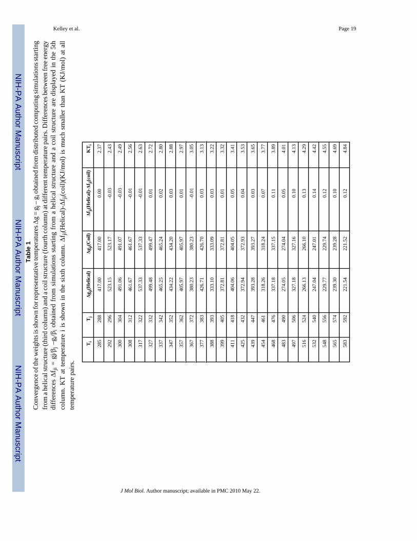
Convergence of the weights—As shown in Table 1, weights obtained from ST simulationsstarting from different initial configurations are converged. Free energy difference betweenneighboring temperatures (gi+1 / βi+1 − gi / βi) is always less than 0.2 KJ/mol, smaller than aKT. As discussed before, converged weights will produce uniform sampling in ST. In Fig. 2(a) and (b), the amount of sampling at each temperature obtained from a series of 2000 4nssimulations is displayed. The sampling is biased to the high temperatures at the beginning,indicating the weights are not converged. After about 24000ns, simulations tend to spendmostly equal time exploring each temperature. We also note that due to the asynchronous natureof FAH (i.e. different nodes have different CPU speed) the order of the trajectories show n inFig 2 is only an estimate.
Convergence of the helical properties—The helix melting curves demonstrate thatsimulations starting from the helical and unfolded states converge excellently as shown in Fig4. The system has a significant fraction of the helical content at low temperatures, e.g. at theroom temperature (300K), the average helical content is about 36%. The average helicalproperties as a function of time at 300K are also plotted in Fig. 3. The simulations starting fromdifferent configurations reach the convergence at the timescale of about 20ns. The averagenumber of helical segments is around 1.5, indicating that this peptide tends not to form onesingle straight helix, but to bend into multiple helix segments. Similar behaviors are also beenobserved for another 22 residue helical peptide.12
Secondary StructureThe finding of multiple helix segments is clearly shown in the secondary structure analysis.The secondary structure as calculated using DSSP13 is shown by residue in Figure 5. Theprobability spike in turn propensity involving residues Lys9-Ala10-Phe11 shows a clear division
Kelley et al. Page 2
J Mol Biol. Author manuscript; available in PMC 2010 May 22.
NIH
-PA Author Manuscript
NIH
-PA Author Manuscript
NIH
-PA Author Manuscript

between the C- and N-terminal helices. (Fig 5) Though alpha helix is clearly the dominantfeature, it is interesting to note the increased presence of 310 helix in the C-terminal region.The presence of both helix geometries implies a higher degree of flexibility in the N-terminalregion. We believe this might facilitate the observed N17Htt-PolyQ domain interactions andhave possible implications in the aggregation mechanism.
Lastly it is interesting to note the helix content diminishes with increasing temperature (asshown in Fig 6), while loop and turn content increases, with the exception of the loop-spike,which remains somewhat independent of temperature. Since we also observe the number ofhelical segments to diminish and the two-helix bundle percentage to decrease both in magnitudeand with respect to the single linear helix, it is likely some of the loop content is attributableto the disordered state.
Structural ClustersVisualizing the structural ensemble via a set of k-means clusters showed a strong agreementwith the numerical findings, and gave additional insight into the driving forces and possibleimplications of the study. Two helical segments, separated by Ala10 as predicted was thedominant feature in over 70% of the structures at low (biological) temperatures (300K) asshown in Fig. 8. The two helices appeared to be stabilized by a large hydrophobic face on oneside, and a grouping of charged residues on the opposing face. Of the ten clusters, five fit intothis category, including cluster 9, the single largest cluster. With ∼35% of the population at300K, and an rmsd of ∼3 Å with respect to the centroid structure, cluster 9 contained twice thepopulation of the others while also retaining the tightest rmsd, suggesting a free energyminimum for the monomeric state. Similar results were obtained using 5 or 20 clusters (datanot shown).
The remaining clusters fell into one of three additional categories: N-terminal helix only,disordered, and linearly oriented helix/helices. The N-terminal helix as seen in these structureswas significantly formed in all clusters excluding the disordered state. The C-terminal helix,which had an increased tendency to occupy the more narrow 3/10 helix than the correspondingN-terminal residues, was only seen disordered in cluster 10. (See Fig. 7) Of the linearly orientedhelix groups, cluster 8 shows something very similar to our initially hypothesized “native-state”, and was shown to be the second largest cluster at room temperature, but with a muchhigher (∼5 Å) rmsd than cluster 9. The two other linearly oriented helix clusters show the singlehelix bending (cluster 5) and then breaking at the high turn propensity Ala10 residue.
At high temperatures (592K) the population of the disordered cluster was observed to increase∼10 fold (17%) placing it among the three most populated clusters. (For the lower temperaturesit had the lowest total population of any cluster with 0.15%.) The two helix bundles retained∼30% of the structural ensemble, however the single linear helices became the most populatedstate with ∼40%, including the single most populated state at high temperature (592K). (SeeFig 8)
There appears to be two dominant factors governing the structural configuration of the system.Some insight can be gained from looking at notable differences between the two-helix clusters.In some cases the charge groups are in very close proximity with an optimal alternating pattern(cluster 2). However, the hydrophobic face is then split apart by Lys6. Likewise, the set ofstructures possessing the hydrophobic region in a single unbroken face show the chargedresidues split into two separate groups. In the cluster with a disordered second helix (cluster10), the charged residues line the inside of a C-terminal J-like turn, which may proveenergetically favorable when considering the addition of the polyQ tail.
Kelley et al. Page 3
J Mol Biol. Author manuscript; available in PMC 2010 May 22.
NIH
-PA Author Manuscript
NIH
-PA Author Manuscript
NIH
-PA Author Manuscript

DiscussionWe have found from our ST simulations that N17Htt forms two primary states: a single extendedhelix and a two-helix bundle of which the c-terminal helix exhibits some degree of flexibility.Both states display a large hydrophobic face opposite a region of alternating charge inamphipathic fashion. Tam et. al10; 11 have previously predicted N17Htt to form an amphipathicalpha helix as a result of experimental data and a sequence comparison search. We have foundthe system can adopt multiple stable thermodynamic states, of which the dominant state atrelevant temperatures is a two-helix bundle. This two-helix bundle state initially seemsinconsistent with experimental findings, but in fact it agrees well with experimental data. Withrespect to the experimental mutation studies, insertion of two Proline “helix-breaker” residueswithin residues L4-K9 was shown to completely halt aggregation, indicating that this region ishighly helical. In our computational study, those same residues are found to adhere almostexclusively to this regime, showing near 90% helicity at 300K mid stretch. Since the two helicalregions are separated by Ala10, our proposed two-helix structure is not in contrast to theexperimental findings.
Our structures agree very well with the main notion that N17Htt is amphipathic. While the polarresidues and the hydrophobic face are the driving factors for the two-helix conformation, theyappear to compete for the precise configuration the two helices will adopt with relation to oneanother. Although in some structural clusters we see the most prominent feature as a salt-bridgenetwork, in the structure characterized as the ensemble's free energy minimum, the two helicescreate a large uninterrupted hydrophobic face. Such a conformation would favor hydrophobicpacking through dimerization to reduce its surface area. This also adheres to Tam et. al'sprevious work10; 11 in which they found the hydrophobic residues as a prerequisite foraggregation, and the polar residues to significantly control the rate and extent of aggregation.
It has been shown that Htt aggregation is nucleation dependent.14 This infers the existence ofa critical transition, which has been hypothesized to be monomeric beta formation in the polyQtail. It has also been shown that the rate limiting step in the aggregation pathway involvesN17Htt 10; 11. Our simulation results provide two possible mechanisms for such a nucleationevent.
It is shown that N17Htt has binding interactions with both itself and the polyQ domains. It isenergetically favorable for N17Htt to dimerize in such a way as to pack the two hydrophobicfaces together, simultaneously exposing the charged residues to solvent while minimizing non-polar surface area. In order for intra-chain N17Htt-polyQ binding interactions to exist, a turnis required to have been formed by N17Htt or PolyQ. A hydrogen bonding network betweenthe charged residues of N17Htt and the polar polyQ side chains would contribute to thesebinding interactions by creating a beta-like strand. Moreover, the correct formation of this beta-like strand might be crucial for the nucleation event. Both mechanisms we propose are basedon the formation of a beta-strand structure, which we feel is the key role N17Htt plays in Httaggregation kinetics. However, they differ in whether N17Htt or PolyQ makes the turn for theinitial beta-strand.
In the first mechanism, the turn connecting two-helix bundle in N17Htt will naturally serve asthe turn. We have observed a degree of flexibility in the C-terminal helix orientation andconformation, (alpha-helix, 3-10-helix, disordered, etc.) The correct C-terminal helixorientation could help to facilitate the polyQ repeat domain interactions with N17Htt byallowing the chain to more easily wrap back on itself and keeping both domains in closeproximity. (See Fig. 9 (b))
Kelley et al. Page 4
J Mol Biol. Author manuscript; available in PMC 2010 May 22.
NIH
-PA Author Manuscript
NIH
-PA Author Manuscript
NIH
-PA Author Manuscript

In the second mechanism, the polyQ chain makes the turn and N17Htt adopts a single helixconformation in the aggregates. In this case, there would be an additional step. An initialtransition is needed for N17Htt from its dominant two-helix bundle state into the single helixstate. Subsequently, the polyQ tail forms the turn for the beta-strand like configuration. Thishas the potential of being a rare event, and also of being able to propagate similar events in anucleating fashion due to the lengthened scaffold of charged residues it presents. In addition,this mechanism puts more emphasis on the polar residue layout, and less on a covalent tetherbetween the two domains. This could prove insightful due to an observed increase inaggregation kinetics with the addition of unbound N17Htt to polyGln10; 11.
A general topology coinciding with these two structures was proposed by Tam et al10; 11.Moreover, the concept of N17Htt as a scaffold fits in nicely with the current hypothesesregarding Htt fibril structure and rate-limiting nucleation events. The end-to-end length of theN17Htt polar region corresponds roughly to one turn of the current model for β-sheet elongation,excluding the turn residues. Wetzel et. al. performed a series of experimental point mutationstudies in which induced β-turns interspersed by 9-10 glutamine residues showed aggregationpotential nearly as efficient as polyGln45
15; 16. In contrast, peptides containing 7-8 glutaminesbetween β-turns aggregated much less readily, indicating periodic β turns staggered every 9-10glutamines in the aggregate form. In addition, we have found previous computational studiesof polyGln to indicate a repeating β-turn topology of similar length17. We feel these matchingdimensions strongly support the N17Htt role as a molecular scaffold.
There are several current ideas representing the rate-limiting nucleation step. Since it isgenerally accepted that monomeric polyglutamine exists as a random coil, a globular to β-sheettransition is required. There are currently two acknowledged possibilities capable of explainingthe free-energetic pathway for this transition8; 18, both of which would benefit from the ideaof N17Htt acting as a molecular scaffold.
Chen et al.8 propose the single chain critical nucleus to be a “compact β-sheet” of roughly fourturns and of very high β content. Such a structure would be a local free energy minimum ormeta-stable state, having each of the four segments stabilized through hydrogen bonds withthe neighboring intra-chain segment. The problem arising in reaching such a conformation isthat initially there are no existing β-segments for an un-coiled stretch to interact with. N17Htt
would facilitate this process by presenting a constant stretch of accessible polar residues forhydrogen bonding by an uncoiled region of glutamines.
Crick et al.18 point out the possibility of the critical nucleus existing as a free energy maximumin the case of a single structure, or a heterogeneous mix of high energy structures. In this casethe stability would come only after fibrillar addition through interchain interactions. This issimilar to the first transition, but differs by existing as a local free energy maximum needinginter-chain interactions to adopt its configuration permanently. Pre-fibril, two or more of theseevents might be required simultaneously and in close spatial proximity to induce fibrilformation. N17Htt would again present a constant source of polar residues, reducing oreliminating the need for simultaneous globule to rod-like transitions by acting as a source ofinterchain interactions. In addition, any hydrophobic induced dimerization caused by N17Htt
can be imagined to be quite beneficial with this type of transition as well, due to its dependenceon the proximity of other chains.
System and MethodsSimulated Tempering (ST)
Computer simulation, such as Molecular Dynamics, is a powerful technique to explore theconformation space. However, those simulations are often trapped in local free energy minima
Kelley et al. Page 5
J Mol Biol. Author manuscript; available in PMC 2010 May 22.
NIH
-PA Author Manuscript
NIH
-PA Author Manuscript
NIH
-PA Author Manuscript

when applied to complex protein systems.19 Generalized ensemble sampling methods such assimulated tempering 20; 21 and parallel tempering (or replica exchange method)19; 22 weredeveloped to overcome this trapping problem by inducing a random walk in temperature space.In ST, configurations are sampled from a mixed canonical ensemble in which the canonicalensembles with different temperatures are weighted differently as defined by a generalizedHamiltonian:
(1)
Where βn =1/(kBTn), H(X, p) is the Hamiltonian for the canonical ensemble at temperatureTn, and the a priori determined constant gn is the weight for the temperature Tn.
ST works as follows: one single simulation starts from a particular temperature and an attemptis made periodically to change the configuration to another temperature according to a welldefined transition probability. The transition probability is shown below,
(2)
where Ui(x) is the potential energy sampled from the canonical ensembles at Ti. A set of weightsneed to be pre-determined to calculate these transition probabilities. Without proper weighing,ST simulations will be constrained to a subset of the temperature space and become inefficient.21; 23 It was shown that weights leading the system to perform a random walk in temperaturespace equal the unit-less free energies at different temperatures.
It is not an easy task to determine these free energy weights enabling system to perform arandom walk in the temperature space. A recent proposed efficient method for determininginitial weights allowing the system based on short trial simulations is adopted in this study.23 These weights are updated throughout the production simulation by an adaptive weightingmethod using adaptive WHAM in a distributed computing environment.24 The initial weightsare calculated based on the property that the “free energy” weights leading to uniform samplingmust yield the same acceptance ratios for both forward and backward transitions from Ti toTj as shown below.
(3)
where
(4)
Where P (Ui) is the potential energy distribution function (PEDF) at Ti. PEDFs for eachtemperature are estimated from the short trial MD simulations by assuming the distributionsare Gaussian. By solving Eq. 3, we can obtain a set of near “free energy” weights.
Kelley et al. Page 6
J Mol Biol. Author manuscript; available in PMC 2010 May 22.
NIH
-PA Author Manuscript
NIH
-PA Author Manuscript
NIH
-PA Author Manuscript

Simulation DetailsIn this study we examined the 17 N-terminal residue headpiece (N17Htt) of the Huntingtinprotein. The AMBER200325 force field was used with a version of the GROMACS moleculardynamics simulation package26; 27 modified to include an ST algorithm and the FAH24infrastructure (http://folding.stanford.edu). Two initial configurations are used: a helixstructure with a helical content of 73.3% and a random coil structure with no helical content.(See Fig. 1) Each system is solvated in a 42 □ cubic box using 2335 TIP3P water molecules.28 1 Na+ and 2 Cl− counter ions were included to neutralize the charged peptide. The simulationsystems were minimized using a steepest descent algorithm, followed by a 100ps MDsimulation applying a position restraint potential to the peptide heavy atoms. All simulationswere conducted using constant NVT with a Nose-Hoover thermostat29 having a couplingconstant of 0.02ps-1. Long-range electrostatic interactions were treated using the reaction fieldmethod with a dielectric constant of 80. 9 □ cutoffs were imposed on non-bonded interactions.Neighbor lists were updated every 10 steps. A 2fs time step was used and covalent bondsinvolving hydrogen atoms were constrained with the LINCS algorithm.30
1000 simulations from each initial configuration were performed on a distributed computingenvironment using ST enhanced sampling method. The total simulation time was aggregatedto more than 40μs. A roughly exponentially distributed temperature list covering a range from285 to 592K was used. 20 simulations started from each of 50 temperatures, each with adifferent set of initial velocities. In ST, swap of temperatures were attempted every 2ps. Theinitial weights were computed using the data obtained from 50 4ns trial simulations.Subsequently, the weights were updated approximately every 400ns by an adaptive weighingscheme.31
Lifson-Roig helix-coil theoryHelical properties were computed using the Lifson-Roig helix-coil counting theory.32 In thismodel, a residue is considered to be helical if φ = −60 ± x and ψ = −47 ± x degrees. x is set to40 degrees, which is shown to give the best agreement with the melting temperature.12 In LRmodel, a helical segment is defined as three or more consecutive helical residues. Each segmenthas a length of n − 2, where n is the total number of consecutive helical residues. Thus, themaximum helical length of our 17 residue peptide system is 15. The helical content is definedas
(5)
Where Nc is the helical content, Ns is the number of helical segments, Nh is the length ofsegment h, and Nmax is the maximum possible helical length.
ClusteringWe use a variation of the K-mean clustering, also named as K-medoids clustering. Thealgorithm works as follows: All conformations are placed in one of K clusters. A conformationfrom each cluster nearest its center of mass is assigned as its centroid. All other conformationsare reassigned to the cluster representing the centroid to which it is nearest. Centroids are thenupdated to the conformation nearest the cluster center of mass taking into account the newassignments. This updating procedure may be continued for some predetermined number ofiterations or until the answer converges.
Kelley et al. Page 7
J Mol Biol. Author manuscript; available in PMC 2010 May 22.
NIH
-PA Author Manuscript
NIH
-PA Author Manuscript
NIH
-PA Author Manuscript
AcknowledgementsNWK is supported by the National Science Foundation Center on Polymer Interfaces and Macromolecular Assemblies(CPIMA), and XH by NIH Roadmap for Medical Research Grant U54 GM072970. Computing resources wereprovided by the Folding@home users and NSF award CNS-0619926. This work is also supported by NIH R01-GM062868 and NIH PN1 EY016525-02. We would also like to thank helpful discussions with Prof. Seokmin Shinand Greg Bowman.
References1. Gusella JF, Wexler NS, Conneally PM, Naylor SL, Anderson MA, Tanzi RE, Watkins PC, Ottina K,
Wallace MR, Sakaguchi AY, et al. A polymorphic DNA marker genetically linked to Huntington'sdisease. Nature 1983;306:234–8. [PubMed: 6316146]
2. Mac Donald ME, et al. A novel gene containing a trinucleotide repeat that is expanded and unstableon Huntington's disease chromosomes. The Huntington's Disease Collaborative Research Group. Cell1993;72:971–83. [PubMed: 8458085]
3. Rubinsztein DC, Leggo J, Coles R, Almqvist E, Biancalana V, Cassiman JJ, Chotai K, Connarty M,Crauford D, Curtis A, Curtis D, Davidson MJ, Differ AM, Dode C, Dodge A, Frontali M, Ranen NG,Stine OC, Sherr M, Abbott MH, Franz ML, Graham CA, Harper PS, Hedreen JC, Hayden MR, et al.Phenotypic characterization of individuals with 30–40 CAG repeats in the Huntington disease (HD)gene reveals HD cases with 36 repeats and apparently normal elderly individuals with 36–39 repeats.Am J Hum Genet 1996;59:16–22. [PubMed: 8659522]
4. Sathasivam K, Amaechi I, Mangiarini L, Bates G. Identification of an HD patient with a (CAG)180repeat expansion and the propagation of highly expanded CAG repeats in lambda phage. Hum Genet1997;99:692–5. [PubMed: 9150744]
5. Masino L, Kelly G, Leonard K, Trottier Y, Pastore A. Solution structure of polyglutamine tracts inGST-polyglutamine fusion proteins. FEBS Lett 2002;513:267–72. [PubMed: 11904162]
6. Chen S, Berthelier V, Yang W, Wetzel R. Polyglutamine aggregation behavior in vitro supports arecruitment mechanism of cytotoxicity. J Mol Biol 2001;311:173–82. [PubMed: 11469866]
7. Altschuler EL, Hud NV, Mazrimas JA, Rupp B. Random coil conformation for extended polyglutaminestretches in aqueous soluble monomeric peptides. J Pept Res 1997;50:73–5. [PubMed: 9273890]
8. Chen S, Ferrone FA, Wetzel R. Huntington's disease age-of-onset linked to polyglutamine aggregationnucleation. Proc Natl Acad Sci U S A 2002;99:11884–9. [PubMed: 12186976]
9. Infante J, Combarros O, Volpini V, Corral J, Llorca J, Berciano J. Autosomal dominant cerebellarataxias in Spain: molecular and clinical correlations, prevalence estimation and survival analysis. ActaNeurol Scand 2005;111:391–9. [PubMed: 15876341]
10. Tam S, Spiess C, Auyeung W, Poirier M, Frydman J. The Chaperonin TRiC Blocks a HuntingtinSequence Element Promoting the Conformational Switch to Aggregation. Nature Structural andMolecular Biology. 2008submitted
11. Tam, S. Dissertation. Stanford University; 2007. Eukaryotic Chaperonin-mediated modulation ofpolyglutamine aggregation and neurotoxicity.
12. Sorin EJ, Pande VS. Exploring the helix-coil transition via all-atom equilibrium ensemble simulations.Biophys J 2005;88:2472–93. [PubMed: 15665128]
13. Kabsch W, Sander C. Dictionary of protein secondary structure: pattern recognition of hydrogen-bonded and geometrical features. Biopolymers 1983;22:2577–637. [PubMed: 6667333]
14. Wetzel R. Nucleation of huntingtin aggregation in cells. Nat Chem Biol 2006;2:297–8. [PubMed:16710335]
15. Ross CA, Poirier MA, Wanker EE, Amzel M. Polyglutamine fibrillogenesis: the pathway unfolds.Proc Natl Acad Sci U S A 2003;100:1–3. [PubMed: 12509507]
16. Thakur AK, Wetzel R. Mutational analysis of the structural organization of polyglutamine aggregates.Proc Natl Acad Sci U S A 2002;99:17014–9. [PubMed: 12444250]
17. Kelley NW, Rajadas J, Kopito R, Pande VS. Testing the relative stability of experimentally proposedpolyGln structures. Prot Sci. 2008submitted
Kelley et al. Page 8
J Mol Biol. Author manuscript; available in PMC 2010 May 22.
NIH
-PA Author Manuscript
NIH
-PA Author Manuscript
NIH
-PA Author Manuscript

18. Crick SL, Jayaraman M, Frieden C, Wetzel R, Pappu RV. Fluorescence correlation spectroscopyshows that monomeric polyglutamine molecules form collapsed structures in aqueous solutions. ProcNatl Acad Sci U S A 2006;103:16764–9. [PubMed: 17075061]
19. Hansmann UH, Okamoto Y. New Monte Carlo algorithms for protein folding. Curr Opin Struct Biol1999;9:177–83. [PubMed: 10322208]
20. Lyubartsev AP, Martsinovski AA, Shevkunov SV, Vorontsov-Velyaminov PN. New approach toMonte Carlo calculation of the free energy: Method of expanded ensembles. The Journal of ChemicalPhysics 1992;96:1776–1783.
21. Marinari E, Parisi G. Simulated Tempering: a New Monte Carlo Scheme. Europhysics Letters1992;19:451–458.
22. Sugita Y, Okamoto Y. Replica-exchange molecular dynamics method for protein folding. Chem PhysLett 1999;314:141–151.
23. Huang X, Bowman GR, Pande VS. Convergence of folding free energy landscapes via applicationof enhanced sampling methods in a distributed computing environment. J Chem Phys2008;128:205106. [PubMed: 18513049]
24. Shirts M, Pande VS. COMPUTING: Screen Savers of the World Unite! Science 2000;290:1903–1904. [PubMed: 17742054]
25. Duan Y, Wu C, Chowdhury S, Lee MC, Xiong G, Zhang W, Yang R, Cieplak P, Luo R, Lee T,Caldwell J, Wang J, Kollman P. A Point-Charge Force Field for Molecular Mechanics Simulationsof Proteins Based on Condensed-Phase Quantum Mechanical Calculations. J Comp Chem2003;24:1999–2012. [PubMed: 14531054]
26. Lindahl E, Hess B, van der Spoel D. GROMACS 3.0: a package for molecular simulation andtrajectory analysis. J Mol Modeling 2001;7:306–317.
27. Berendsen HJC, Van der Spoel D, Van Drunen R. GROMACS: A message-passing parallel moleculardynamics implementation. Comp Phys Comm 1995:43–56.
28. Jorgensen WLCJ, Madura JD, Impey RW, Klein ML. J Chem Phys 1983;7929. Hoover W. Phys Rev A. 1985;31:1695–1697.30. Hess B, Bekker H, Berendsen HJC, Fraaije JGEM. LINCS: a linear constraint solver for molecular
simulations. J Comput Chem 1997;18:1463–1472.31. Bartels C, Karplus M. Multidimensional adaptive umbrella sampling: Applications to main chain and
side chain peptide conformations. J Comp Chem 1997;12:1450–1462.32. Lifson S, Roig A. On the Theory of Helix---Coil Transition in Polypeptides. J Chem Phys
1961;34:1963–1974.
Kelley et al. Page 9
J Mol Biol. Author manuscript; available in PMC 2010 May 22.
NIH
-PA Author Manuscript
NIH
-PA Author Manuscript
NIH
-PA Author Manuscript

Fig 1.The two initial configurations used for simulations of the N17 headpiece: (a). A helical structureand (b). A random-coil structure.
Kelley et al. Page 10
J Mol Biol. Author manuscript; available in PMC 2010 May 22.
NIH
-PA Author Manuscript
NIH
-PA Author Manuscript
NIH
-PA Author Manuscript

Fig 2.(a). Amount of time the ST simulations starting from a helical structure spent at each of the 50temperatures. (b). The same as (a) except that data is collected from simulations starting froma coil structure.
Kelley et al. Page 11
J Mol Biol. Author manuscript; available in PMC 2010 May 22.
NIH
-PA Author Manuscript
NIH
-PA Author Manuscript
NIH
-PA Author Manuscript

Fig 3.Convergence of the average helical properties as a function of time at 300K. Helix propertiesat each conformation is defined according to classical LR counting theory. Plots obtained fromsimulations starting from the helix structure (black, circle) and the coil structure (red, square)are displayed for (a). average helical content. (b). average number of helical segments.
Kelley et al. Page 12
J Mol Biol. Author manuscript; available in PMC 2010 May 22.
NIH
-PA Author Manuscript
NIH
-PA Author Manuscript
NIH
-PA Author Manuscript

Fig 4.Helical content as a function of temperature obtained from ST simulations starting from thehelical structure (black) and those from the coil structure (red). Error bars are calculated byblock averaging over the configurations later than 32ns.
Kelley et al. Page 13
J Mol Biol. Author manuscript; available in PMC 2010 May 22.
NIH
-PA Author Manuscript
NIH
-PA Author Manuscript
NIH
-PA Author Manuscript

Fig 5.Alpha helix (black), 310-helix (red) and loop (green) content by residue at 300K. There is ahigher propensity for alpha for the first 10 n-terminal residues, followed by a sharp peak inloop content and a second area of high probability for alpha helix content at the C-terminus.This indicates a tendency for a 2-helix bundle, and can be visualized in figure 7.
Kelley et al. Page 14
J Mol Biol. Author manuscript; available in PMC 2010 May 22.
NIH
-PA Author Manuscript
NIH
-PA Author Manuscript
NIH
-PA Author Manuscript

Fig 6.(a). Alpha helix content by residue over a series of temperatures. There is a trend over allresidues for helical content to decrease with increased temperature. (b) 310-helix content byresidue over a series of temperatures. (c). Loop content by residue for a series of temperatures.It is interesting to note the dramatically increased fraction of loop in the two helical stretches,but the relatively low temperature dependence of residue 10.
Kelley et al. Page 15
J Mol Biol. Author manuscript; available in PMC 2010 May 22.
NIH
-PA Author Manuscript
NIH
-PA Author Manuscript
NIH
-PA Author Manuscript

Fig 7.Structures closest to cluster center and the population at 300K for each of the 10 clusters areshown. The most populated cluster (cluster 9) is a two-helix bundle structure.
Kelley et al. Page 16
J Mol Biol. Author manuscript; available in PMC 2010 May 22.
NIH
-PA Author Manuscript
NIH
-PA Author Manuscript
NIH
-PA Author Manuscript

Fig 8.Probability for different states of the system: N-terminal helix only, two-helix, one straighthelix and disordered state. N-terminal helix state contains a N-terminal helix, but the C-terminalpart of the peptide is disordered. Two-helix state with a two-helix bundle is the most populatedstate. Plots for three temperatures are shown: 300K (Black circle), 347K (Red triangle), and592K (Green square).
Kelley et al. Page 17
J Mol Biol. Author manuscript; available in PMC 2010 May 22.
NIH
-PA Author Manuscript
NIH
-PA Author Manuscript
NIH
-PA Author Manuscript

Fig 9.Cartoons showing the N17Htt-PolQ structures in two proposed mechanisms described in thetext. Two representative cluster centroid structures are shown with faces paired to minimizeexposed hydrophobic surface area. Each shows a model polyQ tail (cyan) which satisfies theobserved N17Htt – polyQ domain interactions. (a). N17Htt adopts the single straight helixconformation and has the charged residues in a surface geometry which would compliment thepolyQ tail's β-strand configuration. (b). N17Htt adopts a two-helix bundle conformation. Theincreased flexibility in the C-terminus of the two-helix bundle creates the turn necessary forpolyQ – N17Htt interactions.
Kelley et al. Page 18
J Mol Biol. Author manuscript; available in PMC 2010 May 22.
NIH
-PA Author Manuscript
NIH
-PA Author Manuscript
NIH
-PA Author Manuscript
NIH
-PA Author Manuscript
NIH
-PA Author Manuscript
NIH
-PA Author Manuscript
Kelley et al. Page 19Ta
ble
1C
onve
rgen
ce o
f the
wei
ghts
is sh
own
for r
epre
sent
ativ
e tem
pera
ture
s Δg
= g j
– g
i obt
aine
d fr
om d
istri
bute
d co
mpu
ting
sim
ulat
ions
star
ting
from
a he
lical
stru
ctur
e (th
ird co
lum
n) an
d a c
oil s
truct
ure (
four
th co
lum
n) at
diff
eren
t tem
pera
ture
pai
rs. D
iffer
ence
s bet
wee
n fr
ee en
ergy
diff
eren
ces Δf
ji =
gj/β
j –g i
/βi o
btai
ned
from
sim
ulat
ions
sta
rting
from
a h
elic
al s
truct
ure
and
a co
il st
ruct
ure
are
disp
laye
d in
the
5th
colu
mn.
KT
at te
mpe
ratu
re i
is s
how
n in
the
sixt
h co
lum
n. Δ
f ji(H
elic
al)-Δf
ji(co
il)(K
J/m
ol)
is m
uch
smal
ler
than
KT
(KJ/
mol
) at
all
tem
pera
ture
pai
rs.
Ti
Tj
Δgji(
Hel
ical
)Δg
ji(C
oil)
Δfji(
Hel
ical
)-Δf ji
(coi
l)K
Ti
285
288
417.
0041
7.00
0.00
2.37
292
296
523.
1552
3.17
-0.0
32.
43
300
304
491.
0649
1.07
-0.0
32.
49
308
312
461.
6746
1.67
-0.0
12.
56
317
322
537.
3353
7.33
-0.0
12.
63
327
332
499.
4849
9.47
0.01
2.72
337
342
465.
2546
5.24
0.02
2.80
347
352
434.
2243
4.20
0.03
2.88
357
362
405.
9740
5.97
0.01
2.97
367
372
380.
2338
0.23
-0.0
13.
05
377
383
426.
7142
6.70
0.03
3.13
388
393
333.
1033
3.09
0.03
3.22
399
405
372.
8137
2.81
0.01
3.32
411
418
404.
0640
4.05
0.05
3.41
425
432
372.
9437
2.93
0.04
3.53
439
447
393.
2839
3.27
0.03
3.65
454
461
318.
2631
8.24
0.07
3.77
468
476
337.
1833
7.15
0.11
3.89
483
490
274.
0527
4.04
0.05
4.01
497
506
327.
1832
7.16
0.10
4.13
516
524
266.
1326
6.10
0.13
4.29
532
540
247.
0424
7.01
0.14
4.42
548
556
229.
7722
9.74
0.12
4.55
565
574
239.
3023
9.28
0.10
4.69
583
592
221.
5422
1.52
0.12
4.84
J Mol Biol. Author manuscript; available in PMC 2010 May 22.
Related Documents