RESEARCH ARTICLE The positional identity of mouse ES cell-generated neurons is affected by BMP signaling Michele Bertacchi • Luca Pandolfini • Elisa Murenu • Alessandro Viegi • Simona Capsoni • Alessandro Cellerino • Andrea Messina • Simona Casarosa • Federico Cremisi Received: 11 July 2012 / Revised: 24 September 2012 / Accepted: 25 September 2012 / Published online: 16 October 2012 Ó The Author(s) 2012. This article is published with open access at Springerlink.com Abstract We investigated the effects of bone morpho- genetic proteins (BMPs) in determining the positional identity of neurons generated in vitro from mouse embry- onic stem cells (ESCs), an aspect that has been neglected thus far. Classical embryological studies in lower verte- brates indicate that BMPs inhibit the default fate of pluripotent embryonic cells, which is both neural and anterior. Moreover, mammalian ESCs generate neurons more efficiently when cultured in a minimal medium containing BMP inhibitors. In this paper, we show that mouse ESCs produce, secrete, and respond to BMPs during in vitro neural differentiation. After neuralization in a minimal medium, differentiated ESCs show a gene expression profile consistent with a midbrain identity, as evaluated by the analysis of a number of markers of anterior–posterior and dorsoventral identity. We found that BMPs endogenously produced during neural differentiation mainly act by inhibiting the expression of a telencephalic gene profile, which was revealed by the treatment with Noggin or with other BMP inhibitors. To better characterize the effect of BMPs on positional fate, we compared the global gene expression profiles of differen- tiated ESCs with those of embryonic forebrain, midbrain, and hindbrain. Both Noggin and retinoic acid (RA) support neuronal differentiation of ESCs, but they show different effects on their positional identity: whereas RA supports the typical gene expression profile of hindbrain neurons, Noggin induces a profile characteristic of dorsal telence- phalic neurons. Our findings show that endogenously produced BMPs affect the positional identity of the neurons that ESCs spontaneously generate when differen- tiating in vitro in a minimal medium. The data also support the existence of an intrinsic program of neuronal differ- entiation with dorsal telencephalic identity. Our method of ESC neuralization allows for fast differentiation of neural cells via the same signals found during in vivo embryonic development and for the acquisition of cortical identity by the inhibition of BMP alone. Keywords Noggin BMP Cortical identity Embryonic stem cells Introduction Neural inducing signals are proposed to impart both neural and anterior identity to the ectoderm, while the generation of the full range of CNS structures would be the result of later events that posteriorize anterior neural tissue. According to this view, bone morphogenetic proteins (BMPs) play a key role by antagonizing a neural anterior default differentiation program. Antagonists of BMP sig- naling such as Noggin would ensure low levels of BMPs in the presumptive neuroectoderm thus allowing forebrain development in the absence of posteriorizing signals [67]. Electronic supplementary material The online version of this article (doi:10.1007/s00018-012-1182-3) contains supplementary material, which is available to authorized users. M. Bertacchi L. Pandolfini A. Viegi S. Capsoni A. Cellerino F. Cremisi (&) Scuola Normale Superiore di Pisa, Piazza dei Cavalieri 7, 56100 Pisa, Italy e-mail: [email protected] E. Murenu A. Messina S. Casarosa CIBIO, Universita ` di Trento, Trento, Italy A. Cellerino Leibniz Institute for Age Research – Fritz Lipmann Institute, Jena, Germany Cell. Mol. Life Sci. (2013) 70:1095–1111 DOI 10.1007/s00018-012-1182-3 Cellular and Molecular Life Sciences 123

Welcome message from author
This document is posted to help you gain knowledge. Please leave a comment to let me know what you think about it! Share it to your friends and learn new things together.
Transcript
RESEARCH ARTICLE
The positional identity of mouse ES cell-generated neuronsis affected by BMP signaling
Michele Bertacchi • Luca Pandolfini • Elisa Murenu • Alessandro Viegi • Simona Capsoni •
Alessandro Cellerino • Andrea Messina • Simona Casarosa • Federico Cremisi
Received: 11 July 2012 / Revised: 24 September 2012 / Accepted: 25 September 2012 / Published online: 16 October 2012
� The Author(s) 2012. This article is published with open access at Springerlink.com
Abstract We investigated the effects of bone morpho-
genetic proteins (BMPs) in determining the positional
identity of neurons generated in vitro from mouse embry-
onic stem cells (ESCs), an aspect that has been neglected
thus far. Classical embryological studies in lower verte-
brates indicate that BMPs inhibit the default fate of
pluripotent embryonic cells, which is both neural and
anterior. Moreover, mammalian ESCs generate neurons
more efficiently when cultured in a minimal medium
containing BMP inhibitors. In this paper, we show that
mouse ESCs produce, secrete, and respond to BMPs during
in vitro neural differentiation. After neuralization in a
minimal medium, differentiated ESCs show a gene
expression profile consistent with a midbrain identity, as
evaluated by the analysis of a number of markers of
anterior–posterior and dorsoventral identity. We found that
BMPs endogenously produced during neural differentiation
mainly act by inhibiting the expression of a telencephalic
gene profile, which was revealed by the treatment
with Noggin or with other BMP inhibitors. To better
characterize the effect of BMPs on positional fate, we
compared the global gene expression profiles of differen-
tiated ESCs with those of embryonic forebrain, midbrain,
and hindbrain. Both Noggin and retinoic acid (RA) support
neuronal differentiation of ESCs, but they show different
effects on their positional identity: whereas RA supports
the typical gene expression profile of hindbrain neurons,
Noggin induces a profile characteristic of dorsal telence-
phalic neurons. Our findings show that endogenously
produced BMPs affect the positional identity of the
neurons that ESCs spontaneously generate when differen-
tiating in vitro in a minimal medium. The data also support
the existence of an intrinsic program of neuronal differ-
entiation with dorsal telencephalic identity. Our method of
ESC neuralization allows for fast differentiation of neural
cells via the same signals found during in vivo embryonic
development and for the acquisition of cortical identity by
the inhibition of BMP alone.
Keywords Noggin � BMP � Cortical identity �Embryonic stem cells
Introduction
Neural inducing signals are proposed to impart both neural
and anterior identity to the ectoderm, while the generation
of the full range of CNS structures would be the result of
later events that posteriorize anterior neural tissue.
According to this view, bone morphogenetic proteins
(BMPs) play a key role by antagonizing a neural anterior
default differentiation program. Antagonists of BMP sig-
naling such as Noggin would ensure low levels of BMPs in
the presumptive neuroectoderm thus allowing forebrain
development in the absence of posteriorizing signals [67].
Electronic supplementary material The online version of thisarticle (doi:10.1007/s00018-012-1182-3) contains supplementarymaterial, which is available to authorized users.
M. Bertacchi � L. Pandolfini � A. Viegi � S. Capsoni �A. Cellerino � F. Cremisi (&)
Scuola Normale Superiore di Pisa, Piazza dei Cavalieri 7,
56100 Pisa, Italy
e-mail: [email protected]
E. Murenu � A. Messina � S. Casarosa
CIBIO, Universita di Trento, Trento, Italy
A. Cellerino
Leibniz Institute for Age Research – Fritz Lipmann Institute,
Jena, Germany
Cell. Mol. Life Sci. (2013) 70:1095–1111
DOI 10.1007/s00018-012-1182-3 Cellular and Molecular Life Sciences
123

The dissection of diffusible signals that orchestrate
neural induction has recently been made easier by the study
of embryonic stem cells (ESCs) in vitro differentiation. In
recent years, several reports have described methods for the
generation of neural cells from mouse ESCs [5, 15, 19, 65,
66]. Using defined growth media, it has been possible to
investigate the diffusible factors that affect anterior–pos-
terior (A/P) as well as dorsoventral (D/V) identity of
in vitro-generated neurons. Among these, retinoic acid
(RA), BMPs, Wnts, fibroblast growth factors (FGFs) and
sonic hedgehog (SHH) have been described [10, 15, 18, 28,
65].
Conversely, the use of factor-free chemically defined
media has allowed for the investigation of the differentia-
tion fate of ESCs in the absence of exogenous signals,
showing that it is predominantly neural [19, 20, 63]. Effects
of factors endogenously produced by ESCs have also been
suggested. BMPs sustain self-renewal and inhibit neural
differentiation of ESCs [70]. The BMP inhibitor Noggin
triggers in vitro neuronal differentiation of mammalian
ESCs cultured in growth factor-free chemically defined
medium [9, 22]. It was recently shown that the cell-
intrinsic expression of the zinc-finger nuclear protein
Zfp521, which is inhibited by BMPs, plays a pivotal role in
promoting a default neural state of ESCs. Furthermore, a
role of Zfp521 in supporting an anterior identity of neurons
generated by ESCs was hypothesized [33]. These data
suggest that ESCs produce and are sensitive to BMPs with
an autocrine/paracrine mechanism. However, to our
knowledge, there is no direct measurement of BMP pro-
duction by differentiating ESCs.
Early studies in lower vertebrates suggested that BMP
plays a key role in anterior/posterior patterning. BMP
antagonism on pluripotent cells of Xenopus animal caps
induces cement glands, which are the most anterior ecto-
dermal structures in Xenopus, and anterior brain markers
such as the fore-midbrain marker Otx2, but not hindbrain
or spinal cord markers [26, 38, 56]. More recent studies
highlight that Noggin has a dose-dependent patterning
effect on Xenopus animal caps. At lower doses, Noggin
supports neuralization without the expression of dience-
phalic markers, which are instead activated at higher doses
[39]. Moreover, in Xenopus embryos, the specification of
the forebrain requires isolation of its cells from BMP,
Activin/Nodal, and Wnt signaling by high concentrations
of Noggin produced in cells at the anterior margin of the
neural plate [4]. These observations suggest that, in vivo,
the concentration of endogenous BMPs might be relevant
in the control of the positional identity of neurons. It has
also been proposed that BMPs play a role in the regional
morphogenesis of mouse dorsal telencephalon, by the
control of specific gene expression, cell proliferation, and
local cell death [17]. Forebrain truncations were found in
double-mutant mice for both BMP antagonists Noggin and
Chordin [3]. BMP signaling specifies telencephalic pro-
genitor cells toward the most dorsal fate, the choroid
plexus [27], but earlier effects on the anterior–posterior
patterning are not well characterized.
The aim of the present work is to directly show the
endogenous production of BMP by differentiating ESCs
and to characterize the effects of BMP on the differentia-
tion and positional identity of ESC-generated neurons. We,
therefore, established an in vitro differentiation protocol
that minimizes exogenous signals and analyzed ESCs dif-
ferentiation by performing a genome-wide expression
analysis. We report that mouse ESCs produce and release
BMPs, which act on their differentiation in such a minimal
medium. Blocking the BMP pathway by Noggin or by
other inhibitors selectively affects the A/P positional
identity of the generated neurons. At the highest doses of
Noggin that we tested, the fate of neurons produced by
ESCs is predominantly dorsal telencephalic. These neurons
have a gene expression profile that clusters with that of
early cerebral cortical cells and express telencephalic dif-
ferentiation markers.
Materials and methods
Cells cultures
Murine embryonic stem cell (ESC) lines E14Tg2A (pas-
sages 25–38) and 46 C (transgenic Sox1-GFP ESC kindly
provided by A. Smith, University of Cambridge, UK,
passages 33–39) were cultured on gelatin-coated tissue
culture dishes at a density of 40,000 cells/cm2. ESC med-
ium, which was changed daily, contained GMEM (Sigma),
10 % Fetal Calf Serum, 2 mM Glutamine, 1 mM sodium
Pyruvate, 1 mM non-essential amino acids, 0.05 mM
b-mercaptoethanol, 100 U/ml Penicillin/Streptomycin and
1,000 U/ml recombinant mouse LIF (Invitrogen). Cells
were passaged using Trypsin dissociation and re-plated at a
dilution of 1:3–1:4, to avoid cell confluence and to main-
tain pluripotency. RAW 264.7 (mouse leukemic monocyte
macrophage cell line, kindly supplied by Diana Boraschi,
Institute of Medical Biotechnology, CNR of Pisa) were
cultured in Dulbecco’s modified Eagle’s Medium with
4 mM L-glutamine and 4.5 g/L glucose, supplemented with
10 % fetal bovine serum. Cells were split every 2 days at a
confluence of approximately 10 % (1 9 106 and 3 9 106
cells in 100- and 150-mm plates, respectively) and grown
to a confluence of approximately 80 %. Mouse mesen-
chymal stromal cells (MSCs) primary cultures (kindly
supplied by Cristina Magli, CNR of Pisa) were established
from B6D2F1 (BDF1) mice (Charles River) as described
[44].
1096 M. Bertacchi et al.
123
Neural induction
Chemically defined minimal medium (CDMM) consisted
of DMEM/F12 (Invitrogen), 2 mM Glutamine, 1 mM
sodium Pyruvate, 0.1 mM non-essential amino acids,
0.05 mM b-mercaptoethanol, 100 U/ml Penicillin/Strepto-
mycin supplemented with N2/B27 (no vitamin A;
Invitrogen). Step I: dissociated ESCs were washed with
DMEM/F12, aggregated in agar-coated culture dishes
(65,000 cells per cm2) and cultured as floating aggregates
in CDMM for 2 days. The second day 70 % of CDMM was
renewed. Step II: ESCs aggregates were dissociated and
cultured in adhesion (65,000 cells per cm2) on Poly-orni-
thine (Sigma; 20 lg/ml in sterile water, 4 h coating at
37 �C) and natural mouse Laminin (Invitrogen; 5 lg/ml in
PBS, 4 h coating at 37 �C) for 4 days, changing CDMM
daily. Step III: After a second dissociation, ESCs were
cultured 4 additional days in CDMM devoid of B27 sup-
plement to drive terminal differentiation, using the same
type of seeding density and coated surface. Serum
employed for trypsin inactivation was carefully removed
by several washes in DMEM/F12. The following factors
were tested by addition during step II: Recombinant Mouse
Noggin (R&D; ranging from 5 to 400 nM), BMP4 (R&D;
50 ng/ml), Recombinant Mouse BMPRIA/Fc Chimera
(R&D; 3.75 and 37.5 nM), Dorsomorphin (Sigma-Aldrich;
5 lM), Retinoic Acid (Sigma-Aldrich; 0.1–10 lM),
Cyclopamine (Sigma-Aldrich; 10 lM), SAG (Santa Cruz
Biotechnology; 100 nM), SB431542 (Sigma-Aldrich;
10 lM). Cell viability and proliferation, which were
monitored by trypan blue exclusion test and cell counting,
respectively, were not significantly affected by treatments.
Semiquantitative real-time PCR
For each sample, 500 ng of total RNA were reverse-tran-
scribed. Amplified cDNA was quantified using GoTaq
qPCR Master Mix (Promega) on Rotor-Gene 6000
(Corbett) with the primers listed in Supplemental Table 1.
Amplification take-off values were evaluated using the
built-in Rotor-Gene 6000 ‘‘relative quantitation analysis’’
function, and relative expression was calculated with the
2-DDCt method, normalizing to the housekeeping gene
b-Actin. Standard errors were obtained from the error
propagation formula as described in [46], and statistical
significance was probed with randomization test, taking
advantage of REST Software [51].
Immunocytodetection
Cells prepared for immunocytodetection experiments were
cultured on Poly-ornithine/Laminin coated round glass
coverslips. Cells were fixed using 2 % paraformaldehyde
for 15 min, washed twice with PBS, permeabilized using
0.1 % Triton X100 in PBS and blocked using 0.5 % BSA
in PBS. Primary antibodies used for microscopy included
Oct3/4 (1:200; Santa Cruz DBA), Nanog (1:300; Novus
Biologicals), acetylated N-Tubulin (1:500; Sigma), Neu-
ronal Class III b-Tubulin (1:500; Covance), Doublecortin
(1:500; Abcam), Musashi-1 (1:200; Cell Signaling), Nes-
tin (1:200; Millipore), Synaptophysin (1:100; Santa Cruz
DBA), a-Internexin (1:100; Santa Cruz DBA), phospho-
Smad1/5/8 (1:100; Millipore), FoxG1 (1:200; Abcam),
Tbr1 (1:400; Millipore), Satb2 (1:200; Abcam), VGlut2
(1:300; Abcam), GAD65 (1:500; Chemicon), Pax6 (1:400;
Covance), Nkx2.1 (1:400; Abcam) and GFAP (1:100;
Dako). Primary antibodies were incubated 2 h at room
temperature; cells were then washed three times with PBS
(100 each). Alexa Fluor 488 and Alexa Fluor 568 anti-
mouse or anti-rabbit IgG conjugates (Molecular Probes,
1:500) were incubated 1 h at RT in PBS containing 0.1 %
Triton X100 and 0.5 % BSA for primary antibody
detection, followed by three PBS washes (100 each).
Nuclear staining was obtained with DAPI. The protocol
varied for Tbr1, Satb2 and FoxG1, the antibodies of which
were incubated overnight at 4 �C using 0.3 % Triton
X100.
FACS analysis
Adherent cells were detached by trypsinization, washed
and resuspended in PBS at RT, then analyzed with a
FACSCalibur cytometer (BD). At least 10,000 events per
sample were collected. Data were processed with the free
software WinMDI 2.9 (The Scripps Research Institute).
BMP2 ELISA
Cells were seeded into 24-well plates and cultured as
described. When cells reached 70–80 % confluence, each
well was washed with PBS and fresh medium (DMEM/F12
containing 2 mM Glutamine and 1 mM sodium Pyruvate)
was replaced. After 24 h, supernatant was collected, cen-
trifuged (10,000g, 5 min) to remove particulates and
assayed for BMP2 content with a commercially available
ELISA kit (Quantikine, BMP-2 Immunoassay; R&D Sys-
tems, Minneapolis, MN, USA), according to the
manufacturer’s instructions. This assay could measure
BMP-2 concentrations as low as 50 pg/ml in a linear range
(Pearson correlation coefficient of linear regression
R2 = 0.999; see Supplemental Figure SF1). A 1.2 % cross-
reactivity was observed with 50 ng/mL recombinant
human BMP-4. BMP-2 concentrations of triplicate samples
were determined from the optical densities at 450 nm in
relation to standard curves of the recombinant antigen
provided in the kit.
The positional identity of mouse ES cell-generated neurons 1097
123

Microarray hybridization and data analysis
Cortex, midbrain and hindbrain were dissected from n = 3
mouse embryos (C57BL/6 strain) at embryonic day (E)16.
Total RNA was extracted with NucleoSpin RNA II col-
umns (Macherey–Nagel). RNA from three different sets of
experiment was pooled. RNA quality was assessed with
Agilent Bioanalyzer RNA 6000 Nano kit; 500 ng of RNA
were labeled with One Color Quick amp labeling kit
(Agilent), purified and hybridized overnight onto an
Agilent Mouse Gene Expression Microarray chip
(4 9 44 Kv2) before detection, according to the manu-
facturer’s instructions. Three slides were hybridized with
Noggin-treated ESCs RNA and two slides were hybridized
with RNA from all the other conditions. Agilent DNA
Microarray scanner was used for slide acquisition and spot
analysis was performed with Feature Extraction software
(Agilent).
For GSEA analysis, genes differentially expressed
between Noggin treatment and CDMM (Supplemental
Table 2), or between RA treatment and CDMM (Supple-
mental Table 3; fold-change C2), were analyzed by the
GeneSpring GX11.0 software using BROAD Gene Ontol-
ogy collection (C5; http://www.broadinstitute.org/gsea). A
complete GSEA list with enrichment scores of gene sets
with q value \0.3 is shown in Supplemental Table 4.
To select a gene set representing the anterior-posterior
regionalization of the developing brain, we compared gene
expression profiles of E16 cortex and hindbrain using
Genespring GX11.0 software (Agilent). A set of 592 genes
with an absolute fold-change greater or equal than 10
(p \ 0.05) was selected (see Supplemental Table 5). Sig-
nificance of the data was proven by one-way ANOVA and
Tukey post hoc test with Bonferroni correction for multiple
comparisons. The content of this set of genes was explored
by hierarchical clustering and principal component analy-
sis, taking advantage of Cluster software [16]. Single
linkage algorithm was employed for hierarchical cluster-
ing. Trees were generated using absolute correlation for
genes and Euclidean distance for arrays, and visualized
with java TreeView [55].
Results
A chemically defined minimal medium (CDMM)
induces neurogenesis of ESCs
In order to investigate the default positional identity of
neurons generated from ESCs, we established a culture
method that promotes neurogenesis minimizing the influ-
ence of exogenous signals. This method consists of a three-
step procedure of culture in a chemically defined minimal
medium (CDMM; see ‘‘Materials and methods’’; Fig. 1a)
devoid of serum or morphogens but allowing cell survival
by insulin.
Upon LIF and serum withdrawal, dissociated ESCs were
initially grown as aggregates (Fig. 1b) in CDMM for
2 days. This step (step I), which minimizes cell death,
follows a procedure adapted from previously described
methods [5, 40, 65]. As many protocols use serum-con-
taining medium (SCM) during ESCs aggregation, we also
assayed this condition in the preliminary set-up of our
protocol. As additional control, we used undifferentiated
ESCs cultured in LIF ? serum (ESC medium).
ESC aggregates were subsequently dissociated and
cultured in adhesion for 4 days on Poly-ornhitine/Laminin-
coated wells in CDMM (step II). All additional treatments
(e.g., Noggin), when applied, were performed during this
step (Fig. 1a), unless specified. During step II, ESCs turned
off the expression of the stem cell marker Oct4 [45] and
activated the expression of the pan-neuronal markers
b-Tubulin-III and Ncam, as seen by RT-PCR (Fig. 1c).
This activation was higher in ESCs aggregated in CDMM
than in ESCs aggregated in SCM, as the latter still
expressed high levels of Oct4 and activated the mesoder-
mal marker GATA4 (Fig. 1c). Immunostaining showed
much higher expression of the neural progenitor cell mar-
ker Musashi-1 [47] and robust downregulation of Oct4 in
ESCs cultured in CDMM (Fig. 1d) compared to ESCs
cultured in SCM (Fig. 1e). Whereas ESCs aggregated in
CDMM started expressing b-Tubulin-III at step II, ESCs
aggregated in SCM failed to show b-Tubulin-III labeling
(Supplemental Figure SF2A, B). Similar results were
obtained when analyzing ESCs aggregates cultured for
5 days (Supplemental Figure SF2C, D). Our observations
indicate that aggregation (step I) in the absence of serum
facilitates loss of stem cell pluripotency and induces rapid
neural differentiation (as evaluated at the end of step II).
After a second dissociation, cells were cultured for
4 days in CDMM. This additional step (step III) allowed
cells to undergo terminal differentiation. Notably, the
presence of serum during step I profoundly affected the
fate of cells produced at the end of step III, as the ratio of
neural progenitor cells immunostained by Nestin antibody
was lower in cells aggregated in SCM (8 ± 4.8 %;) com-
pared to cells aggregated in CDMM (44.3 ± 8.7 %;
Supplemental Figure SF2E–G). Consistently, mRNA
expression of Nestin and of pan-neuronal markers Ncam
and b-Tubulin-III was significantly lower at the end of step
III in cells that were aggregated in SCM than in cells that
were aggregated in CDMM (Supplemental Figure SF2H).
Moreover, cells cultured in CDMM formed rosette-like
structures at the end of step III, which are typical of neural
progenitors in vitro ([73]; Supplemental Figure SF2F),
and generated high proportions of neuronal cells
1098 M. Bertacchi et al.
123
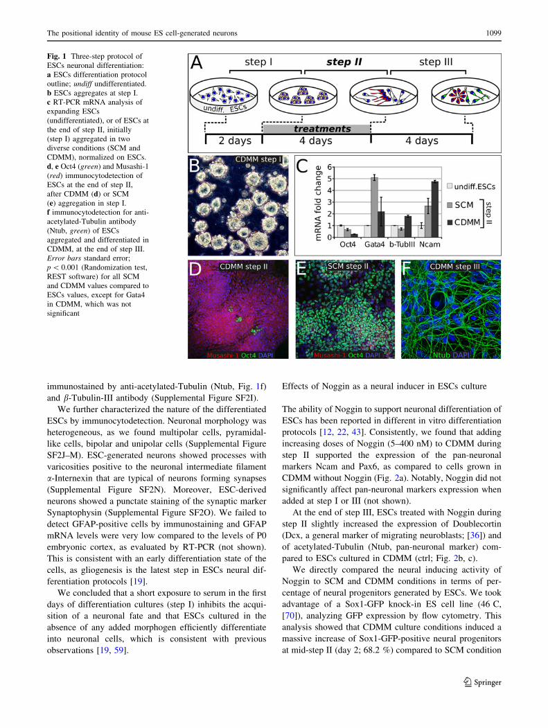
immunostained by anti-acetylated-Tubulin (Ntub, Fig. 1f)
and b-Tubulin-III antibody (Supplemental Figure SF2I).
We further characterized the nature of the differentiated
ESCs by immunocytodetection. Neuronal morphology was
heterogeneous, as we found multipolar cells, pyramidal-
like cells, bipolar and unipolar cells (Supplemental Figure
SF2J–M). ESC-generated neurons showed processes with
varicosities positive to the neuronal intermediate filament
a-Internexin that are typical of neurons forming synapses
(Supplemental Figure SF2N). Moreover, ESC-derived
neurons showed a punctate staining of the synaptic marker
Synaptophysin (Supplemental Figure SF2O). We failed to
detect GFAP-positive cells by immunostaining and GFAP
mRNA levels were very low compared to the levels of P0
embryonic cortex, as evaluated by RT-PCR (not shown).
This is consistent with an early differentiation state of the
cells, as gliogenesis is the latest step in ESCs neural dif-
ferentiation protocols [19].
We concluded that a short exposure to serum in the first
days of differentiation cultures (step I) inhibits the acqui-
sition of a neuronal fate and that ESCs cultured in the
absence of any added morphogen efficiently differentiate
into neuronal cells, which is consistent with previous
observations [19, 59].
Effects of Noggin as a neural inducer in ESCs culture
The ability of Noggin to support neuronal differentiation of
ESCs has been reported in different in vitro differentiation
protocols [12, 22, 43]. Consistently, we found that adding
increasing doses of Noggin (5–400 nM) to CDMM during
step II supported the expression of the pan-neuronal
markers Ncam and Pax6, as compared to cells grown in
CDMM without Noggin (Fig. 2a). Notably, Noggin did not
significantly affect pan-neuronal markers expression when
added at step I or III (not shown).
At the end of step III, ESCs treated with Noggin during
step II slightly increased the expression of Doublecortin
(Dcx, a general marker of migrating neuroblasts; [36]) and
of acetylated-Tubulin (Ntub, pan-neuronal marker) com-
pared to ESCs cultured in CDMM (ctrl; Fig. 2b, c).
We directly compared the neural inducing activity of
Noggin to SCM and CDMM conditions in terms of per-
centage of neural progenitors generated by ESCs. We took
advantage of a Sox1-GFP knock-in ES cell line (46 C,
[70]), analyzing GFP expression by flow cytometry. This
analysis showed that CDMM culture conditions induced a
massive increase of Sox1-GFP-positive neural progenitors
at mid-step II (day 2; 68.2 %) compared to SCM condition
Fig. 1 Three-step protocol of
ESCs neuronal differentiation:
a ESCs differentiation protocol
outline; undiff undifferentiated.
b ESCs aggregates at step I.
c RT-PCR mRNA analysis of
expanding ESCs
(undifferentiated), or of ESCs at
the end of step II, initially
(step I) aggregated in two
diverse conditions (SCM and
CDMM), normalized on ESCs.
d, e Oct4 (green) and Musashi-1
(red) immunocytodetection of
ESCs at the end of step II,
after CDMM (d) or SCM
(e) aggregation in step I.
f immunocytodetection for anti-
acetylated-Tubulin antibody
(Ntub, green) of ESCs
aggregated and differentiated in
CDMM, at the end of step III.
Error bars standard error;
p \ 0.001 (Randomization test,
REST software) for all SCM
and CDMM values compared to
ESCs values, except for Gata4
in CDMM, which was not
significant
The positional identity of mouse ES cell-generated neurons 1099
123

Fig. 2 Effects of Noggin as a neural inducer on ESCs differentia-
tion:a RT-PCR mRNA quantification of pan-neuronal markers Ncam
and Pax6 in expanding ESCs (undifferentiated) and ESCs at the end
of step III after differentiation in CDMM (0) or in CDMM plus
5–400 nM Noggin (expression normalized on undifferentiated ESCs).
b, c Doublecortin (Dcx, red) and acetylated-Tubulin (Ntub, green)
immunocytodetection of ESCs at the end of step III after differen-
tiation in CDMM (b, ctrl) or in CDMM containing 150 nM Noggin
(c). d Flow-cytometry analysis of Sox1-GFP ESCs at day 2 of step II
after culture in different conditions. SCM: ESCs were aggregated
(step I) in serum-containing medium and differentiated (2 days, step
II) without serum. CDMM: both ESCs aggregation (step I) and
differentiation (2 days, step II) were carried out without serum.
CDMM ? Nog 400 nM: as CDMM condition, plus Noggin treatment
during the first two days of step II. e RT-PCR mRNA quantification
(ratio over b-Actin) of Zfp521 at different time points and after
culture in different conditions, as indicated; treatments with Noggin
(400 nM), BMP4 (50 ng/ml), RA (10 nM) and serum (10 %) were
performed at step II. In (a, d, e), error bars show standard error;
*p \ 0.05, ***p \ 0.001 (two-tailed Student’s t test)
1100 M. Bertacchi et al.
123

(14.4 %), whereas Noggin (400 nM) induced a modest,
although significant, increase compared to CDMM
(78.1 %; Fig. 2d). A similar trend was also observed at
early-step II (day1; SCM, 6.4 %; CDMM, 27.9 %; Noggin,
31.1 %; not shown). Ratios of Sox1-GFP positive neural
progenitors obtained in the different culture conditions are
consistent with a differential expression of the key tran-
scription factor of neural commitment Zfp521, which is
highest in cultures with Noggin (400 nM; Fig. 2e). We
concluded that the majority of ESCs cultured in CDMM or
in CDMM plus Noggin become neural progenitors at step
II and established CDMM culture condition as control for
subsequent investigations on BMP inhibition.
CDMM-differentiating ESCs produce
and respond to BMPs
As Noggin affects ESCs neuralization, but BMPs were not
added to culture medium, we assayed for the presence of
BMPs that were endogenously produced by ESCs during
differentiation. We thus compared the mRNA expression
levels of BMP2/4 in proliferating ESCs, in CDMM-differ-
entiating ESCs (during step II), in cells that express high BMP
levels (primary mouse mesenchymal stromal cells, MSCs) or
in cells that express low BMP2/4 levels (macrophage cell line
RAW 264.7; [52]). We found that both undifferentiated and
differentiating ESCs express high BMP2/4 levels (Fig. 3a).
Fig. 3 Endogenous BMP production and BMP activity during ESCs
differentiation in CDMM: a RT-PCR mRNA quantification (ratio
over b-Actin) of BMP2 and BMP4 in expanding ESCs (undifferen-
tiated), ESCs at the second and fourth day of step II, expanding RAW
264.7 cell line (RAW) and mouse mesenchymal stromal cells (MSCs,
passage 3). b Secreted BMP2 quantification by ELISA in cells as in
(a). c–e Ntub (green) and phospho-SMAD1/5/8 immunodetection
(nuclear red staining over DAPI nuclear counterstaining) in ESCs at
step II (day 2) in CDMM (c), 5 h after the addition of 400 nM Noggin
to CDMM (d) and 5 h after the addition of 50 ng/ml BMP4 to
CDMM (e). f Phospho-SMAD1/5/8 immunodetection (red staining
over DAPI) in undifferentiated ESCs. Scale bars 30 lm. g, h Pixel
intensity distribution (fraction of nuclei with given pixel intensity
(g) and mean pixel intensity (h) of immunodetection in nuclei as in
(c–e), respectively. Error bars standard error; *p \ 0.05, **p \ 0.01,
***p \ 0.001 (Student’s t test)
The positional identity of mouse ES cell-generated neurons 1101
123

Notably, ESCs transcribe BMP2/4 also at step II, when
Noggin addition to CDMM exerts its effect on neuronal
differentiation. Moreover, ELISA showed that CDMM-
differentiating ESCs secrete approximately 50 % of the
BMP2 protein secreted by MSCs, but almost ten times
more than the amount secreted by RAW cells (Fig. 3b).
We found that CDMM-differentiating ESCs during step
II express functional BMP receptors. In fact, ESCs at step
II expressed higher levels of BMPR1a-b mRNA than
MSCs, which depend on the binding of BMP2/4 to
BMPR1a-b receptors for osteoblastogenesis [1] (Supple-
mental Figure SF3A). Interestingly, Noggin decreased the
expression of ID1, a downstream effector of the BMP-
responsive pathway [30] in a dose-dependent manner
(Supplemental Figure SF3B). This is in line with the ability
of noggin to block the BMP-responsive pathway in ESCs.
We further investigated the activation of intracellular
transduction pathway in response to BMP signaling by
analyzing SMAD1/5/8 phosphorylation (phospho-SMAD;
Fig. 3c–h). Undifferentiated ESCs were used as a positive
control (Fig. 3f). Most nuclei of both undifferentiated ESCs
and ESCs at step II in different conditions showed phospho-
SMAD immunostaining, with different degrees of intensity.
Figure 3g shows the distribution of the immunostaining
intensity and Fig. 3h reports the mean immunostaining
intensity. We found that control CDMM-differentiating
ESCs show intermediate levels of the phosphorylated form
of SMAD1/5/8 during step II, as compared to ESCs in other
culture conditions (Fig. 3c). This confirms the presence of
an endogenous BMP production and activity. Acute 5 h
treatment with Noggin (400 nM) or BMP4 (50 ng/ml)
during step II significantly decreased or increased SMAD
phosphorylation, respectively (Fig. 3d–g). The pattern of
phospho-SMAD immunodetection showed that virtually all
cells responded to BMP4 addition (Fig. 3e).
Consistently, BMP4 added exogenously to CDMM
throughout step II (50 ng/ml), dramatically repressed the
expression of the pan-neuronal markers Nestin, NFL,
b-Tubulin-III and Pax6 (Supplemental Figure SF3C), thus
confirming the ability of ESCs to specifically respond to
BMP signaling during step II.
Our data thus show that ESCs produce and are sensitive
to BMPs during neuronal differentiation in vitro.
In the absence of exogenous signals, ESCs generate
neurons expressing midbrain dorsal markers
In order to investigate the effect of endogenous BMPs on
ESCs positional identity, we characterized our control
culture (ESCs differentiated in CDMM), by analyzing the
expression of the FoxG1 [69], Wnt7b [49], Six3 [48], Otx2
[2], and En1 [68] genes at the end of step III. These genes
display an ordered (A/P) expression that covers the most
anterior aspect of forebrain (FoxG1, Emx2), entire fore-
brain (Six3), forebrain/midbrain (Otx2), and midbrain
(En1). We also analyzed the expression of HoxB4 [53] and
HoxB9 [11], which mark hindbrain and spinal cord,
respectively (Fig. 4a). We compared the mRNA levels of
these genes in CDMM-differentiated ESCs to the mRNA
levels found in cortex (rostral–dorsal forebrain), mesen-
cephalon (midbrain), rombencephalon (hindbrain), spinal
cord of embryonic day 16 (E16) mouse, and undifferenti-
ated ESCs. Compared to mouse brain, CDMM-
differentiated ESCs expressed very high levels of Otx2 and
En1, low levels of Wnt7b and Six3, and very low levels of
both telencephalic (FoxG1), and posterior markers (HoxB4
and HoxB9) (Fig. 4b).
As ESCs cultured in CDMM failed to express high
levels of hindbrain/spinal cord specific genes, we wanted to
assay their ability to turn on these genes upon induction
with the posteriorizing factor RA. As expected, ESCs
treated with RA during step II, and analyzed at the end of
step III, turned on the posterior markers HoxB4 and HoxB9
and downregulated the anterior markers FoxG1, Six3 and
Otx2 in a dose-dependent fashion (Supplemental Figure
SF4A).
We then analyzed the dorso-ventral (D/V) identity of
ESCs generated cells at the end of step II by comparing the
relative ratios of the cells expressing the dorsal marker Pax6
[60] or the ventral marker Nkx2.1 [50]. A large fraction
(53.1 ± 7.6 %) of control CDMM ESCs expressed Pax6
protein and virtually no cells expressed Nkx2.1 protein
(Fig. 4c, d). As the Pax6/Nkx2.1 D/V gradient is generated
in response to a gradient of sonic hedgehog (Shh) activity
[7], we assayed the effects of a SHH agonist (SAG, [13]) or
of an antagonist (Cyclopamine; [62]) on ESCs. Drugs were
added to CDMM throughout step II. SAG treatment dra-
matically repressed Pax6 (3.9 ± 1.1 %) and activated
Nkx2.1 protein expression in a very large fraction of cells
(79 ± 2.9 %), whereas Cyclopamine affected neither Pax6
nor Nkx2.1 (Fig. 4c, d). RT-PCR analysis confirmed these
results (Supplemental Figure SF4B). Notably, the lack of
any effect of Cyclopamine is consistent with the observa-
tion that Step II ESCs produced very low level of
endogenous SHH (Supplemental Figure SF4C).
These data suggest that in our protocol of differentiation
ESCs change the expression of A/P and D/V markers
accordingly to treatments with morphogens, but mostly
adopt a midbrain dorsal identity when cultured in CDMM
(see ‘‘Discussion’’).
BMP inhibition during differentiation supports
the expression of telencephalic markers
We subsequently investigated if endogenously produced
BMPs can affect the regional identity of ESC-generated
1102 M. Bertacchi et al.
123

neurons. Compared to control, the treatment with increas-
ing doses of Noggin (5–400 nM) during step II induced the
telencephalic marker FoxG1 and repressed the more pos-
terior markers Otx2 and En1 (Fig. 5a) in a dose-dependent
manner, as evaluated at the end of step III. Moreover,
Noggin induced the expression of Wnt7b (a forebrain
marker; [49]), Six3 (prosencephalic marker), Emx2 (early
cortical marker; [58]), Tbr1 and a-CamK-II (late cortical
markers; [8]; [35]; Fig. 5b), and repressed the expression of
the posterior marker Irx3, which is present in midbrain and
more posterior regions ([6]; Fig. 5c). Noggin was ineffec-
tive on the hindbrain/spinal cord markers Gbx2, HoxB4
and HoxB9, leaving their low expression levels almost
unchanged (Fig. 5c). As similar results were obtained when
analyzing cells at earlier or later times of differentiation
(end of step II or step III plus 4 days, respectively; Sup-
plemental Fig. 5), we excluded the possibility that the
effect of Noggin on positional identity may be the result of
an enhancement/acceleration in neural fate induction.
To confirm the specificity of action of Noggin, we used
the chimeric protein BMPR1A-Fc, a BMP inhibitor that
binds to a BMP epitope outside the region recognized by
Noggin ([23, 34]; see Supplemental Figure SF6A) and
Dorsomorphin, a selective inhibitor of the BMP type I
receptors ALK2, ALK3 and ALK6 that blocks BMP-
mediated SMAD1/5/8 phosphorylation ([72]; Supplemen-
tal Figure SF6B). BMPR1A-Fc induced an increase of
Sox1-GFP positive neural progenitors at mid-step II (day 2;
77.6 %; Supplemental Figure SF6D) that is comparable to
the increase induced by Noggin (78.1 %; Fig. 2d). Dorso-
morphin or BMPR1A-Fc treatment during step II also
mimicked Noggin action by inhibiting ID1 expression
(Supplemental Figures SF3B and SF6C), by supporting
FoxG1 expression and by repressing Otx2 and En1 (Sup-
plemental Figure SF6E, F). The specificity of BMPs in
affecting ESCs differentiation fate is also suggested by the
effects exerted by treatment at step II with SB431542.
While Dorsomorphin selectively inhibits the BMP2/4
pathway, SB431542 suppresses the Activin/Tgf-b receptors
ALK4, ALK5 and ALK7 and prevents BMP-independent,
Activin/Tgf-b mediated, SMAD2/3 phosphorylation
(Supplemental Figure SF6B; [9]). Compared to Noggin and
Fig. 4 Regional identity of ESCs differentiated in CDMM: a A/P
(color code) and D/V (white-cyan code) patterning of mouse embryo
as identified by the expression of key patterning genes, elaborated
from EMAP (http://www.emouseatlas.org/emap/home.html) and
articles cited in text. Fb forebrain, Mb midbrain, Hb hindbrain, SCspinal cord, Te telencephalon, Di diencephalon, Met metencephalon,
My myelencephalon. b mRNA relative expression of A/P genes
(as evaluated by RT-PCR, normalized on maximum expression) in
brain tissues of E16 embryos, undifferentiated ESCs and ESCs at the
end of step III. c Immunocytodetection of Pax6 and Nkx2.1 (nuclear
red staining over DAPI nuclear counterstaining) at the end of step II
in ESCs differentiated as indicated. Numbers in (d) show fractions of
Pax6-positive cells (light gray bars) and Nkx2.1-positive cells (darkgray bars), in ESCs differentiated as in (c). Cyc cyclopamine. Errorbars standard error
The positional identity of mouse ES cell-generated neurons 1103
123

Dorsomorphin, SB451243 acted by repressing, rather than
by inducing, FoxG1, slightly inhibited En1 and left Otx2
expression almost unchanged (Supplemental Figure SF6E).
Our results indicate that the inhibition of endogenously
produced BMPs alters the A/P positional identity of the
ESC-generated neurons.
BMP inhibition induces a mixed population of neural
progenitor cells and differentiated neurons expressing
cortical markers
We further investigated the nature of cells generated by
Noggin-treated ESCs. At the end of step II, we found
79.2 % Nestin-positive neural progenitors in CDMM-dif-
ferentiating ESCs, while Noggin treatment (150 nM)
increased this ratio to 90.5 %. Notably, of the Nestin-
positive progenitors in CDMM cultures only 1.2 % were
positive for FoxG1, while Noggin treatment (150 nM)
increased this ratio to 18.2 % (Fig. 6a–c). This implies that
the majority of ESCs in CDMM become neural progenitors
also without Noggin, but Noggin is necessary to acquire a
telencephalic identity (see ‘‘Discussion’’).
The ratio of b-III-Tubulin positive neurons in cultures
treated with Noggin (400 nM) was slightly higher than the
ratio in control cultures at the end of Step III (Fig. 6d). In
both conditions, the majority of cells negative for b-III-
Tubulin staining were Nestin-positive progenitors (not
shown). Both control and Noggin-treated ESCs generated
high ratios of VGlut2-positive glutamatergic neurons
(Fig. 6d, e), whereas the ratios of GAD65-positive GAB-
Aergic neurons either in control or in Noggin-treated ESCs
were lower (Fig. 6d, f). Noggin induced the expression of a
number of genes coding for the isoforms of receptors for
many different neurotransmitters, including GABA (Sup-
plemental Table 6).
To investigate in detail the nature of cells generated by
Noggin-treated ESCs, at the end of Step III, we analyzed at
the cellular level the expression of FoxG1, which labels
telencephalic neuronal progenitors [54], and Tbr1, the
Fig. 5 Effects of BMP
inhibition on the expression of
A/P patterning genes: a A/P
(color code) patterning of
mouse embryonic brain by
FoxG1, Otx2 and En1.
Fb forebrain, Mb midbrain,
Hb hindbrain, SC spinal cord.
Graph shows RT-PCR mRNA
quantification of FoxG1, Otx2
and En1 in ESCs at the end of
step III after differentiation in
CDMM (0) or in CDMM plus
5–400 nM Noggin (normalized
on maximum expression).
b, c RT-PCR mRNA
quantification of forebrain/
cortical markers (b) or
hindbrain/spinal cord markers
(c), in cells as in A (ratio over
b-Actin). Error bars standard
error
Fig. 6 Effects of BMP inhibition on ESCs neural conversion and cell
fate acquisition: a–c double immunocytodetection of Nestin (green)
and FoxG1 (red) at the end of step II in ESCs cultured in CDMM
(a) or in CDMM ? Noggin (150 nM, b). FoxG1-positive cells were
always co-labeled by Nestin. Numbers in (c) show ratios of Nestin-
positive cells among total cells (light blue bars), or ratios of FoxG1-
positive cells among Nestin-positive cells (red bars). d–f VGlut2 (redin e), b-III-Tubulin (green in f), and Gad65 (red in f) immunocytode-
tection and cell counts in Noggin-treated ESCs at step III ? 4 days.
Arrow in (f) indicates a b-III-Tubulin/Gad65 double positive cell.
d The ratios of cells positive for the markers in (e and f). g–oImmunocytodetection of FoxG1 (red in g–j), Tbr1 (red in k–n) and
acetylated-Tubulin (green in g–n) in ESCs cells at the end of step III
after differentiation in CDMM (control; g, k), CDMM plus Noggin
(400 nM; h, l, j, n) and Dorsomorphin (5 uM; i, m). o Cell ratios of
FoxG1-positive and Tbr1-positive cells from culture conditions as in
(g–n), and for ESCs treated with SAG, RA or 150 nM Noggin (not
shown). p A group of neurons almost all positive for Satb2 nuclear
staining. q Numbers show the ratios of Tbr1 or Satb2 positive cells
over time in 150 nM Noggin-treated ESC cultures. Error barsstandard error; *p \ 0.05, **p \ 0.01 (two-tailed Student’s t test)
c
1104 M. Bertacchi et al.
123

The positional identity of mouse ES cell-generated neurons 1105
123

expression of which specifically identifies a sub-set of
cortical neurons (Cajal-Retzius cells, subplate cells and
glutamatergic neurons of the deep layers of the cerebral
cortex; [29]). We compared the expression of the two
proteins in control cells and in cells differentiated in the
presence of Noggin (150 and 400 nM), Dorsomorphin
(5 lM), SAG (100 nM) or of RA (10 lM) during step II.
We found that, compared to control (Fig. 6g, k), ESCs
treated at Step II with Noggin produced a higher ratio of
both FoxG1-positive (Fig. 6h, j, o) and Tbr1-positive cells
at step III (Fig. 6l, n, o). Consistently, the expression of
both proteins was induced by Dorsomorphin and repressed
by RA (Fig. 6i, m, o). Ventralization induced by SAG
inhibited Tbr1 expression and left Foxg1 expression almost
unchanged (Fig. 6o).
Notably, Noggin-induced expression of Tbr1, which
marks earlier cortical neurons, was followed by the acti-
vation of Satb2 (Fig. 6p), which labels late-generated
cortical neurons of layers 2/3. This suggests that Noggin-
treated ESCs follow a differentiation schedule similar to
that of in vivo cortical neurons (Fig. 6q).
We considered the possibility that Noggin acts by
selecting cells committed to a cortical identity, which
might be already present in ESC cultures maintained in
serum ? LIF. We thus assayed the effect of Noggin on
ESCs selected in the absence of signals that might influ-
ence their differentiation potential. ESCs in which
mitogen-activated protein kinase signaling and glycogen
synthase kinase-3 (GSK3) are double-inhibited are homo-
geneous and pluripotent when cultured in a medium
containing LIF but devoid of serum (2i ESCs; [57, 71]). In
our protocol, 2i ESCs neuralization was slightly faster than
the neuralization of ESC maintained in serum ? LIF
(Supplemental Figure SF7A, B). However, the expression
of A/P and D/V markers in neural cells obtained by 2i
ESCs was comparable to the expression in neural cells
obtained by ESCs cultured in serum (Supplemental Figure
SF7C–J). For this reason, we can exclude that our results
might be influenced by some heterogeneity of the starting
ESC population due to culture in serum-containing
medium.
We characterized the identity of Noggin-treated ESCs in
more detail by comparing their global gene expression
profiles to the profiles of ESCs differentiated in other
culture conditions, or to the profiles of embryonic brain
regions. To this purpose, we performed microarray
hybridization (see ‘‘Materials and methods’’).
As RA is a potent inducer of neuronal differentiation
[24], we compared its action to that of Noggin on ESCs
differentiation. We analyzed gene expression profiles using
Gene Set Enrichment Analysis (GSEA). GSEA is a com-
putational method which allows to identify, within
predefined groups of genes (gene sets associated with
particular cellular functions) whether a significant enrich-
ment of regulated genes occurs when comparing two
conditions [61]. Figure 7a shows gene ontology categories
implicated in neuronal function/differentiation and cell
cycle control. The color heat map displays gene set
enrichment scores for Noggin-treated (400 nM) versus
control ESCs (first column) and for RA-treated (10 uM)
versus control ESCs (second column). Comparing Noggin
to RA reveals that both molecules induce highly concor-
dant effects, as seen by the upregulation of gene sets
associated to neuronal differentiation and by the repression
of gene sets related to cell proliferation and cell cycle
progression.
We then investigated the effect of Noggin on positional
identity. Noggin induced the expression of a number of
dorsal–telencephalic markers and left almost unchanged
the expression of intermediate-lateral or ventro-basal
markers. This was evaluated by comparing the mRNA
expression profile of ESCs treated with Noggin (400 nM)
to that of control (CDMM-differentiating ESCs; Fig. 7b).
To study the effects of Noggin and RA on anterior/
posterior (A/P) identity of ESCs, we selected a predefined
subset of developmental genes known to pattern the A/P
axis of the CNS, and we analyzed their expression, using
RA treatment as a control for posteriorization of ESCs. A
number of these genes were coherently regulated in Nog-
gin-treated ESCs and E16 cortex, or in RA-treated ESCs
and E16 hindbrain, suggesting a certain similarity of trea-
ted ESCs and corresponding brain regions (Supplemental
Figure SF8A).
To further characterize ESCs positional identity, we
extracted a list of genes that are differentially expressed
along the A/P axis of developing brain. We chose 592
genes that were differentially expressed between E16 cor-
tex and E16 hindbrain with absolute fold-change greater
than, or equal to, 10-fold. This gene set (Supplemental
Table 5) was first analyzed by principal component anal-
ysis (PCA) to assay the effect of Noggin on ESC positional
identity. PCA is an unbiased method of analysis that pro-
jects data variability on a reduced number of orthogonal
axes, such that the first axis captures the highest degree of
variance in gene expression (Component 1), and sub-
sequent axes (Component 2…n) correspond to successively
decreasing variance. The components capturing the highest
degrees of variance identify the qualities mostly discrimi-
nating among data populations.
Figure 7c shows a plot of the first two principal com-
ponents, which account for 64.15 % (component 1) and for
18.64 % (component 2) of variance between samples.
Component 2 discriminates ESCs (cyan items) from brain
tissues (orange items). As expected by the nature of gene
set selection, component 1 discriminates the A/P identity
(dashed lines in Fig. 7c). Notably, Noggin-treated ESCs
1106 M. Bertacchi et al.
123

have more positive values on component 1 than control
ESCs, confirming the anteriorizing effect of BMP inhibi-
tion. As an internal control of the analysis, RA-treated
ESCs show an opposite trend, consistently with RA pos-
teriorizing effect.
The same gene selection of 592 genes was used for
hierarchical gene clustering analysis (see ‘‘Materials and
Methods’’) of either Noggin-treated or RA-treated ESCs,
with the three brain regions (Fig. 7d; Supplemental Figure
SF8B–D). We found that the gene expression profile of
Noggin-treated ESCs clustered with that of cerebral cortex
(0.44 correlation factor), whereas the RA profile clustered
with midbrain/hindbrain profile, although with a lower
extent (0.14 correlation factor). Regions of high concor-
dance between the differentiated ESCs and the
corresponding brain region are shown in Supplemental
Figure SF8B–D and correspond to known genes of A/P
patterning, including those genes whose expression was
analyzed by RT-PCR.
We concluded that Noggin, in addition to its known role
as neural inducer, plays a major role in establishing an
anterior, cortical fate.
Fig. 7 Gene expression
profiling of differentiated ESCs:
a Gene Set Enrichment Analysis
of 400 nM Noggin-treated
versus control ESCs (Nog/
CDMM, first column) and
10 lM RA-treated versus
control ESCs (RA/CDMM,
second column), filtered for
neuronal function/
differentiation and control of
cell cycle. Heat map color scale
indicates gene set enrichment
scores. b Gene expression fold
change of selected forebrain
markers (see Supplemental
Table 7 for references) at step
III in Noggin-treated ESCs
compared to control (CDMM).
c Principal component analysis
of ESCs and E16 brain regions
(see text for details).
d Hierarchical gene clustering
analysis of ESCs and E16 brain
regions. The first 390 genes are
shown (complete clustering is
displayed in Supplemental
Figure SF7B–D). Numbers over
the branching report Euclidean
distance correlation. Heat map
color scale indicates normalized
gene expression
The positional identity of mouse ES cell-generated neurons 1107
123

Discussion
We have addressed the direct role of BMPs in anterior–
posterior neural patterning. A role for BMP in inhibiting an
anterior identity was suggested by many observations.
Classical studies in lower vertebrates showed that BMP
antagonism on Xenopus animal caps generates anterior
neural structures [26, 38, 56]. In mouse, specific forebrain
defects in mice mutant for BMP antagonists were shown
[3]. However, this is to our knowledge the first study that
directly addresses this issue in a systematic way in neural-
ized ESCs. We have established an original method of ESCs
neuralization that permits to obtain fully differentiated
neurons in a short time through the use of a chemically
defined, minimal medium. These cells respond to RA and
Shh by activating posterior and ventral pathways of dif-
ferentiation, respectively. This is a strong evidence that
in vitro they follow and respond the same signals found
during in vivo embryonic development. We assayed the
effect of BMP endogenously produced by neuralized ESCs
on their own positional identity. The use of this in vitro
differentiation method has allowed us to convincingly show
that BMP signaling can influence the anterior–posterior
neural patterning independently of signals from other germ-
layers. In fact, neuralized ESCs spontaneously acquire a
dorsal–telencephalic identity when deprived of endogenous
BMPs. An important significance of our finding in the stem
cells field consists in the possibility to obtain in vitro cor-
tical neurons from pluripotent ESCs very rapidly and easily,
without the need of any external signaling.
We found that ESCs cultured as adherent cells in a
minimal medium without any added exogenous factors
(CDMM), differentiated as neurons more efficiently than
ESCs cultured in serum-containing medium (SCM) during
the early phase of differentiation (step I). This is consistent
with similar observations reported in the literature ([15, 20,
22, 65, 66]) and confirms the notion that a default program
of neuronal differentiation of ESCs exists and can be
inhibited by factors contained in serum. We do not know to
what extent BMPs, which are present in serum [37], may
account for its inhibitory effects.
Neurons generated by ESCs in CDMM express mid-
brain markers, but we cannot exclude that a portion of them
acquired a diencephalic identity. In fact, these neurons
express Otx2 and Irx3, which are also expressed in caudal
regions of the developing diencephalon. Moreover, BMP
antagonists nearly completely repressed En1 but not Otx2
and Irx3, suggesting that some degree of diencephalic
specification may be retained even following BMP inhi-
bition. In any instances, an accurate comparison of their
global gene expression profile to the global gene expres-
sion profile of dissected embryonic diencephalon is
necessary to definitely address this point.
Noggin inhibited the action of endogenously produced,
secreted BMPs and its action was specific, as confirmed by
control experiments using BMPR1A-Fc and Dorsomor-
phin, which specifically block BMP pathway.
Noggin acted at two distinct levels of ESCs differentia-
tion: it strengthened their spontaneous neural differentiation
in a minimal medium and induced a telencephalic identity.
Zfp521 (see ‘‘Introduction’’) expression was highest in
Noggin-treated cultures compared to any other culture
conditions (Fig. 4b), confirming the crucial role of Noggin
in ESCs neural conversion. However, Noggin induced only
a slight increase of neural progenitor ratio compared to
control, while supporting a dramatic increase of cells
expressing the telencephalic marker FoxG1 (Fig. 6). This
indicates that: (1) the removal of serum from our culture is
per se sufficient to induce a high degree of neuralization,
(2) although significant, the small increase in neural pro-
genitors induced by high doses of Noggin cannot explain
the dramatic increase of telencephalic cells, and (3) these
results suggest a novel mechanism, whose molecular nature
is still unknown, by which BMPs endogenously produced
by differentiating ESCs directly act on the positional
identity of the neural progenitors they spontaneously
generate in a minimal medium.
Notably, we have induced comparable cortical com-
mitment in ESCs which were propagated in chemically-
defined conditions in the absence of serum (2i ESCs;
[57, 71]) before using them for differentiation assays. Thus,
the effect of Noggin on the positional identity of ESC-
generated neurons is not due to the selection of cells
committed to a cortical identity, which might be already
present in ESC cultures maintained in serum ? LIF.
We speculate that the induction of the telencephalic
transcription factors FoxG1 and Emx2 is sufficient to
inhibit the expression of more posterior patterning genes as
En1 and Otx2 through intrinsic molecular mechanisms, but
the nature of such mechanisms has yet to be investigated.
To induce a cortical fate, some procedures make use of a
feeder layer of stromal cells [32], or cell aggregation [15].
In these studies, the factors that were endogenously pro-
duced by cells in culture and that might have influenced
ESCs differentiation were not identified. In one of these
studies, ESCs cultured in a minimal medium at a very low
density generated cells with morphological, electrophysi-
ological, and molecular features of anterior neurons. These
could be directed toward a cortical fate by treatment with
the SHH antagonist Cyclopamine, although neither SHH
secretion nor autocrine action of SHH were directly
investigated [19]. We did not observe any effect of
Cyclopamine on ESCs dorsoventral fate. However, we can
confirm that ESCs can activate SHH signaling, as shown by
the ventralizing effect we describe when adding a SHH
agonist during step II. We hypothesize that, under the low
1108 M. Bertacchi et al.
123

density culture conditions employed by Gaspard et al. [19],
an endogenous production of SHH that was not present in
our culture condition was induced. In any case, ESCs dif-
ferentiating as a monolayer of adherent cells in a minimal
medium devoid of external signals were never able, to our
knowledge, to induce a genuine cortical gene expression
profile, as we on the contrary observed in our Noggin-
treated cells.
The analysis of multiple markers is required to correctly
determine CNS regional identity and exclude possible
alternative fates in ESC-derived neural precursor cells [25].
To this purpose, we carried out a large-scale gene expression
analysis of differentiated ESCs, using principal component
analysis (PCA) and hierarchical clustering. Our main find-
ing is that Noggin has a profound effect on the positional
identity of ESCs-generated neurons, as it up-regulated the
global gene expression of cortical genes and down-regulated
that of midbrain and hindbrain genes. Thus, we reasoned
that a telencephalic, possibly cortical, fate might be the
default, intrinsic differentiation program of pluripotent cells
when endogenous BMP signaling is inhibited. This finding
reinforces the evidence obtained by the immunocytodetec-
tion of cortical cells markers such as Tbr1 and Satb2
(cortical neurons of deep and upper layers, respectively).
The molecular and embryological bases of neural tissue
induction and brain patterning are beginning to emerge,
indicating BMPs as key linking molecules [41, 63]. In our
experimental model, endogenous BMPs were able to
inhibit the expression of telencephalic genes, while at the
same time allowing ESCs neuronal differentiation and high
levels of expression of more posterior markers such as En1
and Otx2. We speculate that BMP activity, which is finely
tuned in mouse developing prosencephalon [17], might
regulate regional differences in embryonic fore-midbrain
as well as it does in ESCs differentiating in a culture dish.
A revisited analysis of mammalian neural induction
points to a model in which neural inducing signals called
‘‘activators’’ are proposed to impart both neural and ante-
rior identity to the ectoderm. In this view, events that
posteriorize the anterior neural tissue to generate the full
range of CNS structures would occur later, by ‘‘tranform-
er’’ molecules [41, 67]. According to such a classical
model, we speculate that a primitive neuronal-telence-
phalic fate of ESCs might be further transformed in
midbrain or hindbrain fate by a secondary signaling of
BMP or RA ‘‘transformers’’, respectively.
Inhibition of BMP signaling appears to be a crucial step
in forebrain induction, as shown by the severe defects in
the development of the prosencephalon of mice double
mutants of the BMP inhibitors chordin and noggin [3].
However, dual inhibition of Wnt and BMP signals has been
proposed to be necessary to confer head organizer activity
both in zebrafish [31], Xenopus [21, 42] and mouse [14].
Although we observed a robust activation of cortical
markers and a strong repression of midbrain genes with the
sole inhibition of BMP, we cannot exclude that differen-
tiating ESCs produce and are sensitive to Wnts and that
Wnt inhibition might be synergistic with BMP inhibition in
inducing a cortical fate in ESCs. Endogenous Wnt activity
might explain why the ratios of ESCs treated with high
doses of Noggin that express markers of cortical progeni-
tors (FoxG1, 22 %) and of cortical neurons (Tbr1 and
Satb2, 30 % in total) at the end of step III do not account
for the total number of Sox1-positive cells neuralized at
step II (90.5 %). Our model of ESCs neuralization might
allow us to experimentally address this point and to dissect
the role of other pathways involved in neural patterning
better than other in vivo systems.
A crucial role for BMP in patterning neural structures
has been recently suggested in vitro, as pluripotent cells of
Xenopus animal caps acquired anterior neural fate when
treated with high doses of Noggin [39, 64]. In fact, our
results are consistent with this observation and point to the
existence of a default, intrinsic program of differentiation
of pluripotent cells that has been conserved through ver-
tebrate evolution and is both neuronal and anterior.
Acknowledgments We are indebted with Diana Boraschi for pro-
viding RAW 264.7 and with Cristina Magli for supplying MS cells.
We thank Paola Italiani for advice on RAW 264.7 cell culture, Cri-
stina Di Primio and Valentina Quercioli for confocal imaging,
Valentina Adami for microarray hybridization and analysis, Maria
Antonietta Calvello for technical assistance and Tania Incitti for
FACS protocol. We are grateful to Austin Smith for the use of the
Sox1-GFP mouse ESCs and to Mario Costa for generously providing
FoxG1 antibody. We thank Elena Cattaneo and Marco Onorati for
their advice on 2i ? LIF culture. We also thank Massimiliano An-
dreazzoli, Antonino Cattaneo, Paolo Malatesta, Roberto Marangoni,
Massimo Pasqualetti and Robert Vignali for discussion and critical
reading of the manuscript. A special mention is due to Alessandro
Vanni who contributed to compare SCM/CDMM ESC cultures. The
Authors acknowledge NHLBI-BayGenomics and NCRR-MMRRC
(UC Davis) for the E14Tg2A cell line. This work was supported by
Grant for Ricerca Interna of Scuola Normale Superiore and by
Fondazione Cassa di Risparmio di Livorno to F.C., by University of
Trento Startup Grant to S.C., and by Grant n. 2011.0251 of Cassa di
Risparmio di Trento e Rovereto to F.C. and S.C.
Conflict of interest We declare that we have no conflicts of interest
in the authorship or publication of this contribution.
Open Access This article is distributed under the terms of the
Creative Commons Attribution License which permits any use, dis-
tribution, and reproduction in any medium, provided the original
author(s) and the source are credited.
References
1. Abe E, Yamamoto M, Taguchi Y, Lecka-Czernik B, OBrien CA,
Economides AN et al (2000) Essential requirement of BMPs-2/4
The positional identity of mouse ES cell-generated neurons 1109
123

for both osteoblast and osteoclast formation in murine bone
marrow cultures from adult mice: antagonism by noggin. J Bone
Miner Res 15(4):663–673
2. Acampora D, Avantaggiato V, Tuorto F, Briata P, Corte G,
Simeone A (1998) Visceral endoderm-restricted translation of
Otx1 mediates recovery of Otx2 requirements for specification of
anterior neural plate and normal gastrulation. Development
125(24):5091–5104
3. Bachiller D, Klingensmith J, Kemp C, Belo JA, Anderson RM, May
SR et al (2000) The organizer factors Chordin and Noggin are
required for mouse forebrain development. Nature 403:658–661
4. Bayramov A, Eroshkin FM, Martynova NY, Ermakova GV,
Solovieva EA, Zaraisky AG (2011) Novel functions of Noggin
proteins: inhibition of Activin/Nodal and Wnt signaling. Devel-
opment 138(24):5345–5356
5. Bibel M, Richter J, Lacroix E, Barde YA (2007) Generation of a
defined and uniform population of CNS progenitors and neurons
from mouse embryonic stem cells. Nat Protoc 2:1034–1043
6. Bosse A, Zulch A, Becker MB, Torres M, Gomez-Skarmeta JL,
Modolell J et al (1997) Identification of the vertebrate Iroquois
homeobox gene family with overlapping expression during early
development of the nervous system. Mech Dev 69(1–2):169–181
7. Briscoe J, Pierani A, Jessell TM, Ericson J (2000) A homeodo-
main protein code specifies progenitor cell identity and neuronal
fate in the ventral neural tube. Cell 101(4):435–445
8. Bulfone A, Smiga SM, Shimamura K, Peterson A, Puelles L,
Rubenstein JL (1995) T-brain-1: a homolog of Brachyury whose
expression defines molecularly distinct domains within the
cerebral cortex. Neuron 15(1):63–78
9. Chambers SM, Fasano C, Papapetrou EP, Tomishima M, Sade-
lain M, Studer L (2009) Highly efficient neural conversion of
human ES and iPS cells by dual inhibition of SMAD signaling.
Nature 27(3):275–280
10. Chatzi C, Brade T, Duester G (2011) Retinoic acid functions as a
key GABAergic differentiation signal in the basal ganglia. PLoS
Biol 9(4):e1000609
11. Chen F, Capecchi MR (1997) Targeted mutations in hoxa-9 and
hoxb-9 reveal synergistic interactions. Dev Biol 181(2):186–196
12. Chiba S, Kurokawa MS, Yoshikawa H, Ikeda R, Takeno M,
Tadokoro M et al (2005) Noggin and basic FGF were implicated
in forebrain fate and caudal fate, respectively, of the neural tube-
like structures emerging in mouse ES cell culture. Exp Brain Res
163(1):86–99
13. DeCamp DL, Thompson TM, de Sauvage FJ, Lerner MR (2000)
Smoothened activates Galphai-mediated signaling in frog mela-
nophores. J Biol Chem 275(34):26322–26327
14. del Barco Barrantes I, Davidson G, Grone H-J, Westphal H,
Niehrs C (2003) Dkk1 and noggin cooperate in mammalian head
induction. Genes Dev 17:2239–2244
15. Eiraku M, Watanabe K, Matsuo-Takasaki M, Kawada M,
Yonemura S, Matsumura M et al (2008) Self-organized formation
of polarized cortical tissues from ESCs and its active manipula-
tion by extrinsic signals. Cell Stem Cell 3(5):519–532
16. Eisen MB, Spellman PT, Brown PO, Botstein D (1998) Cluster
analysis and display of genome-wide expression patterns. Proc
Natl Acad Sci USA 95(25):14863–14868
17. Furuta Y, Piston DW, Hogan BL (1997) Bone morphogenetic
proteins (BMPs) as regulators of dorsal forebrain development.
Development 124(11):2203–2212
18. Gaspard N, Vanderhaeghen P (2010) Mechanisms of neural
specification from embryonic stem cells. Curr Opin Neurobiol
20(1):37–43
19. Gaspard N, Bouschet T, Hourez R, Dimidschstein J, Naeije G,
Passante L et al (2008) An intrinsic mechanism of corticogenesis
from embryonic stem cells. Nature 455:351–358
20. Gaspard N, Bouschet T, Herpoel A, Naeije G, van den Ameele J,
Vanderhaeghen P (2009) Generation of cortical neurons from
mouse embryonic stem cells. Nat Protoc 4(10):1454–1463
21. Glinka A, Wu W, Onichtchouk D, Blumenstock C, Niehrs C
(1997) Head induction by simultaneous repression of Bmp and
Wnt signalling in Xenopus. Nature 389(6650):517–519
22. Gratsch TE, O’Shea KS (2002) Noggin and chordin have distinct
activities in promoting lineage commitment of mouse embryonic
stem (ES) cells. Dev Biol 245(1):83–94
23. Groppe J, Greenwald J, Wiater E, Rodriguez-Leon J, Economides
AN, Kwiatkowski W, Affolter M, Vale WW, Belmonte JC, Choe
S (2002) Structural basis of BMP signalling inhibition by the
cystine knot protein Noggin. Nature 420(6916):636–642
24. Guan K, Chang H, Rolletschek A, Wobus AM (2001) Embryonic
stem cell-derived neurogenesis. Retinoic acid induction and lineage
selection of neuronal cells. Cell Tissue Res 305(2):171–176
25. Hansen DV, Rubenstein JLR, Kriegstein AR (2011) Deriving
excitatory neurons of the neocortex from pluripotent stem cells.
Neuron 70(4):645–660
26. Hawley SH, Wunnenberg-Stapleton K, Hashimoto C, Laurent
MN, Watabe T, Blumberg BW, Cho KW (1995) Disruption of
BMP signals in embryonic Xenopus ectoderm leads to direct
neural induction. Genes Dev 9:2923–2935
27. Hebert JM, Mishina Y, McConnell SK (2002) BMP signaling is
required locally to pattern the dorsal telencephalic midline.
Neuron 35:1029–1041
28. Hendrickx M, Van XH, Leyns L (2009) Anterior-posterior pat-
terning of neural differentiated embryonic stem cells by canonical
Wnts, Fgfs, Bmp4 and their respective antagonists. Dev Growth
Differ 51(8):687–698
29. Hevner RF, Shi L, Justice N, Hsueh Y, Sheng M, Smiga S et al
(2001) Tbr1 regulates differentiation of the preplate and layer 6.
Neuron 29(2):353–366
30. Hollnagel A, Oehlmann V, Heymer J, Ruther U, Nordheim A
(1999) Id genes are direct targets of bone morphogenetic protein
induction in embryonic stem cells. J Biol Chem 274(28):19838–
19845
31. Houart C, Caneparo L, Heisenberg C, Barth K, Take-Uchi M,
Wilson S (2002) Establishment of the telencephalon during
gastrulation by local antagonism of Wnt signaling. Neuron
35:255–265
32. Ideguchi M, Palmer TD, Recht LD, Weimann JM (2010) Murine
embryonic stem cell-derived pyramidal neurons integrate into the
cerebral cortex and appropriately project axons to subcortical
targets. J Neurosci 30(3):894–904
33. Kamiya D, Banno S, Sasai N, Ohgushi M, Inomata H, Watanabe
K et al (2011) Intrinsic transition of embryonic stem-cell differ-
entiation into neural progenitors. Nature 470:503–509
34. Keller S, Nickel J, Zhang JL, Sebald W, Mueller TD (2004)
Molecular recognition of BMP-2 and BMP receptor IA. Nat
Struct Mol Biol 11(5):481–488
35. Kinney JW, Davis CN, Tabarean I, Conti B, Bartfai T, Behrens MM
(2006) A specific role for NR2A-containing NMDA receptors in the
maintenance of parvalbumin and GAD67 immunoreactivity in
cultured interneurons. J Neurosci 26(5):1604–1615
36. Knoth R, Singec I, Ditter M, Pantazis G, Capetian P, Meyer RP
et al (2010) Murine features of neurogenesis in the human hip-
pocampus across the lifespan from 0 to 100 years. PLoS ONE
5(1):e8809
37. Kodaira K, Imada M, Goto M, Tomoyasu A, Fukuda T, Kamijo R
et al (2006) Purification and identification of a BMP-like factor from
bovine serum. Biochem Biophys Res Commun 345(3):1224–1231
38. Lamb T, Knecht A, Smith W, Stachel S, Economides A, Stahl N
et al (1993) Neural induction by the secreted polypeptide noggin.
Science 262:713–718
1110 M. Bertacchi et al.
123

39. Lan L, Vitobello A, Bertacchi M, Cremisi F, Vignali R, Andre-
azzoli M et al (2009) Noggin elicits retinal fate in Xenopus
animal cap embryonic stem cells. Stem Cells 27(9):2146–2152
40. Lee SH, Lumelsky N, Studer L, Auerbach JM, McKay RD (2000)
Efficient generation of midbrain and hindbrain neurons from
mouse embryonic stem cells. Nat Biotechnol 18(6):675–679
41. Levine AJ, Brivanlou AH (2007) Proposal of a model of mam-
malian neural induction. Dev Biol 308(2):247–256
42. Lupo G, Harris WA, Barsacchi G, Vignali R (2002) Induction
and patterning of the telencephalon in Xenopus laevis. Develop-
ment 129:5421–5436
43. McNeish J, Roach M, Hambor J, Mather RJ, Weibley L, Lazzaro
J et al (2010) High-throughput screening in embryonic stem cell-
derived neurons identifies potentiators of AMPA-type glutamate
receptors. J Biol Chem 285(22):17209–17217
44. Nadri S, Soleimani M, Hosseni RH, Massumi M, Atashi A,
Izadpanah R (2007) An efficient method for isolation of murine
bone marrowmesenchymal stem cells. Int J Dev Biol 51:723–729
45. Niwa H, Miyazaki J, Smith AG (2000) Quantitative expression of
Oct-3/4 defines differentiation, dedifferentiation or self-renewal
of ES cells. Nat Genet 24(4):372–376
46. Nordgard O, Kvaløy JT, Farmen RK, Heikkila R (2006) Error
propagation in relative real-time reverse transcription polymerase
chain reaction quantification models: the balance between accu-
racy and precision. Anal Biochem 356(2):182–193
47. Okano H, Kawahara H, Toriya M, Nakao K, Shibata S, Imai T
(2005) Function of RNA-binding protein Musashi-1 in stem cells.
Exp Cell Res 306(2):349–356
48. Oliver G, Mailhos A, Wehr R, Copeland NG, Jenkins NA, Gruss
P (1995) Six3, a murine homologue of the sine oculis gene,
demarcates the most anterior border of the developing neural
plate and is expressed during eye development. Development
121(12):4045–4055
49. Parr BA, Shea MJ, Vassileva G, McMahon AP (1993) Mouse
Wnt genes exhibit discrete domains of expression in the early
embryonic CNS and limb buds. Development 119(1):247–261
50. Pera EM, Kessel M (1997) Patterning of the chick forebrain
anlage by the prechordal plate. Development 124(20):4153–4162
51. Pfaffl MW, Horgan GW, Dempfle L (2002) Relative expression
software tool (REST) for group-wise comparison and statistical
analysis of relative expression results in real-time PCR. Nucleic
Acids Res 30(9):e36
52. Ralph P, Nakoinz I (1977) Antibody-dependent killing of eryth-
rocyte and tumor targets by macrophage-related cell lines:
enhancement by PPD and LPS. J Immunol 119(3):950–954
53. Ramfrez-Solis R, Zheng H, Whiting J, Krumlauf R, Bradley A
(1993) Hoxb-4 (Hox-2.6) mutant mice show homeotic transfor-
mation of a cervical vertebra and defects in the closure of the
sternal rudiments. Cell 73(2):279–294
54. Regad T, Roth M, Bredenkamp N, Illing N, Papalopulu N (2007)
The neural progenitor-specifying activity of FoxG1 is antago-
nistically regulated by CKI and FGF. Nat Cell Biol 9(5):531–540
55. Saldanha AJ (2004) Java Treeview—extensible visualization of
microarray data. Bioinform 20(17):3246–3248
56. Sasai Y, Lu B, Steinbeisser H, De Robertis EM (1995) Regula-
tion of neural induction by the Chd and Bmp-4 antagonistic
patterning signals in Xenopus. Nature 376:333–336
57. Silva J, Barrandon O, Nichols J, Kawaguchi J, Theunissen TW,
Smith A (2008) Promotion of reprogramming to ground state
pluripotency by signal inhibition. PLoS Biol 6(10):e253
58. Simeone A, Gulisano M, Acampora D, Stornaiuolo A, Rambaldi
M, Boncinelli E (1992) Two vertebrate homeobox genes related
to the Drosophila empty spiracles gene are expressed in the
embryonic cerebral cortex. EMBO J 11(7):2541–2550
59. Smukler SR, Runciman SB, Xu S, van der Kooy D (2006)
Embryonic stem cells assume a primitive neural stem cell fate in
the absence of extrinsic influences. J Cell Biol 172(1):79–90
60. Stoykova A, Gruss P (1994) Roles of Pax-genes in developing
and adult brain as suggested by expression patterns. J Neurosci
14(3 Pt 2):1395–1412
61. Subramanian A, Tamayo P, Mootha VK, Mukherjee S, Ebert BL,
Gillette MA et al (2005) Gene set enrichment analysis: a knowl-
edge-based approach for interpreting genome-wide expression
profiles. Proc Natl Acad Sci USA 102(43):15545–15550
62. Taipale J, Chen JK, Cooper MK, Wang B, Mann RK, Milenkovic
L et al (2000) Effects of oncogenic mutations in Smoothened and
Patched can be reversed by cyclopamine. Nature 406(6799):
1005–1009
63. Tropepe V, Hitoshi S, Sirard C, Mak TW, Rossant J, Toronto T
et al (2001) Direct neural fate specification from embryonic stem
cells: a primitive mammalian neural stem cell stage acquired
through a default mechanism. Neuron 30:65–78
64. Viczian AS, Solessio EC, Lyou Y, Zuber ME (2009) Generation
of functional eyes from pluripotent cells. PLoS Biol 7(8):
e1000174
65. Watanabe K, Kamiya D, Nishiyama A, Katayama T, Nozaki S,
Kawasaki H et al (2005) Directed differentiation of telencephalic
precursors from embryonic stem cells. Nat Neurosci 8(3):288–296
66. Wataya T, Ando S, Muguruma K, Ikeda H, Watanabe K, Eiraku
M et al (2008) Minimization of exogenous signals in ES cell
culture induces rostral hypothalamic differentiation. Proc Nat
Acad Sci USA 105(33):11796–11801
67. Wilson SW, Houart C (2004) Early steps in the development of
the forebrain. Dev Cell 6(2):167–181
68. Wurst W, Auerbach AB, Joyner AL (1994) Multiple develop-
mental defects in Engrailed-1 mutant mice: an early mid-
hindbrain deletion and patterning defects in forelimbs and ster-
num. Development 120(7):2065–2075
69. Xuan S, Baptista CA, Balas G, Tao W, Soares VC, Lai E (1995)
Winged helix transcription factor BF-1 is essential for the devel-
opment of the cerebral hemispheres. Neuron 14(6):1141–1152
70. Ying QL, Nichols J, Chambers I, Smith A (2003) BMP induction
of Id proteins suppresses differentiation and sustains embryonic
stem cell self-renewal in collaboration with STAT3. Cell
115(3):281–292
71. Ying QL, Wray J, Nichols J, Batlle-Morera L, Doble B,Woodgett J, Cohen P, Smith A (2008) The ground state of
embryonic stem cell self-renewal. Nature 453:519–523
72. Yu PB, Hong CC, Sachidanandan C, Babitt JL, Deng DY, Hoyng
SA et al (2008) Dorsomorphin inhibits BMP signals required for
embryogenesis and iron metabolism. Nat Chem Biol 4(1):33–41
73. Zhang SC, Wernig M, Duncan ID, Brustle O, Thomson JA (2001)
In vitro differentiation of transplantable neural precursors from
human embryonic stem cells. Nat Biotechnol 19(12):1129–1133
The positional identity of mouse ES cell-generated neurons 1111
123
Related Documents