The Polymorphic N Terminus in Human Vitamin D Receptor Isoforms Influences Transcriptional Activity by Modulating Interaction with Transcription Factor IIB Peter W. Jurutka, Lenore S. Remus, G. Kerr Whitfield, Paul D. Thompson, J.-C. Hsieh, Heike Zitzer, Poupak Tavakkoli, Michael A. Galligan, Hope T. L. Dang, Carol A. Haussler, and Mark R. Haussler Department of Biochemistry College of Medicine University of Arizona Tucson, Arizona 85724 The human vitamin D receptor (hVDR) is a ligand- regulated transcription factor that mediates the actions of the 1,25-dihydroxyvitamin D 3 hormone to effect bone mineral homeostasis. Employing mutational analysis, we characterized Arg-18/Arg- 22, hVDR residues immediately N-terminal of the first DNA binding zinc finger, as vital for contact with human basal transcription factor IIB (TFIIB). Alteration of either of these basic amino acids to alanine also compromised hVDR transcriptional activity. In contrast, an artificial hVDR truncation devoid of the first 12 residues displayed both en- hanced interaction with TFIIB and transactivation. Similarly, a natural polymorphic variant of hVDR, termed F/M4 (missing a FokI restriction site), which lacks only the first three amino acids (including Glu-2), interacted more efficiently with TFIIB and also possessed elevated transcriptional activity compared with the full-length (f/M1) receptor. It is concluded that the functioning of positively charged Arg-18/Arg-22 as part of an hVDR docking site for TFIIB is influenced by the composition of the adjacent polymorphic N terminus. Increased transactivation by the F/M4 neomorphic hVDR is hypothesized to result from its demonstrated en- hanced association with TFIIB. This proposal is supported by the observed conversion of f/M1 hVDR activity to that of F/M4 hVDR, either by over- expression of TFIIB or neutralization of the acidic Glu-2 by replacement with alanine in f/M1 hVDR. Because the f VDR genotype has been associated with lower bone mineral density in diverse popula- tions, one factor contributing to a genetic predis- position to osteoporosis may be the F/f polymor- phism that dictates VDR isoforms with differential TFIIB interaction. (Molecular Endocrinology 14: 401–420, 2000) INTRODUCTION The traditional role of vitamin D, via its active hormonal metabolite 1,25-dihydroxyvitamin D 3 (1,25-(OH) 2 D 3 ), is to regulate calcium and phosphate metabolism to elicit normal bone mineralization and remodeling. 1,25-(OH) 2 D 3 also appears to exert a number of non- classical bioeffects in the immune, central nervous, and endocrine systems, as well as in epithelial cell differentiation (1). The generation of 1,25-(OH) 2 D 3 from vitamin D 3 , obtained initially from diet or derived from sunlight-initiated photobiogenesis in skin, involves se- quential hydroxylations in liver and kidney (1). 1,25- (OH) 2 D 3 ensures that the proper ion product of cal- cium and phosphate exists in the blood for optimal deposition of bone mineral by stimulating intestinal absorption, bone resorption, and renal reabsorption of these ions. A failure to achieve normal bone mineral accretion results in clinical rachitic syndromes, such as nutritional rickets, which arises from the simulta- neous deprivation of sunlight exposure and dietary vitamin D 3 , or hypocalcemic vitamin D-resistant rick- ets (HVDRR), which can be a consequence of inade- quate enzymatic bioactivation of the vitamin. The mo- lecular basis for a rare familial form of HVDRR, in which there is tissue insensitivity to 1,25-(OH) 2 D 3 , has been shown to reside in defects within the gene coding for the nuclear vitamin D receptor (VDR) (2, 3). The phe- notype of these latter HVDRR patients, including hy- pocalcemia, secondary hyperparathyroidism, and se- vere osteopenia, mimics classic nutritional rickets, thus implicating VDR as the mediator of the bone 0888-8809/00/$3.00/0 Molecular Endocrinology Copyright © 2000 by The Endocrine Society 401

Welcome message from author
This document is posted to help you gain knowledge. Please leave a comment to let me know what you think about it! Share it to your friends and learn new things together.
Transcript
The Polymorphic N Terminus inHuman Vitamin D ReceptorIsoforms Influences TranscriptionalActivity by Modulating Interactionwith Transcription Factor IIB
Peter W. Jurutka, Lenore S. Remus, G. Kerr Whitfield,Paul D. Thompson, J.-C. Hsieh, Heike Zitzer, Poupak Tavakkoli,Michael A. Galligan, Hope T. L. Dang, Carol A. Haussler, andMark R. Haussler
Department of BiochemistryCollege of MedicineUniversity of ArizonaTucson, Arizona 85724
The human vitamin D receptor (hVDR) is a ligand-regulated transcription factor that mediates theactions of the 1,25-dihydroxyvitamin D3 hormoneto effect bone mineral homeostasis. Employingmutational analysis, we characterized Arg-18/Arg-22, hVDR residues immediately N-terminal of thefirst DNA binding zinc finger, as vital for contactwith human basal transcription factor IIB (TFIIB).Alteration of either of these basic amino acids toalanine also compromised hVDR transcriptionalactivity. In contrast, an artificial hVDR truncationdevoid of the first 12 residues displayed both en-hanced interaction with TFIIB and transactivation.Similarly, a natural polymorphic variant of hVDR,termed F/M4 (missing a FokI restriction site), whichlacks only the first three amino acids (includingGlu-2), interacted more efficiently with TFIIB andalso possessed elevated transcriptional activitycompared with the full-length (f/M1) receptor. It isconcluded that the functioning of positivelycharged Arg-18/Arg-22 as part of an hVDR dockingsite for TFIIB is influenced by the composition ofthe adjacent polymorphic N terminus. Increasedtransactivation by the F/M4 neomorphic hVDR ishypothesized to result from its demonstrated en-hanced association with TFIIB. This proposal issupported by the observed conversion of f/M1hVDR activity to that of F/M4 hVDR, either by over-expression of TFIIB or neutralization of the acidicGlu-2 by replacement with alanine in f/M1 hVDR.Because the f VDR genotype has been associatedwith lower bone mineral density in diverse popula-tions, one factor contributing to a genetic predis-position to osteoporosis may be the F/f polymor-
phism that dictates VDR isoforms with differentialTFIIB interaction. (Molecular Endocrinology 14:401–420, 2000)
INTRODUCTION
The traditional role of vitamin D, via its active hormonalmetabolite 1,25-dihydroxyvitamin D3 (1,25-(OH)2D3),is to regulate calcium and phosphate metabolism toelicit normal bone mineralization and remodeling.1,25-(OH)2D3 also appears to exert a number of non-classical bioeffects in the immune, central nervous,and endocrine systems, as well as in epithelial celldifferentiation (1). The generation of 1,25-(OH)2D3 fromvitamin D3, obtained initially from diet or derived fromsunlight-initiated photobiogenesis in skin, involves se-quential hydroxylations in liver and kidney (1). 1,25-(OH)2D3 ensures that the proper ion product of cal-cium and phosphate exists in the blood for optimaldeposition of bone mineral by stimulating intestinalabsorption, bone resorption, and renal reabsorption ofthese ions. A failure to achieve normal bone mineralaccretion results in clinical rachitic syndromes, suchas nutritional rickets, which arises from the simulta-neous deprivation of sunlight exposure and dietaryvitamin D3, or hypocalcemic vitamin D-resistant rick-ets (HVDRR), which can be a consequence of inade-quate enzymatic bioactivation of the vitamin. The mo-lecular basis for a rare familial form of HVDRR, in whichthere is tissue insensitivity to 1,25-(OH)2D3, has beenshown to reside in defects within the gene coding forthe nuclear vitamin D receptor (VDR) (2, 3). The phe-notype of these latter HVDRR patients, including hy-pocalcemia, secondary hyperparathyroidism, and se-vere osteopenia, mimics classic nutritional rickets,thus implicating VDR as the mediator of the bone
0888-8809/00/$3.00/0Molecular EndocrinologyCopyright © 2000 by The Endocrine Society
401
mineral homeostatic actions of 1,25-(OH)2D3. Furtherunequivocal evidence for the obligatory role of VDR inskeletal maintenance is detailed in two recent reportsdescribing the VDR null mouse homozygote, whichdisplays a phenotype similar to that of HVDRR patients(4, 5).
The VDR is a member of the nuclear receptorsuperfamily of proteins that contain amino acid ho-mologies within two separate functional domains(6–8). The N-terminal region of VDR is configuredinto two zinc-coordinated fingers responsible forDNA recognition and binding, whereas the C-termi-nal segment binds the 1,25-(OH)2D3 hormone (9).This common modular structure reflects the similarmolecular actions employed by the members of thenuclear receptor superfamily in translating a hor-monal signal into a transcriptional response. Uponbinding 1,25-(OH)2D3, VDR regulates specific genetranscription by binding as a heterodimer with theretinoid X receptor (RXR) (10–13) to a DNA enhancersequence, termed the vitamin D-responsive element(VDRE), that is present within the promoter region ofvitamin D-controlled genes (14–16). Thus, VDR be-longs to the same subgroup of nuclear receptorsthat includes the thyroid hormone receptor (TR) andretinoic acid receptor (RAR), which also het-erodimerize with RXR on their respective DNA-re-sponsive elements (17).
In addition to its interaction with RXR, the VDR hasbeen shown to associate with several additional pro-teins to form the active transcriptional complex re-quired for gene regulation (1). These molecules,termed coactivators, include proteins of the p160class that possess histone acetyl transferase (HAT)activity such as SRC-1 (18), GRIP1 (19), and ACTR(20). Other coactivators postulated to stimulate VDR-mediated transactivation are TIF1 (21), NCoA-62 (22),p65 (23), DRIP205/TRAP220 (24, 25), and componentsof the transforming growth factor-b (TGF-b) signalingpathway, including Smad3 (26, 27). Moreover, VDRhas been reported to interact directly with compo-nents of the basal transcription machinery, such asTATA-binding protein associated factors TAFII135 (28)and TAFII55 (29), with concomitant enhancement inligand-stimulated transcription. Finally, the basal tran-scription factor IIB (TFIIB) has been shown by severallaboratories to interact both physically and functionallywith this receptor (30–33), and one of the regionsrequired for TFIIB association is localized within theC-terminal hormone-binding domain of the VDR (30,31, 34).
The VDR gene harbors several polymorphisms, bothin the coding and noncoding portions of the gene (1,35). However, only one of these polymorphisms re-sults in an actual change in the VDR primary se-quence. This polymorphism occurs within the firstATG start codon of human VDR (hVDR) and contains aFokI restriction endonuclease site (designated f). Ab-sence of the FokI site (denoted F) indicates that thefirst codon is ACG, resulting in translational initiation at
an in-frame ATG three codons downstream (36, 37).Therefore, the FokI polymorphism produces either a424 (F) or a 427 (f) amino acid hVDR protein. These twoisoforms are thus structurally distinct, unlike thosehVDRs that contain polymorphisms present in the 39-portion of the gene that are either silent codonchanges or are found in introns or in the 39-untrans-lated region (1).
Because of the central role of VDR in calcium andphosphate homeostasis to ensure the deposition ofbone mineral, the FokI polymorphism has been stud-ied in the context of its potential influence on bonemineral density (BMD). In several different populations,including American and Japanese premenopausalwomen, as well as Mexican-American and Italian post-menopausal women, an association between en-hanced BMD and the F allele has been reported (36–41), but no mechanism for this relationship has beenproposed.
In the present study, we identify a novel region inthe VDR N-terminal segment required for functionalinteraction with TFIIB and define specific residuesthat participate in transcriptional stimulation medi-ated by 1,25-(OH)2D3 via contact with TFIIB. Thus,similar to the estrogen and glucocorticoid receptors(42, 43), VDR appears to possess an activation func-tion 1-like domain (AF-1) N-terminal of the zinc fin-gers. We also provide unique evidence that, as adirect result of differential interaction of the tworeceptor isoforms with TFIIB at this N-terminal re-gion, the F hVDR possesses more potent transcrip-tional activity. This observation may provide themechanistic basis for the enhanced BMD associ-ated with the FF vs. ff genotype, and intimates that,unlike the inactivating mutations which generate thesevere HVDRR phenotype, smaller differences inVDR activity over a lifetime could significantly im-pact the risk of bone fractures and osteoporosis.
RESULTS
hVDR Mutants Used to Probe Interactionswith TFIIB
Previous reports by us and our collaborators (30)and others (31, 32) have demonstrated that VDRinteracts both physically and functionally with TFIIBin vitro, in transfected mammalian cells, and in theyeast two-hybrid system. Furthermore, one regionof hVDR that appears to be important for contactingTFIIB occurs within the C-terminal hormone bindingdomain (30, 34). In the present study, we endeav-ored to determine whether additional regions inhVDR are important for mediating the interactionbetween the receptor and TFIIB. A series of hVDRmutants was constructed as depicted in Fig. 1.These mutants included truncations of the hormone-binding (D161–427, D134–427, D115–427, D84–427)
MOL ENDO · 2000 Vol 14 No. 3402

or DNA-binding (D1–88) domains, as well as smallerinternal deletions in the N-terminal region of VDR(D5–13, D14–23, D15–17, and D18–21). Specificpoint mutants were also synthesized that resulted inthe loss of a positively charged arginine residue byreplacement with alanine (R18A and R22A) or thatcontained a conservative change from arginine tolysine (R22K), thus maintaining a positive charge atthis position. All of these mutants were constructedfrom the cDNA encoding a polymorphic form ofhVDR known as F/M4, so denoted to indicate thatthis hVDR lacks the first three amino acids and thustranslation begins from the fourth residue, a Kozakconsensus methionine, in the amino acid sequencenumbering convention described previously (44).Another polymorphic variant, termed f/M1, repre-sents the full-length hVDR (amino acids 1–427) withtranslation commencing from the first residue, also aKozak consensus methionine. The f/M1 receptorwas created by mutagenesis using the F/M4 cDNAas a template and inserting the naturally occurringcodons present in the f/M1 receptor [coding for thefirst three amino acids (MEA-) Fig. 1]. Finally, a pointmutant within the context of the f/M1 polymorphicVDR was generated by converting glutamate to ala-nine at position 2.
Mapping Regions of hVDR Required forInteraction with hTFIIB
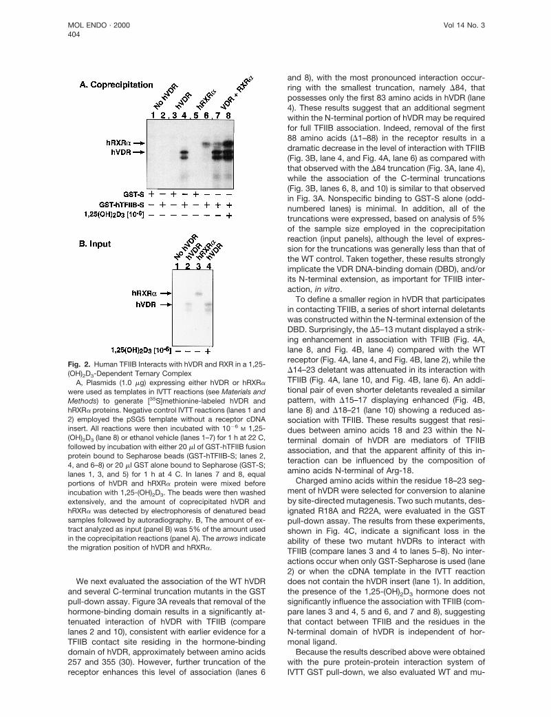
To define regions of hVDR that associate with TFIIB,we employed an in vitro coprecipitation or pull-downassay using glutathione-S-transferase (GST) fusionprotein methodology and in vitro transcribed andtranslated (IVTT) receptor proteins. Figure 2A depictsthe basic interaction between wild-type (WT) hVDRand a GST-TFIIB fusion protein bound to Sepharosebeads (lane 4), with no interaction occurring when onlyGST-Sepharose (GST-S) is used (lane 3) or when theIVTT reaction contains only the pSG5 parent expres-sion vector (lanes 1 and 2). The association betweenRXRa and TFIIB (lane 6) is much weaker than betweenVDR and TFIIB (lane 4), and the amount of RXRa thatis coprecipitated does not increase in the presence ofunliganded VDR (compare lanes 6 and 7). Importantly,inclusion of 1,25-(OH)2D3 markedly stimulates associ-ation of RXRa-VDR and TFIIB (lane 8). These obser-vations indicate that the RXR-VDR heterodimer, whichforms in solution in the presence of 1,25-(OH)2D3 andrepresents the functionally relevant molecular speciesin mediating the activation of VDRE-controlled genes,can readily interact with TFIIB in a presumed ternarycomplex.
Fig. 1. Construction of hVDR Mutants Used to Probe Interactions with TFIIBThe central portion of the figure depicts a schematic representation of full-length hVDR (427 amino acids) containing an
N-terminal DBD and C-terminal ligand binding region. Illustrated directly above the central hVDR molecule are the two polymor-phic designations for the receptor: f/M1 encoding the full-length hVDR and F/M4, which codes for a shorter 424-amino acidprotein isoform. Shown above the F/M4 hVDR are C- and N-terminal truncations that were generated in this receptor isoform bysite-directed mutagenesis. The bottom portion of the panel depicts smaller internal deletions and point mutations (arrows) thatwere generated in the F/M4 hVDR isoform in this study. The numbers after each delta symbol represent the amino acids deletedin each case [using the Baker et al. (44) numbering system]. For point mutations (E2A, R18A, R22A, and R22K), the number ofthe mutated residue is designated along with the corresponding amino acid substitution.
VDR Polymorphisms Modulate Interaction with TFIIB 403
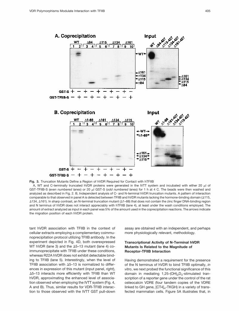
We next evaluated the association of the WT hVDRand several C-terminal truncation mutants in the GSTpull-down assay. Figure 3A reveals that removal of thehormone-binding domain results in a significantly at-tenuated interaction of hVDR with TFIIB (comparelanes 2 and 10), consistent with earlier evidence for aTFIIB contact site residing in the hormone-bindingdomain of hVDR, approximately between amino acids257 and 355 (30). However, further truncation of thereceptor enhances this level of association (lanes 6
and 8), with the most pronounced interaction occur-ring with the smallest truncation, namely D84, thatpossesses only the first 83 amino acids in hVDR (lane4). These results suggest that an additional segmentwithin the N-terminal portion of hVDR may be requiredfor full TFIIB association. Indeed, removal of the first88 amino acids (D1–88) in the receptor results in adramatic decrease in the level of interaction with TFIIB(Fig. 3B, lane 4, and Fig. 4A, lane 6) as compared withthat observed with the D84 truncation (Fig. 3A, lane 4),while the association of the C-terminal truncations(Fig. 3B, lanes 6, 8, and 10) is similar to that observedin Fig. 3A. Nonspecific binding to GST-S alone (odd-numbered lanes) is minimal. In addition, all of thetruncations were expressed, based on analysis of 5%of the sample size employed in the coprecipitationreaction (input panels), although the level of expres-sion for the truncations was generally less than that ofthe WT control. Taken together, these results stronglyimplicate the VDR DNA-binding domain (DBD), and/orits N-terminal extension, as important for TFIIB inter-action, in vitro.
To define a smaller region in hVDR that participatesin contacting TFIIB, a series of short internal deletantswas constructed within the N-terminal extension of theDBD. Surprisingly, the D5–13 mutant displayed a strik-ing enhancement in association with TFIIB (Fig. 4A,lane 8, and Fig. 4B, lane 4) compared with the WTreceptor (Fig. 4A, lane 4, and Fig. 4B, lane 2), while theD14–23 deletant was attenuated in its interaction withTFIIB (Fig. 4A, lane 10, and Fig. 4B, lane 6). An addi-tional pair of even shorter deletants revealed a similarpattern, with D15–17 displaying enhanced (Fig. 4B,lane 8) and D18–21 (lane 10) showing a reduced as-sociation with TFIIB. These results suggest that resi-dues between amino acids 18 and 23 within the N-terminal domain of hVDR are mediators of TFIIBassociation, and that the apparent affinity of this in-teraction can be influenced by the composition ofamino acids N-terminal of Arg-18.
Charged amino acids within the residue 18–23 seg-ment of hVDR were selected for conversion to alanineby site-directed mutagenesis. Two such mutants, des-ignated R18A and R22A, were evaluated in the GSTpull-down assay. The results from these experiments,shown in Fig. 4C, indicate a significant loss in theability of these two mutant hVDRs to interact withTFIIB (compare lanes 3 and 4 to lanes 5–8). No inter-actions occur when only GST-Sepharose is used (lane2) or when the cDNA template in the IVTT reactiondoes not contain the hVDR insert (lane 1). In addition,the presence of the 1,25-(OH)2D3 hormone does notsignificantly influence the association with TFIIB (com-pare lanes 3 and 4, 5 and 6, and 7 and 8), suggestingthat contact between TFIIB and the residues in theN-terminal domain of hVDR is independent of hor-monal ligand.
Because the results described above were obtainedwith the pure protein-protein interaction system ofIVTT GST pull-down, we also evaluated WT and mu-
Fig. 2. Human TFIIB Interacts with hVDR and RXR in a 1,25-(OH)2D3-Dependent Ternary Complex
A, Plasmids (1.0 mg) expressing either hVDR or hRXRawere used as templates in IVTT reactions (see Materials andMethods) to generate [35S]methionine-labeled hVDR andhRXRa proteins. Negative control IVTT reactions (lanes 1 and2) employed the pSG5 template without a receptor cDNAinsert. All reactions were then incubated with 1026 M 1,25-(OH)2D3 (lane 8) or ethanol vehicle (lanes 1–7) for 1 h at 22 C,followed by incubation with either 20 ml of GST-hTFIIB fusionprotein bound to Sepharose beads (GST-hTFIIB-S; lanes 2,4, and 6–8) or 20 ml GST alone bound to Sepharose (GST-S;lanes 1, 3, and 5) for 1 h at 4 C. In lanes 7 and 8, equalportions of hVDR and hRXRa protein were mixed beforeincubation with 1,25-(OH)2D3. The beads were then washedextensively, and the amount of coprecipitated hVDR andhRXRa was detected by electrophoresis of denatured beadsamples followed by autoradiography. B, The amount of ex-tract analyzed as input (panel B) was 5% of the amount usedin the coprecipitation reactions (panel A). The arrows indicatethe migration position of hVDR and hRXRa.
MOL ENDO · 2000 Vol 14 No. 3404
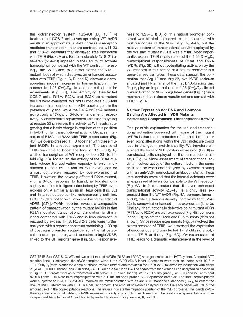
tant hVDR association with TFIIB in the context ofcellular extracts employing a complementary coimmu-noprecipitation protocol utilizing TFIIB antibody. In theexperiment depicted in Fig. 4D, both overexpressedWT hVDR (lane 3) and the D5–13 mutant (lane 4) co-immunoprecipitate with TFIIB under these conditions,whereas R22A hVDR does not exhibit detectable bind-ing to TFIIB (lane 5). Interestingly, when the level ofTFIIB association with D5–13 is normalized to differ-ences in expression of this mutant (input panel, right),D5–13 interacts more efficiently with TFIIB than WThVDR, approximating the enhanced level of associa-tion observed when employing the IVTT system (Fig. 4,A and B). Thus, similar results for VDR-TFIIB interac-tion to those observed with the IVTT GST pull-down
assay are obtained with an independent, and perhapsmore physiologically relevant, methodology.
Transcriptional Activity of N-Terminal hVDRMutants Is Related to the Magnitude ofReceptor-TFIIB Interaction
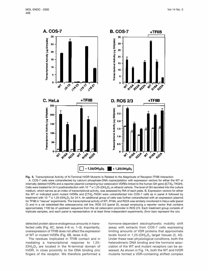
Having demonstrated a requirement for the presenceof the N terminus of hVDR to bind TFIIB optimally, invitro, we next probed the functional significance of thisdomain in mediating 1,25-(OH)2D3-stimulated tran-scription of a reporter gene under the control of the ratosteocalcin VDRE (four tandem copies of the VDRElinked to GH gene, [CT4]4-TKGH) in a variety of trans-fected mammalian cells. Figure 5A illustrates that, in
Fig. 3. Truncation Mutants Define a Region of hVDR Required for Contact with hTFIIBA, WT and C-terminally truncated hVDR proteins were generated in the IVTT system and incubated with either 20 ml of
GST-TFIIB-S (even numbered lanes) or 20 ml GST-S (odd numbered lanes) for 1 h at 4 C. The beads were then washed andanalyzed as described in Fig. 2. B, Independent analysis of C- and N-terminal hVDR truncation mutants. A pattern of interactioncomparable to that observed in panel A is detected between TFIIB and hVDR mutants lacking the hormone-binding domain (D115,D134, D161). In sharp contrast, an N-terminal truncation mutant (D1–88) that does not contain the zinc finger DNA-binding regionand N terminus of hVDR does not interact appreciably with hTFIIB (lane 4), at least under the wash conditions employed. Theamount of extract analyzed as input in each panel was 5% of the amount used in the coprecipitation reactions. The arrows indicatethe migration position of each hVDR protein.
VDR Polymorphisms Modulate Interaction with TFIIB 405
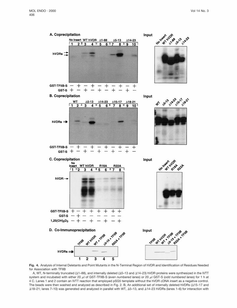
Fig. 4. Analysis of Internal Deletants and Point Mutants in the N-Terminal Region of hVDR and Identification of Residues Neededfor Association with TFIIB
A, WT, N-terminally truncated (D1–88), and internally deleted (D5–13 and D14–23) hVDR proteins were synthesized in the IVTTsystem and incubated with either 20 ml of GST-TFIIB-S (even numbered lanes) or 20 ml GST-S (odd numbered lanes) for 1 h at4 C. Lanes 1 and 2 contain an IVTT reaction that employed pSG5 template without the hVDR cDNA insert as a negative control.The beads were then washed and analyzed as described in Fig. 2. B, An additional set of internally deleted hVDRs (D15–17 andD18–21; lanes 7–10) was generated and analyzed in parallel with WT, D5–13, and D14–23 hVDRs (lanes 1–6) for interaction with
MOL ENDO · 2000 Vol 14 No. 3406
this cotransfection system, 1,25-(OH)2D3 (1028 M)treatment of COS-7 cells overexpressing WT hVDRresults in an approximate 56-fold increase in receptor-mediated transcription. In sharp contrast, the D14–23and D18–21 deletants that displayed little interactionwith TFIIB (Fig. 4, A and B) are moderately (D18–21) orseverely (D14–23) impaired in their ability to activatetranscription compared with the WT control. Interest-ingly, the D5–13 and, to a lesser extent, the D15–17mutant, both of which displayed an enhanced associ-ation with TFIIB (Fig. 4, A, B, and D), showed a corre-sponding modest increase in transactivation in re-sponse to 1,25-(OH)2D3. In another set of similarexperiments (Fig. 5B), also employing transfectedCOS-7 cells, R18A, R22A, and R22K point mutanthVDRs were evaluated. WT hVDR mediates a 23-foldincrease in transcription of the GH reporter gene in thepresence of ligand, while the R18A or R22A mutantsexhibit only a 17-fold or 3-fold enhancement, respec-tively. A conservative replacement (arginine to lysine)at residue 22 preserves the activity at WT levels, sug-gesting that a basic charge is required at this positionin hVDR for full transcriptional activity. Because inter-action of R18A and R22A with TFIIB is attenuated (Fig.4C), we overexpressed TFIIB together with these mu-tant hVDRs in a rescue experiment. The additionalTFIIB was able to boost the level of 1,25-(OH)2D3-elicited transcription of WT receptor from 23- to 34-fold (Fig. 5B). Moreover, the activity of the R18A mu-tant, whose transactivation capacity is only mildlyaffected (17-fold vs. 23-fold for WT hVDR), can bealmost completely restored by overexpression ofTFIIB. However, the severely affected R22A mutant,with a 3-fold response to ligand, is boosted onlyslightly (up to 4-fold ligand stimulation) by TFIIB over-expression. A similar analysis in HeLa cells (Fig. 5C)and in a rat osteoblast-like osteosarcoma cell line,ROS 2/3 (data not shown), also employing the artificialVDRE, [CT4]4-TKGH reporter, reveals a comparablepattern of transactivation by the mutant hVDRs in thatR22A-mediated transcriptional stimulation is dimin-ished compared with R18A and is less successfullyrescued by excess TFIIB. ROS 2/3 cells were furtheranalyzed with a reporter construct containing 1100 bpof upstream promoter sequence from the rat osteo-calcin natural promoter, which contains a single VDRE,linked to the GH reporter gene (Fig. 5D). Responsive-
ness to 1,25-(OH)2D3 of this natural promoter con-struct was blunted compared to that occurring withmultiple copies of the VDRE (Fig. 5, A–C), but therelative pattern of transcriptional activity displayed bythe WT and mutant hVDRs was similar. Most impor-tantly, excess TFIIB nearly restored the 1,25-(OH)2D3
transcriptional responsiveness of R18A and R22AhVDRs (Fig. 5D) without potentiating activation by theWT receptor in this setting of a natural promoter in abone-derived cell type. These data support the con-tention that Arg-18 and Arg-22, two hVDR residuessituated just N-terminal of the first DNA-binding zincfinger, play an important role in 1,25-(OH)2D3-elicitedtransactivation of VDRE-regulated genes (Fig. 5) via amechanism that includes recruitment and contact withTFIIB (Fig. 4).
Neither Expression nor DNA and HormoneBinding Are Affected in hVDR MutantsPossessing Compromised Transcriptional Activity
One possible explanation for the reduced transcrip-tional activation observed with some of the mutanthVDRs is that the introduction of internal deletions oreven point alterations within the VDR molecule couldlead to changes in protein stability. We therefore ex-amined the level of VDR protein expression (Fig. 6) intransfected cells employed for the transcriptional as-says (Fig. 5). Since assessment of transcriptional ac-tivity involves assay of the culture medium, the samecells can be lysed and analyzed by Western blottingwith an anti-VDR monoclonal antibody (9A7g). Theseimmunoblots revealed that the internal deletants wereall expressed at levels comparable to the WT receptor(Fig. 6A). In fact, a mutant that displayed enhancedtranscriptional activity (D5–13) is slightly less ex-pressed than the WT hVDR (Fig. 6A, compare lanes 1and 2), while a transcriptionally inactive mutant (D14–23) is somewhat enhanced in its expression (lane 3).Similarly, the functionally defective point mutant VDRs(R18A and R22A) are well expressed (Fig. 6B, comparelanes 1–3), as are the R22K and E2A mutants (data notshown). Since rescue experiments (Fig. 5) involved theoverexpression of TFIIB, we assessed the expressionof endogenous and transfected TFIIB utilizing a poly-clonal TFIIB antibody (Fig. 6C). Overexpression ofTFIIB leads to a dramatic enhancement in the level of
GST-TFIIB-S or GST-S. C, WT and two point mutant hVDRs (R18A and R22A) were generated in the IVTT system. A control IVTTreaction (lane 1) employed the pSG5 template without the hVDR cDNA insert. Reactions were then incubated with 1026 M
1,25-(OH)2D3 (even numbered lanes) or ethanol vehicle (odd numbered lanes) for 1 h at 22 C followed by incubation with either20 ml GST-TFIIB-S (lanes 1 and 3–8) or 20 ml GST-S (lane 2) for 1 h at 4 C. The beads were then washed and analyzed as describedin Fig. 2. D, Extracts from cells transfected with either TFIIB alone (lane 1), WT hVDR alone (lane 2), or TFIIB and WT or mutanthVDRs (lanes 3–5) were immunoprecipitated with a TFIIB antibody-protein A/G-Sepharose complex. The immunoprecipitateswere subjected to 5–20% SDS/PAGE followed by immunoblotting with an anti-VDR monoclonal antibody (9A7g) to detect thelevel of hVDR interaction with TFIIB in a cellular context. The amount of extract analyzed as input in each panel was 5% of theamount used in the coprecipitation reactions. The arrows indicate the migration position of the hVDR proteins. The bands belowthe migration position of full-length hVDR represent proteolytic products in each reaction. The results are representative of threeindependent trials for panel C and two independent trials each for panels A, B, and D.
VDR Polymorphisms Modulate Interaction with TFIIB 407
detected protein above endogenous amounts in trans-fected cells (Fig. 6C, lanes 4–6 vs. 1–3). Importantly,overexpression of TFIIB does not affect the expressionof WT or mutant hVDRs (Fig. 6B, lanes 4–6).
The residues implicated in TFIIB contact and inmediating a transcriptional response to 1,25-(OH)2D3 are located in the N-terminal domain ofhVDR, in close proximity to the DNA binding zincfingers of the receptor. We therefore performed a
hormone-dependent electrophoretic mobility shiftassay with extracts from COS-7 cells expressinglimiting amounts of VDR proteins that approximatelevels found in 1,25-(OH)2D3 target tissues (3, 45).Under these near-physiological conditions, both theheterodimeric DNA binding and the hormone asso-ciation of the WT and mutant receptors can be as-sessed. As shown in Fig. 7A, both the WT and hVDRmutants formed a VDR-containing shifted complex
Fig. 5. Transcriptional Activity of N-Terminal hVDR Mutants Is Related to the Magnitude of Receptor-TFIIB InteractionA, COS-7 cells were cotransfected by calcium phosphate-DNA coprecipitation with expression vectors for either the WT or
internally deleted hVDRs and a reporter plasmid containing four osteocalcin VDREs linked to the human GH gene ([CT4]4-TKGH).Cells were treated for 24 h posttransfection with 1028 M 1,25-(OH)2D3 or ethanol vehicle. The level of GH secreted into the culturemedium, which serves as an index of transcriptional activity, was assessed by RIA of each plate. B, Expression vectors for eitherthe WT or indicated point mutant hVDRs and [CT4]4-TKGH were cotransfected into COS-7 cells as in panel A followed bytreatment with 1028 M 1,25-(OH)2D3 for 24 h. An additional group of cells was further cotransfected with an expression plasmidfor TFIIB in “rescue” experiments. The transcriptional activity of WT, R18A, and R22A was similarly monitored in HeLa cells (panelC) and in a rat osteoblast-like osteosarcoma cell line, ROS 2/3 (panel D), except employing a reporter vector that containsapproximately 1100 bp of upstream sequence from the rat osteocalcin promoter in ROS 2/3. Each treatment group consists oftriplicate samples, and each panel is representative of at least three independent experiments. Error bars represent the SEM.
MOL ENDO · 2000 Vol 14 No. 3408

(lanes 3, 6, and 8) that was enhanced by the additionof the 1,25-(OH)2D3 ligand (lanes 4, 7, and 9). Sig-nificantly, the level of augmentation by 1,25-(OH)2D3
(;4-fold) based on densitometric scanning of theautoradiographs in Fig. 7A was similar for the WTreceptor and each mutant hVDR (data not shown).Also, the level of expression of each receptor wasessentially equivalent and unmodified by ligand asmonitored by immunoblotting (data not shown). Thenonspecific (NS) nature of the two lower protein-DNA complexes was deduced by their appearancein the lanes that used extracts from non-VDR trans-fected cells (lanes 1 and 2), as well as by their lackof elimination by the VDR antibody (lane 5). Similardata were obtained when employing the R18A orR22A hVDR (Fig. 7B). Taken together, these resultsstrongly suggest that deletion of hVDR residues be-tween positions 15 and 21 or modification of aminoacids 18 or 22 to alanine does not decrease het-erodimeric DNA binding by the VDR protein. There-fore, altered VDRE association cannot account forthe attenuation of transactivation by these mutants.Additionally, the nearly equivalent enhancement inDNA binding elicited by the 1,25-(OH)2D3 hormonesuggests that ligand binding properties of the mu-tant hVDRs remain unchanged.
The Polymorphic N Terminus of hVDR AffectsBoth the Interaction with TFIIB and theLevel of Transactivation
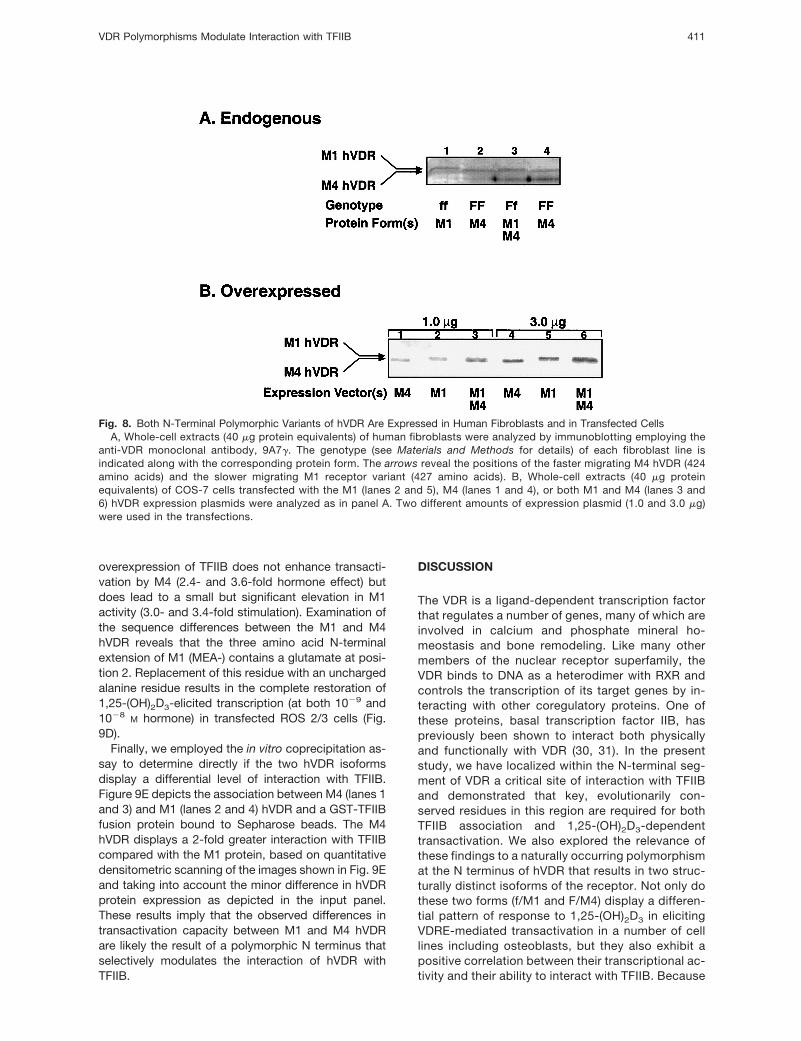
The N-terminal hVDR FokI polymorphism results in theformation of either a full-length 427-amino acid hVDR(denoted either “f” to indicate the presence of the FokIrestriction site or designated “M1” for translation fromthe first methionine in the primary sequence), or ashorter 424-amino acid protein (termed either “F” forthe absence of the FokI site or named “M4” to indicatetranslational initiation from the methionine at the fourthposition in the primary sequence). These isoformshave previously been associated with differences inBMD in diverse populations (36–41). Having identifiedspecific residues located near the N terminus of hVDRas important for TFIIB interaction and transactivation,we next investigated the potential impact, if any, of thispolymorphic N terminus on the TFIIB contact domainnearby in the primary sequence. First, extracts fromhuman fibroblasts were evaluated by Western blottingto determine the extent of expression of the two en-dogenous hVDRs in these cells. As shown in Fig. 8A,both polymorphic forms of the receptor are well ex-pressed (lanes 1 and 2), and in the case of the hetero-zygote, both proteins are apparently synthesized (lane3). The electrophoretic mobility of the M1 and M4hVDRs can be distinguished in these denaturing gels,with the shorter M4 receptor displaying a slightly fastermigration (compare lane 2 to 1). We then constructedexpression vectors containing the coding sequencefor both the M1 and M4 hVDRs. Western blot analysis
of extracts from COS-7 cells transfected with either ofthese plasmids demonstrates the expression of theappropriate hVDR isoform (Fig. 8B), with little differ-ence in the expression levels of either species. As inthe case of the endogenous hVDR, the overexpressedpolymorphic receptors display distinct electrophoreticmobilities (compare lanes 1 and 2 or 4 and 5). Similarto the experiments shown in Fig. 7, we evaluated theheterodimeric DNA binding activity and hormone bind-ing capacity of the M1 and M4 hVDRs in the hormone-dependent gel mobility shift assay and found no de-tectable differences in either DNA or 1,25-(OH)2D3
association between these two receptor isoforms(data not shown).
Since both polymorphic forms of hVDR are wellexpressed, either endogenously in cultured human fi-broblasts, or in transfected cells, we tested the tran-
Fig. 6. N-Terminal hVDR Mutants and hTFIIB Are Well Ex-pressed in Transfected Cells
A, Whole-cell extracts (40 mg protein equivalents) ofCOS-7 cells transfected with the indicated hVDR expressionplasmids were analyzed by immunoblotting employing theanti-VDR monoclonal antibody, 9A7g (64) as described inMaterials and Methods. Note the faster migration of theD5–13 and D14–23 mutants that contain larger deletions. NSdenotes nonspecific bands. B, Immunoblotting was per-formed as in panel A utilizing COS-7 cells transfected withexpression vectors for WT or point mutant hVDRs in theabsence or presence of an additional expression plasmidcontaining TFIIB. C, A parallel set of extracts was probed witha polyclonal antibody directed against TFIIB.
VDR Polymorphisms Modulate Interaction with TFIIB 409

scriptional activity in response to varying doses of1,25-(OH)2D3 of these two receptors in different trans-fected cell lines. Figure 9A illustrates that in trans-fected COS-7 cells, 1,25-(OH)2D3 (1029 and 1028 M)treatment results in an approximate 27- and 78-foldincrease, respectively, in receptor-mediated transcrip-tion with the M4 receptor. In contrast, the M1 proteinonly displayed an 8.5- and 38-fold increase in activityat these two ligand concentrations. Interestingly, over-expression of TFIIB did not lead to a statistically sig-nificant potentiation of transcription with the more ac-tive M4, but did increase the activity of M1 (up to
13.3- and 66-fold). Similar results were obtained withthe [CT4]4-TKGH reporter in HeLa cells (Fig. 9B) and inmurine P19 embryonal carcinoma (EC) cells or a ratosteoblast-like osteosarcoma cell line, ROS 2/3 (datanot shown). ROS 2/3 cells were further analyzed witha reporter construct containing 1100 bp of the ratosteocalcin natural promoter linked to the GH reportergene (Fig. 9C). In this experiment, 1,25-(OH)2D3 (1029
and 1028 M) treatment results in an approximate 3.0-and 3.5-fold increase, respectively, in receptor-medi-ated transcription with the M4 receptor, but only a 1.8-and 2.6-fold effect was noted with M1. Again, the
Fig. 7. DNA and Hormone Binding Are Not Affected in hVDR Mutants Possessing Compromised Transcriptional ActivityA, A gel mobility shift assay was performed using extracts (5 mg total protein) from COS-7 cells transfected with WT or
internally deleted hVDR expression plasmids (0.1 mg) along with a labeled oligonucleotide containing the VDRE from the ratosteocalcin gene as described in Materials and Methods. Extracts were incubated with 1027 M 1,25-(OH)2D3 (lanes 2, 4, 5,7, and 9) or ethanol vehicle (lanes 1, 3, 6, and 8) for 0.5 h at 22 C before incubation with the VDRE oligonucleotide. Lane5 also contained an anti-VDR monoclonal antibody (9A7g) that is directed against an epitope in the DBD and disrupts theinteraction of VDR with the VDRE (12). Lanes 1 and 2 contain extracts from COS-7 cells transfected with the expressionvector lacking the hVDR insert. Arrows indicate the migration of the specific VDR-VDRE complex (as a heterodimer withRXR), nonspecific complexes (NS), or free probe. B, The gel mobility shift assay was performed as in panel A but employingpoint mutant hVDRs.
MOL ENDO · 2000 Vol 14 No. 3410
overexpression of TFIIB does not enhance transacti-vation by M4 (2.4- and 3.6-fold hormone effect) butdoes lead to a small but significant elevation in M1activity (3.0- and 3.4-fold stimulation). Examination ofthe sequence differences between the M1 and M4hVDR reveals that the three amino acid N-terminalextension of M1 (MEA-) contains a glutamate at posi-tion 2. Replacement of this residue with an unchargedalanine residue results in the complete restoration of1,25-(OH)2D3-elicited transcription (at both 1029 and1028 M hormone) in transfected ROS 2/3 cells (Fig.9D).
Finally, we employed the in vitro coprecipitation as-say to determine directly if the two hVDR isoformsdisplay a differential level of interaction with TFIIB.Figure 9E depicts the association between M4 (lanes 1and 3) and M1 (lanes 2 and 4) hVDR and a GST-TFIIBfusion protein bound to Sepharose beads. The M4hVDR displays a 2-fold greater interaction with TFIIBcompared with the M1 protein, based on quantitativedensitometric scanning of the images shown in Fig. 9Eand taking into account the minor difference in hVDRprotein expression as depicted in the input panel.These results imply that the observed differences intransactivation capacity between M1 and M4 hVDRare likely the result of a polymorphic N terminus thatselectively modulates the interaction of hVDR withTFIIB.
DISCUSSION
The VDR is a ligand-dependent transcription factorthat regulates a number of genes, many of which areinvolved in calcium and phosphate mineral ho-meostasis and bone remodeling. Like many othermembers of the nuclear receptor superfamily, theVDR binds to DNA as a heterodimer with RXR andcontrols the transcription of its target genes by in-teracting with other coregulatory proteins. One ofthese proteins, basal transcription factor IIB, haspreviously been shown to interact both physicallyand functionally with VDR (30, 31). In the presentstudy, we have localized within the N-terminal seg-ment of VDR a critical site of interaction with TFIIBand demonstrated that key, evolutionarily con-served residues in this region are required for bothTFIIB association and 1,25-(OH)2D3-dependenttransactivation. We also explored the relevance ofthese findings to a naturally occurring polymorphismat the N terminus of hVDR that results in two struc-turally distinct isoforms of the receptor. Not only dothese two forms (f/M1 and F/M4) display a differen-tial pattern of response to 1,25-(OH)2D3 in elicitingVDRE-mediated transactivation in a number of celllines including osteoblasts, but they also exhibit apositive correlation between their transcriptional ac-tivity and their ability to interact with TFIIB. Because
Fig. 8. Both N-Terminal Polymorphic Variants of hVDR Are Expressed in Human Fibroblasts and in Transfected CellsA, Whole-cell extracts (40 mg protein equivalents) of human fibroblasts were analyzed by immunoblotting employing the
anti-VDR monoclonal antibody, 9A7g. The genotype (see Materials and Methods for details) of each fibroblast line isindicated along with the corresponding protein form. The arrows reveal the positions of the faster migrating M4 hVDR (424amino acids) and the slower migrating M1 receptor variant (427 amino acids). B, Whole-cell extracts (40 mg proteinequivalents) of COS-7 cells transfected with the M1 (lanes 2 and 5), M4 (lanes 1 and 4), or both M1 and M4 (lanes 3 and6) hVDR expression plasmids were analyzed as in panel A. Two different amounts of expression plasmid (1.0 and 3.0 mg)were used in the transfections.
VDR Polymorphisms Modulate Interaction with TFIIB 411

these hVDR isoforms have previously been associ-ated with differences in BMD in certain populations,one factor contributing to a genetic predisposition toosteoporosis therefore may involve the varying po-tency in interaction between polymorphic hVDRsand the basal transcriptional machinery.
Initial Mapping of a TFIIB Contact Site in VDR
Previous studies have identified TFIIB as a VDR-inter-acting protein, employing both pull-down assays (30–34) and the yeast two-hybrid system (31, 34). Yeasttwo-hybrid data from our laboratory confirm this inter-
Fig. 9. Transcriptional Activity of the f/M1 Polymorphic hVDR Is Reduced as Is Association of this Variant Receptor with hTFIIBExpression vectors for either the M1 (427 amino acids) or M4 (424 amino acids) polymorphic hVDR and [CT4]4-TKGH were
cotransfected into COS-7 (panel A) or HeLa (panel B) cells as in Fig. 5 followed by treatment with the indicated concentrationof 1,25-(OH)2D3 for 24 h. A parallel group of cells was further cotransfected with an expression plasmid for human TFIIB in“rescue” experiments (1TFIIB). The transcriptional activity of M1 and M4 hVDRs was also monitored in a rat osteoblast-likeosteosarcoma cell line, ROS 2/3 (panel C), employing a reporter vector that contains approximately 1100 bp of upstreamsequence from the rat osteocalcin promoter linked to the GH reporter gene. D, Expression vectors for M1 and M4polymorphic hVDR, or a mutant M1 (containing an alanine at position 2 instead of a glutamic acid; E2A) along with a naturalosteocalcin promoter-reporter construct were cotransfected into ROS 2/3 cells as in panel C, followed by treatment with theindicated concentration 1,25-(OH)2D3 for 24 h. Each treatment group consists of triplicate samples, and each panel isrepresentative of at least three independent experiments. Error bars represent the SEM. E, M1 (lanes 2 and 4) and M4 (lanes1 and 3) hVDR proteins were generated in the IVTT system and incubated with 20 ml of GST-hTFIIB-S for 1 h at 4 C. Thebeads were then washed and analyzed as described in the legend to Fig. 2. The amount of extract analyzed as input was5% of the amount used in the pull-down reactions.
MOL ENDO · 2000 Vol 14 No. 3412
action (C. Encinas and P. W. Jurutka, unpublisheddata). Furthermore, mapping studies have been per-formed using truncation and deletion methodologywith both the GST pull-down (30) and yeast assays(31, 34). The results of these initial mapping experi-ments pointed to the hormone-binding domain inhVDR as possessing one site of interaction for TFIIB,likely encompassing primarily amino acids 257–355(30). More recently, utilizing an alternative strategy, thehormone-binding domain (hVDR residues 93–427) wasemployed to generate a random point mutant librarythat was used to screen for TFIIB interaction-deficientmutants in the yeast two-hybrid system. The resultsimplicated 11 individual residues between amino acids228 and 345 in VDR, the majority of which are hydro-phobic, as being critical for TFIIB association (46).
Multiple TFIIB Interaction Sites andFunctional Significance
Several factors argue, however, that the C-terminaldomain of hVDR may not be the only site of TFIIBinteraction. The limitation of using gross deletions andtruncations (30, 31) is that, once any critical interactingregion has been removed, other, perhaps equally im-portant, domains may go undetected. Also, in theabove cited point mutant screen with the hVDR hor-mone-binding region (46), the entire N-terminal regionof hVDR (residues 1–92) was not present during theanalysis. These caveats, plus the current results usingsmall N-terminal deletions and point mutations, leadus to propose that, in addition to hydrophobic aminoacids within the hormone-binding region of hVDR,charged residues in the N-terminal domain of the mol-ecule are required for optimal TFIIB association andfunctionally competent VDR-mediated transactivation(see also Fig. 10).
The functional significance of TFIIB interaction withVDR is supported by transactivation studies that dem-onstrate a cooperative effect of these two molecules.In experiments employing P19 EC cells, 1,25-(OH)2D3
treatment results in only a minor stimulation of reportergene expression when either hVDR or TFIIB alone iscotransfected into these cells. Concomitant overex-pression of VDR and TFIIB causes a dramatic (.30-fold) enhancement in reporter gene transcription (30).Another report (34) revealed that expression of a TFIIBmutant that was nonfunctional but still retained theability to interact with hVDR resulted in a selectivedominant negative effect on 1,25-(OH)2D3-elicitedtranscription in an osteoblast-like cell line. This inhibi-tion could be reversed by simultaneous expression ofWT TFIIB. Neither an inhibition nor a stimulation wasobserved with glucocorticoid-induced transcription ofa glucocorticoid responsive element-reporter con-struct, indicating that excess TFIIB did not exert non-specific effects on transcription. Thus, one require-ment for 1,25-(OH)2D3-mediated transcription appearsto be specific physical interaction of hVDR with TFIIB,
which we propose involves both C- and N-terminaldomains in the receptor.
Previous investigations of the putative functionalrole for the N terminus of hVDR have suggested that,unlike some of the other nuclear receptors that pos-sess a larger N-terminal domain, perhaps this region ofthe hVDR may be functionally dispensable. Removal ofN-terminal residues up to and including amino acid 21in hVDR did not have a significant impact on transcrip-tional activity in transfected COS-1 cells using a hu-man osteocalcin gene promoter-reporter construct(47). However, this hVDR construct still possessesArg-22, a residue that is vital for transcriptional activityin all of the cell lines we tested. One might thereforededuce from our results that exposure of Arg-22 at theextreme N terminus of the (D1–21) truncation mutant(47) might accentuate its role, even obviating the needfor Arg-18, the other basic residue we found to play asignificant role in hVDR-TFIIB binding.
Other Nuclear Receptors Bind to TFIIB
The present proposal that hVDR has a second, N-terminal interaction domain with TFIIB, and that thisdomain contains crucial basic residues, has a prece-dent from the results concerning interaction betweenTR and TFIIB. Chicken TRa contains 50 residues N-terminal of the first zinc finger, including a cluster ofbasic amino acids (KRKRK) at positions 23–27 thatwere shown to be crucial for both TFIIB interaction andtransactivation by this receptor (48). Thus, like hVDR,chicken TRa requires N-terminal basic residues forcontacting TFIIB and for transactivation. The fact thatthis cluster of positively charged amino acids is wellconserved among all known TRa homologs from ver-tebrate species [including fish (GenBank accessionno. BAA08201)] is indeed consistent with such animportant functional role for these residues. Signifi-cantly, positively charged residues are conserved atpositions corresponding to 18 and 22 in all publishedVDR sequences, ranging from human to the recentlycloned Xenopus VDR (GenBank accession no.AAB58585), strongly implying that these residuesmust subserve some crucial function.
Moreover, human TRb also interacts specificallywith TFIIB and, similar to our proposal for VDR (Fig.10), deletion analysis revealed two contact sites in theTRb molecule, one located in the N-terminal regionand the other positioned in the ligand-binding domain(49). Each of these two distinct regions in TRb wasdemonstrated to interact with different sites in TFIIB(49). It therefore appears that VDR and TR, in additionto sharing other features, such as heterodimerizationwith RXR on direct-repeat responsive elements as wellas some sequence homology (27%), also possessdual interaction interfaces with TFIIB. Of course, it ispossible that, in the tertiary structures of full-length TRand VDR, these two interaction regions combine toform a single docking scaffold.
VDR Polymorphisms Modulate Interaction with TFIIB 413

A further example of a nuclear receptor that inter-acts with TFIIB via an N-terminal domain is the ho-modimerizing ER (50). This study of ER, as well as asimilar evaluation of the orphan nuclear receptor he-patocyte nuclear factor-4 (HNF-4) (51), concludes thatassociation of TFIIB with the respective receptor facil-itates the assembly of transcriptional preinitiationcomponents, particularly the TATA-box binding pro-tein (TBP). Thus, a picture of nuclear receptor trans-activation is beginning to emerge in which TFIIB playsa crucial role.
Significance of the TFIIB Interaction inTransactivation by VDR
There appear to be at least two distinct sets of protein-protein interactions involved in transcriptional activa-tion by hVDR. One is represented by a group of co-activators that bind to a cleft comprised of the Tyr-236to Lys-246 (helix 3) region combined with the helix 12AF-2 platform in VDR (52) and effect derepression ofchromatin nucleosome organization via HAT-cata-lyzed displacement of histones at the active promotersite. In our current model, contact by these coactiva-tors with components of TBP-TAF and RNA polymer-ase II, with the possible participation of a cointegratoranalogous to CBP/p300 (53), facilitates transcriptional
activation. A second crucial interaction appears to bethe recruitment of TFIIB to the promoter via the VDR-RXR heterocomplex (see Figs. 2 and 10). The deliveryof TFIIB would then stabilize the RNA polymerase IIpreinitiation complex and allow for repeated rounds oftranscription of the regulated target gene.
Clinical Impact and Functional Significance ofhVDR Polymorphisms
The FokI site polymorphism in exon 2 of the hVDRgene is distinct from all other reported VDR polymor-phisms in that the two biallelic variants actually differ inprotein sequence (f/M1 being 3 amino acids longer).The shorter F/M4 receptor apparently arose after thedivergence of hominids from apes and has beendubbed a “neomorph” (1), yet it presently constitutesapproximately 65% of VDR alleles in human subjects(36–41, 54, 55). This predominance of the relativelyrecent F/M4 allele suggests an evolutionary advantagein humans. In support of this notion, the African-Amer-ican population, which has a significantly lower inci-dence of osteoporosis than Caucasians, also has agreater prevalence (.80%) of the F/M4 variant (38).
A direct association between the VDR FokI polymor-phism and BMD has been reported in several studies.In a group of Mexican-American/Caucasian women,
Fig. 10. Model of Interaction between hVDR and TFIIB and Its Consequences for Transactivation by the hVDR-RXR Complex onthe VDRE
M1 and M4 hVDR refer to genetic variants of the hVDR gene, also distinguished by the presence (f) or absence (F) of a FokIsite in the genomic DNA. A, The M4 variant of hVDR, lacking a Glu residue at position 2, can interact optimally with hTFIIB viatwo conserved basic residues (Arg-18 and Arg-22) in the Site II (denoted “II”) N-terminal interaction domain. This interaction withhTFIIB is augmented by a previously described Site I interacting region, denoted “I”, located within the C-terminal ligand-bindingdomain of hVDR (30, 34). TFIIB is thereby efficiently delivered to the transcriptional initiation complex, leading to robusttranscriptional activation. Also part of the activation process is the dissociation of a corepressor (“CoR”), as well as the bindingof a coactivator (CoA), like SRC-1, which aids in derepression of the gene by acetylating histones and reorganizing chromatin inthe promoter region (as suggested by the “dissociation” of histones; see text for references). B, The M1 variant of hVDR containsthree additional amino acids at the extreme N terminus, including a Glu residue (Glu-2), which is proposed to weaken theinteraction of Arg-18 and Arg-22 with hTFIIB. Subsequent steps in transactivation by the hVDR-RXR complex on the VDRE canstill occur but are attenuated by the weaker nature of the f/M1 hVDR contact with hTFIIB in the Site II region.
MOL ENDO · 2000 Vol 14 No. 3414

subjects with the ff genotype had a 12.8% lower BMDat the lumbar spine than FF subjects, with heterozy-gotes possessing an intermediate BMD (36). Thisstudy also showed an increased 2-yr rate of bone lossfrom the femoral neck in the ff vs. FF women. Similarly,in a population of premenopausal Japanese women,BMD in the lumbar spine was 12% less for the ffgenotype vs. FF (37). Recently, in a large cohort thatconsisted of a group of 400 postmenopausal womenof Italian descent, the FF genotype was associatedwith enhanced lumbar BMD (41). Interestingly, the ef-fect of the FF genotype on enhanced lumbar BMD wasgreatest in women within 5 yr of menopause, progres-sively declining afterward, and the low BMD ff geno-type was significantly overrepresented in patients withosteoporotic vertebral fractures (relative risk 2.58). Inyet a fourth population of American premenopausalwomen (both Caucasian and African-American), ffwomen had femoral neck BMD that was 7.4% lowerthan that of FF women (38). The association of BMDand the FokI polymorphism also has been extended toinclude a population of healthy growing children. Sub-jects that were FF homozygotes had a total body BMDthat was 8.2% higher and a mean calcium absorptionthat was 41.5% greater than ff individuals (40). Thus,the FokI polymorphism may be related to several VDR-dependent parameters of bone metabolism, includingintestinal calcium absorption and BMD. However, onestudy found no significant relationship between the F/fgenotype and BMD in a population of French pre-menopausal women (55), although as the authorspoint out, the association of BMD and this polymor-phism may be masked by various regional factors,such as a high calcium diet. Indeed, an influence ofdietary calcium on the impact of the FokI polymor-phism on BMD has been suggested by another group(39).
Comparative activities of the M1 vs. M4 hVDRs havebeen investigated directly via cotransfection of ex-pression plasmids for these isoforms together with1,25-(OH)2D3-responsive reporter vectors. One suchreport (37) suggested a 1.7-fold greater activity ofF/M4 over f/M1 hVDR in transfected HeLa cells. Weobserve (Fig. 9) that the F/M4 hVDR isoform is 1.5- to2.5-fold more transcriptionally active than the f/M1protein, with the most marked difference resultingwhen the isoforms are assayed in osteoblast-like cellsemploying a VDRE in the setting of its natural, bone-specific promoter. However, in another study (56), theactivities of hVDRs were comparable, both in trans-fected COS-7 cells employing reporter gene con-structs and utilizing Northern blot analysis of vitamin D24-hydroxylase mRNA induction by 1,25-(OH)2D3 inhuman fibroblasts. The disparate results observed be-tween Gross et al. (56) and our present report or thatof Arai et al. (37) may be because of differences be-tween the reporter constructs used in each study.While we employed both a synthetic rat osteocalcinVDRE and a construct containing the natural promoterregion from the osteocalcin gene, Gross et al. (56)
used a synthetic human osteocalcin reporter and aconstruct that contained a large portion of the vitaminD 24-hydroxylase promoter. Importantly, the negativestudy of Gross et al. (56), and the earlier work by Araiet al. (37) demonstrating a functional difference be-tween M1 and M4 hVDR, were both conducted usingtransient transfection experiments with a single cellline. In the present study, the functional variance be-tween M1 and M4 hVDR was derived from experi-ments in four different cell lines, including an osteo-blast-like osteosarcoma line (ROS 2/3). In addition, wehave provided a functional linkage between the vari-ance in M1/M4 hVDR activity and the differential in-teraction of these two isoforms with TFIIB, an obser-vation that is consistent with the proximity of the hVDRpolymorphic N terminus and the novel TFIIB interac-tion domain localized within the N-terminal segment ofhVDR described in the present study. Finally, recentinvestigations in our laboratory have found that theF/M4 VDR is more transcriptionally active than thef/M1 receptor by analyzing endogenous VDR functionin 17 different human fibroblast lines with varying ge-notypes at the FokI locus but a constant genotype interms of relevant VDR polymorphisms in the 39-UTR(G. K. Whitfield, L. S. Remus, P. W. Jurutka, H. Zitzer,A. K. Oza, H. T. L. Dang, C. A. Haussler, M. A. Galligan,M. L. Thatcher, and M. R. Haussler, unpublished data).Therefore, we conclude that the F/M4 neomorph rep-resents a more transcriptionally potent VDR isoform.
Integrative Model for Transactivation byPolymorphic hVDRs
Figure 10 represents a schematic working model fortranscriptional control by 1,25-(OH)2D3 that incorpo-rates the new findings presented in this manuscript.The model highlights a novel role for conserved basicresidues in the N-terminal region of VDR for contactingTFIIB and provides an explanation for the differentialeffect of the polymorphic N terminus on both the in-teraction of the receptor with this basal transcriptionfactor and on the activity of VDR. Panel A (far left)depicts the shorter 424 amino acid F/M4 hVDR inassociation with TFIIB at two sites within the receptor(the three molecules of TFIIB do not imply that thisnumber of TFIIB molecules interact with one hVDR butrather are meant to indicate the difference in the levelof interaction between TFIIB and M4 vs. M1 hVDR).Site I is localized in the C-terminal hormone-bindingregion of hVDR (30, 34) while site II represents theN-terminal domain identified in the present results,specifically conserved basic residues Arg-18 andArg-22 (denoted by 11). Also associated with VDR isa corepressor (CoR), likely SMRT (signal mediator andrepressor of transcription), which is thought to contactthe receptor in the C-terminal ligand-binding domain(57). The VDR-TFIIB complex is loosely bound to DNAvia the VDR DNA binding zinc fingers, but the regu-lated gene is illustrated in the repressed state because
VDR Polymorphisms Modulate Interaction with TFIIB 415
of chromatin nucleosome structure (shown schemati-cally as an association of DNA and histones).
Upon binding to the 1,25-(OH)2D3 ligand (panel A,middle), the M4 VDR is postulated to undergo a con-formational change with the following consequences:1) release of TFIIB from site I (34), 2) release of thecorepressor and concomitant association of VDR withthe RXR heteropartner (11, 45, 57) and a coactivator/HAT protein (CoA), the latter interaction being facili-tated by the AF-2 of VDR (18–20, 58), and 3) derepres-sion of the target gene by coactivator/HAT-mediatedchromatin nucleosome reorganization (schematicallydepicted as a release of the histones). As a result ofthis derepressive effect, the active RXR-VDR-TFIIBcomplex can now associate with high-affinity VDREbinding sites located in the promoter region of theregulated gene (panel A, right) and “deliver” TFIIB tothe preinitiation complex (PIC), resulting in stabiliza-tion of the PIC followed by subsequent rounds of RNApolymerase II-directed transcription of the down-stream target gene. It is conceivable that the TFIIBdelivery process is also facilitated by a VDRE (DNA)-induced change in the conformation of the N-terminaldomain of VDR, thereby releasing TFIIB as the rate-limiting factor in the formation of the PIC.
Panel B depicts a similar mechanism of action forthe longer 427-amino acid M1 hVDR. However, thisisoform of VDR does not interact with TFIIB as well asM4 hVDR (schematically denoted as only two mole-cules of TFIIB), presumably because of the presenceof a negatively charged glutamate (minus sign en-closed by circle) localized within the three amino acidN-terminal extension of M1 hVDR. We speculate thatthe molecular mechanism whereby the negativelycharged Glu-2 residue attenuates TFIIB binding couldinvolve either an intermolecular repulsion betweenf/M1 hVDR and presumed negative residues in TFIIBthat bridge to VDR Arg-22/18, or a nonproductive in-tramolecular interaction of Glu-2 in f/M1 hVDR with theArg-22/18 TFIIB site II, thus precluding TFIIB contact.Regardless, the hypothesized net result is that the M1isoform delivers less TFIIB to the PIC, with a subse-quently reduced amount of transcriptional initiationand mRNA synthesis from the target gene. Experimen-tally, the activity of M1 hVDR can be raised to the levelof the more potent M4 receptor either by 1) overex-pression of TFIIB to boost the endogenous levels ofthis protein and thus overcome the lower affinity forhVDR and the resulting diminished local concentra-tions that are delivered by M1 hVDR (Fig. 9), or 2) by“neutralization” of the glutamic acid residue at position2 in M1 hVDR via mutagenesis to an alanine (Fig. 9D).
In summary, we have elucidated a novel domain inhVDR, located near the N terminus and adjacent to theDNA-binding zinc finger motif, that is required for 1,25-(OH)2D3-elicited transcriptional activity. Within this re-gion, two basic residues, Arg-18 and Arg-22, wereidentified as critical for transactivation and contactwith the basal transcription factor IIB. A polymorphicvariant of hVDR that encodes a shorter, 424-amino
acid protein (F/M4), which has been associated withenhanced BMD in diverse populations, is more tran-scriptionally active and is shown herein to associatemore avidly with TFIIB compared with the 427-aminoacid f/M1 isoform. Given the central role of 1,25-(OH)2D3 in calcium and mineral homeostasis, the vary-ing potency of interaction between polymorphichVDRs and components of the basal transcriptionalmachinery is likely one of several factors contributingto a genetic predisposition to osteoporosis.
MATERIALS AND METHODS
Construction of Mutant hVDR Plasmids
The hVDR expression vector, pSG5-hVDR (59), was em-ployed in synthesizing point mutants by in vitro site-directedmutagenesis. Alteration of specific residues, deletions, aswell as truncations of hVDR, were created via the methodoutlined in the Chameleon Double-Stranded, Site-DirectedMutagenesis Kit (Stratagene, La Jolla, CA) employing double-stranded hVDR cDNA [which contains amino acids 4–427,designated M4; see Baker et al. (44)]. The mutations gener-ated in M4 hVDR for this study include the following pointmutations: R18A, R22A, and R22K and internal deletionsD5–13, D4–23, D15–17, and D18–21. Truncations producedwere D161–427 (which contains amino acids 4–160), D134–427 (amino acids 4–133), D115–427 (amino acids 4–114),D84–427 (amino acids 4–83), and D1–88 (amino acids 89–427). Finally, one insertional mutation was created using M4hVDR (amino acids 4–427) cDNA to include all 427 residuesof the f hVDR (designated M1 or full-length hVDR). M1 hVDRcDNA was then used to construct E2A, a point mutant withalanine in place of glutamic acid at position 2.
Transfection of Cultured Cells and TranscriptionalActivation Assay
COS-7 monkey kidney epithelial cells (800,000 cells per60-mm plate) were transfected with 0.1 mg of WT or mutantpSG5-hVDR expression plasmid (59) and 10 mg of a reporterplasmid ([CT4]4-TKGH) containing four copies of the rat os-teocalcin VDRE (16) inserted upstream of the viral thymidinekinase promoter-GH reporter gene (Nichols Institute Diag-nostics, San Juan Capistrano, CA) by the calcium phosphate-DNA coprecipitation method as described previously (60).The pTZ18U plasmid was used as carrier DNA, and eachtransfection contained a constant amount of total DNA (20mg). In TFIIB “rescue” experiments, an additional 0.2 mg ofpSG5-human (h)TFIIB vector was cotransfected into thecells. Sixteen hours later, the transfected cells were washed,then refed in DMEM (Life Technologies, Gaithersburg, MD)supplemented with 10% FBS, 100 U/ml penicillin, 100 mg/mlstreptomycin, and various concentrations of 1,25-(OH)2D3 inethanol vehicle. After 24 h of incubation at 37 C, the level ofGH secreted into the culture medium was assessed by RIAusing a commercial kit (Nichols Institute Diagnostics) accord-ing to the manufacturer’s protocol. Treatment of HeLa cellswas carried out in a similar manner except that they weretransfected with 1.5 mg of pSG5-hVDR (or mutant) expres-sion plasmid and, when indicated, 1.5 mg of pSG5-hTFIIB.These cells were cultured in MEM supplemented with 10%FBS and antibiotics. In some experiments a rat osteoblast-like osteosarcoma cell line, ROS 2/3 (61), which contains only100 VDR molecules per cell (62, 63), was employed. The ROS2/3 cells were transfected with 1.0 mg of WT or mutantpSG5-hVDR in the absence or presence of 1.0 mg pSG5-
MOL ENDO · 2000 Vol 14 No. 3416
hTFIIB. The cells were later washed and then refed in DMEMand Ham’s F-12 (DMEM/F12, 1:1) supplemented with 10%FBS, 100 U/ml penicillin, 100 mg/ml streptomycin, and vari-ous concentrations of 1,25-(OH)2D3 in ethanol vehicle.
Preparation of Cellular Extracts and Immunoblotting
Transfected COS-7 cells (as described above) were lysed di-rectly in loading buffer (2% SDS, 5% b-mercaptoethanol, 125mM Tris-HCl, pH 6.8, and 20% glycerol), and 40 mg of cellularprotein were run on 5–15% gradient SDS/polyacrylamide gels.After electrophoretic fractionation, proteins were electrotrans-ferred to Immobilon-P membranes (Millipore Corp., Bedford,MA) using a Transblot apparatus (Bio-Rad Laboratories, Inc.Richmond, CA) in 25 mM Tris-HCl, pH 7.4, 192 mM glycine,0.01% SDS, and 20% methanol. The membrane was thenblocked by incubation for 3 h with 3% blotto (3% dry milk, 10mM Tris-HCl, pH 7.5, 150 mM NaCl). Immunodetection of boundhVDR or hTFIIB proteins was then performed using the mono-clonal anti-VDR antibody, 9A7g (64) or an anti-TFIIB polyclonalantibody (SI-1; Santa Cruz Biotechnology, Inc., Santa Cruz, CA).After the first antibody treatment, the Immobilon-P membranewas washed and treated at room temperature for 1 h with goatantirat IgG conjugated to biotin followed by four 15-min washes.A 5-ml mixture of biotinylated alkaline phosphatase and neu-travidin (Pierce Chemical Co., Rockford, IL; in a ratio of 1:4) waspreincubated for 45 min at 22 C in 1% blotto. The mixture wasdiluted to 30 ml with 1% blotto and added to the membrane fora 2-h incubation with rocking at room temperature and then waswashed four more times, followed by a fifth wash with biotin blotbuffer (0.1 M Tris-HCl, pH 9.5, 0.1 M NaCl, 2 mM MgCl2, 0.05%Triton X-100). Finally, the blot was exposed to color reagentcontaining 50 mg/ml of 5-bromo-4-chloro-3-indolyl-phosphateand 100 mg/ml of 4-nitroblue tetrazolium chloride. The colorreaction was stopped by washing with distilled water.
Preparation of Cellular Extracts and Gel MobilityShift Assays
The hVDR proteins used for gel mobility shift assays wereobtained from whole-cell extracts of COS-7 cells transfectedwith WT or mutant pSG5-hVDR plasmids. The transfectedcells were washed and then refed in DMEM supplementedwith 10% FBS, 100 U/ml penicillin, and 100 mg/ml strepto-mycin but in the absence of 1,25-(OH)2D3. After 24 h ofincubation at 37 C, the cells were washed twice with PBS(136 mM NaCl, 26 mM KCl, 8 mM Na2HPO4, 1.5 mM KH2PO4,pH 7.2), and scraped into 200 ml of KETZD-0.3 buffer (10 mM
Tris-HCl, pH 7.6, 1 mM EDTA, 0.3 mM ZnCl2, 0.3 M KCl, 10%glycerol, 1 mM dithiothreitol, 0.1 mM phenylmethylsulfonylfluoride, 15 mg/ml aprotinin, 1 mg/ml leupeptin, 1 mg/ml pep-statin A). After sonication, samples were centrifuged at16,000 3 g for 15 min at 4 C and the hVDR-containingsupernatant was used in electrophoretic mobility shift assaysas described previously (45, 65). Briefly, 5 ml of transfectedCOS-7 cell lysate were incubated with 1027 M 1,25-(OH)2D3 inDNA-binding buffer [10 mM Tris-HCI, pH 7.6, 150 mM KCI, 1mg/ml acetylated BSA, 50 mg/ml poly(deoxyinosinic-deoxy-cytidylic acid)] for 30 min at 22 C followed by the addition of0.5 ng of 32P-labeled rat osteocalcin VDRE (59-AGCTGCACT-GGGTGAATGAGGACATTACA-39; half-sites comprising animperfect direct repeat are underlined) and incubation foranother 20 min. Electrophoresis and autoradiography condi-tions were as described elsewhere (66).
GST Coprecipitation andCoimmunoprecipitation Assays
Human transcription factor IIB (hTFIIB)-GST fusion protein wasexpressed from pGEX-2T-hTFIIB (49), and GST alone was ex-pressed from pGEX-4T, both in Escherichia coli strain DH5a.
The overexpressed proteins were coupled to glutathioneSepharose (1.0 mg protein/ml resin) according to the protocol ofthe manufacturer (Pharmacia Biotech, Uppsala, Sweden) andstored as a 50% slurry in KETZD-0.3 buffer (0.3 M KCl) contain-ing 30% glycerol at 220 C. For the GST pull-down assays, thedesired cDNAs for WT or mutant hVDRs or human RXRa wereused to generate [35S]methionine-labeled proteins, utilizing theTNT Coupled Reticulocyte Lysate kit, an in vitro transcription/translation system (Promega Corp., Madison, WI). The GST orGST-TFIIB Sepharose beads (20 ml each) were incubated inKETZD-0.15 buffer containing 0.1% Tween-20, 150 mM KCl, 1mg/ml BSA, and the following protease inhibitors: aprotinin,leupeptin, pefabloc SC, and pepstatin (wash buffer) at 4 C for1 h on a rocking platform. The desired 35S-labeled protein(s)was then incubated with the beads in the absence or presenceof 1,25-(OH)2D3 (1026 M). Next, the unbound proteins werewashed from the beads four times with 1 ml each of wash(KETZD-0.15) buffer. The bound proteins were extracted fromthe beads into 40 ml loading buffer, boiled for 3 min and sepa-rated by gradient (5–20%) SDS-PAGE, and visualized via auto-radiography. The amount of extract analyzed as input was 5%of the amount used in the coprecipitation reactions. For coim-munoprecipitation assays, TFIIB and WT or mutant hVDRs wereoverexpressed in COS-7 cells (as described above), followed bypreparation of cellular extracts in KETZD-0.3 buffer employingsonication. The lysates were incubated with 2 mg of anti-TFIIBpolyclonal antibody and 25 ml of Protein A/G-Plus Agarose(Santa Cruz Biotechnology, Inc.) for 2 h at 4 C. The immuno-precipitates were then washed extensively in wash buffer andresuspended in 50 ml loading buffer, followed by immunode-tection of TFIIB-bound VDRs (as described above).
Genotyping of Human Fibroblasts
DNA was prepared from cultured human fibroblasts (107
cells) using the QIAmp tissue kit (QIAGEN, Valencia, CA)according to the manufacturer’s instructions. The isolatedgenomic DNA (500 ng) was dissolved in a total volume of 50ml that also included 100 ng each of primer 2a and 2b (36), 5ml of 103 buffer (Perkin-Elmer Corp., Norwalk, CT) with 1.5mM MgCl2 and 2.5 mM each of dATP, dCTP, dTTP, and dGTPand 0.25 ml Taq DNA polymerase (Roche Molecular Bio-chemicals, Indianapolis, IN). PCR conditions were 10 cyclesat 94 C for 30 sec, 60 C for 60 sec (with 20.1 C/cycle) and 72C for 60 sec. This was followed by 25 cycles at 94 C for 30sec, 59 C for 60 sec, and 72 C for 60 sec. Approximately 200ng of unpurified PCR product were then incubated with 1 mlFokI enzyme (New England Biolabs, Inc., Beverly, MA) and 1ml 103 buffer in a total volume of 10 ml for 1.5 h at 37 C. Thedigestion mixture was then electrophoresed on a 4%NuSieve (3:1) agarose gel in Tris-borate-EDTA buffer andanalyzed as described previously (36).
Acknowledgments
We thank Milan Uskokovı́c of Hoffmann-LaRoche Inc. forkindly providing us with 1,25-dihydroxyvitamin D3 for ourstudies. We are grateful to Keiko Ozato and Jorge Blanco forsupplying the GST-TFIIB vector used in this study. We alsoacknowledge Michelle Thatcher and Sanford Selznick fortheir technical expertise.
Received September 18, 1999. Revision received Decem-ber 6, 1999. Accepted December 15, 1999.
Address requests for reprints to: Mark R. Haussler, Ph.D.,Department of Biochemistry, College of Medicine, Universityof Arizona, Tucson, Arizona 85724. E-mail: [email protected].
This work was supported by NIH Grants (AR-15781 andDK-33351) to M.R.H.
VDR Polymorphisms Modulate Interaction with TFIIB 417

REFERENCES
1. Haussler MR, Whitfield GK, Haussler CA, Hsieh J-C,Thompson PD, Selznick SH, Encinas Dominguez C, Ju-rutka PW 1998 The nuclear vitamin D receptor: biologicaland molecular regulatory properties revealed. J BoneMiner Res 13:325–349
2. Rut AR, Hewison M, Kristjansson K, Luisi B, Hughes MR,O’Riordan JLH 1994 Two mutations causing vitamin Dresistant rickets: modelling on the basis of steroid hor-mone receptor DNA-binding domain crystal structures.Clin Endocrinol (Oxf) 41:581–590
3. Whitfield GK, Selznick SH, Haussler CA, Hsieh J-C, Gal-ligan MA, Jurutka PW, Thompson PD, Lee SM, ZerwekhJE, Haussler MR 1996 Vitamin D receptors from patientswith resistance to 1,25-dihydroxyvitamin D3: point muta-tions confer reduced transactivation in response to li-gand and impaired interaction with the retinoid X recep-tor heterodimeric partner. Mol Endocrinol 10:1617–1631
4. Li YC, Pirro AE, Amling M, Delling G, Baron R, Bronson R,Demay MB 1997 Targeted ablation of the vitamin Dreceptor: an animal model of vitamin D-dependent rick-ets type II with alopecia. Proc Natl Acad Sci USA 94:9831–9835
5. Yoshizawa T, Handa Y, Uematsu Y, Takeda S, Sekine K,Yoshihara Y, Kawakami T, Arioka K, Sato H, Uchiyama Y,Masushige S, Fukamizu A, Matsumoto T, Kato S 1997Mice lacking the vitamin D receptor exhibit impairedbone formation, uterine hypoplasia and growth retarda-tion after weaning. Nat Genet 16:391–396
6. Beato M 1989 Gene regulation by steroid hormones. Cell56:335–344
7. Evans RM 1988 The steroid and thyroid hormone recep-tor superfamily. Science 240:889–895
8. Haussler MR, Terpening CM, Jurutka PW, Meyer J,Schulman BA, Haussler CA, Whitfield GK, Komm BS1988 Vitamin D hormone receptors: structure, regulationand molecular function. In: Imura H, Shizume K, YoshidaS (eds) Progress in Endocrinology. Elsevier Science Pub-lishers B.V., Amsterdam, pp 763–770
9. Haussler MR, Jurutka PW, Hsieh J-C, Thompson PD,Selznick SH, Haussler CA, Whitfield GK 1995 New un-derstanding of the molecular mechanism of receptor-mediated genomic actions of the vitamin D hormone.Bone 17[Suppl]:33S–38S
10. Liao J, Ozono K, Sone T, McDonnell DP, Pike JW 1990Vitamin D receptor interaction with specific DNA requiresa nuclear protein and 1,25-dihydroxyvitamin D3. ProcNatl Acad Sci USA 87:9751–9755
11. MacDonald PN, Dowd DR, Nakajima S, Galligan MA,Reeder MC, Haussler CA, Ozato K, Haussler MR 1993Retinoid X receptors stimulate and 9-cis retinoic acidinhibits 1,25-dihydroxyvitamin D3-activated expressionof the rat osteocalcin gene. Mol Cell Biol 13:5907–5917
12. MacDonald PN, Haussler CA, Terpening CM, GalliganMA, Reeder MC, Whitfield GK, Haussler MR 1991 Bac-ulovirus-mediated expression of the human vitamin Dreceptor: functional characterization, vitamin D responseelement interactions, and evidence for a receptor auxil-iary factor. J Biol Chem 266:18808–18813
13. Ross TK, Moss VE, Prahl JM, DeLuca HF 1992 A nuclearprotein essential for binding of rat 1,25-dihydroxyvitaminD3 receptor to its response elements. Proc Natl Acad SciUSA 89:256–260
14. Noda M, Vogel RL, Craig AM, Prahl J, DeLuca HF, Den-hardt DT 1990 Identification of a DNA sequence respon-sible for binding of the 1,25-dihydroxyvitamin D3 recep-tor and 1,25-dihydroxyvitamin D3 enhancement ofmouse secreted phosphoprotein 1 (Spp-1 or osteopon-tin) gene expression. Proc Natl Acad Sci USA 87:9995–9999
15. Ozono K, Liao J, Kerner SA, Scott RA, Pike JW 1990 Thevitamin D-responsive element in the human osteocalcingene: association with a nuclear proto-oncogene en-hancer. J Biol Chem 265:21881–21888
16. Terpening CM, Haussler CA, Jurutka PW, Galligan MA,Komm BS, Haussler MR 1991 The vitamin D-responsiveelement in the rat bone gla protein is an imperfect directrepeat that cooperates with other cis-elements in 1,25-dihydroxyvitamin D3-mediated transcriptional activation.Mol Endocrinol 5:373–385
17. Mangelsdorf DJ, Evans RM 1995 The RXR heterodimersand orphan receptors. Cell 83:841–850
18. Masuyama H, Brownfield CM, St-Arnaud R, MacDonaldPN 1997 Evidence for ligand-dependent intramolecularfolding of the AF-2 domain in vitamin D receptor-acti-vated transcription and coactivator interaction. Mol En-docrinol 11:1507–1517
19. Hong H, Kohli K, Garabedian MJ, Stallcup MR 1997GRIP1, a transcriptional coactivator for the AF-2 trans-activation domain of steroid, thyroid, retinoid, and vita-min D receptors. Mol Cell Biol 17:2735–2744
20. Chen H, Lin RJ, Schiltz RL, Chakravarti D, Nash A, NagyL, Privalsky ML, Nakatani Y, Evans RM 1997 Nuclearreceptor coactivator ACTR is a novel histone acetyltrans-ferase and forms a multimeric activation complex withP/CAF and CBP/p300. Cell 90:569–580
21. Le Douarin B, Zechel C, Garnier J-M, Lutz Y, Tora L,Pierrat B, Heery D, Gronemeyer H, Chambon P, LossonR 1995 The N-terminal part of TIF1, a putative mediatorof the ligand-dependent activation function (AF-2) of nu-clear receptors, is fused to B-raf in the oncogenic proteinT18. EMBO J 14:2020–2033
22. Baudino TA, Kraichely DM, Jefcoat Jr SC, MacDonaldPN 1998 Isolation and characterization of NCoA-62, anovel coactivator protein involved in vitamin D-mediatedtranscription. J Biol Chem 273:16431–16441
23. Nakajima S, Yanagihara I, Ozono K 1997 A 65-kilodaltonnuclear protein binds to the human vitamin D receptor: abacterial-expressed histidine-tagged receptor study.Biochem Biophys Res Commun 232:806–809
24. Yuan CX, Ito M, Fondell JD, Fu ZY, Roeder RG 1998 TheTRAP220 component of a thyroid hormone receptor-associated protein (TRAP) coactivator complex interactsdirectly with nuclear receptors in a ligand-dependentfashion [published erratum appears in Proc Natl Acad SciUSA 1998 Nov 24;95(24):14584]. Proc Natl Acad Sci USA95:7939–7944
25. Rachez C, Lemon BD, Suldan Z, Bromleigh V, Gamble M,Naar AM, Erdjument-Bromage H, Tempst P, FreedmanLP 1999 Ligand-dependent transcription activation bynuclear receptors requires the DRIP complex. Nature398:824–828
26. Yanagisawa J, Yanagi Y, Masuhiro Y, Suzawa M, Wa-tanabe M, Kashiwagi K, Toriyabe T, Kawabata M, Miya-zono K, Kato S 1999 Convergence of transforminggrowth factor-b and vitamin D signaling pathways onSMAD transcriptional coactivators. Science 283:1317–1321
27. Yanagi Y, Suzawa M, Kawabata M, Miyazono K, Yanagi-sawa J, Kato S 1999 Positive and negative modulation ofvitamin D receptor function by transforming growth fac-tor-beta signaling through smad proteins. J Biol Chem274:12971–12974
28. Mengus G, May M, Carre L, Chambon P, Davidson I 1997Human TAFII135 potentiates transcriptional activation bythe AF-2s of the retinoic acid, vitamin D3, and thyroidhormone receptors in mammalian cells. Genes Dev 11:1381–1395
29. Lavigne AC, Mengus G, Gangloff YG, Wurtz JM, David-son I 1999 Human TAFII55 interacts with the vitamin D3and thyroid hormone receptors and with derivatives ofthe retinoid X receptor that have altered transactivationproperties. Mol Cell Biol 19:5486–5494
MOL ENDO · 2000 Vol 14 No. 3418
30. Blanco JCG, Wang I-M, Tsai SY, Tsai M-J, O’Malley BW,Jurutka PW, Haussler MR, Ozato K 1995 Transcriptionfactor TFIIB and the vitamin D receptor cooperativelyactivate ligand-dependent transcription. Proc Natl AcadSci USA 92:1535–1539
31. MacDonald PN, Sherman DR, Dowd DR, Jefcoat JrSC, DeLisle RK 1995 The vitamin D receptor interactswith general transcription factor IIB. J Biol Chem 270:4748–4752
32. Guo B, Aslam F, Van Wijnen AJ, Roberts SGE, Frenkel B,Green MR, DeLuca H, Lian JB, Stein GS, Stein JL 1997YY1 regulates vitamin D receptor/retinoid X receptor me-diated transactivation of the vitamin D responsive osteo-calcin gene. Proc Natl Acad Sci USA 94:121–126
33. Nakajima S, Yamagata M, Sakai N, Ozono K 1998 Char-acterization of the activation function-2 domain of thehuman 1,25-dihydroxyvitamin D3 receptor. Mol Cell En-docrinol 139:15–24
34. Masuyama H, Jefcoat Jr SC, MacDonald PN 1997 TheN-terminal domain of transcription factor IIB is requiredfor direct interaction with the vitamin D receptor andparticipates in vitamin D-mediated transcription. Mol En-docrinol 11:218–228
35. Wood RJ, Fleet JC 1998 The genetics of osteoporosis:vitamin D receptor polymorphisms. Annu Rev Nutr 18:233–258
36. Gross C, Eccleshall TR, Malloy PJ, Villa ML, Marcus R,Feldman D 1996 The presence of a polymorphism at thetranslation initiation site of the vitamin D receptor gene isassociated with low bone mineral density in postmeno-pausal Mexican-American women. J Bone Miner Res11:1850–1855
37. Arai H, Miyamoto K-I, Taketani Y, Yamamoto H, Iemori Y,Morita K, Tonai T, Nishisho T, Mori S, Takeda E 1997 Avitamin D receptor gene polymorphism in the translationinitiation codon: effect on protein activity and relation tobone mineral density in Japanese women. J Bone MinerRes 12:915–921
38. Harris SS, Eccleshall TR, Gross C, Dawson-Hughes B,Feldman D 1997 The vitamin D receptor start codonpolymorphism (FokI) and bone mineral density in pre-menopausal American black and white women. J BoneMiner Res 12:1043–1048
39. Ferrari S, Rizzoli R, Manen D, Slosman D, Bonjour JP1998 Vitamin D receptor gene start codon polymor-phisms (FokI) and bone mineral density: interaction withage, dietary calcium, and 39-end region polymorphisms.J Bone Miner Res 13:925–930
40. Ames SK, Ellis KJ, Gunn SK, Copeland KC, Abrams SA1999 Vitamin D receptor gene Fok1 polymorphism pre-dicts calcium absorption and bone mineral density inchildren. J Bone Miner Res 14:740–746
41. Gennari L, Becherini L, Mansani R, Masi L, Falchetti A,Morelli A, Colli E, Gonnelli S, Cepollaro C, Brandi ML1999 FokI polymorphism at translation initiation site ofthe vitamin D receptor gene predicts bone mineral den-sity and vertebral fractures in postmenopausal Italianwomen. J Bone Miner Res 14:1379–1386
42. Tora L, White J, Brou C, Tasset D, Webster N, Scheer E,Chambon P 1989 The human estrogen receptor has twoindependent nonacidic transcriptional activation func-tions. Cell 59:477–487
43. Dahlman-Wright K, Almlof T, McEwan IJ, Gustafsson JA,Wright AP 1994 Delineation of a small region within themajor transactivation domain of the human glucocorti-coid receptor that mediates transactivation of gene ex-pression. Proc Natl Acad Sci USA 91:1619–1623
44. Baker AR, McDonnell DP, Hughes MR, Crisp TM, Man-gelsdorf DJ, Haussler MR, Pike JW, Shine J, O’MalleyBW 1988 Cloning and expression of full-length cDNAencoding human vitamin D receptor. Proc Natl Acad SciUSA 85:3294–3298
45. Thompson PD, Jurutka PW, Haussler CA, Whitfield GK,Haussler MR 1998 Heterodimeric DNA binding by thevitamin D receptor and retinoid X receptors is enhancedby 1,25-dihydroxyvitamin D3 and inhibited by 9-cis reti-noic acid: evidence for allosteric receptor interactions.J Biol Chem 273:8483–8491
46. DeLisle RK, MacDonald PN 1998 Characterization ofdomains of the vitamin D receptor required for interactionwith basal transcription factor II. Bone 23[Suppl]:S199(Abstract)
47. Sone T, Kerner S, Pike JW 1991 Vitamin D receptorinteraction with specific DNA: association as a 1,25-dihydroxyvitamin D3-modulated heterodimer. J BiolChem 266:23296–23305
48. Hadzic E, Desai-Yajnik V, Helmer E, Guo S, Wu S, Koudi-nova N, Casanova J, Raaka BM, Samuels HH 1995 A10-amino-acid sequence in the N-terminal A/B domain ofthyroid hormone receptor alpha is essential for transcrip-tional activation and interaction with the general tran-scription factor TFIIB. Mol Cell Biol 15:4507–4517
49. Baniahmad A, Ha I, Reinberg D, Tsai S, Tsai M-J,O’Malley BW 1993 Interaction of human thyroid-hor-mone receptor-b with transcription factor TFIIB may me-diate target gene derepression and activation by thyroid-hormone. Proc Natl Acad Sci USA 90:8832–8836
50. Sabbah M, Kang KI, Tora L, Redeuilh G 1998 Oestrogenreceptor facilitates the formation of preinitiation complexassembly: involvement of the general transcription factorTFIIB. Biochem J 336:639–646
51. Malik S, Karathanasis SK 1996 TFIIB-directed transcrip-tional activation by the orphan nuclear receptor hepato-cyte nuclear factor 4. Mol Cell Biol 16:1824–1831
52. Feng W, Ribeiro RC, Wagner RL, Nguyen H, Apriletti JW,Fletterick RJ, Baxter JD, Kushner PJ, West BL 1998Hormone-dependent coactivator binding to a hydropho-bic cleft on nuclear receptors. Science 280:1747–1749
53. Janknecht R, Hunter T 1996 A growing coactivator net-work. Nature 383:22–23
54. Haussler MR, Jurutka PW, Haussler CA, Hsieh J-C,Thompson PD, Remus LS, Selznick SH, Encinas C, Whit-field GK 1997 VDR-mediated transactivation: interplaybetween 1,25(OH)2D3, RXR heterodimerization, tran-scription (co)factors and polymorphic receptor variants.In: Norman AW, Bouillon R, Thomasset M (eds) VitaminD: Chemistry, Biology and Clinical Applications of theSteroid Hormone. University of California, Printing andReprographics, Riverside, CA, pp 210–217
55. Eccleshall TR, Garnero P, Gross C, Delmas PD, FeldmanD 1998 Lack of correlation between start codon poly-morphism of the vitamin D receptor gene and bone min-eral density in premenopausal French women: theOFELY study. J Bone Miner Res 13:31–35
56. Gross C, Krishnan AV, Malloy PJ, Eccleshall TR, ZhaoXY, Feldman D 1998 The vitamin D receptor gene startcodon polymorphism: a functional analysis of FokI vari-ants. J Bone Miner Res 13:1691–1699
57. Schulman IG, Juguilon H, Evans RM 1996 Activation andrepression by nuclear hormone receptors: hormonemodulates an equilibrium between active and repressivestates. Mol Cell Biol 16:3807–3813
58. Jurutka PW, Hsieh J-C, Remus LS, Whitfield GK, Thomp-son PD, Haussler CA, Blanco JCG, Ozato K, HausslerMR 1997 Mutations in the 1,25-dihydroxyvitamin D3 re-ceptor identifying C-terminal amino acids required fortranscriptional activation that are functionally dissociatedfrom hormone binding, heterodimeric DNA binding andinteraction with basal transcription factor IIB, in vitro.J Biol Chem 272:14592–14599
59. Hsieh J-C, Jurutka PW, Galligan MA, Terpening CM,Haussler CA, Samuels DS, Shimizu Y, Shimizu N, Haus-sler MR 1991 Human vitamin D receptor is selectivelyphosphorylated by protein kinase C on serine 51, a res-
VDR Polymorphisms Modulate Interaction with TFIIB 419
idue crucial to its trans-activation function. Proc NatlAcad Sci USA 88:9315–9319
60. Jurutka PW, Hsieh J-C, MacDonald PN, TerpeningCM, Haussler CA, Haussler MR, Whitfield GK 1993Phosphorylation of serine 208 in the human vitamin Dreceptor: the predominant amino acid phosphorylatedby casein kinase II, in vitro, and identification as asignificant phosphorylation site in intact cells. J BiolChem 268:6791–6799
61. Majeska RJ, Rodan SB, Rodan GA 1980 Parathyroidhormone-responsive clonal cell lines from rat osteosar-coma. Endocrinology 107:1494–1503
62. Dokoh S, Donaldson CA, Haussler MR 1984 Influence of1,25-dihydroxyvitamin D3 on cultured osteogenic sar-coma cells: correlation with the 1,25-dihydroxyvitamin D3receptor. Cancer Res 44:2103–2109
63. Haussler CA, Marion SL, Pike JW, Haussler MR 19861,25-Dihydroxyvitamin D3 inhibits the clonogenic growth
of transformed cells via its receptor. Biochem BiophysRes Commun 139:136–143
64. Pike JW, Marion SL, Donaldson CA, Haussler MR 1983Serum and monoclonal antibodies against the chick in-testinal receptor for 1,25-dihydroxyvitamin D3. J BiolChem 258:1289–1296
65. Whitfield GK, Hsieh J-C, Nakajima S, MacDonald PN,Thompson PD, Jurutka PW, Haussler CA, Haussler MR1995 A highly conserved region in the hormone bindingdomain of the human vitamin D receptor contains residuesvital for heterodimerization with retinoid X receptor and fortranscriptional activation. Mol Endocrinol 9:1166–1179
66. Nakajima S, Hsieh J-C, MacDonald PN, Galligan MA,Haussler CA, Whitfield GK, Haussler MR 1994 The C-terminal region of the vitamin D receptor is essential toform a complex with a receptor auxiliary factor requiredfor high affinity binding to the vitamin D responsive ele-ment. Mol Endocrinol 8:159–172
MOL ENDO · 2000 Vol 14 No. 3420
Related Documents