Pleura and Lungs

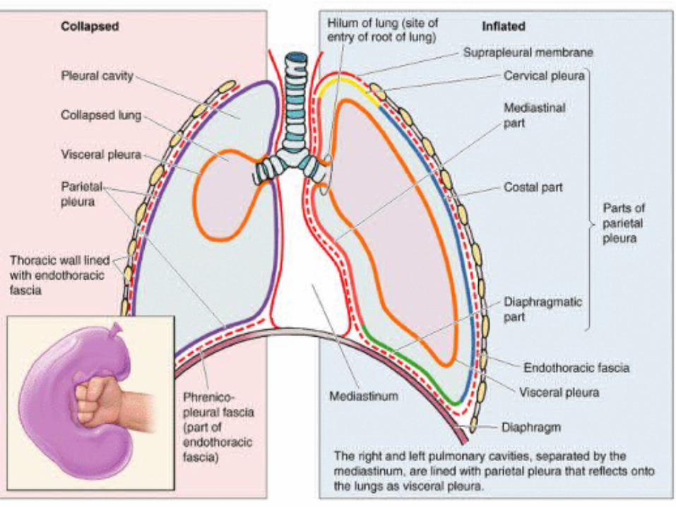
The pleura is divided into two major types, based on location: 1. Parietal pleura 2. Visceral pleura Each pleural cavity is the potential space enclosed.
Dec 25, 2015
Welcome message from author
This document is posted to help you gain knowledge. Please leave a comment to let me know what you think about it! Share it to your friends and learn new things together.
Transcript
Pleura and Lungs
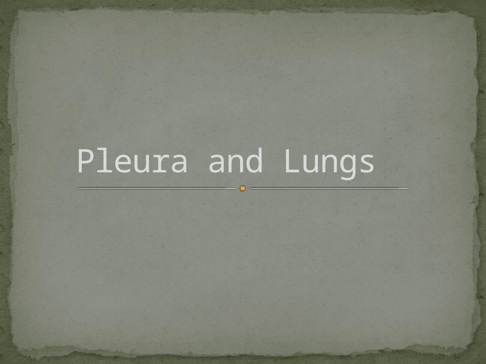
The pleura is divided into two major types, based on location:
1. Parietal pleura2. Visceral pleura
Each pleural cavity is the potential space enclosed between the visceral and parietal pleurae.
Pleura
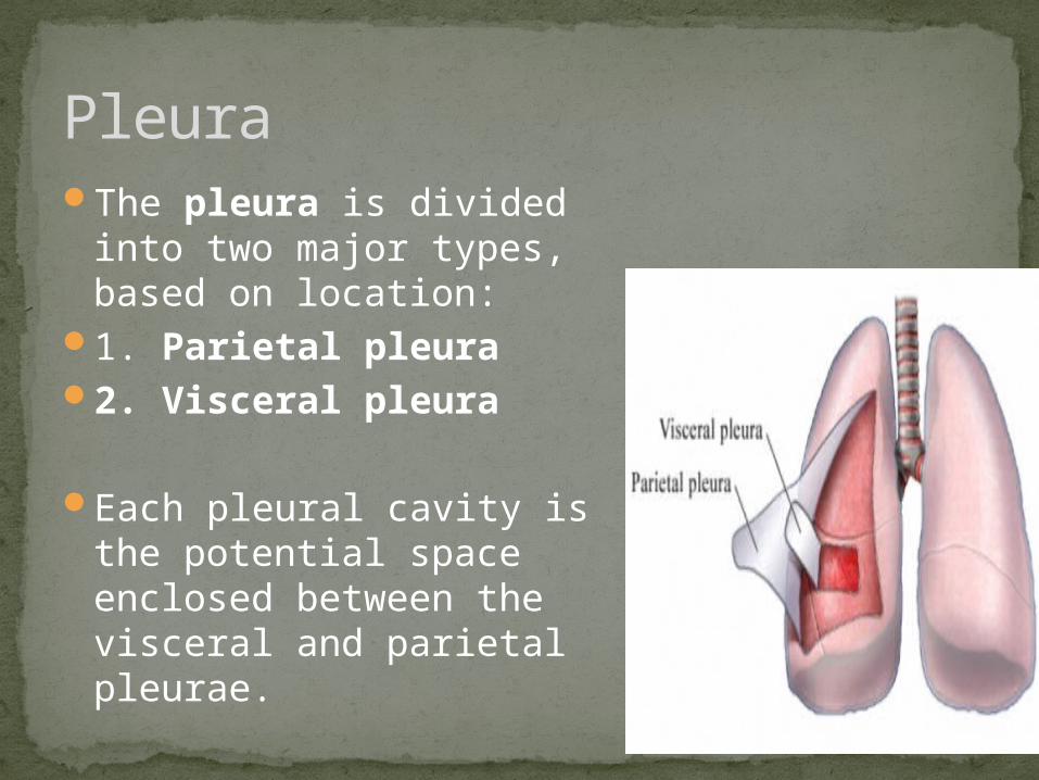
1. Costal part2. Diaphragmatic part3. Mediastinal part4. Cervical part
Parietal pleura
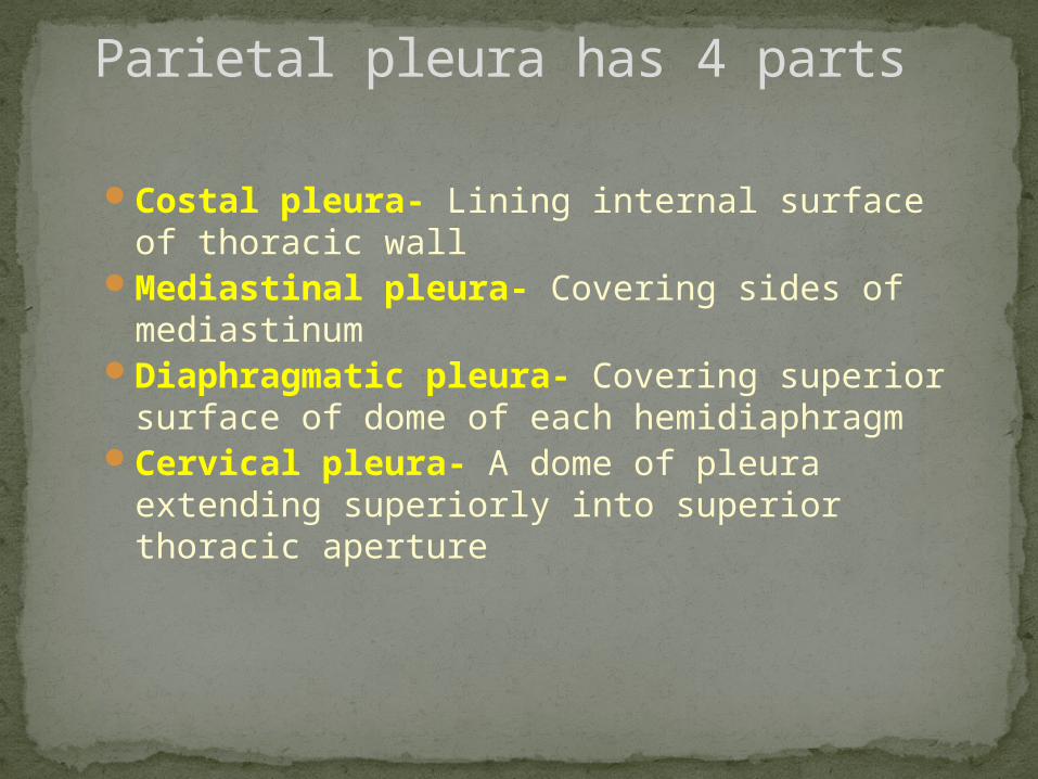
Costal pleura- Lining internal surface of thoracic wall
Mediastinal pleura- Covering sides of mediastinum
Diaphragmatic pleura- Covering superior surface of dome of each hemidiaphragm
Cervical pleura- A dome of pleura extending superiorly into superior thoracic aperture
Parietal pleura has 4 parts
Covers the lungs Cannot be dissected from lungInsensitive to pain
Visceral pleura
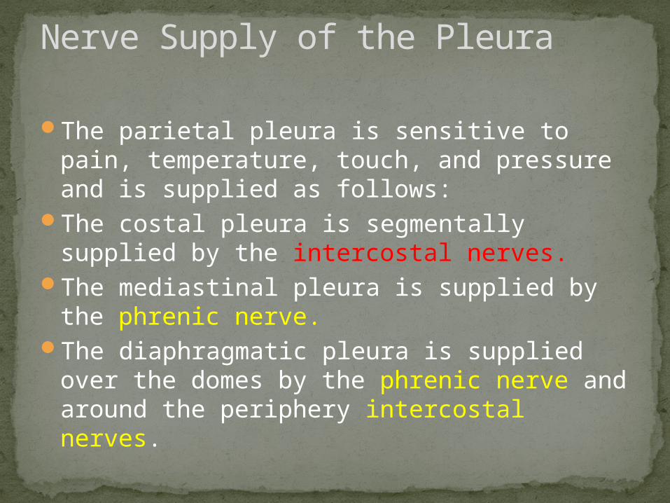
The parietal pleura is sensitive to pain, temperature, touch, and pressure and is supplied as follows:
The costal pleura is segmentally supplied by the intercostal nerves.
The mediastinal pleura is supplied by the phrenic nerve.
The diaphragmatic pleura is supplied over the domes by the phrenic nerve and around the periphery intercostal nerves.
Nerve Supply of the Pleura
The visceral pleura covering the lungs is sensitive to stretch but is insensitive to common sensations such as pain and touch. It receives an autonomic nerve supply from the pulmonary plexus .
visceral pleura
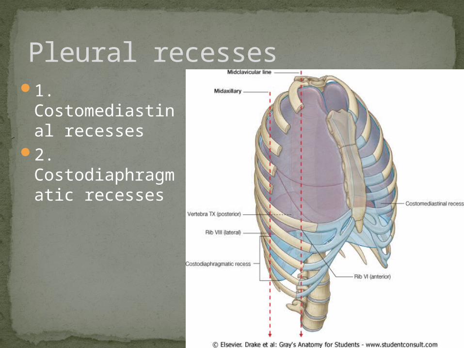
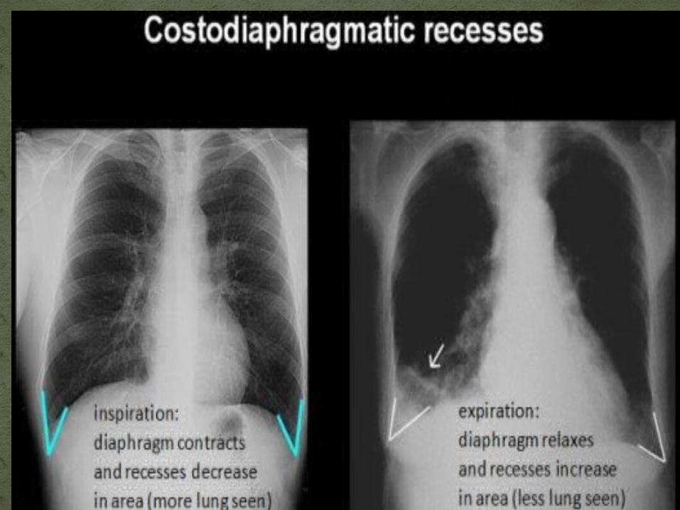
1. Costomediastinal recesses
2. Costodiaphragmatic recesses
Pleural recesses
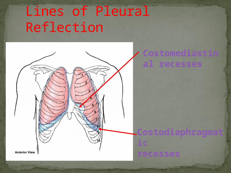
Lines of Pleural Reflection
Costomediastinal recesses
Costodiaphragmaticrecesses
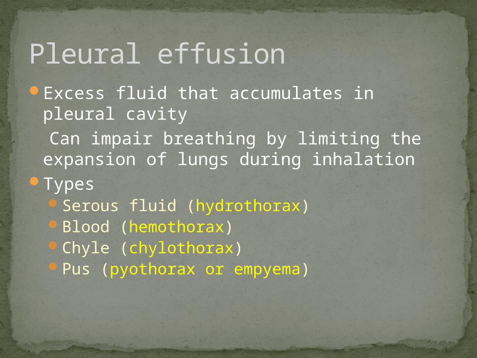
Excess fluid that accumulates in pleural cavity
Can impair breathing by limiting the expansion of lungs during inhalation
TypesSerous fluid (hydrothorax) Blood (hemothorax) Chyle (chylothorax) Pus (pyothorax or empyema)
Pleural effusion
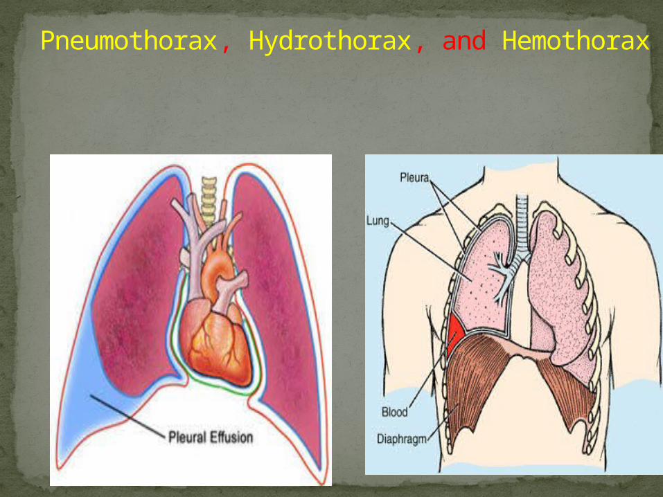
Pneumothorax, Hydrothorax, and Hemothorax
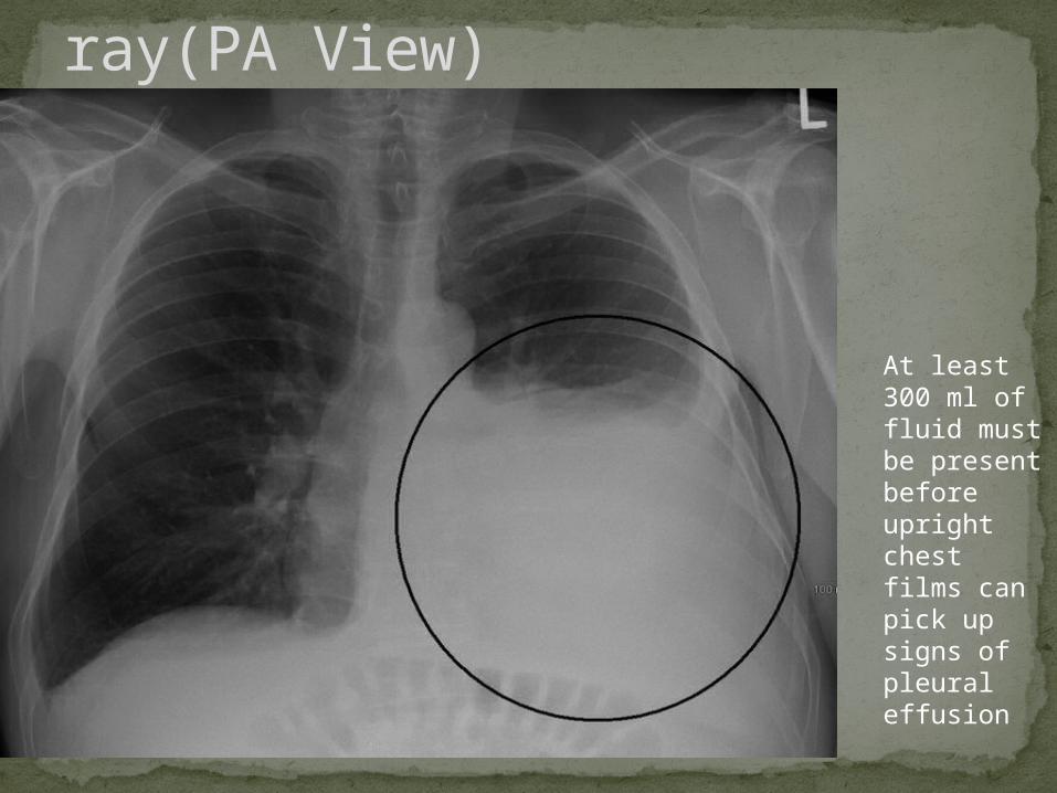
Pleural effusion Chest x ray(PA View)
At least 300 ml of fluid must be present before upright chest films can pick up signs of pleural effusion
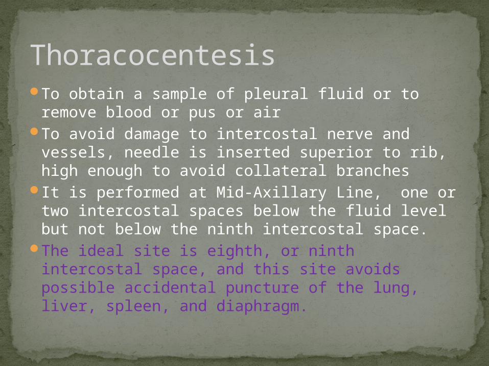
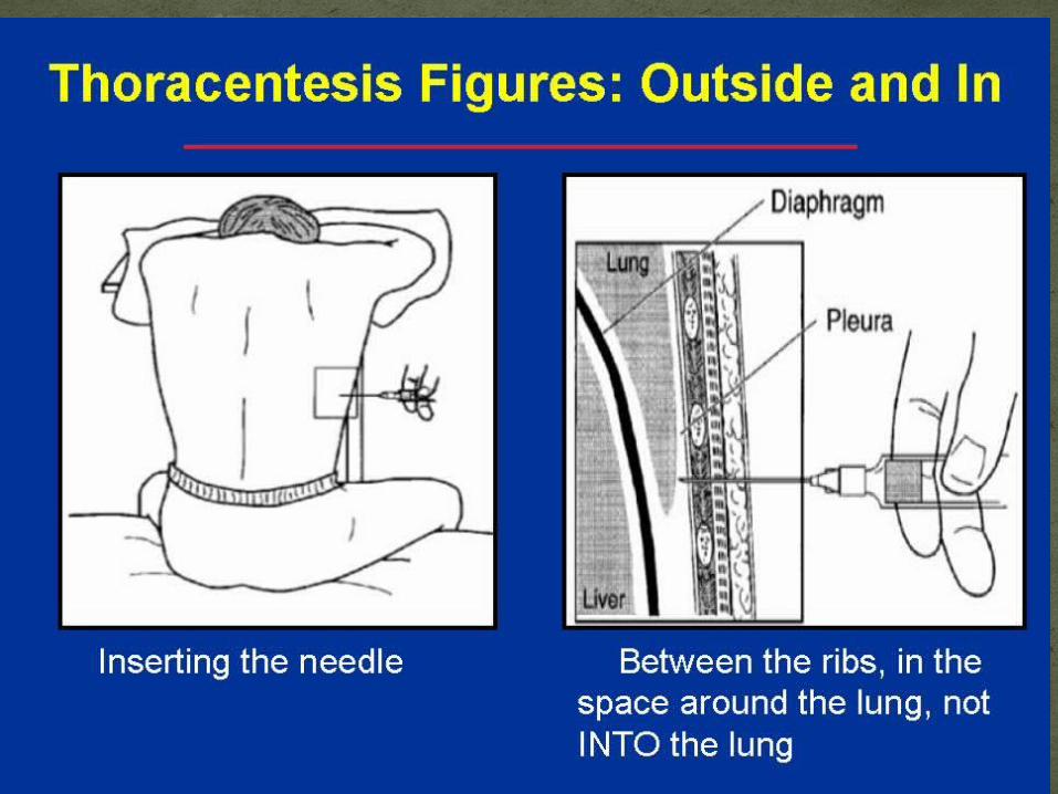
To obtain a sample of pleural fluid or to remove blood or pus or air
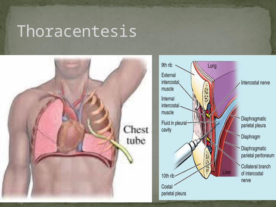
To avoid damage to intercostal nerve and vessels, needle is inserted superior to rib, high enough to avoid collateral branches
It is performed at Mid-Axillary Line, one or two intercostal spaces below the fluid level but not below the ninth intercostal space.
The ideal site is eighth, or ninth intercostal space, and this site avoids possible accidental puncture of the lung, liver, spleen, and diaphragm.
Thoracocentesis
Thoracentesis
Lungs
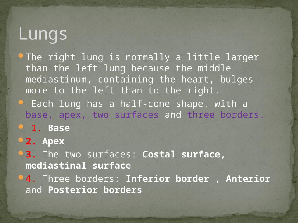
The right lung is normally a little larger than the left lung because the middle mediastinum, containing the heart, bulges more to the left than to the right.
Each lung has a half-cone shape, with a base, apex, two surfaces and three borders.
1. Base2. Apex3. The two surfaces: Costal surface, mediastinal
surface 4. Three borders: Inferior border , Anterior and
Posterior borders
Lungs
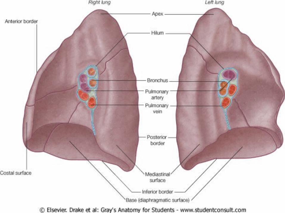
1.Pulmonary artery
2. Two pulmonary veins
3. Main bronchus 4. Bronchial
vessels5. Nerves and
lymphatics.
Hilum of the lung
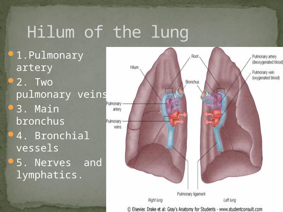
The right lung has three lobes and two fissures.
Fissures1. Oblique fissure2. Horizontal fissure
Right lung
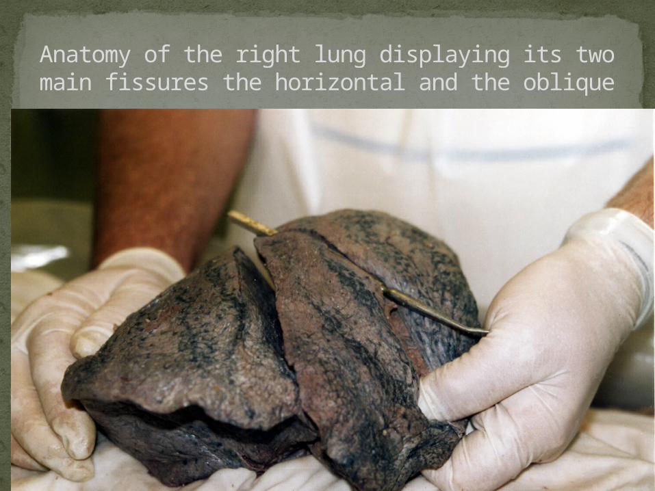
Anatomy of the right lung displaying its two main fissures the horizontal and the oblique
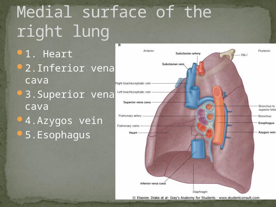
1. Heart 2.Inferior vena
cava 3.Superior vena
cava 4.Azygos vein 5.Esophagus
Medial surface of the right lung
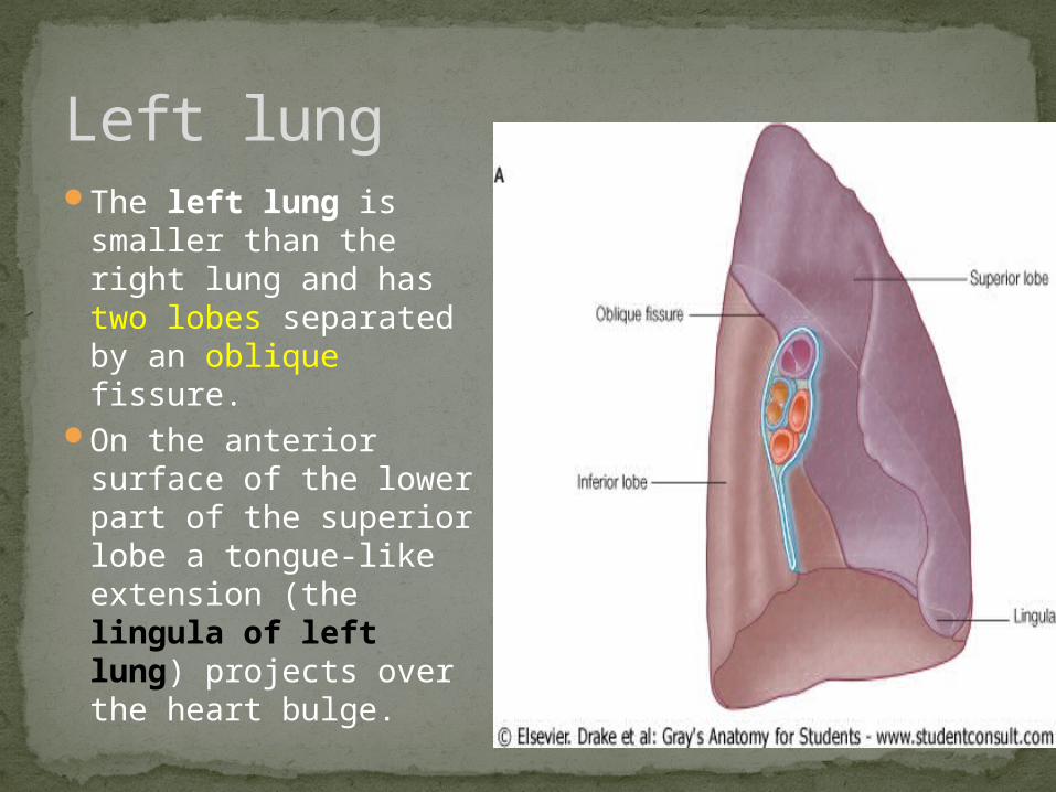
The left lung is smaller than the right lung and has two lobes separated by an oblique fissure.
On the anterior surface of the lower part of the superior lobe a tongue-like extension (the lingula of left lung) projects over the heart bulge.
Left lung
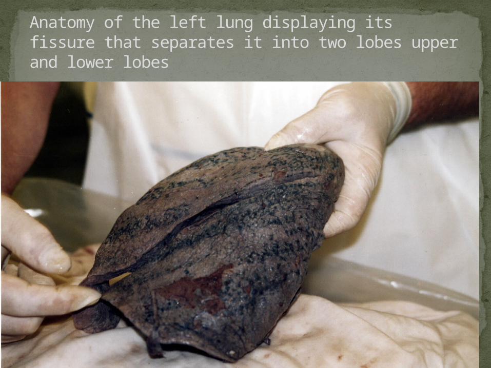
Anatomy of the left lung displaying its fissure that separates it into two lobes upper and lower lobes
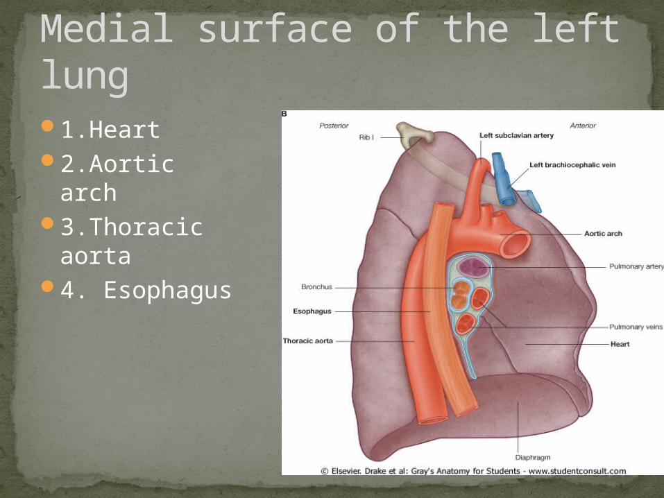
1.Heart 2.Aortic arch 3.Thoracic
aorta4. Esophagus
Medial surface of the left lung
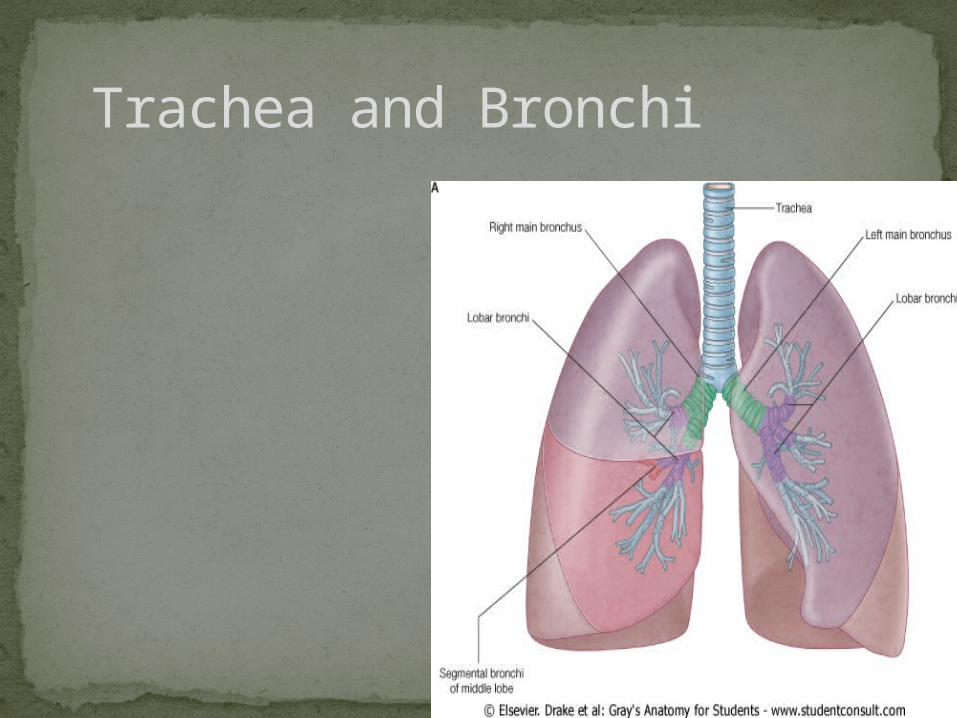
Trachea and Bronchi
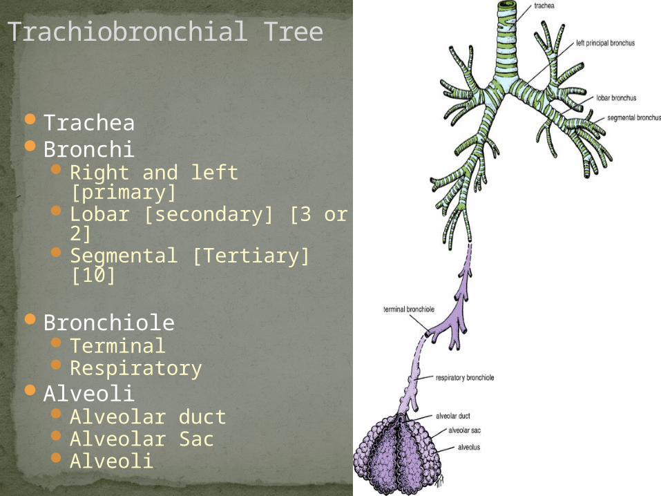
Trachiobronchial Tree
TracheaBronchi
Right and left [primary]Lobar [secondary] [3 or 2]Segmental [Tertiary] [10]
BronchioleTerminalRespiratory
AlveoliAlveolar ductAlveolar SacAlveoli
Trachea bifurcates→ two main stem bronchi, right and leftCarina- keel-like ridge between two openings
of main stem bronchiMain stem bronchus divides into lobar
bronchi3 lobar bronchi on right: upper, middle, and
lower 2 lobar bronchi on left: upper and lower
Each lobar bronchus branches into segmental bronchi that supply a bronchopulmonary segment
Bifurcation of the trachea
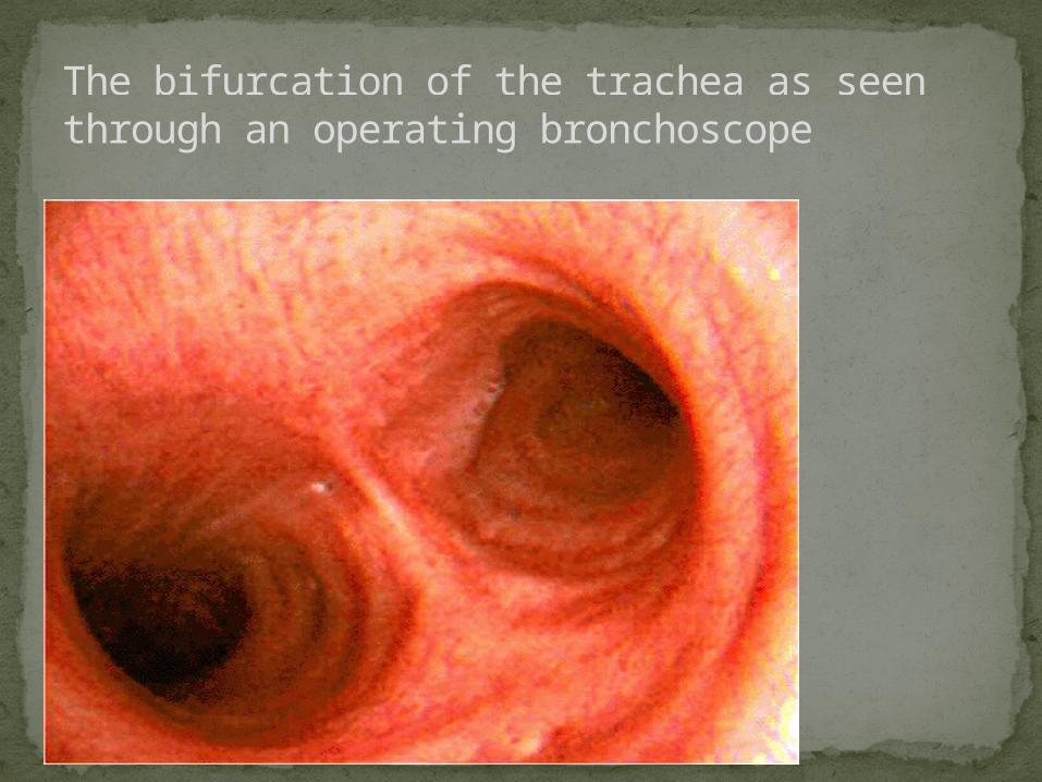
The bifurcation of the trachea as seen through an operating bronchoscope
Aspiration of Foreign BodiesIf food, liquids, or foreign bodies are aspirated,
they often will lodge in the right mainstem bronchus.
Because right bronchus is wider and shorter and runs more vertically than left bronchus
Encountered by dentists Aspiration of piece of tooth, filling
material, or a small instrument.If the endotracheal tube used for
intubation is inserted too far, it usually lodges in the right mainstem bronchus. This allows ventilation of the right lung, but leaves the left lung useless.
2 sets of Blood Supply
1.Pulmonary Vessels: for Gas Exchange
2. Bronchial Vessels: for blood supply to lung substance like any other organ
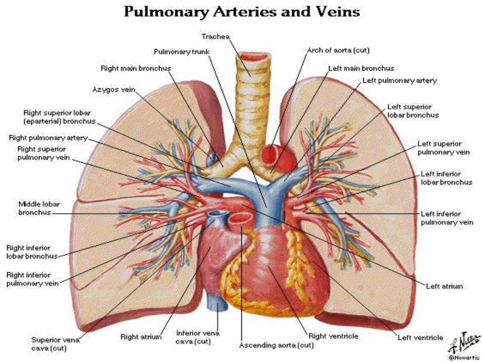
Vasculature of lungs
Pulmonary arteryCarries unoxygenated blood from heart to lungsEach artery gives lobar and segmental arteries
Pulmonary veins Intrasegmental veins drain to intersegmental veins
in pulmonary septa, which drain to two pulmonary veins for each lung
Carry oxygenated blood from lungs to heart
Pulmonary Vessels
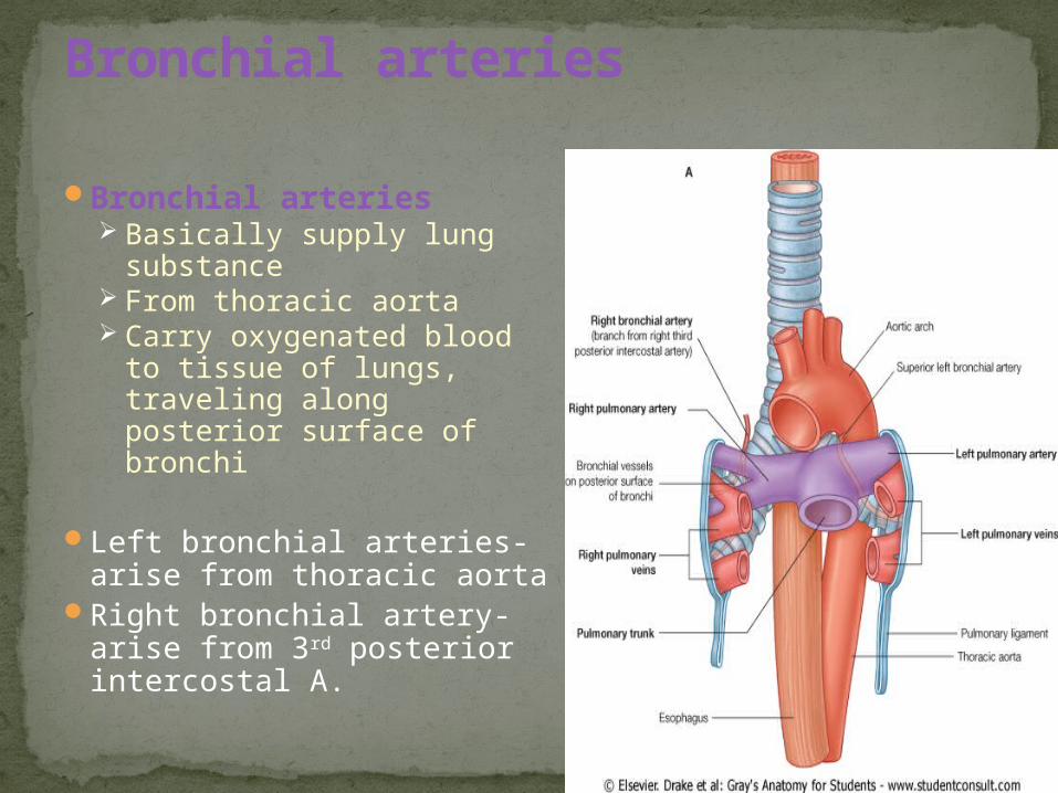
Bronchial arteries Basically supply lung
substance From thoracic aorta Carry oxygenated blood
to tissue of lungs, traveling along posterior surface of bronchi
Left bronchial arteries- arise from thoracic aorta
Right bronchial artery- arise from 3rd posterior intercostal A.
Bronchial arteries
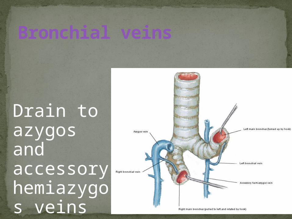
Bronchial veins
Drain to azygos and accessory hemiazygos veins
Related Documents