The Physiological Role of Ascorbate as Photosystem II Electron Donor: Protection against Photoinactivation in Heat-Stressed Leaves 1[C][OA] Szilvia Z. To ´th*, Vale ´ria Nagy, Jos T. Puthur 2 , La ´szlo ´ Kova ´cs, and Gyo ˝zo ˝ Garab Institute of Plant Biology, Biological Research Center Szeged, Hungarian Academy of Sciences, H–6701 Szeged, Hungary Previously, we showed that ascorbate (Asc), by donating electrons to photosystem II (PSII), supports a sustained electron transport activity in leaves in which the oxygen-evolving complexes were inactivated with a heat pulse (49°C, 40 s). Here, by using wild-type, Asc-overproducing, and -deficient Arabidopsis (Arabidopsis thaliana) mutants (miox4 and vtc2-3, respectively), we investigated the physiological role of Asc as PSII electron donor in heat-stressed leaves (40°C, 15 min), lacking active oxygen-evolving complexes. Chlorophyll-a fluorescence transients show that in leaves excited with trains of saturating single- turnover flashes spaced 200 ms apart, allowing continual electron donation from Asc to PSII, the reaction centers remained functional even after thousands of turnovers. Higher flash frequencies or continuous illumination (300 mmol photons m 22 s 21 ) gradually inactivated them, a process that appeared to be initiated by a dramatic deceleration of the electron transfer from Tyr Z to P680 + , followed by the complete loss of charge separation activity. These processes occurred with half-times of 1.2 and 10 min, 2.8 and 23 min, and 4.1 and 51 min in vtc2-3, the wild type, and miox4, respectively, indicating that the rate of inactivation strongly depended on the Asc content of the leaves. The recovery of PSII activity, following the degradation of PSII proteins (D1, CP43, and PsbO), in moderate light (100 mmol photons m 22 s 21 , comparable to growth light), was also retarded in the Asc- deficient mutant. These data show that high Asc content of leaves contributes significantly to the ability of plants to withstand heat-stress conditions. Heat stress is a serious threat to plants, which can lead to a drastic reduction in crop yield (e.g. Wahid et al., 2007; Allakhverdiev et al., 2008). Exposure of plants to elevated temperatures results in the inacti- vation of Rubisco activase (Barta et al., 2010) and the oxygen-evolving complex (OEC) of PSII, including the removal of the extrinsic proteins as well as the release of calcium and manganese ions from their binding sites (Nash et al., 1985; Enami et al., 1994; Yamane et al., 1998; Barra et al., 2005). Heat stress can also damage the D1 and D2 proteins (De Las Rivas and Barber, 1997; Yoshioka et al., 2006). Under natural conditions heat stress mostly occurs together with light stress. Though the effects of both stresses have been studied extensively, there are only a few studies where the mechanism of damage caused by combination of these two stress factors was investi- gated. It has been shown that in the absence of heat- adaptation processes, with yet an unknown mechanism (s), the two stress factors act in strong synergy to inactivate PSII in plants (Havaux, 1994), and in corals they lead to photobleaching (Abrego et al., 2008). Inactivation of PSII by excess light and damage to the D1 protein are most probably induced by singlet oxygen, which is produced via interaction with the triplet reaction center chlorophyll (chl; 3 P680) arising from the recombination of the charge-separated state between P680 and the pheophytin electron acceptor ( 3 [P680 + Phe]; Aro et al., 1993; Vass and Cser, 2009). This acceptor-side damage, as it has recently been shown by point mutation at the quinone-binding pocket of PSII in Synechocystis sp. PCC 6803 (Larom et al., 2010), can be alleviated by efficiently draining the electrons from the primary quinone acceptor Q A to an exoge- nous electron acceptor, cytochrome c. In other terms it appears that photoinhibition can be prevented by warranting a rapid electron transfer from the acceptor side of PSII. A priori, it cannot be ruled out that photoinhibition under heat-stress conditions occurs with the same mechanism since heat stress can lead to 1 This work was supported by the Hungarian Research Founda- tion (grant nos. PD72718 and CNK80345 to S.Z.T. and G.G., respec- tively), by the Bolyai Ja ´nos Research Foundation of the Hungarian Academy of Sciences (research scholarship to S.Z.T.), and by the Department of Science and Technology, Government of India (Better Opportunities for Young Scientists in Chosen Areas of Science and Technology fellowship to J.T.P.). 2 Present address: Department of Botany, Calicut University, C.U. Campus P.O. Kerala 673635, India. * Corresponding author; e-mail [email protected]. The author responsible for distribution of materials integral to the findings presented in this article in accordance with the policy described in the Instructions for Authors (www.plantphysiol.org) is: Szilvia Z. To ´ th ([email protected]). [C] Some figures in this article are displayed in color online but in black and white in the print edition. [OA] Open Access articles can be viewed online without a sub- scription. www.plantphysiol.org/cgi/doi/10.1104/pp.110.171918 382 Plant Physiology Ò , May 2011, Vol. 156, pp. 382–392, www.plantphysiol.org Ó 2011 American Society of Plant Biologists

Welcome message from author
This document is posted to help you gain knowledge. Please leave a comment to let me know what you think about it! Share it to your friends and learn new things together.
Transcript
The Physiological Role of Ascorbate as Photosystem IIElectron Donor: Protection against Photoinactivation inHeat-Stressed Leaves1[C][OA]
Szilvia Z. Toth*, Valeria Nagy, Jos T. Puthur2, Laszlo Kovacs, and Gyozo Garab
Institute of Plant Biology, Biological Research Center Szeged, Hungarian Academy of Sciences, H–6701Szeged, Hungary
Previously, we showed that ascorbate (Asc), by donating electrons to photosystem II (PSII), supports a sustained electrontransport activity in leaves in which the oxygen-evolving complexes were inactivated with a heat pulse (49�C, 40 s). Here, byusing wild-type, Asc-overproducing, and -deficient Arabidopsis (Arabidopsis thaliana) mutants (miox4 and vtc2-3, respectively),we investigated the physiological role of Asc as PSII electron donor in heat-stressed leaves (40�C, 15 min), lacking activeoxygen-evolving complexes. Chlorophyll-a fluorescence transients show that in leaves excited with trains of saturating single-turnover flashes spaced 200 ms apart, allowing continual electron donation from Asc to PSII, the reaction centers remainedfunctional even after thousands of turnovers. Higher flash frequencies or continuous illumination (300 mmol photons m22 s21)gradually inactivated them, a process that appeared to be initiated by a dramatic deceleration of the electron transfer from TyrZto P680+, followed by the complete loss of charge separation activity. These processes occurred with half-times of 1.2 and 10min, 2.8 and 23 min, and 4.1 and 51 min in vtc2-3, the wild type, and miox4, respectively, indicating that the rate of inactivationstrongly depended on the Asc content of the leaves. The recovery of PSII activity, following the degradation of PSII proteins(D1, CP43, and PsbO), in moderate light (100 mmol photons m22 s21, comparable to growth light), was also retarded in the Asc-deficient mutant. These data show that high Asc content of leaves contributes significantly to the ability of plants to withstandheat-stress conditions.
Heat stress is a serious threat to plants, which canlead to a drastic reduction in crop yield (e.g. Wahidet al., 2007; Allakhverdiev et al., 2008). Exposure ofplants to elevated temperatures results in the inacti-vation of Rubisco activase (Barta et al., 2010) and theoxygen-evolving complex (OEC) of PSII, including theremoval of the extrinsic proteins as well as the releaseof calcium and manganese ions from their bindingsites (Nash et al., 1985; Enami et al., 1994; Yamaneet al., 1998; Barra et al., 2005). Heat stress can also
damage the D1 and D2 proteins (De Las Rivas andBarber, 1997; Yoshioka et al., 2006).
Under natural conditions heat stress mostly occurstogether with light stress. Though the effects of bothstresses have been studied extensively, there are only afew studies where the mechanism of damage caused bycombination of these two stress factors was investi-gated. It has been shown that in the absence of heat-adaptation processes, with yet an unknownmechanism(s), the two stress factors act in strong synergy toinactivate PSII in plants (Havaux, 1994), and in coralsthey lead to photobleaching (Abrego et al., 2008).
Inactivation of PSII by excess light and damage tothe D1 protein are most probably induced by singletoxygen, which is produced via interaction with thetriplet reaction center chlorophyll (chl; 3P680) arisingfrom the recombination of the charge-separated statebetween P680 and the pheophytin electron acceptor(3[P680+Phe]; Aro et al., 1993; Vass and Cser, 2009). Thisacceptor-side damage, as it has recently been shownby point mutation at the quinone-binding pocket ofPSII in Synechocystis sp. PCC 6803 (Larom et al., 2010),can be alleviated by efficiently draining the electronsfrom the primary quinone acceptor QA to an exoge-nous electron acceptor, cytochrome c. In other terms itappears that photoinhibition can be prevented bywarranting a rapid electron transfer from the acceptorside of PSII. A priori, it cannot be ruled out thatphotoinhibition under heat-stress conditions occurswith the same mechanism since heat stress can lead to
1 This work was supported by the Hungarian Research Founda-tion (grant nos. PD72718 and CNK80345 to S.Z.T. and G.G., respec-tively), by the Bolyai Janos Research Foundation of the HungarianAcademy of Sciences (research scholarship to S.Z.T.), and by theDepartment of Science and Technology, Government of India (BetterOpportunities for Young Scientists in Chosen Areas of Science andTechnology fellowship to J.T.P.).
2 Present address: Department of Botany, Calicut University, C.U.Campus P.O. Kerala 673635, India.
* Corresponding author; e-mail [email protected] author responsible for distribution of materials integral to the
findings presented in this article in accordance with the policydescribed in the Instructions for Authors (www.plantphysiol.org) is:Szilvia Z. Toth ([email protected]).
[C] Some figures in this article are displayed in color online but inblack and white in the print edition.
[OA] Open Access articles can be viewed online without a sub-scription.
www.plantphysiol.org/cgi/doi/10.1104/pp.110.171918
382 Plant Physiology�, May 2011, Vol. 156, pp. 382–392, www.plantphysiol.org � 2011 American Society of Plant Biologists
a deceleration of the electron transfer rate between QAand QB, the primary and secondary quinone acceptors(Ducruet and Lemoine, 1985). However, elevated tem-peratures affect mainly the donor side of PSII, inparticular the OEC. Experiments on PSII preparationsand on leaves with chemically inactivated OECs indi-cate that the very high sensitivity of PSII to lightappears to be caused by the impaired electron dona-tion from the OEC. This, in turn, results in strongaccumulation of highly oxidizing radicals, P680+, TyrZ
+,and superoxide (Chen et al., 1995) or hydroxyl radicals(Spetea et al., 1997), and leads to a rapid inactivationand degradation of PSII reaction centers (Callahanet al., 1986; Blubaugh and Cheniae, 1990; Jegerscholdand Styring, 1996). This type of photodamage is calledweak light or donor-side-induced photoinhibition. Inanalogy with the acceptor-side photoinhibition, it canbe assumed, in fact in vitro data strongly suggest, thata continuous electron flow to PSII from alternativedonors, such as diphenylcarbazide (DPC) or ascorbate(Asc), can alleviate the photoinactivation of the reac-tion centers (Mano et al., 1997).In this article we provide experimental evidence
that Asc plays a protective role in photoinhibition inheat-stressed leaves. Asc is present in the lumen prob-ably at millimolar concentration (Foyer and Lelandais,1996), and in the absence of active OEC serves as arelatively rapid (t1/2 approximately 25 ms) electrondonor to PSII (Toth et al., 2009), and thus might becapable of protecting PSII by supplying electrons tothe reaction center. To test this hypothesis, we sub-jected intact leaves of wild-type, Asc-overproducing(miox4; Lorence et al., 2004), and -deficient mutant(vtc2-3; Conklin et al., 2000) Arabidopsis (Arabidopsisthaliana) plants to heat stress (40�C, 15 min) and in-vestigated the time course and mechanism of photo-inactivation of PSII.
RESULTS
Electron Donation Rates from Asc to PSII in ArabidopsisGenotypes with Different Asc Contents
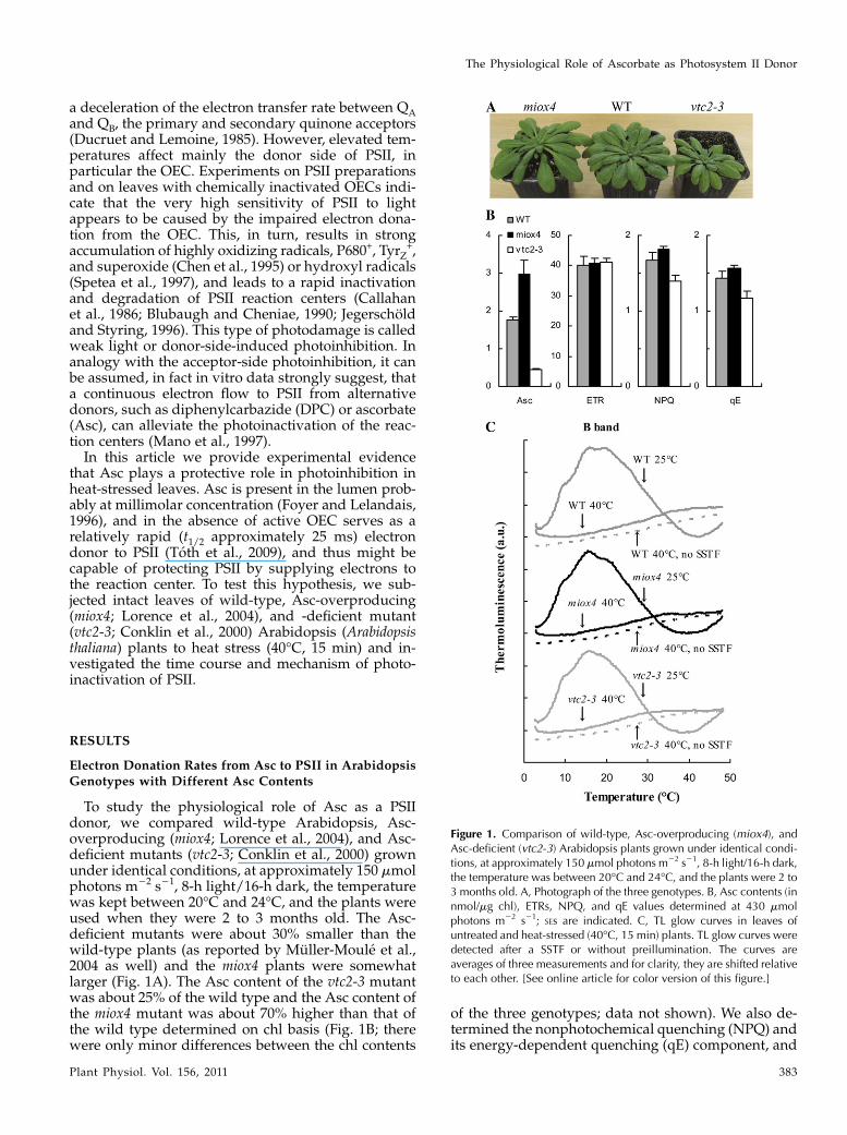
To study the physiological role of Asc as a PSIIdonor, we compared wild-type Arabidopsis, Asc-overproducing (miox4; Lorence et al., 2004), and Asc-deficient mutants (vtc2-3; Conklin et al., 2000) grownunder identical conditions, at approximately 150 mmolphotons m22 s21, 8-h light/16-h dark, the temperaturewas kept between 20�C and 24�C, and the plants wereused when they were 2 to 3 months old. The Asc-deficient mutants were about 30% smaller than thewild-type plants (as reported by Muller-Moule et al.,2004 as well) and the miox4 plants were somewhatlarger (Fig. 1A). The Asc content of the vtc2-3 mutantwas about 25% of the wild type and the Asc content ofthe miox4 mutant was about 70% higher than that ofthe wild type determined on chl basis (Fig. 1B; therewere only minor differences between the chl contents
of the three genotypes; data not shown). We also de-termined the nonphotochemical quenching (NPQ) andits energy-dependent quenching (qE) component, and
Figure 1. Comparison of wild-type, Asc-overproducing (miox4), andAsc-deficient (vtc2-3) Arabidopsis plants grown under identical condi-tions, at approximately 150 mmol photons m22 s21, 8-h light/16-h dark,the temperature was between 20�C and 24�C, and the plants were 2 to3 months old. A, Photograph of the three genotypes. B, Asc contents (innmol/mg chl), ETRs, NPQ, and qE values determined at 430 mmolphotons m22 s21; SEs are indicated. C, TL glow curves in leaves ofuntreated and heat-stressed (40�C, 15 min) plants. TL glow curves weredetected after a SSTF or without preillumination. The curves areaverages of three measurements and for clarity, they are shifted relativeto each other. [See online article for color version of this figure.]
The Physiological Role of Ascorbate as Photosystem II Donor
Plant Physiol. Vol. 156, 2011 383
the rate of electron transport (ETR) in the leaves. NPQand qE were highest in the Asc-overproducing mutant(miox4) and lowest in the Asc-deficient mutant (vtc2-3)and there were no significant differences between thethree genotypes in terms of ETR values (Fig. 1B). Theresults obtained on the vtc2-3mutant are in agreementwith the results of Muller-Moule et al. (2002, 2003), whoshowed that the lower NPQ values are caused by thelower violaxanthin deepoxidase activity due to limita-tion in the available Asc in the lumen. The higher NPQand qE values obtained on the miox4 mutant indicatethat there is more Asc available in the lumen of the Asc-overproducing miox4 mutant than in the wild type.Determination of the lumenal Asc content does notseem to be feasible since upon chloroplast isolation Ascis mostly lost (Ivanov and Edwards, 2000).
Detached leaves of wild-type, Asc-overproducing(miox4), and Asc-deficient (vtc2-3) Arabidopsis plantswere subjected to heat stress at 40�C for 15 min in awater bath in the dark. This treatment was chosen toobtain a homogenous material with completely inacti-vated OECs and to separate the effects of heat andlight stresses. The B thermoluminescence (TL) band(Vass, 2003) arising from recombination reactions be-tween the S2 state of the OEC and QB
2, induced by asaturating single turnover flash (SSTF) was totallyabolished by the heat treatment in all three geno-types, showing a total loss of oxygen-evolving activity(Fig. 1C). A 40�C heat treatment is physiologicallyrelevant since leaf temperatures can easily reach 40�Cin the field, especially under drought-stress conditions(Burghardt et al., 2008; Shahenshah and Isoda, 2010).This treatment leads to a moderate, less than 2-foldacceleration of the decay kinetics of the flash-inducedelectrochromic absorbance transient (D515), an indica-tor of the dissipation of the transmembrane electricalfield (Junge, 1977): The t1/2 of the decay decreasedfrom 55 to 34 ms, with similar behavior in the threegenotypes. These data show that the permeability ofthe thylakoid membranes did not increase drasticallyas a result of the 40�C, 15-min heat treatment (incontrast to a 50�C heat treatment applied earlier thatcaused dissipation of the electric field within a fewms; Toth et al., 2005); these data agree well with theresults obtained on heat-treated Arabidopsis leaves(Krumova et al., 2010). Decrease in the initial amplitudeof the flash-induced electrochromic absorbance tran-sient was also observed, suggesting a partial inacti-vation of PSII reaction centers (data not shown; seebelow).
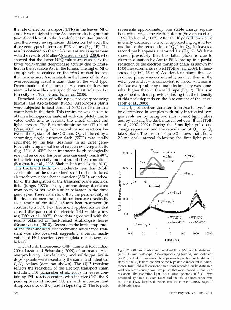
The fast chl afluorescence (OJIP) transients (Govindjee,2004; Lazar and Schansker, 2009) of untreated Asc-overproducing, Asc-deficient, and wild-type Arabi-dopsis plants were essentially the same, with identicalFv/Fm values (data not shown). The OJIP transientreflects the reduction of the electron transport chainincluding PSI (Schansker et al., 2005). In leaves con-taining PSII reaction centers with inactive OEC the Kpeak appears at around 300 ms with a concomitantdisappearance of the J and I steps (Fig. 2). The K peak
represents approximately one stable charge separa-tion, with TyrZ as the electron donor (Srivastava et al.,1997; Toth et al., 2007). After the K peak fluorescenceintensity decreases to a level approaching F0 in a fewms due to the reoxidation of QA
2 by QB. In leaves asecond peak appears at around 1 s (Fig. 2). We haveshown previously that this latter phase is due toelectron donation by Asc to PSII, leading to a partialreduction of the electron transport chain as shown byP700 measurements as well (Toth et al., 2009). In heat-stressed (40�C, 15 min) Asc-deficient plants this sec-ond rise phase was considerably smaller than in thewild type and it was somewhat retarded, whereas inthe Asc-overproducing mutant its intensity was some-what higher than in the wild type (Fig. 2). This is inagreement with our previous finding that the intensityof this peak depends on the Asc content of the leaves(Toth et al., 2009).
The t1/2 of electron donation from Asc to TyrZ+ can
be determined in samples with fully inactivated oxy-gen evolution by using two short (5-ms) light pulsesand by varying the dark interval between them (Tothet al., 2007, 2009). During the 5-ms light pulse onecharge separation and the reoxidation of QA
2 by QBtakes place. The inset of Figure 2 shows that after a2.3-ms dark interval following the first light pulse
Figure 2. OJIP transients in untreated wild type (WT) and heat-stressed(40�C, 15 min) wild-type, Asc-overproducing (miox4), and -deficient(vtc2-3) Arabidopsis mutants. The approximate positions of the differentsteps of the OJIP transient and of the K peak are indicated in paren-theses. Inset: chl a fluorescence transients recorded on heat-stressedwild-type leaves during two 5-ms pulses that were spaced 2.3 and 53.4ms apart. The excitation light (3,500 mmol photons m22 s21) wasproduced by three 650-nm LEDs and the chl a fluorescence wasmeasured at wavelengths above 700 nm. The transients are averages ofsix kinetic traces.
Toth et al.
384 Plant Physiol. Vol. 156, 2011
there is no variable fluorescence, which is due to thefull inactivation of oxygen evolution. However, withlonger dark intervals, the K peak recovers followingsingle exponential kinetics and the t1/2 of the regener-ation of the K peak can be used as the half-time of therereduction of TyrZ
+ by Asc (Toth et al., 2009). The t1/2was approximately 30ms inwild-type andmiox4 leaves,whereas in the Asc-deficient mutant the electron dona-tion wasmuch slower, the t1/2 was approximately 50 ms(Table I). These results on heat-stressed leaves (40�C, 15min) are in a good agreement with our previous find-ings where plants were subjected to a relatively harshheat pulse of 49�C, 40 s (Toth et al., 2009).
Asc-Dependent Multiple Turnover of PSII
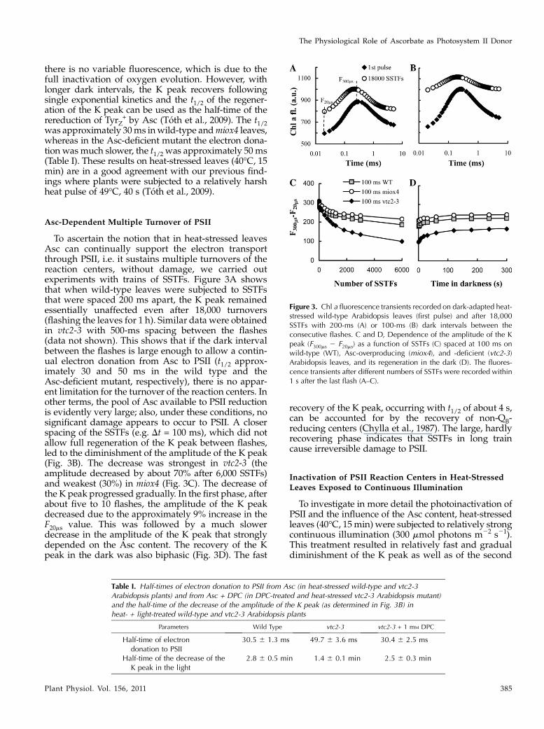
To ascertain the notion that in heat-stressed leavesAsc can continually support the electron transportthrough PSII, i.e. it sustains multiple turnovers of thereaction centers, without damage, we carried outexperiments with trains of SSTFs. Figure 3A showsthat when wild-type leaves were subjected to SSTFsthat were spaced 200 ms apart, the K peak remainedessentially unaffected even after 18,000 turnovers(flashing the leaves for 1 h). Similar data were obtainedin vtc2-3 with 500-ms spacing between the flashes(data not shown). This shows that if the dark intervalbetween the flashes is large enough to allow a contin-ual electron donation from Asc to PSII (t1/2 approx-imately 30 and 50 ms in the wild type and theAsc-deficient mutant, respectively), there is no appar-ent limitation for the turnover of the reaction centers. Inother terms, the pool of Asc available to PSII reductionis evidently very large; also, under these conditions, nosignificant damage appears to occur to PSII. A closerspacing of the SSTFs (e.g. Dt = 100 ms), which did notallow full regeneration of the K peak between flashes,led to the diminishment of the amplitude of the K peak(Fig. 3B). The decrease was strongest in vtc2-3 (theamplitude decreased by about 70% after 6,000 SSTFs)and weakest (30%) in miox4 (Fig. 3C). The decrease ofthe K peak progressed gradually. In the first phase, afterabout five to 10 flashes, the amplitude of the K peakdecreased due to the approximately 9% increase in theF20ms value. This was followed by a much slowerdecrease in the amplitude of the K peak that stronglydepended on the Asc content. The recovery of the Kpeak in the dark was also biphasic (Fig. 3D). The fast
recovery of the K peak, occurring with t1/2 of about 4 s,can be accounted for by the recovery of non-QB-reducing centers (Chylla et al., 1987). The large, hardlyrecovering phase indicates that SSTFs in long traincause irreversible damage to PSII.
Inactivation of PSII Reaction Centers in Heat-StressedLeaves Exposed to Continuous Illumination
To investigate in more detail the photoinactivation ofPSII and the influence of the Asc content, heat-stressedleaves (40�C, 15min) were subjected to relatively strongcontinuous illumination (300 mmol photons m22 s21).This treatment resulted in relatively fast and gradualdiminishment of the K peak as well as of the second
Table I. Half-times of electron donation to PSII from Asc (in heat-stressed wild-type and vtc2-3Arabidopsis plants) and from Asc + DPC (in DPC-treated and heat-stressed vtc2-3 Arabidopsis mutant)and the half-time of the decrease of the amplitude of the K peak (as determined in Fig. 3B) inheat- + light-treated wild-type and vtc2-3 Arabidopsis plants
Parameters Wild Type vtc2-3 vtc2-3 + 1 mM DPC
Half-time of electrondonation to PSII
30.5 6 1.3 ms 49.7 6 3.6 ms 30.4 6 2.5 ms
Half-time of the decrease of theK peak in the light
2.8 6 0.5 min 1.4 6 0.1 min 2.5 6 0.3 min
Figure 3. Chl a fluorescence transients recorded on dark-adapted heat-stressed wild-type Arabidopsis leaves (first pulse) and after 18,000SSTFs with 200-ms (A) or 100-ms (B) dark intervals between theconsecutive flashes. C and D, Dependence of the amplitude of the Kpeak (F300ms 2 F20ms) as a function of SSTFs (C) spaced at 100 ms onwild-type (WT), Asc-overproducing (miox4), and -deficient (vtc2-3)Arabidopsis leaves, and its regeneration in the dark (D). The fluores-cence transients after different numbers of SSTFs were recorded within1 s after the last flash (A–C).
The Physiological Role of Ascorbate as Photosystem II Donor
Plant Physiol. Vol. 156, 2011 385
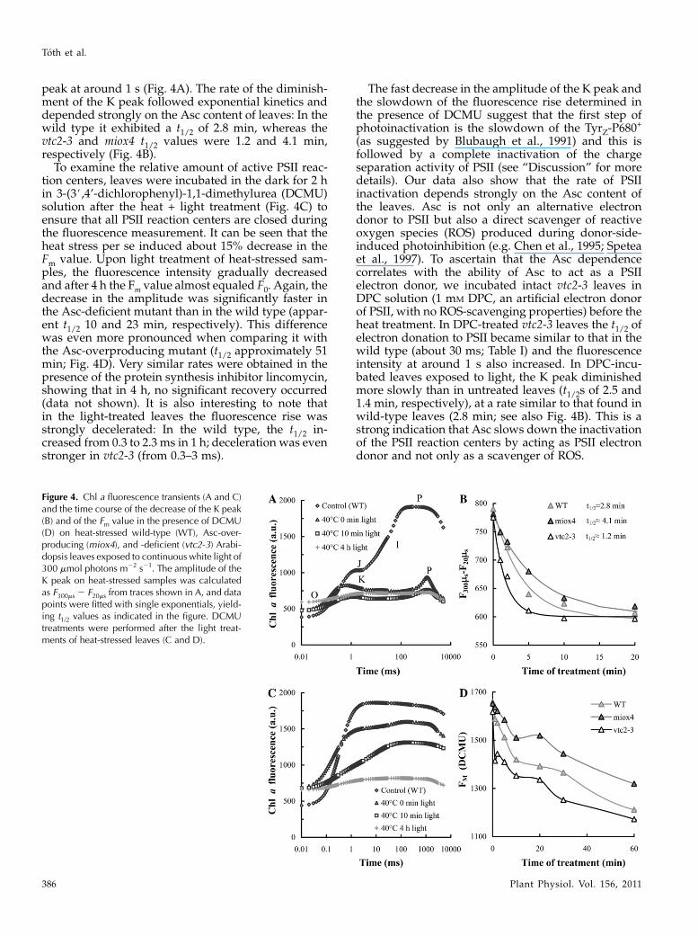
peak at around 1 s (Fig. 4A). The rate of the diminish-ment of the K peak followed exponential kinetics anddepended strongly on the Asc content of leaves: In thewild type it exhibited a t1/2 of 2.8 min, whereas thevtc2-3 and miox4 t1/2 values were 1.2 and 4.1 min,respectively (Fig. 4B).
To examine the relative amount of active PSII reac-tion centers, leaves were incubated in the dark for 2 hin 3-(3#,4’-dichlorophenyl)-1,1-dimethylurea (DCMU)solution after the heat + light treatment (Fig. 4C) toensure that all PSII reaction centers are closed duringthe fluorescence measurement. It can be seen that theheat stress per se induced about 15% decrease in theFm value. Upon light treatment of heat-stressed sam-ples, the fluorescence intensity gradually decreasedand after 4 h the Fm value almost equaled F0. Again, thedecrease in the amplitude was significantly faster inthe Asc-deficient mutant than in the wild type (appar-ent t1/2 10 and 23 min, respectively). This differencewas even more pronounced when comparing it withthe Asc-overproducing mutant (t1/2 approximately 51min; Fig. 4D). Very similar rates were obtained in thepresence of the protein synthesis inhibitor lincomycin,showing that in 4 h, no significant recovery occurred(data not shown). It is also interesting to note thatin the light-treated leaves the fluorescence rise wasstrongly decelerated: In the wild type, the t1/2 in-creased from 0.3 to 2.3 ms in 1 h; deceleration was evenstronger in vtc2-3 (from 0.3–3 ms).
The fast decrease in the amplitude of the K peak andthe slowdown of the fluorescence rise determined inthe presence of DCMU suggest that the first step ofphotoinactivation is the slowdown of the TyrZ-P680
+
(as suggested by Blubaugh et al., 1991) and this isfollowed by a complete inactivation of the chargeseparation activity of PSII (see “Discussion” for moredetails). Our data also show that the rate of PSIIinactivation depends strongly on the Asc content ofthe leaves. Asc is not only an alternative electrondonor to PSII but also a direct scavenger of reactiveoxygen species (ROS) produced during donor-side-induced photoinhibition (e.g. Chen et al., 1995; Speteaet al., 1997). To ascertain that the Asc dependencecorrelates with the ability of Asc to act as a PSIIelectron donor, we incubated intact vtc2-3 leaves inDPC solution (1 mM DPC, an artificial electron donorof PSII, with no ROS-scavenging properties) before theheat treatment. In DPC-treated vtc2-3 leaves the t1/2 ofelectron donation to PSII became similar to that in thewild type (about 30 ms; Table I) and the fluorescenceintensity at around 1 s also increased. In DPC-incu-bated leaves exposed to light, the K peak diminishedmore slowly than in untreated leaves (t1/2s of 2.5 and1.4 min, respectively), at a rate similar to that found inwild-type leaves (2.8 min; see also Fig. 4B). This is astrong indication that Asc slows down the inactivationof the PSII reaction centers by acting as PSII electrondonor and not only as a scavenger of ROS.
Figure 4. Chl a fluorescence transients (A and C)and the time course of the decrease of the K peak(B) and of the Fm value in the presence of DCMU(D) on heat-stressed wild-type (WT), Asc-over-producing (miox4), and -deficient (vtc2-3) Arabi-dopsis leaves exposed to continuouswhite light of300 mmol photons m22 s21. The amplitude of theK peak on heat-stressed samples was calculatedas F300ms 2 F20ms from traces shown in A, and datapoints were fitted with single exponentials, yield-ing t1/2 values as indicated in the figure. DCMUtreatments were performed after the light treat-ments of heat-stressed leaves (C and D).
Toth et al.
386 Plant Physiol. Vol. 156, 2011
It is also known that Asc is required for the forma-tion of NPQ (more specifically, qE), because it is asubstrate for violaxanthin deepoxidase (Hager, 1969).qE plays a photoprotective role and therefore we haveto consider if the faster rate of PSII inactivation ob-served in the vtc2-3mutant might be caused by smallerqE. Determination of NPQ and qE in heat-treatedsamples is not possible because Fm can only be reachedwhen DCMU is present (Fig. 4; see also Toth et al.,2007). However, the data show that the t1/2 of electrondonation from Asc to PSII in heat-treated samples isslow compared to electron donation by active OECs(30–50 ms versus 0.1–1 ms; Babcock et al., 1976) andtherefore, significant and persistent reduction of theelectron transport chain, a prerequisite for the forma-tion of qE, is very unlikely: Illumination of sampleswith completely inactivated OECs at 300 mmol photonsm22 s21 would result in electron transport comparableto that occurring at around 10 mmol photons m22 s21 inthe case of non-heat-treated samples. Light intensitiesbelow 40 mmol photons m22 s21 are insufficient for theformation of qE in any of the three genotypes studied(data not presented; see Muller-Moule et al., 2002),therefore photoprotection by qE formation during ourphotoinactivation experiments is unlikely to occur.As shown in Figure 5, in untreated wild-type leaves,
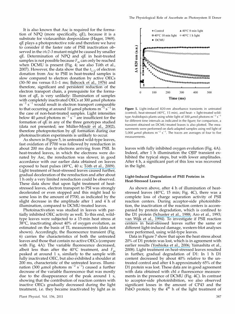
fast oxidation of P700 was followed by rereduction inabout 200 ms due to electrons arriving from PSII. Inheat-treated leaves, in which the electrons were do-nated by Asc, the rereduction was slower, in goodaccordance with our earlier data obtained on leavesexposed to heat pulses (49�C, 40 s; Toth et al., 2009).Light treatment of heat-stressed leaves caused further,gradual deceleration of the rereduction and after about1 h only a very limited rereduction could be observed.These data show that upon light treatment of heat-stressed leaves, electron transport to PSI was stronglydecelerated or even stopped and this might lead tosome loss in the amount of P700, as indicated by theslight decrease in the amplitude after 1 and 4 h ofillumination, compared to DCMU-treated leaves.Photoinactivation was studied in leaves with par-
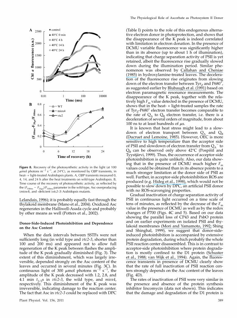
tially inhibited OEC activity as well. To this end, wild-type leaves were subjected to a 15-min heat stress at38�C, inactivating about 40% of oxygen evolution, asestimated on the basis of TL measurements (data notshown). Accordingly, the fluorescence transient (Fig.6) carries the characteristics of both the untreatedleaves and those that contain no active OECs (comparewith Fig. 4A): The variable fluorescence decreased,albeit less than after the 40�C treatment, and Fmpeaked at around 1 s, similarly to the sample withfully inactivated OEC, but also exhibited a shoulder at200 ms, characteristic of the untreated leaves. Illumi-nation (300 mmol photons m22 s21) caused a furtherdecrease of the variable fluorescence that was mostlydue to the disappearance of the peak around 1 s,showing that the contribution of reaction centers withinactive OECs gradually decreased during the lighttreatment, i.e. they became inactivated by light as in
leaves with fully inhibited oxygen evolution (Fig. 4A).Indeed, after 1 h illumination the OJIP transient ex-hibited the typical steps, but with lower amplitudes.After 4 h, a significant part of this loss was recoveredin the light.
Light-Induced Degradation of PSII Proteins in
Heat-Stressed Leaves
As shown above, after 4 h of illumination of heat-stressed leaves (40�C, 15 min; Fig. 4C), there was acomplete loss of charge separation activity of PSIIreaction centers. During acceptor-side photoinhibi-tion, the inactivation of the reaction centers is accom-panied by protein degradation, which is confined tothe D1 protein (Schuster et al., 1988; Aro et al., 1993;van Wijk et al., 1994). To investigate if PSII reactioncenters in heat-stressed leaves suffer the same ordifferent light-induced damage, western-blot analyseswere performed, using wild-type leaves.
Data in Figure 7 show that upon the heat stress about20% of D1 protein was lost, which is in agreement withearlier results (Yoshioka et al., 2006; Yamashita et al.,2008). Light treatment on heat-stressed leaves resultedin further, gradual degradation of D1: In 1 h D1content decreased by about 40% relative to the un-treated control and after 4 h approximately 65% of theD1 protein was lost. These data are in good agreementwith data obtained with chl a fluorescence measure-ments in the presence of DCMU (Fig. 4C). In contrastto acceptor-side photoinhibition, we also observedsignificant losses in the amount of CP43 and thePsbO protein; by the 4th h of the light treatment of
Figure 5. Light-induced 820-nm absorbance transients in untreated(control), heat-stressed (40�C, 15 min), and heat- + light-treated wild-type Arabidopsis plants using white light of 300 mmol photons m22 s21
for different time intervals as indicated in the figure; for comparison, atransient obtained on DCMU-treated leaves is also plotted. The mea-surements were performed on dark-adapted samples using red light of5,000 mmol photons m22 s21. The traces are averages of four to fivemeasurements.
The Physiological Role of Ascorbate as Photosystem II Donor
Plant Physiol. Vol. 156, 2011 387
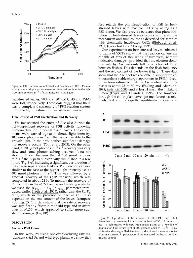
heat-treated leaves, 70% and 80% of CP43 and PsbOwere lost, respectively. These data suggest that therewas a complete disassembly of PSII reaction centersupon the light treatment of heat-stressed leaves.
Time Course of PSII Inactivation and Recovery
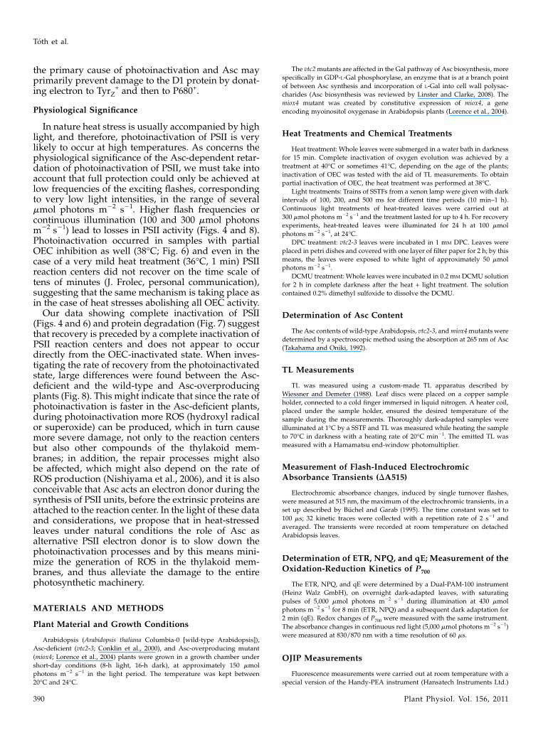
We investigated the effect of Asc also during thelight-dependent recovery of PSII activity followingphotoinactivation in heat-stressed leaves. The experi-ments were carried out at moderate light intensity,100 mmol photons m22 s21 that is comparable to thegrowth light. In the dark neither significant damagenor recovery occurs (Toth et al., 2005). On the otherhand, at 300 mmol photons m22 s21 recovery was veryslow and some photobleaching occurred (data notshown). It can be seen that at 100 mmol photonsm22 s21 the K peak substantially diminished in a fewhours (Fig. 8A), indicating a significant perturbation ofthe charge separation activity of PSII reaction centers,similar to the case at the higher light intensity i.e. at300 mmol photons m22 s21. This was followed by agradual recovery of the OJIP transient, which wascompleted in about 24 h. To monitor the recovery ofPSII activity in the vtc2-3, miox4, and wild-type plants,we used the (F100ms 2 F20ms)/F100ms parameter intro-duced earlier (Toth et al., 2005), rather than the Fv/Fmratio, which in the presence of inactive OEC alsodepends on the Asc content of the leaves (comparewith Fig. 2). Our data show that the rate of recoverywas significantly faster in the wild type and in miox4than in vtc2-3, which appeared to suffer more sub-stantial damage (Fig. 8B).
DISCUSSION
Asc as a PSII Donor
In this work, by using Asc-overproducing (miox4),-deficient (vtc2-3), and wild-type plants, we show that
Asc retards the photoinactivation of PSII in heat-stressed leaves with inactive OECs by acting as aPSII donor. We also provide evidence that photoinhi-bition in heat-stressed leaves occurs with a similarmechanism and time course as described for sampleswith chemically inactivated OECs (Blubaugh et al.,1991; Jegerschold and Styring, 1996).
Our experiments on heat-stressed leaves subjectedto trains of SSTFs show that the reaction centers arecapable of tens of thousands of turnovers, withoutnoticeable damage—provided that the electron dona-tion rate by Asc warrants full rereduction of TyrZ
+
between flashes. This depends on the flash frequencyand the Asc content of the leaves (Fig. 3). These datashow that the Asc pool was capable to support tens ofthousands of stable charge separations in PSII. Indeed,it has been estimated that the Asc content of chloro-plasts is about 25 to 50 mM (Eskling and Akerlund,1998; Smirnoff, 2000) and at least 4 mM in the thylakoidlumen (Foyer and Lelandais, 1996). The transportthrough the chloroplast envelope membranes is rela-tively fast and is rapidly equilibrated (Foyer and
Figure 7. Dependence of the amounts of D1, CP43, and PsbO,determined by western-blot analyses in heat (40�C, 15 min) andheat- + light-treated wild-type Arabidopsis plants as a function ofillumination time (white light of 300 photons mmol m22 s21). Typicalblots (A) and averages (B) determined by densitometry from four to fiveblots as expressed in percentage of the untreated (no heat-, no light-treated) control.
Figure 6. OJIP transients of untreated and heat-treated (38�C, 15 min)wild-type Arabidopsis plants, measured after various times in the light(300 mmol photons m22 s21), as indicated in the figure.
Toth et al.
388 Plant Physiol. Vol. 156, 2011
Lelandais, 1996); it is probably equally fast through thethylakoid membrane (Mano et al., 2004). Oxidized Ascregenerates in the Halliwell-Asada cycle and probablyby other means as well (Potters et al., 2002).
Donor-Side-Induced Photoinhibition and Dependence
on the Asc Content
When the dark intervals between SSTFs were notsufficiently long (in wild type and vtc2-3, shorter than100 and 200 ms) and appeared not to allow fullregeneration of the K peak between flashes the ampli-tude of the K peak gradually diminished (Fig. 3). Theextent of this diminishment, which was largely irre-versible, depended strongly on the Asc content of theleaves and occurred in several minutes (Fig. 3C). Incontinuous light of 300 mmol photons m22 s21, theamplitude of the K peak decreased with 1.2, 2.8, and4.1 min t1/2s in vtc2-3, the wild type, and miox4,respectively. This diminishment of the K peak wasirreversible, indicating damage to the reaction center.The fact that Asc in vtc2-3 could be replaced with DPC
(Table I) points to the role of this endogenous alterna-tive electron donor in photoprotection, and shows thatthe disappearance of the K peak is indeed correlatedwith limitation in electron donation. In the presence ofDCMU variable fluorescence was significantly higherthan in its absence (up to about 1 h of illumination),indicating that charge separation activity of PSII is yetretained, albeit the fluorescence rise gradually sloweddown during the illumination period. Similar phe-nomenon was observed by Callahan and Cheniae(1985) in hydroxylamine-treated leaves. The decelera-tion of the fluorescence rise originates from slowingdown of the electron transfer between TyrZ and P680+,as suggested earlier by Blubaugh et al. (1991) based onelectron paramagnetic resonance measurements. Thedisappearance of the K peak, together with the rela-tively high Fm value detected in the presence of DCMU,shows that in the heat- + light-treated samples the rateof TyrZ-P680
+ electron transfer becomes comparable tothe rate of QA to QB electron transfer, i.e. there is adeceleration of several orders of magnitude, from about100 ns to at least hundreds of ms.
It is known that heat stress might lead to a slow-down of electron transport between QA and QB(Ducruet and Lemoine, 1985). However, OEC is moresensitive to high temperature than the acceptor sideof PSII and slowdown of electron transfer from QA
2 toQB can be observed only above 42�C (Pospısil andTyystjarvi, 1999). Thus, the occurrence of acceptor-sidephotoinhibition is quite unlikely. Also, our data show-ing that in the presence of DCMU much higher Fmvalues could be obtained than in its absence points to amuch stronger limitation at the donor side of PSII aswell. Further, in acceptor-side photoinhibition ROS areproduced (e.g. Hideg et al., 1998), which would not bepossible to slow down by DPC, an artificial PSII donorwith no ROS-scavenging properties.
Gradual inactivation of charge separation activity ofPSII in continuous light occurred on a time scale oftens of minutes, as reflected by the decrease of the Fmvalue in the presence of DCMU as well as by the redoxchanges of P700 (Figs. 4C and 5). Based on our datashowing the parallel loss of CP43 and PsbO proteinand on earlier experiments on isolated PSII and thy-lakoid membranes (Mori and Yamamoto, 1992; Shingand Shinghal, 1999), we suggest that donor-side-induced photoinhibition is accompanied by extensiveprotein degradation, during which probably the wholePSII reaction center disassembled. This is in contrast toacceptor-side photoinhibition where protein degrada-tion is mostly confined to the D1 protein (Schusteret al., 1988; van Wijk et al., 1994). Again, the fluores-cence transients in presence of DCMU clearly showthat the rate of full inactivation of PSII reaction cen-ters strongly depends on the Asc content of the leaves(Fig. 4D).
The rates of inactivation of PSII were very similar inthe presence and absence of the protein synthesisinhibitor lincomycin (data not shown). This indicatesthat the damage and degradation of the D1 protein is
Figure 8. Recovery of the photosynthetic activity in the light (at 100mmol photons m22 s21, at 24�C), as monitored by OJIP transients, inheat- + light-treated Arabidopsis plants. A, OJIP transients measured 0,4, 14, and 24 h after the heat treatments on wild-type Arabidopsis. B,Time course of the recovery of photosynthetic activity, as reflected bythe (F100ms 2 F20ms)/F100ms parameter in the wild-type, Asc-overproducing(miox4), and -deficient (vtc2-3) Arabidopsis mutants.
The Physiological Role of Ascorbate as Photosystem II Donor
Plant Physiol. Vol. 156, 2011 389
the primary cause of photoinactivation and Asc mayprimarily prevent damage to the D1 protein by donat-ing electron to TyrZ
+ and then to P680+.
Physiological Significance
In nature heat stress is usually accompanied by highlight, and therefore, photoinactivation of PSII is verylikely to occur at high temperatures. As concerns thephysiological significance of the Asc-dependent retar-dation of photoinactivation of PSII, we must take intoaccount that full protection could only be achieved atlow frequencies of the exciting flashes, correspondingto very low light intensities, in the range of severalmmol photons m22 s21. Higher flash frequencies orcontinuous illumination (100 and 300 mmol photonsm22 s21) lead to losses in PSII activity (Figs. 4 and 8).Photoinactivation occurred in samples with partialOEC inhibition as well (38�C; Fig. 6) and even in thecase of a very mild heat treatment (36�C, 1 min) PSIIreaction centers did not recover on the time scale oftens of minutes (J. Frolec, personal communication),suggesting that the same mechanism is taking place asin the case of heat stresses abolishing all OEC activity.
Our data showing complete inactivation of PSII(Figs. 4 and 6) and protein degradation (Fig. 7) suggestthat recovery is preceded by a complete inactivation ofPSII reaction centers and does not appear to occurdirectly from the OEC-inactivated state. When inves-tigating the rate of recovery from the photoinactivatedstate, large differences were found between the Asc-deficient and the wild-type and Asc-overproducingplants (Fig. 8). This might indicate that since the rate ofphotoinactivation is faster in the Asc-deficient plants,during photoinactivation more ROS (hydroxyl radicalor superoxide) can be produced, which in turn causemore severe damage, not only to the reaction centersbut also other compounds of the thylakoid mem-branes; in addition, the repair processes might alsobe affected, which might also depend on the rate ofROS production (Nishiyama et al., 2006), and it is alsoconceivable that Asc acts an electron donor during thesynthesis of PSII units, before the extrinsic proteins areattached to the reaction center. In the light of these dataand considerations, we propose that in heat-stressedleaves under natural conditions the role of Asc asalternative PSII electron donor is to slow down thephotoinactivation processes and by this means mini-mize the generation of ROS in the thylakoid mem-branes, and thus alleviate the damage to the entirephotosynthetic machinery.
MATERIALS AND METHODS
Plant Material and Growth Conditions
Arabidopsis (Arabidopsis thaliana Columbia-0 [wild-type Arabidopsis]),
Asc-deficient (vtc2-3; Conklin et al., 2000), and Asc-overproducing mutant
(miox4; Lorence et al., 2004) plants were grown in a growth chamber under
short-day conditions (8-h light, 16-h dark), at approximately 150 mmol
photons m22 s21 in the light period. The temperature was kept between
20�C and 24�C.
The vtc2mutants are affected in the Gal pathway of Asc biosynthesis, more
specifically in GDP-L-Gal phosphorylase, an enzyme that is at a branch point
of between Asc synthesis and incorporation of L-Gal into cell wall polysac-
charides (Asc biosynthesis was reviewed by Linster and Clarke, 2008). The
miox4 mutant was created by constitutive expression of miox4, a gene
encoding myoinositol oxygenase in Arabidopsis plants (Lorence et al., 2004).
Heat Treatments and Chemical Treatments
Heat treatment: Whole leaves were submerged in a water bath in darkness
for 15 min. Complete inactivation of oxygen evolution was achieved by a
treatment at 40�C or sometimes 41�C, depending on the age of the plants;
inactivation of OEC was tested with the aid of TL measurements. To obtain
partial inactivation of OEC, the heat treatment was performed at 38�C.Light treatments: Trains of SSTFs from a xenon lamp were given with dark
intervals of 100, 200, and 500 ms for different time periods (10 min–1 h).
Continuous light treatments of heat-treated leaves were carried out at
300 mmol photons m22 s21 and the treatment lasted for up to 4 h. For recovery
experiments, heat-treated leaves were illuminated for 24 h at 100 mmol
photons m22 s21, at 24�C.DPC treatment: vtc2-3 leaves were incubated in 1 mM DPC. Leaves were
placed in petri dishes and covered with one layer of filter paper for 2 h; by this
means, the leaves were exposed to white light of approximately 50 mmol
photons m22 s21.
DCMU treatment: Whole leaves were incubated in 0.2 mM DCMU solution
for 2 h in complete darkness after the heat + light treatment. The solution
contained 0.2% dimethyl sulfoxide to dissolve the DCMU.
Determination of Asc Content
The Asc contents of wild-type Arabidopsis, vtc2-3, andmiox4mutants were
determined by a spectroscopic method using the absorption at 265 nm of Asc
(Takahama and Oniki, 1992).
TL Measurements
TL was measured using a custom-made TL apparatus described by
Wiessner and Demeter (1988). Leaf discs were placed on a copper sample
holder, connected to a cold finger immersed in liquid nitrogen. A heater coil,
placed under the sample holder, ensured the desired temperature of the
sample during the measurements. Thoroughly dark-adapted samples were
illuminated at 1�C by a SSTF and TL was measured while heating the sample
to 70�C in darkness with a heating rate of 20�C min21. The emitted TL was
measured with a Hamamatsu end-window photomultiplier.
Measurement of Flash-Induced ElectrochromicAbsorbance Transients (DA515)
Electrochromic absorbance changes, induced by single turnover flashes,
were measured at 515 nm, the maximum of the electrochromic transients, in a
set up described by Buchel and Garab (1995). The time constant was set to
100 ms; 32 kinetic traces were collected with a repetition rate of 2 s21 and
averaged. The transients were recorded at room temperature on detached
Arabidopsis leaves.
Determination of ETR, NPQ, and qE; Measurement of the
Oxidation-Reduction Kinetics of P700
The ETR, NPQ, and qE were determined by a Dual-PAM-100 instrument
(Heinz Walz GmbH), on overnight dark-adapted leaves, with saturating
pulses of 5,000 mmol photons m22 s21 during illumination at 430 mmol
photons m22 s21 for 8 min (ETR, NPQ) and a subsequent dark adaptation for
2 min (qE). Redox changes of P700 were measured with the same instrument.
The absorbance changes in continuous red light (5,000 mmol photons m22 s21)
were measured at 830/870 nm with a time resolution of 60 ms.
OJIP Measurements
Fluorescence measurements were carried out at room temperature with a
special version of the Handy-PEA instrument (Hansatech Instruments Ltd.)
Toth et al.
390 Plant Physiol. Vol. 156, 2011
that allows reducing the length of the measurement to 300 ms. Leaf samples
were illuminatedwith continuous red light emitted by three LEDs (3,500 mmol
photons m22 s21, 650 nm peak wavelength; the spectral half-width was 22 nm;
the light is cut off at 700 nm by a near-infrared short-pass filter). The first
reliably measured point of the fluorescence transient is at 20 ms, which was
taken as F0. The length of the measurements was 5 s or 5 ms. In the case of the
double 5-ms pulses, the dark intervals between the light pulses were 2.3, 9.6,
16.9, 31.5, 38.8, 53.4, 75.3, 100, 200, or 500 ms.
Western-Blot Analysis
Leaf discs equivalent to a total area of 3.1 cm2 cut from Arabidopsis leaves
were frozen in liquid nitrogen and ground to a fine powder and then
homogenized in 500 mL Laemmli buffer. The homogenates were incubated at
90�C for 5 min followed by a 20-min incubation at 37�C, and then proteins
were separated by 15% denaturing SDS-PAGE. The proteins were blotted on
nitrocellulose membranes using a semidry blotting system with methanol-
containing buffer. The nitrocellulose membranes were blocked using 5% skim
milk powder in Tris-buffered saline plus Tween 20 (TBST) buffer (10 mM TRIS
pH 8.0, 0.15 M NaCl, 0.1% Tween 20) for 2 h and incubated with primary
antibodies raised against PsbA (D1), PsbC (CP43), and PsbO (33 kD OEC
protein; Agrisera AB) for 2 h in TBST buffer with 5% milk powder. The
membranes were washed three times for 5 min in TBST buffer and incubated
with goat anti-rabbit IgG horseradish peroxidase conjugate (Millipore) at a
1:5,000 dilution in TBST buffer with 5% milk powder for 2 h. Immunoblotted
membranes were incubated for 5 min in ECL plus horseradish peroxidase
substrate (GE Healthcare Bio-Sciences) and chemiluminescence was detected
on Hyperfilm ECL photographic film (GE Healthcare Bio-Sciences). The
developed film was digitalized and analyzed by 1D Scan software package.
Sequence data from this article can be found in the GenBank/EMBL data
libraries under accession numbers NM_118819.2 (vtc2) and NM_118759.4
(miox4).
ACKNOWLEDGMENTS
We thank Dr. Argelia Lorence and Prof. Craig L. Nessler (Arkansas State
University) for the miox4 mutant and Prof. Patricia Conklin (State University
of New York College at Cortland) for providing the vtc2 mutants. We also
thank Dr. Gert Schansker (Biological Research Center Szeged) for critical
reading of the manuscript and helpful suggestions.
Received December 29, 2010; accepted February 27, 2011; published February
28, 2011.
LITERATURE CITED
Abrego D, Ulstrup KE, Willis BL, van Oppen MJH (2008) Species-specific
interactions between algal endosymbionts and coral hosts define their
bleaching response to heat and light stress. Proc Biol Sci 275: 2273–2282
Allakhverdiev SI, Kreslavski VD, Klimov VV, Los DA, Carpentier R,
Mohanty P (2008) Heat stress: an overview of molecular responses in
photosynthesis. Photosynth Res 98: 541–550
Aro EM, Virgin I, Andersson B (1993) Photoinhibition of photosystem II:
inactivation, protein damage and turnover. Biochim Biophys Acta 1143:
113–134
Babcock GT, Blankenship RE, Sauer K (1976) Reaction kinetics for
positive charge accumulation on the water side of chloroplast photo-
system II. FEBS Lett 61: 286–289
Barra M, Haumann M, Dau H (2005) Specific loss of the extrinsic 18 KDa
protein from photosystem II upon heating to 47�C causes inactivation of
oxygen evolution likely due to Ca release from the Mn-complex.
Photosynth Res 84: 231–237
Barta C, Dunkle AM, Wachter RM, Salvucci ME (2010) Structural changes
associated with the acute thermal instability of Rubisco activase. Arch
Biochem Biophys 499: 17–25
Blubaugh DJ, Atamian M, Babcock GT, Golbeck JH, Cheniae GM (1991)
Photoinhibition of hydroxylamine-extracted photosystem II mem-
branes: identification of the sites of photodamage. Biochemistry 30:
7586–7597
Blubaugh DJ, Cheniae GM (1990) Kinetics of photoinhibition in hydrox-
ylamine-extracted photosystem II membranes: relevance to photoacti-
vation and sites of electron donation. Biochemistry 29: 5109–5118
Buchel C, Garab G (1995) Electrochromic absorbance changes in the
chlorophyll-c-containing alga Pleurochloris meiringensis (Xanthophy-
ceae). Photosynth Res 43: 49–56
Burghardt M, Burghardt A, Gall J, Rosenberger C, Riederer M (2008)
Ecophysiological adaptations of water relations of Teucrium chamaedrys
L. to the hot and dry climate of xeric limestone sites in Franconia
(Southern Germany). Flora 203: 3–13
Callahan FE, Becker DW, Cheniae GM (1986) Studies on the photoactiva-
tion of the water-oxidizing enzyme. II. Characterization of weak light
photoinhibition of PSII and its light-induced recovery. Plant Physiol 82:
261–269
Callahan FE, Cheniae GM (1985) Studies on the photoactivation of
the water-oxidizing enzyme. I. Processes limiting photoactivation in
hydroxylamine-extracted leaf segments. Plant Physiol 79: 777–786
Chen GX, Blubaugh DJ, Homann PH, Golbeck JH, Cheniae GM (1995)
Superoxide contributes to the rapid inactivation of specific secondary
donors of the photosystem II reaction center during photodamage of
manganese-depleted photosystem II membranes. Biochemistry 34:
2317–2332
Chylla RA, Garab G, Whitmarsh J (1987) Evidence for slow turnover in a
fraction of photosystem II complexes in thylakoid membranes. Biochim
Biophys Acta 894: 562–571
Conklin PL, Saracco SA, Norris SR, Last RL (2000) Identification of ascor-
bic acid-deficient Arabidopsis thaliana mutants. Genetics 154: 847–856
De Las Rivas J, Barber J (1997) Structure and thermal stability of photo-
system II reaction centers studied by infrared spectroscopy. Biochem-
istry 36: 8897–8903
Ducruet JM, Lemoine Y (1985) Increased heat sensitivity of the photosyn-
thetic apparatus in triazine-resistant biotypes from different plant
species. Plant Cell Physiol 26: 419–429
Enami I, Kitamura M, Tomo T, Isokawa Y, Ohta H, Katoh S (1994) Is the
primary cause of thermal inactivation of oxygen evolution in spinach
PSII membranes release of the extrinsic 33 kDa protein or of Mn?
Biochim Biophys Acta 1186: 52–58
Eskling M, Akerlund H-E (1998) Changes in the quantities of violaxanthin
de-epoxidase, xanthophylls and ascorbate in spinach upon shift from
low to high light. Photosynth Res 57: 41–50
Foyer CH, Lelandais MA (1996) A comparison of the relative rates of
transport of ascorbate and glucose across the thylakoid, chloroplast and
plasmalemma membranes of pea leaf mesophyll cells. J Plant Physiol
148: 391–398
Govindjee (2004) Chlorophyll a fluorescence: a bit of basics and history. In
GC Papageorgiou, Govindjee, eds, Chlorophyll a Fluorescence: A Sig-
nature of Photosynthesis, Advances in Photosynthesis and Respiration,
Vol 19. Springer, Dordrecht, The Netherlands, pp 1–42
Hager A (1969) Lichtbedingte pH-erniedrigung in einem chloroplasten-
kompartiment als ursache der enzymatischen violaxanthin-zeaxanthin-
umwandlung; beziehungen zur photophosphorylierung. Planta 89:
224–243
Havaux M (1994) Temperature-dependent modulation of the photoinhibition-
sensitivity of photosystem II in Solanum tuberosum leaves. Plant Cell Physiol
35: 757–766
Hideg E, Kalai T, Hideg K, Vass I (1998) Photoinhibition of photosynthesis
in vivo results in singlet oxygen production detection via nitroxide-
induced fluorescence quenching in broad bean leaves. Biochemistry 37:
11405–11411
Ivanov B, Edwards G (2000) Influence of ascorbate and the Mehler
peroxidase reaction on non-photochemical quenching of chlorophyll
fluorescence in maize mesophyll chloroplasts. Planta 210: 765–774
Jegerschold C, Styring S (1996) Spectroscopic characterization of interme-
diate steps involved in donor-side-induced photoinhibition of photo-
system II. Biochemistry 35: 7794–7801
Junge W (1977) Membrane potentials in photosynthesis. Annu Rev Plant
Physiol 28: 503–536
Krumova SB, Laptenok SP, Kovacs L, Toth T, van Hoek A, Garab G, van
Amerongen H (2010) Digalactosyl-diacylglycerol-deficiency lowers the
thermal stability of thylakoid membranes. Photosynth Res 105: 229–242
Larom S, Salama F, Schuster G, Adir N (2010) Engineering of an alternative
electron transfer path in photosystem II. Proc Natl Acad Sci USA 107:
9650–9655
The Physiological Role of Ascorbate as Photosystem II Donor
Plant Physiol. Vol. 156, 2011 391
Lazar D, Schansker G (2009) Models of Chlorophyll a fluorescence tran-
sients. In A Laisk, L Nedbal, Govindjee, eds, Photosynthesis in Silico;
Understanding Complexity from Molecules to Ecosystems, Advances in
Photosynthesis and Respiration, Vol 29. Springer, Dordrecht, The
Netherlands, pp 85–123
Linster CL, Clarke SG (2008) L-Ascorbate biosynthesis in higher plants:
the role of VTC2. Trends Plant Sci 13: 567–573
Lorence A, Chevone BI, Mendes P, Nessler CL (2004) Myo-inositol
oxygenase offers a possible entry point into plant ascorbate biosynthe-
sis. Plant Physiol 134: 1200–1205
Mano J, Hideg E, Asada K (2004) Ascorbate in thylakoid lumen functions
as an alternative electron donor to photosystem II and photosystem I.
Arch Biochem Biophys 429: 71–80
Mano J, Ushimaru T, Asada K (1997) Ascorbate in thylakoid lumen as an
endogenous electron donor to photosystem II: protection of thylakoids
from photoinhibition and regeneration of ascorbate in stroma by
dehydroascorbate reductase. Photosynth Res 53: 197–204
Mori H, Yamamoto Y (1992) Deletion of antenna chlorophyll-a-binding
proteins CP43 and CP47 by Tris-treatment of PSII membranes in weak
light: evidence for a photo-degradative effect on the PSII components
other than the reaction center-binding proteins. Biochim Biophys Acta
1100: 293–298
Muller-Moule P, Conklin PL, Niyogi KK (2002) Ascorbate deficiency
can limit violaxanthin de-epoxidase activity in vivo. Plant Physiol 128:
970–977
Muller-Moule P, Golan T, Niyogi KK (2004) Ascorbate-deficient mutants
of Arabidopsis grow in high light despite chronic photooxidative stress.
Plant Physiol 134: 1163–1172
Muller-Moule P, Havaux M, Niyogi KK (2003) Zeaxanthin deficiency
enhances the high light sensitivity of an ascorbate-deficient mutant of
Arabidopsis. Plant Physiol 133: 748–760
Nash D, Miyao M, Murata N (1985) Heat inactivation of oxygen evolution
in photosystem II particles and its acceleration by chloride depletion
and exogenous manganese. Biochim Biophys Acta 807: 127–133
Nishiyama Y, Allakhverdiev SI, Murata N (2006) A new paradigm for the
action of reactive oxygen species in the photoinhibition of photosystem
II. Biochim Biophys Acta 1757: 742–749
Pospısil P, Tyystjarvi E (1999) Molecular mechanism of high-temperature-
induced inhibition of acceptor side of photosystem II. Photosynth Res
62: 55–66
Potters G, De Gara L, Asard H, Horemans N (2002) Ascorbate and
glutathione: guardians of the cell cycle, partners in crime? Plant Physiol
Biochem 40: 537–548
Schansker G, Toth SZ, Strasser RJ (2005) Methylviologen and dibromo-
thymoquinone treatments of pea leaves reveal the role of photosystem I
in the Chl a fluorescence rise OJIP. Biochim Biophys Acta 1706: 250–261
Schuster G, Timber R, Ohad I (1988) Turnover of thylakoid photosystem
II proteins during photoinhibition of Chlamydomonas reinhardtii. Eur J
Biochem 177: 403–410
Shahenshah, Isoda A (2010) Effects of water stress on leaf temperature and
chlorophyll fluorescence parameters in cotton and peanut. Plant Prod
Sci 13: 269–278
Shing AK, Shinghal GS (1999) Formation of cross-linking between pho-
tosystem 2 proteins during irradiation of thylakoid membranes at high
temperature. Photosynthetica 36: 213–223
Smirnoff N (2000) Ascorbate biosynthesis and function in photoprotection.
Philos Trans R Soc Lond B Biol Sci 355: 1455–1464
Spetea C, Hideg E, Vass I (1997) Low pH accelerates light-induced damage
of photosystem II by enhancing the probability of the donor-side
mechanism of photoinhibition. Biochim Biophys Acta 1318: 275–283
Srivastava A, Guisse B, Greppin H, Strasser RJ (1997) Regulation of
antenna structure and electron transport in photosystem II of Pisum
sativum under elevated temperature probed by the fast polyphasic
chlorophyll-a fluorescence transient: OKJIP. Biochim Biophys Acta 1320:
95–106
Takahama U, Oniki T (1992) Regulation of peroxidase-dependent oxida-
tion of phenolics in the apoplast of spinach leaves by ascorbate. Plant
Cell Physiol 33: 379–387
Toth SZ, Puthur JT, Nagy V, Garab G (2009) Experimental evidence for
ascorbate-dependent electron transport in leaves with inactive oxygen-
evolving complexes. Plant Physiol 149: 1568–1578
Toth SZ, Schansker G, Garab G, Strasser RJ (2007) Photosynthetic electron
transport activity in heat-treated barley leaves: the role of internal
alternative electron donors to photosystem II. Biochim Biophys Acta
1767: 295–305
Toth SZ, Schansker G, Kissimon J, Kovacs L, Garab G, Strasser RJ (2005)
Biophysical studies of photosystem II-related recovery processes after a
heat pulse in barley seedlings (Hordeum vulgare L.). J Plant Physiol 162:
181–194
van Wijk KJ, Nilsson LO, Styring S (1994) Synthesis of reaction center
proteins and reactivation of redox components during repair of photo-
system II after light-induced inactivation. J Biol Chem 269: 28382–28392
Vass I (2003) The history of photosynthetic thermoluminescence. Photo-
synth Res 76: 303–318
Vass I, Cser K (2009) Janus-faced charge recombinations in photosystem II
photoinhibition. Trends Plant Sci 14: 200–205
Wahid A, Gelani S, Ashraf M, Foolad MR (2007) Heat tolerance in plants:
an overview. Environ Exp Bot 61: 199–223
Wiessner W, Demeter S (1988) Comparative thermoluminescence study of
autotrophically and photoheterotrophically cultivated Chlamydobotrys
stellata. Photosynth Res 18: 345–356
Yamane Y, Kashino Y, Koike H, Satoh K (1998) Effects of high tem-
peratures on the photosynthetic systems in spinach: oxygen-evolving
activities, fluorescence characteristics and the denaturation process.
Photosynth Res 57: 51–59
Yamashita A, Nijo N, Pospısil P, Morita N, Takenaka D, Aminaka R,
Yamamoto Y, Yamamoto Y (2008) Quality control of photosystem II:
reactive oxygen species are responsible for the damage to photosystem
II under moderate heat stress. J Biol Chem 283: 28380–28391
Yoshioka M, Uchida S, Mori H, Komayama K, Ohira S, Morita N,
Nakanishi T, Yamamoto Y (2006) Quality control of photosystem II:
cleavage of reaction center D1 protein in spinach thylakoids by FtsH
protease under moderate heat stress. J Biol Chem 281: 21660–21669
Toth et al.
392 Plant Physiol. Vol. 156, 2011
Related Documents