The phenotype of a knockout mouse identifies flavin-containing monooxygenase 5 (FMO5) as a regulator of metabolic ageing Sandra G. Gonzalez Malagon a , Anna N. Melidoni b,1 , Diana Hernandez a,2 , Bilal A. Omar b , Lyndsey Houseman a , Sunil Veeravalli a , Flora Scott a , Dorsa Varshavi c , Jeremy Everett c , Yugo Tsuchiya a , John F. Timms d , Ian R. Phillips a,b , Elizabeth A. Shephard a, * a Institute of Structural and Molecular Biology, University College London, London WC1E 6BT, UK b School of Biological and Chemical Sciences, Queen Mary University of London, London E1 4NS, UK c Medway Metabonomics Research Group, University of Greenwich, Chatham Maritime, Kent ME4 4TB, UK d Women’s Cancer, Institute for Women’s Health, University College London, London WC1E 6BT, UK 1. Introduction Flavin-containing monooxygenases (FMOs) (EC 1.14.13.8) of eukaryotes are located in the membranes of the endoplasmic reticulum. Humans possess five functional FMO genes [1,2], four of which, FMO1, 2, 3 and 4, are clustered on chromosome 1, in the region 1q24.3, whereas FMO5 is located at 1q21.1 [1]. In mouse, the genes encoding FMOs 1, 2, 3 and 4 are clustered on Chromosome 1 and Fmo5 is on Chromosome 3 [1]. FMOs 1, 2 and 3 catalyze the NADPH-dependent oxygenation of a wide array of foreign chemicals, including drugs, environmental pollutants and dietary-derived compounds [3]. The FMO1 gene displays little genetic variation, with only a few coding-region single-nucleotide polymorphisms (SNPs), each of which is present at low frequency [2,4] and does not affect catalytic activity [5]. The majority of humans, however, are homozygous for a nonsense mutation of FMO2, which results in the expression of a non- functional protein [6,7], and mutations in FMO3 cause the disorder trimethylaminuria [8], which is characterized by an unpleasant body odour, due to defective N-oxygenation of dietary-derived trimethylamine [9]. FMO4 has not been detected in vivo and little is known of its substrates or activity. Although FMO5 is highly expressed in the liver of mice [10] and humans [11], little is known of the role of this protein. FMO5 displays few characteristics of an enzyme involved in the Biochemical Pharmacology 96 (2015) 267–277 A R T I C L E I N F O Article history: Received 27 March 2015 Accepted 27 May 2015 Available online 4 June 2015 Keywords: Body weight Cholesterol Glucose Malic enzyme 1 White adipose tissue A B S T R A C T We report the production and metabolic phenotype of a mouse line in which the Fmo5 gene is disrupted. In comparison with wild-type (WT) mice, Fmo5 / mice exhibit a lean phenotype, which is age-related, becoming apparent after 20 weeks of age. Despite greater food intake, Fmo5 / mice weigh less, store less fat in white adipose tissue (WAT), have lower plasma glucose and cholesterol concentrations and enhanced whole-body energy expenditure, due mostly to increased resting energy expenditure, with no increase in physical activity. An increase in respiratory exchange ratio during the dark phase, the period in which the mice are active, indicates a switch from fat to carbohydrate oxidation. In comparison with WT mice, the rate of fatty acid oxidation in Fmo5 / mice is higher in WAT, which would contribute to depletion of lipid stores in this tissue, and lower in skeletal muscle. Five proteins were down regulated in the liver of Fmo5 / mice: aldolase B, ketohexokinase and cytosolic glycerol 3-phosphate dehydrogenase (GPD1) are involved in glucose or fructose metabolism and GPD1 also in production of glycerol 3- phosphate, a precursor of triglyceride biosynthesis; HMG-CoA synthase 1 is involved in cholesterol biosynthesis; and malic enzyme 1 catalyzes the oxidative decarboxylation of malate to pyruvate, in the process producing NADPH for use in lipid and cholesterol biosynthesis. Down regulation of these proteins provides a potential explanation for the reduced fat deposits and lower plasma cholesterol characteristic of Fmo5 / mice. Our results indicate that disruption of the Fmo5 gene slows metabolic ageing via pleiotropic effects. ß 2015 The Authors. Published by Elsevier Inc. This is an open access article under the CC BY license (http://creativecommons.org/licenses/by/4.0/). * Corresponding author. Tel.: +44 02076792321. E-mail address: [email protected] (E.A. Shephard). 1 Current address: Department of Biochemistry, University of Cambridge, Cambridge CB2 1QW, UK. 2 Current address: Plasticell Ltd., Stevenage Bioscience Catalyst, Stevenage SG1 2FX, UK. Contents lists available at ScienceDirect Biochemical Pharmacology jo u rn al h om epag e: ww w.els evier.c o m/lo cat e/bio c hem p har m http://dx.doi.org/10.1016/j.bcp.2015.05.013 0006-2952/ß 2015 The Authors. Published by Elsevier Inc. This is an open access article under the CC BY license (http://creativecommons.org/licenses/by/4.0/).

Welcome message from author
This document is posted to help you gain knowledge. Please leave a comment to let me know what you think about it! Share it to your friends and learn new things together.
Transcript

Biochemical Pharmacology 96 (2015) 267–277
The phenotype of a knockout mouse identifies flavin-containingmonooxygenase 5 (FMO5) as a regulator of metabolic ageing
Sandra G. Gonzalez Malagon a, Anna N. Melidoni b,1, Diana Hernandez a,2, Bilal A. Omar b,Lyndsey Houseman a, Sunil Veeravalli a, Flora Scott a, Dorsa Varshavi c, Jeremy Everett c,Yugo Tsuchiya a, John F. Timms d, Ian R. Phillips a,b, Elizabeth A. Shephard a,*a Institute of Structural and Molecular Biology, University College London, London WC1E 6BT, UKb School of Biological and Chemical Sciences, Queen Mary University of London, London E1 4NS, UKc Medway Metabonomics Research Group, University of Greenwich, Chatham Maritime, Kent ME4 4TB, UKd Women’s Cancer, Institute for Women’s Health, University College London, London WC1E 6BT, UK
A R T I C L E I N F O
Article history:
Received 27 March 2015
Accepted 27 May 2015
Available online 4 June 2015
Keywords:
Body weight
Cholesterol
Glucose
Malic enzyme 1
White adipose tissue
A B S T R A C T
We report the production and metabolic phenotype of a mouse line in which the Fmo5 gene is disrupted.
In comparison with wild-type (WT) mice, Fmo5�/� mice exhibit a lean phenotype, which is age-related,
becoming apparent after 20 weeks of age. Despite greater food intake, Fmo5�/�mice weigh less, store less
fat in white adipose tissue (WAT), have lower plasma glucose and cholesterol concentrations and
enhanced whole-body energy expenditure, due mostly to increased resting energy expenditure, with no
increase in physical activity. An increase in respiratory exchange ratio during the dark phase, the period
in which the mice are active, indicates a switch from fat to carbohydrate oxidation. In comparison with
WT mice, the rate of fatty acid oxidation in Fmo5�/� mice is higher in WAT, which would contribute to
depletion of lipid stores in this tissue, and lower in skeletal muscle. Five proteins were down regulated in
the liver of Fmo5�/�mice: aldolase B, ketohexokinase and cytosolic glycerol 3-phosphate dehydrogenase
(GPD1) are involved in glucose or fructose metabolism and GPD1 also in production of glycerol 3-
phosphate, a precursor of triglyceride biosynthesis; HMG-CoA synthase 1 is involved in cholesterol
biosynthesis; and malic enzyme 1 catalyzes the oxidative decarboxylation of malate to pyruvate, in the
process producing NADPH for use in lipid and cholesterol biosynthesis. Down regulation of these
proteins provides a potential explanation for the reduced fat deposits and lower plasma cholesterol
characteristic of Fmo5�/� mice. Our results indicate that disruption of the Fmo5 gene slows metabolic
ageing via pleiotropic effects.
� 2015 The Authors. Published by Elsevier Inc. This is an open access article under the CC BY license
(http://creativecommons.org/licenses/by/4.0/).
Contents lists available at ScienceDirect
Biochemical Pharmacology
jo u rn al h om epag e: ww w.els evier .c o m/lo cat e/b io c hem p har m
1. Introduction
Flavin-containing monooxygenases (FMOs) (EC 1.14.13.8) ofeukaryotes are located in the membranes of the endoplasmicreticulum. Humans possess five functional FMO genes [1,2], four ofwhich, FMO1, 2, 3 and 4, are clustered on chromosome 1, in theregion 1q24.3, whereas FMO5 is located at 1q21.1 [1]. In mouse, thegenes encoding FMOs 1, 2, 3 and 4 are clustered on Chromosome1 and Fmo5 is on Chromosome 3 [1].
* Corresponding author. Tel.: +44 02076792321.
E-mail address: [email protected] (E.A. Shephard).1 Current address: Department of Biochemistry, University of Cambridge,
Cambridge CB2 1QW, UK.2 Current address: Plasticell Ltd., Stevenage Bioscience Catalyst, Stevenage SG1
2FX, UK.
http://dx.doi.org/10.1016/j.bcp.2015.05.013
0006-2952/� 2015 The Authors. Published by Elsevier Inc. This is an open access artic
FMOs 1, 2 and 3 catalyze the NADPH-dependent oxygenation ofa wide array of foreign chemicals, including drugs, environmentalpollutants and dietary-derived compounds [3]. The FMO1 genedisplays little genetic variation, with only a few coding-regionsingle-nucleotide polymorphisms (SNPs), each of which is presentat low frequency [2,4] and does not affect catalytic activity [5]. Themajority of humans, however, are homozygous for a nonsensemutation of FMO2, which results in the expression of a non-functional protein [6,7], and mutations in FMO3 cause the disordertrimethylaminuria [8], which is characterized by an unpleasantbody odour, due to defective N-oxygenation of dietary-derivedtrimethylamine [9]. FMO4 has not been detected in vivo and littleis known of its substrates or activity.
Although FMO5 is highly expressed in the liver of mice [10] andhumans [11], little is known of the role of this protein. FMO5displays few characteristics of an enzyme involved in the
le under the CC BY license (http://creativecommons.org/licenses/by/4.0/).

S.G. Gonzalez Malagon et al. / Biochemical Pharmacology 96 (2015) 267–277268
detoxification of small foreign chemicals, with little or no activitytowards classic FMO substrates, such as methimazole [12],trimethylamine [13] and benzydamine [14]. Knowledge of itssubstrates is limited [2,15,16]: it catalyzes the N-oxygenation ofshort-chain aliphatic primary amines such as N-octylamine [12]and the S-oxygenation of S-methyl-esonarimod, an active metab-olite of the anti-rheumatic esonarimod [15,17]. The ability of FMO5to catalyze a Baeyer–Villiger oxidation reaction was revealed in astudy of an anti-cancer therapeutic [18].
Interindividual variation in the amount of FMO5 in human liverhas been reported [19]. This may be due to physiological effects orto exposure to foreign chemicals, as, unlike other FMOs, FMO5 isinducible by a number of chemicals, e.g., expression is increased ina breast cancer cell line by the synthetic progestin R5020 [20], inhuman hepatocytes by rifampin [21] and in HepG2 cells byhyperforin [22].
Recently, through the use of a knockout (KO) mouse line, wehave shown that FMO1 plays an important role not only in drugmetabolism in vivo [23,24], but also in endogenous metabolism,acting as a regulator of energy homeostasis [25]. A physiologicalrole for FMO5 is also suspected, due to the small number ofxenobiotic substrates for the enzyme and the paucity of coding-region SNPs in the FMO5 gene. To investigate the role of FMO5 weproduced a KO mouse line in which the Fmo5 gene has beendisrupted. Here, we report the metabolic phenotype of Fmo5�/�
mice. This indicates that FMO5 promotes metabolic ageing, viapleiotropic effects, including acting as a positive modulator ofcholesterol biosynthesis, and thus identifies, for the first time, arole for this protein in endogenous metabolism.
2. Materials and methods
2.1. Gene targeting and generation of Fmo5�/� mice
Genomic clones containing Fmo5 were isolated from the RPC1-21 mouse Pac genomic library (Roswell Park Cancer Institute,Buffalo, NY) by screening with a PCR product corresponding to the30-UTR of human FMO5. The identity of the clones was confirmedby hybridization with the 30-UTR of mouse Fmo5 and restrictionmap comparison with mouse genomic DNA. One of the clones (in apPAC4 vector) was analyzed by Southern blotting with Fmo5 exon-specific PCR products as probes. This identified the appropriaterestriction fragments to be incorporated in the targeting vector andthe screening strategy to be employed at the embryonic stem (ES)cell and animal level. A 2-kb KpnI fragment extending upstreamfrom a site located 150 bp upstream of the ATG translationalinitiation codon and a 7-kb ApaI fragment extending downstreamfrom a site located 653 bp downstream of the translationalinitiation codon were used as the short and long arms of homology,respectively. These fragments were inserted on either side of a neor
cassette within a pPN targeting vector to produce the finaltargeting construct (Fig. 1A). pPN was modified from pPNT (a gift ofVLJ Tybulewicz, MRC NIMR, Mill Hill, London) by excision of theHSV-1tk cassette.
E14 ES cells (derived from the 129/Ola mouse strain) weretransfected, by electroporation, with the linearized targetingconstruct. Recombination between the homologous regions ofthe targeting construct and the Fmo5 genomic locus (Fig. 1A)would result in the replacement of exon 2, which encodes the ATGtranslation initiation codon and the FAD-binding domain of FMO5,by the neor cassette, rendering the allele null. After positiveselection of ES cells with G418, correctly targeted clones wereidentified by both PCR and Southern-blot analysis (Fig. 1B). ES cellsin which the Fmo5 gene had been correctly targeted were injectedinto C57BL/6 mouse blastocysts [26]. Blastocysts were thenimplanted into the uterus of a pseudopregnant CD-1 mouse [26].
Male chimeric progeny were crossed with WT female C57BL/6mice to test for germline transmission of the ES cell mutation.Progeny arising from germ cells derived from the injected ES cells,i.e., agouti coloured, were genotyped as described below and inFig. 1C. Progeny that were heterozygous for the disrupted Fmo5
gene were backcrossed with WT C57BL/6 mice for eight genera-tions to establish a congenic KO mouse line. Offspring weregenotyped, by PCR analysis of mouse tail DNA, as describedpreviously [26], using the primer pair 2a-A1r (2a, forward primer 50
CCTTTGCGTTTACGGAAGAAGGGTGC 30; A1r, reverse primer 50
TTGCCTGCATGCATGTTTATGTGC 30), to confirm the presence of thetargeted allele (�2 kb) or the wild-type allele (�1 kb) (Fig. 1C).
2.2. Animal maintenance, body weight and food intake measurements
Males and females from the congenic mouse line, which wereheterozygous for the disrupted Fmo5 gene, were mated to producethe homozygous KO mice (Fmo5�/�) used in this study. Theabsence of the FMO5 protein in the KO animals was confirmed bywestern blotting (Fig. 1D). WT C57BL/6 mice were used as controls.Mice were bred at UCL and fed a standard chow diet (Teklad Global18% Protein Rodent Diet, Harlan Laboratories, Inc., Madison, WI).For body weight measurement, animals were housed at amaximum density of five per cage and weight was recorded upto 52 weeks of age. For food intake measurement, animals werehoused at a density of four per cage and food weighed before andafter each 72-h period for 10 weeks. Tissue, plasma and urinesamples were collected between 10:30 a.m. and 12:00 noon.Animal procedures were carried out in accordance with the UKAnimal Scientific Procedures Act and with local ethics committeeapproval and appropriate Home Office Licences.
2.3. Morphological analysis
Sections of white adipose tissue (WAT) were stained withhaematoxylin and eosin. Diameters of adipocytes were measuredas described previously [25]. Frozen sections (12 mm) of liver werestained with Oil Red O (Sigma–Aldrich, Poole, UK) and counter-stained with haematoxylin.
2.4. Plasma and urine metabolites
After restriction of food for either 4 h or overnight, blood andurine were collected between 10:30 a.m. and 12:00 noon. Bloodsamples were taken from mouse tail tips and plasma was isolated,as described [27]. Concentrations of plasma and urine metabolites,with the exception of pyruvate, were determined at the MRCMammalian Genomics Unit, Harwell, Oxon, UK [27]. Pyruvate wasquantified by NMR spectroscopy in plasma samples diluted 2:1with 0.9% saline in D2O, the latter to provide the field/frequency‘lock’ for the NMR spectrometer. 1H NMR spectra were recorded onan Avance spectrometer (Bruker BioSpin GmbH, Rheinstetten,Germany) operating at 600.44 MHz at a temperature of 300 K. Twotypes of one-dimensional (1D) 1H NMR spectra were acquired. Thefirst was a standard 1D NOESY presaturation (Bruker pulsesequence noesypr1d) spectrum using the pulse sequence (RD-908-t1-908-tm-908-acquire). For each spectrum, a total of eightdummy scans and 128 transients were collected into 65,536 datapoints over a spectral width of 20.0173 ppm, using a relaxationdelay (RD) of 2 s, and a mixing time of 100 ms. To suppress signalsfrom macromolecules, a second set of data was acquired using theCarr–Purcell–Meiboom–Gill (CPMG) spin-echo experiment (Bru-ker pulse sequence cpmgpr). The pulse sequence used was RD[908x � (t � 1808y � t)n � acquire], with RD = 2 s, t, spin-echodelay = 400 ms and n (the number of loops) = 100, and thereforea total spin–spin relaxation delay (2nt) of 80 ms. During the

Fig. 1. Gene targeting and generation of Fmo5�/� KO mice. (A) Recombination between homologous regions (thick black lines) of the targeting construct and the Fmo5 locus
leading to the generation of the Fmo5 targeted allele, in which exon 2 is replaced by the neor cassette (neo). Exons are represented by black boxes. Primers used in PCR
screening of the G418r ES clones are depicted with black arrows (a and b). Primers used to distinguish between the WT and targeted alleles are depicted with white arrows (c
and d) (see also C). Restriction sites and probes used for Southern blot genotyping of the targeted ES clones and the mice are also shown. Not drawn to scale. (B) After PCR
screening (not shown) Southern blot analysis with (i) a neor-specific probe, (ii) a 50-internal probe, (iii) a 30-external probe further confirmed the correct structure of the
targeted allele. Analysis using a vector-specific probe verified the absence of any random integrations in the targeted ES clones (data not shown). (C) Mice were genotyped by
PCR analysis of tail DNA with primers c and d (see A). Het, heterozygous knockout (Fmo5+/�); KO, homozygous knockout (Fmo5�/�); WT, wild type. (D) Western blot of liver
proteins showing lack of FMO5 expression in Fmo5�/� mice (KO) compared with WT mice (WT).
S.G. Gonzalez Malagon et al. / Biochemical Pharmacology 96 (2015) 267–277 269
S.G. Gonzalez Malagon et al. / Biochemical Pharmacology 96 (2015) 267–277270
relaxation delay, low-power irradiation was applied to achievesuppression of the water peak. Other parameters were as above. AllNMR spectra were processed using MNova software v9.1.0(Mestrelab, Santiago de Compostela, Spain) with zero-filling to65,536 data points, apodisation with a line-broadening of 0.3 Hz,automated baseline correction and phasing (with manual overrideas required) and referencing to the alpha anomeric proton ofD-glucose at 5.233 ppm. Quantification of pyruvate in the NOESYpresaturation spectra was made relative to the concentration ofglucose (Cg) in each sample, determined by biochemical analysis.Specifically, the area for the methyl signal for pyruvate at 2.37 ppmwas divided by 3 to obtain the one-proton equivalent signal area,Apyr. This was then compared with the one-proton signal areacalculated for D-glucopyranose, Ag. To obtain the latter figure, thearea of the alpha anomeric proton of D-glucopyranose at 5.233 ppmwas divided by the known fraction of the alpha pyranose insolution: 0.376 [28]. Pyruvate concentration was then calculatedas Cg � Apyr/Ag. All signal area measurements were done bymanual integration in MNova.
2.5. Glycogen measurements
Liver glycogen was measured by the glucose oxidase assaymethod as described by Huijing [29].
2.6. Metabolic rate measurements
30-week-old male mice were housed individually in Oxymaxcages (Columbus Instruments, Columbus, OH) at 27 8C and allowedto acclimatize for 24 h. VO2 and VCO2 were measured, over a 72-hperiod, and heat, respiratory exchange ratio (RER) and restingenergy expenditure (REE) calculated, as described previously [25].
2.7. Assessment of voluntary exercise
30-week-old male mice were housed individually in anactivity wheel chamber system (Lafayette Instrument Co.,Lafayette, IN). Activity was measured every minute during thedark phase and every hour during the light phase for a period ofseven days. Data were analyzed using the Lafayette softwareexcel office add-in and the hourly average of the interval countdata was calculated to determine the total wheel revolutions perhour per mouse.
2.8. Fatty acid oxidation
Fatty acid oxidation was measured using [U-14C]palmitate (GELife Sciences, Little Chalfont, Bucks, UK), essentially as described[30].
2.9. Proteomic analysis
Livers from five KO and five WT mice were perfused in situ, viathe hepatic vein, with cold phosphate-buffered saline to removeexcess haemoglobin. Livers were flash frozen in liquid nitrogen andstored at �80 8C. Samples were prepared, and differential proteinexpression between Fmo5�/� and WT liver samples was quantifiedby fluorescence two-dimensional difference gel electrophoresis(2D-DIGE), followed by image analysis, essentially as described[31]. The criteria for identifying differentially expressed proteinswere >1.5-fold difference in spot intensity and a statistical t-testP < 0.01. Selected protein spots were picked using an Ettan SpotPicker system (GE Life Sciences). Proteins were digested withtrypsin and identified by liquid chromatography tandem massspectrometry (MS), essentially as described [32]. Raw MS datawere searched against the IPI mouse database (20100214; 56,737
sequences) using the Mascot search engine. Identifications wereaccepted when two or more usable unique peptide sequences werematched to a protein, using a significance threshold of >0.05 andan ion score cut off of >20.
2.10. Western blot analysis
Liver tissue was homogenized in ice-cold 1% Triton X-100,140 mM NaCl, 10 mM Tris pH8, 1 mM EDTA, 1 mM PMSFcontaining HaltTM Protease and Phosphatase Inhibitor Cocktail(Thermo Fisher Scientific, Loughborough, Leicestershire, UK) at25 Hz for 30 s in a Tissue Lyser II (Qiagen, Crawley, Surrey, UK).Samples were left to cool on ice for 1 min and homogenized for afurther 30 s at 25 Hz. Homogenates were rotated on a spinningwheel for 15 min at 4 8C, then centrifuged at 12,000 � g for20 min at 4 8C. Supernatants were analyzed by SDS-PAGE andwestern blotting. For FMO5, the blot was incubated with Rabbitanti-FMO5 polyclonal Antibody, 13699-1-AP (Proteintech, Chi-cago, IL) and then with a horse radish peroxidase-conjugatedsecondary antibody (Donkey Anti-Rabbit IgG H&L (HRP)),ab97064 (Abcam, Cambridge, MA). Blots were developed usingenhanced chemiluminescence (ECL), as described [33], using 4-iodophenylboronic acid (Thermo Fisher Scientific) as an enhanc-er. Signal was detected using an LAS-1000 image reader (FujifilmUK, Ltd., Bedford, Beds, UK) with software version 2.6. For malicenzyme 1 (ME1) liver tissue was homogenized at 4 8C in RIPA lysisbuffer (Sigma–Aldrich) containing HaltTM Protease and Phos-phatase Inhibitor Cocktail. Homogenates were agitated continu-ously for 2 h at 4 8C, then centrifuged at 12,000 � g for 20 min at4 8C. Supernatants were analyzed by SDS-PAGE and westernblotting. The blot was incubated with Anti-ME1 antibody,ab84561 (Abcam). The secondary antibody was Alexa Fluor800 goat anti-rabbit IgG (Life Technologies, Paisley, Scotland,UK). For loading control the blot was incubated with amonoclonal anti-mouse beta-actin (Sigma–Aldrich) and ananti-mouse Alexa Fluor 680 secondary antibody (Life Technolo-gies), then imaged using an Odyssey system (LI-COR BiosciencesLtd., Cambridge, Cambs, UK).
2.11. Quantitative real-time (qRT) PCR
Liver RNA was isolated using Tri Reagent (Sigma–Aldrich) andcDNA synthesized using a Precision QScript Reverse Transcriptasekit (Primer Design Ltd, Southampton, Hants, UK). qRT PCR wasperformed as described previously [25] and mRNAs werequantified by the DDCT method [34]. Primer sequences forward(F) and reverse (R) were: Srebp2F 50 TGAAGGACTTAGTCATGGG-GAC 30 and Srebp2R 50 CGCAGCTTGTGATTGACCT 30, Hmgcs1F 50
CCTGGACCGCTGCTATTCT 30 and Hmgcs1R 50 CAGTTTACCAA-TATGGTGAGTGAAAGA 30, HmgcrF 50 CCGAATTGTATGTGGCACTGT30 and HmgcrR 50 TTATCTTTGATCTGTTGTGAACCAT 30, SSF 50
ATGGAGTTCGTCAAGTGTCTAGG 30 and SSR 50 GCTGCCGTAT-GTCCCCATC 30, Cyp7a1F 50 ACACATACCAATAAGAAGAGCATTT 30
and Cyp7a1R 50 GACCAGAATAACCTCAGACTCATA 30, Cyp27a1F 50
GATGAGACAGGAGGGCAAGTA 30 and Cyp27a1R 50 TGCGATGAA-GATCCCATAGGT 30, SRB1F 50 TTCTGGGGTCTTCACTGTCTT 30 andSRB1R 50 TCTTGCTGAGTCCGTTCCAT 30, Abcg5F 50 GTCATCGC-CACGGTCATTT 30 and Abcg5R 50 AGAGCAGCAGAGAAATATCCAAA30, Abcg8F 50 GCAGATTCAATTTAATGGACACCTT 30 and Abcg8R, 50
CATAGAGTGGATGCGAGTTCAG 30, Abcb11F 50 TGGTCAATTCCTT-CACTAACATCT 30 and Abcb11R 50 AAGCGAATCCTGTCAGCATTT 30,ME1F 50 GGCCTGCGGACTGAGACACATCGA 30 and ME1R 50
AAACAGTGGCCATCTTTTCTTTGTA 30.A geNormTM kit and geNorm software (Primer Design Ltd.) were
used to determine the most suitable housekeeping gene for use asinternal reference.

S.G. Gonzalez Malagon et al. / Biochemical Pharmacology 96 (2015) 267–277 271
2.12. Squalene synthase assay
Microsomal membrane vesicles were isolated from liver andsqualene synthase activity was measured using farnesyl pyrophos-phate-[1-3H(N)], triammonium salt (Perkin Elmer, Seer Green,Bucks, UK), as described previously [35], except that the incubationtime was 45 min. Radioactive squalene was resolved by thin-layerchromatography, using 5% toluene in hexane, and revealed byincubating the plate with iodine pearls. The spot was marked and theplate left overnight to release iodine. The spot was then scraped intoscintillation fluid and radioactive squalene measured by liquidscintillation spectrometry. Protein was assayed using the DC proteinassay (Bio-Rad Laboratories Ltd., Hemel Hempstead, Herts, UK).
2.13. Quantification of acetyl-CoA
For total hepatic acetyl-CoA, frozen liver (0.1 g) was homogenizedon ice in 0.5 ml of ice-cold 5% (w/v) perchloric acid (PCA) using anUltraTurrax tissue disintegrator. For cytosolic acetyl-CoA, liver washomogenized in ice-cold 225 mM mannitol, 75 mM sucrose, 0.1 mMEDTA, 5 mM MOPS pH7.4 using a Potter Elvehjem homogenizer. Thehomogenate was centrifuged at 700 � g for 10 min and thesupernatant was centrifuged at 12,000 � g for 10 min to pelletmitochondria. The postmitochondrial supernatant was centrifugedat 100,000 � g for 90 min. All centrifugation steps were performed at4 8C. The resulting supernatant was acidified by adding PCA to a finalconcentration of 5%. Acetyl-CoA was measured as described [36].
2.14. Statistical analysis
Statistical analyses were performed using an unpaired, two-tailed t-test or one-way ANOVA, as appropriate. Significance levelP < 0.05.
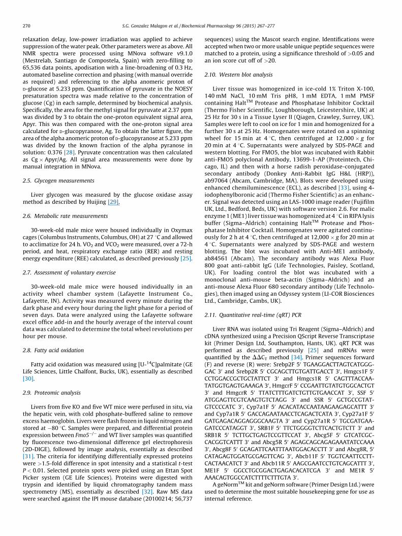
Fig. 2. Fmo5�/�mice exhibit a lean phenotype. (A) Body weight plotted as means � SEM (
WT and Fmo5�/� (KO) mice. (C) Ratio of weight of EWAT to body weight of WT and Fmo5�/�
(WT), 8 (KO). Data are expressed as means � SEM. **P < 0.01; ****P < 0.0001. (D) Sections of e
and eosin. Scale bar = 100 mm. (E) Food intake expressed as cumulative food intake in gra
3. Results
3.1. Fmo5�/� mice exhibit an age-related lean phenotype, with
reduced gains in body weight and fat deposits
Fmo5�/� mice appeared healthy and bred normally. From birthto about 20 weeks of age the weight of male KO and WT mice fed astandard chow diet was similar (Fig. 2A). From 20 weeks of ageboth KO and WT mice continued to gain weight. However, fromthis age the rate of weight gain of the KO mice was less than that ofWT mice (Fig. 2A), and by 30 weeks the weight of KO mice(33.92 � 0.53 g, n = 7) was 10% less than that of WT animals(37.50 � 0.85 g, n = 8) (P < 0.01). The difference in weight increasedwith age and by 52 weeks KO mice (35.46 � 0.50 g, n = 5) weighed17% less than WT mice (42.96 � 0.66 g, n = 5) (P < 0.001). Femalemice displayed a similar age-related difference in weight gain (datanot shown) and at 30 weeks of age the weight of female KO mice(23.79 � 0.71 g, n = 10) was 14% less than that of WT mice(27.75 � 0.81 g, n = 6) (P < 0.01). As age-related differences in weightgain between KO and WT mice were similar in both male and femalemice, subsequent analysis was done on male animals unless statedotherwise.
At 30 weeks of age, the KO mice were leaner than WT mice, hadless subcutaneous and inguinal fat and less fat surroundinginternal organs (Fig. 2B). The epididymal fat pads of KO mice(0.47 � 0.05 g, n = 8) weighed 63% less than those of WT mice(1.27 � 0.11 g, n = 11) (P < 0.0001). At 10 weeks of age the ratio of theweight of epididymal white adipose tissue (EWAT) to body weight ofWT and KO mice was similar (Fig. 2C). In WT mice it increasedsignificantly with age (P < 0.01), reaching 0.036 � 0.002 (n = 11) by30 weeks (Fig. 2C). In contrast, in KO mice the ratio was relativelyconstant with age and at 30 weeks was 0.016 � 0.002 (n = 8), 56% lessthan that of WT mice (P < 0.0001). Inactivation of the Fmo5 gene
WT, n = 8; KO, n = 7), *P < 0.05, **P < 0.01. (B) Internal abdominal view of 30-week-old
(KO) mice. 10 week: n = 4 (WT), 3 (KO); 20 week: n = 10 (WT), 5 (KO); 30 week: n = 11
pididymal WAT of 30-week-old WT and Fmo5�/� (KO) mice stained with haematoxylin
ms per g of body weight of WT and Fmo5�/� (KO) mice. n = 8 (WT), 7 (KO).

Fig. 3. Fmo5�/� mice have lower plasma concentrations of glucose, cholesterol and
pyruvate. (A) Plasma concentration of glucose in 10- and 30-week-old male and 30-
week-old female WT and Fmo5�/� (KO) mice. 10 week: n = 6 (WT), 3 (KO); 30-week
male: n = 19 (WT), 11 (KO); 30-week female: n = 9 (WT), 13 (KO). (B) Plasma
concentration of total cholesterol in 10- and 30-week-old male and 30-week-old
female WT and Fmo5�/� (KO) mice. 10 week: n = 11 (WT), 8 (KO); 30-week male:
n = 23 (WT), 18 (KO); 30-week female: n = 6 (WT), 10 (KO). (C) Plasma
concentration of pyruvate in 15- and 30-week-old male WT and Fmo5 (KO)
mice, n = 4 (15 week), 5 (30 week). Data are expressed as means � SEM. *P < 0.05,
**P < 0.01, ****P < 0.0001.
S.G. Gonzalez Malagon et al. / Biochemical Pharmacology 96 (2015) 267–277272
therefore leads to an age-related reduction in the rate of increase inbody weight, which is accompanied by a lack of increase in fat depotsize as the KO mice age.
At 30 weeks of age the diameter of epididymal adipocytes fromthe KO mice (57.22 � 1.60 mm, n = 3) was less than that of thosefrom WT mice (78.80 � 2.88 mm, n = 3) (P < 0.01) (Fig. 2D), equiva-lent to a decrease in cell volume of 62%. This corresponds closely tothe 63% decrease in the weight of epididymal fat pads of the KO mice,indicating that the difference in weight of epididymal fat between KOand WT mice was due to a difference in adipocyte volume.
Over a 12-week period from 16 weeks of age, the cumulativeintake of food by KO mice was �28% more than by WT mice, whenmeasured per g of body weight (Fig. 2E). The increased intake offood by the KO mice was evident from 20 weeks of age, whichcoincides with the time at which their rate of weight gain begins toslow in comparison with WT mice (Fig. 2A). At 30 weeks of age,although the FMO5 KO mice weighed 10% less than WT animals,their food intake per animal per day was 14% more than that of WTmice (data not shown). Therefore, the reduction in the rate ofweight gain and in the amount of WAT in the KO mice occurreddespite an increase in food intake.
3.2. Fmo5�/� mice have no impairment of lipid import into or export
from adipocytes
There were no significant differences between 30-week-old KOand WT mice in the plasma concentrations of triglycerides (KO,0.83 � 0.03 mmol/l, n = 7; WT, 0.89 � 0.07 mmol/l, n = 10), non-esterified fatty acids (NEFA) (KO, 0.88 � 0.09 mmol/l, n = 4; WT,0.74 � 0.07 mmol/l, n = 4) or glycerol (KO, 0.35 � 0.02 mmol/l, n = 4;WT, 0.38 � 0.01 mmol/l, n = 4), indicating that the reduced accumu-lation in Fmo5�/� mice of adipose tissue triacylglycerol was not theresult of impaired lipid import into or export from adipocytes. Theplasma concentration of NEFA after a 16-h fast increased to1.40 � 0.10 mmol/l (n = 4) in KO mice and to 1.36 � 0.13 mmol/l(n = 4) in WT mice. Thus, despite having less stored fat, Fmo5�/�micewere able to mobilize triglycerides from WAT normally in response toan overnight fast.
3.3. Fmo5�/� mice do not increase ectopic fat stores
Histological analysis revealed that there was no difference inthe amount of lipid present in the livers of KO and WT mice (datanot shown). This indicates that the decreased accumulation oftriglycerides in WAT of Fmo5�/�mice was not a consequence of anincrease in ectopic fat storage and suggests that export oftriglycerides from liver is not impaired in these animals.
3.4. Fmo5�/� mice have reduced plasma glucose and cholesterol
The plasma concentration of glucose in 10-week-old maleFmo5�/� and WT mice was similar (Fig. 3A). At 30 weeks of age itwas unchanged in WT mice (10.92 � 0.23 mmol/l, n = 19), but waslower in KO mice (9.07 � 0.63 mmol/l, n = 11) (P < 0.05). Thirty-week-old female KO mice also had lower plasma glucose than theirWT counterparts (Fig. 3A). At 30 weeks, both KO and WT male miceresponded to a 12-h overnight withdrawal of food by decreasing theirplasma glucose. Again, the plasma concentration of glucose in KOmice (6.60 � 0.09 mmol/l, n = 5) was significantly lower than in WTmice (7.90 � 0.35 mmol/l, n = 8) (P < 0.01). The urinary concentra-tion of glucose of KO (1.69 � 0.15 mmol/l, n = 3) and WT mice(2.12 � 0.51 mmol/l, n = 3) was similar, indicating that the reducedplasma glucose of KO mice was not a consequence of enhancedurinary excretion of glucose.
There was no difference in liver glycogen content of KO(66.83 � 4.88 mg/g liver, n = 5) and WT (63.84 � 5.16 mg/g liver,
n = 6) mice. Thus, the reduction in plasma glucose in the Fmo5�/�
mice is not due to increased storage of glucose in the form of glycogen inthe liver or to decreased mobilization of glucose from liver glycogen.
The plasma concentration of total cholesterol in 10-week-oldmale KO and WT mice was similar (Fig. 3B). At 30 weeks of age theconcentration in WT mice had increased, but in KO animals

S.G. Gonzalez Malagon et al. / Biochemical Pharmacology 96 (2015) 267–277 273
remained the same, 24% less than that in WT animals (Fig. 3B). In30-week-old mice, plasma cholesterol in female WT animals waslower than in male WT animals and similar to that in male KO mice(Fig. 3B). As was the case for male mice, in 30-week-old femalemice plasma cholesterol was lower in KO than in WT animals(Fig. 3B). In 30-week-old male animals plasma concentrations ofboth HDL (KO, 1.61 � 0.21 mmol/l, n = 7; WT, 2.43 � 0.10 mmol/l,n = 10) (P < 0.01) and LDL (KO, 0.36 � 0.02 mmol/l, n = 7; WT,0.48 � 0.02 mmol/l, n = 10) (P < 0.001) were lower, by 34% and25% respectively, in the KO mice. There was no significant differencebetween KO and WT mice in the ratio of total cholesterol to HDL.
3.5. Fmo5�/� mice have enhanced energy expenditure
Total energy expenditure, as measured by oxygen consumption,was significantly higher in Fmo5�/� mice than in WT animals inboth the light and dark phases (Fig. 4A), which suggests thatoxidation of fuel substrates is continuously higher in the KO mice.Resting energy expenditure (REE), a measure of basal metabolicrate plus energy expended in processing food, also is higher in KOthan in WT mice (Fig. 4B). More than 80% of the enhanced whole-body energy expenditure of Fmo5�/�mice is thus attributable to anincrease in REE.
The respiratory exchange ratio (RER) reflects the relativecontributions of carbohydrate and fat oxidation to total energyexpenditure. During the light phase the RER for FMO5 KO mice(0.943 � 0.007) was similar to that for WT mice (0.950 � 0.006)(Fig. 4C). During the dark phase the RER of WT animals remained thesame as that for the light phase. In contrast, in the dark phase theRER of the KO mice increased to 0.989 � 0.006 (Fig. 4C). The results
Fig. 4. Fmo5�/�mice have enhanced energy expenditure, but no increase in voluntary exe
(B) REE. (C) RER for light, dark and combined light and dark phases. Light phase (L, 07:0
period for 30-week-old male WT (n = 6) and Fmo5�/� (KO) (n = 6) mice. Values are means
of 30-week-old male WT and Fmo5�/� (KO) mice was assessed over seven days. Actogram
indicate that during the light phase the proportion of carbohydrateand fat oxidized by KO and WT mice is similar and for WT miceremains the same during the dark phase. However, during the darkphase the KO mice increase the proportion of carbohydrateoxidized. Urinary concentrations of urea and creatinine weresimilar in KO and WT mice (data not shown), indicating that theKO mice did not increase protein breakdown to maintain energybalance.
3.6. Fmo5�/� mice do not exhibit increased physical activity
The physical activity of Fmo5�/� and WT mice, as assessed byvoluntary wheel running over a period of seven days, was similar,with a normal increase in nocturnal activity for both sets of mice(Fig. 4D). There was no significant difference between the KO andWT mice in either the distance run or the speed of running (datanot shown). Therefore the enhanced whole-body energy expendi-ture of Fmo5�/� mice was not a consequence of increased physicalactivity.
3.7. In Fmo5�/� mice fatty acid oxidation is increased in EWAT and
decreased in skeletal muscle
At 20 weeks of age, the time at which KO mice begin to show areduction in weight gain, the rate of oxidation of [14C]palmitate inEWAT of KO animals (219 � 30 mmol palmitate oxidized/fat pad/h,n = 4) was 55% more than that of WT animals (141 � 17 mmolpalmitate oxidized/fat pad/h, n = 10) (P < 0.05). The increased rate offatty acid oxidation would contribute to depletion of lipid stores inEWAT of KO mice.
rcise. (A) Energy expenditure in the light, dark and combined light and dark phases.
0 to 19:00), dark phase (D, 19:00 to 07:00). Parameters were measured over a 72-h
� SEM. *P < 0.05, **P < 0.01, ***P < 0.001, ****P< 0.0001. (D) Voluntary wheel running
shows average interval count/h � SEM for a single day.

Fig. 5. Five key metabolic enzymes are down regulated in the liver of Fmo5�/� mice. Relative abundance of aldolase B (ALDOB) (A), glycerol 3-phosphate dehydrogenase
(GPD1) (B), ketohexokinase (KHK) (C), malic enzyme 1 (ME1) (D) and HMG-CoA synthase 1 (HMGCS1) (E) in liver of WT and Fmo5�/� (KO) mice determined via proteomic
analysis. (F) Western blot analysis of liver lysates of WT and Fmo5�/� (KO) mice. The blot was incubated with antibodies against ME1 and actin, as a loading control, and
developed as described in Section 2.
Table 1Relative difference in the abundance of mRNAs in the liver of WT and Fmo5�/�mice.
mRNA Relative
expression,
KO to WT
Cholesterol biosynthesis
HMG CoA synthase (HMGCS1) 1.71 � 0.47
HMG CoA reductase (HMGR) 2.41 � 0.46*
Squalene synthase (SS) 3.23 � 0.57*
Sterol responsive element binding protein (SREBP-2) 2.45 � 0.28*
Cholesterol uptake and transport
Scavenger receptor (SR-B1) 1.24 � 0.21
Abcg 5 1.74 � 0.26
Abcg 8 1.60 � 0.25
Bile acid synthesis
Cholesterol 7a hydroxylase (CYP7a1) 1.22 � 0.49
Cholesterol 27a hydroxylase (CYP27a1) 0.78 � 0.20
The abundance of each mRNA was determined by qRT-PCR. Data are presented as
relative abundance in Fmo5�/� (KO) compared to WT mice. Values are expressed as
means � S.E.M, n = 6–9.* P < 0.05.
S.G. Gonzalez Malagon et al. / Biochemical Pharmacology 96 (2015) 267–277274
Heart and skeletal muscle are tissues that oxidize lipid as theirmajor energy source. In 20-week-old mice the rate of [14C]palmi-tate oxidation in cardiac muscle was similar in KO and WT mice(data not shown). However, in resting soleus muscle it was 32%lower in KO (1255 � 74 mmol palmitate oxidized/g/h, n = 5) than inWT animals (1843 � 224 mmol palmitate oxidized/g/h, n = 4)(P < 0.05).
3.8. Proteins involved in carbohydrate metabolism or in cholesterol or
lipid synthesis are down regulated in Fmo5�/� mouse liver
The liver plays an important role in metabolic homeostasisand is an organ in which the Fmo5 gene is highly expressed[10]. To gain an insight into the basis of the metabolic phenotypeof the Fmo5�/�mice we performed a proteomic analysis of liver toidentify proteins that differed in abundance between KO and WTmice and, thus, might contribute to the phenotype. Five proteinswere down regulated in KO animals. All are enzymes involved incarbohydrate metabolism or in cholesterol or lipid biosynthesis(Fig. 5): aldolase B and ketohexokinase, involved in glucose andfructose metabolism; glycerol 3-phosphate dehydrogenase(GPD1), a cytosolic protein important for the production ofNAD+ and for transporting reducing equivalents from the cytosolto mitochondria; b-hydroxy-b-methylglutaryl-CoA (HMG-CoA)synthase 1, a cytosolic protein involved in cholesterol biosynthesis;and cytosolic malic enzyme (ME1), a lipogenic enzyme thatcatalyzes the oxidative decarboxylation of malate to pyruvate, inthe process producing NADPH for use in lipid and cholesterolbiosynthesis. Down-regulation of aldolase B, a glycolytic enzyme,and ME1 would be expected to result in a decrease in the productionof pyruvate. This is supported by the finding that the plasmaconcentration of pyruvate in 30-week-old animals is 57% lower inFmo5�/�mice than in WT animals (Fig. 3C). The difference in plasmapyruvate is age related, as in 15-week-old animals the concentrationis the same in KO and WT mice (Fig. 3C).
The lower abundance of HMG-CoA synthase 1 in the liver of KOmice was not the result of a difference in the abundance of thecorresponding mRNA (Table 1). In contrast, the mRNAs for twoother enzymes involved in cholesterol biosynthesis, HMG-CoAreductase and squalene synthase, were more abundant in the liversof KO than of WT mice, as was the mRNA for sterol regulatoryelement-binding protein-2 (SREBP-2), a transcription factor thatup-regulates the transcription of genes encoding several enzymesinvolved in cholesterol synthesis (Table 1). Although the abun-dance of the mRNA for squalene synthase was greater in the liversof KO than of WT mice, neither the amount nor activity of theencoded protein differed significantly between KO and WT mice(data not shown). The abundance in liver of mRNAs for proteins

S.G. Gonzalez Malagon et al. / Biochemical Pharmacology 96 (2015) 267–277 275
involved in cholesterol uptake and transport or in bile acidsynthesis did not differ between KO and WT mice (Table 1).
All the carbon in cholesterol is derived from acetate, via acetyl-CoA. The amounts of total and cytosolic acetyl-CoA in liver were notsignificantly different between KO and WT mice (data not shown).
Western blotting revealed that, in contrast to WT mice, inFmo5�/� mice ME1 was not detectable in the liver (Fig. 5F).However, as was the case for HMG-CoA synthase 1 (see above), theabundance of the corresponding ME1 mRNA in the livers of KO andWT mice was not significantly different. Therefore down regula-tion of the expression of HMG-CoA synthase 1 and ME1 in Fmo5�/�
mice is mediated at the translational or post-translational level.
4. Discussion
Mice in which the Fmo5 gene had been inactivated appearedhealthy but exhibited a lean phenotype, which was age-related,becoming apparent only at about 20 weeks of age. From this ageWT mice continued to increase body weight and this wasaccompanied by increases in the amount of lipid stored in WATand in the plasma concentration of cholesterol. In contrast, as theyaged Fmo5�/� mice, despite eating more than their WT counter-parts, were resistant to weight gain and their fat deposits andplasma cholesterol remained the same as that of 10-week-oldmice. In addition, at 30 weeks of age the plasma concentration ofglucose was lower in KO than in WT mice. The main characteristicsof the phenotype, reduced weight gain and lower plasmaconcentrations of glucose and cholesterol, were evident also infemale Fmo5�/� mice and, thus, the phenotype is gender-independent.
Fmo5�/� mice suffered no loss of appetite or decrease inphysical activity, indicating that the phenotype is unlikely to be aconsequence of illness or general malaise. Indeed, disruption of thegene encoding FMO5 resulted in a relatively ‘healthy’ phenotype,with mice being protected against a number of age-relatedmetabolic changes that could adversely affect wellbeing. Theincreased food consumption of the Fmo5�/� mice was likely aresponse to the lower amounts of fat reserves of these animals.
The lean phenotype of Fmo5�/� mice was associated withenhanced whole-body energy expenditure, most of which was dueto higher REE, with no increase in physical activity. The basis of theenhanced REE is unclear.
The increased rate of fatty acid oxidation in EWAT of KO mice at20 weeks of age would contribute to the depletion of triglyceridestores in this tissue and, hence, to reduction of adipocyte volume.At the same age, the lower rate of fatty acid oxidation in skeletalmuscle of KO than of WT mice suggests a switch to increased use ofcarbohydrate as fuel in this tissue in KO mice. This is supported bythe RER of 30-week-old mice, which indicates that during the darkphase, the period in which the mice are most active, the ratio ofcarbohydrate to fat utilized as fuel was higher in KO than in WTmice. The increased use by KO mice of carbohydrate as fuel ispossibly a response to the diminished stores of lipid in WAT ofthese animals and might also contribute to their lower plasmaconcentration of glucose. In WT mice the Fmo5 gene is notexpressed in WAT or skeletal muscle (data not shown), indicatingthat the effects in these tissues of disruption of Fmo5 are indirect.
Three of the proteins down regulated in the liver of Fmo5�/�
mice, aldolase B, ketohexokinase and GPD1, are involved incarbohydrate metabolism. Aldolase B is a glycolytic enzyme thatcatalyzes the production of glyceraldehyde 3-phosphate anddihydroxyacetone phosphate (DHAP), from fructose 1,6-bispho-sphate. Ketohexokinase (or fructokinase) catalyzes the conversionof fructose to fructose 1-phosphate, which can be converted toglyceraldehyde and DHAP in a reaction catalyzed by aldolase B.Both glyceraldehyde and DHAP can be converted to the glycolytic
intermediate glyceraldehyde 3-phosphate, via reactions catalyzedby triose kinase and triose phosphate isomerase respectively. Forglycolysis to proceed glyceraldehyde 3-phosphate must beconverted to 1,3-bisphosphoglycerate, in a reaction which requiresNAD+ and produces NADH. GPD1 catalyzes the oxidation ofcytosolic NADH by DHAP to produce NAD+, which re-entersglycolysis, and glycerol 3-phosphate, which can be combined withfatty acids to form triglycerides. The expected consequence of thedown regulation of aldolase B, ketohexokinase and GPD1 in theliver of Fmo5�/� mice is slowing of the entry of fructose intoglycolysis and of the metabolism of glucose via glycolysis and, thus,a decrease the amount of pyruvate produced by this pathway. Thisis supported by the finding that the plasma concentration ofpyruvate was lower in KO than in WT mice. In addition, downregulation of GPD1 would be expected to moderate the biosyn-thesis of triglycerides, via its effect on production of glycerol 3-phosphate, providing a potential explanation for the reduced fatdeposits of these animals.
Aldolase B can catalyze the reverse reaction, producing fructose1,6-bisphosphate from DHAP and glyceraldehyde 3-phosphate, akey step in gluconeogenesis. Down regulation of aldolase B would,therefore, be expected to slow both glycolysis and gluconeogene-sis, suggesting that Fmo5�/� mice do not increase gluconeogenesisin response to a lower plasma concentration of glucose. Thesimilarity in liver glycogen content of the KO and WT miceindicates that the former do not respond to lower plasma glucoseby increasing the mobilization of glucose from glycogen.
The lower plasma concentration of cholesterol in Fmo5�/�micewas accompanied by lower abundance in liver of HMG-CoAsynthase 1, which catalyzes the first committed step in isoprenoidbiosynthesis. It has been shown that modulation of HMG-CoAsynthase 1 activity influences cholesterol biosynthesis [37,38] andthe enzyme is negatively regulated, via a feedback mechanism, inresponse to cholesterol feeding [39]. In contrast, in Fmo5�/� micedown regulation of the amount of HMG-CoA synthase 1 occurred inthe context of lower, not higher, plasma cholesterol. However, theinduction in Fmo5�/� mice of mRNAs encoding HMG-CoAreductase, which catalyzes the rate-limiting step in cholesterolbiosynthesis, squalene synthase, which catalyzes the first step inthe pathway that produces cholesterol from farnesyl pyrophos-phate, and the transcription factor SREBP-2 is a normal response tolow plasma cholesterol [40,41]. But, despite this, the concentrationof plasma cholesterol in the KO mice remained low. In Fmo5�/�
mice down regulation of HMG-CoA synthase 1 protein occurred inthe absence of a change in the amount of its mRNA, indicating that,in these animals, regulation of expression of HMG-CoA synthase1 was independent of transcription and also of that of other keyenzymes in the pathway.
Acetyl-CoA is a product of fatty acid and glucose catabolism.Depending on the needs of a cell or organism, acetyl-CoA can beused for energy production, by entering the citric acid cycle, or forthe biosynthesis of fatty acids, ketone bodies or cholesterol. Theconcentration of cytosolic acetyl-CoA in the livers of KO and WTmice was similar, suggesting that the impact on fat and cholesterolproduction in the KO mice was primarily due to down regulation ofkey synthetic enzymes, not to a shortage of the primary buildingblocks.
The lower plasma concentration of pyruvate in the Fmo5�/�
mice could be explained by the down regulation in liver of theglycolytic enzyme aldolase B (see above) and of ME1, whichcatalyzes the production of pyruvate from malate. It has beenshown, in cell culture, that glucose, through the glycolytic productpyruvate, induces the amount and activity of ME1 [42]. Thus, inFmo5�/� mice reduction in the production of pyruvate, as aconsequence of down regulation of aldolase B, might contribute tothe down regulation of ME1.

S.G. Gonzalez Malagon et al. / Biochemical Pharmacology 96 (2015) 267–277276
Expression of ME1 is subject to regulation at several stages:transcription, stabilization of nuclear RNA and degradation ofcytoplasmic RNA; and it is regulated independently in response toa high-carbohydrate diet and by thyroid hormone [43]. In Fmo5�/�
mice the abundance of ME1 is apparently regulated by a differentmechanism: either via a block in the translation of the mRNA or anincrease in the rate of degradation of the protein.
The reaction catalyzed by ME1 is an important source of NADPHfor use in anabolic reactions, including the biosynthesis of fattyacids and cholesterol. Repression of ME1 decreases the cellularconcentration of NADPH [42,44]. Reduction in the concentration ofNADPH, as a consequence of down regulation of ME1, would beexpected to moderate the biosynthesis of both fatty acids andcholesterol, thus providing a potential explanation for the reducedfat deposits and lower plasma cholesterol that are characteristic ofthe metabolic phenotype of Fmo5�/� mice.
In liver biopsies of type-2 diabetics, FMO5 was one of 134 genesfound to be repressed [45]. The phenotype of the Fmo5�/� micereveals that in the absence of FMO5 weight and plasma glucose arereduced. This suggests strongly that the reduction in theexpression of FMO5 in type-2 diabetics is a response to thisaberrant metabolic condition in an attempt to restore homeostasis.
Another FMO KO mouse line, one that lacks genes encodingFMO1, FMO2 and FMO4, also exhibits a lean phenotype, withreduced body weight and fat deposits [25]. However, there aredistinct differences between this KO mouse line and Fmo5�/�mice.The phenotype of mice lacking Fmo1, Fmo2 and Fmo4 genes, whichis attributed to lack of FMO1, is evident from as early as six weeksof age and is characterized by enhanced whole-body energyexpenditure and REE with no change in RER (indicating an increasein both fat and carbohydrate oxidation), increased capacity forexercise, elevated fatty acid oxidation in skeletal muscle, but not inWAT, higher plasma glucose and evidence for the operation of afutile fuel cycle in WAT [25]. In contrast, the phenotype of Fmo5�/�
mice is age-related, becoming apparent only after 20 weeks of age,and is characterized by lower plasma concentrations of glucoseand cholesterol, fatty acid oxidation that is reduced in skeletalmuscle but elevated in WAT, and no increase in physical activity. Incommon with mice that lack FMO1, FMO2 and FMO4, mice thatlack FMO5 exhibit enhanced whole-body energy expenditure andREE, but have increased RER, indicating a switch from fat tocarbohydrate oxidation. Thus, both FMO5 and FMO1 are metabolicregulators. However, whereas FMO1 acts as a regulator of energyhomeostasis [25], our results indicate that FMO5 regulatesmetabolic ageing via pleiotropic effects. These include positivemodulation of cholesterol biosynthesis, via promoting theexpression in liver of enzymes involved in the cholesterolbiosynthetic pathway and the production of NADPH, and alsopromoting the hepatic expression of enzymes involved inglycolysis/gluconeogenesis and in the formation of glycerol 3-phosphate for the biosynthesis of triglycerides.
FMO5 is classified as an oxidoreductase and it is possible that itseffects are mediated via modulation of the cellular redox state.However, in vivo substrates of FMO5 have yet to be identified andthe mechanism by which the enzyme exerts its pleiotropic effectsremains to be established.
Our results have potential implications for humans, indicatingthat interindividual variation in FMO5 expression [20–22] maycontribute to differences in fat deposits and plasma cholesterol andthat induction of FMO5 expression by some therapeutics [19] mayhave adverse effects on the metabolic health of patients.
Acknowledgements
We thank V.L.J. Tybulewicz for pPNT. Work was supported byscholarships or studentships from: CONACYT, Mexico (SGGM),
Barts and the Royal London School of Medicine and Dentistry(ANM), the Drummond Trust (LH), the Wellcome Trust (FS), ORSand UCL graduate School (SV) and Primer Design (SGGM and FS).Work was funded in part by a grant from the Wellcome Trust(053590) to EAS and IRP. The proteomics work was supported bythe National Institute for Health Research (NIHR), UniversityCollege London Hospitals (UCLH) Biomedical Research Centre.
References
[1] D. Hernandez, A. Janmohamed, P. Chandan, I.R. Phillips, E.A. Shephard,Organization and evolution of the flavin-containing monooxygenase genes ofhuman and mouse: identification of novel gene and pseudogene clusters,Pharmacogenetics 14 (2004) 117–130.
[2] I.R. Phillips, A.A. Francois, E.A. Shephard, The flavin-containingmonooxygenases (FMOs): genetic variation and its consequences for themetabolism of therapeutic drugs, Curr. Pharmacogenomics 5 (2007) 292–313.
[3] S.K. Krueger, D.E. Williams, Mammalian flavin-containing monooxygenases:structure/function, genetic polymorphisms and role in drug metabolism,Pharmacol. Ther. 106 (2005) 357–387.
[4] B. Furnes, J. Feng, S.S. Sommer, D. Schlenk, Identification of novel variants ofthe flavin-containing monooxygenase gene family in African Americans, DrugMetab. Dispos. 31 (2003) 187–193.
[5] B. Furnes, Evaluation of xenobiotic N- and S-oxidation by variant flavin-containing monooxygenase 1 (FMO1) enzymes, Toxicol. Sci. 78 (2004)196–203.
[6] C.T. Dolphin, D.J. Beckett, A. Janmohamed, T.E. Cullingford, R.L. Smith, E.A.Shephard, et al., The flavin-containing monooxygenase 2 Gene (FMO2) ofhumans, but not of other primates, encodes a truncated, nonfunctionalprotein, J. Biol. Chem. 273 (1998) 30599–30607.
[7] K.R. Veeramah, M.G. Thomas, M.E. Weale, D. Zeitlyn, A. Tarekegn, E. Bekele,et al., The potentially deleterious functional variant flavin-containingmonooxygenase 2*1 is at high frequency throughout sub-Saharan Africa,Pharmacogenet. Genomics 18 (2008) 877–886.
[8] C.T. Dolphin, A. Janmohamed, R.L. Smith, E.A. Shephard, I.R. Phillips, Missensemutation in flavin-containing mono-oxygenase 3 gene, FMO3, underlies fish-odour syndrome, Nat. Genet. 17 (1997) 491–494.
[9] I.R. Phillips, E.A. Shephard, Flavin-containing monooxygenases: mutations,disease and drug response, Trends Pharmacol. Sci. 29 (2008) 294–301.
[10] A. Janmohamed, D. Hernandez, I.R. Phillips, E.A. Shephard, Cell-, tissue-, sex-and developmental stage-specific expression of mouse flavin-containingmonooxygenases (Fmos), Biochem. Pharmacol. 68 (2004) 73–83.
[11] J.R. Cashman, J. Zhang, Human flavin-containing monooxygenases, Annu. Rev.Pharmacol. Toxicol. 46 (2006) 65–100.
[12] L.H. Overby, A.R. Buckpitt, M.P. Lawton, E. Atta-Asafo-Adjei, J. Schulze, R.M.Philpot, Characterization of flavin-containing monooxygenase 5 (FMO5) clonedfrom human and guinea pig: evidence that the unique catalytic properties ofFMO5 are not confined to the rabbit ortholog, Arch. Biochem. Biophys. 317(1995) 275–284.
[13] D.H. Lang, C.K. Yeung, R.M. Peter, C. Ibarra, R. Gasser, K. Itagaki, et al., Isoformspecificity of trimethylamine N-oxygenation by human flavin-containingmonooxygenase (FMO) and P450 enzymes: selective catalysis by FMO3,Biochem. Pharmacol. 56 (1998) 1005–1012.
[14] D.H.D. Lang, A.E.A. Rettie, In vitro evaluation of potential in vivo probes forhuman flavin-containing monooxygenase (FMO): metabolism of benzydamineand caffeine by FMO and P450 isoforms, Br. J. Clin. Pharmacol. 50 (2000)311–314.
[15] J. Zhang, M.A. Cerny, M. Lawson, R. Mosadeghi, J.R. Cashman, Functionalactivity of the mouse flavin-containing monooxygenase forms 1, 3, and 5, J.Biochem. Mol. Toxicol. 21 (2007) 206–215.
[16] M.S. Motika, J. Zhang, E.C. Ralph, M.A. Dwyer, J.R. Cashman, pH dependence onfunctional activity of human and mouse flavin-containing monooxygenase 5,Biochem. Pharmacol. 83 (2012) 962–968.
[17] N. Ohmi, H. Yoshida, H. Endo, M. Hasegawa, M. Akimoto, S. Higuchi, S-oxidation of S-methyl-esonarimod by flavin-containing monooxygenases inhuman liver microsomes, Xenobiotica 33 (2003) 1221–1231.
[18] W.G. Lai, N. Farh, G.A. Moniz, Y.N. Wong, A Baeyer–Villiger oxidationspecifically catalyzed by human flavin-containing monooxygenase, DrugMetab. Dispos. 5 (39) (2011) 61–70.
[19] G.C. Carver, R.M. Philpot, Quantitation and kinetic properties of hepaticmicrosomal and recombinant flavin-containing monooxygenases 3 and 5 fromhumans, Chem. Biol. Interact. 106 (1997) 29–45.
[20] M.M. Miller, R.A. James, J.K. Richer, D.F. Gordon, W.M. Wood, K.B. Horwitz,Progesterone regulated expression of flavin-containing monooxygenase 5 bythe B-isoform of progesterone receptors: implications for tamoxifencarcinogenicity, J. Clin. Endocrinol. Metab. 82 (1997) 2956–2961.
[21] J.M. Rae, M.D. Johnson, M.E. Lippman, D.A. Flockhart, Rifampin is a selective,pleiotropic inducer of drug metabolism genes in human hepatocytes: studieswith cDNA and oligonucleotide expression arrays, J. Pharmacol. Exp. Ther. 299(2001) 849–857.
[22] S. Krusekopf, I. Roots, St. John’s wort and its constituent hyperforinconcordantly regulate expression of genes encoding enzymes involved in basiccellular pathways, Pharmacogenet. Genomics 15 (2005) 817–829.

S.G. Gonzalez Malagon et al. / Biochemical Pharmacology 96 (2015) 267–277 277
[23] D. Hernandez, A. Janmohamed, P. Chandan, B.A. Omar, I.R. Phillips, E.A.Shephard, Deletion of the mouse Fmo1 gene results in enhancedpharmacological behavioural responses to imipramine, Pharmacogenet.Genomics 19 (2009) 289–299.
[24] E.A. Shephard, I.R. Phillips, The potential of knockout mouse lines in definingthe role of flavin-containing monooxygenases in drug metabolism, ExpertOpin. Drug Metab. Toxicol. 6 (2010) 1083–1094.
[25] S. Veeravalli, B.A. Omar, L. Houseman, M. Hancock, S.G. Gonzalez Malagon, F.Scott, et al., The phenotype of a flavin-containing monooxygenase knockoutmouse implicates the drug-metabolizing enzyme FMO1 as a novel regulator ofenergy balance, Biochem. Pharmacol. 90 (2014) 88–95.
[26] D. Hernandez, A.N. Melidoni, L.R. Phillips, E.A. Shephard, Microinjection oftargeted embryonic stem cells and establishment of knockout mouse lines forFmo genes, Methods Mol. Biol. 320 (2006) 329–341.
[27] T.A. Hough, P.M. Nolan, V. Tsipouri, A.A. Toye, I.C. Gray, M. Goldsworthy, et al.,Novel phenotypes identified by plasma biochemical screening in the mouse,Mamm. Genome 13 (2002) 595–602.
[28] M.U. Roslund, P. Tahtinen, M. Niemitz, R. Sjoholm, Complete assignments ofthe (1)H and (13)C chemical shifts and J(H, H) coupling constants in NMRspectra of D-glucopyranose and all D-glucopyranosyl-D-glucopyranosides,Carbohydr. Res. 343 (2008) 101–112.
[29] F. Huijing, A rapid enzymic method for glycogen estimation in very smalltissue samples, Clin. Chim. Acta 30 (1970) 567–572.
[30] N. Alam, E.D. Saggerson, Malonyl-CoA and the regulation of fatty acidoxidation in soleus muscle, Biochem. J. 334 (1998) 233–241.
[31] S. Gharbi, P. Gaffney, A. Yang, M.J. Zvelebil, R. Cramer, M.D. Waterfield, J.F.Timms, Evaluation of two-dimensional differential gel electrophoresis forproteomic expression analysis of a model breast cancer cell system, Mol. CellProteomics 1 (2002) 91–98.
[32] J. Sinclair, G. Metodieva, D. Dafou, S.A. Gayther, J.F. Timms, Profiling signaturesof ovarian cancer tumour suppression using 2D-DIGE and 2D-LC–MS/MS withtandem mass tagging, J. Proteomics 74 (2011) 451–465.
[33] C. Haan, I. Behrmann, A cost effective non-commercial ECL-solution forWestern blot detections yielding strong signals and low background, J.Immunol. Methods 318 (2007) 11–19.
[34] W. Liu, D.A. Saint, A new quantitative method of real time reversetranscription polymerase chain reaction assay based on simulation ofpolymerase chain reaction kinetics, Anal. Biochem. 302 (2002) 52–59.
[35] R.A. Memon, I. Shechter, A.H. Moser, J.K. Shigenaga, C. Grunfeld, K.R. Feingold,Endotoxin, tumor necrosis factor, and interleukin-1 decrease hepatic squalenesynthase activity, protein, and mRNA levels in Syrian hamsters, J. Lipid Res. 38(1997) 1620–1629.
[36] Y. Tsuchiya, U. Pham, W. Hu, A. Ohnuma, I. Gout, Changes in acetyl CoA levelsduring the early embryonic development of Xenopus laevis, PLOS ONE 9 (2014)e97693.
[37] M. Mehrabian, K.A. Callaway, C.F. Clarke, R.D. Tanaka, M. Greenspan, A.J. Lusis,R.S. Sparkes, T. Mohandas, J. Edmond, A.M. Fogelman, P.A. Edwards, Regulationof rat liver 3-hydroxy-3-methylglutaryl coenzyme A synthase and thechromosomal localization of the human gene, J. Biol. Chem. 261 (1986)16249–16255.
[38] J.S. Chen, A.W. Alberts, V.M. Hunt, M.N. Chang, S.S. Yang, K.L. Thompson, et al.,Inhibition of hydroxymethylglutaryl-coenzyme A synthase by L-659,699, Proc.Natl. Acad. Sci. U. S. A. 84 (1987) 7488–7492.
[39] T. Sugiyama, K. Clinkenbeard, J. Moss, M.D. Lane, Multiple cytosolic forms ofhepatic b-hydroxy-b-methyglutaryl coA synthase: possible regulatoryrole in cholesterol synthesis, Biochem. Biophys. Res. Commun. 48 (1972)255–261.
[40] J.L. Goldstein, M.S. Brown, Regulation of the mevalonate pathway, Nature 343(1990) 425–430.
[41] G.C. Ness, C.M. Chambers, Feedback and hormonal regulation of hepatic3-hydroxy-3-methylglutaryl coenzyme A reductase: the concept of cholesterolbuffering capacity, Proc. Soc. Exp. Biol. Med. 224 (2000) 8–19.
[42] F.-J. Zheng, H.-B. Ye, M.-S. Wu, Y.-F. Lian, C.-N. Qian, Y.-X. Zeng, Repressingmalic enzyme 1 redirects glucose metabolism, unbalances the redox state, andattenuates migratory and invasive abilities in nasopharyngeal carcinoma celllines, Chin. J. Cancer 31 (2012) 519–531.
[43] B. Dozin, J.E. Rall, V.M. Nikodem, Tissue-specific control of rat malic enzymeactivity and messenger RNA levels by a high carbohydrate diet, Proc. Natl.Acad. Sci. U. S. A. 83 (1986) 4705–4709.
[44] P. Jiang, W. Du, A. Mancuso, K.E. Wellen, X. Yang, Reciprocal regulation of p53and malic enzymes modulates metabolism and senescence, Nature 493 (2013)689–693.
[45] T. Takamura, M. Sakurai, T. Ota, H. Ando, M. Honda, S. Kaneko, Genes forsystemic vascular complications are differentially expressed in the livers oftype 2 diabetic patients, Diabetologia 47 (2004) 638–647.
Related Documents