1 The Pathophysiology of Hemorrhagic Shock Richard E. Klabunde, Ph.D. Associate Professor of Physiology Department of Biomedical Sciences Ohio University College of Osteopathic Medicine

Welcome message from author
This document is posted to help you gain knowledge. Please leave a comment to let me know what you think about it! Share it to your friends and learn new things together.
Transcript

1
The Pathophysiology of Hemorrhagic Shock
Richard E. Klabunde, Ph.D.Associate Professor of Physiology
Department of Biomedical SciencesOhio University College of Osteopathic Medicine

3
Learning Objectives
• Describe how acute blood loss leads to hypotension.
• Describe the compensatory mechanisms that operate to restore arterial pressure following hemorrhage.
• Describe the decompensatory mechanisms that lead to irreversible shock.
• Describe the rationale for different medical interventions following hemorrhage.

4
General Definition of Hemorrhagic Shock
A clinical syndrome resulting from decreased blood and oxygen perfusion of vital organs resulting from a loss of
blood volume.

5
Hemorrhagic Shock(Initial Uncompensated Responses)
EDV or EDP or PCWP
SV A
B
LVPress
LV Vol
AB
Blood Loss
↓ CVP ↓ EDV (Preload)
↓ SV↓ CO↓ PA
Frank-StarlingMechanism

6
Effects Blood Volume Loss on Mean Arterial Pressure
Com
pens
ated
0 2 4 60
50
100
Time (hours)
Dec
ompe
nsat
ed
15%
25%
35%
45%
Transfusion60%
Aor
tic P
ress
(mm
Hg)
(Adapted from Guyton & Crowell, 1961)
III
III
IV

7
Classes of Hemorrhagic Shock• Class I hemorrhage (loss of 0-15%)
– Little tachycardia– Usually no significant change in BP, pulse pressure,
respiratory rate• Class II hemorrhage (loss of 15-30%)
– HR >100 beats per minute, tachypnea, decreased pulse pressure
• Class III hemorrhage (loss of 30-40%)– Marked tachycardia and tachypnea, decreased systolic
BP, oliguria • Class IV hemorrhage (loss of >40%)
– Marked tachycardia and decreased systolic BP, narrowed pulse pressure, markedly decreased (or no) urinary output
– Immediately life threatening

8
Compensatory Mechanisms• Baroreceptor reflexes• Circulating vasoconstrictors • Chemoreceptor reflexes• Reabsorption of tissue fluids• Renal reabsorption of sodium and water• Activation of thirst mechanisms• Cerebral ischemia• Hemapoiesis

9
Arterial Baroreceptors
Receptor Firing
Arterial Pressure Pulse
ReceptorFiring Rate
(% max)
100 Carotid Sinus
50
0 100 200MAP (mmHg)
A
B
DecreasedMAP &
Pulse Press
Klabunde, RE, Cardiovascular Physiology Concepts, Lippincott Williams & Wilkins, 2004

10
Autonomic Responses to Baroreceptor Activity
• Arterial baroreceptor firing inhibits sympathetic outflow and stimulates parasympathetic outflow
• Therefore, reduced firing, which occurs during hemorrhage, leads to sympathetic activation and parasympathetic inhibition
Klabunde, RE, Cardiovascular Physiology Concepts, Lippincott Williams & Wilkins, 2004

11
Effects of 8% Blood Loss on AorticPressure in Anesthetized Dogs
(Effects of Baroreceptor Denervation)
-60
-40
-20
0
IntactCarotid sinus onlyAortic arch onlyNo baroreceptors
Mea
n A
ortic
Pre
ss
(% d
ecre
ase)
(Adapted from A.J. Edis, 1971)
12
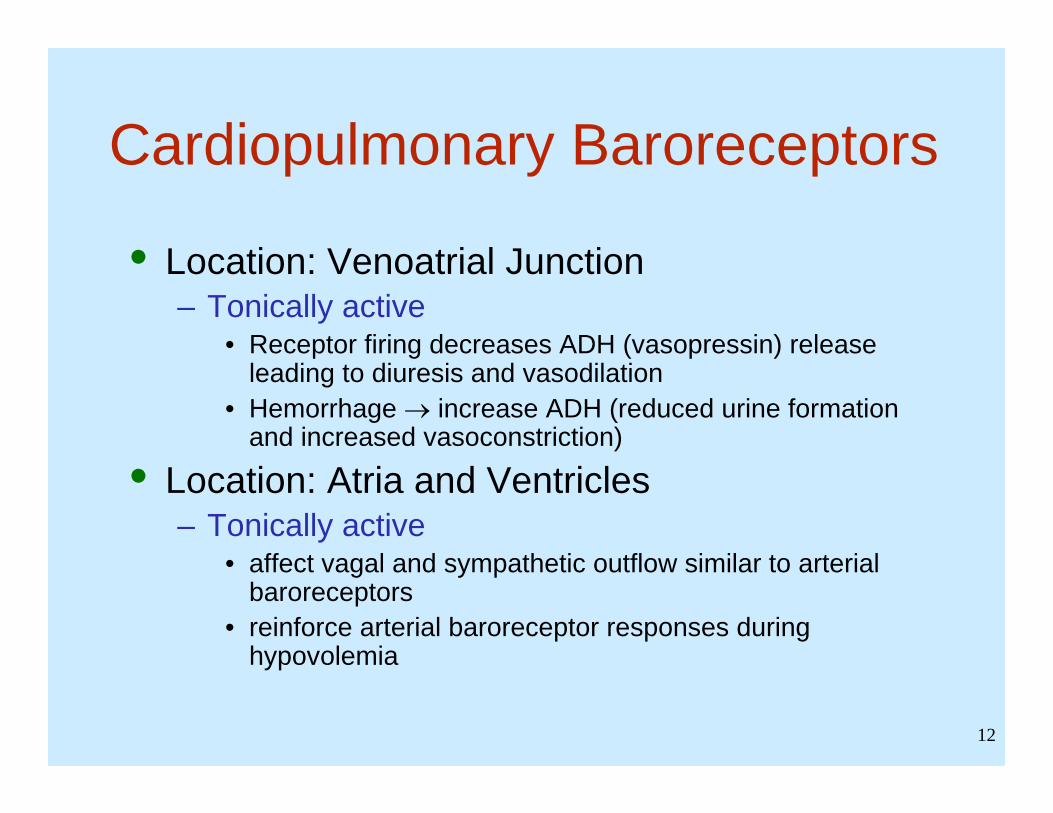
Cardiopulmonary Baroreceptors
• Location: Venoatrial Junction– Tonically active
• Receptor firing decreases ADH (vasopressin) release leading to diuresis and vasodilation
• Hemorrhage → increase ADH (reduced urine formation and increased vasoconstriction)
• Location: Atria and Ventricles– Tonically active
• affect vagal and sympathetic outflow similar to arterial baroreceptors
• reinforce arterial baroreceptor responses during hypovolemia

13
Baroreceptor Reflexes
Klabunde, RE, Cardiovascular Physiology Concepts, Lippincott Williams & Wilkins, 2004

14
• Redistribution of cardiac output– Intense vasoconstriction in skin, skeletal muscle, renal
(during severe hemorrhage) and splanchnic circulations increases systemic vascular resistance, which attenuates the fall in arterial pressure
– Coronary and cerebral circulations spared– Therefore, cardiac output is shunted to essential organs
• Redistribution of blood volume– Strong venoconstriction in GI, hepatic and skin
circulations– Partial restoration of central venous blood volume and
pressure to counteract loss of filling pressure to the heart
Baroreceptor Reflexes Cont.

15
Importance of Changes in Venous Tone

16
Central Venous Pressure During Hemorrhage
• Hemorrhage decreases blood volume and decreases CVP (A→B)
• Peripheral venous constriction decreases venous compliance (B→C),which increases CVP and shifts blood volume toward heart
• Increased CVP increases ventricular preload and force of contraction (Frank-Starling mechanism)
Vol
Press
A
B C
Venous Compliance Curves

17
Humoral Compensatory Mechanisms
Klabunde, RE, Cardiovascular Physiology Concepts, Lippincott Williams & Wilkins, 2004

18
Importance of Humoral Compensatory Mechanisms
• Angiotensin II, vasopressin and catecholamines reinforce sympathetic mediated vasoconstriction to help maintain arterial pressure by – increasing systemic vascular resistance – decreasing venous compliance, which increases
ventricular preload and enhances stroke volume• Angiotensin II, aldosterone and vasopressin
act on the kidneys to increase blood volume

19
Chemoreceptor Reflexes
• Peripheral chemoreceptors– Carotid bodies– Aortic bodies
• Central chemoreceptors– Medulla (associated with cardiovascular
control “centers”)
20
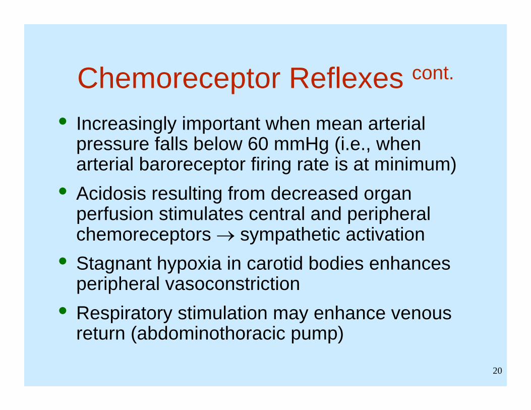
Chemoreceptor Reflexes cont.
• Increasingly important when mean arterial pressure falls below 60 mmHg (i.e., when arterial baroreceptor firing rate is at minimum)
• Acidosis resulting from decreased organ perfusion stimulates central and peripheral chemoreceptors → sympathetic activation
• Stagnant hypoxia in carotid bodies enhances peripheral vasoconstriction
• Respiratory stimulation may enhance venous return (abdominothoracic pump)

21
Reabsorption of Tissue Fluids• Capillary pressure falls
– Reduced arterial and venous pressures– Increased precapillary resistance– Transcapillary fluid reabsorption (up to 1 liter/hr
autoinfused)• Capillary plasma oncotic pressure can fall from 25
to 15 mmHg due to autoinfusion thereby limiting capillary fluid reabsorption
• Hemodilution causes hematocrit to fall which decreases blood viscosity

22
[ ])()( TCTC PPAKFM ππ −−−⋅=
Changes in Starling Forces Following Hemorrhage
Starling Equation for Fluid Balance

23
Cerebral Ischemia• When mean arterial pressure falls below 60
mmHg, cerebral perfusion decreases because the pressure is below the autoregulatory range
• Cerebral ischemia produces very intense sympathetic discharge that is several-fold greater than the maximal sympathetic activation caused by the baroreceptor reflex

24
Decompensatory Mechanisms“Progressive Shock”
• Cardiogenic Shock– Impaired coronary perfusion causing myocardial
hypoxia, systolic and diastolic dysfunction, arrhythmias
• Sympathetic Escape– Loss of vascular tone (↓SVR) causing progressive
hypotension and organ hypoperfusion– Increased capillary pressure causing increased fluid
filtration and hypovolemia• Cerebral Ischemia
– Loss of autonomic outflow due to severe cerebral hypoxia

25
• Metabolic Acidosis• Rheological –
– Increased microvascular viscosity– Microvascular plugging by leukocytes and platelets– Intravascular coagulation
• Systemic Inflammatory Response– Endotoxin release into systemic circulation– Cytokine formation – TNF, IL, etc.– Enhanced nitric oxide formation– Reactive oxygen-induced cellular damage– Increased capillary permeability– Multiple organ failure

26
Decompensatory Mechanisms(Cardiogenic Shock and Sympathetic Escape)
↓ Inotropy
↓ CardiacOutput
+
↓ CoronaryPerfusion
↓ ArterialPressure
↑ SympatheticVasoconstriction
TissueHypoxia
Vasodilation
+

27
Time-Dependent Changes in Cardiac Function
• Dogs hemorrhaged and arterial pressure held at 30 mmHg
• Precipitous fall in cardiac function occurred after 4 hours of severe hypotension
0 5 10Left Atrial Pressure (mmHg)
CardiacOutput
0 2
4.5
5
5.2
4
(adapted from Crowell et al., 1962)
Hours afterHemorrhage

28
Comparison of Different Forms of Shock Cardiogenic
Shock Hemorrhagic
Shock Septic Shock
CV Origin Cardiac Volume Vascular
Cardiac Output
↓ ↓ ↑↓
Vascular Resistance
↑ ↑ ↓
Blood Volume
↑ ↓ ↓
Management Mechanical Inotropes
Vasopressors Vasodilators
IV Fluids/BloodVasopressors
IV Fluids Antibiotics
VasopressorsInotropes

29
Resuscitation Issues
• Reducing reperfusion injury & systemic inflammatory response syndrome (SIRS)– Anti-inflammatory drugs– NO scavenging and antioxidant drugs
• Resuscitation fluids– Crystalloid vs. non-crystalloid solutions– Isotonic vs. hypertonic solutions– Whole blood vs. packed red cells– Hemoglobin-based solutions– Perfluorocarbon-based solutions– Fluid volume-related issues

30
Resuscitation Issues cont.
(Current Research)
• Efficacy of pressor agents• Hypothermic vs. normothermic resuscitation• Tailoring therapy to conditions of shock
– Uncontrolled vs. controlled hemorrhage– Traumatic vs. atraumatic shock

31
Review Learning Objectives
• Describe how acute blood loss leads to hypotension.
• Describe the compensatory mechanisms that operate to restore arterial pressure following hemorrhage.
• Describe the decompensatory mechanisms that lead to irreversible shock.
• Describe the rationale for different medical interventions following hemorrhage.
Related Documents