doi:10.1016/j.jacc.2009.03.037 2009;54;36-46 J. Am. Coll. Cardiol. Kiran Patel, Michael Frenneaux, and John E. Sanderson Yu Ting Tan, Frauke Wenzelburger, Eveline Lee, Grant Heatlie, Francisco Leyva, Longitudinal Motion and Diastolic Ventricular Function Involving Torsion, Untwist, and Exercise Echocardiography Reveals Complex Abnormalities of Both Systolic The Pathophysiology of Heart Failure With Normal Ejection Fraction: This information is current as of May 11, 2011 http://content.onlinejacc.org/cgi/content/full/54/1/36 located on the World Wide Web at: The online version of this article, along with updated information and services, is by on May 11, 2011 content.onlinejacc.org Downloaded from

Welcome message from author
This document is posted to help you gain knowledge. Please leave a comment to let me know what you think about it! Share it to your friends and learn new things together.
Transcript

doi:10.1016/j.jacc.2009.03.037 2009;54;36-46 J. Am. Coll. Cardiol.
Kiran Patel, Michael Frenneaux, and John E. Sanderson Yu Ting Tan, Frauke Wenzelburger, Eveline Lee, Grant Heatlie, Francisco Leyva,
Longitudinal Motionand Diastolic Ventricular Function Involving Torsion, Untwist, and
Exercise Echocardiography Reveals Complex Abnormalities of Both Systolic The Pathophysiology of Heart Failure With Normal Ejection Fraction:
This information is current as of May 11, 2011
http://content.onlinejacc.org/cgi/content/full/54/1/36located on the World Wide Web at:
The online version of this article, along with updated information and services, is
by on May 11, 2011 content.onlinejacc.orgDownloaded from

Ipw(d
FBStCBci
2
Journal of the American College of Cardiology Vol. 54, No. 1, 2009© 2009 by the American College of Cardiology Foundation ISSN 0735-1097/09/$36.00P
Heart Failure
The Pathophysiology of HeartFailure With Normal Ejection FractionExercise Echocardiography Reveals ComplexAbnormalities of Both Systolic and Diastolic VentricularFunction Involving Torsion, Untwist, and Longitudinal Motion
Yu Ting Tan, MBBS,* Frauke Wenzelburger, MD,*† Eveline Lee, MBCHB,†Grant Heatlie, MBBS, PHD,† Francisco Leyva, MD,* Kiran Patel, MBBCHIR, PHD,*Michael Frenneaux, MD,* John E. Sanderson, MD*
Birmingham and Stoke-on-Trent, United Kingdom
Objectives The purpose of this study was to test the hypothesis that in heart failure with normal ejection fraction (HFNEF)exercise limitation is due to combined systolic and diastolic abnormalities, particularly involving ventricular twistand deformation (strain) leading to reduced ventricular suction, delayed untwisting, and impaired early diastolicfilling.
Background A substantial proportion of patients with heart failure have a normal left ventricular ejection fraction. Currentlythe pathophysiology is considered to be due to abnormal myocardial stiffness and relaxation.
Methods Patients with a diagnosis of HFNEF and proven cardiac limitation by cardiopulmonary exercise testing were stud-ied by standard, tissue Doppler, and speckle tracking echocardiography at rest and on submaximal exercise.
Results Fifty-six patients (39 women; mean age 72 � 7 years) with a clinical diagnosis of HFNEF and 27 age-matchedhealthy control subjects (19 women; mean age 70 � 7 years) had rest and exercise images of sufficient qualityfor analysis. At rest, systolic longitudinal and radial strain, systolic mitral annular velocities, and apical rotationwere lower in patients, and all failed to rise normally on exercise. Systolic longitudinal functional reserve wasalso significantly lower in patients (p � 0.001). In diastole, patients had reduced and delayed untwisting, re-duced left ventricular suction at rest and on exercise, and higher end-diastolic pressures. Mitral annular systolicand diastolic velocities, systolic left ventricular rotation, and early diastolic untwist on exercise correlated withpeak VO2max.
Conclusions In HFNEF there are widespread abnormalities of both systolic and diastolic function that become more apparenton exercise. HFNEF is not an isolated disorder of diastole. (J Am Coll Cardiol 2009;54:36–46) © 2009 by theAmerican College of Cardiology Foundation
ublished by Elsevier Inc. doi:10.1016/j.jacc.2009.03.037
fftasmcs
aSi
t is now well-established that at least one-half of theatients presenting with symptoms and signs of heart failureill have a normal left ventricular ejection fraction (LVEF)
1,2). This form of heart failure has been variously labeled asiastolic heart failure, heart failure with preserved systolic
rom the *Department of Cardiovascular Medicine, University of Birmingham,irmingham, United Kingdom; and the †University Hospital of North Staffordshire,toke-on-Trent, United Kingdom. The project was funded by a project grant fromhe British Heart Foundation and an equipment grant from the North Staffs Heartommittee. Dr. Sanderson has received travel grants from Sanofi-Aventis,oehringer-Ingelheim, and Pfizer, and lecture fees from Pfizer. Dr. Frenneaux is aonsultant to Medtronic and St. Jude and has a method-of-use patent for perhexilinen heart failure (no current financial value).
nManuscript received November 18, 2008; revised manuscript received March 13,
009, accepted March 17, 2009.
content.onlinejDownloaded from
unction, or more simply heart failure with a normal ejectionraction (HFNEF). The pathophysiology of HFNEF andhe generation of symptoms remain controversial (3). Zile etl. (4) suggested on the basis of invasive hemodynamictudies that patients with HFNEF have significant abnor-alities in active relaxation and passive stiffness, and con-
luded that the pathophysiological cause of elevated dia-tolic pressures and symptoms is abnormal diastolic function
See page 47
lone, because systolic function was considered normal.imilarly, Westermann et al. (5) demonstrated recently
ncreased LV stiffness at rest in HFNEF, although this did
ot increase with hand-grip or atrial pacing. Both of theseby on May 11, 2011 acc.org
stofusnhHmi3tiimtumti
M
Wsp
YmrpwpwiciicpCrchmrerep2Awad
tBtbclccosVesS4mDvbtadTfiaeat
oltbbtss
Lrdasad((owSpaSa(
37JACC Vol. 54, No. 1, 2009 Tan et al.June 30, 2009:36–46 Pathophysiology of HF With Normal EF
tudies used invasive techniques with the subjects at rest, buthe primary symptom of HFNEF patients is breathlessnessn exercise. Furthermore, the orthodox view that systolicunction is entirely normal has been challenged in studiessing newer echocardiographic techniques, which havehown that systolic function does not seem to be entirelyormal in all subjects with HFNEF (6–8) or those with LVypertrophy (9) and diabetes (10), both etiologic factors forFNEF (11). New developments in echocardiography enable auch fuller assessment of LV systolic and diastolic function,
ncluding measurement of myocardial deformation or strain inplanes, ventricular twist and untwist, annular motion (longi-
udinal function), and LV suction, which is a vital mechanismn early diastolic ventricular filling (12–14). We hypothesized thatn HFNEF there are combined systolic and diastolic abnor-
alities particularly involving ventricular twist and deforma-ion (strain) leading to reduced ventricular suction, delayedntwisting, and impaired early diastolic filling and that theseechanisms are more important in the generation of symp-
oms, especially on exercise when diastole shortens, thanncreased ventricular stiffness alone.
ethods
e assessed LV systolic and diastolic function noninva-ively at rest and on exercise with echocardiography inatients with HFNEF and in healthy control subjects.Patients with signs or symptoms of heart failure (New
ork Heart Association [NYHA] functional class II orore) with an LVEF �50% by transthoracic echocardiog-
aphy who met the criteria of Vasan and Levy (15) forrobable diastolic heart failure and healthy control subjectsith a comparable mean age chosen randomly from localrimary care surgeries were recruited in this study. Patientsere studied on treatment (Table 1). Exclusion criteria
ncluded moderate-to-severe pulmonary disease, significantongenital or valvular heart disease, electrical pacemakers ormplantable cardiac defibrillators, and established history ofschemic heart disease. All subjects gave written informedonsent before their participation, and the study was ap-roved by the institutions’ research ethical committees.ardiopulmonary exercise test. Subjects had standard spi-
ometry before they underwent incremental treadmill exer-ise testing with metabolic gas exchange and simultaneouseart rate, blood pressure (BP), and oxygen saturationonitoring with a modified Bruce protocol (16,17). A
espiratory exchange ratio �1 was taken to indicate maximalffort (18). Breathing reserve �15 l/min was taken asespiratory limitation. A blood sample was taken beforexercise for N-terminal pro-brain natriuretic peptide (NT-roBNP) analysis (19).-dimensional and tissue Doppler echocardiography.ll subjects underwent full echocardiography examinationith a GE Vingmed Vivid Seven scanner (Horton, Norway)
t rest and during exercise. Symptom-limited (fatigue or
yspnea) exercise testing was done on a semi-recumbent and acontent.onlinejDownloaded from
ilting bicycle ergometer (LodeV, Groningen, the Netherlands)
o a maximum heart rate of 100eats/min (i.e., submaximal exer-ise to maximize frame rates). Ateast 3 sets of images with loopsonsisting of at least 3 consecutiveardiac cycles each were stored forffline analysis with a customizedoftware package (EchoPac, GEingmed). The LV volume and
jection fraction (EF) were mea-ured with the modified biplaneimpson’s method from the apical- and 2-chamber views (20). LVass was calculated according toevereux formula (21). Left atrial
olume was calculated with theiplane area-length method fromhe apical 4- and 2-chamber viewsnd indexed to body surface area toerive left atrial volume index (22).he early filling (E) and atrial (A)lling peak velocities, E/A ratio,nd deceleration time (DT) ofarly filling and isovolumic relax-tion time were measured fromransmitral flow.
Color M-mode Doppler wasbtained by positioning the scanine through the mitral valve withhe Nyquist limit and the coloraseline adjusted to obtain theest spatial resolution. The mi-ral flow propagation velocity (Vp) was measured by thelope along the aliasing isovelocity line as previously de-cribed (23,24).
Peak mitral annular myocardial velocity of all 4 walls of theV (septal, lateral, inferior, and anterior) was recorded with
eal time pulsed wave tissue Doppler method as previouslyescribed (25). The sample volume and gain were optimized,nd the Nyquist limit was set to 15 to 20 cm/s. The peakystolic (S=), early diastolic (E=), and late diastolic (A=) mitralnnular velocities were measured, and the ratio of early mitraliastolic inflow velocity to early diastolic mitral annular velocityE/E=), which is an index of LV filling pressure, was calculated26). Color-coded tissue Doppler images were also acquiredver 3 consecutive cardiac cycles for each of the 4 myocardialalls and analyzed offline as previously described (27,28).ystolic (Sm) and diastolic (Em) velocities were measured bylacing a 4 � 4 mm region of interest in the midmyocardialrea of each wall.peckle tracking. The LV longitudinal strain, radial strain,nd rotation were assessed with the speckle tracking method12–14). Offline analysis of apical 4- and 2-chamber images
Abbreviationsand Acronyms
A= � peak late diastolicmyocardial mitral annularvelocity by pulse waveDoppler imaging
Am � peak late diastolicmyocardial mitral annularvelocity by color tissueDoppler imaging
DT � deceleration time
E= � peak early diastolicmyocardial mitral annularvelocity by pulse waveDoppler imaging
Em � peak early diastolicmyocardial mitral annularvelocity by color tissueDoppler imaging
LV � left ventricle/ventricular
NT-proBNP � N-terminalpro-brain natriuretic peptide
S= � peak systolicmyocardial mitral annularvelocity by pulse waveDoppler imaging
Sm � peak systolicmyocardial mitral annularvelocity by color tissueDoppler imaging
VO2max � maximumoxygen consumption
Vp � mitral flowpropagation velocity
nd short-axis images at 3 levels (ba by on May 11, 2011 acc.org
sal, midventricular, and
aeaaqtt1dbDfvttace
cl
P�L
S
D
LVEDDsociatio
38 Tan et al. JACC Vol. 54, No. 1, 2009Pathophysiology of HF With Normal EF June 30, 2009:36–46
pical) were completed by tracing the endocardium innd-diastole and the thickness of the region of interestdjusted to include the entire myocardium. The softwareutomatically tracks the myocardial motion on the subse-uent frame, and results were displayed graphically. Rota-ion and strain in the radial, longitudinal, and circumferen-ial planes were measured as previously described (12–4,29). All echocardiographic measurements were done inuplicate by 2 independent observers (Y.T.T. and F.W.)linded to each other’s results.erived parameters for assessing systolic and diastolic
unction. Stroke volume was calculated by using the aorticalve pulsed wave Doppler method whereby the velocityime integral of aortic annular flow was obtained by tracinghe pulsed Doppler profile and multiplied by the area of theortic annulus as previously described (30). Pre-load re-ruitable stroke work index, peak power index, single beat
Clinical Characteristics and Standard Echocardi
Table 1 Clinical Characteristics and Standa
Patients (n
Age, yrs 72 �
Sex, F/M 39/1
BMI, kg/m2 30 �
NYHA functional class
II 41
III 15
VO2max, ml/min/kg (% of predicted) 17.9 � 4.0 (
Hypertension 45/56 (
Years of hypertension 8.3 �
Diabetes mellitus 12/56 (
Atrial fibrillation 3/56 (
Coronary artery disease 6/56 (
ACE inhibitor 18 (
AT1-blocker 16 (
Beta-blocker 20 (
Calcium-channel blocker 13 (
Diuretic 28 (
Alpha-blocker 11 (
Statin 19 (
LVEDD, cm 4.7 �
Biplane LVEF, % 61 �
FS, % 40 �
IVSd, cm 1.1 �
LVMI, g/m2 96 �
LAVI, ml/m2 32 �
E-wave, cm/s 0.69 �
A-wave, cm/s 0.85 �
E/A 0.83 �
DT, ms 250 �
IVRT, ms 96 �
E/E= 11.4 �
*Fisher exact test between patients and control subjects. †Mann-WhiACE � angiotensin-converting enzyme; AT1 � angiotensin 1; A-wave �
time of peak early Doppler mitral filling velocity; E/A � ratio of early to laearly diastolic mitral annular velocity; E-wave � early mitral diastolic inflowdiastolic interventricular septal thickness; LAVI � left atrial volume index;fraction; LVMI � left ventricular mass index; NYHA � New York Heart As
stimated end-systolic elastance, arterial elastance (31–33),content.onlinejDownloaded from
hamber stiffness (34), pressure volume ratio (35), andongitudinal reserve indexes (36) were calculated:
Arterial elastance � 0.9 � BP systole/stroke volume
End-systolic elastance � 0.9 � BP systole/end-systolic volume
eak power index � peak ejection rateV outflow tract peak velocity� � BP systole/end-diastolic volume
Pressure/volume ratio � �E/E'�/end-diastolic volume
Chamber stiffness � 70/�DT-20�2
ystolic longitudinal function reserve index �
�Sm � �1 � �1/Smrest��
iastolic longitudinal function reserve index �
hic Parameters
hocardiographic Parameters
) Control Subjects (n � 27) p Value
70 � 7 0.195
19/8 0.579*
24 � 4 �0.001
—
—
6) 30.9 � 4.3 (135 � 18) �0.001
0/27 (0%) �0.001*
0 �0.001†
0/27 (0%) 0.006*
0/19 (0%) 0.302*
0/19 (0%) 0.086*
0 �0.001*
0 �0.001*
0 �0.001*
0 0.003*
0 �0.001*
0 0.009*
0 �0.001*
4.6 � 0.5 0.275
62 � 8 0.306
39 � 7 0.558
1.0 � 0.2 0.092
82 � 22 0.081
24 � 9 0.002
0.57 � 0.11 0.003
0.70 � 0.14 0.001
0.84 � 0.19 0.877
258 � 46 0.515
100 � 22 0.569
8.2 � 2.0 0.001
est between patients and control subjects.itral diastolic inflow velocity; BMI � body mass index; DT � decelerationl inflow velocities; E/E= � ratio of early mitral diastolic inflow velocity to
ty; FS � fractional shortening; IVRT � isovolumic relaxation time; IVSd �
� left ventricular end-diastolic diameter; LVEF � left ventricular ejectionn; VO2max � maximum oxygen consumption.
ograp
rd Ec
� 56
7
7
5
78 � 1
80%)
10.0
21%)
5%)
11%)
32%)
29%)
36%)
23%)
50%)
20%)
34%)
0.7
6
10
0.3
34
11
0.19
0.19
0.26
58
27
4.3
tney U tlate m
te mitraveloci
�Em � [1 � �1/Emrest)]
by on May 11, 2011 acc.org

Sddwaw4p(vwHwMCjds
fwpcs
R
Awcchhim1tomitrefa
w(mmafbVws
aurppbisHrstscstawLMapdswrs(c
iraatsifatacccRim(oisLspe
39JACC Vol. 54, No. 1, 2009 Tan et al.June 30, 2009:36–46 Pathophysiology of HF With Normal EF
tatistical analysis. Sample size was estimated with pilotata. For systolic longitudinal function with anticipatedifference in mean of 1.5 and SD 2, a sample size of 30ould provide 99% power for a paired t test and 89% for
n unpaired t test with � � 0.05 (1 tail). For rotationith anticipated difference in mean of 3 degrees and SD, a sample size of 30 would provide 99% power for aaired t test and 89% for an unpaired t test with � � 0.051 tail). Statistical analysis was performed with SPSSersion 15.0 (Chicago, Illinois). Continuous variablesere expressed as mean � SD. Comparisons betweenFNEF patients and control subjects were performedith unpaired t test for normally distributed data and theann-Whitney U test for non-normally distributed data.omparisons within HFNEF patients and control sub-
ects were performed with paired t test only, because allata were normally distributed. Linear regression (Pear-on’s coefficient) was performed to test correlations.
Interobserver and intraobserver agreements were per-ormed with readings of 10 randomly selected subjects andere calculated with � model reliability analysis and re-orted as interclass correlation coefficient (ICC) with a 95%onfidence interval. A value of p � 0.05 was consideredignificant.
esults
total of 121 subjects (74 patients and 47 control subjects)ere recruited in this study. Eighteen patients were ex-
luded: 7 were found to have respiratory limitation onardiopulmonary exercise test, 6 were unable to exercise andad poor picture quality for analysis, 2 had no increase ineart rate on exercise, 1 was found to have evidence of
schemia on cardiopulmonary exercise test, 1 had normalaximum oxygen consumption (VO2max) on exercise, andhad entirely normal echocardiography and symptoms were
hought to be related to atrial fibrillation. Of 47 recruited,nly 27 completely healthy control subjects without pastedical history and receiving no medications could be
ncluded in this study. Twenty subjects were excluded dueo evidence of hypertension (previously undiagnosed) atest (n � 17), reduced VO2max on cardiopulmonaryxercise test (n � 1), fulfilled echocardiographic criteriaor HFNEF (n � 1), and sinus tachycardia at rest due tonxiety (n � 1).
The mean age of the patients was 72 � 7 years, and 70%ere women. Control subjects were of comparable age
70 � 7 years, p � NS) and 70% were women. The pastedical history and drug history of the patients are sum-arized in Table 1. All patients were receiving treatment
nd had had symptoms of heart failure with NYHAunctional class II or III. Patients had a significantly higherody mass index compared with control subjects, butO2max, which accounted for body mass index and age,as significantly lower in patients compared with control
ubjects (Table 1). Thirty-six patients were able to perform ucontent.onlinejDownloaded from
satisfactory full cardiopulmonary exercise test, 11 werenable to perform the test, and 9 did not achieve aespiratory exchange ratio �1. The median NT-proBNP ofatients was 85.8 pg/ml (interquartile range 37.3 to 179.1g/ml). The LVEF and LV dimensions were comparableetween patients and control subjects. Left atrial volumendex, mitral inflow E and A waves, and E/E= wereignificantly higher in the patients (Table 1).
emodynamic changes. The resting and exercise heartate and BP were comparable between patients and controlubjects (Table 2). The increase in aortic outflow velocityime integral, peak left ventricular outflow tract velocity,troke volume, and cardiac output on exercise was signifi-antly higher in control subjects. Even though controlubjects had a slightly lower initial aortic outflow velocityime integral, stroke volume, and cardiac output, they wereble to achieve a significant increase on exercise comparedith the patients (Table 2).ongitudinal function: tissue Doppler and speckle tracking.itral annular velocities in systole (Sm), early diastole (Em),
nd late diastole (Am) at rest were significantly lower inatients compared with control subjects. On exercise, theifferences in Sm and Em between patients and controlubjects became highly significant (Table 2). The Sm and Emere used to calculate the systolic and diastolic longitudinal
eserve indexes. The HFNEF patients had a significantly lowerystolic (0.64 � 0.51 vs. 1.54 � 0.51, p � 0.001) and diastolic1.49 � 0.77 vs. 2.32 � 1.24, p � 0.011) longitudinal reserveompared with control subjects (Table 2).
With 2-dimensional speckle tracking patients had signif-cantly lower longitudinal strain during systole at rest,egardless of the wall used to assess. Calculation of theverage longitudinal strain with 6 segments of the septalnd lateral walls or the average from the 6 segments ofhe inferior and the anterior walls or the average of all 12egments of all 4 walls yielded similar results. Thisndicated a reduction in global longitudinal systolicunction in patients (Table 3). Even though patients had
significant increase in longitudinal strain on exercise,he magnitude of longitudinal strain on exercise onlypproximated to the magnitude of longitudinal strain inontrol subjects at rest. The longitudinal strain on exer-ise was significantly lower in patients compared withontrol subjects (Table 3).adial function: 2-dimensional strain. Radial strain dur-
ng systole was also significantly reduced in patients at rest,ore so on exercise compared with healthy control subjects
Table 3). Interestingly, radial strain increased significantlyn exercise in patients, but the mean radial strain on exercisen patients only equated to the resting radial strain in controlubjects.V rotation and untwist. The ability of the LV to rotate in
ystole and untwist in diastole was significantly reduced inatients compared with control subjects both at rest and onxercise. The percentage of LV untwist in early (25% of
ntwist duration) and late diastole (75% of untwist dura-by on May 11, 2011 acc.org

twowuTbjw
sppses(ar
0nVpfltpwiEmsSlCewc
H
Dow to e
t
40 Tan et al. JACC Vol. 54, No. 1, 2009Pathophysiology of HF With Normal EF June 30, 2009:36–46
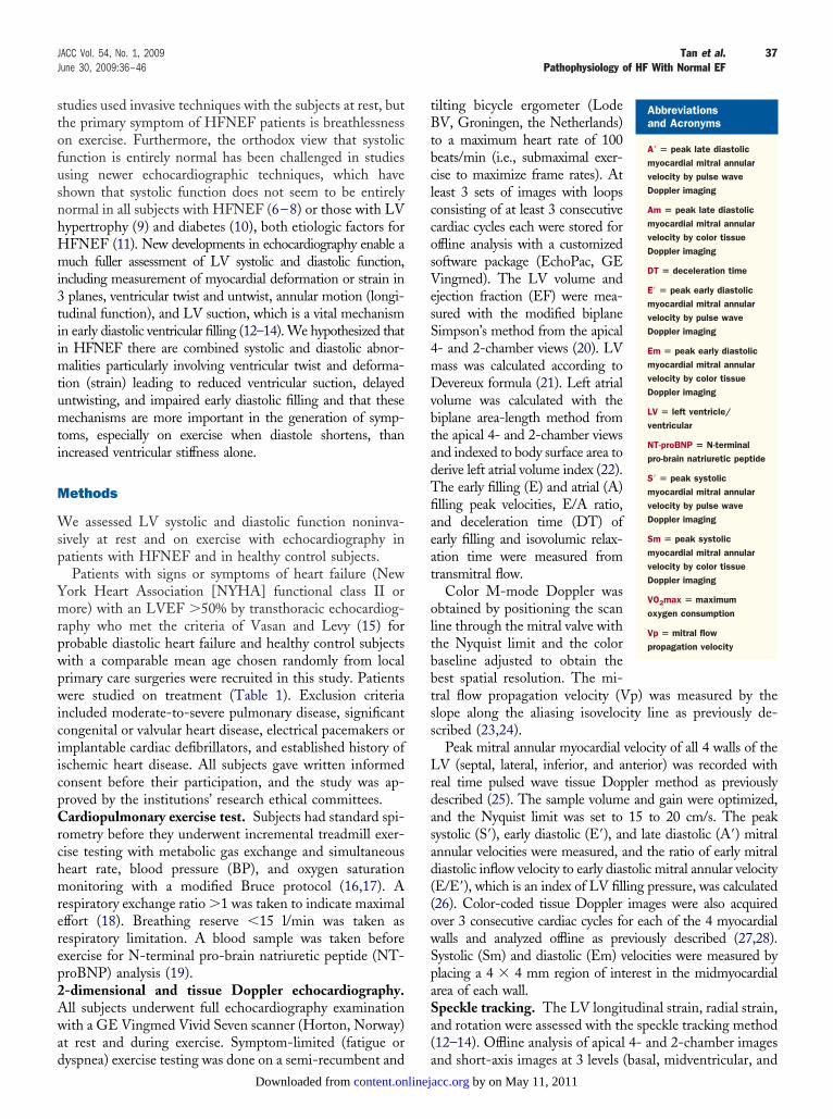
ion) at rest were significantly lower in patients comparedith control subjects. However, on exercise, the percentagef untwist in early and mid-diastole (50% untwist duration)ere significantly lower in patients, and the percentage ofntwist in late diastole showed a trend toward significance.he untwist rate was not significantly different at rest butecame significantly different on exercise with control sub-
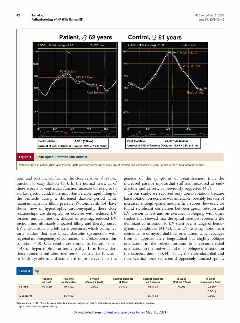
ects having a significantly higher untwist rate comparedith patients (Table 3, Fig. 1).Similar to our findings for longitudinal and radial
train, the magnitude of apical rotation, untwist rate, andercentage of untwist in late diastole on exercise inatients increased only to a level comparable to controlubjects at rest. However, the percentage of untwist inarly and mid-diastole in control subjects at rest remainedignificantly higher than that of patients on exerciseTable 3). There was significant correlation betweenpical rotation and LV torsion at rest and on exercise (at
emodynamic Status and Doppler Echocardiography
Table 2 Hemodynamic Status and Doppler Echocardiography
Patientsat Rest
Patientson Exercise
p Value(Paired t Test)
BP systolic (mm Hg) 146 � 16 168 � 18 �0.001
BP diastolic (mm Hg) 75 � 10 86 � 13 �0.001
Heart rate (beats/min) 69 � 11 89 � 10 �0.001
VTI (cm) 21.5 � 4.8 22.8 � 5.3 0.066
�VTI (cm) 1.3 � 4.2
Stroke volume (ml) 73 � 20 74 � 23 0.239
� stroke volume (ml) 4 � 24
Cardiac output (l/min) 4.8 � 1.5 6.4 � 2.0 �0.001
� cardiac output (l/min) 1.6 � 2.2
Peak LVOT velocity (m/s) 1.1 � 0.2 1.2 � 0.3 0.004
� peak LVOT velocity (m/s) 0.1 � .2
Sm (cm/s) 5.1 � 1.0 6.0 � 1.0 �0.001
Em (cm/s) 4.4 � 1.2 6.4 � 1.5 �0.001
� Em (cm/s) 2.0 � 1.0
E-wave (cm/s) 0.70 � 0.17 0.95 � 0.17
� E-wave 0.27 � 0.19
Am (cm/s) 6.8 � 1.5 8.7 � 1.6 �0.001
E/E= 11.4 � 4.3 11.4 � 4.5 0.298
Systolic reserve index 0.6 � 0.5
Diastolic reserve index 1.5 � 0.8
ata are mean � SD. *t test between patients and control subjects at rest. †t test between patieAm � late diastolic annular velocity; BP � blood pressure; E/E= � ratio of early mitral diastolic infl
ract; Sm � systolic mitral annular velocity; VTI � aortic outflow velocity time integral.
est r � 0.704, p � 0.001; on exercise r � 0.53, p � acontent.onlinejDownloaded from
.016) in those with good basal images (n � 45 at rest,� 20 on exercise).
p. Mitral flow propagation velocity was used as an ap-roximation to the intraventricular pressure gradient, re-ecting ventricular suction. The Vp was comparable be-ween the 2 groups at rest (40 � 10 m/s vs. 39 � 7 m/s,
� 0.52) but became significantly different on exerciseith the control subjects having a higher ability to
ncrease their Vp on exercise compared with patients.ven though patients were able to increase Vp on exercise, theagnitude of the increase was significantly less than control
ubjects (Table 4). The Vp on exercise correlated with peakm on exercise (r � 0.47, p � 0.005), suggesting a mechanistic
ink between systole and diastolic suction.orrelations between peak VO2max on exercise and
chocardiographic parameters. Peak VO2max correlatedith the following echocardiographic parameters on exer-
ise: Sm (r � 0.61, p � 0.003), Em (r � 0.417, p � 0.038),
ontrol Subjectsat Rest
Control Subjectson Exercise
p Value(Paired t Test)
p Value(Unpaired t Test)
140 � 15 163 � 15 �0.001 0.114*
0.298†
78 � 8 87 � 9 �0.001 0.166*
0.687†
67 � 9 92 � 5 �0.001 0.484*
0.157†
19.3 � 3.9 23.3 � 3.4 �0.001 0.045*
0.743†
4.3 � 1.9 0.003
62 � 13 79 � 20 0.004 0.015*
0.364†
18 � 24 0.039
4.2 � 0.9 7.4 � 2.0 �0.001 0.059*
0.097†
3.1 � 2.4 0.027
1.0 � 0.2 1.2 � 0.1 �0.001 0.044
0.682
0.2 � 0.1 0.007
5.7 � 0.9 7.7 � 1.0 �0.001 0.025*
�0.001†
5.4 � 1.1 8.3 � 1.2 �0.001 0.004*
�0.001†
2.9 � 1.6 0.023
0.57 � 0.11 0.95 � 0.12 �0.001
0.853
0.38 � 0.13 0.015
7.8 � 1.4 9.7 � 1.7 �0.001 0.012*
0.063†
8.2 � 2.0 8.8 � 1.8 0.384 �0.001*
0.009†
1.5 � 0.7 �0.001
2.3 � 1.2 0.011
control subjects on exercise.arly diastolic annular velocity; Em � early diastolic annular velocity; LVOT � left ventricular outflow
C
nts and
pical rotation (r � 0.44, p � 0.026), E/E= (r � �0.34, by on May 11, 2011 acc.org

p(DeoHpjaepisIsOhifc
D
Im
npbrafnfuwvsdctmitrtaTs
2
D nts andwist 25
41JACC Vol. 54, No. 1, 2009 Tan et al.June 30, 2009:36–46 Pathophysiology of HF With Normal EF
� 0.04), Vp (r � 0.35, p � 0.03), and 25% untwistingr � 0.53, p � 0.007).
erived measurements. There was no significant differ-nce in arterial elastance or end-systolic elastance (a measuref systolic function and end-systolic stiffness) at rest (Table 5).owever, we observed an increase in arterial elastance in
atients on exercise (not significant), whereas control sub-ects had a reduced arterial elastance on exercise, resulting insignificant difference in the change in arterial elastance on
xercise (Table 5). Stroke work index was similar betweenatients and control subjects at rest and on exercise, risingn both groups to the same degree. Estimated “chambertiffness” was similar to control subjects.nterobserver and intraobserver variability. The interob-erver variability at rest by ICC was between 0.88 and 0.96.n exercise, ICC varied from 0.67 to 0.99; Vp had the
ighest interobserver variability. The intraobserver variabil-ty by ICC at rest varied from 0.88 to 0.98 and on exerciserom 0.66 to 0.98. Again, Vp had the highest variabilityompared with other measurements.
iscussion
n this study, we have performed a comprehensive assess-
-Dimensional Strain and Rotation
Table 3 2-Dimensional Strain and Rotation
Patientsat Rest
Patientson Exercise
p Value(Paired t Test)
Long strain SepLat (%) �19.4 � 3.8 �20.4 � 4.2 0.082
� long strain SepLat (%) �1.2 � 3.9
Long strain InfAnt (%) �18.4 � 3.8 �20.1 � 4.5 0.012
� long strain InfAnt (%) �2.1 � 4.2
Global long strain (%) �18.9 � 3.5 �20.1 � 4.1 0.005
� global long strain (%) �1.9 � 3.3
Radial strain (%) 41.8 � 13.5 49.1 � 15.4 0.02
� radial strain (%) 6.6 � 15.6
Apical rotation (°) 10.4 � 4.0 13.5 � 4.7 0.008
� apical rotation (°) 3.0 � 4.9
Untwist rate (°/s) �80 � 34 �105 � 32 �0.001
� untwist rate (°/s) �33 � 35
Untwist 25% 23.7 � 9.3 21.2 � 8.5 0.465
� untwist 25% �2.0 � 13.1
Untwist 50% 53.9 � 12.6 50.5 � 12.3 0.371
� untwist 50% �3.3 � 17.7
Untwist 75% 77.3 � 6.4 80.3 � 9.8 0.477
� untwist 75% 1.8 � 12.1
ata are mean � SD. *t test between patients and control subjects at rest. †t test between patieInfAnt � inferior and anterior walls; long � longitudinal; SepLat � septal and lateral walls; Unt
ent of systolic and diastolic ventricular function with new dcontent.onlinejDownloaded from
oninvasive techniques at rest and on exercise in HFNEFatients and have demonstrated a variety of abnormalities ofoth systolic and diastolic function. These include reducedadial and longitudinal myocardial systolic strain both at restnd on exercise, reduced systolic and diastolic longitudinalunctional reserve (mitral annular velocities fail to riseormally), reduced ventricular systolic rotation at rest thatails to increase normally on exercise, delayed ventricularntwisting with further worsening on exercise associatedith a reduced LV suction, and a consequent reduced strokeolume rise on exercise. Recent studies (12–14,37) usingimilar echocardiographic techniques to ours have elegantlyemonstrated the close temporal, functional, and tightlyoordinated relationships in normal patients between LVwist during systole, with accompanying mitral annularotion toward the apex, and in early diastole, the untwist-
ng process and recoil that generate the negative intraven-ricular pressure gradient or suction. This is followed by theapid motion of the mitral annulus back toward the base ofhe heart, which also aids filling by simply moving thennulus around the column of the incoming blood (38).hus, LV torsion or twist is a mechanism for generating
tored energy during systole, which is released during early
ontrol Subjectsat Rest
Control Subjectson Exercise
p Value(Paired t Test)
p Value(Unpaired t Test)
�21.5 � 3.0 �24.4 � 2.6 �0.001 0.018*
�0.001†
�3.1 � 2.8 0.051
�20.4 � 3.7 �23.2 � 3.2 0.004 0.042*
0.008†
2.8 � 3.9 0.592
�20.9 � 3.0 �23.8 � 2.5 �0.001 0.018*
�0.001†
�2.8 � 2.7 0.315
49.2 � 12.9 61.9 � 12.8 0.001 0.03*
0.002†
12.4 � 12.3 0.154
13.0 � 2.8 17.7 � 3.6 0.003 0.015*
0.005†
4.9 � 4.1 0.216
�96 � 30 �129 � 32 0.007 0.069*
0.013†
�29 � 39 0.722
30.9 � 9.7 29.2 � 7.7 0.477 0.006*
0.002†
�2.4 � 14.1 0.922
58.8 � 8.4 61.7 � 8.2 0.384 0.101*
0.001†
3.0 � 14.2 0.223
81.8 � 7.1 85.0 � 4.7 0.126 0.016*
0.057†
3.3 � 8.7 0.652
control subjects on exercise.% (50%, 75%) � percentage untwist at 25% (50%, 75%) untwist duration.
C
iastole to produce ventricular recoil, upward annular mo- by on May 11, 2011 acc.org
tftatmsrtsLerc(ti
gid
bifLsddcfoots
V
D
42 Tan et al. JACC Vol. 54, No. 1, 2009Pathophysiology of HF With Normal EF June 30, 2009:36–46
ion, and suction, confirming the close relation of systolicunction to early diastole (39). In the normal heart, all ofhese aspects of ventricular function increase on exercise toid fast ejection and, more important, enable rapid filling ofhe ventricle during a shortened diastole period whileaintaining a low filling pressure. Notomi et al. (14) have
hown how in hypertrophic cardiomyopathy these closeelationships are disrupted on exercise with reduced LVorsion, annular motion, delayed untwisting, reduced LVuction, and ultimately impaired filling and thereby raisedV end-diastolic and left atrial pressures, which confirmedarly studies that also linked diastolic dysfunction withegional inhomogeneity of contraction and relaxation in thisondition (40). Our results are similar to Notomi et al.14) in hypertrophic cardiomyopathy. It is likely thathese fundamental abnormalities of ventricular functionn both systole and diastole are more relevant to the
Figure 1 Peak Apical Rotation and Untwist
Rotation curve of patient (left) and control (right) showing magnitude of peak apic
p
Table 4 Vp
Patientsat Rest
Patientson Exercise
p Value(Paired t Test)
Con
Vp (m/s) 40 � 10 49 � 11 0.001
� Vp (m/s) 11 � 13
ata are mean � SD. *t test between patients and control subjects at rest. †t test between patients andVp � mitral flow propagation velocity.
content.onlinejDownloaded from
enesis of the symptoms of breathlessness than thencreased passive myocardial stiffness measured at end-iastole and at rest, as previously suggested (4,5).In our study, we reported only apical rotation, because
asal rotation on exercise was unreliable, possibly because ofncreased through-plane motion. In a subset, however, weound significant correlation between apical rotation andV torsion at rest and on exercise, in keeping with other
tudies that showed that the apical rotation represents theominant contribution to LV twist over a range of hemo-ynamic conditions (41,42). The LV twisting motion is aonsequence of myocardial fiber orientation, which changesrom an approximately longitudinal but slightly obliquerientation in the subendocardium to a circumferentialrientation in the mid-wall and to an oblique orientation inhe subepicardium (43,44). Thus, the subendocardial andubepicardial fibers represent 2 oppositely directed spirals.
tion and percentage of early untwist (25% of total untwist duration).
ubjectsst
Control Subjectson Exercise
p Value(Paired t Test)
p Value(Unpaired t Test)
7 61 � 14 0.001 0.516*
�0.001†
22 � 14 0.001
al rota
trol Sat Re
39 �
control subjects on exercise.
by on May 11, 2011 acc.org
BdrLairdeduwgupmosrlditsdva
sclm
ttreco
vcmftEsFDsempDtdmenmfadS
D
Der index
43JACC Vol. 54, No. 1, 2009 Tan et al.June 30, 2009:36–46 Pathophysiology of HF With Normal EF
ecause of larger radii, the torque of subepicardial fibersominates and accounts for the normal counterclockwiseotation of the LV apex. Previous studies have found that inV hypertrophy, apical twist and untwisting might beugmented or reduced (45,46), and similarly, torsion isncreased in patients with mild diastolic dysfunction buteduced in those with more severe degrees of diastolicysfunction (47). This anatomical arrangement probablyxplains these variable results: in the early stage the suben-ocardial longitudinal fibers are primarily affected so thatnopposed epicardial torque will increase apical twist,hereas later more widespread fibrosis or damage will affectlobal function and lead to reduced twist with delayedntwisting. This also explains why annular velocities thatrimarily reflect subendocardial fibers are such sensitiveeasures of ventricular function (48) and were reduced in
ur HFNEF patients, particularly on exercise. All previoustudies of torsion and untwisting in HFNEF were done atest, and this is the first study that has assessed bothongitudinal function and apical rotation on exercise. Re-uced and delayed untwisting on exercise will undoubtedly
mpede LV suction in early diastole and delay rapid filling sohat more filling will occur in late diastole during atrialystole, and this will explain the common finding of theelayed relaxation pattern (reduced early but increased atrialelocity) on the mitral inflow velocities associated withging and LV hypertrophy (49,50).
The net differences in global longitudinal strain, radialtrain, and apical rotation from rest to exercise seemedomparable between patients and control subjects, mostikely due to a relatively small sample size. However, the
erived Measurements
Table 5 Derived Measurements
Patientsat Rest
Patientson Exercise
p Value(Paired t Test)
P/V (1/ml) 0.17 � 0.80 0.17 � 0.70 0.135
�P/V (1/ml) 0.01 � 0.05
Ea (mm Hg/ml) 2.0 � 0.7 2.2 � 0.9 0.083
�Ea (mm Hg/ml) 0.22 � 0.71
K (mm Hg/ml) 1.6 � 0.7 3.4 � 1.6 �0.001
�K (mm Hg/ml) 1.9 � 1.7
PPI (mm Hg/s) 2.2 � 0.7 3.1 � 1.1 �0.001
�PPI (mm Hg/s) 1.0 � 1.0
Ees (mm Hg/ml) 4.9 � 1.9 7.4 � 2.7 �0.001
�Ees (mm Hg/ml) 2.5 � 2.6
SWI (mm Hg) 94 � 26 132 � 49 0.001
�SWI (mm Hg) 39.4 � 52.9
ata are mean � SD. *t test between patients and control subjects at rest. †t test between patieEa � arterial elastance; Ees � end-systolic elastance; K � chamber stiffness; PPI � peak pow
agnitude of increase in strain and rotation was consis- pcontent.onlinejDownloaded from
ently higher in control subjects. It is also noteworthyhat strain and apical rotation were significantly lower atest in patients and did not exceed that of control subjectsven on exercise, indicating that patients have a signifi-antly lower functional capacity even at rest and more son exercise.Derived measurements of stiffness, arterial stiffness, and
entricular systolic compliance did not show any majorhanges between patients and control subjects. In part thisight be because these measurements are often derived
rom single beats and have many assumptions involved inheir calculations. Chamber stiffness is derived from the-wave DT but probably only is accurate in those with a
hort DT, whereas some of our patients had a delayed DT.urthermore there are no data on the effects of exercise andT has been shown to be a function of both chamber
tiffness and chamber relaxation viscoelasticity (51). How-ver, the derived pressure-volume index, an indirecteasure of compliance, was higher in patients, as ex-
ected. Clearly other noncardiac factors are also involved.ecreased arterial compliance and ventricular interac-
ions will impair ventricular function (both systolic andiastolic) on exercise. Interestingly, the other derivedeasurements of global systolic function (end-systolic
lastance, stroke work index, and peak power index) wereot significantly different from the control group. Thisight reflect the method or that abnormalities of systolic
unction are subtle and do not affect pump function tony great degree but have a profound influence on earlyiastolic filling.tudy limitations. Our initial power calculation was
rol Subjectst Rest
Control Subjectson Exercise
p Value(Paired t Test)
p Value(Unpaired t Test)
3 � 0.50 0.14 � 0.44 0.543 0.035*
0.039†
0.01 � 0.06 0.744
.1 � 0.5 1.9 � 0.4 0.227 0.403*
0.145†
�0.18 � 0.65 0.044
.4 � 0.6 3.8 � 1.4 �0.001 0.200*
0.380†
2.4 � 1.7 0.249
.2 � 0.7 3.1 � 0.8 �0.001 0.793*
0.947†
0.9 � 0.8 0.702
.6 � 2.2 6.9 � 1.7 �0.001 0.206*
0.461†
1.4 � 2.3 0.138
1 � 36 140 � 46 0.02 0.405*
0.579†
36.6 � 56.2 0.872
control subjects on exercise.; P/V � pressure/volume ratio; SWI � stroke work index.
Conta
0.1
2
1
2
5
10
nts and
erformed with a sample size of 30, which provided by on May 11, 2011 acc.org
acsweeshdotitppctDmvet
C
Wcvastudasiatwa
AT
44 Tan et al. JACC Vol. 54, No. 1, 2009Pathophysiology of HF With Normal EF June 30, 2009:36–46
dequate power. However, we only included 27 healthyontrol subjects, but a repeat power calculation with aample size of 27 yielded comparable power. All patientsere taking medication, because it was considered un-
thical to stop treatment entirely. It is probable that theffect of treatment would be to improve deformation ortrain and rotation, and therefore the differences mightave been greater without treatment although it would beifficult to account for a resulting increase in arterial BPn exercise. Diuretic drugs do reduce symptoms, andhere is a suggestion that angiotensin-converting enzymenhibitors or receptor antagonists might improve longi-udinal function (52). Exercise was submaximal, but theatients were breathless at this level. For a more elderlyopulation, submaximal exercise is more relevant andomparable to usual activities than exercising to exhaus-ion, which is probably done very rarely in daily life.iagnostic criteria for HFNEF patients receiving treat-ent are not available, and the BNP results were quite
ariable, but these were also taken on treatment. How-ver, metabolic exercise testing was used to confirm that
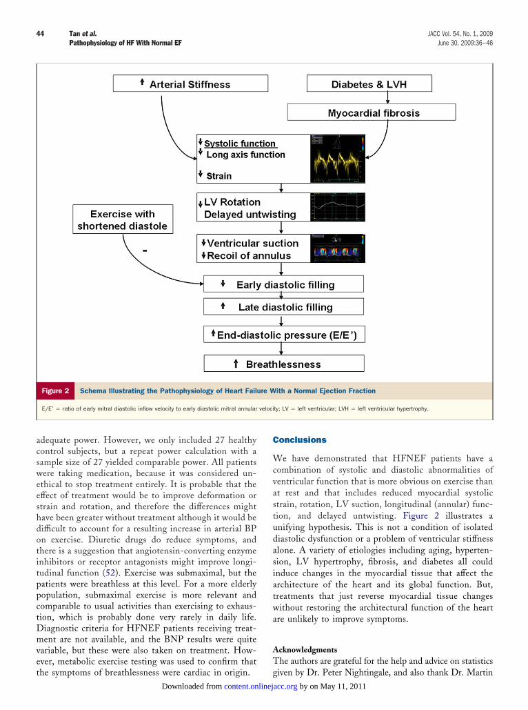
Figure 2 Schema Illustrating the Pathophysiology of Heart Fail
E/E= � ratio of early mitral diastolic inflow velocity to early diastolic mitral annular
he symptoms of breathlessness were cardiac in origin. gcontent.onlinejDownloaded from
onclusions
e have demonstrated that HFNEF patients have aombination of systolic and diastolic abnormalities ofentricular function that is more obvious on exercise thant rest and that includes reduced myocardial systolictrain, rotation, LV suction, longitudinal (annular) func-ion, and delayed untwisting. Figure 2 illustrates anifying hypothesis. This is not a condition of isolatediastolic dysfunction or a problem of ventricular stiffnesslone. A variety of etiologies including aging, hyperten-ion, LV hypertrophy, fibrosis, and diabetes all couldnduce changes in the myocardial tissue that affect therchitecture of the heart and its global function. But,reatments that just reverse myocardial tissue changesithout restoring the architectural function of the heart
re unlikely to improve symptoms.
cknowledgmentshe authors are grateful for the help and advice on statistics
ith a Normal Ejection Fraction
ty; LV � left ventricular; LVH � left ventricular hypertrophy.
ure W
veloci
iven by Dr. Peter Nightingale, and also thank Dr. Martin by on May 11, 2011 acc.org

AS
RDUU
R
1
1
1
1
1
1
1
1
1
1
2
2
2
2
2
2
2
2
2
2
3
3
3
3
3
3
3
3
3
3
4
4
4
45JACC Vol. 54, No. 1, 2009 Tan et al.June 30, 2009:36–46 Pathophysiology of HF With Normal EF
llen, Dr. Paul Foley, Julie Machin, Rebekah Weaver, andtuart Wragg for their assistance.
eprint requests and correspondence: Prof. John E. Sanderson,epartment of Cardiovascular Medicine, The Medical School,niversity of Birmingham, Edgbaston, Birmingham B15 2TT,nited Kingdom. E-mail: [email protected].
EFERENCES
1. Owan TE, Hodge DO, Herges RM, Jacobsen SJ, Roger VL, RedfieldMM. Trends in prevalence and outcome of heart failure with preservedejection fraction. N Engl J Med 2006;355:251–9.
2. Bhatia RS, Tu JV, Lee DS, et al. Outcome of heart failure withpreserved ejection fraction in a population-based study. N Engl J Med2006;355:260–9.
3. Sanderson JE. Heart failure with a normal ejection fraction. Heart2007;93:155–8.
4. Zile MR, Baicu CF, Gaasch WH. Diastolic heart failure: abnormal-ities in active relaxation and passive stiffness of the left ventricle.N Engl J Med 2004;350:1953–9.
5. Westermann D, Kasner M, Steendijk P, et al. Role of left ventricularstiffness in heart failure with normal ejection fraction. Circulation2008;117:2051–60.
6. Yip G, Wang M, Zhang Y, Fung JW, Ho PY, Sanderson JE. Leftventricular long axis function in diastolic heart failure is reduced inboth diastole and systole: time for a redefinition? Heart 2002;87:121–5.
7. Yu CM, Lin H, Yang H, Kong SL, Zhang Q, Lee SW. Progressionof systolic abnormalities in patients with “isolated” diastolic heartfailure and diastolic dysfunction. Circulation 2002;105:1195–201.
8. Bruch C, Gradaus R, Gunia S, Breithardt G, Wichter T. Dopplertissue analysis of mitral annular velocities: evidence for systolic abnor-malities in patients with diastolic heart failure. J Am Soc Echocardiogr2003;16:1031–6.
9. Wang M, Yip GW, Wang AY, et al. Tissue Doppler imaging providesincremental prognostic value in patients with systemic hypertensionand left ventricular hypertrophy. J Hypertens 2005;23:183–91.
0. Fang ZY, Leano R, Marwick TH. Relationship between longitudinaland radial contractility in subclinical diabetic heart disease. Clin Sci(Lond) 2004;106:53–60.
1. Hogg K, Swedberg K, McMurray J. Heart failure with preserved leftventricular systolic function. J Am Coll Cardiol 2004;43:317–27.
2. Becker M, Bilke E, Kuhl H, et al. Analysis of myocardial deformationbased on pixel tracking in two dimensional echocardiographic imagesenables quantitative assessment of regional left ventricular function.Heart 2006;92:1102–8.
3. Helle-Valle T, Crosby J, Edvardsen T, et al. New noninvasive methodfor assessment of left ventricular rotation: speckle tracking echocardi-ography. Circulation 2005;112:3149–56.
4. Notomi Y, Martin-Miklovic MG, Oryszak SJ, et al. Enhancedventricular untwisting during exercise. A mechanistic manifestation ofelastic recoil described by Doppler tissue imaging. Circulation 2006;113:2524–33.
5. Vasan RS, Levy D. Defining diastolic heart failure: a call for stan-dardized diagnostic criteria. Circulation 2000;101:2118–21.
6. Albouaini K, Egred M, Alahmar A, Wright DJ. Cardiopulmonaryexercise testing and its application. Heart 2007;93:1285–92.
7. Arruda ALM, Pellikka PA, Olson TP, et al. Exercise capacity,breathing pattern and gas exchange during exercise for patients withisolated diastolic dysfunction. J Am Soc Echocardiogr 2007;20:838–46.
8. Witte KKA, Nikitin NP, Cleland JGF, et al. Excessive breathlessnessin patients with diastolic heart failure. Heart 2006;92:1425–9.
9. Squire IB, O’Brien RJ, Demme B, Davies JE, Ng LL. N-terminalpro-atrial natriuretic peptide (N-ANP) and N-terminal pro-B-typenatriuretic peptide (N-BNP) in the prediction of death and heartfailure in unselected patients following acute myocardial infarction.
Clin Sci (Lond) 2004;107:309–16.4
content.onlinejDownloaded from
0. Schiller NB, Shah PM, Crawford M, et al. Recommendation forquantitation of the left ventricle by two-dimensional echocardiogra-phy. American Society of Echocardiography Committee on Standards,Subcommittee on Quantitation of Two-Dimensional Echocardio-grams. J Am Soc Echocardiogr 1989;2:258–67.
1. Devereux RB, Alonso DR, Lutas EM, et al. Echocardiographicassessment of left ventricular hypertrophy: comparison to necropsyfindings. Am J Cardiol 1986;57:450–8.
2. Lim TK, Ashrafian H, Dwivedi G, et al. Increased LAVI is anindependent predictor of raised serum natriuretic peptide in patientswith suspected heart failure but normal LVEF: implication fordiagnosis of DHF. Eur J Heart Failure 2006;8:38–45.
3. Garcia MJ, Palac RT, Malenka DJ, et al. Color M-mode Doppler flowpropagation velocity is a relatively preload-independent index of leftventricular filling. J Am Soc Echocardiogr 1999;12:129–37.
4. Garcia MJ, Ares MA, Asher C, et al. An index of early ventricularfilling that combined with pulsed Doppler peak E velocity mayestimate capillary wedge pressure. J Am Coll Cardiol 1997;29:448–54.
5. Oki T, Tabata T, Yamada H, et al. Clinical application of pulsedDoppler imaging for assessing abnormal left ventricular relaxation.Am J Cardiol 1997;79:921–8.
6. Nagueh SF, Middleton KJ, Kopelen HA, et al. Doppler tissue imaginga non-invasive technique for evaluation of LV relaxation and estima-tion of filling pressures. J Am Coll Cardiol 1997;30:1527–33.
7. Wang M, Yip GWK, Wang AYM, et al. Peak early diastolicmitral annulus velocity by tissue Doppler imaging adds independentand incremental prognostic value. J Am Coll Cardiol 2003;41:820 – 6.
8. Wang M, Yip GWK, Yu CM, et al. Independent and incrementalprognosis value of early diastolic annular velocity in patients withimpaired left ventricular systolic function. J Am Coll Cardiol 2005;45:272–7.
9. Notomi T, Lysyansky P, Setser RM, et al. Measurement of ventriculartorsion by two-dimensional ultrasound speckle tracking imaging. J AmColl Cardiol 2005;45:2034–41.
0. Sun JP, Min P, Fouad FM, et al. Automated cardiac output measure-ment by spatiotemporal integration of color Doppler data. Circulation1997;95:932–9.
1. Borlaug BA, Melenovsky V, Redfield MM, et al. Impact of arterialload and loading sequence on left ventricular tissue velocities inhumans. J Am Coll Cardiol 2007;50:1570–7.
2. Baicu CF, Zile MR, Aurigemma GP, Gaasch WH. Left ventricularperformance, function and contractility in patients with diastolic heartfailure. Circulation 2005;111:2306–12.
3. Kelly RP, Ting CT, Yang TM, et al. Effective arterial elastance as indexof arterial vascular load in humans. Circulation 1996;27:168–75.
4. Little WC, Ohno M, Kitzman DW, Thomas JD, Cheng CP.Determination of left ventricular chamber stiffness of early left ven-tricular filling. Circulation 1995;92:1933–9.
5. King GJ, Murphy RT, Almuntaser I, Bennet K, Ho E, Brown AS.Alterations in myocardial stiffness in elite athletes assessed by a newDoppler index. Heart 2008;94:1323–5.
6. Ha JW, Lee HC, Keng ES, et al. Abnormal left ventricular longitu-dinal functional reserve in patients with diabetes mellitus. Heart2007;93:1571–6.
7. Notomi Y, Popovic ZB, Yamada H, et al. Ventricular untwisting: atemporal link between left ventricular relaxation and suction. Am JPhysiol Heart Circ Physiol 2008;294:505–13.
8. Henein MY, Gibson DG. Normal long axis function. Heart 1999;81:111–3.
9. Yip GWK, Zhang Y, Tan PYH, et al. Left ventricular long axischanges in early diastole and systole: impact of systolic function ondiastole. Clin Sci 2002;102:515–22.
0. Gibson DG, Sanderson JE, Traill TA, Brown DJ, Goodwin JF.Regional left ventricular wall movement in hypertrophic cardiomyop-athy. Br Heart J 1978;40:1327–33.
1. Kim HK, Sohn DW, Lee SE, et al. Assessment of left ventricularrotation and torsion with two-dimensional speckle tracking echocar-diography. J Am Soc Echocardiogr 2007;20:45–53.
2. Opdahl A, Helle-Valle T, Remme EW, et al. Apical rotation byspeckle tracking echocardiography: a simplified bedside index of leftventricular twist. J Am Soc Echocardiogr 2008;21:1121–8.
3. Greenbaum RA, Ho SY, Gibson DG, Becker AE, Anderson RH. Leftventricular fibre architecture in man. Br Heart J 1981;45:248–63.
by on May 11, 2011 acc.org

4
4
4
4
4
4
5
5
5
46 Tan et al. JACC Vol. 54, No. 1, 2009Pathophysiology of HF With Normal EF June 30, 2009:36–46
4. Ingels NB Jr., Hansen DE, Daughters GT, Stinson EB, AldermanEL, Miller DC. Relation between longitudinal, circumferential, andoblique shortening and torsional deformation in the left ventricle ofthe transplanted human heart. Circ Res 1989;64:915–27.
5. Wang J, Khoury DS, Yue Y, Torre-Amione G, Nagueh SF. Leftventricular untwisting rate by speckle tracking echocardiography.Circulation 2007;116:2580–6.
6. Takeuchi M, Borden WB, Nakai H, et al. Reduced and delayeduntwisting of the left ventricle in patients with hypertension and leftventricular hypertrophy: a study using tow-dimensional speckle track-ing echocardiography. Eur Heart J 2007;20:36–44.
7. Park SJ, Miyazaki C, Bruce CJ, Ommen S, Miller FA, Oh JK. Leftventricular torsion by two-dimensional speckle tracking echocardiog-raphy in patients with diastolic dysfunction and normal ejectionfraction. J Am Soc Echocardiogr 2008;21:1129–37.
8. Sanderson JE, Wang M, Yu CM. Tissue Doppler imaging forpredicting outcome in cardiovascular disease. Curr Opin Cardiol
content.onlinejDownloaded from
9. Nakai H, Takeuchi M, Nishikage T, Kokumai M, Otani S, Lang RM.Effect of aging on twist-displacement loop by 2-dimensional speckletracking. J Am Soc Echocardiogr 2006;19:880–5.
0. Takeuchi M, Borden WB, Nakai H, et al. Reduced and delayeduntwisting of the left ventricle in patients with hypertension and leftventricular hypertrophy: a study using two-dimensional speckle track-ing imaging. Europ Heart J 2007;28:2756–62.
1. Shmuylovich L, Kovacs SJ. E-wave deceleration time may not providean accurate determination of LV chamber stiffness if LV relaxation/viscoelasticity is unknown. Am J Physiol Heart Circ Physiol 2007;292:H2712–20.
2. Yip GW, Wang M, Wang T, et al. The Hong Kong diastolic heartfailure study: a randomized controlled trial of diuretics, irbesartan andramipril on quality of life, exercise capacity, left ventricular global andregional function in heart failure with a normal ejection fraction. Heart2008;94:573–80.
2004;19:458–63. Key Words: diastolic y heart failure y rotation y strain y untwist.
Call for Papers Prevention / OutcomesThis is a call for submission of manuscripts dealing with Prevention / Outcomes.
In recognition of the increasing research advances and number of manuscriptssubmitted to JACC on certain topics, we are planning to initiate a recurring program of expanded Focus Issues devoted to these topics.
In rotation, the last issue of each month will be expanded to include additional papers related either to Heart Failure or Heart Rhythm Disorders orPrevention/Outcomes. Thus, four issues per year will highlight each of these subjects. The articles accepted for these issues will be in addition to those submittedand published on these topics on a regular basis in our regular issues.
Prevention/Outcomes will be covered in the December 15/22, 2009 Focus Issue and we are soliciting manuscripts on this topic to be submited by July 11, 2009.Please submit manuscripts online in the usual fashion at jaccsubmit.org.
by on May 11, 2011 acc.org

doi:10.1016/j.jacc.2009.03.037 2009;54;36-46 J. Am. Coll. Cardiol.
Kiran Patel, Michael Frenneaux, and John E. Sanderson Yu Ting Tan, Frauke Wenzelburger, Eveline Lee, Grant Heatlie, Francisco Leyva,
Longitudinal Motionand Diastolic Ventricular Function Involving Torsion, Untwist, and
Exercise Echocardiography Reveals Complex Abnormalities of Both Systolic The Pathophysiology of Heart Failure With Normal Ejection Fraction:
This information is current as of May 11, 2011
& ServicesUpdated Information
http://content.onlinejacc.org/cgi/content/full/54/1/36including high-resolution figures, can be found at:
Supplementary Material http://content.onlinejacc.org/cgi/content/full/54/1/36/DC1
Supplementary material can be found at:
References
http://content.onlinejacc.org/cgi/content/full/54/1/36#BIBLfree at: This article cites 52 articles, 30 of which you can access for
Citations
cleshttp://content.onlinejacc.org/cgi/content/full/54/1/36#otherartiThis article has been cited by 30 HighWire-hosted articles:
Rights & Permissions
http://content.onlinejacc.org/misc/permissions.dtltables) or in its entirety can be found online at: Information about reproducing this article in parts (figures,
Reprints http://content.onlinejacc.org/misc/reprints.dtl
Information about ordering reprints can be found online:
by on May 11, 2011 content.onlinejacc.orgDownloaded from
Related Documents