The Origin, Location, and Projections of the Embryonic Abdominal Motorneurons of Drosophila Matthias Landgraf, 1 Torsten Bossing, 2 Gerd M. Technau, 3 and Michael Bate 1 1 Department of Zoology, Cambridge University, Cambridge CB2 3EJ, United Kingdom, 2 Wellcome/CRC Institute, Cambridge CB2 1QR, United Kingdom, and 3 Institut fu ¨ r Genetik, Universita ¨ t Mainz, 55122 Mainz, Germany We have used a retrograde labeling technique to identify mo- torneurons for each of the 30 body wall muscles of an abdom- inal hemisegment in the late stage 16 Drosophila embryo. Each motorneuron has a characteristic cell body position, dendritic arborization, and axonal projection. In addition, we have deter- mined the neuroblasts of origin for most of the motorneurons we describe. Some organizational principles for the neuromus- cular system have become apparent: (1) There is no obvious topographic relationship between the cell body positions of motorneurons and the position or orientation of the muscles they innervate; (2) motorneurons that innervate muscles of similar position and orientation are often clustered and have overlapping dendritic trees; (3) morphologically similar motor- neurons are generally derived from a common neuroblast and innervate operationally related muscles; and (4) neuroblasts can give rise to more than one morphological type of motorneuron. Key words: Drosophila; motorneuron; development; lineage; structure; connectivity; arborization; neuroblast The embryonic nervous system of Drosophila is a powerf ul system in which to analyze the mechanisms that underlie neural devel- opment. Its strength lies in its amenability to genetic, molecular, anatomical, and electrophysiological techniques, whereas its rel- ative simplicity allows manipulations and analyses to be per- formed at the level of single, identified cells (for review, see Goodman and Doe, 1993; Keshishian et al., 1996). During early stages of CNS formation, neuroblasts delaminate from the neuroectoderm and give rise to the full complement of neurons and glia. The spatiotemporal sequence of neuroblast delamination and their distinct patterns of gene expression make each of the neuroblasts a uniquely identifiable cell (Hartenstein and C ampos-Ortega, 1984; Doe, 1992; Broadus et al., 1995; for review, see Doe and Technau, 1993). In addition, the neuronal and glial lineages generated by neuroblasts derived from the ventral half of the neuroectoderm have recently been described in detail (Bossing et al., 1996). As far as the motor system is concerned, the aCC and the RP1-5 neurons have served as ex- amples of motor neuron development and the formation of neu- romuscular connections in the periphery (Halpern et al., 1991; Sink and Whitington, 1991a; Broadie et al., 1993; Chiba et al., 1995; for review, see Goodman and Doe, 1993). However, very little is known about the other motorneurons or the central dendritic arborizations that are fundamental to their function. Here we report the use of a retrograde DiI labeling technique that has enabled us to identif y and characterize the motorneurons that innervate each of the 30 body wall muscles in an abdominal hemisegment in late stage 16 Drosophila embryos. Sink and Whitington (1991a) described the location of all embryonic mo- torneurons in the CNS but were unable to resolve individual central and peripheral projections. We have succeeded in sepa- rately cataloging each of the motorneurons and their projections in the periphery, together with their dendritic arbors within the CNS. Each motorneuron is identifiable by its characteristic posi- tion in the neuromere. In addition, the motorneurons make an invariant set of connections with target muscles, and for most motorneurons we can show that they have characteristic dendritic trees. Interestingly, neurons that innervate muscles that are pu- tatively related in their operation are often clustered and their dendritic trees overlap, thus defining particular areas of the neu- ropile as potential sites of integration for functionally related motorneurons. By using DiI as a lineage tracer we have also been able to determine the neuroblast of origin for most of the embry- onic motorneurons we describe. We find that morphologically similar motorneurons are generally derived from the same neu- roblast, but that a neuroblast can give rise to more than one morphological type of motorneuron. This combination of mor- phological analysis with cell lineage studies enables us to begin an investigation of the relationship between the f unctional architec- ture of the nervous system and the origins and differentiation of its constituent cells. MATERIALS AND METHODS Embr yo preparation. Oregon R flies were maintained on apple juice agar plates at 25°C. Embryos were staged according to Campos-Ortega and Hartenstein (1985). Late stage 16 (14.0 –14.5 hr after egg laying at 25°C) embryos were collected, dechorionated in bleach, and rinsed thoroughly with water. Under saline (0.075 M phosphate buffer, pH 7.2) the embryos were transferred to polylysine-coated coverslips, lifted out of the vitelline membrane with a glass needle, cut open laterally, and then attached to the coverslip. Gut and fat body were removed by gentle suction and the embryos were flattened by blowing a stream of saline over them. Em- bryos were treated with 0.2 mg/ml collagenase (Boehringer Mannheim, Mannheim, Germany) in saline for 1.5 min, rinsed again with saline, fixed with 3.7% formaldehyde in saline for 2.5 min, and then rinsed with saline. Neuronal staining. Lipid-soluble carbocyanine dye 1,19-dioctadecyl- 3,3,39,39-tetramethyl indocarbocyanine perchlorate (DiI) (Molecular Probes, Eugene, OR) was dissolved in vegetable oil as described by Bossing and Technau (1994) and backfilled into sharpened glass capil- Received June 16,1997; revised Sept. 26, 1997; accepted Oct. 6, 1997. This work was supported by grants from the Sir Halley Stewart Trust and the Medical Research Council (M.L.), the Deutsche Forschungsgemeinschaft (Te 130/ 7–1) and the European Economic Community (CT92– 0790) (G.M.T.), and the Wellcome Trust (M.B.). We are grateful to Andreas Prokop, Paul Whitington, and Helen Skaer for their helpful comments. Correspondence should be addressed to Michael Bate, Department of Zoology, Downing Street, Cambridge CB2 3EJ, England. Copyright © 1997 Society for Neuroscience 0270-6474/97/179642-14$05.00/0 The Journal of Neuroscience, December 15, 1997, 17(24):9642–9655

Welcome message from author
This document is posted to help you gain knowledge. Please leave a comment to let me know what you think about it! Share it to your friends and learn new things together.
Transcript

The Origin, Location, and Projections of the Embryonic AbdominalMotorneurons of Drosophila
Matthias Landgraf,1 Torsten Bossing,2 Gerd M. Technau,3 and Michael Bate1
1Department of Zoology, Cambridge University, Cambridge CB2 3EJ, United Kingdom, 2Wellcome/CRC Institute,Cambridge CB2 1QR, United Kingdom, and 3Institut fur Genetik, Universitat Mainz, 55122 Mainz, Germany
We have used a retrograde labeling technique to identify mo-torneurons for each of the 30 body wall muscles of an abdom-inal hemisegment in the late stage 16 Drosophila embryo. Eachmotorneuron has a characteristic cell body position, dendriticarborization, and axonal projection. In addition, we have deter-mined the neuroblasts of origin for most of the motorneuronswe describe. Some organizational principles for the neuromus-cular system have become apparent: (1) There is no obvioustopographic relationship between the cell body positions of
motorneurons and the position or orientation of the musclesthey innervate; (2) motorneurons that innervate muscles ofsimilar position and orientation are often clustered and haveoverlapping dendritic trees; (3) morphologically similar motor-neurons are generally derived from a common neuroblast andinnervate operationally related muscles; and (4) neuroblasts cangive rise to more than one morphological type of motorneuron.
Key words: Drosophila; motorneuron; development; lineage;structure; connectivity; arborization; neuroblast
The embryonic nervous system of Drosophila is a powerful systemin which to analyze the mechanisms that underlie neural devel-opment. Its strength lies in its amenability to genetic, molecular,anatomical, and electrophysiological techniques, whereas its rel-ative simplicity allows manipulations and analyses to be per-formed at the level of single, identified cells (for review, seeGoodman and Doe, 1993; Keshishian et al., 1996).
During early stages of CNS formation, neuroblasts delaminatefrom the neuroectoderm and give rise to the full complement ofneurons and glia. The spatiotemporal sequence of neuroblastdelamination and their distinct patterns of gene expression makeeach of the neuroblasts a uniquely identifiable cell (Hartensteinand Campos-Ortega, 1984; Doe, 1992; Broadus et al., 1995; forreview, see Doe and Technau, 1993). In addition, the neuronaland glial lineages generated by neuroblasts derived from theventral half of the neuroectoderm have recently been described indetail (Bossing et al., 1996). As far as the motor system isconcerned, the aCC and the RP1-5 neurons have served as ex-amples of motor neuron development and the formation of neu-romuscular connections in the periphery (Halpern et al., 1991;Sink and Whitington, 1991a; Broadie et al., 1993; Chiba et al.,1995; for review, see Goodman and Doe, 1993). However, verylittle is known about the other motorneurons or the centraldendritic arborizations that are fundamental to their function.
Here we report the use of a retrograde DiI labeling techniquethat has enabled us to identify and characterize the motorneuronsthat innervate each of the 30 body wall muscles in an abdominalhemisegment in late stage 16 Drosophila embryos. Sink andWhitington (1991a) described the location of all embryonic mo-
torneurons in the CNS but were unable to resolve individualcentral and peripheral projections. We have succeeded in sepa-rately cataloging each of the motorneurons and their projectionsin the periphery, together with their dendritic arbors within theCNS. Each motorneuron is identifiable by its characteristic posi-tion in the neuromere. In addition, the motorneurons make aninvariant set of connections with target muscles, and for mostmotorneurons we can show that they have characteristic dendritictrees. Interestingly, neurons that innervate muscles that are pu-tatively related in their operation are often clustered and theirdendritic trees overlap, thus defining particular areas of the neu-ropile as potential sites of integration for functionally relatedmotorneurons. By using DiI as a lineage tracer we have also beenable to determine the neuroblast of origin for most of the embry-onic motorneurons we describe. We find that morphologicallysimilar motorneurons are generally derived from the same neu-roblast, but that a neuroblast can give rise to more than onemorphological type of motorneuron. This combination of mor-phological analysis with cell lineage studies enables us to begin aninvestigation of the relationship between the functional architec-ture of the nervous system and the origins and differentiation ofits constituent cells.
MATERIALS AND METHODSEmbryo preparation. Oregon R flies were maintained on apple juice agarplates at 25°C. Embryos were staged according to Campos-Ortega andHartenstein (1985). Late stage 16 (14.0–14.5 hr after egg laying at 25°C)embryos were collected, dechorionated in bleach, and rinsed thoroughlywith water. Under saline (0.075 M phosphate buffer, pH 7.2) the embryoswere transferred to polylysine-coated coverslips, lifted out of the vitellinemembrane with a glass needle, cut open laterally, and then attached tothe coverslip. Gut and fat body were removed by gentle suction and theembryos were flattened by blowing a stream of saline over them. Em-bryos were treated with 0.2 mg/ml collagenase (Boehringer Mannheim,Mannheim, Germany) in saline for 1.5 min, rinsed again with saline, fixedwith 3.7% formaldehyde in saline for 2.5 min, and then rinsed with saline.
Neuronal staining. Lipid-soluble carbocyanine dye 1,19-dioctadecyl-3,3,39,39-tetramethyl indocarbocyanine perchlorate (DiI) (MolecularProbes, Eugene, OR) was dissolved in vegetable oil as described byBossing and Technau (1994) and backfilled into sharpened glass capil-
Received June 16,1997; revised Sept. 26, 1997; accepted Oct. 6, 1997.This work was supported by grants from the Sir Halley Stewart Trust and the
Medical Research Council (M.L.), the Deutsche Forschungsgemeinschaft (Te 130/7–1) and the European Economic Community (CT92–0790) (G.M.T.), and theWellcome Trust (M.B.). We are grateful to Andreas Prokop, Paul Whitington, andHelen Skaer for their helpful comments.
Correspondence should be addressed to Michael Bate, Department of Zoology,Downing Street, Cambridge CB2 3EJ, England.Copyright © 1997 Society for Neuroscience 0270-6474/97/179642-14$05.00/0
The Journal of Neuroscience, December 15, 1997, 17(24):9642–9655

laries, which were then bevelled. With use of a 403 water-immersion lenson a Zeiss fixed-stage microscope and a hydraulic micromanipulator(Narishige, Tokyo, Japan), a small droplet of DiI was deposited on aparticular neuromuscular junction (NMJ). Then the dye was left todiffuse overnight at 4°C. The positions of individual NMJs are largelyinvariant and were established by examining a collection of anti-FasciclinII-stained embryos at late stage 16 (see Fig. 1). To access NMJs ofexternal muscles, internal muscle layers were first surgically removedusing a glass needle. Labeling of neuroblasts and all photoconversionswere performed as described by Bossing and Technau (1994) and Bossinget al. (1996). Embryos were either mounted in 80% glycerol or progres-sively dehydrated with ethanol, cleared, and mounted in araldite. Prep-arations were analyzed using a Zeiss Axiophot microscope. Tracingswere made from an attached video monitor.
RESULTSA map of the motorneurons, their centralarborizations, and their muscle targetsAt late stage 16, NMJs have begun to form on the body wallmuscles, and the filopodia of motorneuron growth cones havegenerally been retracted from nontarget muscles (Broadie andBate, 1993). Individual NMJs are reliably located at particularsites on the muscles so that each NMJ can be specifically targetedfor retrograde labeling (Figs. 1A, 2, 3).
To make the results of our analysis as comprehensible andaccessible as possible, we present the bulk of our data as a seriesof tracings and diagrams illustrating for each muscle the neuronor neurons that innervate it (Fig. 3). Throughout we adhere to themuscle nomenclature of Bate (1993). We have structured thefigure so that muscles are listed from dorsal to ventral andgrouped according to their orientation. In addition, we haveincluded descriptions of the ventral unpaired median neurons(VUMs), each of which innervates a particular subset of muscles.
Figure 3 includes details of each motorneuron: its existingname (if any), the neuroblast of origin, the position of its cell bodyrelative to the commissures and connectives, the characteristicarborization pattern of its dendrites, the nerve root throughwhich its axon exits the CNS, and its projection to its targetmuscle(s). To include details of the relative dorsal–ventral posi-tions of motorneuron cell bodies we have arbitrarily subdividedthe ventral nerve cord into five levels, from level 1 (dorsal) to level2 (neuropile) to level 5 (ventral). Throughout we have given cellbodies characteristic shadings that indicate their relative dorsal–ventral positions (Fig. 1B). These diagrams show a representativedye fill for each motorneuron. On average we labeled each mo-torneuron nine times, and in no case did we find evidence forsignificant variability.
Our data appear to be quite compatible with those publishedpreviously by Sink and Whitington (1991a), Cash et al. (1992),Callahan and Thomas (1994), and Thor and Thomas (1997),with two exceptions. First, Sink and Whitington (1991a) de-scribed RP5 as arborizing over muscles VL2 and VO4-6. Wehave been unable to resolve arborizations of RP5 over anyventral muscles other than VL1 at late stage 16. Second, Cashet al. (1992) showed that muscle LO1 and the SBM are inner-vated by two distinct motorneurons. However, we could onlydetect a single motorneuron innervating both muscles. Becausethe NMJs of these two muscles are relatively inaccessible, andbecause the two muscles have different orientations and there-fore probably different functions, we think it likely that theyare innervated by distinct motorneurons, of which we haveidentified only one. Most of the 31 motorneurons that we havemapped are highly invariant in their targets, their positions,and their dendritic arbors. For instance, as shown in Figure 4C,
the motorneuron innervating muscle DT1 was retrogradelylabeled in four adjacent segments. The cell bodies lie at thesame position relative to the neuropile in all four segments,and the main dendritic branch patterns are characteristic andsimilar to one another.
Figure 1. The abdominal larval body wall muscles and peripheralnerve branches. A, Tracings of the larval body wall muscles andperipheral nerve branches in a single abdominal hemisegment (A2–A7 ) in a late stage 16 embryo as viewed from internal (top) andexternal (bottom). NMJs form at late stage 16, and each muscle isinnervated at a characteristic position. Anterior is up, and the ventralmidline is lef t. Muscle nomenclature according to Bate (1993): D,dorsal acute; DO, dorsal oblique; DT, dorsal transverse; LL, laterallongitudinal; LT, lateral transverse; LO, lateral oblique; SBM, segmentborder muscle; VL, ventral longitudinal; VO, ventral oblique; VA,ventral acute; VT, ventral transverse. Nerve branch nomenclatureaccording to Thomas et al. (1984): ISN, intersegmental nerve; SN,segmental nerve; TN, transverse nerve. B, Diagrammatic transversesection of the ventral nerve cord to illustrate the relative dorsoventralpositions of motorneuron cell bodies. We have arbitrarily divided thedorsoventral axis of the ventral nerve cord into five levels from dorsal(Level 1) to the neuropile (Level 2) to ventral (Level 5). We have givena characteristic shading to each of these five levels. To include infor-mation about dorsoventral position in the description of the motor-neurons we have shaded motorneuron cell bodies according to thiscode (Figs. 3, 5). C, Diagrammatic dorsal view of the ventral nerve cord(seen from interior of embryo) to illustrate the organization of nerveroots. AC, Anterior commissure; PC, posterior commissure; ISNa,anterior root of the intersegmental nerve; ISNp, posterior root of theintersegmental nerve; SN, segmental nerve root; TN, transverse nerveroot. Anterior is up.
Landgraf et al. • Embryonic Motorneurons in Drosophila J. Neurosci., December 15, 1997, 17(24):9642–9655 9643

The segmental nerve (SN) branches SNb and SNd arecomposed of intersegmental motor axonsThe labeling technique readily reveals the nerve roots by whichthe axons of individual motorneurons leave the CNS. In abdom-inal hemisegments A2–A7, motor axons project into the periph-ery along six nerves: the transverse nerve (TN), the intersegmen-tal nerve (ISN), and four SN branches (from dorsal to ventral:SNa, SNb, SNc, and SNd) (Fig. 1A). The TN projects along theborders of the abdominal segments (Bodmer and Jan, 1987;Gorczyca et al., 1994). The ISN contains axons from motorneu-rons located in the same and in the anterior segment next to themuscles they innervate. The ISN has two roots (Fig. 1C): axons ofthe motorneurons from the next anterior segment project throughthe anterior root, and axons of the motorneurons located in thesame segment as the muscles they innervate run through theposterior root. The SN has previously been defined as a nerveconsisting exclusively of axons from motorneurons located in thesame segment as the muscles they innervate (Thomas et al.,1984). Our map of the motorneurons shows, however, that onlySNa and SNc are truly segmental nerves in that only thesebranches exclusively contain the axons of motorneurons from thesame segment. It is these axons that exit the CNS through thesegmental nerve root. SNb and SNd, on the other hand, consist
almost entirely of the axons of motorneurons from the nextanterior segment that exit through the anterior root of the ISN(e.g., RP1,- 3, -4, and -5). In addition, SNb and SNd also containone axon of a segmental motorneuron, namely a VUM neuron,which exits through the posterior root of the ISN and projectsthrough both nerve branches (Figs. 3, 4H,I). It may be reasonableto consider SNd a branch of SNb and SNb a branch of the ISN.This view takes account of the fact that SNb and SNd are formedby axons that extend through the anterior root of the ISN beforedefasciculating from the ISN in the periphery (Johansen et al.,1989; Sink and Whitington, 1991b). At the same time, it fits withthe results of experiments which show that increased adhesionbetween motor axons results in a fusion of SNb and SNd with theISN but has no such affect on SNa and SNc (Van Vactor et al.,1993; Lin and Goodman, 1994; Desai et al., 1996; Fambrough andGoodman, 1996).
The distribution of the motorneurons in the CNSOur data show that as in other insects (Burrows, 1996) there is nosystematic mapping of the peripheral muscle targets onto thecentral positions of the motorneuron cell bodies. The distributionof the neurons does not follow any coordinates of the muscle field,dorsal–ventral, anterior–posterior, or internal–external. Thisconfirms the earlier findings of Sink and Whitington (1991a) andextends them to the complete neuron–muscle map. Nonetheless,there are obvious groupings of motorneuron cell bodies in theCNS, often consisting of neurons that innervate operationallyrelated muscles. Because there is no obvious functional signifi-cance of the position of the neuron cell body in the insect CNS,it seems likely [as has been suggested before by Bate (1976)] thatthese groupings reflect an underlying organization of the way inwhich different motorneurons are produced during neuroblastdivisions. We consider these groupings and their developmentalorigins below.
Neuroblasts produce subsets of motorneurons withsimilar morphologies and related muscle targetsBy combining the retrograde labeling of motorneurons withthe analysis of clones generated by individual neuroblasts, wehave been able to assign motorneurons to their neuroblasts oforigin. We have so far been able to do this for 26 of the 31motorneurons we have identified; these were the ones that arederived from the medial and intermediate neuroblasts andmidline progenitors where lineages have been described (Boss-ing and Technau, 1994; Bossing et al., 1996). In the clonesderived from neuroblasts NB 2-2, NB 4-2, and NB 7-1, themorphologies of most motorneurons were obscured by othercells derived from the same lineage. In these instances we hadto rely on the peripheral projections of labeled axons to iden-tif y the motorneurons that these neuroblasts produce. Someexamples of clones from labeled neuroblasts and several of themotorneurons to which they give rise are illustrated in Figure4. Our results show that NB 2-2 produces motorneurons in-nervating muscles LT1-2. NB 3-1 produces the RP1, -3, -4, and-5 motorneurons, which innervate the ventral longitudinal mus-cles VL1-4. NB 3-2 gives rise to two morphologically differentsets of motorneurons. The first set of three to four motorneu-rons innervates the dorsal muscles DO3-4, DT1, and probablyalso muscle DO5. The second set of two motorneurons derivedfrom NB 3-2 innervates muscle LT3 and probably also muscleLT4. NB 4-2 gives rise to the RP2 motorneuron that innervatesmuscle DA2 and a distinct set of two motorneurons that
Figure 2. Photoconverted DiI preparations of retrogradely labeled ab-dominal motorneurons and their target muscles. From dorsal to ventral:(A) U motorneuron innervating muscle DO1; (B) the motorneuron thatinnervates muscle LT1. C, The motorneurons that innervate muscle VT1were labeled in two adjacent segments. Arrows point to the somata andbroad arrows to drops of DiI, which were deposited at the NMJs. Dorsalis right and anterior is up. The ventral midline is indicated by triangles.Scale bar (shown in C): A, 20 mm; B–C, 10 mm.
9644 J. Neurosci., December 15, 1997, 17(24):9642–9655 Landgraf et al. • Embryonic Motorneurons in Drosophila

Figure 3a–g. The embryonic motorneurons, their central and peripheral projections, and their target muscles. The lef t column shows individual butrepresentative tracings of single motorneurons (therefore, dimensions of nerve cords and motorneurons may vary slightly between panels), indicating theposition of the cell body and dendritic arborizations. Motorneuron names (where they exist) are given at the top left, and the neuroblast of origin (whereknown) is given at the bottom left (uncertainties are indicated by question marks). The nerve root through which the axon exits the (Figure legend continues)
Landgraf et al. • Embryonic Motorneurons in Drosophila J. Neurosci., December 15, 1997, 17(24):9642–9655 9645
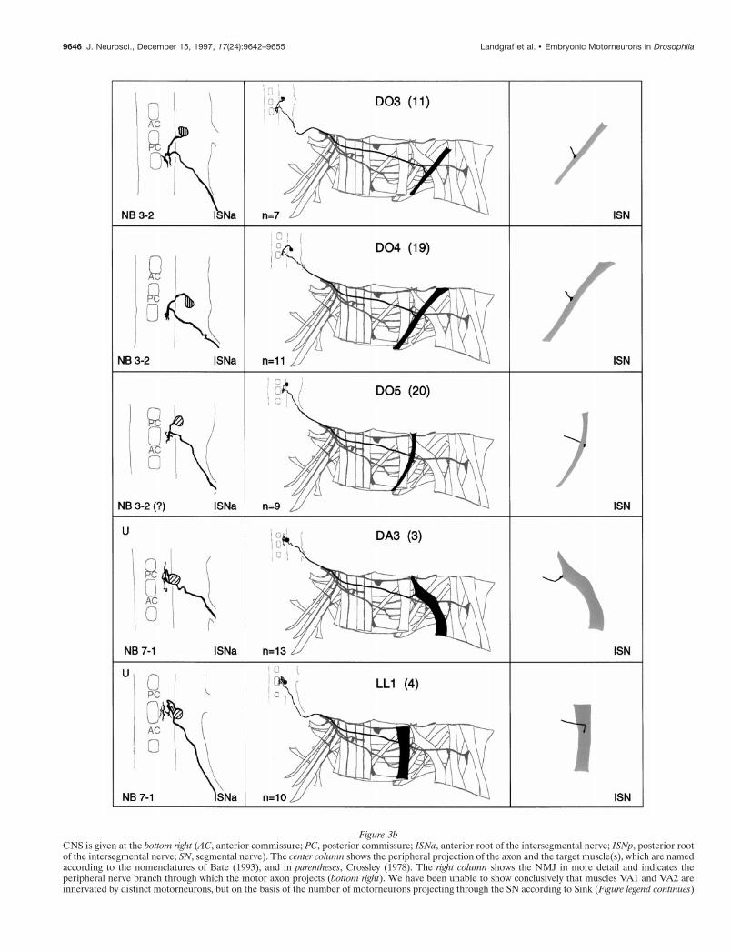
Figure 3bCNS is given at the bottom right (AC, anterior commissure; PC, posterior commissure; ISNa, anterior root of the intersegmental nerve; ISNp, posterior rootof the intersegmental nerve; SN, segmental nerve). The center column shows the peripheral projection of the axon and the target muscle(s), which are namedaccording to the nomenclatures of Bate (1993), and in parentheses, Crossley (1978). The right column shows the NMJ in more detail and indicates theperipheral nerve branch through which the motor axon projects (bottom right). We have been unable to show conclusively that muscles VA1 and VA2 areinnervated by distinct motorneurons, but on the basis of the number of motorneurons projecting through the SN according to Sink (Figure legend continues)
9646 J. Neurosci., December 15, 1997, 17(24):9642–9655 Landgraf et al. • Embryonic Motorneurons in Drosophila

Figure 3cand Whitington (1991a), we propose that they are. To indicate that we are uncertain about which of the dorsal and lateral muscles are innervated by VUMneurons, we have differentially highlighted those muscles that we think are the most likely targets, in agreement with Sink and Whitington (1991a). n 5sample size for each labeled neuron.
Landgraf et al. • Embryonic Motorneurons in Drosophila J. Neurosci., December 15, 1997, 17(24):9642–9655 9647

Figure 3d
9648 J. Neurosci., December 15, 1997, 17(24):9642–9655 Landgraf et al. • Embryonic Motorneurons in Drosophila

Figure 3e
Landgraf et al. • Embryonic Motorneurons in Drosophila J. Neurosci., December 15, 1997, 17(24):9642–9655 9649

Figure 3f
9650 J. Neurosci., December 15, 1997, 17(24):9642–9655 Landgraf et al. • Embryonic Motorneurons in Drosophila

innervate muscles VO1-3. NB 5-2 is the progenitor of theV-motorneuron that innervates muscle VL1. NB 7-1 gives riseto two sets of motorneurons: the four U-motorneurons consti-tute one of these sets and innervate muscles DO1-2, DA3, andLL1. This set could be further subdivided into two, consistingof the DO1-2 neurons on the one hand and the more lateraland ventral DA3/LL1 neurons on the other. A further distinc-tion between the two pairs of U neurons is that it is only theDO1-2 pair that sends a process into the contralateral neuro-mere. The other distinct set of cells derived from 7.1 consistsmost likely of the two motorneurons innervating the ventraloblique muscles VO4-6 (our preparations of NB 7-1 clones didnot conclusively show endplates on muscles VO4-6, but axonsproject to the region that is characteristic for SNd and VO4-6innervation). The origin of aCC from NB 1-1 has already beendescribed (Doe et al., 1988; Udolph et al., 1993), and it isknown that the VUMs arise from midline progenitors (Klambtet al., 1991; Bossing and Technau, 1994).
When lineages and morphologies are compared (Fig. 3), itbecomes clear that morphologically similar motorneurons arisefrom a common neuroblast, but that a single neuroblast may giverise to more than one morphological type. For example, NB 3-2gives rise to two morphologically distinct types of motorneuronsthat innervate muscles in different segments. Those that inner-vate the dorsal oblique muscles of the next posterior segmenthave characteristic axonal projections through the anterior root ofthe ISN and posteriorly running dendritic arbors. Those thatinnervate the lateral transverse muscles of the same segment haveaxonal projections through the SN and anteriorly running den-dritic arbors. When we relate the positions of muscles to themotorneurons that innervate them and their lineages, it becomesclear that muscles that have similar positions and orientations areoften innervated by motorneurons derived from a common neu-roblast. Thus, although there is no obvious relationship betweenthe topography of muscles and motorneurons, there does appearto be a distinct relationship between the progeny of individualneuroblasts and the innervation of particular muscle sets.
The distribution of the dendritic arborsMotorneurons that innervate neighboring muscles have overlap-ping dendritic trees. This is true even for neurons that aremorphologically dissimilar and derived from different neuro-blasts. For example, the motorneurons that innervate the ventrallongitudinal muscles, RP1, - 3, -4, -5 (NB 3-1), and the V-neuron(NB 5-2), project their dendritic arbors to a common region ofthe neuropile located lateral to the posterior commissure (Figs.4H, 5E). Similarly, the dendritic trees of the motorneurons in-
nervating the ventral oblique muscles (VO1-3; NB 4-2 andVO4-6; NB 7-1) are clustered in a region of the neuropile thatoverlaps and is immediately posterior to that defined by thedendritic arbors of the RP1, -3, -4, and -5, and V-neurons (Fig.5F). The dendritic trees of motorneurons that innervate dorsalmuscles are clustered in the neuropile between the commissures(Figs. 4E, 5A–C), and, finally, the dendritic arbors of the segmen-tal motorneurons, which innervate the lateral transverse and theventral acute muscles, are primarily localized lateral to the ante-rior commissure (Figs. 4F, 5D,G). Thus, relationships betweenneuroblasts, the motorneurons they give rise to, their dendriticmorphology, and the muscles they innervate are becoming clear:the cell bodies of motorneurons derived from a common neuro-blast are usually clustered, their axons project to muscles ofsimilar position and orientation, and their dendrites overlap.Motorneurons that are derived from different neuroblasts butinnervate neighboring muscles differ in cell body position andaxonal projection, yet their dendrites branch in a common area ofthe neuropile. The shared feature of most motorneurons withoverlapping dendrites is that they innervate muscles that havesimilar positions and orientations and hence are likely to haverelated functions during muscle contraction. Thus, the differentdomains of dendritic arborization that we describe may representdistinct areas of the neuropile that serve as integrative regions formotorneurons innervating functionally related subsets of thebody wall muscles.
DISCUSSION
The embryonic motorneurons of Drosophilamelanogaster have stereotypic morphologies and areuniquely identifiableTo identify and characterize embryonic motorneurons we haveused the lipophilic fluorescent dye DiI to label motorneuronsretrogradely from their NMJs to their cell bodies. The techniqueallows us to assign motorneurons to each of the 30 abdominalbody wall muscles of the late stage 16 Drosophila embryo. Wehave characterized these motorneurons with respect to the mus-cles they innervate, their positions in the ventral nerve cord, andtheir dendritic arborizations. The map of the motorneurons, theiraxons, dendrites, and target muscles complements the descriptionof the sensory neurons in the embryo (Merritt and Whitington,1995) and provides a baseline from which to investigate thefunctional architecture of the nervous system and its developmen-tal origins.
Figure 3g
Landgraf et al. • Embryonic Motorneurons in Drosophila J. Neurosci., December 15, 1997, 17(24):9642–9655 9651

Additional motorneurons innervate larval musclesAlthough we have described most of the motorneurons, we havenot included all of them, because in late stage 16 embryos wedetect only single axons innervating most body wall muscles.Many of these muscles are known to have multiple innervations inthe third instar larva (Keshishian et al., 1993). Much of thisadditional innervation is accounted for by the maturation of thewidespread terminations of the VUM neurons, which have beententatively identified as the source of the octopaminergic type IIinnervation of the larval body wall muscles (Monastirioti et al.,1995). Our description is limited to the primary innervation bytype I endings (Johansen et al., 1989), present at stage 16. Theidentification of this early differentiating set of motorneuronsreveals interesting new data about the organization of the motorinnervation and its developmental origins.
Organization of the primary motor innervation in thestage 16 embryo and its developmental originsMorphologically similar motorneurons are clonally relatedMost (26 of 31) motorneurons are derived from a few neuroblastslocated ventrally in the neuroectoderm. Each of these neuroblastsproduces one or more sets of motorneurons, and it is an impor-tant finding that morphologically similar motorneurons are ingeneral clonally related and innervate neighboring muscles in theperiphery. For example, considering neurons that innervate ven-tral longitudinal muscles (RP1, -3, -4, and -5 and V) (Fig. 5F), wefind that RP1, -3, -4, and -5 are morphologically similar, withaxons that project contralaterally through the anterior commis-sure, whereas the V-neuron is morphologically distinct andprojects its axon contralaterally through the posterior commis-sure. RP1, -3, -4, and -5 arise from NB 3-1, whereas V is derived
Figure 4. Examples of photoconverted preparations ofclones from individually labeled NBs and of retrogradelylabeled motorneurons. Photoconverted DiI preparations ofclones from individual, identified, labeled NBs (A, D, G),of motorneurons (B, E, F, H ), and of NMJs ( I ), and afluorescent image of DiI-labeled motorneurons ( C) areshown. The images are collages of several focal planes,which were assembled with Photoshop; the dorsal focalplane is uppermost. A, The two medial U motorneuronsand the fpCC interneuron derived from NB 7-1. The othermotorneurons derived from NB 7-1 (the two mediolateralU neurons and the motorneurons that most likely innervatemuscles VO4-6) are not visible in this focal plane. B, Themotorneurons that innervate the four dorsal musclesDA1-2 (aCC and RP2) and DO1-2 (the two medial Uneurons) were retrogradely labeled in one abdominal seg-ment. C, A fluorescent image of the motorneuron inner-vating muscle DT1, which was labeled in four adjacentabdominal segments. Note the characteristic cell body po-sition and dendritic projection. The asterisks indicate T-shaped sensory axons that were labeled by chance in two ofthe segments. The motorneuron that innervates muscleDO4 was labeled in one segment. D, Motorneurons de-rived from two NB 3-2 clones. The anterior clone is tho-racic (T3 ); the posterior clone is abdominal (A2 ). NB 3-2gives rise to two morphological types of motorneurons:first, motorneurons that project through the ISN and inner-vate muscles DT1 (not visible in this focal plane), DO3-4,and probably also DO5 (not visible in this focal plane);second, motorneurons that project through SNa and inner-vate muscle LT3 and probably also LT4. E, A photocon-verted preparation similar to the one shown in C. F, Threemotorneurons that project through SNa are shown. Themotorneurons that innervate muscles LT1-2 are morpho-logically and clonally distinct from the ones innervatingmuscles LO1 and SBM. G, Two NB 3-1 clones, of whichthe anterior clone is thoracic (T3 ) and the posterior cloneis abdominal (A1 ). NB 3-1 gives rise to the four morpho-logically similar RP1, -3, -4, and -5 motorneurons thatinnervate the ventral longitudinal muscles VL1-4. H, Mostof the motorneurons that project through SNb and SNdwere retrogradely labeled in a single abdominal segment toillustrate their relative cell body positions. In addition, themotorneuron that innervates muscles VO1-2 was labeled inthe next posterior segment. Note that the V-neuron ismorphologically distinct from the RP neurons but thattheir dendritic arbors are overlapping (arrowhead) (seetext). The VUM neuron lies in the same segment as themuscles that it innervates, whereas the RP and V-neuronsas well as the motorneurons that innervate muscles VO1-2and VO4-5 lie in the next anterior segment. I, The end-plates of two VUM neurons on the ventral oblique musclesVO1-6 in two adjacent abdominal segments (the VUMswere retrogradely labeled on the contralateral side). Ante-rior is up. The ventral midline is indicated by triangles andis on the lef t in I. Scale bar, 10 mm.
9652 J. Neurosci., December 15, 1997, 17(24):9642–9655 Landgraf et al. • Embryonic Motorneurons in Drosophila

Figure 5. Tracings of groups of motorneurons that innervate muscles that are related in operation by position and orientation. A–G, Composites of tracingsfrom several preparations (A, 2; B, 3; C, 4; D, 3; E, 2; F, 3; G, 2). A, The aCC and RP2 motorneurons that innervate muscles DA1-2. B, The four U neuronsthat innervate the dorsal and lateral muscles DO1-2, DA3, and LL1 (note that this cluster of four neurons may be subdivided further by morphology andtarget muscles; see text for details). C, The motorneurons innervating the dorsal oblique muscles DO3-5 and muscle DT1. D, The motorneurons thatinnervate the lateral muscles LT1-4, LO1, and SBM. Note that muscles LO1 and SBM might be innervated by two distinct motorneurons, one of which isshown here. E, The RP1, -3, -4, -5, and the V-neurons innervate the ventral longitudinal muscles. F, Motorneurons innervating the ventral oblique muscles.G, Motorneurons innervating the ventral acute muscles. Note that there is uncertainty as to whether muscles VA1 and VA2 are innervated by two distinctmotorneurons (Fig. 3). H, All embryonic motorneurons that innervate the larval body wall muscles of abdominal segment A7 at stage 16 (adapted from Sinkand Whitington, 1991a). For details of the individual dendritic arborizations, refer to Figure 3. Anterior is up.
Landgraf et al. • Embryonic Motorneurons in Drosophila J. Neurosci., December 15, 1997, 17(24):9642–9655 9653

from a separate neuroblast, NB 5-2. Similarly, the lateral trans-verse muscles are targeted by two morphologically distinct groupsof motorneurons (Fig. 5E) that arise from two separate neuro-blasts: first, a group of two very ventrally located motorneuronsfrom NB 2-2 that innervate adjacent muscles LT1 and LT2, andsecond, two ventrally located motorneurons, most likely bothfrom NB 3-2, that innervate adjacent muscles LT3 and LT4.
The evolution of the nerve–muscle pairsThese relationships between neuroblasts, the motorneurons theygive rise to, and their target muscles prompt us to speculate aboutthe evolution of neuromuscular networks. The fact that clusters ofpossibly sibling cells derived from a single neuroblast innervaterelated muscles suggests that (1) there is an underlying develop-mental mechanism that links progeny of particular neuroblastswith particular muscle subsets and (2) this relationship may haveinfluenced the formation of new nerve–muscle pairs during theevolution of the neuromuscular system. For example, four ventrallongitudinal muscles could have arisen during the course ofevolution by multiplication of a single functional unit consistingof an RP motorneuron and a ventral longitudinal muscle. Asimple way to bring about such a multiplication of particular setsof interacting cells would be by duplication of their respectiveprecursors, i.e., the RP motorneuron ganglion mother cell and theVL muscle founder cell or its progenitor (Bate, 1990; Carmena etal., 1995). Instances in which we find that two putatively relatedmuscles are co-innervated by a single motorneuron (such as VL3and VL4 or VO1 and VO2), as well as instances in which we findthat two motorneurons related by lineage innervate a singlemuscle (such as RP1 and RP4 innervating muscle VL2), suggestan evolutionary mechanism whereby the progenitors of musclesand motorneurons are duplicated independently of one another.The newly generated motorneuron–muscle units would subse-quently be modified in ways that would make them distinct fromone another, thus allowing for the adaptation of newly formedmotor units for the fine-tuning of movement while retaining aspecific neuromuscular connectivity. Interestingly, the expressionpattern and function of the Drosophila islet gene (Thor andThomas, 1997) supports our hypothesis of the evolution of theneuromuscular network by multiplication of a few ancestralnerve–muscle units: Drosophila islet is expressed in sets of mo-torneurons of common origin that innervate related muscles (e.g.,RP1, -3, -4, and -5 derived from NB 3-1, and motorneurons formuscles VO4-6 derived from NB 7-1) and is required in thesemotorneurons for proper axonal projections to their respectivetargets.
Motorneuron subsets share common domains ofdendritic arborizationIt is also an important aspect of our findings that neurons inner-vating similar muscles are not necessarily related by lineage orposition, but that in these instances they share a common domainof dendritic arborization within the CNS. The most obvious caseis where a single muscle is innervated by two neurons of differentdevelopmental origins. VL1, for example, is innervated by twomotorneurons (V- and RP5 neurons) that are derived from sep-arate neuroblasts. Their cell bodies are not clustered and theyhave different axonal morphologies, but their dendrites project tothe same part of the neuropile. This is a special case of whatappears to be a general rule: muscles that are apparently relatedby function (by proximity and orientation) are innervated by setsof neurons that share a common domain of dendritic arboriza-
tion, whether they are related by lineage or not. We have so faridentified three such domains (which are likely to be furtherrefined by future studies): one for dorsal intersegmental motor-neurons (Fig. 5A–C), one for ventral intersegmental motorneu-rons (Fig. 5E,F), and one for the segmental motorneurons (Fig.5D,G). Provisionally we assume that these regions within thedeveloping neuropile represent sites at which, in the maturesystem, integrative events that are related to the operation ofsubsets of the neuromuscular system can occur.
In some instances, the neurons that contribute to a commondomain of dendritic arborization are not ones innervating musclesthat are obviously functionally related. The fact that the dendritesof the LT1-4 motorneurons and the SBM/LO1 neuron project to acommon neuropile region might be predicted on the grounds ofproximity and the orientation of the muscles concerned or both.However, we also find that VA1-3 motorneurons project dendritesto this domain, and at first sight there seems to be no likelyfunctional relationship between ventral acute and lateral transversemuscles. The dendrites of RP2 represent the converse case andappear to be particularly anomalous (Fig. 5A). We might expect onfunctional grounds that the dendrites of RP2 would cluster withthose of other dorsal intersegmental motorneurons. However, theyactually span the region of the anterior commissure where thedendrites of segmental motorneurons are clustered. One reason forthis may be that RP2, alone among the motorneurons with axons inthe ISN, innervates a muscle in the same segment (apart fromVUMs). This would imply that the position of the dendrites isinfluenced by the segmental origins of the motorneurons and thatthis could be the deciding factor in laying out the foundations ofthe neuropile, rather than any functional relationships between theneurons.
Subdividing the neuropilePerhaps the most interesting question that emerges from thisstudy is the nature of the developmental mechanism that under-lies the apparent partitioning of the neuropile during embryogen-esis. We know very little about dendritic arborization and itscontrol in Drosophila, but we might expect that, like guidance ofgrowing axons, the formation of dendritic branches is regulated inpart by the expression of cell adhesion molecules on the surfacesof neurons and cells with which they come in contact. Themechanisms that underlie the clustering of the dendritic trees offunctionally related motorneurons may be similar to those gov-erning axonal pathfinding and target recognition in the periphery,where axons are ordered into fascicles through a series of adhe-sive preferences (Lin et al., 1994; Lin and Goodman, 1994; Desaiet al., 1996; Fambrough and Goodman, 1996; Krueger et al.,1996). As far as the neuromuscular system is concerned, it isparticularly interesting that the homophilic cell adhesion mole-cule Connectin is expressed on the surfaces of segmental motor-neurons (that is, neurons projecting through SNa or SNc), withthe single exception of the DT1 motorneuron, the cell body ofwhich lies in the next anterior segment with an axon exitingthrough the anterior root of the ISN. In the periphery, axons ofConnectin-expressing motorneurons fasciculate and innervateConnectin-expressing muscles, and in the CNS the dendrites ofthe segmental Connectin-expressing motorneurons (LT1-4, LO1,SBM, VA1-3; see above) cluster together in the region lateral tothe anterior commissure. The DT1 motorneuron is the excep-tional case among the Connectin-expressing motorneurons, be-cause it forms in the next anterior segment. However, the pro-jection of the DT1 motorneuron dendrites supports the notion
9654 J. Neurosci., December 15, 1997, 17(24):9642–9655 Landgraf et al. • Embryonic Motorneurons in Drosophila

that Connectin expression may be one of the properties thatgoverns branching in a common region of neuropile: a branch ofthe DT1 projection runs posteriorly and branches in the regionshared by the other Connectin-expressing motorneurons. Exper-iments are now in progress to test the idea that the patternedexpression of cell adhesion molecules is required for normalpartitioning of dendrites in the developing CNS, as it is for properinnervation of the muscle field.
REFERENCESBate CM (1976) Embryogenesis of an insect nervous system I. A map of
the thoracic and abdominal neuroblasts in Locusta migratoria. J Em-bryol Exp Morphol 35:107–123.
Bate M (1990) The embryonic development of larval muscles in Dro-sophila. Development 110:791–804.
Bate M (1993) The mesoderm and its derivatives. In: The developmentof Drosophila melanogaster (Bate M, Martinez Arias A, eds), pp 1013–1090. Cold Spring Harbor, NY: Cold Spring Harbor Laboratory.
Bodmer R, Jan YN (1987) Morphological differentiation of the embry-onic peripheral neurons in Drosophila. Roux’s Arch Dev Biol196:69–77.
Bossing T, Technau GM (1994) The fate of the CNS midline progenitorsin Drosophila as revealed by a new method for single cell labelling.Development 120:1895–1906.
Bossing T, Udolph G, Doe CQ, Technau GM (1996) The embryoniccentral nervous system lineages of Drosophila melanogaster. I. Theneuroblast lineages derived from the ventral half of the neuroectoderm.Dev Biol 179:41–64.
Broadie KS, Bate M (1993) Development of the embryonic neuromus-cular synapse of Drosophila melanogaster. J Neurosci 13:144–166.
Broadie K, Sink H, Van Vactor D, Fambrough D, Whitington PM, BateM, Goodman CS (1993) From growth cone to synapse: the life historyof the RP3 motor neuron. Development [Suppl] 119:227–238.
Broadus J, Skeath JB, Spana E, Bossing T, Technau GM, Doe CQ (1995)New neuroblast markers and the origin of the aCC/pCC neurons in theDrosophila CNS. Mech Dev 54:1–10.
Burrows M (1996) The neurobiology of an insect brain. Oxford: Ox-ford UP.
Callahan CA, Thomas JB (1994) Tau-b-galactosidase, an axon-targetedfusion protein. Proc Natl Acad Sci USA 91:5972–5976.
Campos-Ortega JA, Hartenstein V (1985) The embryonic developmentof Drosophila melanogaster. Berlin: Springer.
Carmena A, Bate M, Jimenez F (1995) lethal of scute, a proneural gene,participates in the specification of muscle progenitors during Drosophilaembryogenesis. Genes Dev 9:2373–2383.
Cash S, Chiba A, Keshishian H (1992) Alternate neuromuscular targetselection following the loss of single muscle fibers in Drosophila. J Neu-rosci 12:2051–2064.
Chiba A, Snow P, Keshishian H, Hotta Y (1995) Fasciclin III is asynaptic recognition molecule in Drosophila. Nature 374:166–168.
Crossley, A C (1978) The morphology and development of the Drosoph-ila muscular system. In: The genetics and biology of Drosophila, Vol 2b(Ashburner M, Wright T, eds), pp 499–560. New York: Academic.
Desai CJ, Gindhart JG, Goldstein LSB, Zinn K (1996) Receptor ty-rosine phosphatases are required for motor axon guidance in theDrosophila embryo. Cell 84:599–609.
Doe CQ (1992) Molecular markers for identified neuroblasts and gan-glion mother cells in the Drosophila central nervous system. Develop-ment 116:855–863.
Doe CQ, Technau GM (1993) Identification and cell lineage of individ-ual neural precursors in the Drosophila CNS. Trends Neurosci16:510–514.
Doe CQ, Hiromi Y, Gehring WJ, Goodman CS (1988) Expression andfunction of the segmentation gene fushi tarazu during Drosophila neu-rogenesis. Science 239:170–175.
Fambrough D, Goodman CS (1996) The Drosophila beaten path geneencodes a novel secreted protein that regulates defasciculation at motoraxon choice points. Cell 87:1049–1058.
Goodman CS, Doe CQ (1993) Embryonic development of the Drosoph-ila central nervous system. In: The Development of Drosophila mela-nogaster (Bate M, Martinez Arias A, eds), pp 1131–1206. Cold SpringHarbor, NY: Cold Spring Harbor Laboratory.
Gorczyca MG, Randall WP, Budnik V (1994) The role of tinman, amesodermal cell fate gene, in axon pathfinding during the developmentof the transverse nerve in Drosophila. Development 120:2143–2152.
Halpern ME, Chiba A, Johansen J, Keshishian H (1991) Growth conebehavior underlying the development of stereotypic synaptic connec-tions in Drosophila embryos. J Neurosci 11:3227–3238.
Hartenstein V, Campos-Ortega JA (1984) Early neurogenesis in wild-type Drosophila melanogaster. Roux’s Arch Dev Biol 193:308–325.
Johansen J, Halpern ME, Keshishian H (1989) Axonal guidance and thedevelopment of muscle fiber-specific innervation in Drosophila em-bryos. J Neurosci 9:4318–4332.
Keshishian H, Chiba A, Ning Chan T, Halfon M, Harkins E, Jarecki J,Wang L, Anderson MD, Cash S, Halpern ME (1993) Cellular mech-anisms governing synaptic development in Drosophila melanogaster.J Neurobiol 24:757–787.
Keshishian H, Broadie K, Chiba A, Bate M (1996) The Drosophilaneuromuscular junction: a model system for studying synaptic develop-ment and function. Annu Rev Neurosci 19:545–575.
Klambt C, Jacobs JR, Goodman CS (1991) The midline of the Drosoph-ila central nervous system: a model for the genetic analysis of cell fate,cell migration, and growth cone guidance. Cell 64:801–815.
Krueger NX, Van Vactor D, Wan HI, Gelbart WM, Goodman CS, SaitoH (1996) The transmembrane tyrosine phosphatase DLAR controlsmotor axon guidance in Drosophila. Cell 84:611–622.
Lin DM, Goodman CS (1994) Ectopic and increased expression of Fas-ciclin II alters motoneuron growth cone guidance. Neuron 13:507–523.
Lin DM, Fetter RD, Kopczynski C, Grenningloh G, Goodman CS (1994)Genetic analysis of Fasciclin II in Drosophila: defasciculation, refascicu-lation, and altered fasciculation. Neuron 13:1055–1069.
Merritt DJ, Whitington PM (1995) Central projections of sensory neu-rons in the Drosophila embryo correlate with sensory modality, somaposition, and proneural gene function. J Neurosci 15:1755–1767.
Monastirioti M, Gorczyca M, Rapus J, Eckert M, White K, Budnik V(1995) Octopamine immunoreactivity in the fruit fly Drosophila mela-nogaster. J Comp Neurol 356:275–287.
Sink H, Whitington PM (1991a) Location and connectivity of abdominalmotoneurons in the embryo and larva of Drosophila melanogaster.J Neurobiol 22:298–311.
Sink H, Whitington PM (1991b) Pathfinding in the central nervous sys-tem and periphery by identified embryonic Drosophila motor axons.Development 112:307–316.
Thomas JB, Bastiani MJ, Bate M, Goodman CS (1984) From grasshop-per to Drosophila: a common plan for neural development. Nature310:203–206.
Thor S, Thomas JB (1997) The Drosophila islet gene governs axon path-finding and neurotransmitter identity. Neuron 18:397–409.
Udolph G, Prokop A, Bossing T, Technau GM (1993) A common pre-cursor for glia and neurons in the embryonic CNS of Drosophila givesrise to segment-specific lineage variants. Development 118:765–775.
Van Vactor D, Sink H, Fambrough D, Tsoo R, Goodman CS (1993)Genes that control neuromuscular specificity in Drosophila. Cell 73:1137–1153.
Landgraf et al. • Embryonic Motorneurons in Drosophila J. Neurosci., December 15, 1997, 17(24):9642–9655 9655
Related Documents