T his morning Can Ince (Erasmus Medical Center, Rotterdam and Academic Medical Center, Amster- dam, the Netherlands) joins others to discuss transfusions, arguing in their favor in the arena of improving tissue oxygenation in circulatory shock. Dr Ince has published extensively on fundamental aspects of oxygen transport, the microcirculation, and organ function in relation to particular issues that pervade intensive care such as fluid resuscitation, blood transfu- sion and inflammatory modulation. “I’m not a clinician, I’m a physiolo- gist,” he told ISICEM News. “But I’ve worked in intensive care medicine de- partments around for 30 to 40 years. My brief is to think about how things work, why they don’t work, and to identify ways of making physiology kick in to make organs work and hopefully improve outcome.” One of the fundamental issues within intensive care medicine from his vantage point, explained Dr Ince, is that, despite a vast existing knowledge base of the mechanisms involved, hemodynamic management is carried out in practice on the basis of measurable surrogates of tissue perfusion and oxygenation, which are not necessarily related to what is actually going on at the level of the Wednesday 22 March 2017 Day 2 The official daily newsletter of the 37 th ISICEM Reconciling renal replacement data 4 HD monitoring: To measure, perchance to know 6 ICU-acquired weakness: trust the numbers? 10 Protein intake: more or less? 14 ISICEM 2017 opens its doors… On Tuesday morning, the Henry Le Bœuf auditorium welcomed its audience with the sound of music. Piano notes filled the air all the way to the rafters, making way for ISICEM Chairman Jean-Louis Vincent, who took to the stage to usher in the first session of the 37 th ISICEM Symposium. Transfusions are the only way to increase tissue oxygen availability Continued on page 2

Welcome message from author
This document is posted to help you gain knowledge. Please leave a comment to let me know what you think about it! Share it to your friends and learn new things together.
Transcript

T his morning Can Ince (Erasmus Medical Center, Rotterdam and Academic Medical Center, Amster-dam, the Netherlands)
joins others to discuss transfusions, arguing in their favor in the arena of improving tissue oxygenation in
circulatory shock. Dr Ince has published extensively
on fundamental aspects of oxygen transport, the microcirculation, and organ function in relation to particular issues that pervade intensive care such as fluid resuscitation, blood transfu-sion and inflammatory modulation.
“I’m not a clinician, I’m a physiolo-gist,” he told ISICEM News. “But I’ve worked in intensive care medicine de-partments around for 30 to 40 years. My brief is to think about how things work, why they don’t work, and to identify ways of making physiology kick in to make organs work and
hopefully improve outcome.”One of the fundamental issues
within intensive care medicine from his vantage point, explained Dr Ince, is that, despite a vast existing knowledge base of the mechanisms involved, hemodynamic management is carried out in practice on the basis of measurable surrogates of tissue perfusion and oxygenation, which are not necessarily related to what is actually going on at the level of the
Wednesday 22 March 2017 Day 2The official daily newsletter of the 37th ISICEM
Reconciling renal replacement data . . .4
HD monitoring: To measure, perchance to know . . . . . . . 6
ICU-acquired weakness: trust the numbers? . . . . . . 10
Protein intake: more or less? . . . . . . . . . . 14
ISICEM 2017 opens its doors…On Tuesday morning, the Henry Le Bœuf auditorium welcomed its audience with the sound of music. Piano notes filled the air all the way to the rafters, making way for ISICEM Chairman Jean-Louis Vincent, who took to the stage to usher in the first session of the 37th ISICEM Symposium.
Transfusions are the only way to increase tissue oxygen availability
Continued on page 2

2 ISICEM News Wednesday 22 March 2017 Issue 2
microcirculation and tissues of the various organ systems.
“If you ask clinicians as to the purpose of many of the therapeutic interventions related to hemodynamic support of the cardiovascular system, they ultimately agree that it all boils down to improving tissue perfusion and oxygenation. One of the main cornerstones of such therapy in intensive care, for example, is the ad-ministration of fluids. The rationale of giving fluids is to correct hypovolemia with the expectation that increased cardiac output will ensure perfusion of the various tissues. But perfusion of what? Well…oxygen-carrying red blood cells!”
This principle, he stressed, can be contradicted in the practice of administration of fluids (which do not carry oxygen in themselves). Despite the principle of flow generation being generally sound, fluids also reduce the viscosity of existing blood – an impor-tant physiological variable which the vasculature uses to sense shear stress of blood flow, and to accommodate vascular regulation to ensure the op-timal distribution of oxygen transport to the various organ systems to meet various tissues’ metabolic demands.
“At ISICEM, you will hear many lectures about how to give fluids, what type of fluids, when, to what degree, etc. Much detailed informa-tion is related to manipulation of stroke volume, blood pressure in vari-ous compartments, and other issues. However their efficacy to improve tis-sue oxygenation is relatively ignored.
“Many studies in experimental models of shock, where tissue oxy-genation can be precisely measured in different organ systems, have shown that the administra-tion of fluids is highly effective in improving mean arterial pressure or stroke volume, etc., but is ineffective in correcting tissue hypoxia in vulner-able tissue beds such as the kidneys (e.g., Zafrani et al.1). So even though systemic hemodynamic variables can be manipu-lated, the actual purpose of doing so remains wanting – although there are of course boundaries within which it is effective.”
The concept of ‘hemodynamic coherence’ is a requirement in resus-citation, to ensure that changes in global hemodynamic variables cause a parallel improvement in the perfusion and oxygenation of the microcircula-tion in the various organ systems, such as occurs in in healthy individuals where vascular regulation ensures a match between supply and metabolic need of the various organ systems. In conditions such as sepsis such regulation is lost, tissue perfusion is heterogeneous, and the functional capacity of the microcirculation is altered; as such, shunting of vulner-able microcirculatory regions occurs, resulting in them becoming hypoxic while other regions are not.2-4
“Hemodynamic coherence is defined as the expectation in resuscitation that, if you correct the systemic circulation, the microcir-culation follows in parallel.3 A lack of hemodynamic coherence is a dangerous condition; this is what can happen in the unresponsive patient, where despite ever increasing doses of therapy aimed at improving systemic hemodynamic variables, a parallel improvement in surrogates of tissue perfusion and oxygenation perfusion (lactate, oliguria, etc.) does not occur. It is important to know that if one administers fluids, that it is
indeed having the desired benefit at the level of the microcirculation and tissue cells.”
As well as heterogeneous microcir-culation, explained Dr Ince, hemodilu-tion and anemia (which could perhaps be exacerbated by fluid administration), contribute to a loss of hemodynamic coherence. A further two contributors also exist, namely vasoconstriction imposed by excessive use of vasocon-strictor therapy leading to microcircula-tory flow reduction, and tissue edema
resulting in increased diffusion distance of oxygen from the red blood cells in the microcirculation to tissues.
Given all this, how can tissue oxygenation be improved? Dr Ince did not negate existing therapeutic strategies entirely in his response: “Certainly, tissue oxygenation can be improved to a certain extent by using drugs such as vasopressors and fluids, and organs may recover despite the limited efficacy of such therapies.”
A further strategy to combat tissue hypoxia might be to increase the inspired oxygen fraction (fiO2) in mechanical ventilation, he said, al-though this may actually make things worse5. Increasing inspired fractions causes microcirculatory vasoconstric-tion.6 More seriously, high levels of oxygen can induce oxidative stress resulting in reperfusion injury. The only alternative, he concluded, is to give blood as the natural way to give oxygen to the cells. “We have shown, and others have too in many studies, that if you measure the oxygenation of tissues in models of shock where there is tissue hypoxia, blood transfu-sions are the only way to improve that condition. We even showed this to be the case in animal models of sepsis.1 Concomitant to improving the oxygen availability in the tissues, you see a lot of the adverse effects of decreased oxygen in the tissues being reversed when you give blood.”
Dr Ince will begin his presentation with a discussion of anemia. “Anemia is in my opinion one of the biggest problems in intensive care patients, and not enough emphasized when discussing the potential hazards asso-ciated with fluid therapy,” he said.
An overwhelming proportion of in-tensive care patients become anemic during their stay.7 Anemia, explained Dr Ince, is a major risk factor for vulnerable organs such as the brain (cognitive impairment) and kidney
(acute kidney injury) in this group. So why is anemia tolerated, and why are blood transfusions not more pro-fusely used? Dr Ince noted that, along with the current prevailing strategy of focus-ing on global hemodynamic markers, current studies on the use of blood products is lacking in a number of ways, with a lack of appreciation of their role in treating tissue hypoxia, negativity about the way blood is stored, as
much as a lack of clear delineation as to under which pathophysiological and clinical circumstances it should be given.
“Giving blood is an organ trans-plantation, and that is how it should be treated. When you transplant an organ, like a kidney, drugs are given to help the kidney survive in its new environment, as well as to the patient to make sure that a welcome envi-ronment greets the placement of a foreign kidney. In my opinion, such an approach should be applied to blood transfusions as well.”
It should be clear though, he add-ed, that the patient would be amena-ble to blood transfusion, rather than it being deleterious due to the hostile inflammatory milieu characteristic of states of critical illness such as sepsis. It should also be clear, he continued, that the red blood cells themselves should viable and adequately stored and maintain their viability once inside the patient.
Another aspect is ensuring that blood transfused reaches tissues: “In cardiac surgery patients and anemic hematological patients, you see the red blood cells coherent between the systemic and microcirculations [e.g. Yuruk et al8]. But not so in sepsis; despite their always being an increase in arterial hemoglobin levels you often see that the transfused red blood cells do not reach the microcircula-
Transfusions 100 Hall Wednesday 9:00
Transfusions are the only way to increase tissue oxygen availability
“Hemodynamic coherence is defined as the expectation in resuscitation that, if you correct the systemic circulation, the microcirculation follows in parallel.” Can Ince
ISICEM NewsPublishing and ProductionMediFore Limited
Symposium ChairmanJean-Louis Vincent
Editor-in-ChiefPeter Stevenson
EditorsRysia BurmiczTatum AndersonBecky McCall
DesignPeter Williams
Industry Liaison ManagerAmanda D’rojcas
Head Office51 Fox Hill London SE19 2XE Telephone: +44 (0) 208 771 8046 [email protected] www.medifore.co.ukCopyright © 2017: Université Libre de Bruxelles. All rights reserved. No part of this publication may be reproduced, stored in a retrieval system, transmitted in any form or by any other means, electronic, mechanical, photocopying, recording or otherwise without prior permission in writing of ISICEM. The content of ISICEM News does not necessarily reflect the opinion of the ISICEM 2017 Symposium Chairman, the ISICEM Scientific Advisors or Collaborators.

Issue 2 Wednesday 22 March 2017 ISICEM News 3
tion. This is due to the alterations in the microcirculation as well as to altered hemorheological properties of the transfused blood, both of which negatively affect the patency of the microcirculation impeding the en-trance of these cells. Additional drugs to promote their transport to the microcirculation could be considered.”
Data on the use of transfusion in sepsis are numerous over the decades, but disparate in their findings. Dr Ince stressed the importance of personal-izing clinical decisions in transfusion according to each patient’s physiology. “As a physiologist I’m very skeptical about these enormous RCTs, because their design is not interested in the mechanism of why a treatment does or does not work, but simply whether some desired gross outcome (such as mortality) is caused. The complexity of the critically ill patients almost by defi-nition precludes the success of such an approach. This method of provid-ing evidence for various interventions has in my opinion been deleterious to the progress of intensive care medicine because these trials have rarely shown a benefit of anything. If anything they have only shown that something gets worse and most of the time these types of studies have shown no difference between differ-ent groups. Unfortunately such results are often misinterpreted as meaning that ‘it doesn’t really matter which of the interventions you give’.
“Of course though, absence of evidence is not evidence of absence. The only statistic important to a physi-ologist is n=1 – the single patient – because if there is precise knowledge
about the failure of physiology at the bedside, and the underlying mecha-nisms responsible for such failure that need to be fixed, then goal-directed therapy is effective if feedback is ob-tained from the relevant physiological compartments, and treatments can be optimized.”
Notable studies that continue to cloud blood transfusion prac-tice include the iconic Hébert et al. (1999) and CRIT studies (2004).9,10 Whether or not the lack of benefit of transfusion found by such trials in North America and Europe should be interpreted as such was questioned by Vincent et al (2008), wherein they
demonstrated in a small observational cohort that adjustment for cofactors actually conferred a slight mortality advantage when blood transfusions were administered.11 More recent tri-als have explored lower versus higher hemoglobin threshold for transfusion and aged versus fresh blood, showing no clear differences between patients groups.12,13
One issue with some of the older trials, explained Dr Ince, was the fact that blood was not leukocyte-reduced as is now the case in Europe and Canada. In addition, he noted, it must be kept in mind when evaluating the literature that US blood is allowed to
be stored for much longer durations that it is in Europe. “This literature has in my opinion clouded the po-tential importance of the dangers of anemia and its beneficial treatment by blood transfusion,” he said. “It has led to a negative perception of giving blood and there is equal negative perception of giving fluid, actually.”
He summarized: “Because of the costs, and the difficulty of obtaining blood, anemia and the need to cor-rect anemia by blood transfusion have disappeared into the background in the last 10 to 15 years. People have been much more accommodating and accepting of anemia, and have shifted their attention to correcting hemodynamic abnormalities.
“However, recent trials in septic patients are demonstrating the impor-tance of targeting higher hemoglobin levels. Evidence has shown that pa-tients with higher hemoglobin levels as a result of blood transfusions have a better outcome than those that have not [e.g., de Almeida et al14]. It is turning out that giving blood and having high hemoglobin levels is es-pecially important for cognitive func-tion. If you look at elderly patients that have had blood transfusions they do much better on that score as well as in survival [e.g., Sakr et al, Naka-mura et al15,16]. It is time take anemia as a serious avoidable risk factor and to consider more frequently blood transfusion as a means of correct-ing anemia and improving oxygen transport to the tissues of the critically ill patient.”
Dr Ince speaks during ‘Transfusions’, which takes place today from 9:00 in 100 Hall.
Transfusions are the only way to increase tissue oxygen availability
“It is time take anemia as a serious avoidable risk factor.” Can Ince
References
1. Zafrani L et al. Blood transfusion improves renal oxygenation and renal function in sepsis-induced acute kidney injury in rats. Crit Care, 2016;20(1):406.
2. Ince C, and Mik EG. Microcirculatory and mitochondrial hypoxia in sepsis, shock, and resuscitation. J Appl Physiol., 2016;120(2):226-35.
3. Ince C. Hemodynamic coherence and the rationale for monitoring the microcirculation. Crit Care, 2015;19(Suppl 3):S8.
4. Ince C, and Guerci P. Why and when the microcirculation becomes disassociated from the macrocirculation. Intensive Care Med., 2016;42:1645.
5. Villar J, and Kacmarek RM. Oxygen: Breath of Life or Kiss of Death. Crit Care Med., 2017;45(2):368-369.
6. Orbegozo Cortés D et al. Normo-baric hyperoxia alters the micro-circulation in healthy volunteers. Microvasc Res, 2015;98:23-8.
7. Astin R, and Puthucheary Z. Anae-mia secondary to critical illness: an unexplained phenomenon. Extrem
Physiol Med., 2014;3:4.
8. Yuruk K et al. Blood transfusions recruit the microcirculation dur-ing cardiac surgery. Transfusion, 2011;51(5):961-7.
9. Hébert PC et al. A Multicenter, Randomized, Controlled Clinical Trial of Transfusion Requirements in Critical Care. N Engl J Med., 1999;340:409-417.
10. Corwin HL et al. The CRIT Study: Anemia and blood transfusion in the critically ill--current clinical practice in the United States. Crit Care Med., 2004;32(1):39-52.
11. Vincent JL et al. Are Blood Transfu-sions Associated with Greater Mortality Rates?: Results of the Sepsis Occurrence in Acutely Ill Patients Study. Anesthesiology 1, 2008;108:31-39.
12. Holst LB et al. Lower versus Higher Hemoglobin Threshold for Transfu-sion in Septic Shock. N Engl J Med., 2014;371:1381-1391.
13. Alexander PE et al. Transfusion of fresher vs older red blood cells in hospitalized patients: a system-atic review and meta-analysis. Blood, 2016;127:400-410.
14. de Almeida JP et al. Transfusion Requirements in Surgical Oncology Patients A Prospective, Randomized Controlled Trial Anesthesiology, 2015;122:29-38.
15. Sakr Y et al. Anemia and blood transfusion in a surgical intensive care unit. Crit Care, 2010;14(3):R92.
16. Nakamura RE et al. A liberal strategy of red blood cell transfu-sion reduces cardiogenic shock in elderly patients undergoing cardiac surgery. J Thorac Cardiovasc Surg., 2015;150(5):1314-20.

4 ISICEM News Wednesday 22 March 2017 Issue 2
T wo high-profile trials will be under the spotlight today in a presentation by John Kellum (Centre for critical care nephrology,
University of Pittsburgh, PA, USA).Dr Kellum will address a dilemma that has chal-
lenged clinicians for some time: whether and when to start renal replacement therapy (RRT) among critically ill patients with acute kidney injury in the absence of conventional indications. “We know very well who should receive a specific therapy and who should not, but there’s a large group in the middle where we are uncertain,” he explained.
The subject has long been a vexing challenge for clinicians. “Should we do coronary artery bypass surgery or continue medical manage-ment? We can certainly describe patients who need surgery and patients who don’t but there are many where the decision is at best, unclear,” he continued.
Dr Kellum’s specialty in sepsis and organ failure, especially kidney injury, led him to participate in a high-profile randomized trial, the Early Versus Late Initiation of Renal Replace-ment Therapy In Critically Ill Patients With Acute Kid-ney Injury (ELAIN) trial. It reported specifically on the issue of the timing of initia-tion of renal replacement therapy in critically ill patients with acute kidney injury, but with no potentially life-threatening com-plication directly related to renal failure.
And he will look at another study that looked at a similar question: The French Artificial Kidney Initi-ation in Kidney Injury (AKIKI) trial, which compared two strategies for starting RRT in mixed critically-ill patients with acute kidney injury who were receiv-ing mechanical ventilation and/or vasoactives.
There were similarities between the trials, said Dr Kellum: “The primary outcome was survival so it’s not hard to relate this to something important for patients.”
Interestingly, however, the trials differed in terms of the numbers of sites, patients, and even primary endpoints. Hence Dr Kellum will present
the fundamental differences in trial design, sample size, and the put the widely discrepant findings in context.
“The basic message is that two studies can be reconciled by understanding that they enrolled fundamentally differ-ent patients,” he said.
The ELAIN trial was focused on high-risk patients, the vast majority of which will receive RRT. Some were randomized at Stage 2 to receive therapy immediately, and others only after they progressed to Stage 3.
In contrast to ELAIN, AKIKI screened patients that already met the ‘late’ criteria for ELAIN, ex-cluded patients with ‘urgent indications’, and then randomized patients who ‘could wait’ for RRT, or only if they eventually developed urgent indica-tions. In other words, “The AKIKI trial excluded pa-tients who would have been enrolled in ELAIN and instead looked at patients who were able to wait and possibly not receive dialysis,” said Dr Kellum.
The combination of the two trials has led to some interesting conclusions, he added. The ELAIN trial, for instance, showed that early interven-tions are better for some patients. “Compared to starting after getting worse, an early start was better. We can conclude that in patients you are very likely to treat with RRT, it makes sense to start right away.”
And the AKIKI trial produced yet more vital pieces of information, as Dr Kellum explained. “If the likelihood of RRT is lower (around 50% or less; because you have already excluded the high risk patients) it would appear to be safe to wait until patients develop urgent indications for RRT. In this population, waiting was not worse than starting, and it was likely safer and less expensive.”
But there is a caveat, according to Dr Kellum. “If the patient ultimately required RRT they did
worse compared to those getting it right away,” he said, adding that reconciling the two studies is indeed possible: “The two trials really asked different questions. The final results indicate that in patients where RRT is likely, early is better. But when RRT is less likely, it’s probably better to wait. However, determining what is ‘likely’ is still a hard decision.”
That there were two trials has prompted even more questions as to how to determine when to start RRT. Dr Kellum noted that because both stud-ies were rather small, and therefore contributed much in terms of informing practice, it’s hoped that larger multicenter randomized trials can be carried out to address unanswered questions.
Luckily, the issue of when to start renal replace-ment therapy is the subject of another study. Dr Kellum said this new trial is to be carried out by Sean M. Bagshaw, an intensivist and an Associate Professor of Critical Care Medicine at the University of Alberta in Edmonton, Canada and Ron Wald, a nephrologist at St. Michael’s Hospital in Toronto, Canada and an Associate Professor of Medicine at the University of Toronto. This so-called STARRT-AKI research program should provide even more answers to the question of when to treat. “It will seek to include both kinds of patients and provide further information,” he concluded.
When to Start Renal Replacement Therapy Tent Wednesday 16:45
Reconciling different trial outcomes
ISSU
E 3
Ava
ilabl
eto
mor
row
“The final results indicate that in patients where RRT is likely, early is better. But when RRT is less likely, it’s probably better to wait. However, determining what is ‘likely’ is still a hard decision.”John Kellum
“The basic message is that two studies can be reconciled by understanding that they enrolled fundamentally different patients.“John Kellum

Issue 2 Wednesday 22 March 2017 ISICEM News 5
6 ISICEM News Wednesday 22 March 2017 Issue 2
A selection of new hemodynamic monitoring modalities will be presented today, with themes
including non-invasive techniques, peripheral perfusion monitoring, end tidal CO2, computer-assisted and closed-loop systems, and pulmonary edema diagnosis using extravascular lung water.
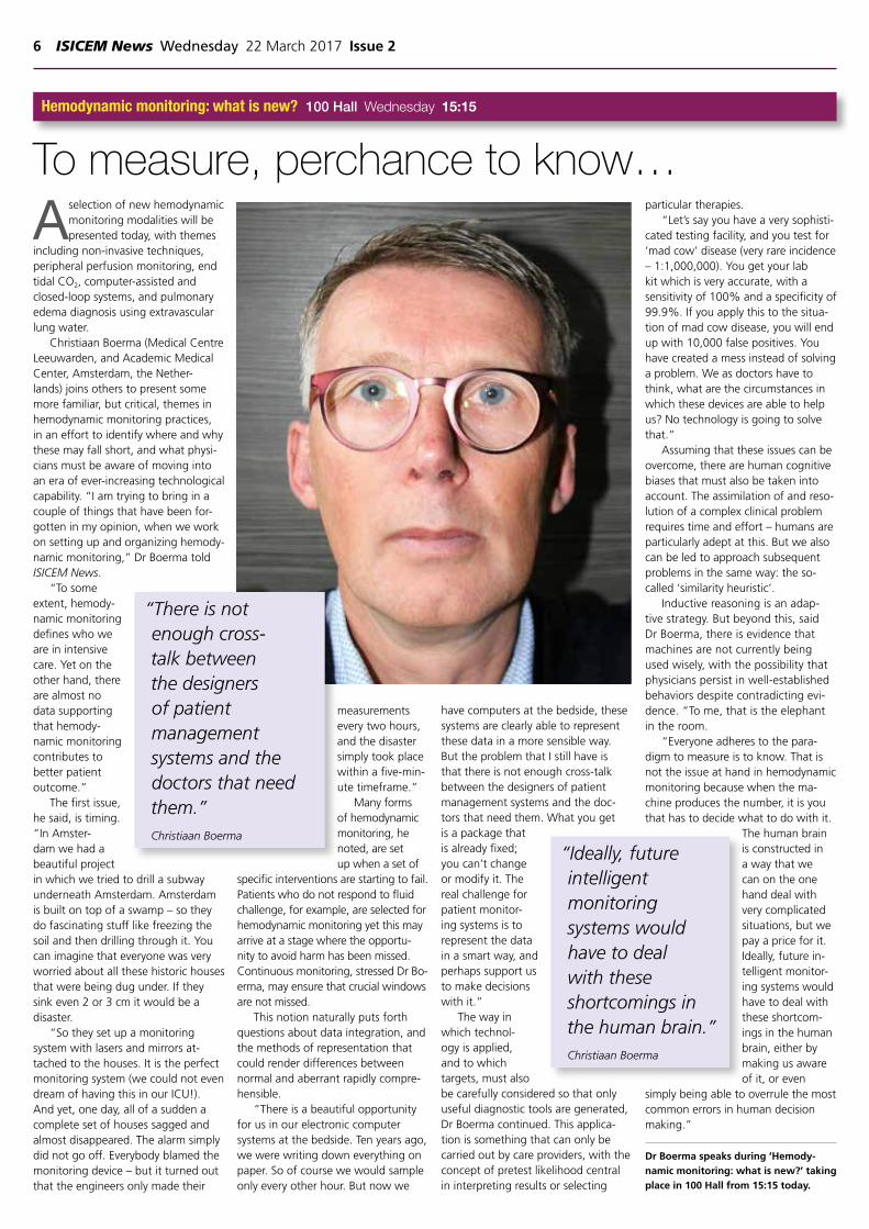
Christiaan Boerma (Medical Centre Leeuwarden, and Academic Medical Center, Amsterdam, the Nether-lands) joins others to present some more familiar, but critical, themes in hemodynamic monitoring practices, in an effort to identify where and why these may fall short, and what physi-cians must be aware of moving into an era of ever-increasing technological capability. “I am trying to bring in a couple of things that have been for-gotten in my opinion, when we work on setting up and organizing hemody-namic monitoring,” Dr Boerma told ISICEM News.
“To some extent, hemody-namic monitoring defines who we are in intensive care. Yet on the other hand, there are almost no data supporting that hemody-namic monitoring contributes to better patient outcome.”
The first issue, he said, is timing. “In Amster-dam we had a beautiful project in which we tried to drill a subway underneath Amsterdam. Amsterdam is built on top of a swamp – so they do fascinating stuff like freezing the soil and then drilling through it. You can imagine that everyone was very worried about all these historic houses that were being dug under. If they sink even 2 or 3 cm it would be a disaster.
“So they set up a monitoring system with lasers and mirrors at-tached to the houses. It is the perfect monitoring system (we could not even dream of having this in our ICU!). And yet, one day, all of a sudden a complete set of houses sagged and almost disappeared. The alarm simply did not go off. Everybody blamed the monitoring device – but it turned out that the engineers only made their
measurements every two hours, and the disaster simply took place within a five-min-ute timeframe.”
Many forms of hemodynamic monitoring, he noted, are set up when a set of
specific interventions are starting to fail. Patients who do not respond to fluid challenge, for example, are selected for hemodynamic monitoring yet this may arrive at a stage where the opportu-nity to avoid harm has been missed. Continuous monitoring, stressed Dr Bo-erma, may ensure that crucial windows are not missed.
This notion naturally puts forth questions about data integration, and the methods of representation that could render differences between normal and aberrant rapidly compre-hensible.
“There is a beautiful opportunity for us in our electronic computer systems at the bedside. Ten years ago, we were writing down everything on paper. So of course we would sample only every other hour. But now we
have computers at the bedside, these systems are clearly able to represent these data in a more sensible way. But the problem that I still have is that there is not enough cross-talk between the designers of patient management systems and the doc-tors that need them. What you get is a package that is already fixed; you can’t change or modify it. The real challenge for patient monitor-ing systems is to represent the data in a smart way, and perhaps support us to make decisions with it.”
The way in which technol-ogy is applied, and to which targets, must also be carefully considered so that only useful diagnostic tools are generated, Dr Boerma continued. This applica-tion is something that can only be carried out by care providers, with the concept of pretest likelihood central in interpreting results or selecting
particular therapies.“Let’s say you have a very sophisti-
cated testing facility, and you test for ‘mad cow’ disease (very rare incidence – 1:1,000,000). You get your lab kit which is very accurate, with a sensitivity of 100% and a specificity of 99.9%. If you apply this to the situa-tion of mad cow disease, you will end up with 10,000 false positives. You have created a mess instead of solving a problem. We as doctors have to think, what are the circumstances in which these devices are able to help us? No technology is going to solve that.”
Assuming that these issues can be overcome, there are human cognitive biases that must also be taken into account. The assimilation of and reso-lution of a complex clinical problem requires time and effort – humans are particularly adept at this. But we also can be led to approach subsequent problems in the same way: the so-called ‘similarity heuristic’.
Inductive reasoning is an adap-tive strategy. But beyond this, said Dr Boerma, there is evidence that machines are not currently being used wisely, with the possibility that physicians persist in well-established behaviors despite contradicting evi-dence. “To me, that is the elephant in the room.
“Everyone adheres to the para-digm to measure is to know. That is not the issue at hand in hemodynamic monitoring because when the ma-chine produces the number, it is you that has to decide what to do with it.
The human brain is constructed in a way that we can on the one hand deal with very complicated situations, but we pay a price for it. Ideally, future in-telligent monitor-ing systems would have to deal with these shortcom-ings in the human brain, either by making us aware of it, or even
simply being able to overrule the most common errors in human decision making.”
Dr Boerma speaks during ‘Hemody-namic monitoring: what is new?’ taking place in 100 Hall from 15:15 today.
Hemodynamic monitoring: what is new? 100 Hall Wednesday 15:15
To measure, perchance to know…
“Ideally, future intelligent monitoring systems would have to deal with these shortcomings in the human brain.”Christiaan Boerma
“There is not enough cross-talk between the designers of patient management systems and the doctors that need them.”Christiaan Boerma


8 ISICEM News Wednesday 22 March 2017 Issue 2
T he urgent need to combat critical illnesses such as acute respiratory distress syndrome (ARDS) and other causes of acute hypoxic
respiratory failure (AHRF) is a key feature of today’s presentation by John G. Laffey (St. Michael’s Hospi-tal and University of Toronto, Canada).
Today, 40% of patients with ARDS die, Profes-sor Laffey explained in conversation with ISICEM News. “There is no direct therapy, with current management focused on supportive therapies and on minimizing the potential for iatrogenic patient harm,” he added.
That’s why he and a team of over 1,000 inves-tigators, from more than 450 ICUs in 50 countries, have undertaken the largest epidemiologic global study of AHRF – including ARDS – to date.
Their Observational Study to UNderstand the Global Impact of Severe Acute Respiratory FailurE (LUNG SAFE) study was intended to focus on some clinically important questions regarding ARDS and other causes of AHRF. “Firstly, the incidence and mortality of ARDS and AHRF in a large interna-tional cohort was not known,” said Professor Laffey. In fact, significant regional differences in ARDS incidence had been suggested, particularly between Europe and North America.
“Given its size and scope LUNG SAFE gives us unparal-leled insights into the burden and current management ap-proaches for ARDS in the 21st century,” he said.
Until recently, it was unclear how evidence-based interventions, such as lower tidal volumes, higher positive end-expiratory pressure (PEEP), and adjuncts such as prone positioning, neuro-muscular blockade, and extracorporeal membrane oxygenation, were being applied in patients with ARDS in routine clinical practice internationally. “As implementation of effective therapies may be limited by lack of recognition of ARDS by clinicians, we also wanted to understand the extent of clinical recognition of ARDS, and understand the factors associated with ARDS recognition and its impact on patient management,” he said.
His team found that that the prevalence of ARDS was 10.4% of ICU admissions, and 23.4% of ventilated patients. And crucially, said Professor Laffey, ARDS is undertreated and continues to be associated with a high mortality. “I think the most surprising findings were the higher than anticipat-ed prevalence of ARDS, and the extent of clinician under-recognition of ARDS,” he explained. “As we were able to independently determine whether or not a patient fulfilled ARDS diagnostic criteria, we were able to accurately assess the true extent of clinician under-recognition.”
What’s more, in recent decades, a number of clinical trials and meta-analyses have demonstrated that several supportive measures, such as lower
tidal volume, higher peak end-expiratory pressure, prone positioning and neuro-muscular blockade can all improve out-come in patients with ARDS. The problem is, they are not used enough, said Profes-sor Laffey: “ARDS is undertreated, in the sense that evidence based measures are under-used in these patients. The relative underuse of lung protective ventilation is highly significant given the clear demon-stration that these strategies save lives in patients with ARDS.”
Despite several decades of research into interventions that improve the outcome of patients with ARDS, more is required to effect implementation. “We need to understand the reasons for the underuse of these relatively simple and – for the most part, cheap – interventions, and then implement strategies that increase their use,” said Professor Laffey.
Turning to potential solutions to this ARDS crisis, Professor Laffey talked about direct therapies in early-phase clinical trials. “I think there is con-siderable potential for cell-based therapies – such
as mesenchymal stromal cells – for patients with ARDS,” he said. “This is an exciting new field of research.”
The LUNG SAFE team has now turned its at-tention to examining the patient group with AHRF. Professor Laffey will speak about recent findings of a brand new analysis form the LUNG SAFE study that compares ARDS with other kinds of acute hy-poxemia respiratory failure. “The evidence base for management of AHRF is largely confined to studies in patients with ARDS,” he said.
Specifically, his team has focused the utility of classifying ARDS compared with other causes of AHRF along with other elements. “We wished to see how patients with other causes of AHRF were managed, and to what extent – if any – that strate-gies proven effective in patients with ARDS were used in these patients. Also we wanted to compare outcomes from ARDS and other causes of AHRF,” he said.
Lastly, his team looked to determine the out-comes from ARDS and other causes of AHRF in a large global patient cohort.
To that end, an important feature of the study was that the LUNG SAFE team recruited all patients that fulfilled specific criteria for acute hypoxemic respiratory failure, including severe hypoxemia and the need for ventilatory support. ARDS patients also fulfilled two additional criteria: the presence of bilateral infiltrates, and with cardiac failure not being the primary cause of respiratory failure. This gave the team access to a large cohort of patients with other causes of AHRF, enabling them to
perform these additional analyses.
The prelimi-nary results of the study will be revealed at ISICEM. So too will findings on outcomes of patients who have a comparable severity with ARDS com-pared with other causes of AHRF.
Professor Laffey and
Dr Giacomo Bellani are now working on the next major prospective cohort study that will look at the process of weaning patients off ventilator support in more detail. “WEAN SAFE will look at how patients are separated from invasive mechanical ventilation, the variations in clinical practice across the world, and how this impacts on outcomes in patients with severe respiratory failure,” he said in closing.
Managing ARDS Gold Hall Wednesday 13:30
Is ARDS really different from other forms of acute hypoxic respiratory failure?
“We need to understand the reasons for the underuse of these relatively simple and – for the most part, cheap – interventions, and then implement strategies that increase their use.”John G. Laffey
“[We] urgently need to discover direct therapies for ARDS.”John G. Laffey
Issue 2 Wednesday 22 March 2017 ISICEM News 9
10 ISICEM News Wednesday 22 March 2017 Issue 2
N euromuscular weakness has been extensively reported in ICU patients. But with its
severity varying along with its dura-tion, how is it possible to distinguish those who could benefit from therapy, and decipher which therapies are most appropriate? This and related issues will be addressed this morning, in a session dedicated to ICU-acquired weakness (ICUAW).
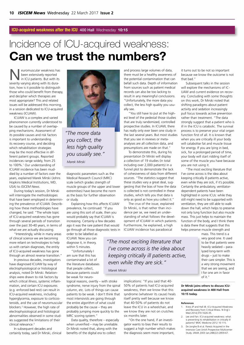
ICUAW is a complex and varied phenomenon currently understood to be caused by a number of overlap-ping mechanisms. Assessment of its possible causes and risk factors is important to understanding its recovery course, and deciding which rehabilitation strategies may be the most effective in dif-ferent patient groups. Reported incidences range widely, from 25 to 100%.1 Our understanding of ICUAW incidence has been mud-died by a number of factors over the years, explained Marek Mirski (Johns Hopkins Medical Institutions, MD, USA) to ISICEM News.
During today’s session, Dr Mirski will interrogate the methodologies that have been employed in determin-ing the prevalence of ICUAW. Describ-ing how these methodologies have changed, he said: “The whole topic of ICU-acquired weakness has gone through several periods of transition over the past 30-40 years, in terms of what we are actually discussing.
“Interestingly, while in many areas of clinical science we are becoming more reliant on technologies to help us with certain diagnoses, the entity of ICU-acquired weakness has gone through an almost reverse transition.”
In previous decades, investigators have diagnosed ICUAW by way of electrophysiological or histological analysis, noted Dr Mirski. Relation-ships were drawn to risk factors by which critical illness, systemic inflam-mation, and certain ICU exposures (e.g. enforced bed rest) can result in ICU-acquired weakness, including hyperglycemia, exposure to corticos-teroids, and the use of neuromuscular blockers.2 However, the high rate of electrophysiological and histological abnormalities observed in some stud-ies suggested the limitations of their clinical relevance.3
In subsequent decades and certainly today, said Dr Mirski, clinical
diagnostic parameters such as the Medical Research Council (MRC) scale (which grades strength of muscle groups of the upper and lower extremities) have become the norm as the basis for further observation or study.
Explaining how this affects ICUAW prevalence, he continued: “If you are using this sort of scale, then you would probably say that ICUAW is increasing. Certainly a few decades ago it was the rare patient that would go through all those diagnostic tests in order to be labelled as ICUAW. Now you can diagnose it, in theory, within 5 minutes.
“Unfortunately I am sure that this has contaminated a lot of the literature databases that people collect, because patients could be weak for neuro-logical reasons, overtly – with stroke syndrome, nerve injury from the spinal column, etc. Lots of things can cause patients to be weak. I don’t think that most intensivists are going through the entire algorithm of what could probably be the cause – they are probably jumping more quickly to the MRC scoring system.”
Data quality, therefore – especially when unverified – may be unreliable. Dr Mirski noted that, along with the benefits of the digital era to collect
and process large volumes of data, there must be a healthy awareness of the potential contamination that can befall such data. Depth of information from sources such as patient medical records can also be too lacking to result in any meaningful conclusions. “Unfortunately, the more data you collect, the less high quality you usu-ally see.
“You still have to put at the high-est level of the pedestal those studies that are truly randomized, controlled prospective studies. In ICUAW, there has really only ever been one study in the last several years. But most studies that you see in reviews or meta-analyses are all collection data, and presumptions are made on that.”
To demonstrate this, during his presentation Dr Mirski will display a collection of 19 studies (in total including over 2,000 patients) in a funnel plot to demonstrate the lack of cohesiveness of data from different sources. “The statistics suggest that these studies vary a great deal, sug-gesting that the bias of how the data is collected is not controlled in these studies, which tells you that data is only as good as how you collect it.”
The crux of the issue, explained Dr Mirski, is that rather than inci-dence per se, we need an under-standing of what follows the devel-opment of ICUAW in which patients. Furthermore, he explained, a high ICUAW incidence has paradoxical
implications: “If you said that 40-50% of patients had ICU-acquired weakness, then we know that this syndrome (whatever its cause) heals itself pretty well because we know that 40-50% of patients do not leave the ICU in a wheelchair, and we know they are not on crutches six months later.
“On the one hand, if an investi-gator wants to bias their results to suggest a high number which makes the diagnosis seem more important,
it turns out to be not so important because we know the outcome is not that bad.”
Subsequent talks in the session will explore the mechanisms of IC-UAW and current evidence on recov-ery. Concluding with some thoughts on this work, Dr Mirski noted that shifting paradigms about patient activity and sedation increasingly pull focus towards active prevention rather than treatment. “The data strongly suggest that a patient who is ill in the ICU is catabolic. The survival process is to preserve your vital organ function first of all. It is known that when you are critically ill your body will catabolise fat and muscle tissue for energy. If you are lying in bed, sick, for a prolonged period of time, your body will start ridding itself of some of the muscle you have because you are not using it.
“The most exciting literature that I’ve come across is the idea about keeping critically ill patients active, even while they are sick, if it’s possible. Certainly the ambulatory, ventilator-dependent patients have been highlighted in some ICUs: while they still might need to be supported with ventilation, they are still able to walk down the hall, which helps to preserve not only lung function but also muscle mass. This just helps to maintain the function of the body, and I think there is data there that suggests that this helps preserve muscle strength and
mass. This trend is a very good one. It used to be that patients were heavily sedated – para-lyzed long-term with drugs – just to make their care simpler. This is a very strong movement that we are seeing, and I for one am in favor of it.”
Dr Mirski joins others to discuss ICU-acquired weakness in 400 Hall from 10:15 today.
References
1. Kress JP and Hall JB. ICU-Acquired Weakness and Recovery from Critical Illness. N Engl J Med 2014;370:1626-35.
2. Lee and Fan: ICU-acquired weakness: what is preventing its rehabilitation in critically ill patients? BMC Medicine 2012 10:115.
3. De Jonghe B et al. Paresis Acquired in the Intensive Care UnitA Prospective Multicenter Study. JAMA 2003 Jan;288(22):2859-67.
ICU-acquired weakness after the ICU 400 Hall Wednesday 10:15
Incidence of ICU-acquired weakness:Can we trust the numbers?
“The most exciting literature that I’ve come across is the idea about keeping critically ill patients active, even while they are sick.”Marek Mirski
“The more data you collect, the less high quality you usually see.”Marek Mirski
Issue 2 Wednesday 22 March 2017 ISICEM News 11
Pre-symposium courses
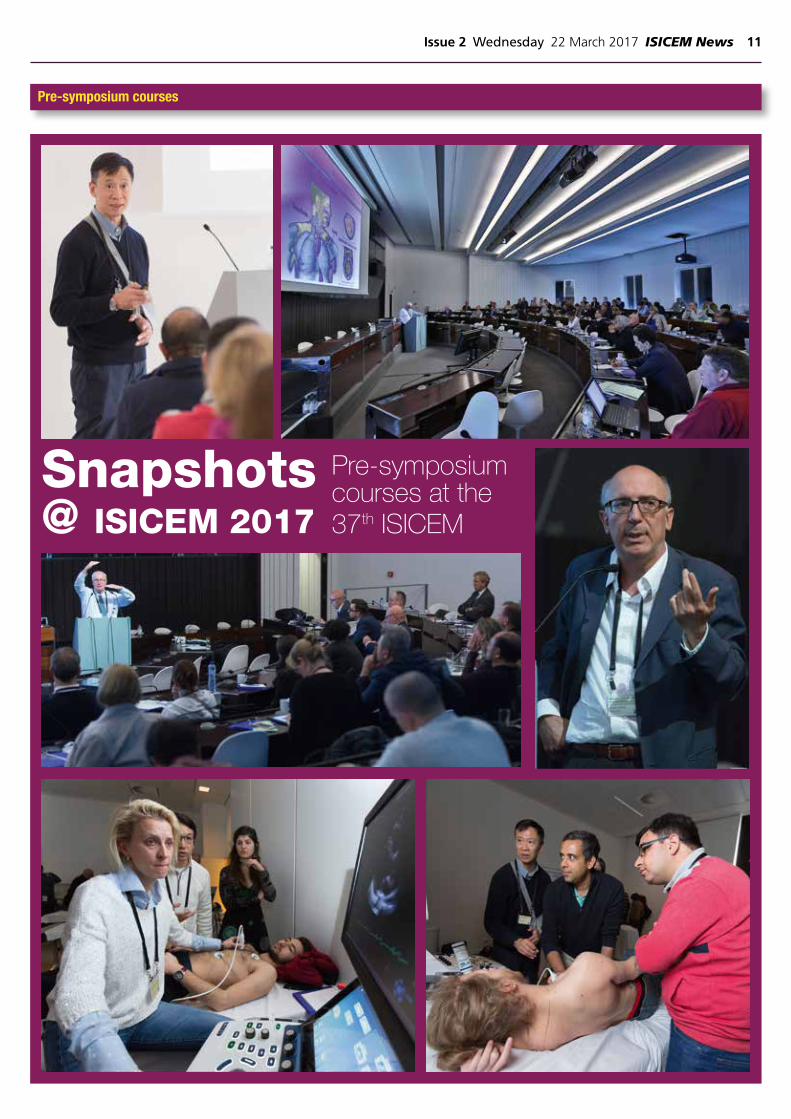
Snapshots @ ISICEM 2017
Pre-symposium courses at the 37th ISICEM

12 ISICEM News Wednesday 22 March 2017 Issue 2
A new study recently presented at the World Congress of An-esthesiologists (WCA) in Hong Kong, researchers concluded that continuous and noninvasive hemoglobin monitoring,
using Masimo SpHb®, may reduce excessive intraoperative red blood cell (RBC) transfusion.1
In a retrospective review of 371 patients who underwent in-traoperative RBC transfusions between 2012 and 2014 at Fuku-shima Medical University in Japan, Dr. Imaizumi and colleagues compared 94 patients who had noninvasive hemoglobin measure-ments to 277 patients who did not (the control group). The total transfusion volumes and transfusion volume per 1 g of blood loss were determined for each group.
Comparing the groups, researchers noted that “a significantly lower mean RBC transfusion volume per 1 g of blood loss was observed in the SpHb group compared with the [control] group (SpHb group, 0.9 ± 1.0 ml/g blood loss vs [control] group, 2.4 ± 5.9 ml/g blood loss, p less than 0.01).” They also observed that there was “no significant difference…in the average RBC transfu-sion volume (SpHb group, 815 ± 819 ml vs. [control] group, 785 ± 773 ml, p=0.75), or the preoperative hemoglobin concentration (SpHb group, 10.4 ± 1.9 g/dL vs. [control] group, 10.2 ± 2.4 g/dL, p=0.27) between the groups.” According to results from this abstract, the authors concluded that “SpHb measurements are as-sociated with reducing excessive intraoperative RBC transfusion.”
“This is the third study, published by different researchers on three continents (US2, Egypt3, and now Japan1) that has shown that in addi-tion to other clinical tools, SpHb may be used to help clinicians make informed transfusion decisions during different types of surgery*,” stated Dr. Steven Barker, Ph.D., M.D., Chief Science Officer, Masimo.
SpHb monitoring may provide additional insight to the directional trend of hemoglobin between invasive blood samplings – when the SpHb trend is stable and the clinician may otherwise think hemoglobin is de-creasing; when the SpHb trend is rising and the clinician may otherwise think hemoglobin is not rising fast enough; or when the SpHb trend is decreasing and the clinician may otherwise think hemoglobin is stable. SpHb with laboratory diagnostic test may thus help clinicians make more timely and informed decisions, and has been shown to help clini-cians provide more timely blood transfusions* and reduce blood transfu-sions in cases such as neurosurgery and orthopedic surgery.2,3
* Clinical decisions regarding red blood cell transfusions should be based on the clinician’s judge-ment considering, among other factors: patient condition, continuous SpHb monitoring, and laboratory diagnostic tests using blood samples.
References1. Imaizumi et al. Continuous and noninvasive hemoglobin monitoring may reduce excessive intraop-
erative RBC transfusion. Proceedings from the 16th World Congress of Anaesthesiologists, Hong Kong. Abstract #PR607.
2. Ehrenfeld JM et al. J Blood Disorders Transf. 2014. 5:9.3. Awada WN et al. J Clin Monit Comput. 2015 Feb 4.
New Study Finds Continuous, Noninvasive Hemoglobin Monitoring Using Masimo SpHb® May Reduce Intraoperative Red Blood Cell Transfusion

Issue 2 Wednesday 22 March 2017 ISICEM News 13
email: [email protected] or call +44 (0) 208 771 8046
We are a full-service medical communications and publishing company, working closely with local and international medical societies and associations, and industry, to develop conference publications, including newsletters and newspapers, as well as reports and medical summaries, medical writing and scientific publications
are the proud publishers of

14 ISICEM News Wednesday 22 March 2017 Issue 2
T he optimal management of nutrition in critical illness still straddles the boundaries of
consensus and controversy, with top-ics such as calorie and protein intake, re-feeding syndrome, gastric residual volume monitoring, parenteral/enter-al nutrition, measurement techniques and pharmaconutrients all represent-ing hot topics in the field.1
Tackling some of these issues, and emphasizing protein intake specifically, will be Jan Wernerman (Karolinska University Hospital, Huddinge, Stockholm, Sweden), who spoke to ISICEM News to share his perspectives ahead of the session.
By way of introduction, Profes-sor Wernerman first commented on the predictive equations used to gauge the appropriate calorie needs of an ICU patient. Com-monly based on weight, height, age, gender and severity of illness, the ac-curacy is known to vary considerably.2 With estimates usually falling between 20 and 35 kcal/kg/day, under- or over-feeding becomes more of an issue with the ongoing variability of need that the patient may exhibit.2
This is particularly troublesome for underweight or overweight patients staying for prolonged periods in the ICU: “Here a daily erroneously intake of > 300 kcal constitutes a real problem,” commented Professor
Wernerman.He added: “Overweight patients
are problematic. They cannot lose weight in the ICU safely, because they will lose muscle and fat on ap-proximate equal weight. However, they really need their muscle (which they have plenty of from the start) to get around. If there is an extensive
vmuscle loss, the rehabilitation will be really problematic.”
The challenge of personalized and accurate nutrition becomes even more complex when estimating protein needs, not least because the neutral tissue protein balance required in healthy individuals is quite different to that of an ICU patient.1 “In ICU patients both synthesis and degrada-tion of proteins is higher than normal, very unevenly distributed in between organs,” continued Professor Werner-man. “Muscle tissue is constantly lost
in the initial 3-4 weeks related to an enhanced degradation, while synthe-sis is broadly speaking on a ‘normal’ level. For the very longstayers, this loss of muscle proteins levels off.
“But the concept of an enhanced ‘need’ for protein intake rests on very weak evidence, most often nitrogen balance data or retrospective obser-
vational data. On a whole-body basis, our research group have shown that extra protein supply up to the level of 1.5-2.0 g/kg/24h gives a better protein balance that is attributable to a higher whole-body protein synthesis (when measure with labelled phenylalanine). However, at this timepoint we cannot say where this protein accretion takes place, and most importantly if it has an impact on outcomes.”
To measure protein need, Professor Wernerman noted that protein turnover assessment using isotopes gives an intriguing ‘snap shot’, achieved by incorporating stable isotopes of carbon, hydrogen or nitrogen into amino acid trac-ers, which can then be infused or ingested to trace the metabolism of the body.3 Whole-body protein synthesis and protein breakdown can then be investigated given that the labelled amino-acid compounds will be incorporated into the muscles at a rate that reflects fractional synthetic
protein rates.3
Alternatively, he noted that “Scanning techniques are attractive as being non-invasive. The resolution, however, is unfortunately not good enough for longitudinal studies. So far scanning techniques have only been applies to muscle tissue.”
Professor Wernerman will present the nuances of protein intake in the ICU in more detail during this afternoon’s session, but for now, he offered some ‘take-home’ messages for the ISICEM audience. “Do not get over-enthusiastic with the high-pro-tein messages. Stay in line with the ESPEN4 guidelines for protein intake. In addition, be conservative with en-ergy intake, in particular during the initial phase of critical illness.
“For the long-stayers in ICU, nu-trition needs to be individualized, and the most important thing is to avoid doing harm.”
References1. Preiser J-C, et al. Metabolic and nutritional
support of critically ill patients: consensus and controversies. Preiser et al. Critical Care, 2015;19:35.
2. Ridley E, Gantner D and Pellegrino V. Nutri-tion therapy in critically ill patients- a review of current evidence for clinicians. Clinical Nutrition ‘xxx’, 2015;1-7.
3. Weijs PJM, et al. Proteins and amino acids are fundamental to optimal nutrition sup-port in critically ill patients. Critical Care, 2014;18:591.
4. The European Society for Clinical Nutrition and Metabolism. http://www.espen.org/education/espen-guidelines
Nutritional support 400 Hall Wednesday 13:45
‘Little evidence, much controversy’ for protein intake in critical illness
“Do not get over-enthusiastic with the high-protein messages.”Jan Wernerman
“In ICU patients both synthesis and degradation of proteins is higher than normal, very unevenly distributed in between organs.”Jan Wernerman
Issue 2 Wednesday 22 March 2017 ISICEM News 15
Y esterday at ISICEM saw an examination of ICU admissions processes, with focus on organizational processes as well as techno-
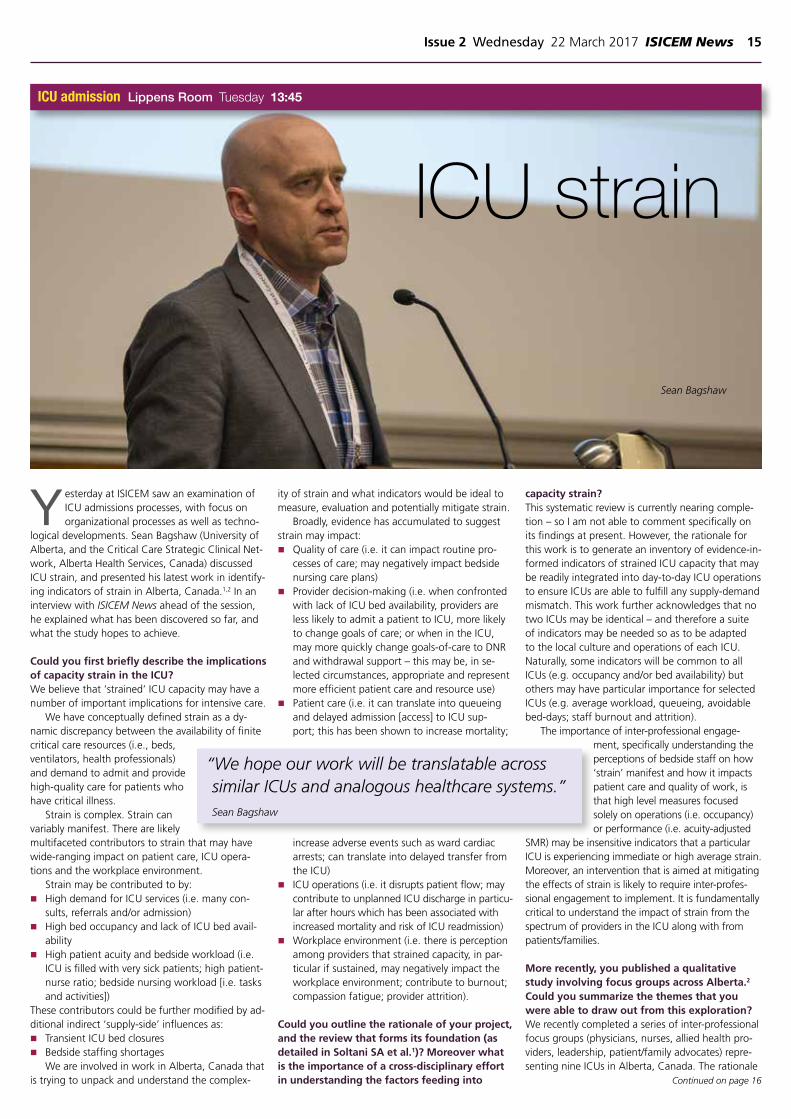
logical developments. Sean Bagshaw (University of Alberta, and the Critical Care Strategic Clinical Net-work, Alberta Health Services, Canada) discussed ICU strain, and presented his latest work in identify-ing indicators of strain in Alberta, Canada.1,2 In an interview with ISICEM News ahead of the session, he explained what has been discovered so far, and what the study hopes to achieve.
Could you first briefly describe the implications of capacity strain in the ICU? We believe that ‘strained’ ICU capacity may have a number of important implications for intensive care.
We have conceptually defined strain as a dy-namic discrepancy between the availability of finite critical care resources (i.e., beds, ventilators, health professionals) and demand to admit and provide high-quality care for patients who have critical illness.
Strain is complex. Strain can variably manifest. There are likely multifaceted contributors to strain that may have wide-ranging impact on patient care, ICU opera-tions and the workplace environment.
Strain may be contributed to by:n High demand for ICU services (i.e. many con-
sults, referrals and/or admission)n High bed occupancy and lack of ICU bed avail-
abilityn High patient acuity and bedside workload (i.e.
ICU is filled with very sick patients; high patient-nurse ratio; bedside nursing workload [i.e. tasks and activities])
These contributors could be further modified by ad-ditional indirect ‘supply-side’ influences as:n Transient ICU bed closuresn Bedside staffing shortages
We are involved in work in Alberta, Canada that is trying to unpack and understand the complex-
ity of strain and what indicators would be ideal to measure, evaluation and potentially mitigate strain.
Broadly, evidence has accumulated to suggest strain may impact:n Quality of care (i.e. it can impact routine pro-
cesses of care; may negatively impact bedside nursing care plans)
n Provider decision-making (i.e. when confronted with lack of ICU bed availability, providers are less likely to admit a patient to ICU, more likely to change goals of care; or when in the ICU, may more quickly change goals-of-care to DNR and withdrawal support – this may be, in se-lected circumstances, appropriate and represent more efficient patient care and resource use)
n Patient care (i.e. it can translate into queueing and delayed admission [access] to ICU sup-port; this has been shown to increase mortality;
increase adverse events such as ward cardiac arrests; can translate into delayed transfer from the ICU)
n ICU operations (i.e. it disrupts patient flow; may contribute to unplanned ICU discharge in particu-lar after hours which has been associated with increased mortality and risk of ICU readmission)
n Workplace environment (i.e. there is perception among providers that strained capacity, in par-ticular if sustained, may negatively impact the workplace environment; contribute to burnout; compassion fatigue; provider attrition).
Could you outline the rationale of your project, and the review that forms its foundation (as detailed in Soltani SA et al.1)? Moreover what is the importance of a cross-disciplinary effort in understanding the factors feeding into
capacity strain? This systematic review is currently nearing comple-tion – so I am not able to comment specifically on its findings at present. However, the rationale for this work is to generate an inventory of evidence-in-formed indicators of strained ICU capacity that may be readily integrated into day-to-day ICU operations to ensure ICUs are able to fulfill any supply-demand mismatch. This work further acknowledges that no two ICUs may be identical – and therefore a suite of indicators may be needed so as to be adapted to the local culture and operations of each ICU. Naturally, some indicators will be common to all ICUs (e.g. occupancy and/or bed availability) but others may have particular importance for selected ICUs (e.g. average workload, queueing, avoidable bed-days; staff burnout and attrition).
The importance of inter-professional engage-ment, specifically understanding the perceptions of bedside staff on how ‘strain’ manifest and how it impacts patient care and quality of work, is that high level measures focused solely on operations (i.e. occupancy) or performance (i.e. acuity-adjusted
SMR) may be insensitive indicators that a particular ICU is experiencing immediate or high average strain. Moreover, an intervention that is aimed at mitigating the effects of strain is likely to require inter-profes-sional engagement to implement. It is fundamentally critical to understand the impact of strain from the spectrum of providers in the ICU along with from patients/families.
More recently, you published a qualitative study involving focus groups across Alberta.2 Could you summarize the themes that you were able to draw out from this exploration?We recently completed a series of inter-professional focus groups (physicians, nurses, allied health pro-viders, leadership, patient/family advocates) repre-senting nine ICUs in Alberta, Canada. The rationale
“We hope our work will be translatable across similar ICUs and analogous healthcare systems.” Sean Bagshaw
Continued on page 16
ICU strain
ICU admission Lippens Room Tuesday 13:45
Sean Bagshaw

16 ISICEM News Wednesday 22 March 2017 Issue 2
for this work is to understand their perception of the contributors to strain, the impacts of strain and to gain insight for potential strategies to mitigate and/or manage strain.
A number of themes and subthemes of con-tributors to strain were identified including:n Patient/family-related: older
and more complex/high acuity patients; inadequate preparation for end-of-life; incomplete understanding of the implications of ICU care; mismatch in patient/family and provider expec-tations
n Provider-related: inexperienced bedside staff due to attrition; inadequate bedside staffing; frequent handovers (contributing to changes in care plan)
n Resource-related: reduced after hours services; lack of inter-professional providers; delays in bed turnovers; inadequate ICU beds and , stepdown beds
n Health-system related: inconsistent triage
decision-making; preferential bed priorities given to other services; under-resourced services con-tributing to bed block in the ICU; limited societal understanding for the potential role of ICU at the end-of-life
The aim of this work is to understand bedside providers’ perspectives, and to gain insight into how
best to measure any particular ICU’s strain, but also design and implement customized strate-gies to avoid, mitigate and effectively manage strain while preserving high-quality ICU care delivery, patient outcomes and family satisfaction with care.
Is there any evidence for variation in ICU ca-pacity strain across different hospitals? And is there perhaps a need to include other depart-ments – and indeed other stakeholders – in the discussion?It is important to recognize that strain is dynamic and can vary hour to hour and day to day. It can also be sustained over time (weeks to months). It can manifest very quickly (a moderate sized ICU
that is at near 100% occupancy receives a large number of high acuity admissions during a single day – this not only fills the ICU and makes for no bed available – but generates significant workload for providers). It is also important to acknowledge that strain is not unique to ICU. Emergency depart-ments, hospital wards and entire hospitals also ex-perience strain. These may have integrated indirect effects on strain in the ICU.
Finally, is there any precedent for what you are working towards in Alberta? And how trans-ferrable are your findings likely to be?My belief is that many jurisdictions are routinely confronted with the challenge of having experi-enced transient or sustained strain on ICU capacity. We hope our work will be translatable across similar ICUs and analogous healthcare systems.
References1. Soltani SA et al. Quality and performance measures of strain
on intensive care capacity: a protocol for a systematic review. Systematic Reviews. 2015;4:158.
2. Bagshaw SM et al. Healthcare Provider Perceptions of Causes and Consequences of ICU Capacity Strain in a Large Publicly Funded Integrated Health Region: A Qualitative Study. Crit Care Med. 2017;45(4):e347-56.
T he intriguing world of target-ed temperature management (TTM) after cardiac arrest will
be explored tomorrow in a presenta-tion by Hans Friberg (University of Skåne, Lund, Sweden).
Speaking on the progress of improving outcomes for unconscious survivors of out-of-hospital cardiac arrest, Professor Friberg will expand on the use of therapeutic hypother-mia to improve outcomes that are usually very poor. “The survival rate for sudden and unexpected cardiac arrest is not very good. If you even make it to the ICU, of those patients that we treat in the ICU, approxi-mately half will eventually die in the hospital or soon thereafter,” he said.
It is well understood that ben-efits of cold water to outcomes are startling. Those who suffer cardiac arrest after drowning in cold water are known to recover better than those who are warm when they suf-fer cardiac arrest, stressed Professor
Friberg. “If that happens, your brain is protected from ischemic injury or lack of oxygen,” he said. “We believe there is a benefit of lowering temperature. It’s been rather clearly shown that a lower temperature protects the brain.”
But outcomes are more mixed in other circumstances: “The evidence is not as clear if you cool down the body after drowning or cardiac arrest. If you are in cardiac arrest when you are warm, it may not be as protective.”
What’s clear is that much work is ongoing to understand the effects
of cooling for all sorts of patients. For instance, research is being conducted by colleagues at Lund into patients who are awake, have not suffered cardiac arrest, but have myocardial infarction. “You cool them down in order to salvage the myocardium,” he said.
Recommendations on how cool-ing might benefit patients in cardiac
arrest have changed dramatically over the last 15 years, said Professor Friberg. That’s because a 2002 paper showed benefits to the recovery of cardiac arrest patients when cooled to 33°C, compared with patients where there was no temperature control. It revived an ancient tradition of cooling for recovery and prompted international guidelines.
Now targeted tempera-ture management for cardiac arrest patients is becoming more
Hypothermia for the Brain Protection 400 Hall Thursday 15:10
Temperature reduction after cardiac arrest: Current status
“Most countries in the Western world do targeted temperature management now, but we do not agree on what temperature is the best.” Hans Friberg
“Strain is complex. Strain can variably manifest.” Sean Bagshaw
ICU admission Lippens Room Tuesday 13:45
ICU strainContinued from page 15
Continued on page 18

Advert
18 ISICEM News Wednesday 22 March 2017 Issue 2
widely practiced, but Professor Friberg cautioned that there are still some unanswered questions. “It’s true most countries in the Western world do TTM now, but we do not agree on what temperature is the best,” he said. “But we agree that we need to control the temperature, and do so during at least the initial phase of ICU care.”
Professor Friberg has managed to develop these original findings by looking in more detail at the benefits of cooling to 33°C. Indeed, a 2013 study, the Target Temperature Management After Cardiac Arrest (TTM) trial,1 showed that patients fared as well at 36°C compared with those cooled to 33°C, said Professor Friberg. The result of the TTM study have been profound, he added, for one because the international recom-
mendations were revised upwards since the study was published. “The recommendations today are that you can treat patients at any temperature between 33°C and 36°C,” he said.
While many physicians may choose 33°C, Professor Friberg noted that it is easier said than done: “It’s actually quite difficult to cool down to 33°C, and there are more poten-tial side effects. At 36°C, it’s not as invasive, it’s less troublesome, easier (in a way), and patients do as well.”
In addition, most drugs have not been tested, nor are metabolized in the same way at lower temperatures.
Professor Friberg also spoke about his research interests in track-ing how brain function changes at different temperatures. “Survival is one thing, but neurological function is much more important. These days we look much more into not just survival and crude neurological func-
tion, we also look at cognitive func-tion, processing speed and quality of life,” he said.
Fortunately, he has managed to establish that there was no differ-ence in crude neurological function, as well as other parameters including quality of life, memory or processing speeds between patients cooled to both 33°C and 36°C. But this should be a key factor in studies he added.
Talking about the importance of adhering to international standards, Professor Friberg commented: “Inter-national recommendations should be followed, and personally I recom-mend 36°C because it makes much so much more sense. It’s less invasive and it makes a lot more sense to keep the patient at this temperature until we know more.”
Professor Friberg also spoke of his interest in knowing whether temperatures greater than 36°C, but below that which signals the onset of fever – around 37.8°C – confer similar protective benefits. That’s why he will talk about an ambitious new trial, TTM-2.2
As he noted, TTM-2 is a large study, and could help establish the optimum temperature conditions under which patients should be kept
to maximize outcomes. It will look, in part, at temperatures at higher levels. As Professor Friberg put it: “Is it okay to allow for a slight temperature rise to 37.8°C? Or is very rapid cooling to 33°C of benefit? Those are the ques-tions we want to answer.”
Professor Friberg reiterated that for the meantime, the message is clear that cooling should be an es-sential component of caring for pa-tients who have had a cardiac arrest. “The message in Brussels is manage the patients according to protocols. Control the temperature, and do not allow for fever until we know more,” he concluded.
References
1. Nielsen N, et al. Targeted Temperature Man-agement at 33°C versus 36°C after Cardiac Arrest. N Engl J Med, 2013;369:2197-2206.
2. ClinicalTrials.gov. Hypothermia or Normother-mia-Targeted Temperature Management After Out-of-hospital Cardiac Arrest-trial (TTM-2).
“The recommendations today are that you can treat patients at any temperature between 33° and 36°C.” Hans Friberg
Hypothermia for the Brain Protection 400 Hall Thursday 15:10
Temperature reduction after cardiac arrest: Current statusContinued from page 16


Related Documents