THE JOURNAL OF BIOLOGICAL CHEMISTRY (0 1991 by The American Society for Biochemistry and Molecular Biology, Inc Vol. 266, No. 36, Issue of December 25. pp. 24398-24403, 1991 Printed in U. S. A. Differential Regulation of Antioxidant Enzymes in Response to Oxidants* (Received for publication, June 24, 1991) Susan Shull$$, Nicholas H. HeintzlI, Muthu Periasamy 11, Muniraj Manohar**, Yvonne M. W. JanssenlI, Joanne P. MarshlI, and Brooke T. Mossmann From the $Department of Biochemistry, TDepartment of Pathology, and )I Department of Physiology and Biophysics, Medical School, university of Vermont, Burlington, Vermont 05405 and the **Laboratory of Molecular Immunology, Harvard Medical School, Dana-Farber Cancer Institute, Boston, Massachusetts 021 15 We have demonstrated the selective induction of manganese superoxide dismutase (MnSOD) or catalase mRNA after exposure of tracheobronchial epithelial cells in vitro to different oxidant stresses. Addition of HzOz caused a dose-dependent increase in catalase mRNA in both exponentially growing and confluent cells. A 3-fold induction of catalase mRNA was seen at a nontoxic dose of 250 p~ H202. Increase in the steady- state mRNA levels of glutathione peroxidase (GPX) and MnSOD were less striking. Expression of catalase, MnSOD, and GPX mRNA was highest in confluent cells. In contrast, constitutive expression of copper and zinc SOD (CuZnSOD) mRNA was greatest in dividing cells and was unaffected by HzOz in both exponentially growing and confluent cells. MnSOD mRNAwas selectively induced in confluent epithelial cells exposed to the reactive oxygen species- generating system, xanthinetxanthine oxidase, while steady-state levels of GPX, catalase, and CuZnSOD mRNA remained unchanged. The %fold induction of MnSOD mRNA was dose-dependent, reaching a peak at 0.2 unitlml xanthine oxidase. MnSOD mRNA in- creases were seen as early as 2 h and reached maximal induction at 24 h. Immunoreactive MnSOD protein was produced in a corresponding dose- and time-de- pendent manner. Induction of MnSOD gene expression was prevented by addition of actinomycin D and cyclo- heximide. These data indicate that epithelial cells of the respiratory tract respond to different oxidant in- sults by selective induction of certain antioxidant en- zymes. Hence, gene expression of antioxidant enzymes does not appear to be coordinately regulated in these cell types. Reactive oxygen species (ROS)’ have been implicated in many lung diseases including those associated with exposure to asbestos, nitrogen dioxide, ozone, paraquat, hyperoxia, carbon tetrachloride, and the anticancer drugs bleomycin and *This work was funded by Grant HL-39469 from the National Heart, Lung and Blood Institute. The costs of publication of this article were defrayed in part by the payment of page charges. This article must therefore be hereby marked “advertisement” in accord- ance with 18 U.S.C. Section 1734 solely to indicate this fact. 8 To whom correspondence should be addressed: Dept. of Biochem- istry, Medical School, University of Vermont, Burlington, VT05405. The abbreviations used are: ROS, reactive oxygen species; SOD, superoxide dismutase; GPX, glutathione peroxidase; AOE, antioxi- dant enzymes; 02, superoxide; ‘OH, hydroxyl radical, HTE, hamster tracheal epithelial cells; CMF-PBS, calcium and magnesium free phosphate-buffered saline; IL-1, interleukin-1; TNF, tumor necrosis factor. adriamycin (for review see Ref. 1). Although sources of ROS and antioxidant enzymes (AOE) have been well-documented in the lung, the individual cell types involved in oxidant injury and defense are poorly understood (2-5). Phagocytic cells have been implicated in the generation of ROS during inflam- mation (reviewed in Ref. 6), but the functional characteristics of the cells of the lung that contribute to lung defense from oxidants are enigmatic. Conceivably, oxidant damage results when the AOE defense mechanisms of the lung are over- whelmed. Eukaryotes have evolved several different AOE to detoxify ROS. Copper, zinc superoxide dismutase (CuZnSOD) is lo- cated primarily in the cytoplasm, whereas manganese SOD (MnSOD), a structurally distinct protein encoded by a differ- ent nuclear gene (7), is located primarily in the mitochondria. Both enzymes catalyze the reaction: 0; + 0, + 2H+ = 0, + H202. H202 is converted to HaO in the peroxisomes by the AOE, catalase, and in the cytoplasm by glutathione peroxidase (GPX). The distribution and regulation of AOE in cellsof the lung are unknown. Tracheal epithelial cells line the airways of the upper res- piratory tract and are the first cells toencounter foreign material impinging uponthe walls of the airways. Their regulation is important in understanding the defense mecha- nisms that occur in response to inhaled oxidants. In this study we examined the in vitro effect of two oxidant stresses: H202 and a xanthinelxanthine oxidase generating system producing a spectrum of ROS (O;, ‘OH, H202) on the levels of steady state mRNA of MnSOD, CuZnSOD, GPX, and catalase in hamster tracheal epithelial cells (HTE; 8). The dose- and time-dependent regulation of these enzymes in response to oxidants was explored initially in both exponentially growing and confluent HTE cells. Our data indicate that the steady state message levels of these enzymes vary independently with the cell cycle. Since more dramatic increases in gene expression were seen with MnSOD in the xanthine/xanthine oxidase generating system, we used actinomycin D and cyclo- heximide, respectively, to explore the dependence of mRNA accumulation on transcription and protein synthesis. Western blot analysis was used to examine whether transcriptional changesinMnSOD were manifested at the protein level. Differential and unique regulation of the AOE message levels occurred in HTE cells after exposure todifferentoxidant stresses. Changes in CuZnSOD mRNA levels were not appar- ent with either oxidant. METHODS AND MATERIALS Cell Culture Methods-HTE cells, isolated and characterized pre- viously (8) as a diploid,nontumorigenic cell line, were grown in Ham’s F12medium (GIBCO-Bethesda Research Laboratories) containing 24398

Welcome message from author
This document is posted to help you gain knowledge. Please leave a comment to let me know what you think about it! Share it to your friends and learn new things together.
Transcript
THE JOURNAL OF BIOLOGICAL CHEMISTRY (0 1991 by The American Society for Biochemistry and Molecular Biology, Inc
Vol. 266, No. 36, Issue of December 25. pp. 24398-24403, 1991 Printed in U. S. A.
Differential Regulation of Antioxidant Enzymes in Response to Oxidants*
(Received for publication, June 24, 1991)
Susan Shull$$, Nicholas H. HeintzlI, Muthu Periasamy 11, Muniraj Manohar**, Yvonne M. W. JanssenlI, Joanne P. MarshlI, and Brooke T. Mossmann From the $Department of Biochemistry, TDepartment of Pathology, and )I Department of Physiology and Biophysics, Medical School, university of Vermont, Burlington, Vermont 05405 and the **Laboratory of Molecular Immunology, Harvard Medical School, Dana-Farber Cancer Institute, Boston, Massachusetts 021 15
We have demonstrated the selective induction of manganese superoxide dismutase (MnSOD) or catalase mRNA after exposure of tracheobronchial epithelial cells in vitro to different oxidant stresses. Addition of HzOz caused a dose-dependent increase in catalase mRNA in both exponentially growing and confluent cells. A 3-fold induction of catalase mRNA was seen at a nontoxic dose of 250 p~ H202. Increase in the steady- state mRNA levels of glutathione peroxidase (GPX) and MnSOD were less striking. Expression of catalase, MnSOD, and GPX mRNA was highest in confluent cells. In contrast, constitutive expression of copper and zinc SOD (CuZnSOD) mRNA was greatest in dividing cells and was unaffected by HzOz in both exponentially growing and confluent cells.
MnSOD mRNA was selectively induced in confluent epithelial cells exposed to the reactive oxygen species- generating system, xanthinetxanthine oxidase, while steady-state levels of GPX, catalase, and CuZnSOD mRNA remained unchanged. The %fold induction of MnSOD mRNA was dose-dependent, reaching a peak a t 0.2 unitlml xanthine oxidase. MnSOD mRNA in- creases were seen as early as 2 h and reached maximal induction at 24 h. Immunoreactive MnSOD protein was produced in a corresponding dose- and time-de- pendent manner. Induction of MnSOD gene expression was prevented by addition of actinomycin D and cyclo- heximide. These data indicate that epithelial cells of the respiratory tract respond to different oxidant in- sults by selective induction of certain antioxidant en- zymes. Hence, gene expression of antioxidant enzymes does not appear to be coordinately regulated in these cell types.
Reactive oxygen species (ROS)’ have been implicated in many lung diseases including those associated with exposure to asbestos, nitrogen dioxide, ozone, paraquat, hyperoxia, carbon tetrachloride, and the anticancer drugs bleomycin and
*This work was funded by Grant HL-39469 from the National Heart, Lung and Blood Institute. The costs of publication of this article were defrayed in part by the payment of page charges. This article must therefore be hereby marked “advertisement” in accord- ance with 18 U.S.C. Section 1734 solely to indicate this fact.
8 To whom correspondence should be addressed: Dept. of Biochem- istry, Medical School, University of Vermont, Burlington, VT 05405.
The abbreviations used are: ROS, reactive oxygen species; SOD, superoxide dismutase; GPX, glutathione peroxidase; AOE, antioxi- dant enzymes; 02, superoxide; ‘OH, hydroxyl radical, HTE, hamster tracheal epithelial cells; CMF-PBS, calcium and magnesium free phosphate-buffered saline; IL-1, interleukin-1; TNF, tumor necrosis factor.
adriamycin (for review see Ref. 1). Although sources of ROS and antioxidant enzymes (AOE) have been well-documented in the lung, the individual cell types involved in oxidant injury and defense are poorly understood (2-5). Phagocytic cells have been implicated in the generation of ROS during inflam- mation (reviewed in Ref. 6), but the functional characteristics of the cells of the lung that contribute to lung defense from oxidants are enigmatic. Conceivably, oxidant damage results when the AOE defense mechanisms of the lung are over- whelmed.
Eukaryotes have evolved several different AOE to detoxify ROS. Copper, zinc superoxide dismutase (CuZnSOD) is lo- cated primarily in the cytoplasm, whereas manganese SOD (MnSOD), a structurally distinct protein encoded by a differ- ent nuclear gene (7 ) , is located primarily in the mitochondria. Both enzymes catalyze the reaction: 0; + 0, + 2H+ = 0, + H202. H202 is converted to HaO in the peroxisomes by the AOE, catalase, and in the cytoplasm by glutathione peroxidase (GPX). The distribution and regulation of AOE in cells of the lung are unknown.
Tracheal epithelial cells line the airways of the upper res- piratory tract and are the first cells to encounter foreign material impinging upon the walls of the airways. Their regulation is important in understanding the defense mecha- nisms that occur in response to inhaled oxidants. In this study we examined the in vitro effect of two oxidant stresses: H202 and a xanthinelxanthine oxidase generating system producing a spectrum of ROS (O;, ‘OH, H202) on the levels of steady state mRNA of MnSOD, CuZnSOD, GPX, and catalase in hamster tracheal epithelial cells (HTE; 8). The dose- and time-dependent regulation of these enzymes in response to oxidants was explored initially in both exponentially growing and confluent HTE cells. Our data indicate that the steady state message levels of these enzymes vary independently with the cell cycle. Since more dramatic increases in gene expression were seen with MnSOD in the xanthine/xanthine oxidase generating system, we used actinomycin D and cyclo- heximide, respectively, to explore the dependence of mRNA accumulation on transcription and protein synthesis. Western blot analysis was used to examine whether transcriptional changes in MnSOD were manifested at the protein level. Differential and unique regulation of the AOE message levels occurred in HTE cells after exposure to different oxidant stresses. Changes in CuZnSOD mRNA levels were not appar- ent with either oxidant.
METHODS AND MATERIALS
Cell Culture Methods-HTE cells, isolated and characterized pre- viously (8) as a diploid, nontumorigenic cell line, were grown in Ham’s F12 medium (GIBCO-Bethesda Research Laboratories) containing
24398
Regulation of Antioxidant Enzymes by Oxidants 24399
10% fetal bovine serum (Sigma, serum endotoxin level <0.125 ng/ ml), 10 units/ml penicillin, and 10 pg/ml streptomycin.
Oxidant Stresses-Two different sources of oxidants were used at nontoxic concentrations as determined previously in HTE cells (9). H202 (Sigma) was diluted in phosphate-buffered saline and the con- centration determined by absorbance at 240 nm (absorbance = 1.31 for 30 mM H202). H202 was added directly to fresh serum-containing medium of designated culture dishes.
ROS were generated continuously by the enzyme-substrate mixture xanthine (Sigma, grade V) and xanthine oxidase (Calbiochem). Xan- thine oxidase, under aerobic conditions, catalyzes the oxidation of xanthine to uric acid with reduction of 0 2 to 0; (10). In a neutral aqueous environment, 0; dismutates rapidly to Hz02 (10). Further- more, the iron catalyzed Haber-Weiss reaction or 0;-driven Fenton reaction will produce ' OH from 0; and H202 under these conditions (11).
Prior to addition of xanthine/xanthine oxidase, confluent cultures were given fresh serum and 50 p~ xanthine containing medium. Xanthine oxidase was added at the indicated concentrations (0.005- 0.4 unit/ml) and times (2-48 h). Controls consisted of cells grown in serum and 50 p~ xanthine containing medium.
Preliminary experiments showed that the induction of MnSOD mRNA using the xanthine/xanthine oxidase system fluctuated de- pending on the sources of the enzyme and fetal bovine serum. Five lots of serum and three lots of xanthine oxidase from different suppliers were assayed for comparative induction of MnSOD mRNA (data not shown). Since production of superoxide by various lots of xanthine oxidase varied, one lot of xanthine oxidase and serum were used for all experiments.
RNA Isolation and Northern Blots-Total RNA was isolated from cells using the procedure of Chomczynski and Sacchi (12). RNA was resolved by electrophoresis in a 1% agarose, 2.25 M formaldehyde gel in a running buffer containing 20 mM MOPS, pH 7.4, and 1 mM EDTA. RNA was transferred to nitrocellulose (Schleicher and Schuell, Keene, NH) according to Maniatis et al. (13). Alternatively, total RNA was applied directly to nitrocellulose using a slot blot apparatus (Schleicher and Schuell; 13). Purified cDNAs were labeled with [ ( u ~ ~ P ] ~ A T P (3,000 Ci/mmol, DuPont-New England Nuclear) by random hexamer priming using a Prime-a-Gene kit (Promega, Madison, WI). Blots were prehybridized 2-12 h in 50% formamide, 0.75 M sodium chloride, 0.075 M sodium citrate, pH 7.0, 5 X Den- hardt's solution, 50 pg/ml salmon testes DNA, and 0.1% SDS at 42 "C. Blots were hybridized with cDNAs, labeled to a specific activity of >lo9 cpm/pg, in hybridization fluid at 42 "C overnight. Hybridized blots were washed in 0.3 M sodium chloride, 0.03 M sodium citrate, 0.1% SDS at 42 "C and monitored with a Geiger counter. More stringent washing conditions were used as needed. Autoradiographs were made by exposing blots to x-ray film (Kodak XAR-5), at -70 "C with an intensifying screen. Radioactivity on blots was quantified directly by detection in a Betascope Blot Analyzer, Model 603, version 2.0 from Betagen, Waltham, MA or by densitometric analysis of the autoradiographs using a Microscan 1000, Technology Resources Inc., Nashville, TN. Samples analyzed using the Betascope are expressed as counts accumulated. These were collected for varying amounts of time, typically 6-14 h, depending upon the length and specific activity of the probe, which is similar to the exposure time frame for the autoradiographs.
Dr. Guy Mullenhach (Chiron Corporation, Emeryville, CA) pro- vided the murine GPX cDNA containing plasmids, pmGPx-SA, and pmGPx-5A (14). From Dr. Ye-Shih Ho (Duke University, Medical Center, Durham, NC) we obtained a 0.65-kb rat CuZnSOD cDNA containing plasmid, pUC13-RSC (15), and a 1.4-kb rat MnSOD cDNA containing plasmid, pSP65-RMS (16). The murine catalase probe, pmCAT-34 (17) was given to us by Dr. Jacquelin Shaffer (State of New York Department of Health, Albany, NY).
Inhibition of Protein and RNA Synthesis-HTE cells were exposed to nontoxic concentrations of inhibitors for 24 h. Fifty p~ cyclohex- imide (Sigma) was used to inhibit protein synthesis and 4 p~ acti- nomycin D (Sigma) was used to inhibit RNA synthesis (18). ROS exposed cells received 0.2 unit/ml xanthine oxidase.
Western Blot Analysis-Confluent epithelial cells were exposed to 50 p M xanthine (control cultures) and 0.05 or 0.2 unit/ml xanthine oxidase in serum containing medium for 24 or 48 h. After aspiration of medium, cell layers were washed 2 X with calcium and magnesium free phosphate buffered saline (GIBCO-BRL; CMF-PBS). Cells were scraped off the dishes with the aid of a rubber policeman in CMF- PBS, a t 4 "C, and washed 2 X more in CMF-PBS. Cells were frozen in equal volumes of deionized water and lyophilized. Prior to electro-
phoresis, samples were dissolved in a sample buffer (62.5 mM Tris- C1, pH 6.8, 2 % SDS, 10% glycerol, and 5% P-mercaptoethanol) and boiled 3 min before loading on the gel. Aliquots saved prior to lyophilization were used for protein determination (19) using bovine serum albumin, Fraction V (Sigma), as a standard. Samples including prestained molecular weight standards (GIBCO-BRL) were electro- phoresed in a 15% SDS-polyacrylamide gel using a Mini-protean I1 Cell apparatus (Bio-Rad) and transferred to nitrocellulose (Schleicher and Schuell) as described previously (20). The nitrocellulose blot was stained with anti-MnSOD antibody diluted in 0.05% Tween-20 (Sigma) in CMF-PBS. Secondary antibody, biotinylated goat anti- rabbit IgG, peroxidase-conjugated avidin, and 4-chloro-1-naphthol, were supplied in kit form (Vector Laboratories Inc., Burlingame, CA) and used according to the recommendation of the supplier. Bands were quantified by densitometric analysis.
Anti-human kidney MnSOD antibody (21) was generously pro- vided by Dr. Larry w. Oberley (University of Iowa, Iowa City, IA).
RESULTS
Effect of H20, on AOE mRNA Levels-The addition of H202 to HTE cells caused a dose-dependent increase in the levels of steady state message for catalase (Fig. 1). This response was observed in exponentially growing cells as well as in cells which had reached confluence prior to exposure to HzOz. The greatest induction was observed with 100 and 250 pM H202 which resulted in increases of 2.5- and 3-fold, respectively, in the levels of catalase mRNA. The message levels of GPX, which also catalyzes the reduction of H202, were less respon- sive to H202. Cells growing exponentially did not demonstrate any change in the levels of GPX mRNA in response to H202, but at concentrations of 100 and 250 p~ H202 the levels of GPX mRNA increased by as much as 50% in confluent cells. Exposure of HTE cells to Hz02 (100 p ~ ) during log phase- growth had relatively little effect on the steady state levels of MnSOD mRNA (Fig. 1). However, 250 p~ HzOz caused a 40% increase in the level of MnSOD mRNA in confluent HTE cells. The CuZnSOD message level was not modified by 100- 250 p~ H202 whether cells were growing exponentially or had reached confluence prior to exposure.
Expression of AOE mRNA Levels during the Cell Cycle- Comparing unexposed HTE cultures at log phase and conflu- ence revealed unique and differential expression of AOE mRNA levels (Fig. 1). GPX mRNA levels were comparable in rapidly growing and confluent epithelial cells. By contrast, the levels of mRNA for catalase increased 4-5-fold when actively growing cells reached confluence. Similarly, MnSOD mRNA increased 3-fold as HTE cells advanced from log phase to confluence. In contrast, levels of CuZnSOD mRNA dis- played an opposite pattern. Upon reaching confluence, the steady-state level of message for this AOE decreased to ap- proximately half the level observed during exponential growth.
The cell cycle variation in steady state message levels of AOE did not appear to influence patterns of regulation when HTE cells were challenged by an oxidant stress such as exposure to H20z. For example, catalase mRNA was induced by Hz02 in both exponentially growing and confluent cells. Based on these results and the higher overall levels of AOE mRNA in confluent cells, the following experiments were conducted with confluent cell cultures, a system which mimics the contiguous cell layer of the tracheal epithelium in uiuo.
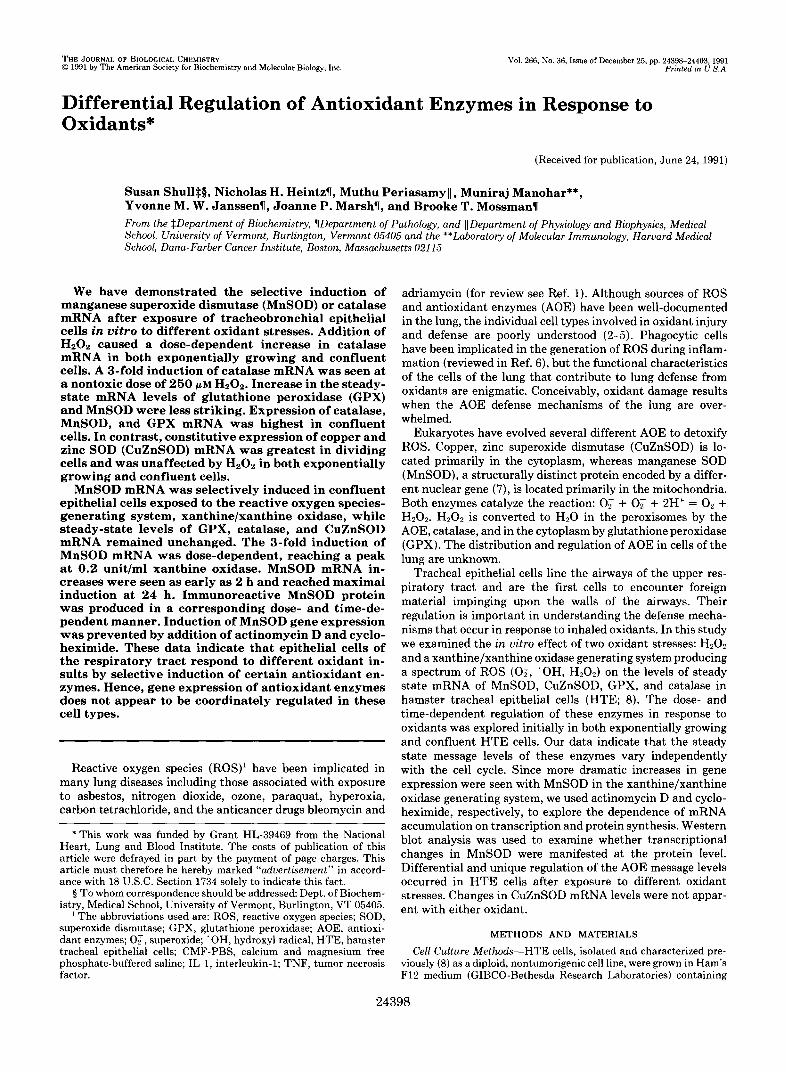
Effect of XanthinelXanthine Oxidase on AOE mRNA Leu- els-The production of ROS during the oxidation of xanthine by xanthine oxidase was exploited as a source for the contin- uous generation of these species, including 0; (10). The steady state levels of MnSOD mRNA demonstrated a dose- dependent increase after exposure of HTE cells to xanthine/ xanthine oxidase (Fig. 2). Peak induction of MnSOD mRNA was seen with addition of 0.2 unit/ml xanthine oxidase where

24400 Regulation of Antioxidant Enzymes by Oxidants
FIG. 1. Northern analysis of mRNA from HTE cells exposed to H202. Cells were exposed for 18 h to varying concentrations of H202 during log phase of growth or at confluence. Fifteen pg of total RNA per lane were electrophoresed, transferred to nitrocel- lulose, and hybridized as described under "Materials and Methods." A, autoradi- ographs of Northern blots hybridized se- quentially with cDNAs to catalase, GPX, MnSOD, and CuZnSOD. Duplicate sam- ples represent separate cell culture dishes. B, quantitative data from Beta- scope analysis or densitometric scanning (catalase). The bars represent the aver- age of the duplicate samples seen in A.
A. CATALASE
0 25 100 250 I 0 25 100 250 U~ ~~0
CuZnSOD I
- 285
18s
B.
6 CuZnSOD
0
$0.0
0 I x v " C 3 0 0
10
n 0 0
X
Y c 3 0 V
- 5 a
0 0 0.005 0.01 0.05 0.1 0.2 0.3 0.4 0 0.005 0.01 0.05 0.1 0.2 0.3 0.4
lJle~WrJ.OmC11d 1Q 10
n 0 0 0 7
5 5 a Y C 3 0 V
0 0 0.005 0.01 0.05 0.1 0.2 0.3 0.4 0 0.005 0.01 0.05 0.1 0.2 0.3 0.4
m m y o u r m m r r I I I B U ~ I d @ l Units/ml Xanthine Oxidase
Log Phase Confluent Hz02
in MnSOD mRNA in HTE cells ex- FIG. 2. Dose-dependent changes
posed to xanthinelxanthine oxidase. Confluent cells were exposed for 18 h to varying concentrations of xanthine oxi- dase in serum and 50 p~ xanthine-con- taining medium. Fifteen pg of total RNA per lane were electrophoresed, trans- ferred to nitrocellulose, and hybridized as described under "Materials and Meth- ods." Samples were analyzed in dupli- cate. The quantitative data represent sample averages from Northern blots analyzed using a Betascope. The auto- radiograph images beneath each graph are representative samples from North- ern blots.
levels increased 2-3-fold after 18 h in comparison to unex- posed cells.
Examination of the other AOE revealed that this induction was unique to MnSOD mRNA. The message levels of the other three AOE did not increase, but rather showed a slight downward trend. These data indicate that this oxidant stress is specific in its selective induction of MnSOD mRNA. Be- cause MnSOD has been implicated in inflammatory disease and is induced by endotoxin and cytokines such as TNF and IL-1 (18, 22, 23), induction of MnSOD mRNA in HTE cells was studied further. Time Course of MnSOD mRNA Induction-A time course
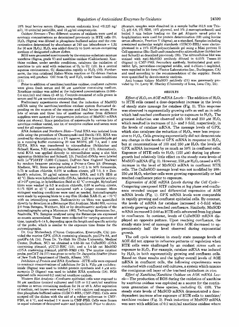
was performed using the most effective concentration of 0.2 unitslml xanthine oxidase. HTE cells demonstrated induction of MnSOD mRNA which peaked at 24 h and returned to control levels by 48 h (Fig. 3). With the exception of a transient spike in catalase mRNA levels a t 2 h, the other three AOE message levels remained constant after exposure to xanthinelxanthine oxidase.
Effects of Actinomycin D and Cycloheximide on MnSOD mRNA-In order to evaluate the contribution of transcription to the induction of MnSOD mRNA, we used the RNA syn- thesis inhibitor, actinomycin D. Addition of actinomycin D to control HTE cells (without oxidant exposure) resulted in a

Regulation of Antioxidant Enzymes by Oxidants 24401
of MnSOD mRNA in HTE cells. Con- FIG. 3. Time course of induction
fluent cells were exposed to 50 pM xan- thine plus 0.2 unit/ml xanthine oxidase for 2,4,8, 24, and 48 h. One pg of total RNA was applied directly to nitrocellu- lose using a slot blot apparatus as de- scribed under “Materials and Methods.” Quantitative data were collected from Betascope analysis of slot blots hybrid- ized with cDNAs to MnSOD, CuZnSOD, GPX, or catalase. Autoradiograph im- ages of representative slots are beneath each corresponding graph. Control = 50 p~ xanthine exposed cells; X / X O = xan- thine/xanthine oxidase exposed cells.
.U 1- 1 I
2s: 0 .
. CuZnSOD - 0 2 4 Control o 0
XlXO r) ,-
FIG. 4. Effect of actinomycin D and cycloheximide on the induction of MnSOD mRNA in HTE cells. Confluent cells were exposed to 50 p~ xanthine plus 0.2 unit/ml xanthine oxidase with the further addition in the indicated samples of 50 p~ cycloheximide or 4 p~ actinomycin D, for 24 h. One pg of total RNA was applied to nitrocellulose using a slot blot apparatus.
4.5-fold increase in MnSOD mRNA levels (Fig. 4). This was an unexpected finding and might suggest that the turnover of MnSOD mRNA requires mRNA synthesis. Exposure of cells to actinomycin D concurrently with xanthinelxanthine oxi- dase resulted in a 20% decrease in MnSOD mRNA levels when compared with HTE cell exposed to actinomycin D alone. This suggests that the induction of MnSOD mRNA observed with xanthinelxanthine oxidase requires active tran- scription of mRNA.
The involvement of protein synthesis was examined by addition of cycloheximide to HTE cells (Fig. 4). Addition of cycloheximide resulted in a 10-fold increase in message levels of MnSOD. However, addition of both cycloheximide and xanthine/xanthine oxidase to HTE cells did not cause a further increase in the level of MnSOD mRNA when com- pared with cells exposed to cycloheximide alone. Xanthine/ xanthine oxidase and cycloheximide exposed cells contained 20% of the MnSOD message compared with control HTE cells (cycloheximide alone). These observations suggest in- volvement of protein synthesis in the induction of MnSOD message.
HTE cells exposed to ROS generated by the xanthine/ xanthine oxidase system demonstrated an induction of MnSOD immunoreactive protein (Fig. 5). Amounts of MnSOD protein increased in a dose- and time-dependent manner. The 2-%fold increase in immunoreactive MnSOD
6 24 is I CATALASE
0 , I 0 2 4 8 24 48
A. 1 2 3 4 5 8 : 7 8 910 1112
29 - ””“ ”“.-
16.4 -
B. 12 n
3 8 .- Y
C
.- Y 9 - 0 4 0 U
0 0.05 0.2 0 0.05 0.2
Units/rnl Xanthine Oxidase
FIG. 5. Western blot analysis of MnSOD in epithelial cells exposed to xanthine/xanthine oxidase. HTE cells grown to con- fluence were exposed in 50 p~ xanthine containing medium to either 0.05 or 0.2 unit/ml xanthine oxidase for 24 or 48 h. Twenty pg of total protein per lane were electrophoresed in a 15% SDS-polyacryl- amide gel. Samples were electrophoretically transferred to nitrocel- lulose, and the blot was stained with anti-MnSOD antibody, as described under “Materials and Methods.” A, Western blot stained with anti-MnSOD antibody. Lanes, lanes 1, 2, and 7, 8 are control samples; lanes 3,4 and 9,lO are cells exposed to 0.05 unit/ml xanthine oxidase, and lanes 5 ,6 and 11,12 are cell samples exposed to 0.2 unit/ ml xanthine oxidase. Lanes 1-6 are cells harvested after 24 h and lanes 7-12 are cells collected after 48 h. Molecular weight standards are indicated 29, M, 29,000; 18.4, M, 18,400. B, quantitation of the Western blot using densitometric scanning analysis. Each bar repre- sents the sample average of separate cell culture samples. W, 24-h exposure; Ed, 48-h exposure.
correlated well with the 2-%fold increase in MnSOD mRNA (Figs. 2 and 3). This implies that induction of MnSOD mRNA results in a corresponding increase in MnSOD protein.
DISCUSSION Epithelial cells of the respiratory tract function in an oxy-
gen-rich environment and must deal continuously with air-

24402 Regulation of Antioxidant Enzymes by Oxidants
borne foreign material. These cells must maintain a balance between the normal encounter of oxidants (e.g. through re- lease by phagocytic cells) and antioxidant defense mecha- nisms (9). Removal of toxic oxygen metabolites is the putative function of AOE such as CuZnSOD, MnSOD, GPX, and catalase. The regulation of these enzymes is critical to our understanding of inflammation and oxidant-associated lung disease.
In our studies, HTE cells responded to different oxidants by differential regulation of AOE. In the presence of H2Oz, these cells showed a 3-fold increase in catalase mRNA (Fig. l), in both exponentially growing and confluent cells.
We were also interested in the effects of a spectrum of ROS on AOE regulation, using a xanthine/xanthine oxidase gen- erating system. Fridovich (10) first characterized the univa- lent reduction of O2 by xanthine oxidase, demonstrating si- multaneous one- and two-electron reactions under physiolog- ical conditions. Approximately 70% of the O2 consumed was observed to be reduced to Hz02 and 30% of the O2 reduced univalently to 0; (10). The xanthine/xanthine oxidase system has been re-examined more recently by Kuppusamy and Zweier (24). Using electron paramagnetic resonance spectros- copy, these authors demonstrated the generation of 'OH through the non-iron mediated univalent reduction of H2O2. Thus, the xanthinelxanthine oxidase mixture has been em- ployed in studies here for the continuous generation of short lived oxygen radicals, 0; and 'OH, along with the oxygen metabolite, H202.
The response to oxidants by HTE cells is discriminating since the challenge presented by the xanthine/xanthine oxi- dase generating system results in a completely different pat- tern of AOE mRNA levels than that of H2O2. After exposure to xanthine/xanthine oxidase, MnSOD mRNA is selectively increased in a dose- and time-dependent manner (Figs. 2 and 3). In contrast, the other three AOE examined remained unchanged or decreased slightly when their steady state mRNA levels were measured.
Numerous mechanisms could account for these differences. The involvement of cytokines in the regulation of MnSOD has been documented in recent years. For example, human melanoma A375 cells, skin fibroblasts, and peripheral blood monocytes increase their synthesis of MnSOD when exposed to interleukin-1 (IL-1; 25). MnSOD mRNA is selectively induced in a variety of cell types by tumor necrosis factor a and p (TNF) as well as IL-la and -0 (22). In these studies, catalase, CuZnSOD, and GPX mRNAs were unchanged by
The role of oxidant stress in the regulation of message levels of MnSOD has recently been examined by Visner et al. (18), who demonstrated the selective induction of MnSOD mRNA, but not CuZnSOD mRNA, in rat pulmonary epithelial cells exposed to lipopolysaccharide (endotoxin), IL-1, or TNF. A 24-h exposure of epithelial or fibroblast cells to 95% 0 2 did not alter the mRNA levels of either MnSOD or CuZnSOD. These observations are in agreement with Iqbal et al. (26), who examined hyperoxic exposure of rats and found an in- crease in CuZnSOD mRNA only occurred in animals pre- exposed to endotoxin for 48 h prior to exposure to 95% 0 2 .
In our work and in studies by others (la), actinomycin D was employed as a general RNA synthesis inhibitor in order to dissect the transcriptional dependence of the induction of MnSOD mRNA (Fig. 4). Interestingly, actinomycin D caused an increase in MnSOD mRNA in control HTE cell cultures. This suggests RNA synthesis is required to regulate the turnover of the message for MnSOD. Perhaps a regulatory RNA molecule or nuclease exists and inhibiting its synthesis
TNF-a.
stabilizes the MnSOD message or inhibits MnSOD mRNA turnover. No further induction of MnSOD mRNA by the xanthine/xanthine oxidase generating system was observed in actinomycin D-exposed cells. This observation suggests that transcription is required for the increase of MnSOD mRNA synthesis in this system.
Inhibition of protein synthesis by cycloheximide caused an even greater increase in MnSOD mRNA (Fig. 4). Comparable induction of MnSOD mRNA by cycloheximide was also seen when rat pulmonary epithelial-like cells were exposed to endotoxin (18). This phenomenon suggests a protein compo- nent is involved in the turnover of this message. There are several possibilities to consider. A repressor protein with a short half-life may actively regulate the concentration of MnSOD mRNA. Inhibition of its synthesis would account for the accumulation of MnSOD mRNA observed. Alternatively, proteins may be involved in the degradation of MnSOD mRNA. Thus, inhibition of protein synthesis would allow for the accumulation of this message. No further increase in MnSOD mRNA was observed when cycloheximide and xan- thine/xanthine oxidase were added simultaneously to HTE cells, indicating requirement of protein synthesis for induc- tion of MnSOD mRNA. A combination of agents resulted in only a %fold induction of message which is less than that observed with cycloheximide alone (Fig. 4). Thus, it is possible that the cadre of ROS released by the xanthine/xanthine oxidase mixture results in an attenuation of cycloheximide's inhibition of protein synthesis.
The induction of MnSOD mRNA observed with xanthine/ xanthine oxidase was coupled to a functional increase in MnSOD protein which was dose and time-dependent (Fig. 5). The pattern of induction of the immunoreactive protein par- alleled increases in steady-state levels of mRNA. However, increases in immunoreactive MnSOD persisted after 48-h exposure to ROS, whereas the MnSOD mRNA steady state levels were returning to control levels by 48 h. Allowing for lag time following transcriptional increases in MnSOD gene expression, the MnSOD data correlate well with the mRNA data (compare Figs. 2, 3, and 5) .
The work presented here demonstrates the selective induc- tion of mRNA for specific AOE with different oxidants. Exposure of epithelial cells to H202 caused a significant induction of catalase mRNA, and at higher concentrations, a detectable increase in GPX mRNA. Alternatively, ROS gen- erated by xanthinelxanthine oxidase were associated with the specific induction of MnSOD mRNA. Although H202 is gen- erated from this system, H202 alone accounts for only a 40% increase in MnSOD mRNA over control values (see Fig. 11, implicating the other ROS generated in the %fold increase in MnSOD mRNA observed (Figs. 2 and 3). In contrast to studies by others using cytokines, endotoxin, or hyperoxia as an oxidant stress (18, 22, 23), we used direct addition of oxidants to evaluate the effect on AOE gene regulation. Our data indicate that the CuZnSOD isoform is not inducible by ROS nor coordinately regulated with MnSOD in epithelial cells of the respiratory tract.
Recently the rat gene for MnSOD has been isolated, and some of its molecular structure described ( 7 ) . No mention was made regarding the presence or absence of previously char- acterized elements responsive to oxidative stress in prokar- yotes or the antioxidant response element described recently in eukaryotes (27, 28). The oxyR gene encodes a protein regulator controlling nine proteins with expression inducible in bacteria following exposure to H202 (29). This oxyR protein acts as a transcriptional activator only in its oxidized form (29). In addition, the soxR regulon has been described in

Regulation of Antioxidant Enzymes by Oxidants 24403
bacteria, as responsible for the positive control of nine pro- 13. Maniatis, T., Fritsch, E. F., and Sambrook, J. (1980) M~lecular teins induced by O;, and distinct from those induced by H202 Cloning: A Laboratory Manual. pp. 201, 383-386, Cold Spring
(30)* With the MnSoD sequence the redatow 14. Mullenbach, G. T., Tabrizi, A., Irvine, B. D., Bell, G. I., Trainer, Harbor Laboratory, Cold Spring Harbor, NY
ments responsible for induction of MnSOD gene expression by various oxidant stresses can now be identified, using the
J. A., and Hallewell, R. A. (1988) Oxy-radicals in Molecular Biology and Pathology (Cemtti, P. A., Fridovich, I., and Mc-
prokaryotic systems by way of example. Cord, J. M., eds) pp. 313-326, Alan R. Liss, Inc., New York 15. Ho. Y.-S.. and CraDo. J. D. (1987) Nucleic Acids Res. 15, 6746
Ackmuledgments"We like to thank Kenneth R. cu- 17. Shaffer, J. B., Sutton, R. B., and Bewley, G. C. (1987) J . Biol.
18. Visner. G. A.. Douaall. W. C.. Wilson. J. M.. Burr, I. A.. and
16. Ho; Y.-S.; and Crapo; J. D. (1987) Nucleic Acids Res. 15; 10070
troneo and Jacquelin Shaffer for helpful discussion and critical review of the manuscript and Judy Kessler for the illustrations.
Chem. 262,12908-12911
1.
2.
3. 4.
5.
6.
7.
8.
9.
10. 11.
12.
REFERENCES
Mossman. B. T.. and Marsh, J. P. (1989) Enuiron. Health Per- spc t . si, 91-94
. .
Fantone, J. C., and Ward, P. A. (1982) Am. J . Pathol. 107,397- 418
Freeman, B. A., and Crapo, J. D. (1982) Lab. Invest. 47, 5-18 Heffner, J . E., and Repine, J. E. (1989) Am. Reu. Respir. Dis.
Sibelle, Y., and Reynolds, H. Y. (1990) Am. Reu. Respir. Dis.
Baggiolini, M., and Wymann, M. P. (1990) Trends Biochem. Sci.
Ho, Y.-S., Howard, A. J., and Crapo, J. D. (1991) Am. J. Respir.
Mossman, B. T., Ezerman, E. B., Adler, K. B., and Craighead, J .
Marsh, J. P., and Mossman, B. T. (1991) Cancer Res. 51, 167-
140,531-554
141,471-501
16,69-72
Cell Mol. Biol. 4, 278-286
E. (1980) Cancer Res. 40,4403-4409
Nick, H. S. '(1990y J.'Biol. Chem. 265, 285612864 '
(1951) J . Biol. Chem. 193, 265-275 19. Lowry, 0. H., Rosebrough, N. J., Farr, A. L., and Randall, R. J.
20. Edmondson. S. W.. Wu. R.. and Mossman. B. T. (1990) J . Cell. I I ,
Physiol. 142,21-30 . ,
21. Oberlev. T. D.. Oberlev. L. W.. Slatterv. A. F.. Lauchner. L. J.. and Elwell, J. H. (1990) Am. >. Path?.' 137,'199-214 '
22. Wong, G. H. W., and Goeddel, D. V. (1988) Science 242, 941- 944
23. Wong, G. H. W., Elwell, J. H., Oberley, L. W., and Goeddel, D. V. (1989) Cell 58,923-931
24. Kuppusamy, P., and Zweier, J. L. (1989) J. Biol. Chem. 264,
25. Masuda, A., Longo, D. L., Kobayashi, Y., Apella, E., Oppenheim, J. J., and Matsushima, K. (1988) FASEB J. 2, 3087-3091
26. Iqbal, J., Clerch, L. B., Hass, M. A., Frank, L., and Massaro, D.
27. Rushmore, T. H., and Pickett, C. B. (1990) J. Biol. Chem. 265,
9880-9884
(1989) Am. J. Physiol. 257, L61-L64
14648-14653 173 28. Favreau, L. V., and Pickett, C. B. (1991) J. Biol. Chem. 266,
Fridovich, I. (1970) J. Biol. Chem. 245,4053-4057 4556-4561 Grisham, M. B., and McCord, J. M. 11986) Physiology of Oxygen 29. Storz, G., Tartaglia, L. A., and Ames, B. N. (1990) Science 248,
Radicals (Taylor, A. E., Matalon, S., and Ward, P. A., eds) pp. 189-194 1-18, Waverly Press, Baltimore
..
Chomczynski, P., and Sacchi, N. (1987) Anal. Biochem. 162, 30. Greenberg, J. T., Monach, P., Chou, J. H., Josephy, P. D., and
Demple, B. (1990) Proc. Natl. Acad. Sci. U. S. A. 87, 6181- 6185 156-159
Related Documents