GHENT UNIVERSITY FACULTY OF VETERINARY MEDICINE Academic year 2010-2011 THE OCCURRENCE OF VAGINAL PROLAPSE IN SHEEP AND CATTLE By Margaux KUIJLAARS Supervisor: Prof. Dr. Aart De Kruif Case Study

Welcome message from author
This document is posted to help you gain knowledge. Please leave a comment to let me know what you think about it! Share it to your friends and learn new things together.
Transcript
1
GHENT UNIVERSITY
FACULTY OF VETERINARY MEDICINE
Academic year 2010-2011
THE OCCURRENCE OF VAGINAL
PROLAPSE
IN SHEEP AND CATTLE
By
Margaux KUIJLAARS
Supervisor: Prof. Dr. Aart De Kruif Case Study

2
3
GHENT UNIVERSITY
FACULTY OF VETERINARY MEDICINE
Academic year 2010-2011
THE OCCURANCE OF VAGINAL
PROLAPSE
IN SHEEP AND CATTLE
By
Margaux KUIJLAARS
Supervisor: Prof. Dr. Aart De Kruif Case Study
4
Tabel of Content
Summary ................................................................................................................................................. 1
1. Introduction .......................................................................................................................................... 2
2. The First Case Report ......................................................................................................................... 3
2.1 The Patient .................................................................................................................................... 3
2.2 The Initial Course of Action ........................................................................................................... 4
2.3 The Fetus....................................................................................................................................... 5
2.4 Placing of the Buhner Suture ........................................................................................................ 5
2.5 Awaiting Parturition ........................................................................................................................ 6
2.6 Partus and Postpartum .................................................................................................................. 8
3. The Second Case Report .................................................................................................................. 11
3.1 The Patient .................................................................................................................................. 11
3.2 The Partus and Postpartum......................................................................................................... 11
3.3 Amputation of the Vaginal Prolapse ............................................................................................ 13
4. A Case of Mismanagement ............................................................................................................... 14
5. Literature Review/Discussion ............................................................................................................ 17
5.1 Anatomy and Fixation of the Vagina ........................................................................................... 17
5.1.1 The Vagina ........................................................................................................................... 17
5.1.2 The Ligaments ...................................................................................................................... 17
5.1.3 Muscles and Fascia .............................................................................................................. 18
5.2 Causes of Vaginal Prolapse ........................................................................................................ 19
5.2.1 Hormone Profile’s and Vaginal Prolapse. ............................................................................. 19
5.2.2 Vaginal Prolapse a Hereditary Trait ..................................................................................... 22
5.2.3 Collagen metabolism and Vaginal Prolapse ......................................................................... 23
5.2.4 Vaginal Prolapse and Nutrition ............................................................................................. 24
5.3 Assessment of the Vaginal Prolapse ........................................................................................... 27
5.4 Replacement of the Vaginal prolapse ......................................................................................... 29
5.4.1 Acute Vaginal Prolapse ........................................................................................................ 30
5.4.1.1 Epidural Anesthesia ........................................................................................................... 30
5.4.1.2 Retention Sutures .............................................................................................................. 31
5.4.2 Chronic Vaginal Prolapse ..................................................................................................... 33
6. Conclusion ......................................................................................................................................... 36
References ............................................................................................................................................ 37

5
The author gives permission for this study to be consulted for personal use. Every other use is subject to the limitations of
copyright, particularly concerning the obligations to specifically mention the source when citing results of this thesis. The
copyright concerning the data mentioned in this literature analyses rests with the supervisor. The original copyright of
individually cited studies and possible associated documentation, such as tables and figures, remains protected. The author and
the supervisor are not to be held responsible for possible treatments and dosages cited and described in this study.
1
Summary
Eversion and prolapse of the vagina is a problem most frequently found affecting both sheep and
cattle. The following paper, will examine three different cases of vaginal prolapse: two concerning
sheep and one concerning a cow. Case one is that of a cow treated for her vaginal prolapse at the
department of obstetrics and reproduction at the faculty of veterinary medicine in Ghent. Case two is
about a sheep that was treated by a local veterinarian after her vaginal prolapse became necrotic. The
third and final case illustrates the violation of animal welfare in a sheep suffering from a vaginal
prolapse. Hereafter, a discussion and literature review follows in which the vaginal prolapse as a
concept is more thoroughly examined. First of all the causes of vaginal prolapse will be explored. Then
a more practical section on the assessment and treatment of both acute and chronicle vaginal
prolapse rounds of this paper.
2
1. Introduction
Eversion and prolapse of the vagina is a problem frequently found affecting both cattle and sheep.
Most commonly the problem presents itself in mature females during their last trimester of pregnancy.
Although vaginal prolapse occurs mainly in pregnant cattle and sheep, the condition is also seen in
non pregnant ewes and heifers (Kahn, 2005). In addition, post partum vaginal prolapse has also been
documented (Hosie, 1989).
The hormonal changes that occur during this last trimester of pregnancy, especially the increase of
estrogen and the production of relaxin, cause a relaxation of the pelvic ligaments and surrounding soft
tissue structures (Wolfe, 2009). The combination of this tissue relaxation with the increased intra-
abdominal pressure caused by the pregnant uterus, is considered the number one predispositioning
factor for vaginal prolapse (Kahn 2005). Other factors capable of increasing this intra-abdominal
pressure such as intra-abdominal fat accumulation, rumen distention, large fetuses, more than one
fetus and the occasionally hilly terrains also make their contribution to the occurrence of vaginal
prolapses’ (Drost, 2007). A various number of dietary factors such as hypocalcemia and the grazing
on pastures with an abundance of clover have also been linked to the disorder (Miesner and
Anderson, 2008). In addition to this it is assumed that the occurrence of vaginal prolapse has a
genetic foundation in both cattle (Brahman, Brahman crossbreds and Herford) and sheep (Kerry Hill,
Romney Marsh) (Kahn, 2005).
A vaginal prolapse begins just cranially of the vestibulovaginal junction as a folding of the vaginal floor.
The discomfort caused by this eversion in addition to the resulting irritation and swelling of the vaginal
mucosa, is the start of a vicious cycle characterized by increased straining and the formation of a more
extensive prolapse (Kahn 2005). In extreme cases the entire vagina may prolapse with the cervix
displaying itself at the most caudal part of the prolapse.
A vaginal prolapse may not directly be considered an emergency but, if not treated, the vagina
becomes swollen, edematous and congested and is therefore very susceptible to injury. With the
vaginal mucosa compromised a spontaneous rupture of the vaginal wall is not unheard of (Hosie,
1989). An addition complication that may require urgent attention, is the containment of the bladder
within the prolapsed vagina. Not only does the bladder potentially hinder repositioning of the prolapse,
it is quite likely that a consequential obstruction of the urethra results in the distention of the bladder
and in a worst case scenario a bladder rupture. Other structures such as the intestines and the uterus
may also be contained in the vaginal prolapse. A spontaneous rupture of the vaginal wall with
herniation of the intestines, bladder or uterus, therefore also belongs on the list of complications to be
associated with vaginal prolapse (Veeraiah and Srinivas, 2010).
In this thesis two case reports will be fully examined. The first case report concerns a Belgian Blue
cow treated at the Department of Obstetrics and reproduction for a vaginal prolapse. The second case
study is that of a Zwartbles sheep, treated for a vaginal prolapse by a local veterinarian. Following this
a case of maltreatment of vaginal prolapse in a sheep will be briefly discussed. Hereafter a literary
analysis follows, assessing what is known today with respect to vaginal prolapse.
3
2. The First Case Report
2.1 The Patient
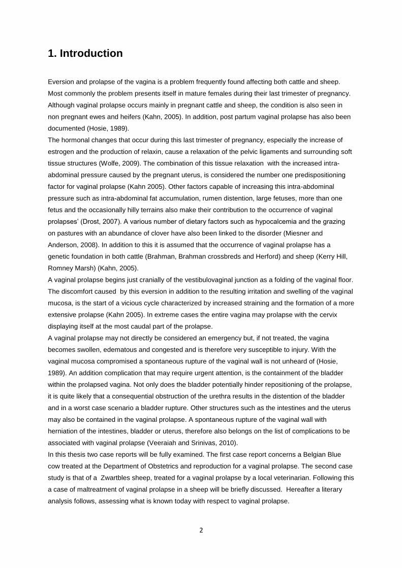
On the first of July 2010 a five year old Belgian Blue cow was brought to the Department of Obstetrics
and reproduction, Faculty of Veterinary Medicine, Ghent University. In addition to the many horses that
are annually brought to this department, Belgian Blue heifers and cows constitute a large part of the
department’s patient database.
The Belgian White Blue breed is a double muscled breed. This double muscled phenotype, is caused
by a genetic mutation in the gene for myostatin, a negative regulator of muscle growth (Thomas et al.,
2000). More specifically Belgian Blue cattle are homozygous for an 11-bp deletion in the gene coding
region that is not detected in cDNA of any normally muscled animals examined. This deletion results in
a unique frame-shift mutation in the part of the myostatin gene most highly conserved in our more
traditionally muscled animals (Kambadur et al., 1997). Piedmontese cattle also displays the double
muscled phenotype and upon examination of their genotype, alterations where found in the same
gene coding region (Kambadur et all., 1997). The result of this mutation is a muscular hyperplasia.
The expression of the myostatin gene is present from early fetal development onwards in both
phenotypically normal and phenotypically double muscled animals (Thomas et al., 2000).
Consequently the instigated muscular hyperplasia leads to calves with twice the muscular volume and
in the case of the Belgian Blue an entire breed of cattle no longer capable of calving naturally. Hence
the reason why Belgian Blue heifers and cows constitute a large part of the departments patient
database. The five year old Belgian Blue cow that concerns us in this particular case was no
exception, she had already undergone two cesarean sections and was due to calve again on the 11th
of July 2010 undoubtedly by means of yet another cesarean section.
Figure 1: The Patient
What set this patient aside from the other Belgian Blue’s was the pink volleyball shaped tissue mass
protruding from her vulva. This was clearly a case of vaginal prolapse.
4
2.2 The Initial Course of Action
On the day of arrival, after a short clinical examination, the decision was made to undertake as little
action as possible. After taking the necessary hygienic measures a small epidural using 2cc’s of
procaine (4%) was placed between the first and the second coccygeal vertebrate and the prolapse
was gentle massaged back into the pelvic cavity. The choice to undertake such limited actions was
based upon the following: the patient was not straining vigorously and she was due to calve shortly.
For the time being the situation appeared stable: when the cow laid down the vagina had a tendency
to prolapse but when encouraging the cow to stand up the prolapse would spontaneously resolve
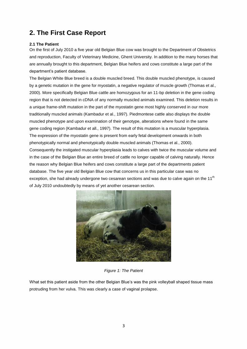
itself. Twenty-four hours later the situation however worsened; the patient began straining more
forcefully and the prolapsed vagina no longer spontaneously resolved when the cow stood up. On the
2nd
of July at 20:00 a new epidural was given and the vagina was repositioned as described above.
Figure 2: The vaginal prolapse
Over the course of two weeks the problem did not diminish. After three days of struggle the cow was
placed on a wedge in order to elevate her hindquarter. The hope was that positioning the cow as such
would better facilitate for the repositioned vagina to stay in the pelvic cavity. In addition to this, the
anesthetic used to bring about the epidural anesthesia was switched from procaine to marcaine
(bupivacaine), a longer acting anesthetic. Because the results obtained after administering epidurals
with procaine had been insufficient it was hoped that the use of a longer acting anesthetic would prove
superior. The result was unfortunately not as good as desired.
A next, and only slightly more invasive measure, was going to be the placement of a Buhner suture. A
Buhner suture is a continues circular suture that can be pulled together to invert or close an opening.
In this case it would be required to place the suture deeply in vulvar tissue in order to mimic the effect
of the constrictor vestibulae muscle (Pitmann, 2010). The problem with this patient was that her
quickly approaching due date made it attractive to wait with such measures, as a Buhner suture must
be removed prior to calving to prevent extensive laceration (Kahn, 2005).
5
2.3 The Fetus
On 16/7/2010, five days after the expected due date questions began to arise concerning the age and
livelihood of the fetus. Whilst an early stage pregnancy is most commonly confirmed via a rectal
exam, possible with the aid of a transrectal ultrasound, both techniques pose limitations when it comes
to the diagnosis of mid and late term pregnancies. Between day 90 and 120 of pregnancy the gravid
horn sinks from within the pelvic cavity to below the cranial pelvic rim . By the 6th month of pregnancy
the gravid horn has sunken deep enough to fill the space between the right flank and the abdominal
floor (Hunnam et al., 2009). As a result of the shifting of the gravid uterus horn most structures
required for a positive pregnancy diagnose are beyond the reach of a transrectal probe (Hunnam et
al., 2009). Fortunately the rectal exam of a cow potentially already three days overdue is more likely to
confirm a pregnancy and this through the palpation of both parts of the fetus and the placentomes, in
addition to the chance of detecting fremitus in both the ipsilateral and contralateral middle uterine
artery (Intervet International, 2011).
Strangely enough the records kept on this patient do not document that a rectal exam was carried out
at this stage. Instead, it appears that a transabdominal echo was made to evaluate the situation. On
the transabdominal echo the calf’s head and extremities where visualized, but neither the ribcage nor
the heartbeat where seen. This information really does not lead us very far. Without seeing the
heartbeat one cannot confirm that the fetus is still alive and seeing bone structures only confirms that
the cow has been carrying for over two months (Purohit, 2010).
Although maybe not as accurate at this stage of the pregnancy, it would have been possible to use the
images obtained via the echo, in a more productive way. By taking certain specific measurements,
such as the crown-rump length and the crown-nose length, one could have calculate an approximate
fetal age. This is simple done by means of a set of standard regression formulas created to utilize
specific measurements to calculate the fetal age (Rexroad et al., 1974 and Riding et al., 2007).
With the many options available to better estimate the potential due date it leaves me guessing why
there was not more done or documented with respect to this dilemma. What the patient’s records do
make clear is that the results of the transabdominal echo lead to the general impression that this cow
was not going to be calving any time shortly. With this in mind and due to the failure of alternative
methods, it was finally decided to place a Buhner suture.
2.4 Placing of the Buhner Suture
In order to place the Buhner suture, the patient first received an epidural using 12cc of procaine. After
washing the involved tissues and repositioning the vaginal prolapse as described above, a Buhner
needle was inserted through the vulva. The needle entered the vulva through the fine-haired skin
below and slightly lateral to the ventral commissure of the vulva and reappeared at an equal distance
above the dorsal vulvar commissure. It is important that the Buhner suture is placed sufficiently deep
into the perivaginal tissue in order to give strength to the suture. After placing the needle, a piece of
umbilical tape was placed through the loop eye of the needle and the needle was removed via the
dorsal opening, hereby threading the umbilical tape through the full length of the right vulvar lip. The
same course of action was then repeated, this time inserting the needle a little bit to the left of where
6
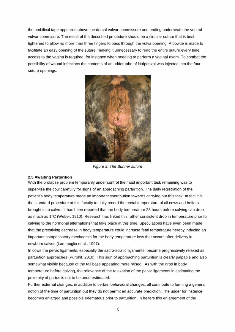
the umbilical tape appeared above the dorsal vulvar commissure and ending underneath the ventral
vulvar commisure. The result of the described procedure should be a circular suture that is best
tightened to allow no more than three fingers to pass through the vulva opening. A bowtie is made to
facilitate an easy opening of the suture, making it unnecessary to redo the entire suture every time
access to the vagina is required, for instance when needing to perform a vaginal exam. To combat the
possibility of wound infections the contents of an udder tube of Nafpenzal was injected into the four
suture openings.
Figure 3: The Buhner suture
2.5 Awaiting Parturition
With the prolapse problem temporarily under control the most important task remaining was to
supervise the cow carefully for signs of an approaching parturition. The daily registration of the
patient’s body temperature made an important contribution towards carrying out this task. In fact it is
the standard procedure at this faculty to daily record the rectal temperature of all cows and heifers
brought in to calve. It has been reported that the body temperature 28 hours before calving can drop
as much as 1°C (Weber, 1910). Research has linked this rather consistent drop in temperature prior to
calving to the hormonal alternations that take place at this time. Speculations have even been made
that the precalving decrease in body temperature could increase fetal temperature hereby inducing an
important compensatory mechanism for the body temperature loss that occurs after delivery in
newborn calves (Lammoglia et al., 1997).
In cows the pelvic ligaments, especially the sacro-sciatic ligaments, become progressively relaxed as
parturition approaches (Purohit, 2010). This sign of approaching parturition is clearly palpable and also
somewhat visible because of the tail base appearing more raised. As with the drop in body
temperature before calving, the relevance of the relaxation of the pelvic ligaments in estimating the
proximity of partus is not to be underestimated.
Further external changes, in addition to certain behavioral changes, all contribute to forming a general
notion of the time of parturition but they do not permit an accurate prediction. The udder for instance
becomes enlarged and possible edematous prior to parturition. In heifers this enlargement of the
7
udder may already be noticed midway the pregnancy, whilst in pluriparous cows it may only become
apparent two to four weeks before parturition (Purohit, 2010). The vulva is also susceptible to swelling
before parturition, but the timing of this event varies greatly amongst individuals. This swelling of the
vulva may be pared with the formation of edema. Especially in heifers this edema can become so
extreme that it extends from the vulva down to the udder sometimes even reaching the umbilical
region. In such cases the edema is considered pathological and one must differentiate it from a
potential hernia or hematoma (de Kruif and van Soom, 2009). Vulvar discharge is another sign of
approaching parturition. Due to the liquefaction of the cervical seal and mucus production in the
vagina, vaginal discharge may be seen one or two days before parturition. Unfortunately in some
cases this discharge may already become apparent as early as two weeks before parturition. If the
discharge is mixed with blood it is an indication of the fact that the accessory placentomes close to
the cervical opening are detaching and parturition is close by (de Kruif and van Soom, 2009).
Several times in the course of the months that followed it was believed that the cow was going into
labor. Naturally these falls alarms where fueled by to the presence of a Buhner suture and the lack of
knowledge concerning the cows gestation time. On the 3rd
of august the cow was found in lateral
decubitus exhibiting clear signs of discomfort. Due to the passing of time the bowtie initially holding the
two ends of umbilical tape together had transformed itself into a knot and it ended up being necessary
to remove the entire suture before vaginal exploration was possible. The vaginal exam taught us that
the cervix was not yet passable by more than one finger and the cow, much to her displeasure,
received a new Buhner suture without first being given an epidural.
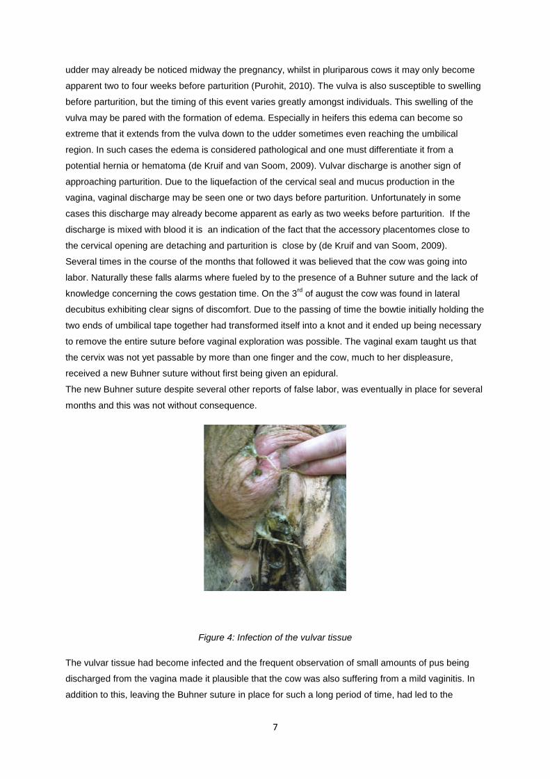
The new Buhner suture despite several other reports of false labor, was eventually in place for several
months and this was not without consequence.
Figure 4: Infection of the vulvar tissue
The vulvar tissue had become infected and the frequent observation of small amounts of pus being
discharged from the vagina made it plausible that the cow was also suffering from a mild vaginitis. In
addition to this, leaving the Buhner suture in place for such a long period of time, had led to the
8
formation of scar tissue around the umbilical tape. Initially this may be seen as an advantage as the
scar tissue ring will continue to combat the vaginal prolapse long after the removal of the Buhner
suture (Pittman, 2010). However a vulva with a scar tissue ring will not be able to stretch enough to
allow for a smooth natural calving, fortunately for this patient that was of little concern.
In an attempt to combat the infection the wounds were washed on occasion with chlorhexidine and the
content of a nafpenzal uddertube was once again injected into the four penetration points of the
Buhner needle. Despite these measures, it was reported that the vulvar tissue had become necrotic to
the extent that the Buhner suture tore out.
Luckily this incident occurred around the time that it became clear to everybody that the patient was
finally going to calve very shortly. In preparation for this long awaited moment the cow had already
been removed from her wedge in the hope that this would allow for the calve to gain better access to
the birth canal. In fact this was done a few weeks earlier on the 12th of October. On this day a rectal
exam had been carried out that finally gave some concrete information, the calf was most likely alive
as fremitus was felt both contralaterally and ipsilaterally and he/she was lying in an anterior
presentation with both front legs and head palpable.
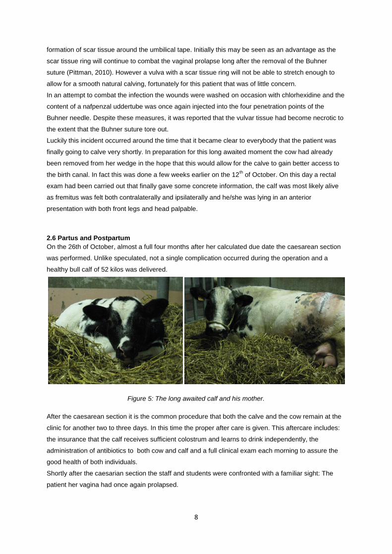
2.6 Partus and Postpartum
On the 26th of October, almost a full four months after her calculated due date the caesarean section
was performed. Unlike speculated, not a single complication occurred during the operation and a
healthy bull calf of 52 kilos was delivered.
Figure 5: The long awaited calf and his mother.
After the caesarean section it is the common procedure that both the calve and the cow remain at the
clinic for another two to three days. In this time the proper after care is given. This aftercare includes:
the insurance that the calf receives sufficient colostrum and learns to drink independently, the
administration of antibiotics to both cow and calf and a full clinical exam each morning to assure the
good health of both individuals.
Shortly after the caesarian section the staff and students were confronted with a familiar sight: The
patient her vagina had once again prolapsed.
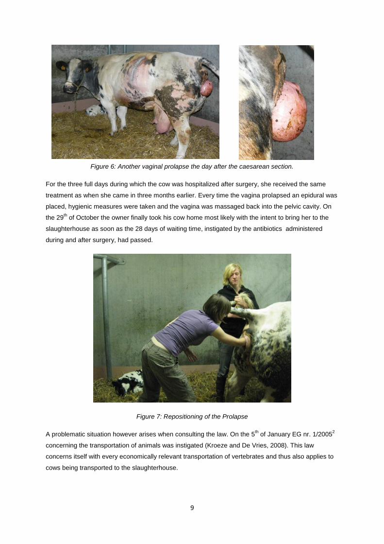
9
Figure 6: Another vaginal prolapse the day after the caesarean section.
For the three full days during which the cow was hospitalized after surgery, she received the same
treatment as when she came in three months earlier. Every time the vagina prolapsed an epidural was
placed, hygienic measures were taken and the vagina was massaged back into the pelvic cavity. On
the 29th of October the owner finally took his cow home most likely with the intent to bring her to the
slaughterhouse as soon as the 28 days of waiting time, instigated by the antibiotics administered
during and after surgery, had passed.
Figure 7: Repositioning of the Prolapse
A problematic situation however arises when consulting the law. On the 5th of January EG nr. 1/2005
2
concerning the transportation of animals was instigated (Kroeze and De Vries, 2008). This law
concerns itself with every economically relevant transportation of vertebrates and thus also applies to
cows being transported to the slaughterhouse.
10
EG nr. 1/20052 states that only animals suitable for transportation are to be transported. This same
law clearly states that animals with a prolapse of any sort are not considered suitable for
transportation. Fortunately there is an alternative way to bring an animal unsuitable for transportation
to the slaughterhouse and this after performing an emergency slaughter. the law concerning the
concept emergency slaughter was however revised and reinstated on the first of January 2006
(Kroeze and De Vries, 2008). EG 853/2004 in its new form dictates that the maximum time between
the occurrence of the prolapse and the emergency slaughter constitutes three days. This would mean
that our patient does not qualify for the emergency slaughter.
The above two laws teach us that a cow with a chronic prolapse simply does not enter the
slaughterhouse. It must however be mentioned that an exception is made for animals whom are
delivered to the slaughterhouse and appear to have prolapsed during transportation (Anonymous,
2010).
What exactly became of the vaginal prolapse cow is not known for certain but an educated guess
would be that she is no longer alive today. Although not frequently applied, one must realize that there
are options to more adequately treat chronic cases of vaginal prolapse as the methods applied to this
patient where only capable of giving temporary relief. This temporary relief was in this case sufficient,
as the patient was a commercial cow at the end of her career. However, had the patient been of higher
value a more permanent fixation technique such as the Minchev suture, Johnson button or
cervicopexy could have been carried out (Miesner and Anderson, 2008).
As for the healthy bull calf, his false due date was probably a simple administrative mistake and he is
most likely growing rapidly to fulfill his destiny as beef cattle.
11
3. The Second Case Report
3.1 The Patient
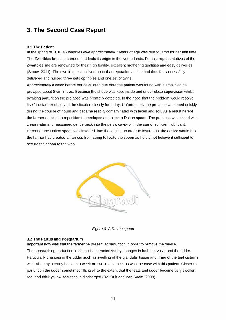
In the spring of 2010 a Zwartbles ewe approximately 7 years of age was due to lamb for her fifth time.
The Zwartbles breed is a breed that finds its origin in the Netherlands. Female representatives of the
Zwartbles line are renowned for their high fertility, excellent mothering qualities and easy deliveries
(Stouw, 2011). The ewe in question lived up to that reputation as she had thus far successfully
delivered and nursed three sets op triples and one set of twins.
Approximately a week before her calculated due date the patient was found with a small vaginal
prolapse about 8 cm in size. Because the sheep was kept inside and under close supervision whilst
awaiting parturition the prolapse was promptly detected. In the hope that the problem would resolve
itself the farmer observed the situation closely for a day. Unfortunately the prolapse worsened quickly
during the course of hours and became readily contaminated with feces and soil. As a result hereof
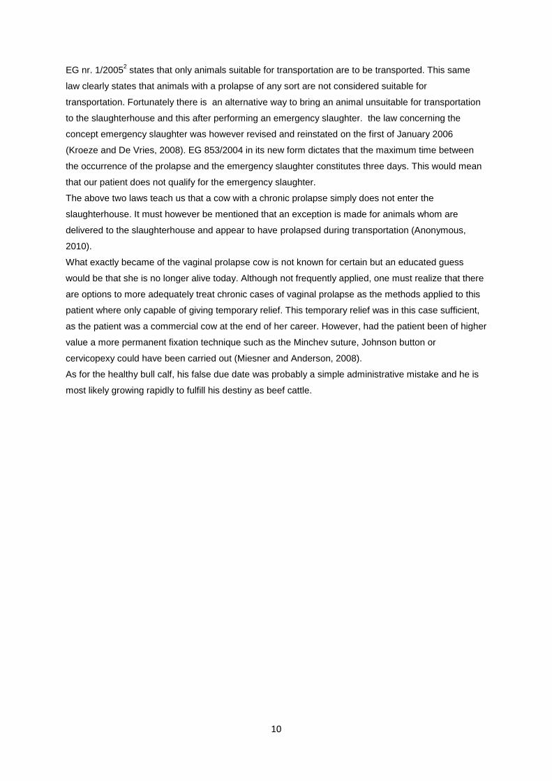
the farmer decided to reposition the prolapse and place a Dalton spoon. The prolapse was rinsed with
clean water and massaged gentle back into the pelvic cavity with the use of sufficient lubricant.
Hereafter the Dalton spoon was inserted into the vagina. In order to insure that the device would hold
the farmer had created a harness from string to fixate the spoon as he did not believe it sufficient to
secure the spoon to the wool.
Figure 8: A Dalton spoon
3.2 The Partus and Postpartum
Important now was that the farmer be present at parturition in order to remove the device.
The approaching parturition in sheep is characterized by changes in both the vulva and the udder.
Particularly changes in the udder such as swelling of the glandular tissue and filling of the teat cisterns
with milk may already be seen a week or two in advance, as was the case with this patient. Closer to
parturition the udder sometimes fills itself to the extent that the teats and udder become very swollen,
red, and thick yellow secretion is discharged (De Kruif and Van Soom, 2009).
12
The first sign of approaching parturition with respect to the vulva is that of slight swelling. Hereafter the
vulva starts to become red only to be both red and swollen on the day of parturition (De Kruif and Van
Soom, 2009).
In addition the physiological changes that occur prepartum certain behavioral signs are also an
important indication for approaching parturition. At the start of parturition an ewe may display the
following restless behavior: she will seclude herself from the others, find a sheltered area, alternate
lying/standing and paw at the ground frequently sniffing at this area. These periods of activity often
occur at 15 minute intervals accompanying abdominal contractions that last approximately 30 seconds
(Scott, 2010).
Figure 9: The Patient
Unfortunately the farmer came back from school one day to find the Dalton spoon in a most awkward
position with a lamb in anterior position, partially hanging out from the vulva. The spoon had without
doubt hindered this lambs expulsion and by the time the farmer came to the scene and removed the
spoon the lamb was dead. As not to waste any more time the farmer quickly delivered the two
remaining lambs, both where still lying quite deep in the uterus, one in anterior and one in posterior
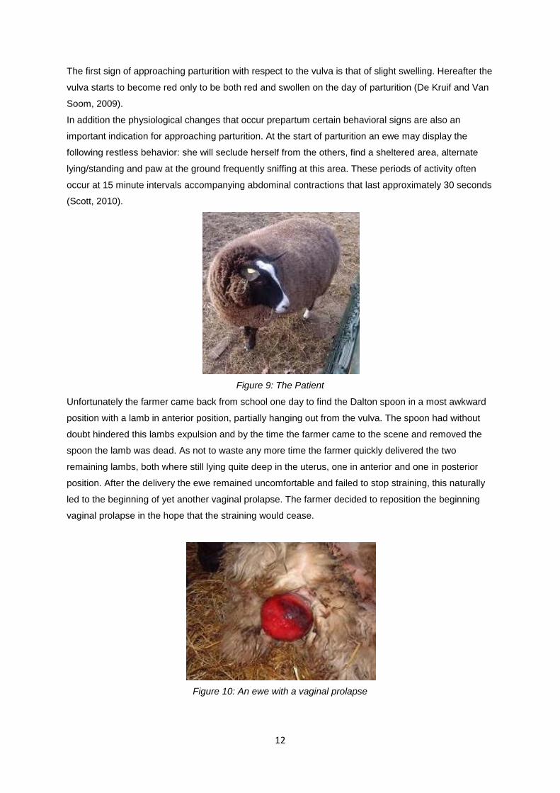
position. After the delivery the ewe remained uncomfortable and failed to stop straining, this naturally
led to the beginning of yet another vaginal prolapse. The farmer decided to reposition the beginning
vaginal prolapse in the hope that the straining would cease.
Figure 10: An ewe with a vaginal prolapse
13
This method however proved inadequate as the vagina almost immediately prolapsed again. Although
the prolapse appeared less extensive then before and despite genuine believe of the farmer that his
ewe was not suffering too much discomfort, this would have probably been a good moment to consult
a veterinarian. The farmer however waited and observed as the vaginal tissue slowly became necrotic
and the prolapse grew in size. After three days of observation and with a clear demarcation line
formed between the life and necrotic vaginal tissue the farmer consulted his veterinarian.
3.3 Amputation of the Vaginal Prolapse
It was clear to both parties involved that the presence of necrotic tissue excluded the simple
repositioning of the vaginal prolapse from the list of options. After taking the necessary hygienic
measures and examining the prolapse more closely the veterinarian had formed an alternative plan. A
more thorough exploration of the prolapse had led her to conclude that it was only really the vaginal
floor that had become necrotic. She therefore decided to simple remove the necrotic tissue, suture the
created defect back together and reposition the vagina. In order for her plan to succeed it was
important that she worked in healthy vaginal tissue and that the opening of the urethra stayed intact.
To find the perfect place for her incision the veterinarian first exerted traction on the vaginal prolapse,
causing for the more healthy tissue to become exposed. She then used about 10cc’s of procaine and
injected small dosages of the anesthetic into the vaginal tissue where she was going to make her
incision. With this done she simple took a scalpel and cut of the stump of necrotic tissue. The defect
created was sutured back together using a simple continuous stitch and the vagina was repositioned.
No additional measures were taken to secure the vagina in place. The ewe was treated with antibiotics
(Neopen) for 5 days and the problem appeared to be resolved. It must be mentioned that throughout
her ordeal the ewe continued to mother her lambs enthusiastically. Four months after the procedure,
the lambs where weaned and the sheep was transported to the slaughterhouse along with the other
reform ewes.
14
4. A Case of Mismanagement
In his recent book, Sheep Medicine, Philip Scott expresses his concern for the welfare of sheep. In his
many years of experience practicing veterinary medicine he has come to the conclusion that the care
and welfare of sheep in the United Kingdom has gradually deteriorated and this largely as a
consequence of the poor economical returns from sheep farming. The sensitive balance that exists
between animal welfare and economical gain is naturally not only a dilemma in the United Kingdom,
but rather a global concern.
The following case that I would briefly like to present is a case of mismanagement of vaginal prolapse
in a Texel ewe.
The Dalton Spoon, as discussed above, is a minimally invasive method for treating vaginal prolapse,
and placement of the device can easily be carried out by the sheep farmer himself. Other, more
invasive methods are however also to the disposal of our farmers. The Texel ewe under discussion
suffered a vaginal prolapse several months before her expected lambing date. The farmer in question,
a strong believer in the Dalton spoon, decided to combat the vaginal prolapse by inserting this device.
Unfortunately the ewe did not respond well to the taken measure, as she continued to strain and
prolapse. After several fruitless attempts to keep the vaginal prolapse in place with the aid of a Dalton
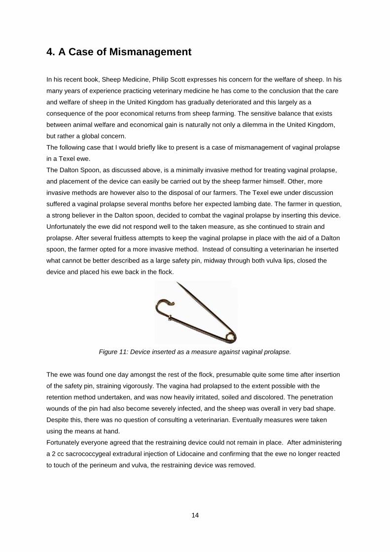
spoon, the farmer opted for a more invasive method. Instead of consulting a veterinarian he inserted
what cannot be better described as a large safety pin, midway through both vulva lips, closed the
device and placed his ewe back in the flock.
Figure 11: Device inserted as a measure against vaginal prolapse.
The ewe was found one day amongst the rest of the flock, presumable quite some time after insertion
of the safety pin, straining vigorously. The vagina had prolapsed to the extent possible with the
retention method undertaken, and was now heavily irritated, soiled and discolored. The penetration
wounds of the pin had also become severely infected, and the sheep was overall in very bad shape.
Despite this, there was no question of consulting a veterinarian. Eventually measures were taken
using the means at hand.
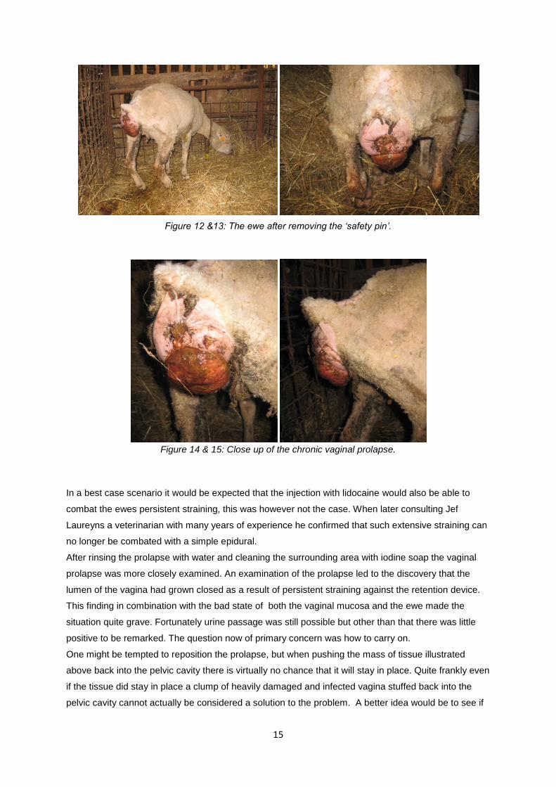
Fortunately everyone agreed that the restraining device could not remain in place. After administering
a 2 cc sacrococcygeal extradural injection of Lidocaine and confirming that the ewe no longer reacted
to touch of the perineum and vulva, the restraining device was removed.
15
Figure 12 &13: The ewe after removing the ‘safety pin’.
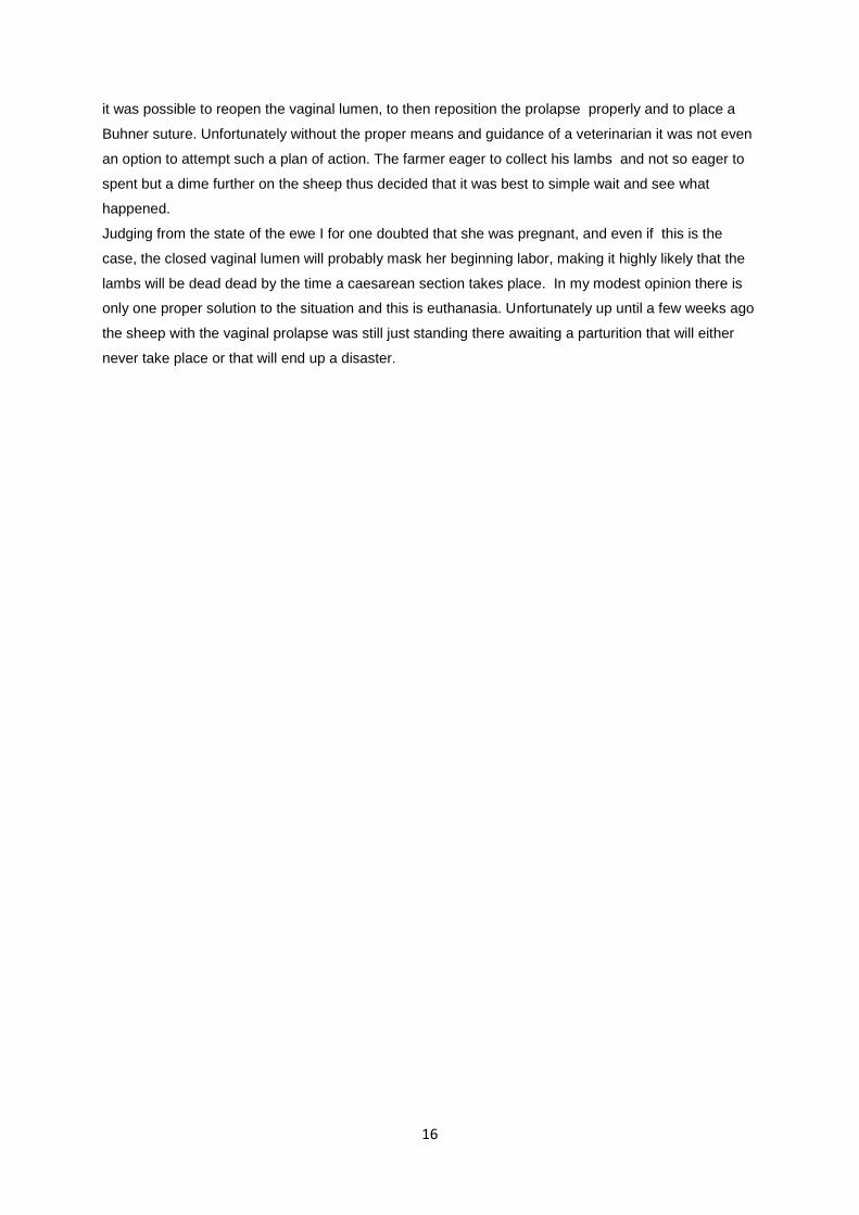
Figure 14 & 15: Close up of the chronic vaginal prolapse.
In a best case scenario it would be expected that the injection with lidocaine would also be able to
combat the ewes persistent straining, this was however not the case. When later consulting Jef
Laureyns a veterinarian with many years of experience he confirmed that such extensive straining can
no longer be combated with a simple epidural.
After rinsing the prolapse with water and cleaning the surrounding area with iodine soap the vaginal
prolapse was more closely examined. An examination of the prolapse led to the discovery that the
lumen of the vagina had grown closed as a result of persistent straining against the retention device.
This finding in combination with the bad state of both the vaginal mucosa and the ewe made the
situation quite grave. Fortunately urine passage was still possible but other than that there was little
positive to be remarked. The question now of primary concern was how to carry on.
One might be tempted to reposition the prolapse, but when pushing the mass of tissue illustrated
above back into the pelvic cavity there is virtually no chance that it will stay in place. Quite frankly even
if the tissue did stay in place a clump of heavily damaged and infected vagina stuffed back into the
pelvic cavity cannot actually be considered a solution to the problem. A better idea would be to see if
16
it was possible to reopen the vaginal lumen, to then reposition the prolapse properly and to place a
Buhner suture. Unfortunately without the proper means and guidance of a veterinarian it was not even
an option to attempt such a plan of action. The farmer eager to collect his lambs and not so eager to
spent but a dime further on the sheep thus decided that it was best to simple wait and see what
happened.
Judging from the state of the ewe I for one doubted that she was pregnant, and even if this is the
case, the closed vaginal lumen will probably mask her beginning labor, making it highly likely that the
lambs will be dead dead by the time a caesarean section takes place. In my modest opinion there is
only one proper solution to the situation and this is euthanasia. Unfortunately up until a few weeks ago
the sheep with the vaginal prolapse was still just standing there awaiting a parturition that will either
never take place or that will end up a disaster.

17
5. Literature Review/Discussion
5.1 Anatomy and Fixation of the Vagina
Vaginal prolapse most frequently affects ewes and cattle in their last trimester of pregnancy . It is
believed that the vagina undergoes a varying degree of relaxation as a result of the hormonal changes
ascribed to late term pregnancy. The same theory upholds for the soft tissues associated with the
vagina, more specifically the related muscles, ligaments, synovial membranes and fascia, undergo a
varying degree of relaxation as a result of the hormonal changes ascribed to late term pregnancy
(Kahn, 2009). When combining this tissue relaxation with an increased abdominal pressure brought
about by the pregnant uterus, a risk for vaginal prolapse is created.
With the believe that soft tissue relaxation plays a dominant part in the pathogenesis of vaginal
prolapse, a better understanding of the anatomy of the vagina and its related soft tissues appears
relevant.
5.1.1 The Vagina
The vagina, with its tube like structure, is considered the cranial part of the female copulatory organ
extending from the external uterine ostium to the entrance of the urethra. The vagina is located in a
medial position in the pelvic cavity underneath the rectum and above the bladder. For the most part
the vagina is located retroperitoneally but the most cranial part is covered in peritoneum. The cervix
located cranially to the vagina restricts the lumen of the cranial vagina to a ring-like space known as
the fornix (König and Liebich, 2004).
The vestibulum vaginae constitutes the caudal part of the female copulatory organ extending from the
ostium urethral opening to the external vulva. In cows the urethra forms a central evagination, the
suburethral diverticulum which opens together with the urethra into the vagina, complicating
catheterization of the urinary bladder. The vestibulum lies for the most part behind the ischial arch and
slopes ventrally towards its opening in the vulva, this ventral sloping must be taken into consideration
when inserting a speculum or any other device into the vagina. Vestibular glands are contained in the
wall of the vestibulum and the secretion thereof facilitates parturition and coitus. Minor vestibular
glands are also present in the sheep and cow additionally In both the cow and the ewe a large
glandular mass is present on both sides of the vestibulum (König and Liebich, 2004).
Anatomically speaking the vulva and the vestibulum vaginae are well fixated whilst the contrary can be
said about the vagina. The caudal part of the vagina is surrounded by lose fatty tissue and connective
tissue and is hereby fixated to a certain extent, whilst the cranial part of the vagina is subject to very
limited fixation (De Kruif and Van Soom, 2009).
5.1.2 The Ligaments
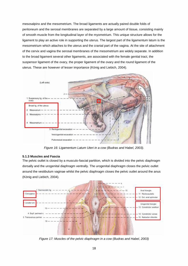
The main attachment of the female genital organs is provided by the ligamentum latum uteri, also
known as the broad ligaments of the uterus. Seeing as the broad ligament suspends the uterus in
addition to both ovaries and ovarian ducts, it can be divided into three parts: the mesovarium, the
18
mesosalpinx and the mesometrium. The broad ligaments are actually paired double folds of
peritoneum and the serosal membranes are separated by a large amount of tissue, consisting mainly
of smooth muscle from the longitudinal layer of the myometrium. This unique structure allows for the
ligament to play an active role in supporting the uterus. The largest part of the ligamentum latum is the
mesometrium which attaches to the uterus and the cranial part of the vagina. At the site of attachment
of the cervix and vagina the serosal membranes of the mesometrium are widely separate. In addition
to the broad ligament several other ligaments, are associated with the female genital tract, the
suspensor ligament of the ovary, the proper ligament of the ovary and the round ligament of the
uterus. These are however of lesser importance (König and Liebich, 2004).
Figure 16: Ligamentum Latum Uteri in a cow (Budras and Habel, 2003).
5.1.3 Muscles and Fascia
The pelvic outlet is closed by a musculo-fascial partition, which is divided into the pelvic diaphragm
dorsally and the urogenital diaphragm ventrally. The urogenital diaphragm closes the pelvic outlet
around the vestibulum vaginae whilst the pelvic diaphragm closes the pelvic outlet around the anus
(König and Liebich, 2004).
Figure 17: Muscles of the pelvic diaphragm in a cow (Budras and Habel, 2003)
19
The pelvic diaphragm, also named the anal triangle is composed of the m. coccygeus the levator ani
muscle and the external anal sphincter. The urogenital diaphragm or triangle, is composed of a strong
perineal membrane which extends from the ischial arch to the ventral and lateral walls of the
vestibulum vaginae attaching cranially to the constrictor vestibule and caudally of the major vestibular
gland. The perineal membrane together with the urogenital muscles (m. constrictor vestibule m.
constrictor vulvae and m retractor clitoris) forms the urogenital triangle and fuses with the pelvic
triangle at the level of the perineal body hereby anchoring the genital tract to the ischial arch (Budras
and Habel, 2003).
5.2 Causes of Vaginal Prolapse
Vaginal prolapse is a frequently occurring phenomena in both sheep and cattle. When it comes to the
causes of vaginal prolapse genetic, nutritional and hormonal components have all been linked with the
problem in question. Unfortunately there is a lot that still remains unknown about the causes of vaginal
prolapse.
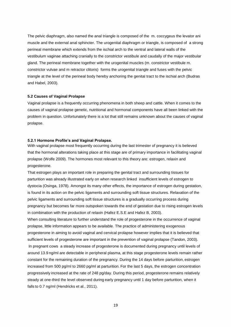
5.2.1 Hormone Profile’s and Vaginal Prolapse.
With vaginal prolapse most frequently occurring during the last trimester of pregnancy it is believed
that the hormonal alterations taking place at this stage are of primary importance in facilitating vaginal
prolapse (Wolfe 2009). The hormones most relevant to this theory are: estrogen, relaxin and
progesterone.
That estrogen plays an important role in preparing the genital tract and surrounding tissues for
parturition was already illustrated early on when research linked insufficient levels of estrogen to
dystocia (Osinga, 1978). Amongst its many other effects, the importance of estrogen during gestation,
is found in its action on the pelvic ligaments and surrounding soft tissue structures. Relaxation of the
pelvic ligaments and surrounding soft tissue structures is a gradually occurring process during
pregnancy but becomes far more outspoken towards the end of gestation due to rising estrogen levels
in combination with the production of relaxin (Hafez E.S.E and Hafez B, 2003).
When consulting literature to further understand the role of progesterone in the occurrence of vaginal
prolapse, little information appears to be available. The practice of administering exogenous
progesterone in aiming to avoid vaginal and cervical prolapse however implies that it is believed that
sufficient levels of progesterone are important in the prevention of vaginal prolapse (Tandon, 2003).
In pregnant cows a steady increase of progesterone is documented during pregnancy until levels of
around 13.9 ng/ml are detectable in peripheral plasma, at this stage progesterone levels remain rather
constant for the remaining duration of the pregnancy. During the 14 days before parturition, estrogen
increased from 500 pg/ml to 2660 pg/ml at parturition. For the last 5 days, the estrogen concentration
progressively increased at the rate of 248 pg/day. During this period, progesterone
remains relatively
steady at one-third the level observed during early pregnancy until 1 day before parturition, when it
falls to 0.7 ng/ml (Hendricks et al., 2011).
20
In ewes a more gradual increase of the progesterone level in peripheral plasma is observed during
pregnancy. The maximum progesterone plasma levels are reached at about day 140, and are logically
dependent upon the amount of fetuses being carried. The mean progesterone plasma value in an
ewe, carrying one lamb lies around 13.2 ng/ml, and in twin pregnancies values of around 20.0 ng/ml
have been documented. During the last week of pregnancy a marked decline in plasma progesterone
levels is observed with a mean value of 2.1 ng/ml at parturition (Fylling, 1970). As described in cows,
plasma estrogen levels in sheep also rise drastically towards the end of gestation in order to peak at
parturition (Sjaastad et al., 2003).
A study of the progesterone and estrogen concentrations in blood plasma of ewes suffering from
vaginal prolapse revealed the following: “The average progesterone concentration of affected ewes
was above those of control animals ante and intra partum. However, no significant differences were
observed. The plasma levels of 17β-estradiol in animals suffering from ante partum vaginal prolapse
were increased in comparison to the values of the pregnant control group, but were without statistical
significance.” (Ennen et al., 2011).
The raised progesterone levels in affected ewes may lead one to question the initial proposal that
progesterone plays a preventative role in the pathogenesis of vaginal prolapse. However the
registered progesterone and estrogen concentrations are the results of a single study. The found
hormone levels are not even of statistical significance and they definitely do not help us any further
with the question of how these two hormones do or do not contribute to the pathogenesis of vaginal
prolapse.
In an attempt to clarify the role of estrogen in the occurrence of vaginal prolapse, a theory was formed
suggesting that an increased expression of estrogen receptor α in the genital tract may facilitate an
increased estrogenic effect resulting in vaginal prolapse. Ennen and her research group however
found that the expression rate of estrogen receptor α was lower in the animals suffering a vaginal
prolapse compared to the healthy control group. Although there is potentially only one study
contradicting the hypothesis that an increased expression of estrogen receptor α in the genital tract
may be the route by which estrogen facilitates vaginal prolapse, there is no study found confirming the
theory either. The only conclusion that can be drawn from this is that the answer still remains
unknown.
At this stage it is important that one remains objective and realizes that there is simple to little research
done with respect to the role of hormones in the pathogenesis of vaginal prolapse, to enable concrete
conclusions to be drawn. When looking at the situation from a practical point of view, although maybe
not scientifically proven, the general believe still rests that high levels of estrogen facilitate vaginal
prolapse. In the end this is not such a foolish thought as one can come to the conclusion using pure
logics. The hormone estrogen relaxes the pelvic ligaments and surrounding soft tissue, hereby
causing a decreased fixation of the vagina. When combining this decreased vaginal fixation with an
increase of intra abdominal pressure such as found in highly pregnant animals, the vagina is more
inclined to prolapse. Another, more concrete argument illustrating the importance of estrogen in the
occurrence of vaginal prolapse, is the fact that cows suffering from cystic ovarian disease are more
susceptible to developing a vaginal prolapse. More specifically cows suffering from follicular cysts,

21
especially those exhibiting extreme signs of nymphomania, have been known to suffer a vaginal
prolapse. The follicular cyst creates a hormonal environment in which progesterone is limited whilst
the production of estrogen exceeds the norm (De Kruif and Van Soom, 2009). Well developed
follicular cysts such as the ones present in nymphomaniac cows thus provide the estrogen rich
environment believed to facilitate the occurrence of vaginal prolapse.
When examining the role of relaxin in the cause of vaginal prolapse, the hormone can be linked to the
pathogenesis in a way similar to that of the hormone estrogen, namely through the weakening of the
soft tissues associated with the vagina.
Relaxin is a hormone that remodels connective tissue hereby inducing cervical dilation, pelvic
relaxation, and separation of interpubic ligaments in several mammalian species (Lloyd, 2011).
Experiments performed on rats acknowledged significant decrease in collagen levels during
pregnancy and parturition coincide with a rapid increase in serum relaxin levels during this time.
(Sherwood & Crnekovic 1979, Sherwood et al. 1980). The reduction in collagen content
may be attributed to collagen degradation through activation of the collagenolytic
but the spectrum of collagen types remains unchanged throughout gestation (Samuel et al., 1998).
The production of this hormone in pregnant ruminants is low. Non ruminants also maintain low relaxin
levels during most of the pregnancy. Analysis of the plasma samples of sows, however illustrates a
surge of relaxin production during the last week of gestion, peaking at levels of 60-90 ng/ml on the day
of parturition (Lloyd, 2011).
When carrying out the same analysis in blood plasma samples of pregnant cattle we learn that levels
of relaxin secreted during the last week of pregnancy are still quite low with values of 0.20 ng/ml
representing the mean. On the day of the partus relaxin however also increases and peaks at
concentrations greater than 0.800 ng/ml (Lloyd, 2011).
In sheep, relaxin plasma concentrations increase with advancing gestation, from an average of .60
ng/ml reaching a peak of 3.90 ng/ml four days before parturition. At parturition however, relaxin
concentration averaged only .80 ng/ml (Lloyd, 2011).
From this data we learn that although an antipartum relaxin surge does occur in sheep, the relaxin
peak occurs earlier then in non ruminants. When comparing the plasma concentration of relaxin
registered in cattle with the relaxin surge and peak undergone by non ruminants, the term hardly
seems applicable to cattle.
Despite completely different measures of relaxin in the blood for different species, the above
information does illustrate that both cattle and ewes are influenced by relaxin during pregnancy.
Knowing a little bit better the mechanism by which relaxin functions, yet again lacking evidence linking
relaxin to vaginal prolapse in a more concrete way, we end up with a conclusion much like the one
drawn earlier on concerning our hormone estrogen. Relaxin breaks down collagen in the pelvic
ligaments and surrounding soft tissue, hereby weakening the connective tissue and thus causing a
decreased fixation of the vagina. When combining this decreased vaginal fixation with an increase of
intra abdominal pressure such as found in highly pregnant animals, the vagina is more inclined to
prolapse.
22
5.2.2 Vaginal Prolapse a Hereditary Trait
It would be nice to be able to say that vaginal prolapse is a hereditary trait located on chromosome x
more specifically in gene coding region y. Unfortunately the basis for the believe that vaginal prolapse
has a genetic foundation is not quite this concrete. It is however assumed that the occurrence of
vaginal prolapse has a genetic foundation in both cattle and sheep (Kahn, 2005). This assumption is
based upon the more frequent observation of the pathology in certain breeds and bloodlines. In cattle
for instance Brahman, Brahman crossbreds and Hereford appear predisposition, whilst in sheep Kerry
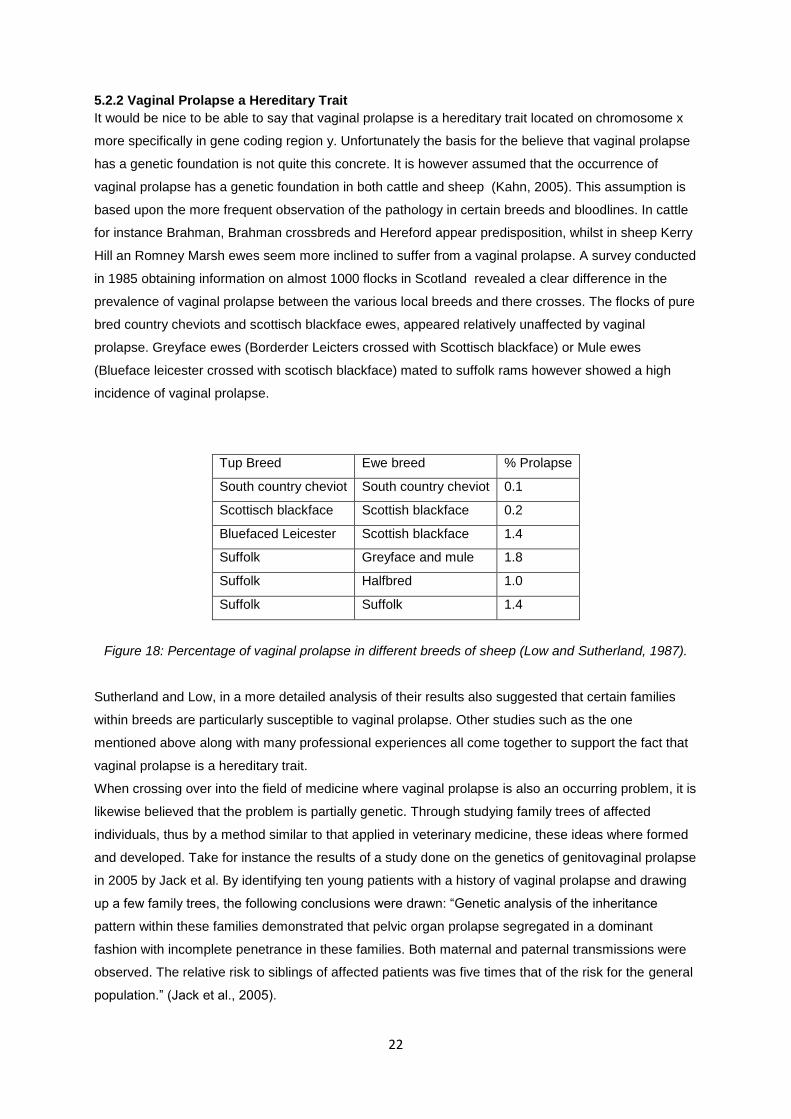
Hill an Romney Marsh ewes seem more inclined to suffer from a vaginal prolapse. A survey conducted
in 1985 obtaining information on almost 1000 flocks in Scotland revealed a clear difference in the
prevalence of vaginal prolapse between the various local breeds and there crosses. The flocks of pure
bred country cheviots and scottisch blackface ewes, appeared relatively unaffected by vaginal
prolapse. Greyface ewes (Borderder Leicters crossed with Scottisch blackface) or Mule ewes
(Blueface leicester crossed with scotisch blackface) mated to suffolk rams however showed a high
incidence of vaginal prolapse.
Tup Breed Ewe breed % Prolapse
South country cheviot South country cheviot 0.1
Scottisch blackface Scottish blackface 0.2
Bluefaced Leicester Scottish blackface 1.4
Suffolk Greyface and mule 1.8
Suffolk Halfbred 1.0
Suffolk Suffolk 1.4
Figure 18: Percentage of vaginal prolapse in different breeds of sheep (Low and Sutherland, 1987).
Sutherland and Low, in a more detailed analysis of their results also suggested that certain families
within breeds are particularly susceptible to vaginal prolapse. Other studies such as the one
mentioned above along with many professional experiences all come together to support the fact that
vaginal prolapse is a hereditary trait.
When crossing over into the field of medicine where vaginal prolapse is also an occurring problem, it is
likewise believed that the problem is partially genetic. Through studying family trees of affected
individuals, thus by a method similar to that applied in veterinary medicine, these ideas where formed
and developed. Take for instance the results of a study done on the genetics of genitovaginal prolapse
in 2005 by Jack et al. By identifying ten young patients with a history of vaginal prolapse and drawing
up a few family trees, the following conclusions were drawn: “Genetic analysis of the inheritance
pattern within these families demonstrated that pelvic organ prolapse segregated in a dominant
fashion with incomplete penetrance in these families. Both maternal and paternal transmissions were
observed. The relative risk to siblings of affected patients was five times that of the risk for the general
population.” (Jack et al., 2005).
23
A simple observation of trends has for instance taught us that daughters of mother cattle suffering
from vaginal prolapse have an increased likelihood of suffering a vaginal prolapse themselves
(Powell, 2005).The advice to cull both mother and daughter would therefore by many be considered
the correct measure. In addition, bull calves of affected mother animals have been suggested capable
of passing on the trait to female offspring and should therefore not be used for breeding (Powell,
2005). Over the years it has become common practice to cull all ewes with a vaginal prolapse (Scott,
2010). Culling every effected ewe hardly seems like the most economical method of action and I
hardly believe farmers would apply it, had experience not taught them that it is simple the right thing to
do.
Maybe we cannot quite pinpoint the exact location of the disease on a chromosome but in my opinion
the information known to us right now sufficiently pleas for the fact that the occurrence of vaginal
prolapse has a hereditary component.
5.2.3 Collagen Metabolism and Vaginal Prolapse
In 2004 a study by Söderberg et al., revealed that human patients suffering from vaginal prolapse
appeared to have up to a 30 percent lower concentration of collagen. This conclusion was drawn after
the analysis of punch biopsies taken from the paraurethral ligaments of 22 prolapse patients
undergoing surgery. In 2007 Twiss et al., published an article believing to have identified the gene
responsible for the genetical foundation playing a part in many vaginal prolapse cases. The gene in
question was LAMC-1 and it codes for a subunit of the laminin protein, that amongst other things,
contributes to the structural integrity of the extracellular matrix (Twiss et al., 2008).
In 2011 Ennen and her research group made a similar breakthrough concerning the occurrence of
vaginal prolapse in ewes. By doing a biomolecular analysis of the connective tissue of several ewes,
with and without vaginal prolapse, it was hoped to illustrate that the occurrence of vaginal prolapse in
these animals could also be linked to alternations in vaginal connective tissue.
Connective tissue consists of both a cellular and an extracellular matrix. It is mainly this extracellular
matrix held responsible for giving stability. More specifically, collagen I and III of the extracellular
matrix make up an important part of vaginal and cervical tissue (Jeffrey, 1991).
Biopsies where taken from the vaginal tissue and analyzed for the mRNA-expression and
transcription levels of the α2-chain of collagen I, the collagenolytic metalloproteinase 1 (MMP-1) and
the tissue inhibitor of MMP-1 (TIMP-1).
The Matrix Metalloproteinase’s such as MMP-1 play a role in the rearrangement of connective tissue
as they are capable of degrading collagen fibers. Tissue Inhibitor proteins such as TIMP-1 are in turn
inhibitors of MMP’s. TIMP-1 for instance actively binds to MMP-1 hereby inhibiting its collagenolytic
function (Timmerman, 2005).
The results of the analyses of mRNA expression and transcription levels of, MMP-1 and TIMP-1 where
as followed: “The average mRNA synthesis of MMP-1 and TIMP-1 in affected ewes varied from those
in healthy, pregnant sheep by a higher and/or lower level respectively, but significant differences were
not apparent.” (Ennen et al., 2011). Although the author is aware that the results obtained where not
statistically significant, she does recognize that the increased expression of MMP-1 and the decreased
expression of TIMP-1 are indicative for an increased catabolism of collagen. Caution is however
24
required in drawing up this conclusion as enzymes and there inhibitors are capable of undergoing
posttranscriptional modifications. The MMP-1’s are for instance secreted in there inactive form only to
be activated at their target destination. In addition to this, the TIMP-1 molecules, although most
commonly functioning as inhibitor, are also capable of having activating effects depending upon their
concentration (Clark et al., 1994). Nevertheless Ennen concludes that: “the mRNA expression rate of
TIMP-1 and MMP-1 can be seen as an indicator for the dysfunction of the collagen metabolites.”.
In addition the result concerning the mRNA expression of α2-chain of collagen 1 indicated a statistical
difference between afflicted animals and the pregnant control group. More specifically the expression
of α2-chain of collagen 1 was lower in ewes with prolapsed vaginal tissue. Because of the fact that
collagen one is actually composed of two α1, and one α2-chains connected to a triple helix the author
warns that the decreased expression of the α2-chain of collagen 1 does not necessarily indicate a
reduced protein biosynthesis and therefore a reduction of collagen in the vaginal wall. Because single
collagen chains undergo translational modification before able to form a triple helix, regulation not only
occurs at the level of transcription but also at the level of protein synthesis (Olsen and Ninomiya,
1999).
Despite some potential reservations, the research that Enen et al., conducted led to the following
general conclusion: “Affected ewes show alterations in the antepartal metabolism of vaginal
connective tissue… Further research involving expression studies might offer perspectives concerning
therapy and prevention of this economically important clinical condition.”(Ennen et al., 2011).
5.2.4 Vaginal Prolapse and Nutrition
Nutrition is commonly listed as a factor contributing to the prevalence of vaginal prolapse. More
specifically, poor quality forage, high levels of concentrate, high estrogenic-content feeds and
hypocalcaemia have all been associated with the pathology ( Miesner and Anderson, 2008).
Additionally there appears to be a correlation between obesity and the occurrence of vaginal prolapse
(Hosie et al., 1999).
Distributing poor quality forage will require for ewes to increase their feed uptake in order to meet their
energy requirements. This increased uptake of forage however results in an increased ruminal filling,
which in turn contributes to a higher intra-abdominal pressure hereby predisposing for vaginal
prolapse (Pelzer, 2008).
The same disadvantage however has been experienced by herders feeding alfalfa hay (Umburger,
1991). Due to the products high quality and palatability, ewes consume more alfalfa hay than is
needed. The bulkiness of the hay in the rumen can, by the same mechanism as poor quality forage,
place pressure on the reproductive tract, resulting in a vaginal prolapse.
The feeding of excessive amounts of concentrates has also been associated with the occurrence of
vaginal prolapse. The Initial concern that one may have when it comes to the feeding of concentrates
to cattle and sheep, is that of overfeeding, as this may lead to ruminal acidosis.
Due to the uptake of rapidly fermentable carbohydrates the ruminal pH shall, as a result of the
products of fermentation, quickly begin to drop. This drop in pH causes a shift in the ruminal flora from
25
cellulolytic to lactic acid producing bacteria, hereby further accelerating the prior established decline in
ruminal pH.
The realized pH change directly undermines the integrity of the ruminal wall. Meanwhile the rapid
fermentation of food causes the ruminal content to become more osmolar. The damage caused to the
ruminal wall, in combination with the hyperosmolarity of the ruminal content, instigates a diffusion of
fluid from the circulation into the rumen. This diffusion brings about a ruminal distention that is further
enhanced by the accumulation of gas formed during fermentation (Deprez, 2009).
In addition to the many complications and symptoms that ruminal acidosis presents, vaginal prolapse
is a side effect that is maybe not directly considered but that can also be associated with the
pathology. It is the ruminal distention resulting from acidosis, that once again contributes to the
formation of that higher intra-abdominal pressure known to facilitate vaginal prolapse. Of course in a
case of acute ruminal acidosis one is more likely to fear for the life of the sheep than for a vaginal
prolapse, yet in more chronic cases of ruminal acidosis one can imagine that vaginal prolapse may
become a part of the pathogenesis. Alternatively one may consider that a vaginal prolapse severe
enough to compromise a patient’s health may actually be the cause of sub acute ruminal acidosis.
Unwell patients often decrease their uptake of roughage whilst still eating their initial amount of
concentrates an amount that suddenly constitutes a much larger percent of their diet.
When following the general trend it becomes clear that any cause for an increased abdominal
pressure appears to predispose for the development of a vaginal prolapse. Aside from the nutritional
factors listed above, other factors capable of increasing intra-abdominal such as intra-abdominal fat
accumulation, large fetuses, multigravid uterus, hilly terrains, the manipulation of sheep before shaving
or claw treatment, limited exercise and lameness leading to long periods of sternal recumbency
therefore all also make their contribution to the occurrence of vaginal prolepses’ (Drost, 2007 and
Scott, 2010).
Having previously elaborated upon the role that estrogen plays in the pathogenesis of vaginal
prolapse, it is a logical next step to link feeds high in phytoestrogens with the occurrence of vaginal
prolapse.
Phytoestrogens, are plant derived components that structurally or functionally mimic mammalian
estrogen (Osoki and Kennelli, 2003). The biological activity of phytoestrogens is diverse. This diversity
can be partially attributed to the ability of phytoestrogens to act as both estrogen agonists and
antagonists thus causing either an estrogenic or antiestrogenic effect. The means by which this is
accomplished varies, thus far both genomic and nongenomic pathways of action have been identified
(Anderson et al., 1999). It has been suggested that the levels of endogenous estrogen contribute to
determining the actions of phytoestrogen as it is believed that phytoestrogens go into competition with
the endogenously available estrogens (Folman and Pope, 1966). In ewes for instance, the activity of
endogenous estrogen is considered low and phytoestrogens have been reported to function primarily
as estrogen agonist, whilst in humans whom are considered to have high levels of endogenous
estrogen, the antiestrogenic effect of phytoestrogen appears to predominate (Adlercreutz et al., 1991).
Cattle also exhibit low levels of estrogen and one shall thus expect phytoestrogens to act primarily as
estrogen agonists thereby causing an estrogenic effect (Adams, 1995).
26
The presence of phytoestrogens has been reported in many legumes some of which are frequently
present on pastures . Prime examples of estrogenic feeds are alfalfa, red clover, white clover,
subterranean clover and soybean (Adams, 1995). Signs of estrogenism such as a swollen vulva,
cervical mucus discharge, behavioral changes and mammary development seen in cattle consuming,
what was later to be discovered as being high estrogenic feeds, first drew attention to phytoestrogens.
At this stage the consumption of these phytoestrogens was linked to infertility based on a pure
confirmation of trends: fertility was low when animals consumed phytoestrogens but the problem
resolved after removing the estrogenic feeds. An extensive scientific approach to the problem has
years later formed a more solid explanation for earlier accounted events.
In a normal situation estradiol-17-β is, amongst other things, responsible for modulating the uterine
prostaglandin production (mainly PGF2α luteolytic and PGE luteotropic) (Woclawek-Potocka et al.,
2005). These prostaglandins in turn play a crucial part in modulating the normal cyclicity of the bovine
reproductive organs. Phytoestrogens where more concretely linked to infertility because they are, just
like estradiol-17-β, capable of promoting Prostoglandin synthesis in the endometrium. Woclawek-
Potocka and her research group extensively examined the effect of phytoestrogens on prostogladin
production and found the following: “Phytoestrogens stimulate both PGF2α and PGE2 in both cell
types of bovine endometrium via an estrogenreceptor-dependent genomic pathway. However,
because phytoestrogens preferentially stimulated PGF2α synthesis in epithelial cells of bovine
endometrium, they may disrupt uterus function by altering the PGF2α to PGE2 ratio. This action of
phytoestrogens on PGF2α may account, at least in part, for the reproductive disorders observed in
ruminants.” (Woclawek-Potocka et al., 2005).
In sheep the consumption of phytoestrogens was historically also linked to a range of abnormalities
including a decreased fertility rate and vaginal prolapse (Pugh, 2002). More extensive research
eventually recognized that a segregation must be made between temporary and permanent infertility
both of which are brought about by the consumption of phytoestrogens by ewes. Ewes suffering from
temporary infertility generally undergo a decrease in ovulation and conception rate. In addition to this
swelling of the udder or reddening of the vulva may be noticed. Nevertheless, the pathology is
frequently subclinical. Temporary infertility will resolve itself after several weeks when moving the
ewes to non-estrogenic pastures (Adams, 1995).
Permanent infertility is brought about by a prolonged exposure of ewes to estrogenic pastures. The
explanation for this is found in the histological alterations that take place in the cervical tissue of the
ewe. In ewes unlike in most mammels the genes controlling sexual differentiation are not fully
deactivated at birth, when exposed to estrogen for a longer period of time, the adult ewe gradually
loses its sexual characteristics in a manner similar to the sexual differentiation process undergone by
male lambs in utero (Adams, 1990). More specifically the cervix seems to undergo a uterus like
differentiation. This differentiation is histologically characterized by an increase in both glandular
tissue, lamina propria and stratified epithelium cells (Adams and Saunders, 1993). Consequently the
characteristic cervical folds diminish hereby hindering the cervix in the normally transportation of
spermatozoa after insemination (Lightfoot et al., 1967).
27
Despite the fact that it is a logical next step to link feeds high in phytoestrogens with the occurrence of
vaginal prolapse concrete scientific research on the matter is not currently available. The above
information however explores the research that is available concerning phytoestrogens and their ability
to affect both sheep and cattle. What we can learn from this information is that phytoestrogens are
definitely capable of interfering with the reproductive capacities of our target species. In cattle it has
even been proven that phytoestrogens utilize the endogenously present estrogen receptors to realize
there estrogenic effect. The thought that phytoestrogens are therefore capable of causing a
weakening of the pelvic ligaments and surrounding soft tissues by similar mechanisms is thus not such
a random thought when knowing what phytoestrogens have proven to be capable of.
To conclude this section on the nutritional causes of vaginal prolapse it is necessary to mention one
more potential cause of vaginal prolapse associated with feed: mycotoxines. More specifically it is the
toxin zearalenone that is linked to estrogenism and vulvovaginitis in both sheep and cattle (Kahn,
2005). Zearalenone are toxins produced by various species of Fusarium molds. Fusarium species are
very common in moderate climates under humid weather conditions and often contaminate growing
plants or stored feeds such as corn, wheat and barley. Zearalenone and its metabolites bind to the
estradiol-17-β receptor and this complex binds to the estradiol site on the DNA where specific RNA
synthesis leads to signs of hyperestrogenism. An intoxication with zearalenone toxin has signs
identical to an over administration of estrogen such as swelling of the vulva, prolapse of the vagina
uterus or rectum and reproductional dysfunctions (Croubels and De Backer, 2009). In order to
diagnose the pathology a chemical analysis of the suspected feed for zearalenones is required.
Unless the animals are chronically affected signs have a tendency to regress up until a month after
exposure to the toxin (Kahn, 2005).
5.3 Assessment of the Vaginal Prolapse
As already mentioned in the introduction, a vaginal prolapse begins just cranially of the
vestibulovaginal junction as a folding of the vaginal floor. The discomfort caused by this eversion in
addition to the resulting irritation and swelling of the vaginal mucosa, is the start of a vicious cycle
characterized by increased straining and the formation of a more extensive prolapse (Kahn 2005).
In mild cases one may simply see an intermittent prolapse of the vagina, with the vagina most
commonly protruding from between the vulva lips when the animal is lying down (Miesner and
Anderson, 2008). In extreme cases the entire vagina may prolapse with the cervix displaying itself at
the most caudal part of the prolapse.
Before further examining the vaginal prolapse, it is required to take certain hygienic measures. The
area surrounding the prolapse must be washed and disinfected using a mild form of antiseptic (e.g.
isobetadine soap or chlorhexidine) Hands must also be washed and disinfected, if preferred gloves
may be worn. Furthermore the prolapsed tissue must be rinsed clean with tepid water also containing
a mild antiseptic. When the necessary hygienic measures have been taken the vaginal prolapse can
be more extensively examined in order to gain a better understanding of the situation.
A mild vaginal prolapse may not directly be considered an emergency, but if not treated, the vagina
becomes swollen, edematous and congested and is therefore very susceptible to injury (Hosie, 1989).
Considering the fact that the vaginal mucosa is easily undermined, a seemingly innocent prolapse
28
therefore has the potential to rapidly progress towards a rupture of the vaginal wall. Additionally, as the
duration of the prolapse increases vascular compromise, trauma and fecal contamination may result in
an increased uptake of toxins via the vaginal mucosa, hereby undermining the patients general clinical
condition (Scott and Gessert, 1998). Seeing as the prognosis of a vaginal prolapse can quickly
become solemn, it appears sound advice to respond to any vaginal prolapse timely and with care.
Additional complications such as the containment of the bladder within the prolapsed vagina may
also require more urgent attention. Not only does the bladder potentially hinder repositioning of the
prolapse, it is quite likely that a consequential obstruction of the urethra results in the distention of the
bladder and in a worst case scenario a bladder rupture. Other structures such as the intestines and
the uterus may also be contained in the vaginal prolapse. A spontaneous rupture of the vaginal wall
with herniation of the intestines, bladder or uterus, therefore also belongs on the list of complications
to be associated with vaginal prolapse (Veeraiah and Srinivas, 2010). Additionally when persistent
straining occurs a vaginal prolapse is sporadically accompanied by a rectum prolapse (De Kruif and
Van Soom, 2009). It is important to note that sheep more frequently suffer from complications in
association with vaginal prolapse than cattle (De Kruif and Van Soom, 2009).
Although not commonly carried out in cattle, in ewes the ultrasound is occasionally consulted to
analyze the content of the vaginal prolapse. With the hygienic measures still in place contact gel or
even obstetric lubricant can simply be applied to the prolapse and a quick ultrasound examination can
be performed. Not only does the visualization of the content of the vaginal prolapse allow for a better
assessment of the situation, the information obtained with this exam is especially relevant if needle
decompression of a suspected fluid filled viscus is to be attempted before replacement of the vaginal
prolapse. Care must be taken when undergoing such measures as puncturing the allantochorion
could introduce infection hereby endanger the pregnancy. Additionally if the uterine horn is contained
within the prolapse the risk of puncturing a major blood vessel exists (Scott, 2010).
What must not be overlooked when confronted with a case of vaginal prolapse is the general clinical
condition of the patient. To what extend does the patient appear hindered by the vaginal prolapse? Is
the patient straining heavily? Does the patient appear to be suffering from additional clinical
abnormalities? Especially when dealing with ewes, the time must be taken to perform a quick general
exam. Findings such as; an increased pulse, inappetite, an increased respiration rate, signs of toxemia
and congested mucous membranes may indicate fetal death and impending abortion, both known
complications of vaginal prolapse in ewes (Scott and Gessert, 1998).
If an individual fails to notice that an ewe is threatening to abort and that individual repositions the
vaginal prolapse whilst it is heavily contaminated and edematous it is possible that abortion of the
fetuses is hindered and that fetal autolysis will rapidly lead to toxemia and death of the ewe. The same
theory upholds when further measures are taken to retain a fresh vaginal prolapse. To prevent such
complications the farmer should keep his ewe under close supervision, paying special attention to the
cervix and the nature of any discharge. An additional measure undertaken by some practicing
veterinarians is the assessment of the viability of the fetuses. Using echography an attempt can be
made at detecting a fetal heartbeat and fetal movement. Because the fetal heartbeat can be difficult
to detect in near-term fetuses it is said that if no fetal movement is detected after five minutes of
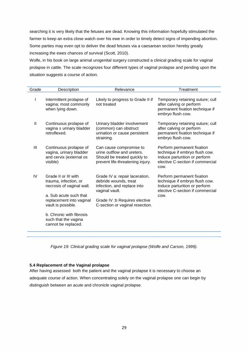
29
searching it is very likely that the fetuses are dead. Knowing this information hopefully stimulated the
farmer to keep an extra close watch over his ewe in order to timely detect signs of impending abortion.
Some parties may even opt to deliver the dead fetuses via a caesarean section hereby greatly
increasing the ewes chances of survival (Scott, 2010).
Wolfe, in his book on large animal urogenital surgery constructed a clinical grading scale for vaginal
prolapse in cattle. The scale recognizes four different types of vaginal prolapse and pending upon the
situation suggests a course of action.
Grade Description Relevance Treatment
I
Intermittent prolapse of vagina; most commonly when lying down.
Likely to progress to Grade II if not treated
Temporary retaining suture; cull after calving or perform permanent fixation technique if embryo flush cow.
II Continuous prolapse of vagina ± urinary bladder retroflexed.
Urinary bladder involvement (common) can obstruct urination or cause persistent straining.
Temporary retaining suture; cull after calving or perform permanent fixation technique if embryo flush cow.
III Continuous prolapse of vagina, urinary bladder and cervix (external os visible)
Can cause compromise to urine outflow and ureters. Should be treated quickly to prevent life-threatening injury.
Perform permanent fixation technique if embryo flush cow. Induce parturition or perform elective C-section if commercial cow.
IV Grade II or III with trauma, infection, or necrosis of vaginal wall. a. Sub acute such that replacement into vaginal vault is possible. b. Chronic with fibrosis such that the vagina cannot be replaced.
Grade IV a: repair laceration, debride wounds, treat infection, and replace into vaginal vault. Grade IV :b Requires elective C-section or vaginal resection.
Perform permanent fixation technique if embryo flush cow. Induce parturition or perform elective C-section if commercial cow.
Figure 19: Clinical grading scale for vaginal prolapse (Wolfe and Carson, 1999).
5.4 Replacement of the Vaginal prolapse
After having assessed both the patient and the vaginal prolapse it is necessary to choose an
adequate course of action. When concentrating solely on the vaginal prolapse one can begin by
distinguish between an acute and chronicle vaginal prolapse.
30
5.4.1 Acute Vaginal Prolapse
If the verdict sounds that the prolapse is acute and the vaginal mucosa is in good condition it is time
to reposition the vaginal prolapse and to select the most appropriate fixation technique. As discussed
in case one, it is sometimes desirable to simple reposition the vaginal prolapse and to combat any
recurrence by minimally invasive methods, such as raising the hindquarter of the patient, placing a
Dalton spoon or placing the patient in a harness. Most frequently however, the utilization of a more
permanent retention technique is desired
5.4.1.1 Epidural Anesthesia
We have already emphasized various times the need to take certain hygienic measures before
handling the vaginal prolapse. When it comes to repositioning of the prolapse a next point of attention
arises: epidural anesthesia.
Naturally when a farmer decides to take matters into his own hands an epidural is not amongst the
options. Veterinarians however do have the option to perform an epidural anesthesia before
repositioning and this should be seriously considered. In many cases, even when deciding to simple
reposition the vaginal prolapse without the intention to utilize a more permanent retention techniques,
an epidural can be advised, as this does not only simplify the reposition, it also combats the direct
reoccurrence of the vaginal prolapse.
In cows a caudal epidural is considered an easy and inexpensive method of analgesia. This form of
anesthesia is thus commonly utilized for obstetric manipulations. In most cases and definitely in the
case of an acute vaginal prolapse desensitization of the 3rd
forth and 5th sacral nerve is sufficient and
thus one will always opt for a low caudal epidural between the 1st and second coccygeal space. Ideally
the hair dorsal to the site where one is planning on administering the anesthetic should be clipped and
the skin scrubbed and disinfected. In order to locate the area between the 1st and 2
nd coccygeal space
the tail should be moved up and down. An 18 gauge needle is placed in the located fossa at a 45
degree angle and then pushed forward to penetrate the skin and enter the epidural space. Once the
needle is in place the syringe may be attached and if no resistance is felt the anesthetic can be slowly
injected. To determine whether the needle is placed correctly one may put a drop of anesthetic on it
and observe whether it is drawn into the epidural space by negative pressure (Edmondson, 2008). The
ability to inject through the needle without any pressure is by many considered sign enough that the
needle is placed correctly. The dosage of local anesthetic to be used naturally depends upon the
product, when using procaine 4% a 2 cc injection will for instance suffice.
In sheep an effective caudal analgesia can be realized by placing an epidural injection with xylazine
and Lidocaine in between the sacrococcygeal articulation. Lidocaine alone can provide caudal
analgesia for up to 4 hours following epidural injection, but when injecting too high volumes paresis
and recumbence may result. Xylazine on the other hand has the capacity to desensitize sensory nerve
fibers without effecting motor fibers to the same extend, hereby leaving the patient standing. Two
milliliters of solution containing 0.07mg/kg xylazine and 0.5mg/kg of lidocaine is the recommended
dosage to bring about caudal analgesia for approximately 24 hours (Scott et al., 1995). The
sacrococcygeal articulation can be identified by slight vertical movement of the base of the tail. The
31
site must also be clipped and the skin scrubbed and disinfected. A 20 gauge needle may then be
inserted at an angle of 20 degrees to the horizontal plane. The lack of contact with bone during needle
insertion combined with the fact that there is no resistance to injection of the solution indicate the
correct placements of the epidural (Scott, 1996).
Not only does the epidural combat straining in both sheep and cattle, it also desensitizes the perineal
area and vulva lips making it possible to painlessly place a retention suture.
5.4.1.2 Retention Sutures
There are many different suture techniques that one may encounter in practice. With this said, there
are few suture techniques that are considered good. The insertion of metal pins through the vulva lip,
in any form shape or size is best to be avoided and the same goes for suturing closed the vulva with a
diverse range of creative suture patterns. What is wrong with these techniques is that they penetrate
the vulvar and/or vaginal mucosa. The combination of a mucosa penetrating wound with urine
scalding and bacterial contamination will at its best lead to a mild infection but is more likely to lead to
large diphtheritic areas that cause considerable discomfort and tenesmus (Scott, 2010).
In recent years the Buhner suture has proven itself by far the most secure method for the retainment
of a vaginal prolapse in both cows and ewes. The procedure for placing such a suture is simple, the
method is effective and the mucosa is left intact.
In case one we already described how to place a Buhner suture in a cow. After washing the involved
tissues, placing an epidural and repositioning the vaginal prolapse a Buhner needle is inserted through
the vulva. The needle enters the vulva through the fine-haired skin below and slightly lateral to the
ventral commissure of the vulva and reappeares at an equal distance above the dorsal vulvar
commissure. It is important that the Buhner suture is placed sufficiently deep into the perivaginal
tissue in order to give strength to the suture. After placing the needle, a piece of umbilical tape is
placed through the loop eye of the needle and the needle is removed via the dorsal opening, hereby
threading the umbilical tape through the full length of the right vulvar lip. The same course of action is
then repeated, this time inserting the needle a little bit to the left of where the umbilical tape appeared
above the dorsal vulvar commissure and ending underneath the ventral vulvar commisure. The result
of the described procedure should be a circular suture that is best tightened to allow no more than two
to three fingers to pass through the vulva opening.
A bowtie is made to facilitate an easy opening of the suture, making it unnecessary to redo the entire
suture every time access to the vagina is required, for instance when needing to perform a vaginal
exam.
The method described above is actually one of two traditional forms of the Buhner suture. When it
comes to Buhner sutures we can in fact distinguish between a perivaginal totally imbedded closing or
the perivaginal-percutaneous method. In the perivaginal-percutaneous method the knot present
underneath the ventral vulvar commisure rests on a strip of intact skin. To achieve this it is important
to make two small vertical incisions underneath the ventral vulva commisure several centimeters
apart. A single horizontal incision is made above the dorsal vulvar commisure The Buhner suture is
then placed using the incisions as points of entrance and exit for the needle and umbilical tape.
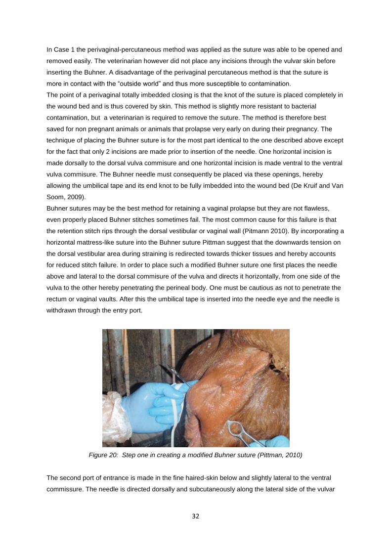
32
In Case 1 the perivaginal-percutaneous method was applied as the suture was able to be opened and
removed easily. The veterinarian however did not place any incisions through the vulvar skin before
inserting the Buhner. A disadvantage of the perivaginal percutaneous method is that the suture is
more in contact with the “outside world” and thus more susceptible to contamination.
The point of a perivaginal totally imbedded closing is that the knot of the suture is placed completely in
the wound bed and is thus covered by skin. This method is slightly more resistant to bacterial
contamination, but a veterinarian is required to remove the suture. The method is therefore best
saved for non pregnant animals or animals that prolapse very early on during their pregnancy. The
technique of placing the Buhner suture is for the most part identical to the one described above except
for the fact that only 2 incisions are made prior to insertion of the needle. One horizontal incision is
made dorsally to the dorsal vulva commisure and one horizontal incision is made ventral to the ventral
vulva commisure. The Buhner needle must consequently be placed via these openings, hereby
allowing the umbilical tape and its end knot to be fully imbedded into the wound bed (De Kruif and Van
Soom, 2009).
Buhner sutures may be the best method for retaining a vaginal prolapse but they are not flawless,
even properly placed Buhner stitches sometimes fail. The most common cause for this failure is that
the retention stitch rips through the dorsal vestibular or vaginal wall (Pitmann 2010). By incorporating a
horizontal mattress-like suture into the Buhner suture Pittman suggest that the downwards tension on
the dorsal vestibular area during straining is redirected towards thicker tissues and hereby accounts
for reduced stitch failure. In order to place such a modified Buhner suture one first places the needle
above and lateral to the dorsal commisure of the vulva and directs it horizontally, from one side of the
vulva to the other hereby penetrating the perineal body. One must be cautious as not to penetrate the
rectum or vaginal vaults. After this the umbilical tape is inserted into the needle eye and the needle is
withdrawn through the entry port.
Figure 20: Step one in creating a modified Buhner suture (Pittman, 2010)
The second port of entrance is made in the fine haired-skin below and slightly lateral to the ventral
commissure. The needle is directed dorsally and subcutaneously along the lateral side of the vulvar
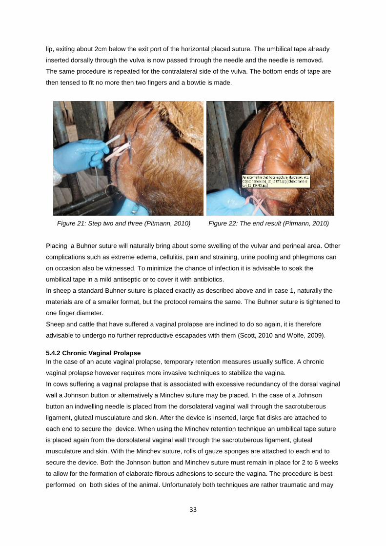
33
lip, exiting about 2cm below the exit port of the horizontal placed suture. The umbilical tape already
inserted dorsally through the vulva is now passed through the needle and the needle is removed.
The same procedure is repeated for the contralateral side of the vulva. The bottom ends of tape are
then tensed to fit no more then two fingers and a bowtie is made.
Figure 21: Step two and three (Pitmann, 2010) Figure 22: The end result (Pitmann, 2010)
Placing a Buhner suture will naturally bring about some swelling of the vulvar and perineal area. Other
complications such as extreme edema, cellulitis, pain and straining, urine pooling and phlegmons can
on occasion also be witnessed. To minimize the chance of infection it is advisable to soak the
umbilical tape in a mild antiseptic or to cover it with antibiotics.
In sheep a standard Buhner suture is placed exactly as described above and in case 1, naturally the
materials are of a smaller format, but the protocol remains the same. The Buhner suture is tightened to
one finger diameter.
Sheep and cattle that have suffered a vaginal prolapse are inclined to do so again, it is therefore
advisable to undergo no further reproductive escapades with them (Scott, 2010 and Wolfe, 2009).
5.4.2 Chronic Vaginal Prolapse
In the case of an acute vaginal prolapse, temporary retention measures usually suffice. A chronic
vaginal prolapse however requires more invasive techniques to stabilize the vagina.
In cows suffering a vaginal prolapse that is associated with excessive redundancy of the dorsal vaginal
wall a Johnson button or alternatively a Minchev suture may be placed. In the case of a Johnson
button an indwelling needle is placed from the dorsolateral vaginal wall through the sacrotuberous
ligament, gluteal musculature and skin. After the device is inserted, large flat disks are attached to
each end to secure the device. When using the Minchev retention technique an umbilical tape suture
is placed again from the dorsolateral vaginal wall through the sacrotuberous ligament, gluteal
musculature and skin. With the Minchev suture, rolls of gauze sponges are attached to each end to
secure the device. Both the Johnson button and Minchev suture must remain in place for 2 to 6 weeks
to allow for the formation of elaborate fibrous adhesions to secure the vagina. The procedure is best
performed on both sides of the animal. Unfortunately both techniques are rather traumatic and may
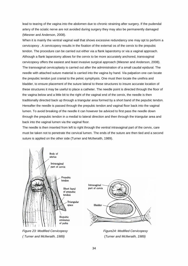
34
lead to tearing of the vagina into the abdomen due to chronic straining after surgery. If the pudendal
artery of the sciatic nerve are not avoided during surgery they may also be permanently damaged
(Miesner and Anderson, 2008).
When it is mainly the ventral vaginal wall that shows excessive redundancy one may opt to perform a
cervicopexy . A cervicopexy results in the fixation of the external os of the cervix to the prepubic
tendon. The procedure can be carried out either via a flank laparotomy or via a vaginal approach.
Although a flank laparotomy allows for the cervix to be more accurately anchored, transvaginal
cervicopexy offers the easiest and least invasive surgical approach (Miesner and Anderson, 2008).
The transvaginal cervicoplasty is carried out after the administration of a small caudal epidural. The
needle with attached suture material is carried into the vagina by hand. Via palpation one can locate
the prepubic tendon just cranial to the pelvic symphysis. One must then locate the urethra and
bladder, to ensure placement of the suture lateral to these structures to insure accurate location of
these structures it may be useful to place a catheter. The needle point is directed through the floor of
the vagina below and a little bit to the right of the vaginal end of the cervix, the needle is then
traditionally directed back up through a triangular area formed by a short band of the pepubic tendon.
Hereafter the needle is passed through the prepubic tendon and vaginal floor back into the vaginal
lumen. To avoid breaking of the needle it can however be adviced to first pass the needle down
through the prepubic tendon in a medial to lateral direction and then through the triangular area and
back into the vaginal lumen via the vaginal floor.
The needle is then inserted from left to right through the ventral intravaginal part of the cervix, care
must be taken not to penetrate the cervical lumen. The ends of the suture are then tied and a second
suture is applied on the other side (Turner and McIlwraith, 1989).
Figure 23: Modified Cervicopexy Figure24: Modified Cervicopexy
( Turner and McIlwraith, 1989) (Turner and McIlwraith, 1989)
35
Disadvantages associated with cervicopexy include entrapment of the urethra, bladder or intestines,
increased risk for bacterial infection, compromise of the cervical lumen and suboptimal positioning of
the vagina (Miesner and Anderson, 2008).
Vaginoplasty and vaginal resection are effective methods for combating vaginal prolapse and they
may be applied to both dorsal and ventral wall prolapse (Miesner and Anderson, 2008). As with all the
techniques discussed previously these techniques are also carried out on the standing animal, after
placing of a small epidural. In the case of a vaginoplasty a triangular segment of the dorsolateral wall
is removed on both sides, with the base of the triangle facing the dorsal midline. The sides are then
sutured together. This method although effective in combating vaginal prolapse, makes the animal
unsuitable for natural servicing and delivery.
When performing a vaginal resection, a complete segment of the vagina is generally removed. A
vaginal speculum is inserted into the lumen of the prolapsed vagina and pins are placed through the
vaginal prolapse to stabilize it for surgery. The injures portion of the prolapse is then removed and an
end to end anastomosis is performed. The procedure can be compared to a rectum amputation
Complications such as stricture, dehiscence, bleedings and abscesses are not uncommon. Once
again the patient is made unfit for natural servicing and delivery.
In sheep one can apply methods such as the Minchev suture, or Johnson button, but this is hardly
ever done. Realistically speaking, chronic vaginal prolepses in both cattle and sheep are usually not
treated, instead the effected animals are culled.
36
6. Conclusion
When it comes to the causes of vaginal prolapse there is still a lot that remains unknown. Although
there are plenty of theories regarding the cause of vaginal prolapse, the scientific evidence supporting
these theories is frequently lacking. Newfound insight concerning the altered metabolism of collagen in
patients suffering from a vaginal prolapse, is probably the beginning of a more concrete scientific
understanding of the pathology.
With respect to the treatment of vaginal prolapse there are a lot of alternatives, nevertheless I find that
there are only a few good options.
Strictly speaking the management of vaginal prolapse in both cows and ewes can be narrowed down
to a few basic steps: assessment, replacement and retention. All these steps should be undertaken
swiftly but only after instigating the appropriate hygienic measures. Additionally I believe that an
epidural ought to be utilized when replacing a vaginal prolapse of any significance. When it comes to
retaining the vaginal prolapse an acute vaginal prolapse should be combated with either a noninvasive
retention method, such as a Dalton spoon or a harness, or if more thorough measures are necessary
a Buhner suture should be placed. A chronic vaginal prolapse is best not treated, unless of course this
is specially requested by the owner. Naturally in practice every case is unique and thus every case
should be approached with an open mind and a healthy dose of logic.
37
References
Adams, N. R. 1990. Permanent infertility in ewes exposed to plant oestrogens. Australian Veterinary
Journal 67, 197.
Adams N.R. and Saunders M.R. (1993). Development of uterus-like redifferentiation in the cervix of
the ewe after exposure to estradiol-17 beta. Biology of Reproduction. 48(2), 357-362.
Adams N.R. (1995). Detection of the effects of phytoestrogens on sheep and cattle. Journal of Animal
Science 73, 1509-1515.
Anonymous. (2010). Vervoerder van Dieren. Course Dierengezondheidzorg, Drongen, p15.
Adlercreutz H., Honjo H., Higashi A., Fotsis T., Hamalainen E., Hasegawa T., Okada H. (1991).
Urinary excretion of lignans and isoflavonoid phytoestrogens in Japanese men and women consuming
a traditional Japanese diet. Journal of Clinical Nutrition 54, 1093.
Anderson J.J.B., Anthony M., Messina M., Garner S.C. (1999). Effects of phyto-oestrogens on tissues.
Nutrition Research Reviews 12, 75–116.
Budras K.D. and Habel R.E. (2003). Perineum, Pelvic Diaphragm, Ischiorectal fossa and Tail. In:
Budras K.D (editor). Bovine anatomy. Schlutersche, Hannover, Germany, p. 92-96.
Clark I.M., Powell L.K., Cawston T.E. (1994). Tissue inhibitor of metaloproteinases (TIMP-1)
stimulates the secretion of collagenase from human skin fibroblast. Biochemical and Biophysical
research communications 203, 874-880.
Croubels S. and De Backer P. (2009) Diergeneeskundige Toxicologie. Course faculty of veterinary
medicine, Ghent, 185.
De Kruif A. and Van Soom A.(2009). Verloskunde van de Huisdier 2. Course faculty of veterinary
medicine, Ghent.
Deprez P. (2009). Propedeutica van de grote huisdieren. Course Faculty of Veterinary Medicine,
Ghent.
Drost M. (2007). Complications during gestion in the cow. Theriogenology 68, 487-491.
Edmondson M.A (2008). Local and Regional Anesthesia in Cattle. Veterinary Clinics Food Animal
Practice 24, 211-226.
38
Ennen S., Kloss S., Scheiner-Bobis G., Failing K., Wehrend A. (2011). Histological, hormonal and
biomolecular analysis of the pathogenesis of ovine Prolapsus vaginae ante partum. Theriogenology
75, 212-219.
Folman Y. and Pope G.S. (1966). The interaction in the immature mouse of potent oestrogens with
coumestrol, genistein and other uterovaginotrophic compounds of low potency. Journal of
Endocrinology 34, 215.
Fylling Petter. (1970). The effect of pregnancy, ovariectomie and parturition on plasma progesterone
level in sheep. Acta Endocrinologica, 65(2), 273-283.
Hafez E.S.E. and Hafez B. (2000). Gestion, Prenatal physiology and Parturition. In: Balado D. (editor)
Reproduction of Farm Animals. 7th edition, Williams & Wilkins, Maryland, USA, p 141.
Henricks D.M., Dickey J.F., Hill J.R., Johnston W.E. (2011). Plasma Estrogen and Progesterone
Levels After Mating, and During Late Pregnancy and Postpartum in Cows. Endocrinology 90(5), 1336-
1342.
Hosie B. (1989). Vaginal rolapse and rupture in sheep. In Practice 11(5), 215-218.
Hosie B.D., Low J.C., Bradley H.K., Robb J. (1991). Nutritional factors associated with vaginal
prolapse in ewes. Veterinary Record 128, 204-208.
Hunnam J.C., Parkinson T.K., McDougall S. (2009). Transcutaneous ultrasound over the right flank to
diagnose mid- to late-pregnancy in the dairy cow. Australian Veterinary Journal 87(8), 313-317.
Intervet International. (2011). Pregnancy diagnosis in heifers and cows. Internet reference:
http://www.partners-in-reproduction.com/reproduction-cattle/pregnancy-diagnosis.asp (consulted on
the 20th of March 2011).
Jack G.S., Nikolova G., Vilain E., Raz S., Rodriguez L.V. (2005). Familiar transmission of
genitovaginal prolapse. Internation Urogynecology Journal 17(5), 498-501.
Jeffrey J.J. (1991) Collagen and collagenase: Pregnancy and parturition. Seminars in Perinatology 15,
118-126.
Kahn C. ed. Merck veterinary manual. 9th ed. Rahway, NJ: Merck, 2005.
Kambadur R., Sharma M., Smith T.P.L., Bass J.J. (1997). Mutations in myostatin (GDF8) in Double-
Muscled Belgian Blue and Piedmontese Cattle. Genome Research 7, 910-915.
König H.E. and Liebich H.G. (2004). Female Genital Organs. In: König H.E. and Liebich H.G. (editors)
Veterinary Anatomy of Domestic Mammels. First edition, Schattauer, Stuttgart, Germany. p. 407-413.
39
Kroeze C.J and De Vries C.J. (2008). VWA toelichting op de transportverordening en de hygiene
verordeningen en de gevolgen voor de noodslachting van als landbouwhuisdier gehouden hoefdieren.
Internet reference: www.vwa.nl (consulted on the 30th of march 2011).
Lammoglia M.A., Bellows R.A., Short R.E., Bellows S.E., Bighorn E.G., Stevenson J.S., Randel R.D.
(1997). Body Temperature and Endocrine Interactions Before and After Calving in Beef Cows. Journal
of Animal Science 75, 2526-2534.
Lightfoot R. J., Croker K.P., Neil H.G. (1967). Failure of sperm transport in relation to ewe infertility
following prolonged grazing on oestrogenic pastures. Australian Journal of Agricultural Research 18,
755.
Lloyd A.L. (2011) Effect of relaxin on parturition in ruminants. Internet reference:
http://www.extension.iastate.edu/Pages/ansci/beefreports/asl-1465.pdf (consulted on april 2nd 2011).
Low J.C. and Sutherland H.K (1987). A census of the prevalence of vaginal prolapse in sheep flocks in
the Borders region of Scotland. The Veterinary Record 120(24), 571-575.
Miesner M.D., Anderson D.E., (2008). Management of Uterine and Vaginal Prolapse in the Bovine.
Veterinary Clinics Food Animal Practice 24, 409-419.
Olsen B.R. and Ninomiya Y. (1999). Fibrillar collagens. In: Kreis T. and Vale R. (editors) Guidebook to
the extracellular matrix, anchor and adhesion proteins. Sambook and Tooze Publications at Oxford
University Press, Oxford, UK, p. 383-387.
Ososki A. L. and Kennelly E. J. (2003). Phytoestrogens: a review of the present state of research.
Phytotherapy Research 17, 845–869.
Osinga A. (1978). Endocrine aspects of bovine dystocia with special reference to estrogens.
Theriogenology 10 (2-3), 149-166.
Pelzer D.K. (2008). Vaginal Prolapse. Internet reference:
http://www.danekeclublambs.com/VaginalProlapse.html (Consulted on April 5th 2011).
Pittman T. (2010). Practice Tips. Canadian Veterinary Journal 51(12), 1347-1348.
Powell J. (2005). Livestock Health Series: Reproductie Prolapses of Cattle. Fact Sheet # FSA 3102.
Arkansas Cooperative Extension Service, Little Rock, AR. 7-05.
Pugh D.G. (2002). Theriogenology of Sheep and Goats. In: Schrefer J. A. (editor) Sheep and Goat
Medicine. first edition, Saunders, Philadelphia, USA, p. 173.
Purohit G. (2010). Methodes of Pregnancy Diagnosis in Domestic Animals: the Current Status.
WebmedCentral REPRODUCTION 2010;1(12):WMC001305
Purohit G. (2010). Parturition in Domestic Animals: A Review. WebmedCentral REPRODUCTION 2010;1(10):WMC00748
40
Rexroad C.E., Casida L.E., Tyler W.J. (1974). Crown-Rump Length in Purebred Holstein-Friesian
Cows. Journal of Dairy Science 57(3), 346-347.
Riding G.A., Lehnert S.A., French A.J., Hill J.R. (2007) Conceptus-related measurements during the
first trimester of bovine pregnancy. The Veterinary Journal 175, 266-272.
Samual C.S., Coghlan J.P., Bateman J.F. (1998). Effects of relaxin, pregnancy and parturition on
collagen metabolism in the rat public symphysis. Journal of Endocrinology 159, 117-125.
Scott P. (1996). Caudal analgesia in sheep. In Practice 18, 383-384.
Scott P.R. (2010) Sheep Medicine. Mason publishing Ltd, London.
Scott P. and Gessert M.(1998). Management of ovine vaginal prolapse. In Practice 20(1), 28-34.
Scott P.R., Sargison N.D., Penny C.D., and Strachan W.D. (1995). The Use of Combined Xylazine
and Lignocaine epidural injection in ewes with vaginal or uterine prolapses. Theriogenology 43, 1175-
1178.
Sherwood O.D., Crnekovic V.E. (1979). Development of a homologous radioimmunoassay for rat
relaxin. Journal of Endocrinology 104, 893–897.
Sherwood O.D., Crnekovic V.E., Gordon .W.L., Rutherford J.E. (1980). Radioimmunoassay of relaxin
throughout pregnancy and during parturition in the rat. Journal of Endocrinology 107, 691–698.
Sjaastad O., Hove K., Sand O. (2003). The Kidneys and the Urinary Tract. In: Steel C. (editor)
Physiology of Domestic Animals. First edition, Gummerus printing, Finland, p. 447.
Söderberg, M. W., Falcone C., Byström B., Malmström A., Ekman G. (2004). Young women with
genital prolapse have a low collagen concentration. Acta Obstetricia et Gynecologica Scandinavica,
83, 1193–1198.
Stouw C.J. (2011). Zwartblesschaap. Internet reference: www.zwartblesschaap.nl (consulted on the
31st of March 2011).
Tandon B. (2004) Abortion in Dairy Cows New Insights and Economic Impacts. Vetcare Update
Bulletin 12(2) 4.
Thomas M., Langley B., Berry C., Sharma M., Kirk S., Bass J., Kambadur R. (2000). Myostatin, a
Negative Regulator of Muscle Broth, Functions by Inhibiting Myoblast Proliferation. The Journal of
Biological Chemistry 275, 40235-40243.
Timmermans J.P.(2005). Cel en Weefselleer 1. Course, Faculty of Veterinarian Medicine, Antwerp.
41
Turner S.A. and McIlwraith W.C. (1989). Cervicopexy For Vaginal Prolapse. In: Truner S.A and
McIlwraith W.C. (editors) Techniques in Large Animal Surgery. Second edition, Lippincott Williams &
Wilkins, Philidelphia, ISA, p.330-333.
Twiss C., Triaca V., Bergman J., Rodriguez L.V. (2007). The epidemiology, social burden, and
genetics of pelvic organ prolapse. Current Bladder Dysfunction Reports 3(2), 90-94.
Twiss C., Triaca V., Rodriguez V.L. (2007) Familial transmission of urogenital prolapse and
incontinence. Current Opinion in Obstetrics & Gynecology 19(5) 464-468.
Umburger H.S. (1991). Feeding Sheep. Virginia Cooperative Extension Publication, USA, 410-853.
Veeraiah G., Srinivas M. (2010). Spontaneous extrusion of the intestines and uterus as a squeal to
vaginal prolapse in a buffalo heifer: a case report. Buffalo Bulletin 29(1), 60-64.
Weber E. (1910) Beobachtung uber die rektaltemperatur des gesunden rindes, zugleich ein beitrag
zur frage der vorausbestimmung der zeit der geburt bel der kuh. Dtsch. Tieraerztl. Wochenschr. 18,
143.
Woclawek-Potocka I., Okuda K., Acosta T.J., Korzekwa A., Pilawski W., Skarzynskia D.J. (2005).
Phytoestrogen metabolites are much more active than phytoestrogens themselves in increasing
prostaglandin F2α synthesis via prostaglanin F2α synthase-like 2 stimulation in bovine endometrium.
Prostagladines and other Lipid Mediators 78(1-4), 202-217.
Woclawek-Potocka I., Acosta T.J., Korzekwa A., Bah M.M., Shibaya M., Okuda K., Skarzynski D.J.
(2005). Phytoestrogens modulate prostaglandin production in bovine endometrium: cell type specificity
and intracellular mechanisms. Experimental Biology and Medicine 230, 366-333.
Wolfe D.F. (2009) Medical and Surgical Management of Vaginal Prolapse in Cattle. Presentation “81st
western veterinary conference”, Auburn, Februari 15th 2009.
Wolfe D.F. and Carson R.L. (1999). Surgery of the vestibule, vagina, and cervix: cattle, sheep and
goats. In: Wolfde D.F. and Moll H.D. (editors) Large animal urogenital surgery. 1st edition, Williams and
Wilkins, Baltimore, USA, p. 421-424.
Related Documents