GUIDELINES The obstetric and gynaecological management of women with inherited bleeding disorders – review with guidelines produced by a taskforce of UK Haemophilia Centre DoctorsÕ Organization C. A. LEE,* C. CHI, S. R. PAVORD, à P. H. B. BOLTON-MAGGS,§ D. POLLARD,* A. HINCHCLIFFE-WOOD – and R. A. KADIR – *Katharine Dormandy Haemophilia Centre and Haemostasis Unit, Royal Free Hospital; Department of Obstetrics and Gynaecology, Royal Free Hospital, London; àDepartment of Haemostasis and Thrombosis, Directorate of Haematology, Leicester Royal Infirmary, Leicester; §Manchester Haemophilia Comprehensive Care Centre, Manchester Royal Infirmary, Manchester; and –Haemophilia Society, Women Bleed Too, London, UK Summary. The gynaecological and obstetric manage- ment of women with inherited coagulation disorders requires close collaboration between obstetrician/ gynaecologists and haematologists. Ideally these women should be managed in a joint disciplinary clinic where expertise and facilities are available to provide comprehensive assessment of the bleeding disorder and a combined plan of management. The haematologist should arrange and interpret laborat- ory tests and make provision for appropriate replace- ment therapy. These guidelines have been provided for healthcare professionals for information and guidance and it is also intended that they are readily available for women with bleeding disorders. Keywords: carrier of haemophilia, guidelines, inher- ited bleeding disorders, UKHCDO, von Willebrand’s disease, women Introduction Women with inherited bleeding disorders are particularly at risk of bleeding complications from regular haemostatic challenges during menstruation and childbirth. In the last decade, there has been an international research interest in women with inherited bleeding disorders. This has led to considerable progress in the identification of obstetric and gynaecological problems in these women and raising clinical awareness amongst their care providers. Menorrhagia is the common- est bleeding symptoms in women with inherited bleeding disorders and could be the first or only presenting symptom. Childbirth also presents an intrinsic haemostatic challenge to these women. They require specialized and individualized care during pregnancy. Particular aspects of their obstetric management include preconceptual coun- selling, prenatal diagnosis and antenatal, intrapar- tum and postpartum care. We provide these guidelines based on collective clinical experience and the published literature. It is aimed to provide guidance for healthcare professionals in the man- agement of women with inherited bleeding dis- orders and is also available as information for the women. Search strategy Each section has been written by one or more mem- bers of the group who have a particular knowledge of the bleeding disorder or the gynaecological or obstetric management. The medical literature was searched using key words and cross-reference was Correspondence: Professor C. A. Lee, Katharine Dormandy Hae- mophilia Centre and Haemostasis Unit, Royal Free Hospital, Pond Street, London NW3 2QG, UK. Tel.: +44 7977 038202; fax: +44 1285 644126; e-mail: [email protected] Accepted after revision 8 June 2006 Haemophilia (2006), 12, 301–336 DOI: 10.1111/j.1365-2516.2006.01314.x Ó 2006 Blackwell Publishing Ltd 301

Welcome message from author
This document is posted to help you gain knowledge. Please leave a comment to let me know what you think about it! Share it to your friends and learn new things together.
Transcript
GUIDELINES
The obstetric and gynaecological management of womenwith inherited bleeding disorders – review with guidelinesproduced by a taskforce of UK Haemophilia Centre Doctors�Organization
C. A. LEE,* C. CHI,� S. R. PAVORD,� P. H. B. BOLTON-MAGGS,§ D. POLLARD,*
A. HINCHCLIFFE-WOOD– and R. A. KADIR�–*Katharine Dormandy Haemophilia Centre and Haemostasis Unit, Royal Free Hospital; �Department of Obstetrics and
Gynaecology, Royal Free Hospital, London; �Department of Haemostasis and Thrombosis, Directorate of Haematology,
Leicester Royal Infirmary, Leicester; §Manchester Haemophilia Comprehensive Care Centre, Manchester Royal Infirmary,
Manchester; and –Haemophilia Society, Women Bleed Too, London, UK
Summary. The gynaecological and obstetric manage-ment of women with inherited coagulation disordersrequires close collaboration between obstetrician/gynaecologists and haematologists. Ideally thesewomen should be managed in a joint disciplinaryclinic where expertise and facilities are available toprovide comprehensive assessment of the bleedingdisorder and a combined plan of management. Thehaematologist should arrange and interpret laborat-
ory tests and make provision for appropriate replace-ment therapy. These guidelines have been providedfor healthcare professionals for information andguidance and it is also intended that they are readilyavailable for women with bleeding disorders.
Keywords: carrier of haemophilia, guidelines, inher-ited bleeding disorders, UKHCDO, von Willebrand’sdisease, women
Introduction
Women with inherited bleeding disorders areparticularly at risk of bleeding complications fromregular haemostatic challenges during menstruationand childbirth. In the last decade, there has beenan international research interest in women withinherited bleeding disorders. This has led toconsiderable progress in the identification ofobstetric and gynaecological problems in thesewomen and raising clinical awareness amongsttheir care providers. Menorrhagia is the common-est bleeding symptoms in women with inheritedbleeding disorders and could be the first or only
presenting symptom. Childbirth also presents anintrinsic haemostatic challenge to these women.They require specialized and individualized careduring pregnancy. Particular aspects of theirobstetric management include preconceptual coun-selling, prenatal diagnosis and antenatal, intrapar-tum and postpartum care. We provide theseguidelines based on collective clinical experienceand the published literature. It is aimed to provideguidance for healthcare professionals in the man-agement of women with inherited bleeding dis-orders and is also available as information for thewomen.
Search strategy
Each section has been written by one or more mem-bers of the group who have a particular knowledgeof the bleeding disorder or the gynaecological orobstetric management. The medical literature wassearched using key words and cross-reference was
Correspondence: Professor C. A. Lee, Katharine Dormandy Hae-
mophilia Centre and Haemostasis Unit, Royal Free Hospital, Pond
Street, London NW3 2QG, UK.Tel.: +44 7977 038202; fax: +44 1285 644126;
e-mail: [email protected]
Accepted after revision 8 June 2006
Haemophilia (2006), 12, 301–336 DOI: 10.1111/j.1365-2516.2006.01314.x
� 2006 Blackwell Publishing Ltd 301
made to already published relevant guidelines inorder to achieve conformity. A draft copy of theguideline was sent for consultation to the RoyalCollege of Obstetricians and Gynaecologists, RoyalCollege of Physicians, Royal College of Pathologists,Royal College of Anaesthetists, Royal College ofNursing, Royal College of Midwives, Royal Collegeof Paediatrics and Child Health, British Society forHaematology the Obstetric Haematology Group,World Federation of Haemophilia, The HaemophiliaSociety, Women Bleed Too, UKHCDO and theNational Institute of Clinical Excellence. The re-sponses were noted. Recommendations have beenbased on reports with the highest levels of evidence(Appendix 1).
Useful websites
1 Haemophilia society: http://www.haemophi-lia.org.uk; Women Bleed Too: http://www.wom-enbleedtoo.org.uk.
2 Royal College of Obstetricians and Gynaecolo-gists: http://www.rcog.org.uk.
3 United Kingdom Haemophilia Centre Doctors�Organisation: http://www.ukhcdo.org.
4 The Cochrane Library: http://www.cochrane.org.5 National Institute for Health and Clinical Excel-
lence: http://www.nice.org.uk.
Obstetrics
Haemophilia
Genetic counselling and pre-pregnancy careInheritance. Haemophilia A and B are sex-linked
recessive disorders with an incidence of one in 5000and one in 30 000 male births respectively. Thegenes for factor VIII (FVIII) and factor IX (FIX) arelocated on the X-chromosome. Carriers of haemo-philia have a 50% chance of passing on the genedefect to their offspring; in each pregnancy there is a50% chance of having an affected son and a 50%chance of having a daughter who will also be acarrier of the condition.
As the nature of haemophilia is consistent amongstfamily members, carriers can be informed whethertheir risk is for severe, moderate or mild haemophi-lia. There is also a genetic element to the develop-ment of inhibitors which is partly due to the effect ofthe same mutation, although other genetic factorsmay be involved [1].Genetic counselling. In families at risk of having a
child with haemophilia, assessment of carrier statusand counselling should ideally be carried out before
conception to allow considerations for suitablereproductive options. The aims of counselling areto provide prospective parents with adequate infor-mation that enables them to reach a decision that isappropriate to their situation and to provide themwith support throughout the process. Counsellingshould include assessment and discussion of thegenetic risk, the options of prenatal testing that areavailable with the limitations and potential compli-cations, and discussions on the subsequent choices ifthe fetus is found to be affected. This should beundertaken by a team of appropriate staff from thehaemophilia centre, the fetal medicine unit and theclinical genetics team. UKHCDO has producedguidelines on the framework of genetic serviceprovision for haemophilia and other inherited bleed-ing disorders [2].Reproductive options. Families known to be at risk
of transmitting an inherited bleeding disorder to theirchildren may have the options of: (i) conceivingnaturally and having prenatal diagnosis duringpregnancy with the option of termination if the fetusis found to be affected; (ii) declining prenatal testingand accepting the risk and consequences of having anaffected child; (iii) not having a child; (iv) adopting achild; (v) considering assisted conception techniquesusing donor egg or sperm as indicated; and (vi)considering preimplantation genetic diagnosis (PGD)with embryo selection. The decision is influenced byethnic and cultural issues, the severity of haemophiliain the family, and the personal/family experience ofthe disorder.Preimplantation diagnosis. PGD uses in vitro
fertilization (IVF) to create embryos, tests one ortwo cells from each embryo for the specific geneticabnormality and identifies unaffected embryos forthe transfer to the uterus, obviating the need forprenatal diagnosis and birth of an affected child. It isa relatively new technique and currently, evidence ofits effectiveness and safety is still limited [3]. Inhaemophilia, initially PGD only provided diagnosisof fetal sex but there have been reports of specificdiagnosis more recently [4]. It is likely to become arealistic option for more couples at risk of having achild affected by haemophilia in the near future.However, IVF is costly, stressful and the success rates(overall live birth rate �22%) are much lower thanspontaneous conception [5]. On current evidencePGD may be indicated in some individual cases aftercareful counselling and assessment but should not beregarded as a standard service [6]. It is required bythe Human Fertilisation and Embryology Authorityfor all PGD to be carried out in licensed centres. Bestpractice guidelines for clinical PGD and screening
302 C. A. LEE et al.
Haemophilia (2006), 12, 301–336 � 2006 Blackwell Publishing Ltd
have been produced by the European Society ofHuman Reproduction and Embryology PGD Con-sortium [7].
Pre-pregnancy counselling should be offered tocarriers of haemophilia to discuss suitablereproductive options and methods of prenataldiagnosis (grade C, level IV).
Pregnancy in carriers of haemophilia should bemanaged by a multidisciplinary team includingan obstetrician, haematologist and anaesthetist(grade C, level IV).
Prenatal diagnosis. This should be undertaken incentres with full genetic, haematological and obstet-ric expertise. There are invasive and non-invasivemethods available for prenatal diagnosis of haemo-philia.(i) Invasive methods for specific diagnosis. Chori-
onic villus sampling (CVS) is the principal methodused for prenatal diagnosis of haemophilia. Theprocedure is performed at 11–14 weeks of gestationunder ultrasound guidance to obtain a sample ofchorionic villi for analysis. It is recommended thatCVS should not be performed prior to 10 weeks ofgestation due to reports of its association with limbdefects when carried out before this gestation [8]. Ithas the advantage over amniocentesis of permittingdiagnosis in the first trimester. If termination ofpregnancy is opted for, it is possibly less traumaticand acceptable to the patient. Transabdominal CVScarries a similar risk of miscarriage as amniocentesiswhich is approximately 1–2% [9]. However, eachunit should audit the outcomes of invasive proce-dures performed and advise patients of the respectivecomplication rates.
Currently fetal sex cannot be determined with100% accuracy in the first trimester by ultrasound,which means female fetuses cannot be excluded andare exposed to the risk. Amniocentesis, which canalso be used for prenatal diagnosis, allows exclusionof female fetuses identified by ultrasound, but isperformed later than CVS (after 15 weeks of gesta-tion). This procedure is also performed under ultra-sound guidance to obtain a sample of amniotic fluidfor cell karyotyping and source of DNA. The rate ofmiscarriage associated with amniocentesis isapproximately 1% [10].
Cordocentesis, ultrasound guided fetal bloodsampling, was previously performed to obtain fetalblood for clotting factor assay. However, it is veryrarely performed today. It should only be consideredif all other possible techniques cannot be used or donot give conclusive results. This option may be
considered if a woman wishes to ensure that she doesnot have a child affected with severe haemophiliaand the causative mutation cannot be identified. Theprocedure is reported to have a 1.25% risk ofprocedure-related fetal loss when performed for non-chromosomal indications and by an experiencedoperator [11]. This risk is possibly higher if the fetusis affected with haemophilia due to the risk of cordbleeding. The levels of FVIII and FIX in a normalfetus at 19 weeks gestation are approximately 40and 10 IU dL)1, respectively [12,13], which aresignificantly lower than in an adult. It is thereforeimportant to ensure no maternal blood contamin-ation in the sample by checking the mean corpuscu-lar volume (MCV) of the erythrocytes (>120 fL forfetal MCV and � 90 fL for maternal MCV) or by theKleihauer technique showing resistance of fetalhaemoglobin to acid elution.Prophylaxis for invasive testing. As these proce-
dures are carried out early in pregnancy, the FVIIIlevel is unlikely to have risen significantly. Thereforeit is essential to check the mother’s clotting factorlevel (FVIII or FIX) and arrange prophylactic treat-ment for any invasive prenatal diagnostic test if thelevel is <50 IU dL)1. All invasive testing may causefeto-maternal haemorrhage; therefore, anti-D immu-noglobulin should be given to Rhesus D-negativemothers.
Chorionic villus sampling is the method of choicefor specific prenatal diagnosis of haemophilia.Maternal clotting factor level should be checkedprior to any invasive procedures and prophylactictreatment arranged if the level is <50 IU dL)1
(grade C, level IV).
Detailed guidelines on amniocentesis and CVS [14]and on the provision of clinical and laboratorygenetic services for haemophilia [2] are available.(ii) Non-invasive methods for determination of
fetal sex. Non-invasive methods are at presentlimited to fetal gender determination. Knowledge offetal sex is beneficial in pregnancies at risk ofhaemophilia. Its importance should be emphasizedto couples who do not wish to have prenataldiagnosis. If the fetus is identified as female, themother can be reassured especially when specificmutational diagnosis is not possible or when themother is unsure of her feelings towards terminationof pregnancy. The parents will then have theoption of avoiding invasive testing in these cases.Knowledge of fetal sex is also helpful for labourmanagement as invasive monitoring techniques,vacuum extraction and difficult forceps deliveries
INFORMATION AND GUIDANCE ON INHERITED BLEEDING DISORDERS 303
� 2006 Blackwell Publishing Ltd Haemophilia (2006), 12, 301–336

should be avoided when the coagulation status of themale fetus is unknown in order to minimize the riskof haemorrhagic complications. If the parents do notwish to know the sex of the baby, this informationshould be made available to the midwives andobstetricians caring for the women in labour.
Ultrasound assessment in the second and thirdtrimesters can accurately determine fetal sex [15–19].Improvements in the resolution of ultrasound equip-ments have now provided the possibility of detailedvisualization of the fetus in early pregnancy. Themorphological aspect of the external genitalia isidentical in both sexes until 11 weeks of gestation,but after this stage, there is rapid differentiation ofthe genitalia [20]. The direction at which the fetalphallus points in the mid-sagittal plan is differentbetween male (caudally) and female (anteriorly).These findings have been utilized by several groups toassess fetal gender from 11 weeks of gestation[21,22]. The sensitivity of this technique, however,is limited especially at 11–12 weeks of gestation. Inthe study by Whitlow et al. [21], the overall successof correctly assigning fetal gender increased withgestational age from 46% to 75%, 79% and 90% at11, 12, 13 and 14 weeks respectively. In the study byEfrat et al. [22], the accuracy of sex determinationalso increased with gestation from 70.3% to 98.7%and 100% at 11, 12 and 13 weeks respectively. Thesensitivity of fetal gender determination in the firsttrimester is limited by two factors. First, visualizationof the genital tubercle is not always possible at suchearly gestations. Secondly, differentiation of thegenital tubercle into the male or female phallus onlybegins at 11 weeks of gestation, so this sign cannotbe used prior to this stage. Due to these limitations,this technique has not been introduced into routinepractice and is currently only performed in special-ized centres.
An alternative non-invasive method of determiningfetal sex is by assessing free fetal DNA in thematernal circulation for the presence or the absenceof Y-chromosome-specific DNA sequence. Technicaladvances, such as the development of quantitativereal-time polymerase chain reaction, have allowedseveral groups to demonstrate a 100% sensitivity andspecificity in the detection of male fetuses [23–25].Free fetal DNA has been detected as early as the fifthweek of pregnancy [24], but concentrations, andsubsequently the sensitivity of the test, increase withadvancing gestation. There is the potential to deter-mine fetal sex using this technique prior to 11 weeks,the earliest gestation at which CVS would beperformed, hence avoiding invasive testing in femalefetuses. This technique, however, is at present still
being performed in research settings. The combineduse of two independent non-invasive techniques,ultrasound and analysis of free fetal DNA in mater-nal blood, as method of assessing fetal sex to increaseconfidence in the reliability of these tests has beenproposed recently [26].
Knowledge of fetal gender allows invasive testingto be avoided in female pregnancies and enablesappropriate management of labour and delivery;therefore, the importance of establishing fetal sexin pregnancies at risk of haemophilia should beconveyed to the parents (grade C, level IV).
Management plan. The patient’s choice and theresults of prenatal testing should be clearly docu-mented in the case note alongside the managementplan for pregnancy, labour and delivery. A multidis-ciplinary team of obstetricians, haematologists, nursespecialists and anaesthetists should be involved in theformulation of this plan.
Antenatal management. Factor VIII levels have beenshown to increase significantly in carriers of haemo-philia A during pregnancy. Although the majority ofpatients will develop levels within the normal range,the rise is variable, and a small proportion maystill have low levels at term [27,28]. In contrast,FIX levels do not rise significantly in carriers ofhaemophilia B [28].
The risk of bleeding in early pregnancy andmiscarriage is unknown in carriers of haemophilia,but there is evidence that the risk of antepartumhaemorrhage (bleeding from the genital tract afterthe 22nd week of pregnancy) is not increased[27,28].
Women may be exposed to various haemostaticchallenges during pregnancy such as invasive prena-tal diagnostic tests, termination of pregnancy andspontaneous miscarriage. All of these may be com-plicated by excessive and prolonged bleeding.
Factors VIII and IX levels should be checked atbooking, and at 28 and 34 weeks of gestation,especially in those with low pre-pregnancy levels(<50 IU dL)1). Monitoring in the third trimester isessential in order to plan the management of labourand the provision of prophylactic treatment tominimize the risk of postpartum haemorrhage (PPH).
Carriers of haemophilia should have their clottingfactor level (FVIII or FIX) checked at booking andat 28 and 34 weeks of gestation to allow appro-priate management of labour and delivery andto assess the need for prophylactic treatment(grade C, level IV).
304 C. A. LEE et al.
Haemophilia (2006), 12, 301–336 � 2006 Blackwell Publishing Ltd
Treatment Due to the significant rise of FVIII levelduring pregnancy, treatment with coagulation factorconcentrate is rarely required during pregnancy incarriers of haemophilia A [28]. On the contrary,carriers of haemophilia B with a low baseline levelare more likely to require haemostatic support tocover delivery, especially if Caesarean section isrequired as FIX level does not rise significantly inpregnancy.
If treatment is required in carriers of eitherhaemophilia A or B, recombinant products shouldbe regarded as the products of choice [29]. Plasma-derived clotting factor concentrates, treated with thecurrently available virucidal method have no risk oftransmitting the hepatitis B and C virus and humanimmunodeficiency virus [30]. However, they havethe potential to transmit hepatitis A and parvovirusB19 [31,32]. While not normally a serious infectionin non-immunocompromised adults, parvovirusinfection of the fetus may result in hydrops fetalisand fetal death.
Desmopressin [1-desamino-8-d-arginine vasopres-sin (DDAVP)] increases plasma levels of vonWillebrand factor (VWF) and FVIII in the bloodand thus has potential use in carriers of haemo-philia A. It has no effect on FIX levels, hence is ofno value in carriers of haemophilia B. The use ofDDAVP during pregnancy is controversial becauseof the potential risks of placental insufficiency dueto arterial vasoconstriction and of miscarriage orpreterm labour due to an oxytoxic effect [33].However, in contrast to naturally occurring vaso-pressin, DDAVP has minimal vasoconstrictive andoxytocic effects, consistent with its predominantV2 vasopressin receptor activity. There is a risk ofmaternal and/or neonatal hyponatraemia as a resultof DDAVP’s more potent and prolonged antidiu-retic effect, compared with that of natural hor-mone vasopressin [33]. Therefore, restriction offluid intake is required to accompany its use. Theefficacy and safety of DDAVP for prophylaxis ortreatment of pregnancy-associated bleeding havenot been systematically studied, but evidence of itssafety during pregnancy in women with diabetesinsipidus using smaller doses is available [34]. In aseries of 27 haemophilia A carriers with low FVIIIlevels who received DDAVP for coverage ofinvasive prenatal diagnostic procedures, there wasno serious side effect other than mild facialflushing and headache [35]. DDAVP does not passinto breast milk in significant amounts. Hence,DDAVP may be used in labour and during thepostpartum period.
Recombinant FVIII and FIX should be used as thetreatment of choice in pregnant carriers of hae-mophilia A and B (grade C, level IV).
Intrapartum management. Labour and delivery arecritical times for carriers of haemophilia and theiraffected child when they are exposed to varioushaemostatic challenges. Pregnancies in carriers ofhaemophilia should be managed in collaboration witha haemophilia centre and arrangement for deliveryshould be made in advance. It is recommended forwomen carrying an affected fetus to delivery at a unitwhere the necessary expertise in the management ofthis disorder and resources for laboratory testing andclotting factor treatments are readily available.
A delivery plan should be made in advance and forwomen carrying an affected fetus, delivery shouldbe planned at a unit where the necessary expertisein the management of this disorder and resourcesfor laboratory testing and clotting factor treat-ments are readily available (grade C, level IV).
Labour. During labour, the maternal coagulationscreen and appropriate factor assays should bechecked, as well as saving serum for cross-matching.If it is difficult to access factor levels in labour, it isacceptable to rely on the third trimester levels toformulate a plan. When the factor level is<50 IU dL)1, an intravenous line should be estab-lished and prophylactic treatment given for labourand the postpartum period.
Spontaneous labour should be allowed wherepossible and special consideration should be givenwhen labour is to be induced. Induced labour is likelyto be prolonged and associated with the need forinstrumental delivery or emergency caesarean sectionparticularly in primigravida women with unfavour-able cervix at the start of induction. In these cases, amultidisciplinary team of obstetrician and haematol-ogist along with the mother should perform a carefulrisk assessment. In some circumstances, electivecaesarean section could be considered less traumaticto both the mother and her affected son.Regional analgesia and anaesthesia. A plan of
management should be devised with the anaesthetistprior to labour/delivery and discussed with thepatient. The use of regional block in patients withbleeding disorders is controversial because of thepotential risk of epidural or spinal haemorrhage andhaematoma, which may lead to permanent neuro-logical damage. However, provided the coagulationscreen is normal and the relevant factor level is above50 IU dL)1, regional block is not contraindicated
INFORMATION AND GUIDANCE ON INHERITED BLEEDING DISORDERS 305
� 2006 Blackwell Publishing Ltd Haemophilia (2006), 12, 301–336
[28,36]. In situations where factor levels cannot beassessed due to advanced labour, provided the factorlevel is >50 IU dL)1 in the third trimester, it is thensufficient to assess the platelet count, partial throm-boplastin time (PTT) and prothrombin time (PT).Regional block in carriers of haemophilia should beperformed by an expert anaesthetist with the help ofa specialized haematologist for assessment of coagu-lation status and arrangement of treatment whenneeded. The epidural should be placed in the midline,which decreases the chance of intravascular puncturewith the epidural catheter [37], and the lowestconcentration of a local anaesthetic and narcoticmixture should be used to achieve analgesia so as tomaintain motor function [38]. The extent of motorblock should be assessed frequently until the anaes-thetic has worn off and the catheter removed. If thedegree of motor block is more than that expected orif the anaesthetic appears to be prolonged, magneticresonance imaging should be arranged to check fordevelopment of an epidural haematoma. It is import-ant to check factor levels prior to the removal ofepidural catheter as the pregnancy-induced rise infactor levels may quickly reverse after birth andbleeding in the spinal canal may then arise. Intra-muscular analgesia and non-steroidal anti-inflamma-tory drugs (NSAID) should be avoided if factor levelsare below normal.
Regional block in carriers of haemophilia is notcontraindicated if the coagulation screen is normaland the relevant factor level is above 50 IU dL)1
(or raised to >50 IU dL)1 by prophylactic treat-ment). It should be performed by an expertanaesthetist with the help of a specialized haema-tologist for assessment of coagulation status andarrangement of treatment if required (grade C,level IV).
Fetal monitoring. The use of fetal scalp electrodesand fetal blood sampling should be avoided inaffected male fetuses or when fetal sex or coagulationstatus of male fetus is unknown. Although nobleeding complication in affected fetuses and neo-nates has been reported so far from these procedures,it is advisable to avoid their use due to the potentialrisk of scalp haemorrhage in affected male fetuses.
The use of fetal scalp electrodes and fetal bloodsampling should be avoided in affected malefetuses or when fetal sex or coagulation status ofmale fetus is unknown (group C, level IV).
Delivery. Affected fetuses are at risk of serioushead bleeding, including cephalohaematoma andintracranial haemorrhage (ICH), from the process
of birth. The safest method of delivery for fetuses atrisk is controversial. In a survey in the USA, 11% ofobstetricians preferred to deliver pregnant carriersof haemophilia by caesarean section [39]. Ljunget al. [40] reviewed 117 children with moderate tosevere haemophilia born between 1970 and 1990and found 23 neonatal bleedings associated withdelivery. The risk of haemorrhage was 10% withvaginal delivery, 64% with vacuum extraction and23% with caesarean section. The risk of headbleeding specifically was 3% with vaginal delivery,64% with vacuum extraction and 15% withcaesarean section. It was concluded that the riskof serious bleeding during normal vaginal delivery issmall and that delivery of all fetuses at risk ofhaemophilia by caesarean section is not expected toeliminate this risk. However, the use of vacuumextraction or forceps, or prolonged labour especi-ally prolonged second stage of labour, should beavoided as they are associated with an increasedrisk of cephalohaematoma or intracranial bleeding[28,40].
In principle, fetuses at risk of haemophilia shouldbe delivered by the least traumatic method and earlyrecourse to caesarean section should be considered.Although vacuum extraction should not be used, lowforceps delivery may be considered less traumaticthan caesarean section when the head is deeplyengaged in the pelvis and it is expected to be an easyoutlet procedure. In these cases, the procedureshould be performed by an experienced obstetrician.Care should be taken in minimizing maternal genitaland perineal trauma in order to reduce the risk ofPPH.
Vacuum extraction, mid-cavity forceps and pro-longed labour should be avoided in affected malefetuses or when fetal sex or coagulation status ofmale fetus is unknown. Delivery should beachieved by the least traumatic method and earlyrecourse to caesarean section should be considered(grade C, level IV).
Postpartum management. The pregnancy-inducedrise in clotting factors (FVIII and VWF, not FIX)falls rapidly after delivery. Carriers of haemophiliaare at increased risk of both primary (defined as>500 ml blood loss in the first 24 h) and secondary(after the first 24 h) PPH. The incidences of primaryand secondary PPH in the general population areapproximately 5–8% and 0.8%, respectively[41,42]. In a study amongst haemophilia carriersthe incidence of primary and secondary PPH wereincreased at 22% and 11%, respectively [28].
306 C. A. LEE et al.
Haemophilia (2006), 12, 301–336 � 2006 Blackwell Publishing Ltd
Another study reported a significantly higher inci-dence of prolonged bleeding after delivery amonghaemophilia carriers (22%) in comparison with thecontrol group (6%) [43]. Five PPHs and a largeperineal haematoma were reported among 43 preg-nancies in haemophilia carriers [27]. It seems thatFVIII or FIX activity has a significant influence on therisk of bleeding in haemophilia carriers [28,43]. Thefactor level should ideally be checked daily afterdelivery. It should be maintained above 50 IU dL)1
for at least 3 days, or 5 days if caesarean section hasbeen performed, in order to minimize the risk ofprimary and secondary PPH. The risk of PPH can befurther reduced by active management of the thirdstage of labour [44] and minimizing maternal genitaland perineal trauma.
Active management of third stage should bepractised in carriers of haemophilia (grade C, levelIV).
In the event of PPH, after correction of hypovol-aemia, factor replacement therapy or treatment withDDAVP should be instituted in close collaborationwith the local haemophilia centre. Obstetric causesfor excessive bleeding should not be overlooked.Consideration should be given to tranexamic acid toreduce bleeding in cases of heavy lochia.
Factor levels should be monitored postdeliveryand maintained above 50 IU dL)1 for at least3 days, or 5 days if caesarean section has beenperformed (grade C, level IV).
Neonates. Neonates affected with haemophiliamay be at risk of bleeding at puncture sites, fromsurgical interventions (circumcision being the com-monest), and spontaneous bleeding such as bruising,organ and joint bleeding. If necessary, heel pricksshould be carried out carefully with pressure appliedto the site for a full 5 min afterwards. Any prolongedbleeding or excessive bruising at the site of the heelprick should be reported to the haemophilia teamimmediately.
Intramuscular injections and venepunctures shouldbe avoided in neonates affected with haemophiliaor whose coagulation status is unknown. VitaminK should be given orally and routine immuniza-tions should be given intradermally or subcutane-ously. Circumcision should be delayed until thecoagulation status of the neonate is known andappropriate management can be arranged by thehaematologist (grade C, level IV).
Cord blood sample. After delivery, a cord bloodsample should be obtained in a citrated tube and
transferred to coagulation laboratory for coagulationfactor assay within two hours. The result should beconveyed to the parents by an appropriate memberof the haemophilia team, usually one of the staff ofthe haemophilia centre. Early diagnosis allows forprevention of bleeds and appropriate management ofsuspected or documented bleeds.
Cord blood should be collected from all maleoffspring of carriers of haemophilia to assessclotting factor levels for identification and earlymanagement of newborns at risk. The results ofthe tests should be conveyed to the parents by anappropriate member of the haemophilia team(grade C, level IV).
Intracranial haemorrhage. The incidence of ICH inneonates with haemophilia has been estimated at 1–4% and these events are typically associated withsignificant morbidity and mortality [45]. A review ofbleeding episodes in 349 newborns with haemophiliain 66 publications found 366 bleeding episodes; headbleeding being the most common with intracranialbleeding and subgaleal/cephalohaematomas account-ing for 27% and 13% of all bleeding episodes [46]. Itis debated as to whether all haemophilic infantsshould have a routine cranial ultrasound. A normalscan does not exclude all bleeds. It is recognized thatcranial ultrasound is a relatively poor investigationfor the detection of subdural haemorrhage, which isthe commonest site of ICH in the neonates. Inaddition, the optimal time of scanning is unclearreflecting the typically delayed presentation of intra-cranial bleeding. A survey of haemophilia centres inthe UK showed that only 41% of respondents wouldroutinely perform cranial ultrasound on neonateswith severe haemophilia and 21% would performultrasound in the presence of clinical signs suggestiveof bleeding [47]. No evidence currently exists tosupport the routine use of cranial ultrasound. How-ever, it is recommended that if labour has beentraumatic, e.g. following forceps delivery or pro-longed labour, preterm or there are any clinical signssuggestive of bleeding, a cranial ultrasound scanshould be performed.
Community midwives should be informed ofaffected babies. They and the mothers should bemade aware of the early signs of ICH (e.g. lethargy,vomiting, seizures and poor feeding) as the reportedmean age at which the haemorrhage occurred is4.5 days (range, birth to 1 month) [45] when boththe mother and baby are usually discharged home.
The administration of routine prophylaxis postde-livery is controversial. It has been argued by those infavour of early prophylaxis that it is illogical to
INFORMATION AND GUIDANCE ON INHERITED BLEEDING DISORDERS 307
� 2006 Blackwell Publishing Ltd Haemophilia (2006), 12, 301–336
manage potential cranial trauma in neonates expect-antly [48]. However, recently, there have beenreports of an increased risk of inhibitor developmentwhen clotting factor is administered in the earlyneonatal period [49,50]. This association remains tobe clarified by larger studies as these studies did notspecifically address those children treated during thefirst few days of life and did not take into accountother potential risk factors for inhibitor develop-ment. Nevertheless, it is advisable to consider pro-phylaxis in traumatic or premature deliveries and ifthere is any suspicion of bleeding.
Cranial ultrasound/computed tomography (CT)scans should be arranged for all neonates withhaemophilia if labour had been premature, trau-matic, e.g. following forceps delivery or prolongedlabour, or if there are any clinical signs suggestiveof bleeding. In these cases it is recommended thatrecombinant clotting factor should be adminis-tered to raise the plasma clotting factor to100 IU dL)1 (grade C, level IV).
von Willebrand’s disease
Pre-pregnancy counselling and prenatal diagnosis.
von Willebrand’s disease is the commonest inheritedbleeding disorder with a prevalence of approximately1% in the general population [51]. It is generallyinherited as an autosomal condition, and thuschildren of either sex may inherit the condition.There are three main types of VWD. Type 1 is thecommonest accounting for � 70% of all cases. Types1 and 2 are transmitted as an autosomal dominanttrait. The risk of a woman with type 1 VWDtransmitting the disease to her child is 50%. How-ever, only 33% of children born to these women areclinically affected, probably because of variablepenetrance and expression of the abnormal gene[52]. The same is true for type 2A and most cases oftype 2B. However, the situation is more complicatedfor the other subtypes of type 2 VWD. Extensivefamily studies are required to assess this risk. Type 3VWD is an autosomal recessive disorder, and affec-ted individuals are either homozygotes or compoundheterozygotes. If a child with type 3 has already beenborn in the family, the risk of a subsequent childbeing affected is 25%.
Preconceptual counselling should include the op-tion of prenatal diagnosis. Antenatal diagnosis is notusually required or requested in types 1 and 2 VWDas the bleeding tendency is relatively mild. However,if the fetus is at risk of type 3 (severe) VWD, theparents may wish to consider antenatal diagnosis.
This should be planned in advance to allow thecausative mutation(s) or informative polymorphismsto be identified using one of the invasive methods ofprenatal diagnosis described above.
The option of prenatal diagnosis should bediscussed and offered to women with VWDwhose genetic mutation is identifiable, partic-ularly to those at risk of having a child with type3 VWD (grade C, level IV).
Antenatal management. Haemostatic response topregnancy is variable in different types and sub-types of VWD. In type I VWD there is usually aprogressive increase in FVIII coagulant activity(FVIII:C), VWF antigen (VWF:Ag) and VWF activity(VWF:AC), and correction of the bleeding time(BT) during pregnancy [27,53–55]. Most womenwith type 1 VWD achieve VWF levels in the normal(non-pregnant) range by the third trimester [56].Failure of primary haemostasis to improve signifi-cantly in pregnancy, especially in severely affectedtype I women, has been reported [57,58]. In aseries of 24 pregnancies in 13 women studiedretrospectively, it was noted that FVIII:C andVWF:Ag rose above baseline values by a factor ofat least 1.5 during pregnancy in most cases [58].However, a baseline VWF:AC of <15 IU dL)1 (4/14cases) was predictive of a third trimester level of<50 IU dL)1 and less marked improvements inVWF:AC [58].
In type 2 VWD, FVIII and VWF antigen levelsoften increase during pregnancy, but most studiesshow minimal or no increase in VWF activity levels,and a persistently abnormal pattern of multimers,reflecting the increased production of abnormal VWF[27,56,58]. Despite increased FVIII and VWF pro-duction during pregnancy, FVIII:C levels are oftenlow in women with subtype 2N VWD because ofimpaired binding by the abnormal VWF [59]. Insubtype 2B VWD thrombocytopenia may develop orworsen during pregnancy due to increased produc-tion of the abnormal intermediate VWF multimers,which bind to platelets and induce spontaneousplatelet aggregation [60].
Women with type 3 VWD show little or noincrease in their FVIII and VWF plasma levels[54,61].
Due to the great variability of haemostaticresponse of VWD to pregnancy, regular monitoringof VWF:Ag and VWF:AC together with FVIII:C isessential. This should be performed at presentation,prior to any invasive procedures, and in the thirdtrimester. The platelet count should also be monit-ored in women with type 2B VWD [60]. If VWF:AC
308 C. A. LEE et al.
Haemophilia (2006), 12, 301–336 � 2006 Blackwell Publishing Ltd
is <50 IU dL)1, consideration should be made forprophylactic treatment with a clotting factor con-centrate containing VWF to cover any invasiveprocedures and delivery.
A review of 84 pregnancies from 1980 to 1996 ofwomen with all types of VWD showed that 33%(28) reported vaginal bleeding in the first trimester.The overall spontaneous miscarriage rates were 21%in this study [54] and 22% in another study [62].This is not significantly different from the normalrate of 15% [63]. Even though there appears to be ahigher incidence of vaginal bleeding in the firsttrimester in women with VWD, there is no increasein the miscarriage rate [54,59]. It is likely that thesewomen present more readily if they experiencebleeding in early pregnancy. However, there is anincreased risk of bleeding complications associatedwith spontaneous miscarriage or elective termination[54,61,62]. In one study, 10% of spontaneous orelective abortions were complicated by excessivebleeding requiring transfusion. In addition, intermit-tent bleeding two weeks after miscarriage occurredin 30% of cases [54]. FVIII and VWF do not risesignificantly until the second trimester [58] by whichstage most miscarriages have already occurred.Therefore, factor levels should be checked in womenpresenting with spontaneous miscarriages and inthose opting for termination of pregnancy. Prophy-lactic treatment should be given when factor levelsare <50 IU dL)1.
Pregnancy in women with VWD should be man-aged by a multidisciplinary team including anobstetrician, haematologist and anaesthetist(grade C, level IV).
Factor levels including VWF:Ag, VWF:AC andFVIII:C should be checked at booking, 28 and34 weeks and prior to invasive procedures. Pro-phylactic treatment should be given when factorlevels are <50 IU dL)1 to cover invasive proce-dures and delivery (grade C, level IV).
Treatment. Desmopressin increases plasma FVIIIand VWF levels. However, its use in pregnancy iscontroversial as discussed previously in its use incarriers of haemophilia. Multiple reports havesuggested its effectiveness in the prevention orcontrol of bleeding at the time of abortion anddelivery without complications [56,58,64,65]. In onesurvey, 50% and 34% of haematologists reportedusing intravenous and intranasal DDAVP, respect-ively, for PPH in women with type 1 VWD. And only31% considered pregnancy as a contraindication[66]. DDAVP is not contraindicated in uncompli-
cated pregnancy although like all drugs it should beused with caution. Prolonged administration inpregnancy should be avoided and close monitoringfor water retention is important [67]. A fluidrestriction of 1 L for 24 h after DDAVP shouldminimize the risk of fluid overload and consequenthyponatraemia.
Desmopressin can be used in pregnancy, butrepeated administration or use in pregnanciescomplicated with pre-eclampsia must be avoided.Close monitoring for water retention must accom-pany its use (grade C, level IV).
Virally inactivated concentrate containing VWF isthe treatment of choice in women with VWDunresponsive to DDAVP for preventing or control-ling pregnancy-associated bleeding.
If FVIII:C or VWF:AC is <50 IU dL)1, prophy-lactic administration of concentrate containing VWFshould be given to minimize maternal haemorrhagiccomplications [27,54]. Prophylactic infusion shouldstart at the onset of labour, with the aim of raisingFVIII:C and VWF:AC to >50 IU dL)1; this should bemaintained for at least 3 days after vaginal deliveryand at least 5 days after caesarean section [68].DDAVP can be used in women with type 1 VWDand in some with type 2A, as there is usually a goodresponse to this treatment with no risk of viraltransmission. DDAVP should generally be avoidedin women with type 2B VWD because it canprecipitate thrombocytopenia [69]; platelet transfu-sions have been given when the platelet count was<20 · 109 L in type 2B [60]. Management of type2N VWD using recombinant FVIII has been repor-ted [70]. Women with type 3 VWD do not respondto DDAVP and as FVIII:C and VWF:AC do notincrease in these cases during pregnancy, treatmentis required with concentrate containing VWF tocover delivery.
Women with type I VWD generally do not requireprophylactic treatment for delivery. In type 2VWD, treatment is required for operative deliveryor if there is perineal trauma. Women with type 3VWD require treatment for all types of delivery(grade C, level IV).
Tranexamic acid can also be used in the preventionor control of PPH. Although there have not beenstudies of antifibrinolytic therapy during pregnancyin women with VWD, tranexamic acid has been usedto control or prevent bleeding from placental abrup-tion, caesarean section or other obstetric causeswithout apparent maternal or fetal adverse effect[71–74].
INFORMATION AND GUIDANCE ON INHERITED BLEEDING DISORDERS 309
� 2006 Blackwell Publishing Ltd Haemophilia (2006), 12, 301–336
Intrapartum management Management of labour anddelivery are similar to that of carriers of haemophilia.It is important to establish both the type and plasmalevels of FVIII and VWF so as to plan the manage-ment of labour and the provision of prophylactictreatment.
Arrangements for delivery should be made inadvance. It is recommended for women with severeVWD to delivery at a unit where the necessaryexpertise in the management of this disorder andresources for laboratory testing and clotting factortreatments are readily available.
A delivery plan should be made in advance. Forwomen with severe VWD, delivery should beplanned at a unit where the necessary expertise inthe management of this disorder and resources forlaboratory testing and clotting factor treatmentsare readily available (grade C, level IV).
Epidural anaesthesia. Several case reports haveshown women with VWD received epidural anaes-thesia without bleeding complications [61,75–77]. Ina series, eight women with VWD received regionalanaesthesia during labour and delivery withoutbleeding complications and only one woman receivedprophylactic therapy as the clotting factor levels were>50 IU dL)1 in the other cases [54]. Epidural anaes-thesia may be considered for use in the majority ofwomen with type 1 VWD whose levels have risen to>50 IU dL)1. However, the decision of its use needsto be made jointly by an experienced anaesthetist,obstetrician and haematologist after considerationsare given to haemostatic concerns such as the degreeof correction of the plasma FVIII:C and VWF levels,possible degree of residual platelet impairment,possible rate of postpartum decline of VWF and theconsequent risks of bleeding/spinal haematoma. Therisks of an epidural or spinal anaesthetic for caesar-ean section should be balanced against the risk of ageneral anaesthetic. In all cases the epidural should beinserted by an experienced anaesthetist. Epiduralanaesthesia is generally not recommended for use intype 2 or 3 VWD [67].
Epidural anaesthesia can be offered for use inmajority of women with type 1 VWD whose VWFactivity is >50 IU dL)1 (or raised to >50 IU dL)1
by prophylactic treatment). It should be carriedout by an experienced anaesthetist. It is generallynot recommended for use in type 2 or 3 VWD(grade C, level IV).
Delivery. In patients with types 1 and 2 VWD,normal vaginal delivery and Caesarean section areregarded as safe if VWF:AC is >50 IU dL)1 [67].
Treatment with DDAVP or VWF-containing con-centrates may be needed. DDAVP is useful in thetreatment of type 1 and some type 2 VWD but is ofno value in type 3 VWD. In type 3 disease, VWF-containing concentrates are required to cover alltypes of delivery. Vacuum extraction and mid-cavity/rotational forceps should be avoided, especially infetuses at risk of having types 2 or 3 VWD, due to therisk of intracranial bleeding. It is recommended thatdelivery should be achieved by the least traumaticmethod. Prolonged labour, especially prolongedsecond stage of labour, should be avoided and earlyrecourse to caesarean section should be considered.
Women with VWF activity <50 IU dL)1 shouldreceive prophylactic treatment at the onset oflabour or prior to planned caesarean section(grade C, level IV).
Postpartum management. Postpartum managementof women with VWD is similar to that of carriers ofhaemophilia as they are also at increased risk fromprimary (>500 ml blood loss in the first 24 h) andsecondary (24 h to 6 weeks postpartum) PPH due tothe rapid fall in FVIII and VWF levels after delivery.In three series, which included a total of 51 womenwith information on 92 deliveries, primary PPHcomplicated 16–29% of pregnancies whilst secon-dary PPH complicated 20–29% of pregnancies[27,54,58]. The risk of PPH can be reduced by activemanagement of third stage of labour [44] andminimizing maternal genital and perineal trauma.
Active management of third stage should bepractised in women with VWD (grade C, level IV).
It is important to check VWF levels postdelivery inwomen with VWD, particularly those with signifi-cantly low pre-pregnancy baseline levels. The risk ofPPH appears to be relatively higher in women withtypes 2 and 3 VWD, especially those with low FVIIIand VWF levels (<50 IU dL)1) at term. In these casesit is important to maintain VWF level within thenormal range with either DDAVP or VWF-contain-ing concentrates, and for at least 3 days postdelivery,or 5 days if following caesarean section.
There is variable fall in VWF levels to baselinepostdelivery. There are anecdotal reports of adecrease from 41 to 9 IU dL)1 over the course of aweek [58] and a further case where there was a fall tohalf values with 24 h postdelivery [55]. On the otherhand, the average time of presentation of PPH inwomen with VWD was found to be 15.7 ± 5.2 days[78]. This implies the potential need for prophylaxisand/or close observation for up to several weeks
310 C. A. LEE et al.
Haemophilia (2006), 12, 301–336 � 2006 Blackwell Publishing Ltd
postpartum. Prolonged and/or intermittent secon-dary PPHs have been reported in women with VWD[54,58]. Tranexamic acid or combined oral contra-ceptive (COC) pills can be used to control postpar-tum bleeding in these cases. It is recommended thatpatients are encouraged to report excessive bleedingand haemoglobin levels should be documented.
Factor levels should be monitored postdelivery andprophylaxis given to maintain VWF activity andFVIII levels >50 IU dL)1 for at least 3 days, or5 days following caesarean section. Tranexamicacid or COC pill should be considered to controlprolonged and/or intermittent secondary PPH(grade C, level IV).
Neonates. Neonates with severe VWD are at riskof head bleedings (scalp haematoma and ICH)during labour and delivery. Hence the use of invasivemonitoring techniques (fetal scalp electrodes andfetal blood sampling) and instrumental deliveries(vacuum extraction or mid-cavity/rotational forceps)should be avoided in fetuses at risk of having type 2,type 3, or moderately severe type 1 VWD. Inneonates affected with mild type I VWD, this riskis very small. In addition, neonates may have someprotection from the increase in VWF and FVIII levelsinduced by the stress of labour [79].Cord blood sample. After delivery, a cord blood
sample should be obtained from newborns at risk oftype 3 VWD for assessment. When assessing theneonatal clotting factor levels, it should be appreci-ated that these correlate with gestational age andreach adult levels at age of 6 months. Thus, althoughsevere forms of these disorders can be diagnosed at thetime of birth, mild forms are not always reliablydiagnosed due to the increase in VWF and FVIII levelsinduced by the stress of labour [79]. The child shouldbe screened later during the first year of age. The resultshould be conveyed to the parents by an appropriatemember of staff who is most involved in counselling.
For fetuses at risk of types 2 and 3, or moderatelysevere type 1 VWD, invasive monitoring tech-niques, vacuum extraction and rotational/mid-cavity forceps should be avoided and a cord bloodsample should be sent for assessment (grade C,level IV).
Intramuscular injections should be avoided untilcoagulation status is known and vitamin K givenorally. Immunization for hepatitis B should beconsidered and given by intradermal route. Circum-cision should also be postponed until the coagulationstatus is known. If necessary, heel pricks should be
carried out carefully with pressure applied to the sitefor a full 5 min afterwards. Any prolonged bleedingor excessive bruising at the site of the heel prickshould be reported to the haemophilia team imme-diately.
Intramuscular injections and venepunctures shouldbe avoided in neonates whose coagulation status isunknown. Vitamin K should be given orally androutine immunizations should be given intrader-mally or subcutaneously. Circumcision should bedelayed until the coagulation status of the neonateis known and appropriate management can bearranged by the haematologist (grade C, level IV).
Factor XI deficiency
The inheritance of factor XI (FXI) deficiency isautosomal with severely low levels of <15 IU dL)1 inhomozygotes or compound heterozygotes and partialdeficiency in heterozygotes, with levels between 15and 70 IU dL)1 [80,81]. The condition is associatedwith a much more variable bleeding tendency thanhaemophilia A or B. Spontaneous bleeding is rare buthaemorrhage can occur at sites prone to fibrinolysisand women with partial as well as severe deficiencyare at risk of excessive uterine bleeding [82,83]. Thebleeding is inconsistent within a family and is alsonot clearly related to factor levels as in haemophiliaA and B [81–84]. Neither does the abnormalgenotype causing the condition appear to bear anyrelationship to bleeding tendency [85]. This unpre-dictable nature of FXI deficiency makes managementfor pregnancy and delivery much more difficult andattempts should be made to identify whether thepatient has a clinical bleeding tendency and whetherother factors are involved, such as coexistence ofVWD and platelet malfunction [86].
Pregnancy in women with FXI deficiency requiresspecialized and individualized care provided col-laboratively by an obstetrician, haematologist andanaesthetist (grade C, level IV).
Due to the unpredictability of the condition,attempts should be made to identify the individ-ual’s clinical bleeding tendency and the coexistenceof confounding factors (grade C, level IV).
Genetic counselling and prenatal diagnosis. Pre-conceptual genetic counselling should be offered toall patients to ascertain whether the mutation isknown and discuss the options of prenatal diagnosis.However, due to the large number of different
INFORMATION AND GUIDANCE ON INHERITED BLEEDING DISORDERS 311
� 2006 Blackwell Publishing Ltd Haemophilia (2006), 12, 301–336
genetic mutations implicated in FXI deficiency,prenatal diagnosis is often difficult, except in Ash-kenazi Jews in whom two different mutationspredominate: type II, a stop codon in exon 5 andtype III, a missense mutation in exon 9 leading toreduced expression of FXI. Prenatal diagnosis shouldbe offered to patients where there is a risk of severeFXI deficiency.
Prenatal diagnosis should be discussed and offeredto patients where there is a risk of severe FXIdeficiency (grade C, level IV).
Antenatal manangement. FXI usually remains con-stant throughout pregnancy but studies have alsoshown inconsistencies in levels, with increases ordecreases as pregnancy advances [54,87–89]. As it isoften not feasible to check levels in an acutesituation, routine monitoring should be carried outat booking, 28 and 34 weeks. Invasive proceduressuch as CVS, amniocentesis and termination ofpregnancy can be complicated by excessive orprolonged haemorrhage. Levels should be checkedprior to these procedures and prophylaxis with FXIconcentrate or, if unavailable, virally inactivatedfresh frozen plasma given for patients with severelylow levels or a history of bleeding. Probable �non-bleeders� can be managed expectantly with treatmentavailable on stand-by should bleeding occur.
Factor XI levels should be checked at booking, 28and 34 weeks and prior to invasive procedures.Many patients can be managed expectantly butpatients with severely low levels or a positivebleeding history should be given prophylaxis tocover invasive procedures (grade C, level IV).
Intrapartum management. Patients with FXI levels<15 IU dL)1 have a 16–30% risk of excessivebleeding during delivery [54,85] and should receiveprophylactic FXI concentrate or, if unavailable, freshfrozen plasma at the onset of labour or prior toplanned induction or caesarean section, unless thereis a history of uneventful major surgical procedureswithout prior treatment. In view of the thromboticpotential of FXI concentrate [90,91], peak levelsshould not exceed 70 IU dL)1 (normal range 70–150). As for all blood products there are concernsabout the risk of transfusion-transmitted infections.Recombinant factor VIIa (rFVIIa) has been usedsuccessfully in these patients [92–94], although it isas yet unlicensed in this setting. Its short half-lifemakes it unsuitable for use to cover labour, but couldbe used for management of elective caesareansection. The dose used may be lower than the
standard 90 lg kg)1 used in haemophilia patientswith inhibitors, but further studies are needed toassess the optimal dose [95].
In patients with levels between 15 and 70 IU dL)1,tranexamic acid should be given if there is a bleedinghistory or if this is unknown due to the absence ofprevious haemostatic challenges [96]. Otherwise it isreasonable to keep fresh frozen plasma or FXIconcentrate available for patients who developexcessive bleeding [85].Regional anaesthesia. Epidural anaesthesia has
been carried out without complication in FXI-defici-ent patients [54,97]; however, the consequences of aspinal haematoma with compression of the spinalcord makes it an unacceptable risk and epiduralshould be avoided in severe cases or women withsignificant bleeding history. If epidural is necessary, itshould be covered with FXI concentrate. FFP con-tains variable levels of FXI and is not recommendedto cover epidural. Recombinant FVIIa may findincreased use in the future especially to coverprocedures such as caesarean section.
A delivery plan should be made in advance. It isrecommended for women with FXI deficiency todeliver at a unit where the necessary expertise inthe management of this disorder and resources forlaboratory testing and clotting factor treatmentsare readily available (grade C, level IV).
Women with severe deficiency and/or a bleedinghistory, should receive prophylactic treatment atthe onset of labour or prior to planned inductionor caesarean section (grade C, level IV).
Postpartum management. The incidence of primaryand secondary PPH has been reported to be 16% and24%, respectively, in untreated patients with FXIdeficiency [54]. Active management of third stageshould be practised in women with FXI deficiency.Prophylactic treatment with tranexamic acid shouldbe considered postdelivery. When given, it should beextended for 3 days postpartum or 5 days followingcaesarean section. The standard dose is 1 g 6–8 hourly. The concomitant use of tranexamic acidand FXI concentrate should be avoided. The half-lifeof FXI, as determined in 19 patients, was 52 ± 22 h(mean ± SD) [98], and therefore subsequent doses areseldom needed. Recombinant FVIIa has a half-life oftwo hours and repeat doses may be required ortreatment continued with tranexamic acid. Althoughthe incidence of thrombosis in patients receiving theseproducts is low, attention should be given to simplethromboprophylactic measures including ensuranceof adequate hydration and early mobilization.
312 C. A. LEE et al.
Haemophilia (2006), 12, 301–336 � 2006 Blackwell Publishing Ltd
Active management of third stage should bepractised in women with FXI deficiency (grade C,level IV).
Where prophylaxis has been given, it should beextended to 3 days postpartum or 5 days follow-ing caesarean section (grade C, level IV).
Neonate. Neonatal haemorrhage due to peripar-tum events is rare; however, care should be taken toavoid unnecessary trauma to the baby, includingavoidance of ventouse extraction, rotational forcepsand invasive monitoring techniques.
Spontaneous bleeding or ICH in the neonatalperiod, have not been reported but a cord bloodsample should be taken at birth for FXI level todetermine the potential for bleeding during high-risksituations such as circumcision. However, as neona-tal levels are reduced to approximately 50% of adultlevels, mild FXI deficiency cannot be reliably diag-nosed and repeat testing during the first year of lifemay be indicated.
Intramuscular injections should be avoided, untilcoagulation status is known, and vitamin K givenorally if levels are reduced.
Care should be taken to avoid unnecessary traumato the baby at delivery and a cord blood sample forFXI level should be obtained (grade C, level IV).
Rare bleeding disorders
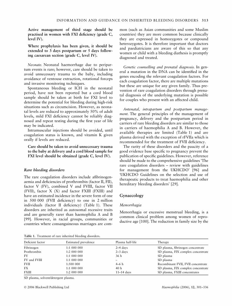
The rare coagulation disorders include afibrinogen-aemia and deficiencies of prothrombin (factor II, FII),factor V (FV), combined V and FVIII, factor VII(FVII), factor X (X) and factor FXIII (FXIII) andhave an estimated incidence in the severe form of onein 500 000 (FVII deficiency) to one in 2 millionindividuals (factor II deficiency) (Table 1). Thesedisorders are inherited as autosomal recessive traitsand are generally rarer than haemophilia A and B[99]. However, in racial groups, communities orcountries where consanguineous marriages are com-
mon (such as Asian communities and some Muslimcountries) they are more common because clinicallythey are expressed in homozygotes or compoundheterozygotes. It is therefore important that doctorsand paediatricians are aware of this so that anywomen or child with a bleeding diathesis is promptlydiagnosed and treated.
Genetic counselling and prenatal diagnosis. In gen-eral a mutation in the DNA can be identified in thegenes encoding the relevant coagulation factors. Foreach coagulation factor, there are multiple mutationsbut these are unique for any given family. Thus pre-vention of rare coagulation disorders through prena-tal diagnosis of the underlying mutation is possiblefor couples who present with an affected child.
Antenatal, intrapartum and postpartum manage-
ment. The general principles of the management ofpregnancy, delivery and the postpartum period incarriers of rare bleeding disorders are similar to thosein carriers of haemophilia A and B. However, theavailable therapies are limited (Table 1) and areplasma derived with the exception of rFVIIa which isrecommended for the treatment of FVII deficiency.
The rarity of these disorders and the paucity of agood evidence base specific to pregnancy prevent thepublication of specific guidelines. However, referenceshould be made to the comprehensive guidelines �Therare coagulation disorders – review with guidelinesfor management from the UKHCDO� [96] and�UKHCDO Guidelines on the selection and use oftherapeutic products to treat haemophilia and otherhereditary bleeding disorders� [29].
Gynaecology
Menorrhagia
Menorrhagia or excessive menstrual bleeding, is acommon clinical problem among women of repro-ductive age [100]. The reduction in family size by the
Table 1. Treatment of rare inherited bleeding disorders.
Deficient factor Estimated prevalence Plasma half-life Therapy
Fibrinogen 1:1 000 000 2–4 days SD plasma, fibrinogen concentrate
Prothrombin 1:2 000 000 2–3 days SD plasma, FIX complex concentrate
FV 1:1 000 000 36 h SD plasma
FV and FVIII 1:1 000 000 – SD plasma
FVII 1:500 000 4–6 h Recombinant FVII, FVII concentrate
FX 1:1 000 000 40 h SD plasma, FIX complex concentrate
FXIII 1:2 000 000 11–14 days SD plasma, FXIII concentrates
SD plasma, solvent/detergent plasma.
INFORMATION AND GUIDANCE ON INHERITED BLEEDING DISORDERS 313
� 2006 Blackwell Publishing Ltd Haemophilia (2006), 12, 301–336
widespread use of contraception and sterilization hasresulted in a significant increase in the number ofperiods women experience during their reproductivelife. This in turns amplifies the problem of menor-rhagia today. Menorrhagia is subjectively defined asa complaint of heavy cyclical menstrual bleedingoccurring over several consecutive cycles [101].Objectively, it is defined as a total menstrual bloodloss of >80 mL per menstruation (using the alkalinehaematin method) [102]. It is difficult to quantifymenstrual blood loss objectively because it involvestechniques that are specialized, time-consuming andrequire collection of sanitary material by women. Asa result, assessment of menorrhagia in clinicalpractice is usually subjective and often relies on thedescription provided by the patient. This method isunfortunately inaccurate and there is lack of corre-lation between patient’s impression and the objectiveassessment of actual volume of blood loss [102]. Thepictorial blood assessment chart (PBAC) is a semi-objective method of assessing menstrual bleedingbased on the number and saturation of sanitary padsand tampons used (Appendix 2). A pictorial chartscore of >100 was shown to have a reasonableaccuracy for identifying menorrhagia with a sensi-tivity of 86% and a specificity of 89% [103].Although the validity of this chart has been debated[104], it is simple to use and is at present the bestpractical tool available for the assessment of men-strual blood loss.
It is estimated that approximately 30% of womencomplain of menorrhagia [105]. Annually about onein 20 women aged 30–49 years consult their generalpractitioner for this reason [106]. It is the mainpresenting complaint in women referred to gynae-cologists and accounts for two-thirds of all hyster-ectomies [107]. Menorrhagia could be due to local orsystemic causes; however, the underlying pathologyremains unidentified in approximately 50% of cases[108,109].
Disorders of haemostasis are associated withmenorrhagia. Although this association has longbeen recognized, the magnitude of these disorders asa possible cause of idiopathic menorrhagia has beenunderestimated. Women who are referred for inves-tigation of menorrhagia are not routinely screenedfor coagulation disorders even when no pelvicpathology is identified. In a recent UK survey, only13% of gynaecologists would request a standardclotting screen and 2% would test for VWD in a35 year old with menorrhagia and no pelvic pathol-ogy [110]. Similarly, a US survey found that 17% ofphysicians would order BT determination for repro-ductive age women with menorrhagia and only 4%
would consider VWD as the cause of menorrhagia[111].
The use of PBAC should be considered in theassessment of menstrual blood loss for the diag-nosis of menorrhagia and evaluation of the treat-ment outcomes (grade C, level IV).
Prevalence of inherited bleeding disorders in women
with menorrhagia. Over the last decade, there havebeen international efforts to assess the prevalence ofinherited bleeding disorders in women with menor-rhagia. VWD is the most common inherited bleedingdisorder worldwide with a prevalence of 1–2% in thegeneral population [51,112]. A recent systemicreview of 11 prevalence studies of VWD in 988women with menorrhagia reported an overall pre-valence of 13% (95% CI, 11–15.6%), with preval-ence in individual studies ranging from 5% to 24%[113]. The prevalence was higher in the Europeanstudies compared with that in the North Americanstudies [18% (95% CI 15–23%) vs. 10% (95% CI7.5–13%); P ¼ 0.007]. This was explained by theauthors to be likely the result of differences betweenthe studies in the method of assessing menstrualblood loss and of recruitment of the study popula-tion, their ethnic composition, and the laboratorycriteria for diagnosis of VWD.
In addition to VWD, platelet disorders and clottingfactor deficiencies have also been identified in womenpresenting with menorrhagia [114–117].
A significant proportion of women with idiopathicmenorrhagia have an underlying bleeding disorder,the commonest being VWD. Therefore, testing forthese disorders should be considered (grade B,level III).
Heavy, prolonged and irregular menstrual periodsare frequent complaints in adolescent girls. This islikely to be due to immaturity of the hypothalamic–pituitary–ovarian axis resulting in anovulatory cycles[118–120]. However, in a significant proportion ofthese cases, an underlying bleeding disorder may beidentified [121–124]. Eight of 14 adolescents pre-senting with menorrhagia without thrombocytopeniawho underwent comprehensive testing for a bleedingdisorder had an underlying bleeding disorder [121].In a retrospective study of 106 adolescents withmenorrhagia, 11 (10.4%) patients were found tohave an underlying bleeding disorder. A positivefamily history of bleeding symptoms was a signifi-cant predictor of a bleeding disorder in this study[124]. Acute adolescent menorrhagia requiringhospital admission is also a strong predictor of a
314 C. A. LEE et al.
Haemophilia (2006), 12, 301–336 � 2006 Blackwell Publishing Ltd
bleeding disorder. A primary coagulation disorderwas found in 19% of those admitted to a children’shospital for acute menorrhagia [125]. One quarter ofthose with haemoglobin <10 g dL)1, one-third ofthose requiring transfusion, and one-half of thosepresenting at menarche had such an underlyingdisorder. In another study of adolescents withmenorrhagia requiring hospitalization, 15 (33%) of46 cases had an underlying haematological disease,with VWD accounting for five cases [126].
Whether adolescent menorrhagia is more likely tobe associated with bleeding disorders is uncertain. Ina study of 115 women with menorrhagia, thefrequency of an underlying bleeding disorder inadolescent, reproductive age, and perimenopausalwomen presenting with menorrhagia was found tobe similar [127].
Menstruation may be the first haemostatic chal-lenge faced by girls with an inherited bleedingdisorder. Menorrhagia in these girls is usually severeand they often present acutely. This could beexplained by the findings of increased VWF andFVIII levels with age [128,129].
Adolescents presenting with acute menorrhagiashould be investigated for bleeding disorders. Forprepubertal girls either known to have a bleedingdisorder or a positive family history, plans shouldbe made in anticipation of the possibility of acutemenorrhagia at the onset of menarche (grade C,level IV).
Testing for inherited bleeding disorders in women with
menorrhagia
Bleeding history.Testing for VWD and other bleed-ing disorders in all women with menorrhagia is neitherpractical nor necessary. The first step in investigatingfor an underlying bleeding disorder in these women isto take a focused personal and family history ofbleeding symptoms. This can be used to identifywomen with significant additional bleeding symptomssuggestive of an underlying bleeding disorder [130].The frequency of bleeding symptoms has been showedto be significantly higher in patients with VWD than inwomen without a bleeding disorder [115]. Signifi-cantly more women with VWD had menorrhagia sincemenarche (65%), compared with women without ableeding disorder (9%). Bleeding after tooth extrac-tion, and postpartum and postoperative bleeding werealso more common among women with VWD [115].
Bleeding history should include [131]:1 Excessive menstrual bleeding since menarche;2 History of PPH, surgery-related bleeding or
bleeding associated with dental work;
3 History of bruising >5 cm one to two times permonth, epistaxis one to two times per month,frequent gum bleeding, family history of bleedingsymptoms.[A �positive screen� entails any one from 1 or 2, ortwo or more from 3.]
Initial investigation for an underlying bleedingdisorder in women with menorrhagia shouldconsist of a focused personal and family historyof bleeding symptoms (grade C, level IV).
Laboratory testing:1. Full blood count and ferritin.The initial laboratory evaluation for an underlying
bleeding disorder should consist of full blood count(FBC) and serum ferritin, which will exclude thromb-ocytopenia and assess the degree of anaemia. If thedetailed history identified a �positive screen� and theplatelet count is normal, then a stepwise sequence oftesting for various bleeding disorders should becarried out. This also applies to women who areconsidering major surgical intervention, regardless ofthe presence of a positive screen [130]. It is essentialfor these testing to be undertaken in settings wherenecessary expertise and resources are available on-site to ensure appropriate and accurate diagnosis.2. PT and activated PTT.PT and activated PTT (APTT) are standard tests of
haemostasis, which are carried out as part of theinitial evaluation. However, they have poor sensitiv-ity, specificity and both positive and negative pre-dictive values for an underlying bleeding disorder[132]. Normal PT and APTT do not exclude anunderlying bleeding disorder, but they should beadequate for screening for the severe rare clottingfactor deficiencies [133]. A mixing study should beperformed in cases of prolonged APTT to distinguishbetween a deficiency state and an inhibitor.3. von Willebrand screen.This includes VWF:Ag, VWF:AC and FVIII levels
(FVIII:C). FVIII level can be reduced in VWD asVWF protects FVIII from proteolytic cleavage [134].Comprehensive guidelines on the diagnosis of VWDhave been produced by the UKHCDO [135].
Difficulties in diagnosing type 1 VWD because ofits variable expression influenced by genetic andenvironmental factors are well established [136,137].These factors include women’s race, ABO bloodgroup, phase of the menstrual cycle and the use oforal contraceptives.
At present, there is a lack of consensus in theliterature supporting a true variation of VWF levelsduring the menstrual cycle. Initial studies havesuggested a decrease in VWF and FVIII levels during
INFORMATION AND GUIDANCE ON INHERITED BLEEDING DISORDERS 315
� 2006 Blackwell Publishing Ltd Haemophilia (2006), 12, 301–336
menstruation [138–140]. A longitudinal study of 39healthy Caucasian women sampled on days 2, 8, 15and 21 showed a strong cyclic variation with adecrease in VWF values in the early follicular phaseand peak values in the luteal phase [129]. However,another longitudinal study of 95 healthy womensampled serially at days 4–7, 11–15 and 21–28, didnot found this variation [141]. A recent cross-sectional study of 90 controls and 85 women withmenorrhagia, assigned into groups that were morefinely divided in the sampling time during themenstrual cycle, found the lowest VWF levels ondays 1–4 and the highest on days 9–10 [142]. Due tothe inconsistencies in these results, recommendationsfor testing exclusively during menses cannot bemade. However, the time of testing in relation tothe menstrual cycle should be noted, and if the resultsfrom the initial testing were borderline or just belowthe normal range, repeat testing during days 1–4 ofthe menstrual cycle would be advisable.
It is currently not clear whether the concurrentuse of COC would obscure the diagnosis of VWD.It has been observed that oestrogen increases VWFlevels [143]. There has been report of COC usemasking the diagnosis of VWD [144]. A review ofthe literature on the effect of COCs on coagulationfactors reveals large variations of study designs andresults [145]. The majority of the studies demon-strated an increase in fibrinogen, prothrombin,FVII, FVIII and VWF whilst other factors, suchas FV, FIX and FXIII, did not seem to be affected.The effect appears to be related to oestrogen, dosedependent, and appreciable above a dose of 50 lgof ethynylestradiol. The effect of smaller doses ofoestrogen, which are commonly used today, is lessand may not be significant. In a study of 20 non-pill users and 20 women on low-dose COC(£30 lg), no significant difference between themean values of APTT, fibrinogen, FVIII:C,VWF:Ag, VWF:AC and FXI were found [129].Due to lack of evidence presently demonstrating adefinite effect of COC on VWF levels, a practicalapproach would be to still test women when onCOC, especially if they are still experiencingmenorrhagia. Repeat testing off COC should how-ever be considered if results are borderline or in thelower half of the normal range.
Patients with blood type O have 25% lower VWFlevels compared with other blood types [146]. Thismeans if VWD testing is adjusted for the ABO bloodtype, a lower VWF level would be required as a cutoff for the diagnosis of VWD for blood type Opatients. Consequently, this would exclude bloodtype O patients with subnormal level defined as two
standard deviations below the mean of the totalpopulation (i.e. <50 IU dL)1), but a level within thenormal range for blood type O patients (i.e.>35 IU dl)1). However, type O patients with VWFlevels between 35 and 50 IU dL)1 were found tohave similar bleeding symptoms as non-O patients inthat range [147]. Therefore, the use of ABO adjustedranges for VWF levels may not be necessary becausebleeding symptoms seem to depend on the VWF levelregardless of the blood type.4. Platelet function analysis.Neither BT nor platelet function analyser (PFA-
100) are effective screening tests for mild VWD andplatelet function disorders [148–150]. However, theycan be performed concurrently with VWF testing as abaseline and used for monitoring postintervention toensure adequate haemostasis.
If all initial haemostasis testing and completegynaecological evaluation are normal, platelet aggre-gation and release studies should be considered. Arelatively high prevalence of platelet function abnor-malities was recently reported in women withmenorrhagia [117,127]. Amongst 74 women withunexplained menorrhagia, platelet aggregation andplatelet ATP release were decreased with one ormore agonist in 35 (47.3%) and 43 (58.1%) womenrespectively. The prevalence odds of platelet aggre-gation abnormalities was found to be 4.2-fold higheramong women with menorrhagia than controlwomen [117].5. Other clotting factor levels.If the VWD (including FVIII) screen and platelet
function analysis are both normal, depending on thedegree and severity of additional personal bleedingsymptoms and the family history, testing for addi-tional coagulation deficiencies (FV, FVII, FIX, FXIand FXIII) should be considered.6. Non-haematological tests.There is some evidence linking hypothyroidism
with menorrhagia [151–153]. Thyroid function testsshould be considered if the patient is symptomatic.
Laboratory testing for an inherited bleeding dis-order should be carried out if the woman present-ing with menorrhagia has a positive bleedinghistory or if surgical intervention is planned(grade C, level IV).
Laboratory testing should only be undertaken insettings where necessary expertise and resourcesare available on-site to ensure appropriate andaccurate diagnosis (grade C, level IV).
Laboratory testing should be carried out in thefollowing stepwise sequence (grade C, level IV):
316 C. A. LEE et al.
Haemophilia (2006), 12, 301–336 � 2006 Blackwell Publishing Ltd
1 FBC and ferritin;2 PT and APTT;3 von Willebrand screen (VWF:Ag, VWF:RCo
and FVIII:C) Testing exclusively during men-struation, off COC or adjusted for blood type isnot necessary at the initial evaluation. However,these details should be documented and repeattesting should be considered if the results areborderline.
4 Platelet function analysis (platelet aggregationand release studies);
5 Other clotting factor levels;6 Thyroid function tests if symptomatic.
Menorrhagia in women with inherited bleeding disor-
ders Menorrhagia has been well recognized as acommon symptom in women with inherited bleedingdisorders including VWD, other clotting factordeficiencies and platelet disorders [27,99,154–156].In a review of the obstetric and gynaecologicalmanifestations of bleeding disorders, menorrhagiawas reported in 32–100% of women with type IVWD and in 10–70% of women with other bleedingdisorders [157]. In a study of 38 females with type IVWD, menorrhagia was the most common bleedingsymptoms, occurring in 93% of adult women [155].Using the PBAC to assess menstrual blood loss,menorrhagia (PBAC score >100) was confirmed in74%, 59% and 57% of women with VWD, FXIdeficiency and who were carriers of haemophiliarespectively [154]. The duration of menstruation wasalso found to be significantly longer and episodes offlooding more common in women with inheritedbleeding disorders compared with the control group(P ¼ 0.001).
Menstruation has a major influence on women’slifestyle and employment. Menorrhagia has beenfound to have a negative effect on the quality of lifein women with inherited bleeding disorders [158]. Inthe same study, 39% of these women had to cutdown on the time they spent on their work or otheractivities, 47% felt they had accomplished less thanthey would like during this period, and 38% felt theywere limited in the kind of work they could do [158].The impact of menstruation on young women interms of restrictions in sport activity, travel, work,studies and sex is also substantial [159]. Althoughthese comments were made in regard to the impact ofmenses in general, this effect is likely to be evengreater in women with an inherited bleeding disorderand excessive menstrual bleeding. Early recognition,accurate diagnosis and appropriate management willhelp to improve not only the quality of care for thesewomen but also their quality of life.
Menorrhagia is one of the most common bleedingmanifestations in women with inherited bleedingdisorders and has a negative effect on their qualityof life (grade B, level III).
Management of menorrhagia in women with inherited
bleeding disorders A multidisciplinary clinic, inclu-ding both haematologist and gynaecologist, set upwithin the network of haemophilia treatment centres(HTCs) is ideal for providing comprehensive care forthe management of menorrhagia in women withinherited bleeding disorders. This ensures appropri-ate and accurate on-site haemostasis testing; avoidscommunication problems between professionals;allows clear management plans to be made (andcompetent completion of desmopressin test dose);and can address the psychosocial aspects related tobleeding disorders.
In a survey by the Centers for Disease Control andPrevention in the USA, 95% (71 of 75) of womenreceiving care in HTCs reported a strong positiveopinion and satisfaction [160]. Similar positivefindings were found among patients of the multidis-ciplinary clinic at the Katharine Dormandy Haemo-philia Centre of the Royal Free Hospital in London[161].
Menorrhagia in women with an underlying bleed-ing disorder is likely, but not exclusively, to be due toa defect in haemostasis. The cause of menorrhagiamay be multifactorial in these women. In a survey ofwomen with VWD, half of the women undergoinghysterectomy for menorrhagia had additional uterinepathology such as fibroids or endometriosis [162].Therefore, a thorough gynaecological evaluationshould be performed to exclude pelvic pathology,especially the possibility of malignancy in olderwomen.
The Royal College of Obstetricians and Gynaecol-ogists has produced management guidelines ofmenorrhagia in general [102,107]. Most of thesehave not been assessed in menorrhagia related to anunderlying bleeding disorder, but for best clinicalpractice, the results for general menorrhagia patientscan be extrapolated.
Management of bleeding disorder-related menor-rhagia involves consideration of the patient’s age,childbearing status and preference in terms ofperceived efficacy and side-effect profile. Therapeuticoptions for the control of menorrhagia in womenwith underlying bleeding disorders include medicaltreatments [such as anti-fibrinolytics (tranexamicacid), intranasal and subcutaneous DDAVP, oralcontraceptives, levonorgestrel (LNG) intrauterine
INFORMATION AND GUIDANCE ON INHERITED BLEEDING DISORDERS 317
� 2006 Blackwell Publishing Ltd Haemophilia (2006), 12, 301–336
device and clotting factor replacement] and surgicaltreatments (such as endometrial ablation and hyster-ectomy). They are similar to the treatment optionsfor menorrhagia in general with the exception ofDDAVP and clotting factor replacement. However,management of women with inherited bleedingdisorders requires additional monitoring of thehaemostatic parameters and awareness of the in-creased risk of bleeding with any surgical interven-tions.
Management of menorrhagia in women withinherited bleeding disorders should be providedby a multidisciplinary team including a haematol-ogist and gynaecologist (grade C, level IV).
Medical treatment of menorrhagia
Tranexamic acid Increased uterine fibrinolytic activ-ity is found in women with menorrhagia comparedto those with normal menstrual loss; this increasedfibrinolysis is most likely due to high levels ofendometrium-derived plasmin and plasminogen acti-vators [163,164]. Tranexamic acid is an antifibrin-olytic agent that reversibly blocks lysine-binding siteson plasminogen and prevents fibrin degradation[165]. Endometrial tissue plasminogen activator (t-PA) levels were significantly lower after three treat-ment cycles in 12 menorrhagic women who receivedtranexamic acid 500 mg four times daily for 5 days[166]. In another study, tranexamic acid, 1 g threetimes daily for 5 days, was also shown to signifi-cantly reduced t-PA and plasmin activity in themenstrual as well as in the peripheral blood ofmenorrhagic women, compared with pretreatmentvalues [163].
Several studies of variable design have been carriedout to assess the therapeutic efficacy of tranexamicacid in the treatment of idiopathic menorrhagia[166–170]. Using oral tranexamic acid ranging from500 mg or 1 g four times daily for 4–7 days percycle, the menstrual blood loss was reduced by 34–59% over two to three cycles in women withmenorrhagia. Tranexamic acid has been shown tobe more effective than NSAIDs in the reduction ofmenstrual loss; 54% vs. 20% when compared withmefenamic acid [170], and 44% vs. 20% whencompared with flurbiprofen [168]. However, none ofthem affected the duration of menses [168,170].
Oral tranexamic acid is generally well tolerated bywomen with menorrhagia. Nausea and diarrhoea arethe most common side effects. The Cochrane sys-tematic review of antifibrinolytics for heavy men-strual bleeding concluded that adverse events during
treatment with tranexamic acid were no higher thanwith placebo, NSAIDs, cyclic progestogens or et-hamsylate [171]. There have been some concernswith the use of tranexamic acid due to isolatedreports of thromboembolic complications, whichmay have led to the reluctance in its use. However,the incidence of thrombosis, over a 19-years timeframe and 238 000 patient-years of treatment withtranexamic acid, has been shown to be similar to thespontaneous frequency of thrombosis in women inthe general population [172]. Nonetheless, the use oftranexamic acid is contraindicated in patients with ahistory of thromboembolic disease.
Royal College of Obstetricians and Gynaecologists(RCOG) guidelines recommend tranexamic acid for3 months as a first-line medical treatment for menor-rhagia [101]. If successful, tranexamic acid can beused indefinitely in patients not requiring contracep-tion or who prefer non-hormonal treatment. Inwomen with bleeding disorder, tranexamic acid hasbeen widely used (orally, intravenously, topicallyalone or as an adjuvant therapy) in the preventionand management of oral cavity bleeding, epistaxis,gastrointestinal bleeding and menorrhagia. However,there is a lack of objective data on its efficacy in thereduction of menstrual loss in this population.
The recommended oral dose of tranexamic acid forthe treatment of menorrhagia is 1 g three to fourtimes daily for 3–4 days (maximum total daily doseof 4 g) [173]. It should be initiated once menstru-ation has started. There has been report on successfuluse of a single high-dose (4 g daily) therapy in threetypes 2A and 2B VWD patients, but this regime canbe associated with severe nausea and vomiting [174].
Combined oral contraceptives Combined oral contra-ceptives are a highly reliable method of birth control.They are also useful for cycle regulation and reduc-tion in the incidence of dysmenorrhea and premen-strual tension. There is some evidence that COCsignificantly reduces menstrual blood loss in womenwith and without menorrhagia [175,176]. In a trialof a 50 lg ethinylestradiol pill, menstrual loss wasreduced by 52.6% in 68% of 164 women withobjective menorrhagia [177]. The efficacy of low-dose COC in reducing menstrual blood loss has beenassessed in only one randomized controlled trial.However, the trial was not placebo controlled andwas relatively small (45 women, seven did notcomplete the trial) [178]. There was a significantreduction in menstrual loss in the COC group (43%,P £ 0.001), as well as in the low-dose danazol (49%,P ¼ 0.006) and the mefenamic acid groups (38–
318 C. A. LEE et al.
Haemophilia (2006), 12, 301–336 � 2006 Blackwell Publishing Ltd
39%, P ¼ 0.002). FVIII and VWF levels have beenshown to increase with the use of high-dose (50 lgoestradiol) pills [145] but not with the currentlystandard, lower-dose (30 lg oestradiol) pills [129].
The efficacy of COC in reducing menstrual bloodloss in women with bleeding disorders is unknown.In a survey of women with VWD (types 2 and 3)unresponsive to DDAVP, COC was reported to be aneffective treatment of menorrhagia in 88% of thewomen [62]. On the other hand, in type I patients, astandard dose and a higher dose COC were effectivein only 24% and 37% of the cases respectively [162].
Traditionally COC is administered daily for21 days, followed by a pill-free week during whichuterine bleeding occurs. In recent years, the extendedor continuous administration of COC (>28 days ofactive pills) has been reported as a successful regimein the treatment of endometriosis, dysmenorrhea,and other menstruation-association symptoms [179–182]. Avoidance of menstruation through continu-ous dosing of COC has potential benefits whichinclude less interference with daily activities orspecial events and less menstruation-related absen-teeism from work or school [183,184]. The Cochra-ne systematic review of continuous versus cyclicaluse of COC concluded that both regimes have similarpatient satisfaction and discontinuation rates [185].It also showed that continuous COC administrationhas similar, if not better, effect on improvingbleeding patterns and may improve menstruation-associated symptoms. This regime could be veryhelpful in the management of menorrhagia in womenwith bleeding disorders as this regime allows womento control the timing and reduce the frequency oftheir menstruation. It may also be more effectivethan the cyclical regime in reducing menstrual bloodloss.
Increased risk of thrombosis is the main concernassociated with the use of COC. However, womenwith bleeding disorders have a low inherited throm-botic risk. Serious side effects of COC includehypertension and, rarely, impaired liver functionand hepatic tumours. Other less serious side effectsinclude nausea, vomiting, headache, breast tender-ness, breakthrough bleeding, fluid retention, depres-sion and skin reactions.
Desmopressin Desmopressin (1-desamino-8-d-argi-nine vasopression, DDAVP) is a synthetic analogueof the antidiuretic hormone vasopressin. It increasesplasma concentration of FVIII and VWF throughendogenous release [186–188]. It is an importanttherapeutic alternative to plasma-derived coagula-tion products because it is effective in selected cases
and avoids the risks of infection with blood-borneviruses. A test dose is recommended before treatmentto assess its effectiveness as the response is notuniversal. DDAVP has been shown to be effective inthe prevention and treatment of bleeding episodes inpatients with mild VWD and haemophilia A. It canalso be used in patients with platelet disordersbecause of its effect on increasing platelet adhesive-ness [189].
Desmopressin is available in several formulations.It can be administered parenterally via intravenousor subcutaneous injection, or nasally as a spray. Thechosen route of administration is dependent on thepurpose of its use and the magnitude of the effectrequired. The optimal haemostatic effect of DDAVPis achieved with a dosage of 0.3 lg kg)1, whichincreases plasma FVIII and VWF levels two- tosixfold after intravenous administration [190]. It isusually given over 20 min as slow intravenousinfusion. Typically the 4 lg mL)1 solution is madeup in 50–100 mL normal saline for infusion. Whengiven subcutaneously in the same dose (0.3 lg kg)1),the response is of similar magnitude to that ofintravenous administration, but peak levels arereached later [191]. A more concentrated solution(15 lg mL)1) is available for subcutaneous usealthough it does not have a product license in theUK. Intranasal administration of DDAVP is anattractive route because it allows patients to treatthemselves at home without delay in the event ofbleeding episodes, e.g. at the onset of menstruation,and without the use of needles [192,193]. The effectof intranasal administration of 300 lg with ametered-dose spray is comparable with that of0.2 lg kg)1 intravenous injection, and the reproduc-ibility of the haemostatic effect is similar to that ofintravenous injection [186,194].
There are few side effects of DDAVP related toits vasomotor effects. They include mild tachycar-dia, headache and flushing. Due to the antidiureticeffect of DDAVP, there is a small risk of hypo-natraemia and potentially water intoxication. Thiscomplication can be greatly reduced by restrictionof fluid intake and close electrolyte monitoring,especially in those receiving multiple doses ofDDAVP.
The influence of nasal DDAVP on the localfibrinolytic activity in the endometrium and onmenstrual blood loss is interesting. Increased localfibrinolytic activity in the endometrium and men-strual fluid has been demonstrated in women withmenorrhagia [195,196]. Tissue-PA is the main phy-siological activator of the fibrinolytic system in bloodand in the endometrium. DDAVP has been shown to
INFORMATION AND GUIDANCE ON INHERITED BLEEDING DISORDERS 319
� 2006 Blackwell Publishing Ltd Haemophilia (2006), 12, 301–336
increase t-PA in the circulation [187,188], but in onestudy, it did not increase the fibrinolytic activity(plasmin levels) in the menstrual fluid [195].
Case series have shown that subcutaneous [197]and intranasal DDAVP [198] are effective in themanagement of menorrhagia in women with bleed-ing disorders who respond to DDAVP. Based onwomen’s subjective assessment of reduction inmenstrual flow, the efficacy of DDAVP was repor-ted as excellent or good in 86% and 92% of womentreated with subcutaneous and intranasal DDAVP,respectively [197,198]. However, in a randomizedplacebo-controlled crossover trial using PBAC, in-tranasal DDAVP was not significantly (P ¼ 0.51)different from placebo in reducing PBAC scores[199]. There was a statistically significant reductionin the PBAC score (P ¼ 0.0001) in both groupscompared with pretreatment score and a trendtowards a lower score when using intranasalDDAVP. It is possible that this difference did notreach statistical significance due to small samplesize. No statistically significant difference was foundin measures of quality of life for either group in thisstudy. In another randomized crossover study, thereduction in menstrual blood loss with DDAVP, inwomen with menorrhagia and prolonged BT, wasnot significantly different from either baseline orplacebo [200]. However, there was a significantreduction in the mean blood flow during the 2 daysof DDAVP treatment and in the total menstrualblood loss when DDAVP was combined withtranexamic acid. It is possible that tranexamic acidcounteracts the increased fibrinolytic activity (t-PA)induced by DDAVP.
Currently, there is no consensus regarding theoptimum dose and duration of nasal DDAVP fortreatment of menorrhagia in women with bleedingdisorders. Most studies have used high-dose DDAVPintranasal spray (1.5 mg mL)1) once daily; adminis-tered as 150 lg (one intranasal spray) for patientsweighing £50 kg and 300 lg (two intranasal sprays;one per nostrils) for patients weighing >50 kg.Additional doses within a 24-h period will requirespecialized and individual assessment. The responseto test dose should be clearly documented. DDAVP isusually given during the first 2–3 days of the men-strual period. This is based on the finding that 90%of all menstrual flow occurs during the first 3 days[201]. However, the pattern of menstruation maybedifferent in women with bleeding disorders. In onestudy, these women bleed heavily throughout theirmenstruation [154]. As a result, dose and duration oftreatment may have to be adjusted depending on thePBAC scores.
Other medical treatments Progestogens. Oral pro-gestogens are one of the most commonly prescribedmedications for menorrhagia [202]. There are twodifferent cyclical regimes of oral progestogens: ashort luteal phase treatment (days 19–26 or 15–25)or a longer 21-days course starting from day 5 ofthe cycle. A Cochrane systematic review, includingsix randomized controlled trials of the short lutealphase oral progestogen therapy, showed this regimeto be significantly less effective in reducing men-strual blood loss when compared with danazol,tranexamic acid and progesterone releasing intra-uterine system (IUS) [203]. A single trial comparinga long 21-day course (norethisterone 5 mg threetimes daily) with LNG-IUS showed significantreduction in menstrual blood loss in both group,but oral progestogen was less effective and accept-able by patients when compared with LNG-IUS[204]. No randomized, controlled trials compar-ing progestogen treatment with placebo were iden-tified.
Cyclical 21-days progesterone therapy can beconsidered as a second-line treatment for patientswho do not respond to other medical therapiespreviously discussed or in whom such treatments arecontraindicated. However, the compliance of thisregime is usually not good due to progestogenic sideeffects. These include fatigue, mood changes, weightgain, bloating, headaches, depression, irregularbleeding and adverse effect on bone density andlipid profile.
Oral progestogens in high doses alone, or incombination with DDAVP or clotting factor concen-trate, may be useful in the treatment of acutemenorrhagia in women with inherited bleedingdisorders. Other forms of progestogens, injectionsor implants, have not been evaluated in women withmenorrhagia, but studies in women without menor-rhagia have shown that a considerable proportion ofwomen experience amenorrhoea with such therapies.More research is needed before recommendation canbe made for women with menorrhagia with orwithout associated bleeding disorders.Nonsteroidal anti-inflammatory drugs. NSAIDs
(e.g. mefenamic acid and naproxen) have beenshown to be more effective than placebo in reducingmenstrual blood loss but less effective than eithertranexamic acid or danazol [205]. NSAIDs have theadded advantage of reducing menstrual pain andmenstrual migraine. However, their use is contrain-dicated in women with inherited bleeding disordersdue to their anti-aggregator effect on platelet func-tion. This highlights the need for awareness ofinherited bleeding disorder as a potential cause of
320 C. A. LEE et al.
Haemophilia (2006), 12, 301–336 � 2006 Blackwell Publishing Ltd
menorrhagia in order to avoid the use of NSAIDs insuch cases.Ethamsylate. Ethamsylate is a haemostatic agent
that maintains platelet and capillary integrity andaffects prostaglandin synthesis. In the only random-ized controlled trial in women with menorrhagia ingeneral, this treatment showed no reduction inmenstrual blood loss when used during menstru-ation, compared with 20% and 54% reductionfound with mefenamic acid and tranexamic acidrespectively [170]. There are no data on the efficacyof this treatment in women with bleeding disorders,but in view of its mode of action, it may have adifferent effect on heavy menstrual loss in thesewomen, which warrants further studies.Danazol and gonadotrophin-releasing hormone.
Danazol and gonadotrophin-releasing hormone(GnRH) agonists are effective in reducing menstrualloss and duration of menstruation. Data on theireffectiveness in the treatment of menorrhagia relatedto bleeding disorders are lacking. Their side effectsand risks, because of the resulting hypoestrogenicstate, make them less acceptable for long-term use.They are more expensive than other medical treat-ments of menorrhagia. GnRH agonists with simul-taneous add-back therapy with either tibolone orcombined oestrogen/progesterone may be an alter-native option to surgery for women with severebleeding disorders, e.g. type 3 VWD, and menorrha-gia not responding to other treatments.
Levonorgestrel-releasing intrauterine device
The LNG-IUS (trade name Mirena) is an intrauterinesystem with a T-shaped plastic frame 32-mm longand a reservoir on the vertical stem of the IUScontaining 52 mg of LNG. It releases 20 lg of LNGevery 24 h over a recommended duration of use of5 years. It suppresses endometrial growth causing theglands of the endometrium to become atrophic andthe epithelium inactive [206]. It was originallydeveloped as a contraceptive device. Despite it beingsuch a novel intervention, it has rapidly become verypopular. In a systematic review, LNG-IUS wasshown to reduce the average menstrual blood lossby 74–97% after durations of use between 3 and12 months [207]. It is an attractive treatment optionfor women who require contraception and wish topreserve their fertility and for those who are unlikelyto comply with drug treatments.
The Cochrane systematic review revealed LNG-IUS to be more effective in the treatment of menor-rhagia in general compared with oral norethisteronegiven during days 5–26 of the menstrual cycle (94%
reduction in menstrual blood by LNG-IUS vs. 87%by oral norethisterone) and associated with higherrates of satisfaction and continuation with treatment(77% vs. 22%) [204,208]. The LNG-IUS was alsofound to produce significantly greater reductions inmenstrual blood loss (measured by the alkalinehaematin method, weighing sanitary materials beforeand after use, and PBAC scores) than mefenamic acid[209]. Another trial compared LNG-IUS with theexisting medical treatment (prostaglandin synthesisinhibitors or fibrinolysis inhibitors) the women werereceiving while waiting to undergo hysterectomy fortreatment of excessive menstrual bleeding [210];64% of women in the LNG-IUS group decided tocancel hysterectomy at 6 months compared with14% in the group who continued with their existingmedical treatment (P ¼ 0.001). All quality of lifescores were significantly higher in the LNG-IUSgroup at 6 months (P ¼ 0.002).
When compared with endometrial ablation (eithertranscervical resection or balloon ablation), LNG-IUS appears to be less effective in the treatment ofmenorrhagia [208]. A significantly larger proportionof women with ablation had successful treatment (asmeasured by a PBAC score of <75) than those withLNG-IUS (92% versus 75%; P ¼ 0.0006). However,there was no significant difference in rates ofsatisfaction with treatment (LNG-IUS: 77.3% andablation: 84.3%) or haemoglobin values after treat-ment [208]. Endometrial ablation may provide aquicker resolution to menstrual symptoms, but itrequires the use of operation facilities and anaesthe-sia, and is not suitable for women who want topreserve their fertility. The LNG-IUS requires lessskill, can be inserted in outpatient, and is an effectivereversible contraception.
The LNG-IUS was compared with hysterectomy inone trial to assess the effects on quality of life andcost-effectiveness [211,212]. The health-related qual-ity of life improved significantly in both groups andthere were no significant differences in outcomemeasures at 12 months except that women withhysterectomy suffered less pain (P ¼ 0.01). Although20% and 42% of women in the LNG-IUS group hadundergone hysterectomy by 12 months and 5 yearsof follow up, respectively, the mean costs of treat-ment for this group of women were significantlylower than the group who had hysterectomy.
The side effects include those related to anintrauterine device, for example, irregular bleedingand expulsion of the device [208]. The commonestside effect is irregular bleeding or spotting in thefirst 3–6 months after insertion. It usually settlesafter 6–12 months in the majority of cases. Amen-
INFORMATION AND GUIDANCE ON INHERITED BLEEDING DISORDERS 321
� 2006 Blackwell Publishing Ltd Haemophilia (2006), 12, 301–336
orrhoea is also commonly reported; 44% of thewomen had no bleeding after 6 months in onestudy [213]. Therefore, appropriate counselling onthe potential changes in the bleeding pattern mayhelp to improve its acceptability. The expulsionrates (5–10%) are similar to other intrauterinedevices [214,215]. The overall discontinuation ratefor the LNG-IUS was 20% in randomized con-trolled trials and 17% in the case series [207].Other side effects of LNG-IUS include those relatedto the systematic effects of progestogens, such asweight gain, bloating, breast tenderness, greasy hairor skin, acne, nausea, mood changes and depres-sion. Nevertheless, it is better tolerated thansystemic progestogens.
The long-term (5 years) efficacy of LNG-IUS forcontraception is well established [214]. However,data on its long-term efficacy in the treatment ofmenorrhagia is limited. The LNG-IUS was found toinduce atrophy of the endometrial epithelium formore than 5 years [206], which provides a rationalefor the reduction of menstrual blood loss for aprolonged period of time. An observational study hasdemonstrated a continuation rate of 50% after amean 54 months of follow up. Amongst thesewomen, 57% have occasional bleeding and 35%have amenorrhoea [216]. Significant reduction inmenstrual blood loss and increase in haemoglobinand serum ferritin levels were shown to sustain for aslong as 36 months in women with idiopathic menor-rhagia [217]. Further studies are needed to establishthe long-term efficacy of LNG-IUS in the reductionof menstrual blood loss.
The use of LNG-IUS in women with inheritedbleeding disorders has been evaluated in 16 women(13 VWD, two FXI deficiency and one Hermansky–Pudlak syndrome) with menorrhagia not respondingto medical treatment [218]. After 9 months, thePBAC score had decreased significantly from amedian of 213 (range 98–386) to 47 (range 24–75)and the haemoglobin concentrations had increasedsignificantly from a median of 12.1 (range 8.0–13.2 g dL)1) to 13.1 (range 12.3–14 g dL)1) (P ¼0.0001). Nine women became amenorrhoeic. Noside effects were reported, except irregular spotting,which ranged from 30 to 90 days (median 42 days).Most (11) women did not feel that the irregularbleeding affected their life and the remaining fivewere only slightly affected. Prior to the insertion ofthe LNG-IUS, all women had at least 1 day permonth where their life was significantly affected bytheir periods, with six women reporting at least3 days were affected. Nine months after insertion ofLNG-IUS, none of them had any days of the month
when their menstruation significantly affected theirlife. All women in this trial had mild to moderatebleeding disorders; therefore, further studies arerequired to evaluate the effectiveness of LNG-IUSin women with severe factor deficiencies.
Women with bleeding disorders could potentiallybe at risk of bleeding at the time of insertion.Adequate haemostatic coverage is recommendedespecially in women with severe forms of bleedingdisorders.
Clotting factor Clotting factor replacement, witheither recombinant or plasma-derived factor concen-trate, will be required in some women with bleedingdisorders, especially in adolescents [122] and thosewith severe deficiency or when they present acutely.A multidisciplinary assessment approach by haema-tologists and gynaecologists is crucial in the man-agement of these women.
Medical treatment of menorrhagia in womenwith inherited bleeding disorders include tranex-amic acid, COCs, DDAVP, cyclical 21-days oralprogesterone, and the LNG-IUS. The treatmentchoice depends on the type of bleeding disorder,and patient’s age, childbearing status and prefer-ences in terms of the perceived efficacy and sideeffects (grade C, level IV).
Specific haemostatic therapy will be required insome women with inherited bleeding disorders tocontrol menorrhagia. A multidisciplinary approachin the management of these individuals is essentialto ensure optimal outcomes (grade C, level IV).
Surgical treatment for menorrhagia
Surgical intervention may be required insome women who do not tolerate medical treat-ments or where such treatments have failed. Anunderlying bleeding disorder can potentially be thereason for failed medical therapy [219]. Surgerymay also be indicated in the presence of a pelvicpathology.
Women with inherited bleeding disorders are atgreater risk of bleeding complications from surgery,including increased perioperative and delayed (7–10 days after surgery) bleeding. Prophylactic treat-ment is recommended in women with bleedingdisorders. Close follow up for at least 10 dayspostsurgery is also recommended to monitor specificfactor levels and to assess for delayed bleedingcomplications such as wound breakdown. Multidis-ciplinary care by haematological, gynaecological/surgical and anaesthetic teams is essential to ensure
322 C. A. LEE et al.
Haemophilia (2006), 12, 301–336 � 2006 Blackwell Publishing Ltd
an optimal outcome. Any surgical intervention inwomen with bleeding disorders should be carried outby experienced professionals. Extra care should betaken in ensuring adequate haemostasis and the useof surgical drains should be considered.
Hysterectomy Hysterectomy is an established, effect-ive and definitive treatment for menorrhagia associ-ated with high patient satisfaction. However,hysterectomy is a major surgical procedure withsignificant physical and emotional complications andsocial and economic costs. A large cohort studyevaluated 37 298 hysterectomies performed forbenign indications in the UK in 1994 and 1995 witha 6 week postsurgery follow up and found theassociated mortality rate to be 0.38 per 1000 (95%CI, 0.25–0.64) [220]. The overall operative compli-cation rate was 3.5% (3% severe) and postoperativecomplication rate was 9% (1% severe) [220,221].Hysterectomy also has a risk of long-term complica-tions, including early ovarian failure, and urinaryand sexual problems. For these reasons, less invasivealternatives (e.g. endometrial resection and ablation)have been developed for the treatment of menorrha-gia.
Endometrial ablation Endometrial ablative tech-niques, which destroy the lining of the uterus, areincreasingly used today as an effective alternative tohysterectomy for the management of heavy men-strual bleeding. They have a shorter operating timeand hospital stay, quicker recovery and fewer post-operative complications than hysterectomy [222]. Inrecent years various endometrial ablative techniqueshave evolved. The first generation techniques (resec-tion, laser and rollerball) were introduced in the1980s. They are performed under direct vision andrequire specialized surgical skills. The second gen-eration techniques are designed to ablate the fullthickness of the endometrium by controlled applica-tion of heat, cold, microwave or other forms ofenergy. They require sophisticated equipment andare mostly performed blindly. A systematic reviewreported that, in general, they are technically simplerand quicker to perform than first generation tech-niques, while satisfaction rates and reduction inmenstrual blood loss are similar [223]. Secondgeneration techniques are also less invasive, there-fore, they may be a safer option for women withbleeding disorders. Although existing evidence sug-gests that the complication profiles of the newertechniques compare favourably to the first generationtechniques, long-term data on the safety of thesenewer techniques are still unavailable.
One study evaluated thermal balloon ablation in70 women with severe menorrhagia and severesystematic disease, including 25 with �coagulopathy�[224]. The procedure was performed under localanaesthesia and the success rate was over 90% at3 years follow up. The findings of a smaller retro-spective study, which assessed the efficacy of en-dometrial ablation in seven women with VWD-related menorrhagia, were less favourable [225].Four women experienced recurrence of menorrhagiaafter a median of 8 months postablation, and threeof them eventually underwent hysterectomy at amedian of 11 months postablation.
Consideration for the presence of an underlyingbleeding disorder may be warranted in women who�fail� endometrial ablations or continue to haveexcessive menstrual bleeding after an ablation. Pro-phylactic treatment should always be considered toprevent bleeding complications. Guidance regardingthe selection and use of therapeutic products forsurgery in patients with VWD and other hereditarybleeding disorders can be obtained from the UKHC-DO guidelines [29,67].
Prophylactic treatment should be considered inwomen with inherited bleeding disorders under-going surgery for treatment of menorrhagia. Thesurgery should be performed by experiencedprofessionals with special attention to haemosta-sis. The use of a surgical drain should beconsidered (grade C, level IV).
Other gynaecological problems in women withinherited bleeding disorders
Women with bleeding disorders are at risk of thesame gynaecological problems that affect all women,possibly with higher risks for conditions that areassociated with bleeding [157].
Haemorrhagic ovarian cysts A recent review identified15 studies, mostly case reports and case series,reporting one or more haemorrhagic ovarian cystsin women with inherited bleeding disorders [157]. Inone study, the prevalence was found to be 6.6%(nine of 136 women with VWD) [226]. In anotherstudy, two cases of spontaneous broad ligamenthaematomas were reported in a series of eightwomen with VWD [27]. Women with bleedingdisorders appear to be more vulnerable to bleedingfrom ruptured ovarian follicles. During ovulation,the ovum is expelled from the ovarian follicle intothe peritoneal cavity. This process is usually notassociated with any significant amount of bleeding,
INFORMATION AND GUIDANCE ON INHERITED BLEEDING DISORDERS 323
� 2006 Blackwell Publishing Ltd Haemophilia (2006), 12, 301–336
but this risk is likely to be increased in women withbleeding disorders. As a result excessive bleeding canoccur into the peritoneal cavity, the residual follicleor corpus luteum forming a haemorrhagic ovariancyst, or the broad ligament resulting in a retroperi-toneal haematoma.
In a survey of 81 menstruating women with type IVWD, 60 reported mid-cycle Mittelschmerz pain at amedian intensity of 4 on a scale of 1–10, similar tothe pain that accompanied their menstrual cycle[162]. Two-thirds of the cycles associated with mid-cycle pain have ultrasonically demonstrated pelvicfluid [227]. This suggests that Mittelschmerz isassociated with bleeding at ovulation and couldexplain the large proportion of women with VWDsuffering from it.
Haemorrhagic ovarian cysts in women with bleed-ing disorders may respond to clotting factor replace-ment therapy [228]. Conservative management ispreferable in these women as surgery can lead tofurther damage, especially in cases with broad liga-ment haematoma. Surgical intervention, however,may be necessary in some cases and it is important togive appropriate prophylaxis in such cases.
Recurrences of haemorrhagic ovarian cysts canitself be a manifestation of a bleeding disorder [229].COCs have been shown to inhibit ovulation [230];therefore, can be used to prevent recurrences ofhaemorrhagic ovarian cysts [229,231,232]. In wo-men with recurrent ovulation bleeding who wish toconceive, ovarian tracking with replacement therapyat the time of ovulation can be considered.
Endometriosis Endometriosis is a painful inflamma-tory condition characterized by the presence ofendometrial tissue at ectopic sites. Several studieshave demonstrated increased rates of endometriosisin women with heavier menses [233] presumably dueto the increased rate of retrograde menstruation, aprobable aetiology of endometriosis [234]. Thuswomen with bleeding disorders who have menor-rhagia would similarly be at increased risk. In asurvey of 102 women with VWD, 30% reported ahistory of endometriosis compared with 13% ofcontrols [250]. Whether identification of bleedingdisorders and appropriate intervention to reduce themenstrual loss would have an impact on the rate ofendometriosis is currently unknown.
Dysmenorrhoea Dysmenorrhoea (pain or discomfortduring or just before a menstrual period) is acommon gynaecological complaint. It has beenclassified into primary or secondary dysmenorrhoea.Primary dysmenorrhoea occurs in the absence of
organic pathology and usually begins during adoles-cence. The prevalence of dysmenorrhoea is highestamongst adolescent women, with estimates rangingfrom 60% to 93%, depending on the measurementmethod used [235–238].
Secondary dysmenorrhoea is caused by pelvicpathology (e.g. endometriosis, adenomyosis andfibroids). It is important to differentiate betweenthese two types of dysmenorrhoea to enable appro-priate management.
Women with bleeding disorders commonly experi-ence dysmenorrhoea. This could be explained by theincreased prevalence of menorrhagia amongst them.The severity of dysmenorrhoea and its interferenceon daily work have been shown to be significantlygreater in women with bleeding disorders than thecontrol group [158]; 52% of women with bleedingdisorders reported moderate, severe or very severepain during their period compared with 28% in thecontrols. In another study, 86% (70/81) of the type IVWD patients reported menstrual pain rated atmedian score of 4 on a scale of 1–10 [162].
Non-steroidal anti-inflammatory drugs are com-monly used and effective in the treatment of dysmen-orrhoea. Their use, however, is contraindicated inwomen with bleeding disorders because of the anti-platelet aggregating effect, making the management ofdysmenorrhoea in these women difficult. Other treat-ment options include other types of analgesia (such asparacetamol and codeine), oral contraceptives andsurgery in the presence of pelvic pathology.
Miscarriages Several case reports and case series havedocumented a significant risk of miscarriage andplacental abruption resulting in fetal loss or prema-ture delivery among women with FXIII deficiency[239–241] and congenital afibrinogenaemia [242–244].
Whether women with other bleeding disordershave an increased risk of miscarriage is unclear.Miscarriage is common in the general population,with 12–13.5% of recognized pregnancies resultingin spontaneous miscarriage [245,246]. When womenwere followed with serial serum human chorionicgonadotrophin measurements, a high miscarriagerate of 31% was found [247]. In two series of womenwith VWD [54,62], the reported prevalence ofmiscarriage was 22% and 25% respectively. Anotherstudy reported a miscarriage rate of 31% among 72intended pregnancies in 32 haemophilia carriers [28].Further studies are needed to confirm whetherinherited bleeding disorders, other than deficiencyof FXIII or fibrinogen, are associated with a higherrate of miscarriage.
324 C. A. LEE et al.
Haemophilia (2006), 12, 301–336 � 2006 Blackwell Publishing Ltd
Factor replacement has been used to reduce theprobability of miscarriage in women with deficiencyof FXIII and fibrinogen [240,248,249]. However,there is currently no evidence for its role in womenwith other bleeding disorders.
Other gynaecological problems In the survey of 102women with VWD, a higher prevalence of endome-trial hyperplasia (10% vs. 1%), endometrial polyps(8% vs. 1%) and fibroids (32% vs. 17%) werereported amongst women with VWD compared withcontrols [250]. As these conditions can manifest withbleeding symptoms, it is likely that more womenwith bleeding disorders become symptomatic. How-ever, there is currently insufficient evidence tosuggest that these conditions are more prevalentamongst women with bleeding disorders and furtherstudies are required in this area.
Women with inherited bleeding disorders are morelikely to be symptomatic from gynaecologi-cal problems that are associated with bleeding.Awareness of an underlying bleeding disorder willallow appropriate management. A multidiscipli-nary approach is essential in the managementof women with inherited bleeding disorders(grade C, level IV).
References
1 Gill JC. The role of genetics in inhibitor formation.Thromb Haemost 1999; 82: 500–504.
2 Ludlam CA, Pasi KJ, Bolton-Maggs P et al. Aframework for genetic service provision for haemo-philia and other inherited bleeding disorders. Hae-mophilia 2005; 11: 145–163.
3 McIntyre L. Pre-implantation diagnosis. STEER:Succinct Timely Eval Evid Rev 2001; 1: 1–9.
4 Michaelides K, Tuddenham EG, Turner C, Laven-der B, Lavery SA. Live birth following the firstmutation specific pre-implantation genetic diagnosisfor haemophilia A. Thromb Haemost 2006; 95:373–379.
5 Human Fertilisation and Embryology Authority.HFEA Guide to Infertility and Directory of Clinics2005/06. London: Human Fertilisation and Embry-ology Authority, 2006.
6 Department of Health UK. Preimplantation GeneticDiagnosis (PGD) – Guiding Principles for Commis-sioners of NHS Services. UK: Department of HealthUK, 2002.
7 Thornhill AR, de Die-Smulders CE, Geraedts JP et al.ESHRE PGD Consortium �Best practice guidelines forclinical preimplantation genetic diagnosis (PGD) andpreimplantation genetic screening (PGS)�. HumReprod 2005; 20: 35–48.
8 Firth HV, Boyd PA, Chamberlain PF, MacKenzie IZ,Morriss-Kay GM, Huson SM. Analysis of limbreduction defects in babies exposed to chorionic villussampling. Lancet 1994; 343: 1069–1071.
9 Nicolaides K, Brizot ML, Patel F, Snijders R. Com-parison of chorionic villus sampling and amniocen-tesis for fetal karyotyping at 10–13 weeks� gestation.Lancet 1994; 344: 435–439.
10 Tabor A, Philip J, Madsen M, Bang J, Obel EB,Norgaard-Pedersen B. Randomised controlled trial ofgenetic amniocentesis in 4606 low-risk women. Lan-cet 1986; 1: 1287–1293.
11 Wilson RD, Farquharson DF, Wittmann BK, Shaw D.Cordocentesis: overall pregnancy loss rate asimportant as procedure loss rate. Fetal Diagn Ther1994; 9: 142–148.
12 Forestier F, Daffos F, Rainaut M, Sole Y, Amiral J.Vitamin K dependent proteins in fetal hemostasis atmid trimester of pregnancy. Thromb Haemost 1985;53: 401–403.
13 Forestier F, Daffos F, Galacteros F, Bardakjian J,Rainaut M, Beuzard Y. Hematological values of 163normal fetuses between 18 and 30 weeks of gestation.Pediatr Res 1986; 20: 342–346.
14 Royal College of Obstetricians and Gynaecologists.Amniocentesis and Chorionic Villus Sampling.Guideline No. 8. London: RCOG Press, 2005.
15 Stephens JD, Sherman S. Determination of fetal sexby ultrasound. N Engl J Med 1983; 309: 984.
16 Plattner G, Renner W, Went J, Beaudette L, Viau G.Fetal sex determination by ultrasound scan in thesecond and third trimesters. Obstet Gynecol 1983;61: 454–458.
17 Birnholz JC. Determination of fetal sex. N Engl J Med1983; 309: 942–944.
18 Natsuyama E. Sonographic determination of fetal sexfrom twelve weeks of gestation. Am J Obstet Gynecol1984; 149: 748–757.
19 Elejalde BR, de Elejalde MM, Heitman T. Visualiza-tion of the fetal genitalia by ultrasonography: a re-view of the literature and analysis of its accuracy andethical implications. J Ultrasound Med 1985; 4: 633–639.
20 Ammini AC, Sabherwal U, Mukhopadhyay C, Vija-yaraghavan M, Pandey J. Morphogenesis of the hu-man external male genitalia. Pediatr Surg Int 1997;12: 401–406.
21 Whitlow BJ, Lazanakis MS, Economides DL. Thesonographic identification of fetal gender from 11 to14 weeks of gestation. Ultrasound Obstet Gynecol1999; 13: 301–304.
22 Efrat Z, Akinfenwa OO, Nicolaides KH. First-tri-mester determination of fetal gender by ultrasound.Ultrasound Obstet Gynecol 1999; 13: 305–307.
23 Costa JM, Benachi A, Gautier E, Jouannic JM, Ern-ault P, Dumez Y. First-trimester fetal sex determin-ation in maternal serum using real-time PCR. PrenatDiagn 2001; 21: 1070–1074.
INFORMATION AND GUIDANCE ON INHERITED BLEEDING DISORDERS 325
� 2006 Blackwell Publishing Ltd Haemophilia (2006), 12, 301–336
24 Honda H, Miharu N, Ohashi Y et al. Fetal genderdetermination in early pregnancy through qualitativeand quantitative analysis of fetal DNA in maternalserum. Hum Genet 2002; 110: 75–79.
25 Hromadnikova I, Houbova B, Hridelova D et al.Replicate real-time PCR testing of DNA in maternalplasma increases the sensitivity of non-invasive fetalsex determination. Prenat Diagn 2003; 23: 235–238.
26 Chi C, Hyett JA, Finning KM, Lee CA, Kadir RA.Non-invasive first trimester determination of fetalgender: a new approach for prenatal diagnosis ofhaemophilia. BJOG 2006; 113: 239–242.
27 Greer IA, Lowe GD, Walker JJ, Forbes CD. Haem-orrhagic problems in obstetrics and gynaecology inpatients with congenital coagulopathies. Br J ObstetGynaecol 1991; 98: 909–918.
28 Kadir RA, Economides DL, Braithwaite J, Goldman E,Lee CA. The obstetric experience of carriers of hae-mophilia. Br J Obstet Gynaecol 1997; 104: 803–810.
29 United Kingdom Haemophilia Centre Doctors’ Or-ganisation. Guidelines on the selection and use oftherapeutic products to treat haemophilia and otherhereditary bleeding disorders. Haemophilia 2003; 9:1–23.
30 Mannucci PM. The choice of plasma-derived clottingfactor concentrates. Baillieres Clin Haematol 1996; 9:273–290.
31 Mannucci PM, Gdovin S, Gringeri A et al. Trans-mission of hepatitis A to patients with hemophilia byfactor VIII concentrates treated with organic solventand detergent to inactivate viruses. The Italian Col-laborative Group. Ann Intern Med 1994; 120: 1–7.
32 Azzi A, Ciappi S, Zakvrzewska K, Morfini M, Mari-ani G, Mannucci PM. Human parvovirus B19 infec-tion in hemophiliacs first infused with two high-purity, virally attenuated factor VIII concentrates. AmJ Hematol 1992; 39: 228–230.
33 Mannucci PM. Desmopressin (DDAVP) in the treat-ment of bleeding disorders: the first 20 years. Blood1997; 90: 2515–2521.
34 Ray JG. DDAVP use during pregnancy: an analysis ofits safety for mother and child. Obstet Gynecol Surv1998; 53: 450–455.
35 Mannucci PM. Use of desmopressin (DDAVP) duringearly pregnancy in factor VIII-deficient women. Blood2005; 105: 3382.
36 Letsky EA. Haemostasis and epidural anaesthesia. IntJ Obstet Anesth 1991; 1: 51–54.
37 Abramovitz S, Beilin Y. Thrombocytopenia, lowmolecular weight heparin, and obstetric anesthesia.Anesthesiol Clin North America 2003; 21: 99–109.
38 Dhar P, Abramovitz S, DiMichele D, Gibb CB,Gadalla F. Management of pregnancy in a patientwith severe haemophilia A. Br J Anaesth 2003; 91:432–435.
39 Kulkarni R, Lusher JM, Henry RC, Kallen DJ. Cur-rent practices regarding newborn intracranial hae-morrhage and obstetrical care and mode of delivery of
pregnant haemophilia carriers: a survey of obstetri-cians, neonatologists and haematologists in the Uni-ted States, on behalf of the National HemophiliaFoundation’s Medical and Scientific Advisory Coun-cil. Haemophilia 1999; 5: 410–415.
40 Ljung R, Lindgren AC, Petrini P, Tengborn L. Nor-mal vaginal delivery is to be recommended for hae-mophilia carrier gravidae. Acta Paediatr 1994; 83:609–611.
41 El-Refaey H, Rodeck C. Postpartum haemorrhage:definitions, medical and surgical management. A timefor change. Br Med Bull 2003; 67: 205–217.
42 Hoveyda F, MacKenzie IZ. Secondary postpartumhaemorrhage: incidence, morbidity, and currentmanagement. BJOG 2001; 108: 927–930.
43 Mauser Bunschoten EP, van Houwelingen JC,Sjamsoedin Visser EJ, van Dijken PJ, Kok AJ, SixmaJJ. Bleeding symptoms in carriers of hemophilia A andB. Thromb Haemost 1988; 59: 349–352.
44 Prendiville WJ, Elbourne D, McDonald S. Activeversus expectant management in the third stage oflabour. Cochrane Database Syst Rev 2000;CD000007.
45 Kulkarni R, Lusher JM. Intracranial and extracranialhemorrhages in newborns with hemophilia: a reviewof the literature. J Pediatr Hematol Oncol 1999; 21:289–295.
46 Kulkarni R, Lusher J. Perinatal management ofnewborns with haemophilia. Br J Haematol 2001;112: 264–274.
47 Chalmers EA, Williams MD, Richards M et al.Management of neonates with inherited bleedingdisorders – a survey of current UK practice. Haemo-philia 2005; 11: 186–187.
48 Buchanan GR. Factor concentrate prophylaxis forneonates with hemophilia. J Pediatr Hematol Oncol1999; 21: 254–256.
49 Lorenzo JI, Lopez A, Altisent C, Aznar JA. Incidenceof factor VIII inhibitors in severe haemophilia: theimportance of patient age. Br J Haematol 2001; 113:600–603.
50 van der Bom JG, Mauser-Bunschoten EP, Fischer K,van den Berg HM. Age at first treatment and immunetolerance to factor VIII in severe hemophilia. ThrombHaemost 2003; 89: 475–479.
51 Rodeghiero F, Castaman G, Dini E. Epidemiologicalinvestigation of the prevalence of von Willebrand’sdisease. Blood 1987; 69: 454–459.
52 Miller CH. Genetics of hemophilia and von Wille-brand’s disease. In: Hilgartner MW, ed. Hemophilia inthe Child andAdult. New York: Masson, 1982: 29–62.
53 Punnonen R, Nyman D, Gronroos M, Wallen O. VonWillebrand’s disease and pregnancy. Acta ObstetGynecol Scand 1981; 60: 507–509.
54 Kadir RA, Lee CA, Sabin CA, Pollard D, EconomidesDL. Pregnancy in women with von Willebrand’s dis-ease or factor XI deficiency. Br J Obstet Gynaecol1998; 105: 314–321.
326 C. A. LEE et al.
Haemophilia (2006), 12, 301–336 � 2006 Blackwell Publishing Ltd
55 Hanna W, McCarroll D, McDonald T et al. Variantvon Willebrand’s disease and pregnancy. Blood 1981;58: 873–879.
56 Conti M, Mari D, Conti E, Muggiasca ML, MannucciPM. Pregnancy in women with different types of vonWillebrand disease. Obstet Gynecol 1986; 68: 282–285.
57 Adashi EY. Lack of improvement in von Willebrand’sdisease during pregnancy. N Engl J Med 1980; 303:1178.
58 Ramsahoye BH, Davies SV, Dasani H, Pearson JF.Obstetric management in von Willebrand’s disease:a report of 24 pregnancies and a review of theliterature. Haemophilia 1995; 1: 140–144.
59 Kujovich JL. von Willebrand disease and pregnancy.J Thromb Haemost 2005; 3: 246–253.
60 Rick ME, Williams SB, Sacher RA, McKeown LP.Thrombocytopenia associated with pregnancy in apatient with type IIB von Willebrand’s disease. Blood1987; 69: 786–789.
61 Caliezi C, Tsakiris DA, Behringer H, Kuhne T,Marbet GA. Two consecutive pregnancies and deliv-eries in a patient with von Willebrand’s disease type 3.Haemophilia 1998; 4: 845–849.
62 Foster PA. The reproductive health of women withvon Willebrand Disease unresponsive to DDAVP:results of an international survey. On behalf of theSubcommittee on von Willebrand Factor of the Sci-entific and Standardization Committee of the ISTH.Thromb Haemost 1995; 74: 784–790.
63 Steer C, Campbell S, Davies M, Mason B, CollinsW. Spontaneous abortion rates after natural andassisted conception. Br Med J 1989; 299: 1317–1318.
64 Castaman G, Eikenboom JC, Contri A, Rodeghiero F.Pregnancy in women with type 1 von Willebranddisease caused by heterozygosity for von Willebrandfactor mutation C1130F. Thromb Haemost 2000; 84:351–352.
65 Ito M, Yoshimura K, Toyoda N, Wada H. Preg-nancy and delivery in patients with von Wille-brand’s disease. J Obstet Gynaecol Res 1997; 23:37–43.
66 Cohen AJ, Kessler CM, Ewenstein BM. Managementof von Willebrand disease: a survey on current clinicalpractice from the haemophilia centres of NorthAmerica. Haemophilia 2001; 7: 235–241.
67 Pasi KJ, Collins PW, Keeling DM et al. Managementof von Willebrand disease: a guideline from the UKHaemophilia Centre Doctors� Organization. Haemo-philia 2004; 10: 218–231.
68 Walker ID, Walker JJ, Colvin BT, Letsky EA, RiversR, Stevens R. Investigation and management ofhaemorrhagic disorders in pregnancy. Haemostasisand Thrombosis Task Force. J Clin Pathol 1994; 47:100–108.
69 Holmberg L, Nilsson IM, Borge L, Gunnarsson M,Sjorin E. Platelet aggregation induced by 1-desamino-
8-d-arginine vasopressin (DDAVP) in Type IIB vonWillebrand’s disease. N Engl J Med 1983; 309: 816–821.
70 Dennis MW, Clough V, Toh CH. Unexpected pres-entation of type 2N von Willebrand disease in preg-nancy. Haemophilia 2000; 6: 696–697.
71 Walzman M, Bonnar J. Effects of tranexamic acid onthe coagulation and fibrinolytic systems in pregnancycomplicated by placental bleeding. Arch ToxicolSuppl 1982; 5: 214–220.
72 Lindoff C, Rybo G, Astedt B. Treatment with tra-nexamic acid during pregnancy, and the risk ofthrombo-embolic complications. Thromb Haemost1993; 70: 238–240.
73 Svanberg L, Astedt B, Nilsson IM. Abruptio placen-tae–treatment with the fibrinolytic inhibitor tranexa-mic acid. Acta Obstet Gynecol Scand 1980; 59: 127–130.
74 Gai MY, Wu LF, Su QF, Tatsumoto K. Clinicalobservation of blood loss reduced by tranexamic acidduring and after caesarian section: a multi-center,randomized trial. Eur J Obstet Gynecol Reprod Biol2004; 112: 154–157.
75 Jones BP, Bell EA, Maroof M. Epidural labor anal-gesia in a parturient with von Willebrand’s diseasetype IIA and severe preeclampsia. Anesthesiology1999; 90: 1219–1220.
76 Milaskiewicz RM, Holdcroft A, Letsky E. Epiduralanaesthesia and von Willebrand’s disease. Anaesthe-sia 1990; 45: 462–464.
77 Cohen S, Daitch JS, Amar D, Goldiner PL. Epiduralanalgesia for labor and delivery in a patient with vonWillebrand’s disease. Reg Anesth 1989; 14: 95–97.
78 Roque H, Funai E, Lockwood CJ. von Willebranddisease and pregnancy. J Matern Fetal Med 2000; 9:257–266.
79 Kulkarni A, Riddell A, Lee CA, Kadir RA. Assessmentof Factor VIII and von Willebrand Factor Levels inCord Blood. RCOG 6th International ScientificMeeting, Cairo, Egypt, 2005.
80 Rapaport SI, Proctor RR, Patch MJ, Yettra M. Themode of inheritance of PTA deficiency: evidence forthe existence of major PTA deficiency and minor PTAdeficiency. Blood 1961; 18: 149–165.
81 Leiba H, Ramot B, Many A. Heredity and coagula-tion studies in ten families with Factor XI (plasmathromboplastin antecedent) deficiency. Br J Haematol1965; 11: 654–665.
82 Bolton-Maggs PH, Young Wan-Yin B, McCrawAH, Slack J, Kernoff PB. Inheritance and bleedingin factor XI deficiency. Br J Haematol 1988; 69:521–528.
83 Bolton-Maggs PH, Patterson DA, Wensley RT, Tud-denham EG. Definition of the bleeding tendency infactor XI-deficient kindreds–a clinical and laboratorystudy. Thromb Haemost 1995; 73: 194–202.
84 Ragni MV, Sinha D, Seaman F, Lewis JH, Spero JA,Walsh PN. Comparison of bleeding tendency, factor
INFORMATION AND GUIDANCE ON INHERITED BLEEDING DISORDERS 327
� 2006 Blackwell Publishing Ltd Haemophilia (2006), 12, 301–336
XI coagulant activity, and factor XI antigen in 25factor XI-deficient kindreds. Blood 1985; 65: 719–724.
85 Salomon O, Steinberg DM, Tamarin I, Zivelin A,Seligsohn U. Plasma replacement therapy during laboris not mandatory for women with severe factor XIdeficiency. Blood Coagul Fibrinolysis 2005; 16: 37–41.
86 Bolton-Maggs PH. The management of factor XIdeficiency. Haemophilia 1998; 4: 683–688.
87 Condie RG. A serial study of coagulation factors XII,XI and X in plasma in normal pregnancy and inpregnancy complicated by pre-eclampsia. Br J ObstetGynaecol 1976; 83: 636–639.
88 Hilgartner MW, Smith CH. Plasma thromboplastinantecedent (Factor XI) in the neonate. J Pediatr 1965;66: 747–752.
89 Nossel HL, Lanzkowsky P, Levy S, Mibashan RS,Hansen JD. A study of coagulation factor levels inwomen during labour and in their newborn infants.Thromb Diath Haemorrh 1966; 16: 185–197.
90 Mannucci PM, Bauer KA, Santagostino E et al.Activation of the coagulation cascade after infusion ofa factor XI concentrate in congenitally deficientpatients. Blood 1994; 84: 1314–1319.
91 Bolton-Maggs PH, Colvin BT, Satchi BT, Lee CA,Lucas GS. Thrombogenic potential of factor XI con-centrate. Lancet 1994; 344: 748–749.
92 Billon S, Le Niger C, Escoffre-Barbe M, Vicariot M,Abgrall JF. The use of recombinant factor VIIa (No-voSeven) in a patient with a factor XI deficiency and acirculating anticoagulant. Blood Coagul Fibrinolysis2001; 12: 551–553.
93 O’Connell NM. Factor XI deficiency–from moleculargenetics to clinical management. Blood CoagulFibrinolysis 2003; 14(Suppl. 1): S59–S64.
94 Bern MM, Sahud M, Zhukov O, Qu K, Mitchell WJr. Treatment of factor XI inhibitor using recombin-ant activated factor VIIa. Haemophilia 2005; 11: 20–25.
95 Brown SA. What dose of recombinant activated factorVII should be used in patients with factor XI defici-ency? Haemophilia 2005; 11: 430–431.
96 Bolton-Maggs PH, Perry DJ, Chalmers EA et al. Therare coagulation disorders – review with guidelinesfor management from the United Kingdom Haemo-philia Centre Doctors� Organisation. Haemophilia2004; 10: 593–628.
97 David AL, Paterson-Brown S, Letsky EA. Factor XIdeficiency presenting in pregnancy: diagnosis andmanagement. BJOG 2002; 109: 840–843.
98 Bolton-Maggs PH, Wensley RT, Kernoff PB et al.Production and therapeutic use of a factor XI con-centrate from plasma. Thromb Haemost 1992; 67:314–319.
99 Peyvandi F, Duga S, Akhavan S, Mannucci PM. Rarecoagulation deficiencies. Haemophilia 2002; 8: 308–321.
100 Oehler MK, Rees MC. Menorrhagia: an update. ActaObstet Gynecol Scand 2003; 82: 405–422.
101 Royal College of Obstetricians and Gynaecologists.The Initial Management of Menorrhagia – NationalEvidence-based Clinical Guidelines. London: RCOGPress, 1998.
102 Hallberg L, Hogdahl AM, Nilsson L, Rybo G. Men-strual blood loss – a population study. Variation atdifferent ages and attempts to define normality. ActaObstet Gynecol Scand 1966; 45: 320–351.
103 Higham JM, O’Brien PM, Shaw RW. Assessment ofmenstrual blood loss using a pictorial chart. Br JObstet Gynaecol 1990; 97: 734–739.
104 Reid PC, Coker A, Coltart R. Assessment of men-strual blood loss using a pictorial chart: a validationstudy. BJOG 2000; 107: 320–322.
105 Rees MC. Role of menstrual blood loss measure-ments in management of complaints of excessivemenstrual bleeding. Br J Obstet Gynaecol 1991; 98:327–328.
106 Royal College of Obstetricians and Gynaecologists.The Management of Menorrhagia in Secondary Care– National Evidence-based Clinical Guidelines. Lon-don: RCOG Press, 1999.
107 Warner P, Critchley HO, Lumsden MA, Campbell-Brown M, Douglas A, Murray G. Referral for men-strual problems: cross sectional survey of symptoms,reasons for referral, and management. Br Med J 2001;323: 24–28.
108 Rees M. Menorrhagia. Br Med J (Clin Res Ed) 1987;294: 759–762.
109 Clarke A, Black N, Rowe P, Mott S, Howle K.Indications for and outcome of total abdominalhysterectomy for benign disease: a prospectivecohort study. Br J Obstet Gynaecol 1995; 102:611–620.
110 Chi C, Shiltagh N, Kingman CEC, Economides DL,Lee CA, Kadir RA. Identification and mangement ofwomen with inherited bleeding disorder: A survey ofobstetrician and gynaecologists in the United King-dom. Haemophilia 2006; 12: 405–412.
111 Dilley A, Drews C, Lally C, Austin H, Barnhart E,Evatt B. A survey of gynecologists concerningmenorrhagia: perceptions of bleeding disorders as apossible cause. J Womens Health Gend Based Med2002; 11: 39–44.
112 Werner EJ, Broxson EH, Tucker EL, Giroux DS,Shults J, Abshire TC. Prevalence of von Willebranddisease in children: a multiethnic study. J Pediatr1993; 123: 893–898.
113 Shankar M, Lee CA, Sabin CA, Economides DL,Kadir RA. Willebrand disease in women withmenorrhagia: a systematic review. BJOG 2004; 111:734–740.
114 Dilley A, Drews C, Miller C et al. Willebrand diseaseand other inherited bleeding disorders in women withdiagnosed menorrhagia. Obstet Gynecol 2001; 97:630–636.
328 C. A. LEE et al.
Haemophilia (2006), 12, 301–336 � 2006 Blackwell Publishing Ltd
115 Kadir RA, Economides DL, Sabin CA, Owens D, LeeCA. Frequency of inherited bleeding disorders in wo-men with menorrhagia. Lancet 1998; 351: 485–489.
116 Edlund M, Blomback M, von Schoultz B, AnderssonO. On the value of menorrhagia as a predictor forcoagulation disorders. Am J Hematol 1996; 53: 234–238.
117 Philipp CS, Dilley A, Miller CH et al. Platelet func-tional defects in women with unexplained menor-rhagia. J Thromb Haemost 2003; 1: 477–484.
118 Neinstein LS. Menstrual problems in adolescents.Med Clin North Am 1990; 74: 1181–1203.
119 Caufriez A. Menstrual disorders in adolescence:pathophysiology and treatment. Horm Res 1991; 36:156–159.
120 Falcone T, Desjardins C, Bourque J, Granger L, Hem-mings R, Quiros E. Dysfunctional uterine bleeding inadolescents. J Reprod Med 1994; 39: 761–764.
121 Bevan JA, Maloney KW, Hillery CA, Gill JC, Mont-gomery RR, Scott JP. Bleeding disorders: a commoncause of menorrhagia in adolescents. J Pediatr 2001;138: 856–861.
122 Kadir RA, Lee CA. Menorrhagia in adolescents. Pe-diatr Ann 2001; 30: 541–546.
123 Oral E, Cagdas A, Gezer A, Kaleli S, Aydin Y, Ocer F.Hematological abnormalities in adolescent menor-rhagia. Arch Gynecol Obstet 2002; 266: 72–74.
124 Jayasinghe Y, Moore P, Donath S, Campbell J, Mo-nagle P, Grover S. Bleeding disorders in teenagerspresenting with menorrhagia. Aust N Z J ObstetGynaecol 2005; 45: 439–443.
125 Claessens EA, Cowell CA. Acute adolescent menor-rhagia. Am J Obstet Gynecol 1981; 139: 277–280.
126 Smith YR, Quint EH, Hertzberg RB. Menorrhagia inadolescents requiring hospitalization. J Pediatr Ado-lesc Gynecol 1998; 11: 13–15.
127 Philipp CS, Faiz A, Dowling N et al. Age and theprevalence of bleeding disorders in women withmenorrhagia. Obstet Gynecol 2005; 105: 61–66.
128 Conlan MG, Folsom AR, Finch A et al. Associationsof factor VIII and von Willebrand factor with age,race, sex, and risk factors for atherosclerosis. TheAtherosclerosis Risk in Communities (ARIC) Study.Thromb Haemost 1993; 70: 380–385.
129 Kadir RA, Economides DL, Sabin CA, Owens D, LeeCA. Variations in coagulation factors in women: ef-fects of age, ethnicity, menstrual cycle and combinedoral contraceptive. Thromb Haemost 1999; 82:1456–1461.
130 Kouides PA, Conard J, Peyvandi F, Lukes A, Kadir R.Hemostasis and menstruation: appropriate investiga-tion for underlying disorders of hemostasis in womenwith excessive menstrual bleeding. Fertil Steril 2005;84: 1345–1351.
131 Kadir RA, Lukes AS, Kouides PA, Fernandez H,Goudemand J. Management of excessive menstrualbleeding in women with hemostatic disorders. FertilSteril 2005; 84: 1352–1359.
132 Fricke W, Kouides P, Kessler C et al. A multicenterclinical evaluation of the Clot Signature Analyzer.J Thromb Haemost 2004; 2: 763–768.
133 Mannucci PM, Duga S, Peyvandi F. Recessivelyinherited coagulation disorders. Blood 2004; 104:1243–1252.
134 Federici AB. The factor VIII/von Willebrand factorcomplex: basic and clinical issues. Haematologica2003; 88: EREP02.
135 Laffan M, Brown SA, Collins PW et al. The diagnosisof von Willebrand disease: a guideline from the UKHaemophilia Centre Doctors� Organization. Haemo-philia 2004; 10: 199–217.
136 Sadler JE, Mannucci PM, Berntorp E et al. Impact,diagnosis and treatment of von Willebrand disease.Thromb Haemost 2000; 84: 160–174.
137 Abildgaard CF, Suzuki Z, Harrison J, Jefcoat K,Zimmerman TS. Serial studies in von Willebrand’sdisease: variability versus �variants�. Blood 1980; 56:712–716.
138 Blomback M, Eneroth P, Andersson O, Anvret M. Onlaboratory problems in diagnosing mild von Wille-brand’s disease. Am J Hematol 1992; 40: 117–120.
139 Mandalaki T, Louizou C, Dimitriadou C, SymeonidisP. Variations in factor VIII during the menstrual cyclein normal women. N Engl J Med 1980; 302: 1093–1094.
140 Kouides P, Phatak P, Sham RL et al. Adjusting thereference range of von Willebrand factor levels andfactor VIII:C levels in relation to the menstrual cycle.Blood 1997; 90: 32a.
141 Onundarson PT, Gudmundsdottir BR, ArnfinnsdottirAV, Kjeld M, Olafsson O. Von Willebrand factordoes not vary during normal menstrual cycle. ThrombHaemost 2001; 85: 183–184.
142 Miller CH, Dilley AB, Drews C, Richardson L, EvattB. Changes in von Willebrand factor and factor VIIIlevels during the menstrual cycle. Thromb Haemost2002; 87: 1082–1083.
143 Alperin JB. Estrogens and surgery in women with vonWillebrand’s disease. Am J Med 1982; 73: 367–371.
144 Mangal AK, Naiman SC. Oral contraceptives and vonWillebrand’s disease. Can Med Assoc J 1983; 128:1274.
145 Beller FK, Ebert C. Effects of oral contraceptives onblood coagulation. A review. Obstet Gynecol Surv1985; 40: 425–436.
146 Gill JC, Endres-Brooks J, Bauer PJ, Marks WJ Jr,Montgomery RR. The effect of ABO blood group onthe diagnosis of von Willebrand disease. Blood 1987;69: 1691–1695.
147 Nitu-Whalley IC, Lee CA, Griffioen A, Jenkins PV,Pasi KJ. Type 1 von Willebrand disease – a clinicalretrospective study of the diagnosis, the influence ofthe ABO blood group and the role of the bleedinghistory. Br J Haematol 2000; 108: 259–264.
148 Philipp CS, Miller CH, Faiz A et al. Screeningwomen with menorrhagia for underlying bleeding
INFORMATION AND GUIDANCE ON INHERITED BLEEDING DISORDERS 329
� 2006 Blackwell Publishing Ltd Haemophilia (2006), 12, 301–336
disorders: the utility of the platelet functionanalyser and bleeding time. Haemophilia 2005; 11:497–503.
149 Posan E, McBane RD, Grill DE, Motsko CL, NicholsWL. Comparison of PFA-100 testing and bleedingtime for detecting platelet hypofunction and vonWillebrand disease in clinical practice. Thromb Hae-most 2003; 90: 483–490.
150 Quiroga T, Goycoolea M, Munoz B et al. Templatebleeding time and PFA-100 have low sensitivity toscreen patients with hereditary mucocutaneous hem-orrhages: comparative study in 148 patients. JThromb Haemost 2004; 2: 892–898.
151 Scott JC, Mussey E. Menstrual patterns in myxoede-ma. Am J Obstet Gynecol 1964; 90: 161–165.
152 Wilansky DL, Greisman B. Early hypothyroidism inpatients with menorrhagia. Am J Obstet Gynecol1989; 160: 673–677.
153 Blum M, Blum G. The possible relationship be-tween menorrhagia and occult hypothyroidism inIUD-wearing women. Adv Contracept 1992; 8:313–317.
154 Kadir RA, Economides DL, Sabin CA, Pollard D, LeeCA. Assessment of menstrual blood loss and gynae-cological problems in patients with inherited bleedingdisorders. Haemophilia 1999; 5: 40–48.
155 Ragni MV, Bontempo FA, Hassett AC. von Wille-brand disease and bleeding in women. Haemophilia1999; 5: 313–317.
156 Hayward CP. Inherited platelet disorders. Curr OpinHematol 2003; 10: 362–368.
157 James AH. More than menorrhagia: a review of theobstetric and gynaecological manifestations of bleed-ing disorders. Haemophilia 2005; 11: 295–307.
158 Kadir RA, Sabin CA, Pollard D, Lee CA, EconomidesDL. Quality of life during menstruation in patientswith inherited bleeding disorders. Haemophilia 1998;4: 836–841.
159 Kristjansdottir J, Johansson ED, Ruusuvaara L. Thecost of the menstrual cycle in young Swedish women.Eur J Contracept Reprod Health Care 2000; 5: 152–156.
160 Kirtava A, Crudder S, Dilley A, Lally C, Evatt B.Trends in clinical management of women with vonWillebrand disease: a survey of 75 women enrolled inhaemophilia treatment centres in the United States.Haemophilia 2004; 10: 158–161.
161 Khan A, Pollard D, Lee CA, Kadir RA. A first yearclinical review of the multidisciplinary haemophilia/obstetrics and gynaecology clinic at the Royal FreeHospital Haemophilia Centre. Haemophilia 2004;10(Suppl. 3): 143–145.
162 Kouides PA, Phatak PD, Burkart P et al. Gynaecolo-gical and obstetrical morbidity in women with type Ivon Willebrand disease: results of a patient survey.Haemophilia 2000; 6: 643–648.
163 Dockeray CJ, Sheppard BL, Daly L, Bonnar J. Thefibrinolytic enzyme system in normal menstruation
and excessive uterine bleeding and the effect of tra-nexamic acid. Eur J Obstet Gynecol Reprod Biol1987; 24: 309–318.
164 Gleeson NC. Cyclic changes in endometrial tissueplasminogen activator and plasminogen activatorinhibitor type 1 in women with normal menstruationand essential menorrhagia. Am J Obstet Gynecol1994; 171: 178–183.
165 Wellington K, Wagstaff AJ. Tranexamic acid: a re-view of its use in the management of menorrhagia.Drugs 2003; 63: 1417–1433.
166 Gleeson NC, Buggy F, Sheppard BL, Bonnar J. Theeffect of tranexamic acid on measured menstrual lossand endometrial fibrinolytic enzymes in dysfunctionaluterine bleeding. Acta Obstet Gynecol Scand 1994;73: 274–277.
167 Callender ST, Warner GT, Cope E. Treatment ofmenorrhagia with tranexamic acid. A double-blindtrial. Br Med J 1970; 4: 214–216.
168 Milsom I, Andersson K, Andersch B, Rybo G. Acomparison of flurbiprofen, tranexamic acid, and alevonorgestrel-releasing intrauterine contraceptivedevice in the treatment of idiopathic menorrhagia.Am J Obstet Gynecol 1991; 164: 879–883.
169 Preston JT, Cameron IT, Adams EJ, Smith SK.Comparative study of tranexamic acid and norethis-terone in the treatment of ovulatory menorrhagia. BrJ Obstet Gynaecol 1995; 102: 401–406.
170 Bonnar J, Sheppard BL. Treatment of menorrhagiaduring menstruation: randomised controlled trial ofethamsylate, mefenamic acid, and tranexamic acid. BrMed J 1996; 313: 579–582.
171 Lethaby A, Farquhar C, Cooke I. Antifibrinolytics forheavy menstrual bleeding. Cochrane Database SystRev 2000; CD000249.
172 Rybo G. Tranexamic acid therapy: effective treatmentin heavy menstrual bleeding: clinical update on safety.Therapeutic Adv 1991; 4: 1–8.
173 British Medical Association and the Royal Pharma-ceutical Society of Great Britain. British NationalFormulary. London: British Medical Association andthe Royal Pharmaceutical Society of Great Britain,2004.
174 Ong YL, Hull DR, Mayne EE. Menorrhagia in vonWillebrand disease successfully treated with singledaily dose tranexamic acid. Haemophilia 1998; 4: 63–65.
175 Callard GV, Litofsky FS, DeMerre LJ. Menstruationin women with normal or artificially controlled cy-cles. Fertil Steril 1966; 17: 684–688.
176 Ramcharan S, Pellegrin FA, Ray RM, Hsu JP. TheWalnut Creek Contraceptive Drug Study. A pros-pective study of the side effects of oral contraceptives.Volume III, an interim report: a comparison of diseaseoccurrence leading to hospitalization or death in usersand nonusers of oral contraceptives. J Reprod Med1980; 25: 345–372.
330 C. A. LEE et al.
Haemophilia (2006), 12, 301–336 � 2006 Blackwell Publishing Ltd
177 Nilsson L, Rybo G. Treatment of menorrhagia. Am JObstet Gynecol 1971; 110: 713–720.
178 Fraser IS, McCarron G. Randomized trial of 2 hor-monal and 2 prostaglandin-inhibiting agents in wo-men with a complaint of menorrhagia. Aust N Z JObstet Gynaecol 1991; 31: 66–70.
179 Vercellini P, De Giorgi O, Mosconi P, Stellato G,Vicentini S, Crosignani PG. Cyproterone acetate ver-sus a continuous monophasic oral contraceptive in thetreatment of recurrent pelvic pain after conservativesurgery for symptomatic endometriosis. Fertil Steril2002; 77: 52–61.
180 Sulak PJ, Cressman BE, Waldrop E, Holleman S,Kuehl TJ. Extending the duration of active oral con-traceptive pills to manage hormone withdrawalsymptoms. Obstet Gynecol 1997; 89: 179–183.
181 Sulak PJ, Scow RD, Preece C, Riggs MW, Kuehl TJ.Hormone withdrawal symptoms in oral contraceptiveusers. Obstet Gynecol 2000; 95: 261–266.
182 Kwiecien M, Edelman A, Nichols MD, Jensen JT.Bleeding patterns and patient acceptability of stand-ard or continuous dosing regimens of a low-dose oralcontraceptive: a randomized trial. Contraception2003; 67: 9–13.
183 Miller L, Notter KM. Menstrual reduction withextended use of combination oral contraceptive pills:randomized controlled trial. Obstet Gynecol 2001;98: 771–778.
184 Miller L, Hughes JP. Continuous combination oralcontraceptive pills to eliminate withdrawal bleeding:a randomized trial. Obstet Gynecol 2003; 101: 653–661.
185 Edelman AB, Gallo MF, Jensen JT, Nichols MD,Schulz KF, Grimes DA. Continuous or extended cyclevs. cyclic use of combined oral contraceptives forcontraception. Cochrane Database Syst Rev 2005;CD004695.
186 Lethagen S, Harris AS, Sjorin E, Nilsson IM. Intra-nasal and intravenous administration of desmopres-sin: effect on F VIII/vWF, pharmacokinetics andreproducibility. Thromb Haemost 1987; 58: 1033–1036.
187 Cash JD, Gader AM, da Costa J. Proceedings: therelease of plasminogen activator and factor VIII tolysine vasopressin, arginine vasopressin, I-desamino-8-d-arginine vasopressin, angiotensin and oxytocin inman. Br J Haematol 1974; 27: 363–364.
188 Mannucci PM, Aberg M, Nilsson IM, Robertson B.Mechanism of plasminogen activator and factor VIIIincrease after vasoactive drugs. Br J Haematol 1975;30: 81–93.
189 Sakariassen KS, Cattaneo M, vd Berg A, Ruggeri ZM,Mannucci PM, Sixma JJ. DDAVP enhances plateletadherence and platelet aggregate growth on humanartery subendothelium. Blood 1984; 64: 229–236.
190 Mannucci PM, Canciani MT, Rota L, Donovan BS.Response of factor VIII/von Willebrand factor toDDAVP in healthy subjects and patients with hae-
mophilia A and von Willebrand’s disease. Br J Hae-matol 1981; 47: 283–293.
191 Mannucci PM, Vicente V, Alberca I et al. Intravenousand subcutaneous administration of desmopressin(DDAVP) to hemophiliacs: pharmacokinetics andfactor VIII responses. Thromb Haemost 1987; 58:1037–1039.
192 Rose EH, Aledort LM. Nasal spray desmopressin(DDAVP) for mild hemophilia A and von Willebranddisease. Ann Intern Med 1991; 114: 563–568.
193 Lethagen S, Ragnarson TG. Self-treatment with des-mopressin intranasal spray in patients with bleedingdisorders: effect on bleeding symptoms and socioe-conomic factors. Ann Hematol 1993; 66: 257–260.
194 Lethagen S, Harris AS, Nilsson IM. Intranasal des-mopressin (DDAVP) by spray in mild hemophilia Aand von Willebrand’s disease type I. Blut 1990; 60:187–191.
195 Edlund M, Blomback M, He S. On the correlationbetween local fibrinolytic activity in menstrual fluidand total blood loss during menstruation and effectsof desmopressin. Blood Coagul Fibrinolysis 2003; 14:593–598.
196 Bonnar J, Sheppard BL, Dockeray CJ. The haemo-static system and dysfunctional uterine bleeding. ResClin Forum 1983; 5: 27–36.
197 Rodeghiero F, Castaman G, Mannucci PM. Pros-pective multicenter study on subcutaneous concen-trated desmopressin for home treatment of patientswith von Willebrand disease and mild or moderatehemophilia A. Thromb Haemost 1996; 76: 692–696.
198 Leissinger C, Becton D, Cornell C Jr, Cox GJ. High-dose DDAVP intranasal spray (Stimate) for the pre-vention and treatment of bleeding in patients withmild haemophilia A, mild or moderate type 1 vonWillebrand disease and symptomatic carriers of hae-mophilia A. Haemophilia 2001; 7: 258–266.
199 Kadir RA, Lee CA, Sabin CA, Pollard D, EconomidesDL. DDAVP nasal spray for treatment of menorrha-gia in women with inherited bleeding disorders: arandomized placebo-controlled crossover study.Haemophilia 2002; 8: 787–793.
200 Edlund M, Blomback M, Fried G. Desmopressin inthe treatment of menorrhagia in women with nocommon coagulation factor deficiency but with pro-longed bleeding time. Blood Coagul Fibrinolysis2002; 13: 225–231.
201 Janssen CA, Scholten PC, Heintz AP. A simple visualassessment technique to discriminate betweenmenorrhagia and normal menstrual blood loss. Ob-stet Gynecol 1995; 85: 977–982.
202 Coulter A, Kelland J, Peto V, Rees MC. Treatingmenorrhagia in primary care. An overview of drugtrials and a survey of prescribing practice. Int JTechnol Assess Health Care 1995; 11: 456–471.
203 Lethaby A, Irvine G, Cameron I. Cyclical progesto-gens for heavy menstrual bleeding. Cochrane Data-base Syst Rev 2000; CD001016.
INFORMATION AND GUIDANCE ON INHERITED BLEEDING DISORDERS 331
� 2006 Blackwell Publishing Ltd Haemophilia (2006), 12, 301–336
204 Irvine GA, Campbell-Brown MB, Lumsden MA,Heikkila A, Walker JJ, Cameron IT. Randomisedcomparative trial of the levonorgestrel intrauterinesystem and norethisterone for treatment of idiopathicmenorrhagia. Br J Obstet Gynaecol 1998; 105: 592–598.
205 Lethaby A, Augood C, Duckitt K. Nonsteroidal anti-inflammatory drugs for heavy menstrual bleeding.Cochrane Database Syst Rev 2000; CD000400.
206 Silverberg SG, Haukkamaa M, Arko H, NilssonCG, Luukkainen T. Endometrial morphology duringlong-term use of levonorgestrel-releasing intra-uterine devices. Int J Gynecol Pathol 1986; 5: 235–241.
207 Stewart A, Cummins C, Gold L, Jordan R, Phillips W.The effectiveness of the levonorgestrel-releasingintrauterine system in menorrhagia: a systematic re-view. BJOG 2001; 108: 74–86.
208 Lethaby AE, Cooke I, Rees M. Progesterone/proges-togen releasing intrauterine systems versus eitherplacebo or any other medication for heavy menstrualbleeding. Cochrane Database Syst Rev 2000;CD002126.
209 Reid PC, Virtanen-Kari S. Randomised comparativetrial of the levonorgestrel intrauterine system andmefenamic acid for the treatment of idiopathicmenorrhagia: a multiple analysis using total men-strual fluid loss, menstrual blood loss and pictorialblood loss assessment charts. BJOG 2005; 112:1121–1125.
210 Lahteenmaki P, Haukkamaa M, Puolakka J et al.Open randomised study of use of levonorgestrelreleasing intrauterine system as alternative to hyster-ectomy. Br Med J 1998; 316: 1122–1126.
211 Hurskainen R, Teperi J, Rissanen P et al. Quality oflife and cost-effectiveness of levonorgestrel-releasingintrauterine system versus hysterectomy for treatmentof menorrhagia: a randomised trial. Lancet 2001;357: 273–277.
212 Hurskainen R, Teperi J, Rissanen P et al. Clinicaloutcomes and costs with the levonorgestrel-releasingintrauterine system or hysterectomy for treatment ofmenorrhagia: randomized trial 5-year follow-up.JAMA 2004; 291: 1456–1463.
213 Hidalgo M, Bahamondes L, Perrotti M, Diaz J,Dantas-Monteiro C, Petta C. Bleeding patterns andclinical performance of the levonorgestrel-releasingintrauterine system (Mirena) up to two years. Con-traception 2002; 65: 129–132.
214 Andersson K, Odlind V, Rybo G. Levonorgestrel-releasing and copper-releasing (Nova T) IUDs duringfive years of use: a randomized comparative trial.Contraception 1994; 49: 56–72.
215 Sivin I, el Mahgoub S, McCarthy T et al. Long-termcontraception with the levonorgestrel 20 mcg day)1
(LNg 20) and the copper T 380Ag intrauterine de-vices: a five-year randomized study. Contraception1990; 42: 361–378.
216 Nagrani R, Bowen-Simpkins P, Barrington JW. Canthe levonorgestrel intrauterine system replace surgicaltreatment for the management of menorrhagia?BJOG 2002; 109: 345–347.
217 Xiao B, Wu SC, Chong J, Zeng T, Han LH, Lu-ukkainen T. Therapeutic effects of the levonorgestrel-releasing intrauterine system in the treatment of idi-opathic menorrhagia. Fertil Steril 2003; 79: 963–969.
218 Kingman CE, Kadir RA, Lee CA, Economides DL.The use of levonorgestrel-releasing intrauterine sys-tem for treatment of menorrhagia in women withinherited bleeding disorders. BJOG 2004; 111: 1425–1428.
219 Lukes AS, Kouides PA. Hysterectomy versus expan-ded medical treatment for abnormal uterine bleeding:clinical outcomes in the medicine or surgery trial.Obstet Gynecol 2004; 104: 864–865.
220 Maresh MJ, Metcalfe MA, McPherson K et al. TheVALUE national hysterectomy study: description ofthe patients and their surgery. BJOG 2002; 109: 302–312.
221 McPherson K, Metcalfe MA, Herbert A et al. Severecomplications of hysterectomy: the VALUE study.BJOG 2004; 111: 688–694.
222 Lethaby A, Shepperd S, Cooke I, Farquhar C. En-dometrial resection and ablation versus hysterectomyfor heavy menstrual bleeding. Cochrane DatabaseSyst Rev 2000; CD000329.
223 Lethaby A, Hickey M. Endometrial destruction tech-niques for heavy menstrual bleeding: a Cochrane re-view. Hum Reprod 2002; 17: 2795–2806.
224 Toth D, Gervaise A, Kuzel D, Fernandez H. Thermalballoon ablation in patients with multiple morbidity:3-year follow-up. J Am Assoc Gynecol Laparosc2004; 11: 236–239.
225 Rubin G, Wortman M, Kouides PA. Endometrialablation for von Willebrand disease-related menor-rhagia – experience with seven cases. Haemophilia2004; 10: 477–482.
226 Silwer J. von Willebrand’s disease in Sweden. ActaPaediatr Scand Suppl 1973; 238: 1–159.
227 Hann LE, Hall DA, Black EB, Ferrucci JT Jr. Mitt-elschmerz. Sonographic demonstration. JAMA 1979;241: 2731–2732.
228 O’Brien PM, DiMichele DM, Walterhouse DO.Management of an acute hemorrhagic ovarian cyst ina female patient with hemophilia A. J Pediatr He-matol Oncol 1996; 18: 233–236.
229 Jarvis RR, Olsen ME. Type I von Willebrand’s diseasepresenting as recurrent corpus hemorrhagicum. Ob-stet Gynecol 2002; 99: 887–888.
230 Mishell DR Jr. Noncontraceptive health benefits oforal steroidal contraceptives. Am J Obstet Gynecol1982; 142: 809–816.
231 Bottini E, Pareti FI, Mari D, Mannucci PM, Mug-giasca ML, Conti M. Prevention of hemoperitoneumduring ovulation by oral contraceptives in womenwith type III von Willebrand disease and afibrino-
332 C. A. LEE et al.
Haemophilia (2006), 12, 301–336 � 2006 Blackwell Publishing Ltd
genemia. Case reports. Haematologica 1991; 76:431–433.
232 Ghosh K, Mohanty D, Pathare AV, Jijina F. Recur-rent haemoperitoneum in a female patient with typeIII von Willebrand’s disease responded to adminis-tration of oral contraceptive. Haemophilia 1998; 4:767–768.
233 Vercellini P, De Giorgi O, Aimi G, Panazza S, UgliettiA, Crosignani PG. Menstrual characteristics in wo-men with and without endometriosis. Obstet Gynecol1997; 90: 264–268.
234 D’Hooghe TM, Bambra CS, Raeymaekers BM, Kon-inckx PR. Increased prevalence and recurrence ofretrograde menstruation in baboons with sponta-neous endometriosis. Hum Reprod 1996; 11: 2022–2025.
235 Klein JR, Litt IF. Epidemiology of adolescent dys-menorrhea. Pediatrics 1981; 68: 661–664.
236 Campbell MA, McGrath PJ. Use of medication byadolescents for the management of menstrual dis-comfort. Arch Pediatr Adolesc Med 1997; 151: 905–913.
237 Davis AR, Westhoff CL. Primary dysmenorrhea inadolescent girls and treatment with oral contracep-tives. J Pediatr Adolesc Gynecol 2001; 14: 3–8.
238 Banikarim C, Chacko MR, Kelder SH. Prevalence andimpact of dysmenorrhea on Hispanic female adoles-cents. Arch Pediatr Adolesc Med 2000; 154: 1226–1229.
239 Burrows RF, Ray JG, Burrows EA. Bleeding risk andreproductive capacity among patients with factor XIIIdeficiency: a case presentation and review of the lit-erature. Obstet Gynecol Surv 2000; 55: 103–108.
240 Rodeghiero F, Castaman GC, Di Bona E, Ruggeri M,Dini E. Successful pregnancy in a woman with con-genital factor XIII deficiency treated with substitutivetherapy. Report of a second case. Blut 1987; 55: 45–48.
241 Inbal A, Muszbek L. Coagulation factor deficienciesand pregnancy loss. Semin Thromb Hemost 2003; 29:171–174.
242 Kobayashi T, Kanayama N, Tokunaga N, Asahina T,Terao T. Prenatal and peripartum management ofcongenital afibrinogenaemia. Br J Haematol 2000;109: 364–366.
243 Evron S, Anteby SO, Brzezinsky A, Samueloff A, El-dor A. Congenital afibrinogenemia and recurrentearly abortion: a case report. Eur J Obstet GynecolReprod Biol 1985; 19: 307–311.
244 Grech H, Majumdar G, Lawrie AS, Savidge GF.Pregnancy in congenital afibrinogenaemia: report of asuccessful case and review of the literature. Br JHaematol 1991; 78: 571–572.
245 Everett C. Incidence and outcome of bleeding beforethe 20th week of pregnancy: prospective study fromgeneral practice. Br Med J 1997; 315: 32–34.
246 Nybo Andersen AM, Wohlfahrt J, Christens P, OlsenJ, Melbye M. Maternal age and fetal loss: population
based register linkage study. Br Med J 2000; 320:1708–1712.
247 Wilcox AJ, Weinberg CR, O’Connor JF et al. Inci-dence of early loss of pregnancy. N Engl J Med 1988;319: 189–194.
248 Trehan AK, Fergusson IL. Congenital afibrinogenae-mia and successful pregnancy outcome. Case report.Br J Obstet Gynaecol 1991; 98: 722–724.
249 Kobayashi T, Asahina T, Maehara K, Itoh M, Ka-nayama N, Terao T. Congenital afibrinogenemia withsuccessful delivery. Gynecol Obstet Invest 1996; 42:66–69.
250 Kirtava A, Drews C, Lally C, Dilley A, Evatt B.Medical, reproductive and psychosocial experiencesof women diagnosed with von Willebrand’sdisease receiving care in haemophilia treatmentcentres: a case-control study. Haemophilia 2003; 9:292–297.
Appendix 1
The definitions of types of evidence used in thisguideline originate from the US Agency for HealthCare Policy and Research. Where possible, recom-mendations are based on the evidence that supportsthem.
Classification of evidence levels
Ia Evidence obtained from meta-analysis of rand-omized controlled trials.
Ib Evidence obtained from at least one randomizedcontrolled trial.
IIa Evidence obtained from at least one well-de-signed controlled study without randomization.
IIb Evidence obtained from at least one other type ofwell-designed quasi-experimental study.
III Evidence obtained from well-designed non-experimental descriptive studies, such as com-parative studies, correlation studies and casestudies.
IV Evidence obtained from expert committee reportsor opinions and/or clinical experience of respec-ted authorities.
Grades of recommendation
A. Requires at least one randomized controlled trialas part of a body of literature of overall good qualityand consistency addressing the specific recommen-dation (evidence levels Ia and Ib).
B. Requires the availability of well-documentedclinical studies but no randomized clinical trials onthe topic of recommendation (evidence levels IIa, IIband III).
INFORMATION AND GUIDANCE ON INHERITED BLEEDING DISORDERS 333
� 2006 Blackwell Publishing Ltd Haemophilia (2006), 12, 301–336
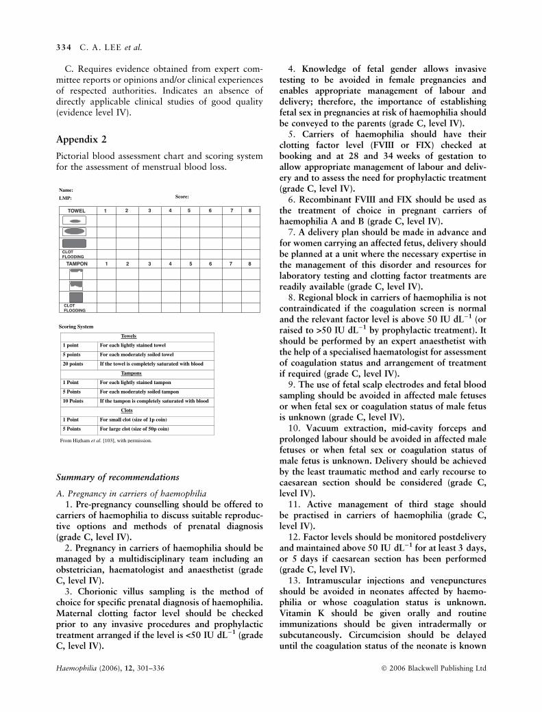
C. Requires evidence obtained from expert com-mittee reports or opinions and/or clinical experiencesof respected authorities. Indicates an absence ofdirectly applicable clinical studies of good quality(evidence level IV).
Appendix 2
Pictorial blood assessment chart and scoring systemfor the assessment of menstrual blood loss.
Summary of recommendations
A. Pregnancy in carriers of haemophilia
1. Pre-pregnancy counselling should be offered tocarriers of haemophilia to discuss suitable reproduc-tive options and methods of prenatal diagnosis(grade C, level IV).
2. Pregnancy in carriers of haemophilia should bemanaged by a multidisciplinary team including anobstetrician, haematologist and anaesthetist (gradeC, level IV).
3. Chorionic villus sampling is the method ofchoice for specific prenatal diagnosis of haemophilia.Maternal clotting factor level should be checkedprior to any invasive procedures and prophylactictreatment arranged if the level is <50 IU dL)1 (gradeC, level IV).
4. Knowledge of fetal gender allows invasivetesting to be avoided in female pregnancies andenables appropriate management of labour anddelivery; therefore, the importance of establishingfetal sex in pregnancies at risk of haemophilia shouldbe conveyed to the parents (grade C, level IV).
5. Carriers of haemophilia should have theirclotting factor level (FVIII or FIX) checked atbooking and at 28 and 34 weeks of gestation toallow appropriate management of labour and deliv-ery and to assess the need for prophylactic treatment(grade C, level IV).
6. Recombinant FVIII and FIX should be used asthe treatment of choice in pregnant carriers ofhaemophilia A and B (grade C, level IV).
7. A delivery plan should be made in advance andfor women carrying an affected fetus, delivery shouldbe planned at a unit where the necessary expertise inthe management of this disorder and resources forlaboratory testing and clotting factor treatments arereadily available (grade C, level IV).
8. Regional block in carriers of haemophilia is notcontraindicated if the coagulation screen is normaland the relevant factor level is above 50 IU dL)1 (orraised to >50 IU dL)1 by prophylactic treatment). Itshould be performed by an expert anaesthetist withthe help of a specialised haematologist for assessmentof coagulation status and arrangement of treatmentif required (grade C, level IV).
9. The use of fetal scalp electrodes and fetal bloodsampling should be avoided in affected male fetusesor when fetal sex or coagulation status of male fetusis unknown (grade C, level IV).
10. Vacuum extraction, mid-cavity forceps andprolonged labour should be avoided in affected malefetuses or when fetal sex or coagulation status ofmale fetus is unknown. Delivery should be achievedby the least traumatic method and early recourse tocaesarean section should be considered (grade C,level IV).
11. Active management of third stage shouldbe practised in carriers of haemophilia (grade C,level IV).
12. Factor levels should be monitored postdeliveryand maintained above 50 IU dL)1 for at least 3 days,or 5 days if caesarean section has been performed(grade C, level IV).
13. Intramuscular injections and venepuncturesshould be avoided in neonates affected by haemo-philia or whose coagulation status is unknown.Vitamin K should be given orally and routineimmunizations should be given intradermally orsubcutaneously. Circumcision should be delayeduntil the coagulation status of the neonate is known
Name:
LMP:
TOWEL 1 2 3 4 5 6 7 8
CLOTFLOODING
TAMPON 1 2 3 4 5 6 7 8
CLOTFLOODING
Score:
Scoring System
Towels
1 point For each lightly stained towel
5 points For each moderately soiled towel
20 points If the towel is completely saturated with blood
Tampons
1 Point For each lightly stained tampon
5 Points For each moderately soiled tampon
10 Points If the tampon is completely saturated with blood
Clots
1 Point For small clot (size of 1p coin)
5 Points For large clot (size of 50p coin)
From Higham et al. [103], with permission.
334 C. A. LEE et al.
Haemophilia (2006), 12, 301–336 � 2006 Blackwell Publishing Ltd
and appropriate management can be arranged bythe haematologist (grade C, level IV).
14. Cord blood should be collected from all maleoffspring of carriers of haemophilia to assess clottingfactor levels for identification and early managementof newborns at risk. The results of the tests should beconveyed to the parents by an appropriate memberof the haemophilia team (grade C, level IV).
15. Cranial ultrasound/CT scans should be ar-ranged for all neonates with haemophilia if labourhad been premature, traumatic, e.g. following for-ceps delivery or prolonged labour, or if there are anyclinical signs suggestive of bleeding. In these cases itis recommended that recombinant clotting factorshould be administered to raise the plasma clottingfactor to 100 IU dL)1 (grade C, level IV).
B. Pregnancy in women with VWD
1. Prenatal diagnosis is not required in the major-ity of families with VWD, but should be discussedand offered to women at risk of having a child withtype 3 VWD (grade C, level IV).
2. Pregnancy in women with VWD should bemanaged by a multidisciplinary team including anobstetrician, haematologist and anaesthetist (gradeC, level IV).
3. Factor levels including VWF:Ag, VWF:AC andFVIII:C should be checked at booking, 28 and34 weeks and prior to invasive procedures. Prophy-lactic treatment should be given when factor levelsare <50 IU dL)1 to cover invasive procedures anddelivery (grade C, level IV).
4. DDAVP can be used in pregnancy, but repeatedadministration or use in pregnancies complicatedwith pre-eclampsia must be avoided. Closemonitoring for water retention must accompany itsuse (grade C, level IV).
5. Women with type I VWD generally do notrequire prophylactic treatment for delivery. In type 2VWD, treatment is required for operative delivery orif there is perineal trauma. Women with type 3 VWDrequire treatment for all types of delivery (grade C,level IV).
6. A delivery plan should be made in advance.For women with severe VWD, delivery should beplanned at a unit where the necessary expertise in themanagement of this disorder and resources forlaboratory testing and clotting factor treatments arereadily available (grade C, level IV).
7. Epidural anaesthesia can be offered for use inmajority of women with type 1 VWD whoseVWF:AC is >50 IU dL)1 (or raised to >50 IU dL)1
by prophylactic treatment). It should be carried out
by an experienced anaesthetist. It is not recommen-ded for use in type 2 or 3 VWD (group C, level IV).
8. Women with VWF:AC <50 IU dL)1 shouldreceive prophylactic treatment at the onset of labouror prior to planned caesarean section (grade C, levelIV).
9. Active management of third stage should bepractised in women with VWD (grade C, level IV).
10. Factor levels should be monitored post-deliv-ery and prophylaxis given to maintain von Wille-brand factor activity and factor VIII levels>50 IU dL)1 for at least 3 days, or 5 days followingcaesarean section. Tranexamic acid or combined oralcontraceptive pill should be considered to controlprolonged and/or intermittent secondary PPH (gradeC, level IV).
11. For fetuses at risk of type 2, type 3 ormoderately severe type 1 VWD, invasive monitoringtechniques, vacuum extraction and rotational/mid-cavity forceps should be avoided and a cord bloodsample should be sent for assessment (grade C, levelIV).
12. Intramuscular injections and venepuncturesshould be avoided in neonates whose coagulationstatus is unknown. Vitamin K should be given orallyand routine immunizations should be given intrad-ermally or subcutaneously. Circumcision should bedelayed until the coagulation status of the neonate isknown and appropriate management can be ar-ranged by the haematologist (grade C, level IV).
C. Pregnancy in women with FXI deficiency
1. Pregnancy in women with FXI deficiencyrequires specialised and individualized care providedcollaboratively by an obstetrician, haematologist andanaesthetist (grade C, level IV).
2. Due to the unpredictability of the condition,attempts should be made to identify the individual’sclinical bleeding tendency and the coexistence ofconfounding factors (grade C, level IV).
3. Prenatal diagnosis should be discussed andoffered to patients where there is a risk of severefactor XI deficiency (grade C, level IV).
4. Factor XI levels should be checked at booking,28 and 34 weeks and prior to invasive procedures.Many patients can be managed expectantly butpatients with severely low levels or a positivebleeding history should be given prophylaxis tocover invasive procedures (grade C, level IV).
5. A delivery plan should be made in advance. It isrecommended for women with FXI deficiency todeliver at a unit where the necessary expertise in themanagement of this disorder and resources for
INFORMATION AND GUIDANCE ON INHERITED BLEEDING DISORDERS 335
� 2006 Blackwell Publishing Ltd Haemophilia (2006), 12, 301–336
laboratory testing and clotting factor treatments arereadily available (grade C, level IV).
6. Women with severe deficiency and/or a bleedinghistory, should receive prophylactic treatment at theonset of labour or prior to planned induction orcaesarean section. Where prophylaxis has been given,it should be extended to 3 days postpartum or 5 daysfollowing caesarean section (grade C, level IV).
7. Active management of third stage should bepractised in women with FXI deficiency (grade C,level IV).
8. Care should be taken to avoid unnecessarytrauma to the baby at delivery and a umbilical cordblood sample for FXI level should be obtained(grade C, level IV).
D. Menorrhagia in women inherited bleeding disorders
1. The use of PBAC should be considered in theassessment of menstrual blood loss for the diagnosisof menorrhagia and evaluation of the treatmentoutcomes (grade C, level IV).
2. A significant proportion of women with idio-pathic menorrhagia have an underlying bleedingdisorder, the commonest being VWD. Thereforetesting for these disorders should be considered(grade B, level III).
3. Adolescents presenting with acute menorrhagiashould be investigated for bleeding disorders. Forprepubertal girls either known to have a bleedingdisorder or a positive family history, plans should bemade in anticipation of the possibility of acutemenorrhagia at the onset of menarche (grade C, levelIV).
4. Initial investigation for an underlying bleedingdisorder in women with menorrhagia should consistof a focus personal and family history of bleedingsymptoms (grade C, level IV).
5. Laboratory testing for an inherited bleedingdisorder should be carried out if thewomanpresentingwith menorrhagia has a positive bleeding history orif surgical intervention is planned (grade C, level IV).
6. Laboratory testing should only be undertaken insettings where necessary expertise and resources areavailable on-site to ensure appropriate and accuratediagnosis (grade C, level IV).
7. Laboratory testing should be carried out in thefollowing stepwise sequence (grade C, level IV):
1 FBC and Ferritin;2 PT and APTT;3 Von Willebrand screen (VWF:Ag, VWF:RCo,
FVIII:C). Testing exclusively during menstruation,off COC, or adjusted for blood type is not neces-sary at the initial evaluation. However, these de-tails should be documented and repeat testingshould be considered if the results are borderline.
4 Platelet function analysis (platelet aggregation andrelease studies);
5 Other clotting factor levels;6 Thyroid function tests if symptomatic.
8. Menorrhagia is one of the most commonbleeding manifestations in women with inheritedbleeding disorders and has a negative effect on theirquality of life (grade B, level III).
9. Management of menorrhagia in women withinherited bleeding disorders should be provided by amultidisciplinary team including a haematologist andgynaecologist (grade C, level IV).
10. Medical treatment of menorrhagia in womenwith inherited bleeding disorders include tranexamicacid, combined oral contraceptives, DDAVP, cyclical21-days oral progesterone, and the LNG-IUS. Thetreatment choice depends on the type of bleedingdisorder, and patient’s age, childbearing status andpreferences in terms of the perceived efficacy and sideeffects (grade C, level IV).
11. Specific haemostatic therapy will be required insome women with inherited bleeding disorders tocontrol menorrhagia. A multidisciplinary approachin the management of these individuals is essential toensure optimal outcomes (grade C, level IV).
12. Prophylactic treatment should be considered inwomen with inherited bleeding disorders undergoingsurgery for treatment of menorrhagia. The surgeryshould be performed by experienced professionalswith special attention to haemostasis. The use of asurgical drain should be considered (grade C, levelIV).
13. Women with inherited bleeding disorders aremore likely to be symptomatic from gynaecologicalproblems that are associated with bleeding. Aware-ness of an underlying bleeding disorder will allowappropriate management. A multidisciplinary ap-proach is essential in the management of womenwith inherited bleeding disorders (grade C, level IV).
336 C. A. LEE et al.
Haemophilia (2006), 12, 301–336 � 2006 Blackwell Publishing Ltd
Related Documents