RESEARCH Open Access The novel protein kinase C epsilon isoform at the adult neuromuscular synapse: location, regulation by synaptic activity-dependent muscle contraction through TrkB signaling and coupling to ACh release Teresa Obis † , Núria Besalduch † , Erica Hurtado, Laura Nadal, Manel M Santafe, Neus Garcia, Marta Tomàs, Mercedes Priego, Maria A Lanuza *† and Josep Tomàs *† Abstract Background: Protein kinase C (PKC) regulates a variety of neural functions, including neurotransmitter release. Although various PKC isoforms can be expressed at the synaptic sites and specific cell distribution may contribute to their functional diversity, little is known about the isoform-specific functions of PKCs in neuromuscular synapse. The present study is designed to examine the location of the novel isoform nPKCε at the neuromuscular junction (NMJ), their synaptic activity-related expression changes, its regulation by muscle contraction, and their possible involvement in acetylcholine release. Results: We use immunohistochemistry and confocal microscopy to demonstrate that the novel isoform nPKCε is exclusively located in the motor nerve terminals of the adult rat NMJ. We also report that electrical stimulation of synaptic inputs to the skeletal muscle significantly increased the amount of nPKCε isoform as well as its phosphorylated form in the synaptic membrane, and muscle contraction is necessary for these nPKCε expression changes. The results also demonstrate that synaptic activity-induced muscle contraction promotes changes in presynaptic nPKCε through the brain-derived neurotrophic factor (BDNF)-mediated tyrosine kinase receptor B (TrkB) signaling. Moreover, nPKCε activity results in phosphorylation of the substrate MARCKS involved in actin cytoskeleton remodeling and related with neurotransmission. Finally, blocking nPKCε with a nPKCε-specific translocation inhibitor peptide (εV1-2) strongly reduces phorbol ester-induced ACh release potentiation, which further indicates that nPKCε is involved in neurotransmission. Conclusions: Together, these results provide a mechanistic insight into how synaptic activity-induced muscle contraction could regulate the presynaptic action of the nPKCε isoform and suggest that muscle contraction is an important regulatory step in TrkB signaling at the NMJ. Keywords: PKC, PKC epsilon, Neuromuscular junction, Neurotransmission, Immunofluorescence, Electrical stimulation, Muscle contraction, TrkB * Correspondence: [email protected]; [email protected] † Equal contributors Unitat d’Histologia i Neurobiologia (UHN). Facultat de Medicina i Ciències de la Salut, Universitat Rovira i Virgili, Sant Llorenç 21, 43201 Reus, Spain © 2015 Obis et al.; licensee BioMed Central. This is an Open Access article distributed under the terms of the Creative Commons Attribution License (http://creativecommons.org/licenses/by/4.0), which permits unrestricted use, distribution, and reproduction in any medium, provided the original work is properly credited. The Creative Commons Public Domain Dedication waiver (http://creativecommons.org/publicdomain/zero/1.0/) applies to the data made available in this article, unless otherwise stated. Obis et al. Molecular Brain (2015) 8:8 DOI 10.1186/s13041-015-0098-x

Welcome message from author
This document is posted to help you gain knowledge. Please leave a comment to let me know what you think about it! Share it to your friends and learn new things together.
Transcript
Obis et al. Molecular Brain (2015) 8:8 DOI 10.1186/s13041-015-0098-x
RESEARCH Open Access
The novel protein kinase C epsilon isoform at theadult neuromuscular synapse: location, regulationby synaptic activity-dependent muscle contractionthrough TrkB signaling and coupling to ACh releaseTeresa Obis†, Núria Besalduch†, Erica Hurtado, Laura Nadal, Manel M Santafe, Neus Garcia, Marta Tomàs,Mercedes Priego, Maria A Lanuza*† and Josep Tomàs*†
Abstract
Background: Protein kinase C (PKC) regulates a variety of neural functions, including neurotransmitter release.Although various PKC isoforms can be expressed at the synaptic sites and specific cell distribution may contributeto their functional diversity, little is known about the isoform-specific functions of PKCs in neuromuscular synapse.The present study is designed to examine the location of the novel isoform nPKCε at the neuromuscular junction(NMJ), their synaptic activity-related expression changes, its regulation by muscle contraction, and their possibleinvolvement in acetylcholine release.
Results: We use immunohistochemistry and confocal microscopy to demonstrate that the novel isoform nPKCε isexclusively located in the motor nerve terminals of the adult rat NMJ. We also report that electrical stimulation ofsynaptic inputs to the skeletal muscle significantly increased the amount of nPKCε isoform as well as its phosphorylatedform in the synaptic membrane, and muscle contraction is necessary for these nPKCε expression changes. The resultsalso demonstrate that synaptic activity-induced muscle contraction promotes changes in presynaptic nPKCε throughthe brain-derived neurotrophic factor (BDNF)-mediated tyrosine kinase receptor B (TrkB) signaling. Moreover, nPKCεactivity results in phosphorylation of the substrate MARCKS involved in actin cytoskeleton remodeling and related withneurotransmission. Finally, blocking nPKCε with a nPKCε-specific translocation inhibitor peptide (εV1-2) strongly reducesphorbol ester-induced ACh release potentiation, which further indicates that nPKCε is involved in neurotransmission.
Conclusions: Together, these results provide a mechanistic insight into how synaptic activity-induced musclecontraction could regulate the presynaptic action of the nPKCε isoform and suggest that muscle contraction is animportant regulatory step in TrkB signaling at the NMJ.
Keywords: PKC, PKC epsilon, Neuromuscular junction, Neurotransmission, Immunofluorescence, Electrical stimulation,Muscle contraction, TrkB
* Correspondence: [email protected]; [email protected]†Equal contributorsUnitat d’Histologia i Neurobiologia (UHN). Facultat de Medicina i Ciències dela Salut, Universitat Rovira i Virgili, Sant Llorenç 21, 43201 Reus, Spain
© 2015 Obis et al.; licensee BioMed Central. This is an Open Access article distributed under the terms of the CreativeCommons Attribution License (http://creativecommons.org/licenses/by/4.0), which permits unrestricted use, distribution, andreproduction in any medium, provided the original work is properly credited. The Creative Commons Public DomainDedication waiver (http://creativecommons.org/publicdomain/zero/1.0/) applies to the data made available in this article,unless otherwise stated.
nPKC pnPKC
Adult
P6 Adult
pnPKC
nPKC
Actin
0
20
40
60
80
% o
f cha
nge
(P6/
Adu
lt)
* *
A B
pnPKCnPKC
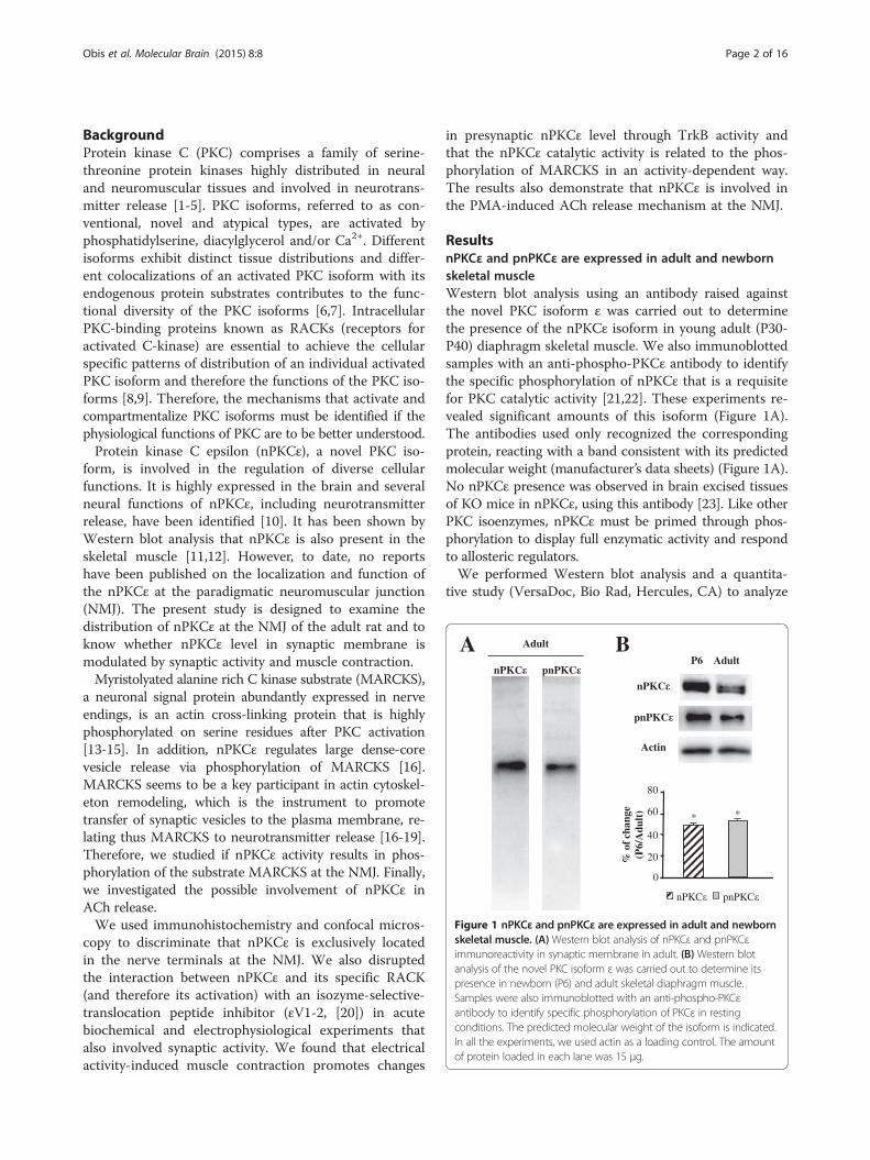
Figure 1 nPKCε and pnPKCε are expressed in adult and newbornskeletal muscle. (A)Western blot analysis of nPKCε and pnPKCεimmunoreactivity in synaptic membrane in adult. (B) Western blotanalysis of the novel PKC isoform ε was carried out to determine itspresence in newborn (P6) and adult skeletal diaphragm muscle.Samples were also immunoblotted with an anti-phospho-PKCεantibody to identify specific phosphorylation of PKCε in restingconditions. The predicted molecular weight of the isoform is indicated.In all the experiments, we used actin as a loading control. The amountof protein loaded in each lane was 15 μg.
Obis et al. Molecular Brain (2015) 8:8 Page 2 of 16
BackgroundProtein kinase C (PKC) comprises a family of serine-threonine protein kinases highly distributed in neuraland neuromuscular tissues and involved in neurotrans-mitter release [1-5]. PKC isoforms, referred to as con-ventional, novel and atypical types, are activated byphosphatidylserine, diacylglycerol and/or Ca2+. Differentisoforms exhibit distinct tissue distributions and differ-ent colocalizations of an activated PKC isoform with itsendogenous protein substrates contributes to the func-tional diversity of the PKC isoforms [6,7]. IntracellularPKC-binding proteins known as RACKs (receptors foractivated C-kinase) are essential to achieve the cellularspecific patterns of distribution of an individual activatedPKC isoform and therefore the functions of the PKC iso-forms [8,9]. Therefore, the mechanisms that activate andcompartmentalize PKC isoforms must be identified if thephysiological functions of PKC are to be better understood.Protein kinase C epsilon (nPKCε), a novel PKC iso-
form, is involved in the regulation of diverse cellularfunctions. It is highly expressed in the brain and severalneural functions of nPKCε, including neurotransmitterrelease, have been identified [10]. It has been shown byWestern blot analysis that nPKCε is also present in theskeletal muscle [11,12]. However, to date, no reportshave been published on the localization and function ofthe nPKCε at the paradigmatic neuromuscular junction(NMJ). The present study is designed to examine thedistribution of nPKCε at the NMJ of the adult rat and toknow whether nPKCε level in synaptic membrane ismodulated by synaptic activity and muscle contraction.Myristolyated alanine rich C kinase substrate (MARCKS),
a neuronal signal protein abundantly expressed in nerveendings, is an actin cross-linking protein that is highlyphosphorylated on serine residues after PKC activation[13-15]. In addition, nPKCε regulates large dense-corevesicle release via phosphorylation of MARCKS [16].MARCKS seems to be a key participant in actin cytoskel-eton remodeling, which is the instrument to promotetransfer of synaptic vesicles to the plasma membrane, re-lating thus MARCKS to neurotransmitter release [16-19].Therefore, we studied if nPKCε activity results in phos-phorylation of the substrate MARCKS at the NMJ. Finally,we investigated the possible involvement of nPKCε inACh release.We used immunohistochemistry and confocal micros-
copy to discriminate that nPKCε is exclusively locatedin the nerve terminals at the NMJ. We also disruptedthe interaction between nPKCε and its specific RACK(and therefore its activation) with an isozyme-selective-translocation peptide inhibitor (εV1-2, [20]) in acutebiochemical and electrophysiological experiments thatalso involved synaptic activity. We found that electricalactivity-induced muscle contraction promotes changes
in presynaptic nPKCε level through TrkB activity andthat the nPKCε catalytic activity is related to the phos-phorylation of MARCKS in an activity-dependent way.The results also demonstrate that nPKCε is involved inthe PMA-induced ACh release mechanism at the NMJ.
ResultsnPKCε and pnPKCε are expressed in adult and newbornskeletal muscleWestern blot analysis using an antibody raised againstthe novel PKC isoform ε was carried out to determinethe presence of the nPKCε isoform in young adult (P30-P40) diaphragm skeletal muscle. We also immunoblottedsamples with an anti-phospho-PKCε antibody to identifythe specific phosphorylation of nPKCε that is a requisitefor PKC catalytic activity [21,22]. These experiments re-vealed significant amounts of this isoform (Figure 1A).The antibodies used only recognized the correspondingprotein, reacting with a band consistent with its predictedmolecular weight (manufacturer’s data sheets) (Figure 1A).No nPKCε presence was observed in brain excised tissuesof KO mice in nPKCε, using this antibody [23]. Like otherPKC isoenzymes, nPKCε must be primed through phos-phorylation to display full enzymatic activity and respondto allosteric regulators.We performed Western blot analysis and a quantita-
tive study (VersaDoc, Bio Rad, Hercules, CA) to analyze

Obis et al. Molecular Brain (2015) 8:8 Page 3 of 16
the density of the band and evaluate the relative amountof the kinase isoform (and its phosphorylated form) inboth the newborn (P6) and the adult. The results showthat the expression of nPKCε isoform appears to be age-dependent being nPKCε more abundant in the newbornthan in the adult (Figure 1B). Under resting conditions(isolated muscle does not receive action potentials fromthe motor neuron soma), we also found quite an abundantlevel of phosphorylated nPKCε and also pnPKCε is moreabundant in the newborn than in the adult (Figure1B).In conclusion, nPKCε is abundantly expressed and
phosphorylated in the skeletal muscle. Subsequently, im-munohistochemical analyses were performed to identifythe cellular localization of the nPKCε isoform at theadult neuromuscular junction components (ie. musclecells, Schwann cells or nerve terminals).
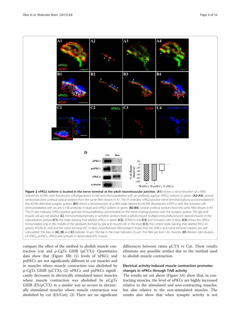
nPKCε isoform is located in the nerve terminal at theneuromuscular junctionImmunofluorescence staining coupled with confocal mi-croscopy analysis was performed to determine the pres-ence and localization of this isoform at the adult NMJ.Precise knowledge of the cellular localization of thenPKCε in pre-, post- and/or perisynaptic elements iscrucial for elucidating the function/s of this protein. Im-munofluorescence experiments were performed in thediaphragm and LAL muscles and immunoreactivity fornPKCε was identical in both muscles. All pictures inFigure 2 show intense immunoreactivity for nPKCε in thesynaptic area identified with AChR labeling. Figure 2Ashows a NMJ with double labeling: AChRs in red andnPKCε in green. Figure 2B shows a NMJ with triple label-ing: AChRs in red, the Schwann cell in blue and nPKCε ingreen. These figures show nPKCε-positive green immuno-labeling concentrated at the presynaptic position over thered postsynaptic gutters (asterisks). This can be seen bothin the semiconsecutive confocal sections (A2-4, B2-4) andin the maximum projection (A1, B1) from the NMJs. Theglia and muscle cell do not seem to be labeled (Figure 2B).In Figure 2A, it appears that pre-terminal axon is alsonPKCε-positive.To improve the analysis of the cellular distribution of
the kinase at the NMJ we obtained semithin cross-sections from whole-mount multiple-immunofluorescentstained muscles [24]. We performed a triple staining inwhich nPKCε isoform (in green in Figure 2C2) was colo-calized with molecular markers of the three cellular ele-ments in the NMJ: muscle cell (nAChR marked withfluorescently labeled α-bungarotoxin, in red, Figure 2C3),Schwann cell (S-100, in blue, Figure 2C4) and nerve ter-minal (syntaxin and neurofilament, in blue, inset in C1)(Figure 2C). When the nPKCε was co-stained with α-BTX and S-100 (C1, colocalization picture), the nPKCεimmunofluorescence (in green) was shown as several
small granular areas located over the postsynaptic line ofthe nAChRs (in red) and externally surrounded by theSchwann cell (in blue). This label corresponds to theaxonal buttons of the nerve terminal because it is colo-calized with the syntaxin/neurofilament stain (labeled inblue in the inset in C1, Figure 2C) and also with syn-taxin alone (not shown). Thus, the results indicate thatnPKCε has a unique location in the synapse: it is exclu-sively expressed in the nerve terminals at the NMJ.Denervation of the extensor digitorum longus muscle
(EDL) was performed to confirm that nPKCε is locatedin the nerve. We denervated the muscle, waited 1–3days, and studied the level of nPKCε by Western blots.The results show that nPKCε protein almost completelydisappear after 1 day of denervation (ratio denervated/control 0.11 ± 0.08, p < 0.05) indicating that the mostpart of nPKCε is located in the nerve (Figure 2D). Bycontrast, cPKCα, an isoform ubiquitously located [25],slightly decreased (Figure 2D). Syntaxin has been usedas a control of denervation. The residual presence ofnPKCε may be related with blood vessels [11].
nPKCε expression is modulated by synaptic activitySynaptic function of PKC family has been shown to beregulated by activity [25], so we tested whether nPKCεsynaptic level is modulated by synaptic activity. There-fore, we studied whether there was any change in theamount of nPKCε and its phosphorylated form in thesynaptic membrane after electrical stimulation (with andwithout muscle contraction). Electrical stimulation ofsynaptic inputs to the diaphragm skeletal muscle (1 Hzfor 30 minutes) and the resulting muscle contractionsignificantly increased the amount of nPKCε isoformand its phosphorylated form in the synaptic membrane(Figure 3A).To separate the effect of the presynaptic stimulation
(and synaptic transmission) from the effect of the musclecell contraction, we performed experiments in whichcontractions were inhibited. When the contraction wasinhibited by cutting the muscle fibers [26-28] or by usingμ-CgTx-GIIIB [28,29], the electrical stimulation signifi-cantly decreased nPKCε and its phosphorylated form inthe synaptic membrane (Figure 3A). Thus, nerve stimu-lation influences the kinase level in the synaptic mem-brane and this suggest that, with stimulation, the kinasecould be detached from the membrane (and thereforestop its catalytic function), or alternatively stimulationentails some consumption of the enzyme. These resultsclearly indicate that muscle contractions induce an im-portant change that reverses the effect of nerve stimula-tion by itself on nPKCε level in the synaptic membrane,suggesting that muscle contraction is necessary for nPKCεmaintenance and its catalytic activity. Moreover, we per-formed several Western blots of nPKCε and pnPKCε to
D
Figure 2 nPKCε isoform is located in the nerve terminal at the adult neuromuscular junction. (A1) shows a reconstruction of a NMJstained for AChRs with fluorescent α-Bungarotoxin in red and immunolabeled with an antibody against nPKCε isoform in green. (A2-A4), severalsemiconsecutive confocal optical sections from the same NMJ shown in A1. The (*) indicates nPKCε-positive nerve terminal buttons accommodated inthe AChRs delimited synaptic gutters. (B1) shows a reconstruction of a NMJ triple labeled for AChRs (fluorescent α-BTX in red), the Schwann cell(immunolabeled with an anti S-100 antibody in blue) and nPKCε isoform in green. (B2-B4), several confocal sections from the same NMJ shown in B1.The (*) also indicates nPKCε-positive granular immunolabeling concentrated on the nerve endings position over the synaptic gutters. The glia andmuscle cell are not labeled. (C), Immunohistochemistry in semithin sections from a whole-mount multiple-immunofluorescent stained muscle. In thecolocalization picture (C1), the triple staining that labeled nPKCε in green (C2), AChRs in red (C3) and Schwann cells in blue, (C4) shows the nPKCεimmunolabel only in the middle of the sandwich formed by glia and muscle cell. In the inset (C1), the control triple staining that labeled PKCε (ingreen), AChRs (in red) and the nerve terminal (NT, in blue, neurofilament-200/syntaxin) shows that the nPKCε and nerve terminal markers are wellcolocalized. The bars in (A), (B) and (C) indicate 10 μm. The bar in the inset indicates 2.5 μm. The NMJ are from LAL muscles. (D) Western blot analysisof nPKCε, pnPKCε, cPKCα and syntaxin in denervated EDL muscle.
Obis et al. Molecular Brain (2015) 8:8 Page 4 of 16
compare the effect of the method to abolish muscle con-traction (cut and μ-CgTx GIIIB (μCTX)). Quantitativedata show that (Figure 3B): (1) levels of nPKCε andpnPKCε are not significantly different in cut muscles andin muscles where muscle contraction was abolished byμ-CgTx GIIIB (μCTX); (2) nPKCε and pnPKCε signifi-cantly decreases in electrically stimulated intact muscleswhere muscle contraction was abolished by μCgTxGIIIB (ES/μCTX) in a similar way as occurs in electric-ally stimulated muscles where muscle contraction wasabolished by cut (ES/Cut); (3) There are no significant
differences between ratios μCTX vs Cut. These resultseliminate any possible artifact due to the method usedto abolish muscle contraction.
Electrical activity-induced muscle contraction promoteschanges in nPKCε through TrkB activityThe results set out above (Figure 3A) show that, in con-tracting muscles, the level of nPKCε are highly increasedrelative to the stimulated and non-contracting muscles,but also relative to the non-stimulated muscles. Theresults also show that when synaptic activity is not

A
B
ES (ES+C)Ctr
nPKC
actin
pnPKC
nPKC
-50
0
50
100
150
-100 *
*
*
% o
f cha
nge
pnPKC
*
*
*
Actin
nPKC
-50
0
50
-100
% o
f ch
ange
% o
f ch
ange
Ctr (ES+C) Cut µCTX ES
Cut / CTX
ES / CTX
ES / Cut
**
P>0,05
ES/Ctr ES+C/ES ES+C/Ctr
Figure 3 nPKCε expression is modulated by synaptic activity. (A) Western blot analysis of nPKCε and pnPKCε immunoreactivity levels insynaptic membrane in adult stimulated muscles with (ES + C) and without (ES) muscle contraction. (B) shows Western blots of nPKCε to comparethe effect of the method to abolish muscle contraction (cut and μ-CgTx GIIIB (μCTX)). Quantitative data eliminate any possible artifact due to themethod used to abolish muscle contraction. There are no significant differences between ratios μCTX vs Cut. The Western blot shows: (1) levelsof nPKCε in non-stimulated muscles where no method was performed to abolish muscle contraction (Ctr), in cut muscles (Cut) and in muscleswhere muscle contraction was abolished by μ-CgTx GIIIB (μCTX); (2) nPKCε in electrically stimulated intact muscles where muscle contraction wasabolished by μCgTx GIIIB (ES,μCTX); and (3) nPKCε in stimulated muscles resulting in contraction (ES + C). Experiments were performed usingdiaphragm muscle.
Obis et al. Molecular Brain (2015) 8:8 Page 5 of 16
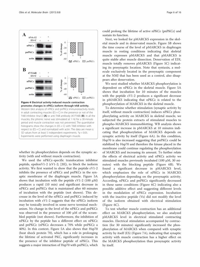
accompanied by muscle contractile activity, the level ofnPKCε is decreased. Thus, we hypothesize that musclecontraction induces a change that can reverse the effectof nerve stimulation itself and a contraction-dependentneurotrophic support from postsynaptic site may make acontribution to this through the tyrosine kinase receptorB (TrkB). To demonstrate whether the activation ofTrkB, as a result of muscle contraction, is critical toaffect nPKCε’s level, we selectively suppressed TrkB ac-tivity in contracting diaphragm muscles using selectiveTrkB inhibitors. We used ANA-12 which showed directand selective binding to TrkB and inhibits processesdownstream of TrkB without altering TrkA and TrkCfunctions [30]. ANA-12 fully inhibits BDNF-inducedTrkB phosphorylation and therefore prevents receptoractivation. We also used an anti-TrkB antibody (47/TrkB)which effectively inhibits BDNF binding to TrkB recep-tors [31]. We measured resultant nPKCε and pnPKCεlevels and found that TrkB blockade resulted in a signifi-cant decrease in the isoform (Figure 4). We obtained thesame result with the two blockade methods. These re-sults demonstrate that electrical activity-induced musclecontraction promotes increase in nPKCε and pnPKCε
through TrkB activity suggesting that muscle contractioninduce an increase of the interaction of a TrkB-specificneurothrophic factor with TrkB that enhance the intra-cellular signaling to increase the isoform levels in thesynaptic membrane. Accordingly, exogenously addedBDNF (10 nM, 30 minutes) in stimulated muscles with-out contraction significantly increased the amount ofpnPKCε (58.78% ± 2.1% of change, p < 0.05). This resultindicates that exogenous BDNF mimics muscle contrac-tion effect on nPKCε levels. We choose the BDNF dosebased in a previous dose–response and time-coursestudy in the same muscle to determine their effect onthe size of the evoked EPP [32].
nPKCε regulates phosphorylation of MARCKS in skeletalmuscle, in an activity-dependent wayTo gain insight into the role of nPKCε we analyzed pos-sible substrate phosphorylation. Although the transloca-tion and phosphorylation of PKC is indicative of PKCactivation [33], a more direct measure of the PKC activ-ity is to detect PKC substrate phosphorylation in cells.Therefore, we wanted to know whether MARCKS phos-phorylation is dependent on nPKCε at the NMJ and
*
B
AANA-12
ES+C + +
- +
nPKC
pnPKC
0
-20
-40
-60%
of
ch
an
ge
*
47/TrkB
ES+C
- +
+ +
nPKC
pnPKC
% o
f ch
an
ge 0
-20
-40
-60
*
*
pnPKCnPKC
pnPKCnPKC
actin
actin
Figure 4 Electrical activity-induced muscle contractionpromotes changes in nPKCε isoform through trkB activity.Western blot analysis of nPKCε and pnPKCε immunoreactivity levelsin adult contracting muscles (ES + C) in the presence or not of theTrkB inhibitor Ana12 (A) or anti-TrkB antibody (47/TrkB) (B). In all themuscles, the phrenic nerve was stimulated at 1 Hz for a 30-minuteperiod and muscle contraction was not prevented. The quantitativehistograms show the changes in (ES + C) with TrkB inhibitor withrespect to (ES + C) and normalized with actin. The data are mean ±SD values from at least 5 independent experiments. *p < 0.05.Experiments were performed using diaphragm muscle.
Obis et al. Molecular Brain (2015) 8:8 Page 6 of 16
whether its phosphorylation depends on the synaptic ac-tivity (with and without muscle contraction).We used the nPKCε-specific translocation inhibitor
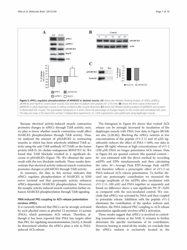
peptide, epsilonV1-2 (εV1-2; [20]), to block the isoformactivity. We first wanted to show that the peptide εV1-2inhibits the presence of nPKCε and pnPKCε in the syn-aptic membrane of the diaphragm muscle. Figure 5Ashows that incubation with the peptide εV1-2 (100 μM)produces a rapid (10 min) and significant decrease innPKCε and pnPKCε that is maintained after 60 minutesof incubation with the peptide (not shown). This de-crease in the level of the nPKCε and pnPKCε induced byincubation with εV1-2 suggests that the nPKCε isoformmay be tonically involved in some nerve terminal mech-anism. No change in the level of the nPKCε and pnPKCεwas observed in the presence of 100 μM of the scram-bled peptide (not shown). Furthermore, the inhibition ofnPKCε by the peptide has a different effect on nPKCεand pnPKCε (nPKCε decreases a 70% while pnPKCε a40%). In this context, Figure 5A also shows that Hsp70(heat shock protein 70), which has a role in prolongingthe lifetime of activated PKC, significantly increases inthe presence of the inhibitor peptide of nPKCε. Thissuggests a major interaction of Hsp70 with pnPKCε, which
could prolong the lifetime of active nPKCε (pnPKCε) andsustain its function.Next, we looked for pMARCKS expression in the skel-
etal muscle and in denervated muscle. Figure 5B showsthe time course of the level of pMARCKS in diaphragmmuscle in resting conditions indicating that skeletalmuscle expresses pMARCKS and that pMARCKS isquite stable after muscle dissection. Denervation of EDLmuscle totally removes pMARCKS (Figure 5C) indicat-ing its presynaptic location. Note that syntaxin, a mol-ecule exclusively located in the presynaptic componentat the NMJ that has been used as a control, also disap-pears after denervation.We next studied whether MARCKS phosphorylation is
dependent on nPKCε in the skeletal muscle. Figure 5Ashows that incubation for 10 minutes of the muscleswith the peptide εV1-2 produces a significant decreasein pMARCKS indicating that nPKCε is related to thephosphorylation of MARCKS in the skeletal muscle.To determine whether stimulation (synaptic activity by
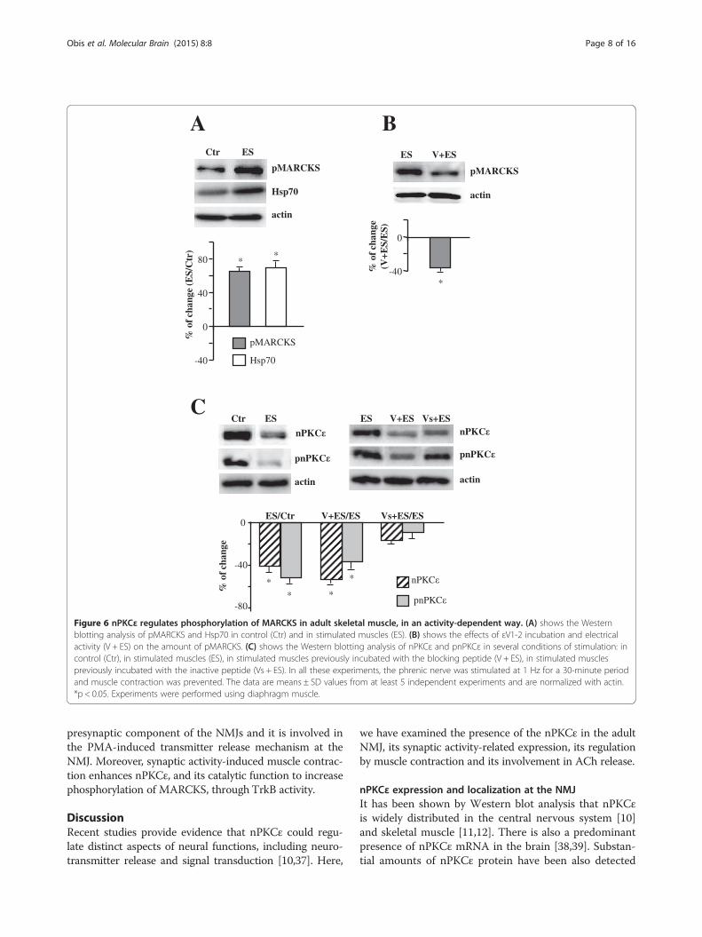
itself, without muscle contraction) induces nPKCε phos-phorylating activity on MARCKS in skeletal muscle, wesubjected the protein extracts of stimulated muscles tophospho-MARCKS immunoblotting. WB analysis showsa significant increase in pMARCKS at 10 minutes indi-cating that phosphorylation of MARCKS depends onsynaptic activity by itself (Figure 6A). In this condition,Hsp70 is also increased suggesting that pnPKCε could bestabilized by Hsp70 and therefore the kinase placed in themembrane could continue regulating the phosphorylationof MARCKS and increasing its amount. To further relatethe effects of electrical activity and nPKCε activity westimulated muscles previously incubated (100 μM, 30 mi-nutes) with the blocking peptide (Figure 6B). Wefound a significant decrease in pMARCKS level,which emphasizes the role of nPKCε in MARCKSphosphorylation depending on the presynaptic activity.According, nPKCε and pnPKCε significantly decreasedin these same conditions (Figure 6C) indicating also apossible additive effect and suggesting different levelsin the modulation of nPKCε expression. Incubationwith the inactive peptide (Vs) did not modify the levelof the isoform obtained with electrical stimulation(Figure 6C).To test whether muscle contraction has an additional
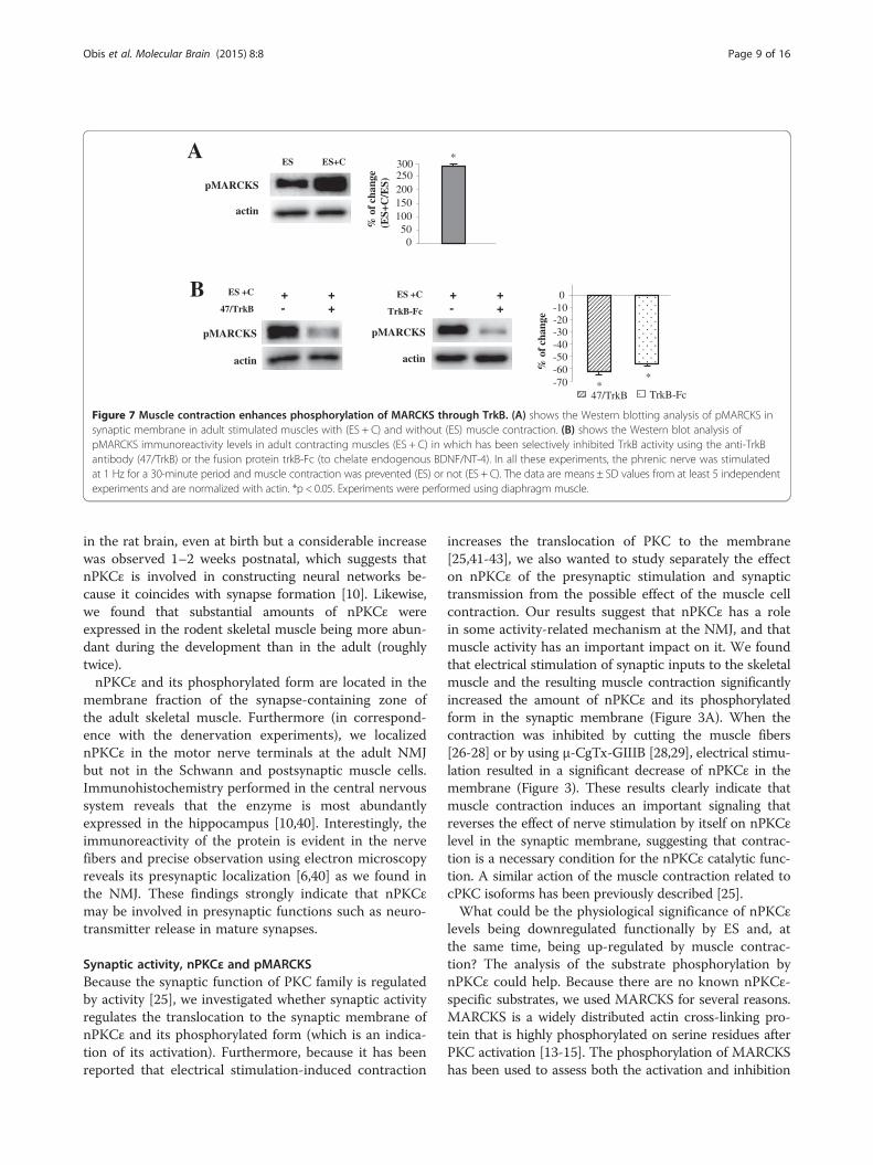
effect on MARCKS phosphorylation, we also analyzedpMARCKS level in electrical stimulated contractingmuscles. Electrical stimulation accompanied by contrac-tion (for 30 minutes) significantly increased the phos-phorylation of MARCKS when compared with synapticactivity by itself (ES) (Figure 7A), indicating that synapticactivity with muscle contraction has a higher effect onthe MARCKS phosphorylation than presynaptic activityby itself.
B Control
0’ 10’ 30’ 60’ 90’
pMARCKS
actin
C
% o
f cha
nge
(Den
/Ctr
)
A
Syntaxin
Ctr
pMARCKS
Den
pMARCKSSyntaxin
200
-20
-40-60
-80
-100**
% o
f cha
nge
(V
1-2/
Ctr
)
Ctr εV1-2
nPKCε
pnPKCε
actin
pMARCKS
Hsp70
*-80
-40
0
* *
*
nPKCε
pnPKCε
pMARCKS
Hsp70
80
4020
60
-20
-60
Figure 5 nPKCε regulates phosphorylation of MARCKS in skeletal muscle. (A) shows the Western blotting analysis of nPKCε, pnPKCε,pMARCKS and Hsp70 in control adult muscles (Ctr) and after incubation with peptide εV1-2 (10 min). (B) shows the time course of the level ofpMARCKS in adult diaphragm muscle in resting conditions after muscle dissection. (C) shows the Western blotting analysis of pMARCKS and syntaxinin denervated EDL muscle. The quantitative histograms in A and C show the percentage of changes respect to the control and normalized with actin.The data are mean ± SD values from at least 5 independent experiments. *p < 0.05. Experiments were performed using diaphragm muscle.
Obis et al. Molecular Brain (2015) 8:8 Page 7 of 16
Because electrical activity-induced muscle contractionpromotes changes in nPKCε through TrkB activity, next,we plan to know whether muscle contraction could affectMARCKS phosphorylation through TrkB activity. Thus,we analyzed the amount of pMARCKS in contractingmuscles in which has been selectively inhibited TrkB ac-tivity using the anti-TrkB antibody (47/TrkB) or the fusionprotein trkB-Fc (to chelate endogenous BDNF/NT-4). Wefound that TrkB blockade resulted in a significant de-crease in pMARCKS (Figure 7B). We obtained the sameresult with the two blockade methods. These results dem-onstrate that electrical activity-induced muscle contractionpromotes changes in pMARCKS through TrkB activity.In summary, the data in this section indicates that
nPKCε regulates phosphorylation of MARCKS in NMJnerve terminal and that presynaptic activity enhancesnPKCε-dependent MARCKS phosphorylation. Moreover,the synaptic activity-induced muscle contraction further en-hances MARCKS phosphorylation throughTrkB signaling.
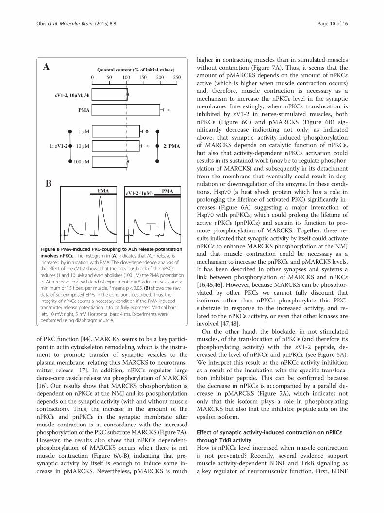
PMA-induced PKC-coupling to ACh release potentiationinvolves nPKCεIt is currently believed that PKCs can be strongly activatedby such phorbol esters as phorbol 12-myristate 13-acetate(PMA), which potentiates ACh release. Therefore, al-though it has been reported that PMA has targets otherthan PKC for regulating neurotransmission [34,35], it shouldbe determined whether the nPKCε plays a role in PMA-induced ACh release.
The histogram in Figure 8A shows that evoked AChrelease can be strongly increased by incubation of thediaphragm muscle with PMA (raw data in Figure 8B left;see also [5,28,36]). Blocking the nPKCε isoform at lowconcentrations of the peptide εV1-2 (1 and 10 μM) sig-nificantly reduces the effect of PMA (~60%; raw data inFigure 8B right) whereas at high concentrations of εV1-2(100 μM) PMA no longer potentiates ACh release. Datain Figure 8A are quantal content (the quantal content –M- was estimated with the direct method by recordingmEPPs and EPPs simultaneously and then calculatingthe ratio: M = Average Peak EPP/Average Peak mEPP)and therefore reflects a presynaptic effect of εV1-2 onPMA-induced ACh release potentiation. To further dis-card any postsynaptic contribution we measured theaverage amplitude of the mEPPs after incubation withεV1-2 (1–100 μM) and PMA together or separately andfound no difference above a non-significant 9% (P > 0.05)as compared with the non-incubated control. We con-clude that nPKCε was activated by PMA and then coupledto potentiate release. Inhibition with the peptide εV1-2eliminates the contribution of the epsilon isoform and,therefore, the PMA-induced PKC-coupling to ACh releasepotentiation significantly involves nPKCε activation.These results suggest that nPKCε is involved in control-
ling transmitter release at the NMJ. It remains to furtherdetermine the specific mechanism of its contribution.However, bearing in mind all the results, we conclude thatthe nPKCε isoform is exclusively located in the
C
A
ES V+ESnPKC
pnPKC
actin
Vs+ES
-40
0ES/Ctr
nPKC
pnPKC**
% o
f cha
nge
-80
V+ES/ES
*
*
Vs+ES/ES
Ctr ES
nPKC
pnPKC
actin
BCtr ES
actin
pMARCKS
-40
0
40
80
% o
f cha
nge
(ES/
Ctr
)
**
pMARCKS
Hsp70
Hsp70
ES V+ES
actin
pMARCKS
*-40
0
% o
f cha
nge
(V+E
S/E
S)
Figure 6 nPKCε regulates phosphorylation of MARCKS in adult skeletal muscle, in an activity-dependent way. (A) shows the Westernblotting analysis of pMARCKS and Hsp70 in control (Ctr) and in stimulated muscles (ES). (B) shows the effects of εV1-2 incubation and electricalactivity (V + ES) on the amount of pMARCKS. (C) shows the Western blotting analysis of nPKCε and pnPKCε in several conditions of stimulation: incontrol (Ctr), in stimulated muscles (ES), in stimulated muscles previously incubated with the blocking peptide (V + ES), in stimulated musclespreviously incubated with the inactive peptide (Vs + ES). In all these experiments, the phrenic nerve was stimulated at 1 Hz for a 30-minute periodand muscle contraction was prevented. The data are means ± SD values from at least 5 independent experiments and are normalized with actin.*p < 0.05. Experiments were performed using diaphragm muscle.
Obis et al. Molecular Brain (2015) 8:8 Page 8 of 16
presynaptic component of the NMJs and it is involved inthe PMA-induced transmitter release mechanism at theNMJ. Moreover, synaptic activity-induced muscle contrac-tion enhances nPKCε, and its catalytic function to increasephosphorylation of MARCKS, through TrkB activity.
DiscussionRecent studies provide evidence that nPKCε could regu-late distinct aspects of neural functions, including neuro-transmitter release and signal transduction [10,37]. Here,
we have examined the presence of the nPKCε in the adultNMJ, its synaptic activity-related expression, its regulationby muscle contraction and its involvement in ACh release.
nPKCε expression and localization at the NMJIt has been shown by Western blot analysis that nPKCεis widely distributed in the central nervous system [10]and skeletal muscle [11,12]. There is also a predominantpresence of nPKCε mRNA in the brain [38,39]. Substan-tial amounts of nPKCε protein have been also detected
30025020015010050%
of c
hang
e (E
S+C
/ES)pMARCKS
ES ES+C
actin
*
0
A
B ES +C
47/TrkB
pMARCKS
actin
+ +- +
pMARCKS
actin
ES +C
TrkB-Fc
+ +- +
% o
f cha
nge
TrkB-Fc
0-10-20-30-40-50-60-70 *
*
47/TrkB
Figure 7 Muscle contraction enhances phosphorylation of MARCKS through TrkB. (A) shows the Western blotting analysis of pMARCKS insynaptic membrane in adult stimulated muscles with (ES + C) and without (ES) muscle contraction. (B) shows the Western blot analysis ofpMARCKS immunoreactivity levels in adult contracting muscles (ES + C) in which has been selectively inhibited TrkB activity using the anti-TrkBantibody (47/TrkB) or the fusion protein trkB-Fc (to chelate endogenous BDNF/NT-4). In all these experiments, the phrenic nerve was stimulatedat 1 Hz for a 30-minute period and muscle contraction was prevented (ES) or not (ES + C). The data are means ± SD values from at least 5 independentexperiments and are normalized with actin. *p < 0.05. Experiments were performed using diaphragm muscle.
Obis et al. Molecular Brain (2015) 8:8 Page 9 of 16
in the rat brain, even at birth but a considerable increasewas observed 1–2 weeks postnatal, which suggests thatnPKCε is involved in constructing neural networks be-cause it coincides with synapse formation [10]. Likewise,we found that substantial amounts of nPKCε wereexpressed in the rodent skeletal muscle being more abun-dant during the development than in the adult (roughlytwice).nPKCε and its phosphorylated form are located in the
membrane fraction of the synapse-containing zone ofthe adult skeletal muscle. Furthermore (in correspond-ence with the denervation experiments), we localizednPKCε in the motor nerve terminals at the adult NMJbut not in the Schwann and postsynaptic muscle cells.Immunohistochemistry performed in the central nervoussystem reveals that the enzyme is most abundantlyexpressed in the hippocampus [10,40]. Interestingly, theimmunoreactivity of the protein is evident in the nervefibers and precise observation using electron microscopyreveals its presynaptic localization [6,40] as we found inthe NMJ. These findings strongly indicate that nPKCεmay be involved in presynaptic functions such as neuro-transmitter release in mature synapses.
Synaptic activity, nPKCε and pMARCKSBecause the synaptic function of PKC family is regulatedby activity [25], we investigated whether synaptic activityregulates the translocation to the synaptic membrane ofnPKCε and its phosphorylated form (which is an indica-tion of its activation). Furthermore, because it has beenreported that electrical stimulation-induced contraction
increases the translocation of PKC to the membrane[25,41-43], we also wanted to study separately the effecton nPKCε of the presynaptic stimulation and synaptictransmission from the possible effect of the muscle cellcontraction. Our results suggest that nPKCε has a rolein some activity-related mechanism at the NMJ, and thatmuscle activity has an important impact on it. We foundthat electrical stimulation of synaptic inputs to the skeletalmuscle and the resulting muscle contraction significantlyincreased the amount of nPKCε and its phosphorylatedform in the synaptic membrane (Figure 3A). When thecontraction was inhibited by cutting the muscle fibers[26-28] or by using μ-CgTx-GIIIB [28,29], electrical stimu-lation resulted in a significant decrease of nPKCε in themembrane (Figure 3). These results clearly indicate thatmuscle contraction induces an important signaling thatreverses the effect of nerve stimulation by itself on nPKCεlevel in the synaptic membrane, suggesting that contrac-tion is a necessary condition for the nPKCε catalytic func-tion. A similar action of the muscle contraction related tocPKC isoforms has been previously described [25].What could be the physiological significance of nPKCε
levels being downregulated functionally by ES and, atthe same time, being up-regulated by muscle contrac-tion? The analysis of the substrate phosphorylation bynPKCε could help. Because there are no known nPKCε-specific substrates, we used MARCKS for several reasons.MARCKS is a widely distributed actin cross-linking pro-tein that is highly phosphorylated on serine residues afterPKC activation [13-15]. The phosphorylation of MARCKShas been used to assess both the activation and inhibition
PMA V1-2 (1µM) PMAB
A Quantal content (% of initial values)
PMA
0 50 100 150 200 250
*
*
*
-2, 10µM, 3h
2: PMA
100 µM
10 µM
1 µM
V1
1: V1-2
Figure 8 PMA-induced PKC-coupling to ACh release potentiationinvolves nPKCε. The histogram in (A) indicates that ACh release isincreased by incubation with PMA. The dose-dependence analysis ofthe effect of the εV1-2 shows that the previous block of the nPKCεreduces (1 and 10 μM) and even abolishes (100 μM) the PMA potentiationof ACh release. For each kind of experiment: n = 5 adult muscles and aminimum of 15 fibers per muscle. *means p < 0.05. (B) shows the rawdata of superimposed EPPs in the conditions described. Thus, theintegrity of nPKCε seems a necessary condition if the PMA-inducedtransmitter release potentiation is to be fully expressed. Vertical bars:left, 10 mV; right, 5 mV. Horizontal bars: 4 ms. Experiments wereperformed using diaphragm muscle.
Obis et al. Molecular Brain (2015) 8:8 Page 10 of 16
of PKC function [44]. MARCKS seems to be a key partici-pant in actin cytoskeleton remodeling, which is the instru-ment to promote transfer of synaptic vesicles to theplasma membrane, relating thus MARCKS to neurotrans-mitter release [17]. In addition, nPKCε regulates largedense-core vesicle release via phosphorylation of MARCKS[16]. Our results show that MARCKS phosphorylation isdependent on nPKCε at the NMJ and its phosphorylationdepends on the synaptic activity (with and without musclecontraction). Thus, the increase in the amount of thenPKCε and pnPKCε in the synaptic membrane aftermuscle contraction is in concordance with the increasedphosphorylation of the PKC substrate MARCKS (Figure 7A).However, the results also show that nPKCε dependent-phosphorylation of MARCKS occurs when there is notmuscle contraction (Figure 6A-B), indicating that pre-synaptic activity by itself is enough to induce some in-crease in pMARCKS. Nevertheless, pMARCKS is much
higher in contracting muscles than in stimulated muscleswithout contraction (Figure 7A). Thus, it seems that theamount of pMARCKS depends on the amount of nPKCεactive (which is higher when muscle contraction occurs)and, therefore, muscle contraction is necessary as amechanism to increase the nPKCε level in the synapticmembrane. Interestingly, when nPKCε translocation isinhibited by εV1-2 in nerve-stimulated muscles, bothnPKCε (Figure 6C) and pMARCKS (Figure 6B) sig-nificantly decrease indicating not only, as indicatedabove, that synaptic activity-induced phosphorylationof MARCKS depends on catalytic function of nPKCε,but also that activity-dependent nPKCε activation couldresults in its sustained work (may be to regulate phosphor-ylation of MARCKS) and subsequently in its detachmentfrom the membrane that eventually could result in deg-radation or downregulation of the enzyme. In these condi-tions, Hsp70 (a heat shock protein which has a role inprolonging the lifetime of activated PKC) significantly in-creases (Figure 6A) suggesting a major interaction ofHsp70 with pnPKCε, which could prolong the lifetime ofactive nPKCε (pnPKCε) and sustain its function to pro-mote phosphorylation of MARCKS. Together, these re-sults indicated that synaptic activity by itself could activatenPKCε to enhance MARCKS phosphorylation at the NMJand that muscle contraction could be necessary as amechanism to increase the pnPKCε and pMARCKS levels.It has been described in other synapses and systems alink between phosphorylation of MARCKS and nPKCε[16,45,46]. However, because MARCKS can be phosphor-ylated by other PKCs we cannot fully discount thatisoforms other than nPKCε phosphorylate this PKC-substrate in response to the increased activity, and re-lated to the nPKCε activity, or even that other kinases areinvolved [47,48].On the other hand, the blockade, in not stimulated
muscles, of the translocation of nPKCε (and therefore itsphosphorylating activity) with the εV1-2 peptide, de-creased the level of nPKCε and pnPKCε (see Figure 5A).We interpret this result as the nPKCε activity inhibitionas a result of the incubation with the specific transloca-tion inhibitor peptide. This can be confirmed becausethe decrease in nPKCε is accompanied by a parallel de-crease in pMARCKS (Figure 5A), which indicates notonly that this isoform plays a role in phosphorylatingMARCKS but also that the inhibitor peptide acts on theepsilon isoform.
Effect of synaptic activity-induced contraction on nPKCεthrough TrkB activityHow is nPKCε level increased when muscle contractionis not prevented? Recently, several evidence supportmuscle activity-dependent BDNF and TrkB signaling asa key regulator of neuromuscular function. First, BDNF
Obis et al. Molecular Brain (2015) 8:8 Page 11 of 16
has been identified as a contraction-inducible protein inhuman skeletal muscle [49]. Secondly, basal levels ofneuromuscular activity are required to maintain normallevels of BDNF in the neuromuscular system [50]. Fi-nally, BDNF and TrkB contribute to the neuromuscularjunction transmission [32,51]. Accordingly, we aimed toinvestigate whether nerve induced-muscle activity wouldproduce a neurotrophin inducing a signaling pathwaythrough TrkB to affect presynaptic nPKCε and its cata-lytic activity. Our results show that blockade of TrkBprevents muscle contraction-induced nPKCε increase(Figure 4A-B) and phosphorylation of MARCKS (Figure 7B)indicating that a neurotrophin acts through TrkB to in-crease nPKCε and pMARCKS on the nerve terminal.Moreover, we found that phosphorylation of nPKCε in-creases significantly when stimulated muscles were incu-bated with BDNF exogenous, indicating that BDNF couldbe the neurothrophin that activates TrkB to enhancenPKCε action and pMARCKS on the nerve terminal. Itcould be speculated that TrkB works to regulate nPKCε(and pnPKCε) by phosphorylating PLCγ (phosphoinosi-tide-specific phospholipase C γ). Phosphorylation of TrkBon Tyr785 recruits PLCγ to the receptors, and the enzymebecomes activated upon tyrosine phosphorylation [52,53].Activated PLCγ hydrolyses PI(4,5)P2 (phosphatidylinositol4,5-biphosphate) to generate inositol tris-phosphate (IP3)and DAG, which activates nPKCε (once the isoform hasbeen previously phosphorylated by PDK (3-phosphoinosi-tide dependent protein kinase) and autophosphorylated[54-56]. These findings provide mechanistic insight intohow synaptic activity induced-muscle contraction couldregulate the presynaptic action of the nPKCε and suggestthat muscle contraction is an important regulatory step inTrkB signaling at the NMJ.
Effect of nPKCε on PMA-induced PKC-coupling to AChrelease potentiationOne aim of the present study is to determine whethernPKCε is involved in the neurotransmission mechanism.Although it has been reported that PMA has targets otherthan PKC for affecting neurotransmission (Munc13)[34,35], PMA strongly activates PKC family to enhanceneurotransmission [5,36,57-60]. Here, we show that block-ing the nPKCε with εV1-2 fully inhibited (Figure 8) thePMA-induced pharmacologic potentiation of ACh releasein a concentration-dependent way indicating that nPKCεplays a role in neurotransmission at the NMJ. Therefore,nPKCε has a key role regulating the ACh release by itselfor by modulating the action of other PKCs isoforms. Ithas been demonstrated that PKC family is involved in themodulation of ACh release at the NMJ [60]. Moreover, ex-ogenously added BDNF potentiates evoked ACh release ina TrkB receptor dependent manner [32]. Furthermore, theTrkB signaling needs an operative PKC pathway to couple
to the release mechanism and potentiate it [61]. nPKCεcould be related with this PKC-TrkB mechanism thatmodulates ACh release at the NMJ. Future experimentswill be necessary to determine how nPKCε is critical tothe maintenance of transmitter release and to betterunderstand how muscle contraction-induced phosphoryl-ation of MARCKS, regulated by nPKCε, is involved inneuromuscular transmission. Furthermore, nPKCε maywork in parallel with the active zone positioning and prim-ing protein Munc13 to enhance neurotransmission be-cause recent studies proposed two probably convergingpathways stimulated by phorbol esters (and DAG) to in-duce potentiation (PKC- and Munc13-dependent) [62,63].It has been proposed that a PKC/Munc18-1 dependent-redistribution of synaptic vesicles after phorbol esterstimulation (via PKC phosphorylation of the SNARE/SMfusion protein Munc18-1), may be the morphologicalcorrelate of the increased release efficiency during potenti-ation [62]. In this redistribution, MARCKS may be in-volved because their role in actin cytoskeleton remodeling,which is the instrument to promote transfer of synapticvesicles to the plasma membrane.In physiological conditions, nerve action is linked to
muscle contractions, therefore it is important to providea mechanism linking muscle contraction to nerve func-tion. Figure 9 shows a diagram illustrating how synapticactivity-induced muscle contraction could regulate thepresynaptic action of the nPKCε through TrkB signalingto enhance phosphorylation of MARCKS that could berelated to the neurotransmitter release.
ConclusionIn summary, our results indicate that nPKCε is exclusivelylocated in the presynaptic component of the NMJs, is reg-ulated by synaptic activity involving muscle contractionthrough TrkB function to enhance phosphorylation ofMARCKS and finally, nPKCε is involved in ACh release.
Material and methodsAnimalsDiaphragm and levator auris longus (LAL) muscles ofyoung adult and postnatal male Sprague–Dawley rats (30–40 and 6 days; Criffa, Barcelona, Spain) were used toperform stimulation experiments, immunohistochemistryand electrophysiological experiments. Denervation experi-ments were performed in the extensor digitorum longusmuscle (EDL). The animals were cared for in accordancewith the guidelines of the European Community CouncilDirective for the humane treatment of laboratory animals.
MaterialsAntibodiesPrimary antibodies used for Western blot and immuno-histochemistry analysis were obtained from different
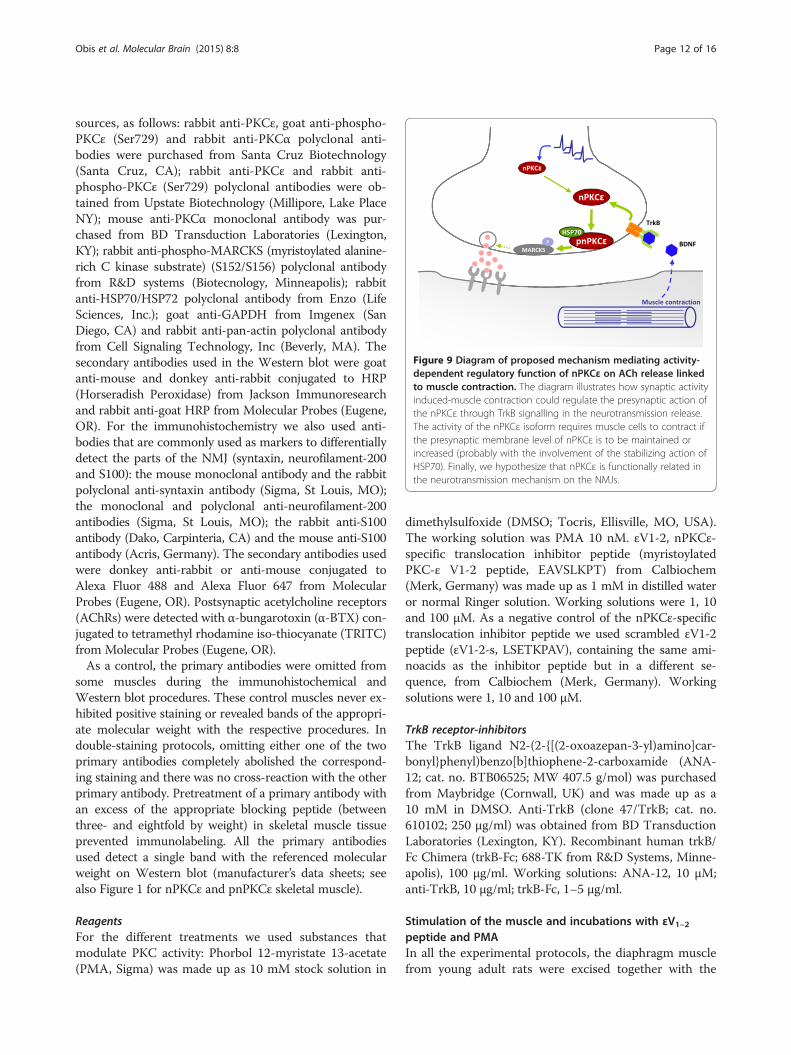
Figure 9 Diagram of proposed mechanism mediating activity-dependent regulatory function of nPKCε on ACh release linkedto muscle contraction. The diagram illustrates how synaptic activityinduced-muscle contraction could regulate the presynaptic action ofthe nPKCε through TrkB signalling in the neurotransmission release.The activity of the nPKCε isoform requires muscle cells to contract ifthe presynaptic membrane level of nPKCε is to be maintained orincreased (probably with the involvement of the stabilizing action ofHSP70). Finally, we hypothesize that nPKCε is functionally related inthe neurotransmission mechanism on the NMJs.
Obis et al. Molecular Brain (2015) 8:8 Page 12 of 16
sources, as follows: rabbit anti-PKCε, goat anti-phospho-PKCε (Ser729) and rabbit anti-PKCα polyclonal anti-bodies were purchased from Santa Cruz Biotechnology(Santa Cruz, CA); rabbit anti-PKCε and rabbit anti-phospho-PKCε (Ser729) polyclonal antibodies were ob-tained from Upstate Biotechnology (Millipore, Lake PlaceNY); mouse anti-PKCα monoclonal antibody was pur-chased from BD Transduction Laboratories (Lexington,KY); rabbit anti-phospho-MARCKS (myristoylated alanine-rich C kinase substrate) (S152/S156) polyclonal antibodyfrom R&D systems (Biotecnology, Minneapolis); rabbitanti-HSP70/HSP72 polyclonal antibody from Enzo (LifeSciences, Inc.); goat anti-GAPDH from Imgenex (SanDiego, CA) and rabbit anti-pan-actin polyclonal antibodyfrom Cell Signaling Technology, Inc (Beverly, MA). Thesecondary antibodies used in the Western blot were goatanti-mouse and donkey anti-rabbit conjugated to HRP(Horseradish Peroxidase) from Jackson Immunoresearchand rabbit anti-goat HRP from Molecular Probes (Eugene,OR). For the immunohistochemistry we also used anti-bodies that are commonly used as markers to differentiallydetect the parts of the NMJ (syntaxin, neurofilament-200and S100): the mouse monoclonal antibody and the rabbitpolyclonal anti-syntaxin antibody (Sigma, St Louis, MO);the monoclonal and polyclonal anti-neurofilament-200antibodies (Sigma, St Louis, MO); the rabbit anti-S100antibody (Dako, Carpinteria, CA) and the mouse anti-S100antibody (Acris, Germany). The secondary antibodies usedwere donkey anti-rabbit or anti-mouse conjugated toAlexa Fluor 488 and Alexa Fluor 647 from MolecularProbes (Eugene, OR). Postsynaptic acetylcholine receptors(AChRs) were detected with α-bungarotoxin (α-BTX) con-jugated to tetramethyl rhodamine iso-thiocyanate (TRITC)from Molecular Probes (Eugene, OR).As a control, the primary antibodies were omitted from
some muscles during the immunohistochemical andWestern blot procedures. These control muscles never ex-hibited positive staining or revealed bands of the appropri-ate molecular weight with the respective procedures. Indouble-staining protocols, omitting either one of the twoprimary antibodies completely abolished the correspond-ing staining and there was no cross-reaction with the otherprimary antibody. Pretreatment of a primary antibody withan excess of the appropriate blocking peptide (betweenthree- and eightfold by weight) in skeletal muscle tissueprevented immunolabeling. All the primary antibodiesused detect a single band with the referenced molecularweight on Western blot (manufacturer’s data sheets; seealso Figure 1 for nPKCε and pnPKCε skeletal muscle).
ReagentsFor the different treatments we used substances thatmodulate PKC activity: Phorbol 12-myristate 13-acetate(PMA, Sigma) was made up as 10 mM stock solution in
dimethylsulfoxide (DMSO; Tocris, Ellisville, MO, USA).The working solution was PMA 10 nM. εV1-2, nPKCε-specific translocation inhibitor peptide (myristoylatedPKC-ε V1-2 peptide, EAVSLKPT) from Calbiochem(Merk, Germany) was made up as 1 mM in distilled wateror normal Ringer solution. Working solutions were 1, 10and 100 μM. As a negative control of the nPKCε-specifictranslocation inhibitor peptide we used scrambled εV1-2peptide (εV1-2-s, LSETKPAV), containing the same ami-noacids as the inhibitor peptide but in a different se-quence, from Calbiochem (Merk, Germany). Workingsolutions were 1, 10 and 100 μM.
TrkB receptor-inhibitorsThe TrkB ligand N2-(2-{[(2-oxoazepan-3-yl)amino]car-bonyl}phenyl)benzo[b]thiophene-2-carboxamide (ANA-12; cat. no. BTB06525; MW 407.5 g/mol) was purchasedfrom Maybridge (Cornwall, UK) and was made up as a10 mM in DMSO. Anti-TrkB (clone 47/TrkB; cat. no.610102; 250 μg/ml) was obtained from BD TransductionLaboratories (Lexington, KY). Recombinant human trkB/Fc Chimera (trkB-Fc; 688-TK from R&D Systems, Minne-apolis), 100 μg/ml. Working solutions: ANA-12, 10 μM;anti-TrkB, 10 μg/ml; trkB-Fc, 1–5 μg/ml.
Stimulation of the muscle and incubations with εV1–2peptide and PMAIn all the experimental protocols, the diaphragm musclefrom young adult rats were excised together with the
Obis et al. Molecular Brain (2015) 8:8 Page 13 of 16
phrenic nerve and placed in oxygenated Ringer solution(see below) continuously bubbled with 95% O2/5% CO2
at room temperature. To stimulate the muscle, thephrenic nerve was stimulated at 1 Hz for a 10- or 30-minute period by an A-M Systems 2100 isolated pulsegenerator (A-M System, Carlsborg, WA). To study sep-arately the effect of the presynaptic stimulation (andsynaptic transmission) from the effect of the muscle cellcontraction, we performed experiments in which con-tractions were prevented (ES) or not (ES + C). Themuscle was prevented from contracting by cutting on ei-ther side of the main intramuscular nerve branch (seebelow). We also performed experiments to discard anypossible artifact due to the muscle fiber cutting. Cutmuscles were compared with muscles where musclecontraction was abolished by using μ-conotoxin GIIIB(μ-CgTx-GIIIB, 3 μM −1.5 μM, from ICS, InternationalClinical Service GmbH, München). The two methodsused to prevent muscle contraction did not show differ-ences with regard to electrophysiological parameters ofthe neurotransmission (see also later in Results).We used a nonpharmacological tool to inhibit specific-
ally nPKCε isoform. We used the nPKCε-specific trans-location inhibitor peptide, epsilonV1-2 (εV1-2; [20]), toblock the isoform activity so that we could analyze itspossible constitutive involvement in cell functions in theresting and stimulated NMJs. This peptide interferes inthe nPKCε interaction with the anchoring proteinepsilon-RACK and, therefore, inhibits the anchoring ofnPKCε near its substrates (and translocation to themembrane of the active isoform) and prevents any sub-sequent substrate phosphorylation and activity [64]. FornPKC, the PKC–RACK interaction occurs via a C2-likedomain that does not bind Ca2+ [64,65]. Thus, the clas-sic calcium-dependent cPKCs are not inhibited with thepeptide εV1-2 because they do not possess this C2-likeregion. In addition, studies using peptide translocationinhibitors of nPKCε (εV1-2) and nPKCδ (δV1) demon-strated that these isoforms (nPKCε and nPKCδ) canhave opposing actions within a given cell type (cardio-myocyte) [66-68] and also showed the isoform specificityof the peptides. There are several evidence that showthat the effects found using the nPKCε-specific transloca-tion inhibitor peptide can be confirmed when nPKCεknockout mice are used [69,70]. The effect of εV1-2(100 μM) was studied in parallel to the effect of the scram-bled version of this peptide (εV1-2-s, 100 μM). In someexperiments, stimulated muscles were previously incu-bated with the peptide εV1-2 or with its inactive form(30 minutes).To activate PKC family with PMA, a potent but non-
selective-isoform PKC activator, the diaphragm muscleswere incubated for 10, 30 or 60 minutes on a Sylgard-coated Petri dish containing Ringer solution for the
control muscles, or εV1-2 (100 μM), scrambled εV1-2(100 μM) or PMA (10 nM).
Western blot analysisDiaphragm muscles from newborn and adult rat weredissected, frozen in liquid nitrogen, and stored at −80°Cbefore use. The muscles were homogenized using ahigh-speed homogenizer (overhead stirrer, VWR Inter-national, Clarksburg, MD) in lysis buffer containing150 mM NaCl, 20 mM Tris–HCl, pH 7.5, 2 mM EGTA,and 5 mM EDTA supplemented with 1% Triton X-100,1 mM PMSF, 50 mM NaF, and 1 mM sodium orthova-nadate from Sigma, (St. Louis, MO) and protease inhibi-tor cocktail (Sigma-Aldrich Corp., Saint Louis, MO,USA). Insoluble material was removed by centrifugationat 1000 g for 10 minutes. The supernatants were col-lected and centrifuged at 15000 g for 20 minutes. Finally,the resulting supernatants (total protein lysates) werecollected. Protein concentrations were determined byusing the Bio-Rad DC protein assay (Bio-Rad, Hercules,CA). Experimental procedures were performed to deter-mine the linear and quantitative dynamic range for eachtarget protein and the appropriate dilutions of sampleswere used for accurate and normalized quantitation bymeans of densitometric analysis. Protein samples of 15or 30 μg were separated by 8% SDS-polyacrylamide elec-trophoresis and electrotransferred to PVDF membranes(Hybond™-P; Amersham, GE Healthcare). The membraneswere blocked in Tris-buffered saline-0.1% Tween-20(TBS-T) containing 5% (W/V) nonfat dry milk or in ablocking reagent to preserve phosphoprotein antigens(PhosphoBLOCKER™; Cell Biolabs, Inc.) and probed withthe primary antibody overnight at 4°C. The membraneswere then incubated with the secondary antibody and vi-sualized enhanced chemiluminescence with the ECL kit(Amersham Life Science, Arlington Heights, IL).In treated and/or stimulated muscles, the synaptic
membranes were obtained. Synaptic and extrasynapticparts of the diaphragm muscle were separated as previ-ously described [25]. We performed control experimentsto check that our protocol for dividing the diaphragmmuscle into synaptic and extrasynaptic region was accur-ate. In some muscles, we repeated the process of separ-ation and detected NMJs with TRITC-conjugated α-BTX.We also stained the nerves with an antibody against anti-neurofilament-200 and did not detect any nerves in extra-synaptic regions. The muscles were homogenized using ahigh-speed homogenizer (overhead stirrer, VWR Inter-national, Clarksburg, MD) in lysis buffer (see above) andthe insoluble material was removed in the same way (bycentrifugation at 1000 g for 10 minutes) but now theresulting supernatant was collected and centrifuged at130000 g for 1 hour. The supernatant was the cytosolicfraction, and the pellet was the membrane fraction. To
Obis et al. Molecular Brain (2015) 8:8 Page 14 of 16
assess the separation of the membrane fraction from thecytosol, we used a goat antibody directed against Glyceral-dehyde 3-phosphate dehydrogenase (GAPDH), a proteinspecific to the cytosolic fraction. GADPH immunoreactiv-ity was not observed in any case in the membrane fraction.The samples were processed the same way as anothersample of total protein (see below).The blots were visualized with aVersaDoc 3000 (Bio-Rad,
Hercules, CA). The densitometry of different bands wasanalyzed with the MetaMorph software. The integratedoptical density of the bands was normalized to the back-ground values and by actin protein. Also, as another load-ing control we used a total protein analysis (Sypro Rubyprotein blot Stain, Bio Rad) to measure the total proteintransferred on polyvininylidene difluoride (PVDF) mem-branes. In all the cases, the quantitative results obtainedby using actin or total protein analysis were no different.The relative variations between the bands in the experi-mental samples and the control samples were calculatedin the same image. The data were taken from densitom-etry measurements made in at least five separate experi-ments, plotted against controls. Data are mean values ±SD. Differences between groups were tested using thet Student test or U test (Mann–Whitney), and the normal-ity of the distributions was tested with the Kolmogorov–Smirnov test. The criterion for statistical significance wasp < 0.05 versus the control.
Immunohistochemistry and confocal microscopyWhole muscle mounts were processed by immunohisto-chemistry to detect the localization of the nPKCε iso-form at the NMJ. LAL and diaphragm muscles fromyoung adult rats were fixed with 4% paraformaldehydefor 30 minutes. After fixation, the muscles were rinsedwith PBS and incubated in 0.1 M glycine in PBS. Themuscles were permeabilized with 0.5% Triton X-100 inPBS, and nonspecific binding was blocked with 4% bo-vine serum albumin (BSA). Then, muscles were incu-bated overnight at 4°C in mixtures of three primaryantibodies raised in different species (anti-nPKCε iso-form antibody and anti-syntaxin and anti-neurofilamentor syntaxin or anti-S100) and then rinsed. The muscleswere then incubated for four hours at room temperaturein a mixture of appropriate secondary antibodies. TheAChRs were detected with α-BTX conjugated with TRITC.At least three muscles were used as negative controls asdescribed above. For a better analysis of the localization ofthe nPKCε isoform at the NMJ, some muscles were proc-essed to obtain semithin cross-sections from whole-mountmultiple-immunofluorescent stained muscles. This methodprovided a simple and sensitive procedure for analyzingthe cellular distribution of molecules at the NMJ [24].Labeled NMJs from the whole-mount muscles and the
semithin cross-sections were viewed with a laser-scanning
confocal microscope (Nikon TE2000-E). Special consider-ation was given to the possible contamination of onechannel by another. In experiments involving negativecontrols, the photomultiplier tube gains and blacklevels were identical to those used for a labeled prep-aration made in parallel with the control preparations.At least 25 endplates per muscle were observed, andat least six muscles were studied. Images were assem-bled using Adobe PhotoShop software (AdobeSystems, San Jose, CA) and neither the contrast norbrightness were modified.
ElectrophysiologyDiaphragm muscles from adult rats were removed surgi-cally and incubated in a Sylgard-Petri dish containingnormal Ringer solution (in mM) – NaCl 135, KCl 5,CaCl2 2.5, MgSO4 1, NaH2PO4 1, NaHCO3 15, glucose11 – which was bubbled continuously with 95% O2, 5%CO2. Temperature and humidity were regulated at 26°Cand 50%, respectively. Spontaneous miniature endplatepotentials (MEPPs) and evoked endplate potentials(EPPs) were recorded intracellularly with conventionalglass microelectrodes filled with 3 M KCl (resistance:20–40 MW). Recording electrodes were connected to anamplifier (Tecktronics, AMS02), and a distant Ag-AgClelectrode connected to the bath solution via an agarbridge (agar 3.5% in 137 mM NaCl) was used as a refer-ence. The signals were digitized (DIGIDATA 1322AInterface, Axon Instruments Inc, CA, USA), stored andcomputer-analyzed. The software Axoscope 9.0 (AxonInstruments Inc, CA, USA) was used for data acquisitionand analysis. To prevent muscle contraction during EPPrecordings, we used μ-conotoxin GIIIB (μ-CgTx-GIIIB,3 μM −1.5 μM, from ICS, International Clinical ServiceGmbH, München). After a muscle fiber had been im-paled, the phrenic nerve was continuously stimulated(70 stimuli at 0.5 Hz) using two platinum electrodes thatwere coupled to a pulse generator (CIBERTEC CS-20)linked to a stimulus isolation unit. The last 50 EPPs wererecorded. We selected fibers with membrane potentialsof no less than -70 mV and used only those results frompreparations which did not deviate by more than 5 mVduring the recording. The mean amplitude (mV) perfiber was calculated and corrected for non-linear summa-tion (EPPs were usually more than 4 mV) [71] assuming amembrane potential of – 80 mV. Quantal content (M)was estimated by the direct method, which consists of re-cording mEPPs and EPPs simultaneously and then calculat-ing the ratio: M =Average Peak EPP/Average Peak mEPP.We studied a minimum of 15 fibers per muscle and usuallya minimum of 5 muscles in each type of experiment. Thedata given in Results are mean values ± SEM. Only onehemidiaphragm was used from each animal for a given ex-periment. We used the two-tailed Welch’s t-test (for
Obis et al. Molecular Brain (2015) 8:8 Page 15 of 16
unpaired values and variances were not assumed to beequal). Differences were considered significant at P < 0.05(*).
DenervationExtensor digitorum longus (EDL) denervation. Young adultrats (30–40 months of age, male) were anesthetized withketamine/xylazine (K/X; 0.1 ml/10 g body weight intraper-itoneal injection of a solution containing 10 mg/ml keta-mine and 1 mg/ml xylazine). To isolate the sciatic nerve, a0.5 cm excision was made on the exterior side of the leg atapproximately 1 cm over the level of knee. The excisionwas made carefully avoiding tissue injury, the sciatic nervewas cut about 1 mm from the nerve’s entrance into themuscle and a 1 cm segment was excised. The wound wasthen closed. At the times desired (1–3 days), the rats wereanesthetized with pentobarbital and the EDL muscle wasexcised.
AbbreviationsACh: Acetylcholine; LAL: Levator auris longus muscle; nAChR: Nicotinicacetylcholine receptor; NMJ: Neuromuscular junction; PKC: Protein Kinase C;nPKCε: Epsilon PKC; PMA: Phorbol 12-myristate 13-acetate; RACKs: Receptorsfor activated C-kinase; PS: Phosphatidylserine; DAG: Diacylglycerol;εV1-2: Isozyme-selective translocation inhibitor; α-BTX: α-bungarotoxin conjugatedTRITC; TrkB: Tyrosine kinase receptor B; BDNF: Brain derived neurotrophic factor;EDL: Extensor digitorum longus muscle; PLCγ1: Phosphoinositide-specificphospholipase C γ1; PI(4,5)P2: Phosphatidylinositol 4,5-biphosphate; IP3: Inositoltris-phosphate; NT: Nerve terminal; PDK: 3-phosphoinositide dependent proteinkinase.
Competing interestsThe authors declare that they have no competing interests.
Authors’ contributionsTO and NB: data collection, quantitative analysis; literature search, datainterpretation, statistics; EH and LN: data collection, quantitative analysis;statistics. MP and MT: data collection; NG and MMS: literature search, datainterpretation; MAL and JT: conception and design, literature search, datainterpretation, manuscript preparation. JT and MAL contributed equally tothis work. All authors read and approved the final manuscript.
AcknowledgementsThis work was supported by a grant from MEC (SAF2011-23711) and a grantfrom the Catalan Government (Generalitat) (2009SGR01248) (JT). The authorsdeclare no competing financial interests.
Received: 10 November 2014 Accepted: 16 January 2015
References1. West JW, Numann R, Murphy BJ, Scheuer T, Catterall WA. A phosphorylation
site in the Na + channel required for modulation by protein kinase C.Science. 1991;254:866–8.
2. Numann R, Hauschka SD, Catterall WA, Scheuer T. Modulation of skeletalmuscle sodium channels in a satellite cell line by protein kinase C. J Neurosci.1994;14:4226–36.
3. Byrne JH, Kandel ER. Presynaptic facilitation revisited: state and timedependence. J Neurosci. 1996;16:425–35.
4. Catterall WA. Interactions of presynaptic Ca2+ channels and snare proteinsin neurotransmitter release. Ann N Y Acad Sci. 1999;868:144–59.
5. Santafé MM, Lanuza MA, Garcia N, Tomàs J. Calcium inflow-dependentprotein kinase C activity is involved in the modulation of transmitter releasein the neuromuscular junction of the adult rat. Synapse. 2005;57:76–84.
6. Tanaka C, Nishizuka Y. The protein kinase C family for neuronal signaling.Annu Rev Neurosci. 1994;17:551–67.
7. Mochly-Rosen D. Localization of protein kinases by anchoring proteins: atheme in signal transduction. Science. 1995;268:247–51.
8. Mochly-Rosen D, Khaner H, Lopez J. Identification of intracellular receptor proteinsfor activated protein kinase C. Proc Natl Acad Sci U S A. 1991;88:3997–4000.
9. Mochly-Rosen D, Khaner H, Lopez J, Smith BL. Intracellular receptors foractivated protein kinase C. Identification of a binding site for the enzyme.J Biol Chem. 1991;266:14866–8.
10. Shirai Y, Adachi N, Saito N. Protein kinase Cepsilon: function in neurons.FEBS J. 2008;275:3988–94.
11. Moraczewski J, Nowotniak A, Wróbel E, Castagna M, Gautron J, Martelly I.Differential changes in protein kinase C associated with regeneration of ratextensor digitorum longus and soleus muscles. Int J Biochem Cell Biol.2002;34:938–49.
12. Vary TC, Goodman S, Kilpatrick LE, Lynch CJ. Nutrient regulation ofPKCepsilon is mediated by leucine, not insulin, in skeletal muscle. Am JPhysiol Endocrinol Metab. 2005;289:E684–94.
13. Stumpo DJ, Graff JM, Albert KA, Greengard P, Blackshear PJ. Molecularcloning, characterization, and expression of a cDNA encoding the “80- to87-kDa” myristoylated alanine-rich C kinase substrate: a major cellularsubstrate for protein kinase C. Proc Natl Acad Sci U S A. 1989;86:4012–6.
14. Aderem A. The Marcks brothers: a family of protein kinase C substrates. Cell.1992;71:713–6.
15. Aderem A. Signal transduction and the actin cytoskeleton: the roles ofMARCKS and profilin. Trends Biochem Sci. 1992;17:438–43.
16. Park Y-S, Hur E-M, Choi B-H, Kwak E, Jun D-J, Park S-J, et al. Involvement ofprotein kinase C-epsilon in activity-dependent potentiation of large dense-core vesicle exocytosis in chromaffin cells. J Neurosci. 2006;26:8999–9005.
17. Mosevitsky MI. Nerve ending “signal” proteins GAP-43, MARCKS, and BASP1.Int Rev Cytol. 2005;245:245–325.
18. Lu D, Yang H, Lenox RH, Raizada MK. Regulation of angiotensin II-inducedneuromodulation by MARCKS in brain neurons. J Cell Biol. 1998;142:217–27.
19. Rosé SD, Lejen T, Zhang L, Trifaró JM. Chromaffin cell F-actin disassemblyand potentiation of catecholamine release in response to protein kinase Cactivation by phorbol esters is mediated through myristoylated alanine-richC kinase substrate phosphorylation. J Biol Chem. 2001;276:36757–63.
20. Johnson JA, Gray MO, Chen C-H, Mochly-Rosen D. A Protein Kinase Ctranslocation inhibitor as an Isozyme-selective antagonist of cardiac function.J Biol Chem. 1996;271:24962–6.
21. Bornancin F, Parker PJ. Phosphorylation of protein kinase C-alpha on serine657 controls the accumulation of active enzyme and contributes to itsphosphatase-resistant state. J Biol Chem. 1997;272:3544–9.
22. Dutil EM, Toker A, Newton AC. Regulation of conventional protein kinase Cisozymes by phosphoinositide-dependent kinase 1 (PDK-1). Curr Biol.1998;8:1366–75.
23. Hafeez BB, Zhong W, Weichert J, Dreckschmidt NE, Jamal MS, Verma AK.Genetic ablation of PKC epsilon inhibits prostate cancer development andmetastasis in transgenic mouse model of prostate adenocarcinoma. CancerRes. 2011;71:2318–27.
24. Lanuza MA, Besalduch N, Garcia N, Sabaté M, Santafé MM, Tomàs J. Plastic-embedded semithin cross-sections as a tool for high-resolutionimmunofluorescence analysis of the neuromuscular junction molecules: specificcellular location of protease-activated receptor-1. J Neurosci Res. 2007;85:748–56.
25. Besalduch N, Tomàs M, Santafé MM, Garcia N, Tomàs J, Lanuza MA. Synapticactivity-related classical protein kinase C isoform localization in the adult ratneuromuscular synapse. J Comp Neurol. 2010;518:211–28.
26. Santafé MM, Garcia N, Lanuza MA, Uchitel OD, Tomás J. Calcium channelscoupled to neurotransmitter release at dually innervated neuromuscularjunctions in the newborn rat. Neuroscience. 2001;102:697–708.
27. Santafé MM, Salon I, Garcia N, Lanuza MA, Uchitel OD, Tomàs J. Muscarinicautoreceptors related with calcium channels in the strong and weak inputsat polyinnervated developing rat neuromuscular junctions. Neuroscience.2004;123:61–73.
28. Santafé MM, Garcia N, Lanuza MA, Tomàs M, Tomàs J. Interaction betweenprotein kinase C and protein kinase A can modulate transmitter release atthe rat neuromuscular synapse. J Neurosci Res. 2009;87:683–90.
29. Santafé MM, Lanuza MA, Garcia N, Tomàs M, Tomàs J. Coupling of presynapticmuscarinic autoreceptors to serine kinases in low and high release conditionson the rat motor nerve terminal. Neuroscience. 2007;148:432–40.
30. Cazorla M, Prémont J, Mann A, Girard N, Kellendonk C, Rognan D.Identification of a low-molecular weight TrkB antagonist with anxiolytic andantidepressant activity in mice. J Clin Invest. 2011;121:1846–57.
Obis et al. Molecular Brain (2015) 8:8 Page 16 of 16
31. Balkowiec A, Katz DM. Activity-dependent release of endogenous brain-derived neurotrophic factor from primary sensory neurons detected byELISA in situ. J Neurosci. 2000;20:7417–23.
32. Garcia N, Tomàs M, Santafé MM, Besalduch N, Lanuza MA, Tomàs J. Theinteraction between tropomyosin-related kinase B receptors and presynapticmuscarinic receptors modulates transmitter release in adult rodent motornerve terminals. J Neurosci. 2010;30:16514–22.
33. Kraft AS, Anderson WB, Cooper HL, Sando JJ. Decrease in cytosolic calcium/phospholipid-dependent protein kinase activity following phorbol estertreatment of EL4 thymoma cells. J Biol Chem. 1982;257:13193–6.
34. Betz A, Ashery U, Rickmann M, Augustin I, Neher E, Südhof TC, et al.Munc13-1 is a presynaptic phorbol ester receptor that enhancesneurotransmitter release. Neuron. 1998;21:123–36.
35. Brose N, Rosenmund C. Move over protein kinase C, you’ve got company:alternative cellular effectors of diacylglycerol and phorbol esters. J Cell Sci.2002;115(Pt 23):4399–411.
36. Santafé MM, Lanuza MA, Garcia N, Tomàs J. Muscarinic autoreceptorsmodulate transmitter release through protein kinase C and protein kinase Ain the rat motor nerve terminal. Eur J Neurosci. 2006;23:2048–56.
37. Chen Y, Tian Q. The role of protein kinase C epsilon in neural signaltransduction and neurogenic diseases. Front Med. 2011;5:70–6.
38. Ono Y, Fujii T, Ogita K, Kikkawa U, Igarashi K, Nishizuka Y. The structure,expression, and properties of additional members of the protein kinase Cfamily. J Biol Chem. 1988;263:6927–32.
39. Wetsel WC, Khan WA, Merchenthaler I, Rivera H, Halpern AE, Phung HM,et al. Tissue and cellular distribution of the extended family of proteinkinase C isoenzymes. J Cell Biol. 1992;117:121–33.
40. Saito N, Itouji A, Totani Y, Osawa I, Koide H, Fujisawa N, et al. Cellular andintracellular localization of epsilon-subspecies of protein kinase C in the rat brain;presynaptic localization of the epsilon-subspecies. Brain Res. 1993;607:241–8.
41. Richter EA, Cleland PJ, Rattigan S, Clark MG. Contraction-associated translocationof protein kinase C in rat skeletal muscle. FEBS Lett. 1987;217:232–6.
42. Cleland PJ, Appleby GJ, Rattigan S, Clark MG. Exercise-induced translocationof protein kinase C and production of diacylglycerol and phosphatidic acidin rat skeletal muscle in vivo. Relationship to changes in glucose transport.J Biol Chem. 1989;264:17704–11.
43. Antipenko A, Frı́as JA, Parra J, Cadefau JA, Cussó R. Effect of chronicelectrostimulation of rabbit skeletal muscle on calmodulin level and proteinkinase activity. Int J Biochem Cell Biol. 1999;31:303–10.
44. Brandman R, Disatnik M-H, Churchill E, Mochly-Rosen D. Peptides derivedfrom the C2 domain of protein kinase C epsilon (epsilon PKC) modulateepsilon PKC activity and identify potential protein-protein interactionsurfaces. J Biol Chem. 2007;282:4113–23.
45. Heidkamp MC, Iyengar R, Szotek EL, Cribbs LL, Samarel AM. Protein kinaseCepsilon-dependent MARCKS phosphorylation in neonatal and adult ratventricular myocytes. J Mol Cell Cardiol. 2007;42:422–31.
46. Garczarczyk D, Toton E, Biedermann V, Rosivatz E, Rechfeld F, Rybczynska M,et al. Signal transduction of constitutively active protein kinase C epsilon.Cell Signal. 2009;21:745–52.
47. Arbuzova A, Schmitz AAP, Vergères G. Cross-talk unfolded: MARCKS proteins.Biochem J. 2002;362(Pt 1):1–12.
48. Sasaki Y. New aspects of neurotransmitter release and exocytosis: Rho-kinase-dependent myristoylated alanine-rich C-kinase substratephosphorylation and regulation of neurofilament structure in neuronal cells.J Pharmacol Sci. 2003;93:35–40.
49. Matthews VB, Aström M-B, Chan MHS, Bruce CR, Krabbe KS, Prelovsek O,et al. Brain-derived neurotrophic factor is produced by skeletal muscle cellsin response to contraction and enhances fat oxidation via activation ofAMP-activated protein kinase. Diabetologia. 2009;52:1409–18.
50. Gómez-Pinilla F, Ying Z, Roy RR, Molteni R, Edgerton VR. Voluntary exerciseinduces a BDNF-mediated mechanism that promotes neuroplasticity.J Neurophysiol. 2002;88:2187–95.
51. Kulakowski SA, Parker SD, Personius KE. Reduced TrkB expression results inprecocious age-like changes in neuromuscular structure, neurotransmission,and muscle function. J Appl Physiol. 2011;111:844–52.
52. Pereira DB, Rebola N, Rodrigues RJ, Cunha RA, Carvalho AP, Duarte CB. Trkbreceptors modulation of glutamate release is limited to a subset of nerveterminals in the adult rat hippocampus. J Neurosci Res. 2006;83:832–44.
53. Reichardt LF. Neurotrophin-regulated signalling pathways. Philos Trans RSoc Lond B Biol Sci. 2006;361:1545–64.
54. Newton AC. Regulation of the ABC kinases by phosphorylation: proteinkinase C as a paradigm. Biochem J. 2003;370(Pt 2):361–71.
55. Rybin VO, Sabri A, Short J, Braz JC, Molkentin JD, Steinberg SF. Cross-regulation of novel protein kinase C (PKC) isoform function in cardiomyocytes.Role of PKC epsilon in activation loop phosphorylations and PKC delta inhydrophobic motif phosphorylations. J Biol Chem. 2003;278:14555–64.
56. Hansra G, Garcia-Paramio P, Prevostel C, Whelan RD, Bornancin F, Parker PJ.Multisite dephosphorylation and desensitization of conventional proteinkinase C isotypes. Biochem J. 1999;342(Pt 2):337–44.
57. Malenka RC, Madison DV, Nicoll RA. Potentiation of synaptic transmission inthe hippocampus by phorbol esters. Nature. 1986;321:175–7.
58. Shapira R, Silberberg SD, Ginsburg S, Rahamimoff R. Activation of proteinkinase C augments evoked transmitter release. Nature. 1987;325:58–60.
59. Capogna M, Gahwiler B, Thompson S. Presynaptic enhancement ofinhibitory synaptic transmission by protein kinases A and C in the rathippocampus in vitro. J Neurosci. 1995;15:1249–60.
60. Hori T, Takai Y, Takahashi T. Presynaptic mechanism for phorbol ester-induced synaptic potentiation. J Neurosci. 1999;19:7262–7.
61. Santafé MM, Garcia N, Tomàs M, Obis T, Lanuza MA, Besalduch N, et al. Theinteraction between tropomyosin-related kinase B receptors and serinekinases modulates acetylcholine release in adult neuromuscular junctions.Neurosci Lett. 2014;561:171–5.
62. Wierda KDB, Toonen RFG, de Wit H, Brussaard AB, Verhage M.Interdependence of PKC-dependent and PKC-independent pathways forpresynaptic plasticity. Neuron. 2007;54:275–90.
63. Rhee JS, Betz A, Pyott S, Reim K, Varoqueaux F, Augustin I, et al. Betaphorbol ester- and diacylglycerol-induced augmentation of transmitterrelease is mediated by Munc13s and not by PKCs. Cell. 2002;108:121–33.
64. Mochly-Rosen D, Gordon AS. Anchoring proteins for protein kinase C: ameans for isozyme selectivity. FASEB J. 1998;12:35–42.
65. Way KJ, Chou E, King GL. Identification of PKC-isoform-specific biologicalactions using pharmacological approaches. Trends Pharmacol Sci. 2000;21:181–7.
66. Gray MO, Karliner JS, Mochly-Rosen D. A selective epsilon-protein kinase Cantagonist inhibits protection of cardiac myocytes from hypoxia-inducedcell death. J Biol Chem. 1997;272:30945–51.
67. Csukai M, Mochly-Rosen D. Pharmacologic modulation of protein kinase Cisozymes: the role of RACKs and subcellular localisation. Pharmacol Res.1999;39:253–9.
68. Dorn GW, Souroujon MC, Liron T, Chen CH, Gray MO, Zhou HZ, et al.Sustained in vivo cardiac protection by a rationally designed peptide thatcauses epsilon protein kinase C translocation. Proc Natl Acad Sci U S A.1999;96:12798–803.
69. Di-Capua N, Sperling O, Zoref-Shani E. Protein kinase C-epsilon is involvedin the adenosine-activated signal transduction pathway conferringprotection against ischemia-reperfusion injury in primary rat neuronalcultures. J Neurochem. 2003;84:409–12.
70. Khasar SG, Lin YH, Martin A, Dadgar J, McMahon T, Wang D, et al. A novelnociceptor signaling pathway revealed in protein kinase C epsilon mutantmice. Neuron. 1999;24:253–60.
71. McLachlan EM, Martin AR. Non-linear summation of end-plate potentials inthe frog and mouse. J Physiol. 1981;311:307–24.
Submit your next manuscript to BioMed Centraland take full advantage of:
• Convenient online submission
• Thorough peer review
• No space constraints or color figure charges
• Immediate publication on acceptance
• Inclusion in PubMed, CAS, Scopus and Google Scholar
• Research which is freely available for redistribution
Submit your manuscript at www.biomedcentral.com/submit
Related Documents